DE102019103848A1 - Method for controlling an ophthalmic laser and treatment device - Google Patents
Method for controlling an ophthalmic laser and treatment deviceDownload PDFInfo
- Publication number
- DE102019103848A1 DE102019103848A1DE102019103848.0ADE102019103848ADE102019103848A1DE 102019103848 A1DE102019103848 A1DE 102019103848A1DE 102019103848 ADE102019103848 ADE 102019103848ADE 102019103848 A1DE102019103848 A1DE 102019103848A1
- Authority
- DE
- Germany
- Prior art keywords
- cornea
- laser
- eye
- interface
- predefined
- Prior art date
- Legal status (The legal status is an assumption and is not a legal conclusion. Google has not performed a legal analysis and makes no representation as to the accuracy of the status listed.)
- Granted
Links
- 238000000034methodMethods0.000titleclaimsabstractdescription51
- 210000004087corneaAnatomy0.000claimsabstractdescription68
- 239000007787solidSubstances0.000claimsabstractdescription20
- 238000000926separation methodMethods0.000claimsabstractdescription11
- 241001465754MetazoaSpecies0.000claimsabstractdescription10
- 230000003287optical effectEffects0.000claimsdescription15
- 208000029091Refraction diseaseDiseases0.000claimsdescription9
- 230000004430ametropiaEffects0.000claimsdescription9
- 208000014733refractive errorDiseases0.000claimsdescription9
- 238000004590computer programMethods0.000claimsdescription7
- 210000004045bowman membraneAnatomy0.000claimsdescription3
- 238000007493shaping processMethods0.000claimsdescription2
- 238000003860storageMethods0.000claimsdescription2
- 238000002679ablationMethods0.000description2
- 238000005259measurementMethods0.000description2
- 238000012876topographyMethods0.000description2
- BUHVIAUBTBOHAG-FOYDDCNASA-N(2r,3r,4s,5r)-2-[6-[[2-(3,5-dimethoxyphenyl)-2-(2-methylphenyl)ethyl]amino]purin-9-yl]-5-(hydroxymethyl)oxolane-3,4-diolChemical compoundCOC1=CC(OC)=CC(C(CNC=2C=3N=CN(C=3N=CN=2)[C@H]2[C@@H]([C@H](O)[C@@H](CO)O2)O)C=2C(=CC=CC=2)C)=C1BUHVIAUBTBOHAG-FOYDDCNASA-N0.000description1
- 206010020675HypermetropiaDiseases0.000description1
- 201000009310astigmatismDiseases0.000description1
- 230000015572biosynthetic processEffects0.000description1
- 238000010586diagramMethods0.000description1
- 238000005516engineering processMethods0.000description1
- 201000006318hyperopiaDiseases0.000description1
- 230000004305hyperopiaEffects0.000description1
- 238000011065in-situ storageMethods0.000description1
- 208000001491myopiaDiseases0.000description1
- 230000004379myopiaEffects0.000description1
- 201000010041presbyopiaDiseases0.000description1
- 230000005855radiationEffects0.000description1
- 238000001356surgical procedureMethods0.000description1
Images

Classifications
- A—HUMAN NECESSITIES
- A61—MEDICAL OR VETERINARY SCIENCE; HYGIENE
- A61F—FILTERS IMPLANTABLE INTO BLOOD VESSELS; PROSTHESES; DEVICES PROVIDING PATENCY TO, OR PREVENTING COLLAPSING OF, TUBULAR STRUCTURES OF THE BODY, e.g. STENTS; ORTHOPAEDIC, NURSING OR CONTRACEPTIVE DEVICES; FOMENTATION; TREATMENT OR PROTECTION OF EYES OR EARS; BANDAGES, DRESSINGS OR ABSORBENT PADS; FIRST-AID KITS
- A61F9/00—Methods or devices for treatment of the eyes; Devices for putting in contact-lenses; Devices to correct squinting; Apparatus to guide the blind; Protective devices for the eyes, carried on the body or in the hand
- A61F9/007—Methods or devices for eye surgery
- A61F9/008—Methods or devices for eye surgery using laser
- A61F9/00825—Methods or devices for eye surgery using laser for photodisruption
- A61F9/00827—Refractive correction, e.g. lenticle
- A—HUMAN NECESSITIES
- A61—MEDICAL OR VETERINARY SCIENCE; HYGIENE
- A61F—FILTERS IMPLANTABLE INTO BLOOD VESSELS; PROSTHESES; DEVICES PROVIDING PATENCY TO, OR PREVENTING COLLAPSING OF, TUBULAR STRUCTURES OF THE BODY, e.g. STENTS; ORTHOPAEDIC, NURSING OR CONTRACEPTIVE DEVICES; FOMENTATION; TREATMENT OR PROTECTION OF EYES OR EARS; BANDAGES, DRESSINGS OR ABSORBENT PADS; FIRST-AID KITS
- A61F9/00—Methods or devices for treatment of the eyes; Devices for putting in contact-lenses; Devices to correct squinting; Apparatus to guide the blind; Protective devices for the eyes, carried on the body or in the hand
- A61F9/007—Methods or devices for eye surgery
- A61F9/008—Methods or devices for eye surgery using laser
- A61F9/00825—Methods or devices for eye surgery using laser for photodisruption
- A61F9/0084—Laser features or special beam parameters therefor
- A—HUMAN NECESSITIES
- A61—MEDICAL OR VETERINARY SCIENCE; HYGIENE
- A61F—FILTERS IMPLANTABLE INTO BLOOD VESSELS; PROSTHESES; DEVICES PROVIDING PATENCY TO, OR PREVENTING COLLAPSING OF, TUBULAR STRUCTURES OF THE BODY, e.g. STENTS; ORTHOPAEDIC, NURSING OR CONTRACEPTIVE DEVICES; FOMENTATION; TREATMENT OR PROTECTION OF EYES OR EARS; BANDAGES, DRESSINGS OR ABSORBENT PADS; FIRST-AID KITS
- A61F9/00—Methods or devices for treatment of the eyes; Devices for putting in contact-lenses; Devices to correct squinting; Apparatus to guide the blind; Protective devices for the eyes, carried on the body or in the hand
- A61F9/007—Methods or devices for eye surgery
- A61F9/008—Methods or devices for eye surgery using laser
- A61F2009/00861—Methods or devices for eye surgery using laser adapted for treatment at a particular location
- A61F2009/00872—Cornea
Landscapes
- Health & Medical Sciences (AREA)
- Ophthalmology & Optometry (AREA)
- Heart & Thoracic Surgery (AREA)
- Vascular Medicine (AREA)
- Optics & Photonics (AREA)
- Surgery (AREA)
- Engineering & Computer Science (AREA)
- Biomedical Technology (AREA)
- Physics & Mathematics (AREA)
- Nuclear Medicine, Radiotherapy & Molecular Imaging (AREA)
- Life Sciences & Earth Sciences (AREA)
- Animal Behavior & Ethology (AREA)
- General Health & Medical Sciences (AREA)
- Public Health (AREA)
- Veterinary Medicine (AREA)
- Laser Surgery Devices (AREA)
Abstract
Translated fromGermanDescription
Translated fromGermanDie vorliegende Erfindung betrifft Verfahren zur Steuerung eines augenchirurgischen Lasers für die Abtrennung eines Volumenkörpers mit vordefinierten Grenzflächen aus einer menschlichen oder tierischen Hornhaut. Die Erfindung betrifft weiterhin eine Behandlungsvorrichtung mit mindestens einem augenchirurgischen Laser für die Abtrennung eines vordefinierten Hornhautvolumens mit vordefinierten Grenzflächen eines menschlichen oder tierischen Auges mittels Photodisruption und mindestens einer Steuereinrichtung für den oder die Laser sowie ein Computerprogramm und ein computerlesbares Medium.The present invention relates to a method for controlling an ophthalmic laser for the separation of a solid with predefined interfaces from a human or animal cornea. The invention further relates to a treatment device with at least one ophthalmic laser for the separation of a predefined corneal volume with predefined interfaces of a human or animal eye by means of photodisruption and at least one control device for the laser or lasers as well as a computer program and a computer-readable medium.
Vorrichtungen und Verfahren zur Steuerung eines photoablativen ophthalmologischen Lasers sind bekannt. So beschreibt die internationale Patentanmeldung
Nachteilig an diesen bekannten Verfahren und Vorrichtungen ist jedoch, dass bei den bekannten ablativen Verfahren und Vorrichtungen der gesamte Volumenkörper mittels Laserenergie verdampft wird. Einerseits erfordert dies relativ lange Behandlungszeiten, andererseits erfolgt ein relativ hoher Energieeintrag durch den Laser in die Hornhaut. Insbesondere letzteres kann zu Komplikationen bei der Entfernung des vorbestimmten Volumenkörpers der Hornhaut führen. Diese Nachteile bekannter photoablativer Laser, beispielsweise von Excimer-Lasern, die überwiegen im ultravioletten Bereich emittieren, konnten bisher noch nicht zufriedenstellend gelöst werden.A disadvantage of these known methods and devices, however, is that in the known ablative methods and devices the entire volume body is vaporized by means of laser energy. On the one hand, this requires relatively long treatment times; on the other hand, the laser introduces a relatively high amount of energy into the cornea. The latter, in particular, can lead to complications when removing the predetermined volume of the cornea. These disadvantages of known photoablative lasers, for example excimer lasers, which predominantly emit in the ultraviolet range, have not yet been satisfactorily resolved.
Es ist daher Aufgabe der vorliegenden Erfindung ein Verfahren und eine Behandlungsvorrichtung zur Steuerung eines augenchirurgischen Lasers für die Abtrennung eines Volumenkörpers mit vordefinierten Grenzflächen aus einer menschlichen oder tierischen Hornhaut bereitzustellen, mit denen die Nachteile des Standes der Technik überwunden werden.It is therefore the object of the present invention to provide a method and a treatment device for controlling an ophthalmic laser for the separation of a solid with predefined interfaces from a human or animal cornea, with which the disadvantages of the prior art are overcome.
Zur Lösung dieser Aufgabe dient ein gattungsgemäßes Verfahren gemäß den Merkmalen des Anspruchs 1, eine gattungsgemäße Behandlungsvorrichtung mit den Merkmalen des Anspruchs 13 sowie ein Computerprogramm gemäß den Merkmalen des Anspruchs 16 und ein computerlesbares Medium gemäß den Merkmalen des Anspruchs 17.A generic method according to the features of claim 1, a generic treatment device with the features of claim 13 and a computer program according to the features of
Vorteilhafte Ausgestaltungen mit zweckmäßigen Weiterbildungen der Erfindung sind in den jeweiligen Unteransprüchen angegeben, wobei vorteilhafte Ausgestaltungen des Verfahrens als vorteilhafte Ausgestaltungen der Behandlungsvorrichtung, des Computerprogramms und des computerlesbaren Mediums und umgekehrt anzusehen sind.Advantageous refinements with expedient refinements of the invention are specified in the respective subclaims, with advantageous refinements of the method being regarded as advantageous refinements of the treatment device, the computer program and the computer-readable medium and vice versa.
Ein erster Aspekt der Erfindung betrifft ein Verfahren zur Steuerung eines augenchirurgischen Lasers für die Abtrennung eines Volumenkörpers mit vordefinierten Grenzflächen aus einer menschlichen oder tierischen Hornhaut. Das erfindungsgemäße Verfahren umfasst dabei ein Steuern des Lasers mittels einer Steuereinrichtung derart, dass dieser gepulste Laserpulse in einem vordefinierten Muster in die Hornhaut abgibt, wobei die Grenzflächen des abzutrennenden Volumenkörpers durch das vordefinierte Muster und eine Oberfläche der Hornhaut definiert sind und die in der Hornhaut liegenden Grenzflächen mittels Photodisruption erzeugt werden. Dadurch, dass der abzutrennende Volumenkörper lediglich durch die Grenzflächen beschrieben und definiert ist und diese Grenzflächen mittels Photodisruption erzeugt werden, kann auf einen vollflächigen beziehungsweise vollvolumigen Abtrag des Volumenkörpers verzichtet werden. Es werden lediglich die Grenzflächen mittels Photodisruption erzeugt, sodass anschließend der vordefinierte Volumenkörper aus der Hornhaut entnommen werden kann. Unter dem Begriff „Grenzflächen“ ist auch zu verstehen, dass der Volumenkörper gegebenfalls mittels einer einzigen in der Hornhaut liegenden Grenzfläche und der Oberfläche der Hornhaut definiert und abgetrennt werden kann. Durch das erfindungsgemäße Verfahren wird einerseits die Behandlungsdauer für die Abtrennung des Volumenkörpers verkürzt, andererseits wird auch der Energieeintrag in die Hornhaut des Patienten signifikant reduziert. Komplikationen, die sich insbesondere durch einen erhöhten Energieeintrag in die Hornhaut ergeben könnten, werden zuverlässig verhindert.A first aspect of the invention relates to a method for controlling an ophthalmic laser for the separation of a solid with predefined interfaces from a human or animal cornea. The method according to the invention comprises controlling the laser by means of a control device in such a way that it emits pulsed laser pulses into the cornea in a predefined pattern, the boundary surfaces of the solid to be separated being defined by the predefined pattern and a surface of the cornea and those lying in the cornea Boundaries are generated by means of photodisruption. Because the volume body to be separated is only described and defined by the interfaces and these interfaces are generated by means of photodisruption, it is possible to dispense with a full-area or full-volume removal of the volume body. Only the interfaces are generated by means of photodisruption, so that the predefined volume can then be removed from the cornea. The term “interfaces” is also to be understood as meaning that the volume body can optionally be defined and separated by means of a single interface located in the cornea and the surface of the cornea. The method according to the invention on the one hand shortens the treatment time for the separation of the volume body, on the other hand the energy input into the cornea of the patient is also significantly reduced. Complications that could result from increased energy input into the cornea are reliably prevented.
Dabei besteht die Möglichkeit, dass der Laser derart gesteuert wird, dass das vordefinierte Muster ausgehend von einer von der Oberfläche der Hornhaut beabstandeten Grenzfläche des Volumenkörpers in Richtung der Oberfläche der Hornhaut abgearbeitet wird. Es besteht aber auch die Möglichkeit, dass der Laser derart gesteuert wird, dass das vordefinierte Muster ausgehend von der Oberfläche der Hornhaut in Richtung einer von der Oberfläche der Hornhaut beabstandeten Grenzfläche des Volumenkörpers abgearbeitet wird. Vorteilhafterweise kann entsprechend den topografischen und/oder pachymetrischen Ausgestaltungen der zu behandelnden Hornhaut entschieden werden, in welcher Reihenfolge die Grenzflächen des abzutrennenden Volumenkörpers abgearbeitet werden.There is the possibility that the laser is controlled in such a way that the predefined pattern is processed in the direction of the surface of the cornea starting from a boundary surface of the volume body which is spaced from the surface of the cornea. However, there is also the possibility that the laser is controlled in such a way that the predefined pattern is processed starting from the surface of the cornea in the direction of an interface of the solid which is spaced from the surface of the cornea. Advantageously can In accordance with the topographical and / or pachymetric configurations of the cornea to be treated, a decision is made as to the order in which the interfaces of the volume body to be separated are processed.
In weiteren vorteilhaften Ausgestaltungen des erfindungsgemäßen Verfahrens verläuft die von der Oberfläche der Hornhaut beabstandete Grenzfläche im Wesentlichen quer zu einer optischen Achse des Auges. Unter dem Begriff „quer“ ist dabei nicht zu verstehen, dass diese Grenzfläche immer senkrecht zur optischen Achse des Auges verlaufen muss. Vielmehr ist unter dem Begriff „quer“ zu verstehen, dass die entsprechende Grenzfläche in unterschiedlichsten Winkeln auf die optische Achse auftreffen kann. So kann die Grenzfläche, die im Wesentlichen quer zur optischen Achse des Auges verläuft, in einem Winkel zwischen 45° und 135° an der optischen Achse anliegen. Des Weiteren kann diese Grenzfläche zumindest teilweise gerade und/oder gekrümmt und/oder wellenartig und/oder gezackt und/oder glatt ausgebildet sein. Auch andere topografische Ausgestaltungen der Grenzfläche quer zur optischen Achse des Auges wie auch den an diese Grenzfläche anschließenden seitlichen Grenzflächen, welche ebenfalls als Grenzflächen zur Definition des Volumenkörpers dienen, sind denkbar.In further advantageous refinements of the method according to the invention, the interface spaced from the surface of the cornea runs essentially transversely to an optical axis of the eye. The term “transversely” does not mean that this interface must always run perpendicular to the optical axis of the eye. Rather, the term “transversely” means that the corresponding interface can impinge on the optical axis at a wide variety of angles. The interface, which runs essentially transversely to the optical axis of the eye, can thus rest on the optical axis at an angle between 45 ° and 135 °. Furthermore, this interface can be at least partially straight and / or curved and / or wave-like and / or jagged and / or smooth. Other topographical configurations of the interface transverse to the optical axis of the eye as well as the lateral interfaces adjoining this interface, which also serve as interfaces for defining the solid, are also conceivable.
In weiteren vorteilhaften Ausgestaltungen des erfindungsgemäßen Verfahrens wird der Laser derart gesteuert, dass das vordefinierte Muster zumindest teilweise kreis- und/oder spiralförmig abgetragen wird. Dabei kann der Start der Photodisruption durch die einzelnen Laserpulse im Zentrum der jeweiligen Grenzfläche oder auch am Rand der jeweiligen Grenzfläche erfolgen.In further advantageous refinements of the method according to the invention, the laser is controlled in such a way that the predefined pattern is at least partially removed in a circular and / or spiral shape. The photodisruption can be started by the individual laser pulses in the center of the respective interface or at the edge of the respective interface.
In weiteren vorteilhaften Ausgestaltungen des erfindungsgemäßen Verfahrens ist das vordefinierte Muster anhand eines oder mehrerer Steuerdatensätze definiert, wobei der oder die Steuerdatensätze Steuerdaten zur Positionierung und/oder zur Fokussierung einzelner Laserpulse in der Hornhaut umfassen. Die Ermittlung der Steuerdatensätze ist bekannt und ergeben sich insbesondere aus der topografischen und/oder pachymetrischen Vermessung der zu behandelnden Hornhaut sowie der Art und des Umfangs der zu korrigierenden Fehlsichtigkeit.In further advantageous refinements of the method according to the invention, the predefined pattern is defined on the basis of one or more control data sets, the control data set or sets comprising control data for positioning and / or for focusing individual laser pulses in the cornea. The determination of the control data records is known and results in particular from the topographical and / or pachymetric measurement of the cornea to be treated and the type and extent of the ametropia to be corrected.
In weiteren vorteilhaften Ausgestaltungen des erfindungsgemäßen Verfahrens ist der abzutrennende Volumenkörper derart ausgebildet, dass durch die Entfernung des Volumenkörpers eine refraktive Korrektur des Auges erfolgt. Weiterhin besteht die Möglichkeit, dass die Grenzfläche, die im Wesentlichen quer zu der optischen Achse des Auges liegt, unmittelbar unterhalb der Bowman-Membran erzeugt wird. Bei den genannten Fehlsichtigkeiten des Auges kann es sich um Myopie, Hyperopie, Presbyopie, Astigmatismus oder auch andere Fehlsichtigkeiten des Auges handeln.In further advantageous refinements of the method according to the invention, the volume body to be separated is designed in such a way that the removal of the volume body results in a refractive correction of the eye. Furthermore, there is the possibility that the interface, which lies essentially transversely to the optical axis of the eye, is created directly below the Bowman membrane. The aforementioned ametropia of the eye can be myopia, hyperopia, presbyopia, astigmatism or other ametropia of the eye.
In weiteren vorteilhaften Ausgestaltungen des erfindungsgemäßen Verfahrens ist die Steuereinrichtung derart ausgebildet, dass der Laser Laserpulse in einem Wellenlängenbereich zwischen 300 nm und 1400 nm, vorzugsweise zwischen 900 nm und 1200 nm, bei einer jeweiligen Pulsdauer zwischen 1 fs und 1 ns, vorzugsweise zwischen 10 fs und 10 ps, und einer Wiederholungsfrequenz größer 10 KHz, vorzugsweise zwischen 100 KHz und 100 MHz, abgibt. Derartige Laser werden bereits für photodisruptive Verfahren in der Augenchirurgie verwendet. So beschreibt beispielsweise die
In weiteren vorteilhaften Ausgestaltungen des erfindungsgemäßen Verfahrens ist die Oberfläche der Hornhaut eine natürliche Oberfläche des Auges oder eine mittels Abtragen oder Verschieben einer obersten Hornhautschicht und/oder mittels Herstellung einer Hornhautklappe künstlich erzeugte Oberfläche des Auges. Damit ist das erfindungsgemäße Verfahren für eine Vielzahl von Verfahren bei der Korrektur von Fehlsichtigkeiten des Auges verwendbar. Insbesondere bei der photorefraktiven Keratektomie (PRK), bei der Laser-epithelialen Keratomileusis (LASIK), der epithelialen Laser-in-situ-Keratomileusis (Epi-LASIK) oder der transepithelialen photorefraktiven Keratektomie (Trans-PRK) kann das erfindungsgemäße Verfahren zur Steuerung eines augenchirurgischen Lasers verwendet werden. Es handelt sich dabei um ein Verfahren, bei dem ein Gewebeabtrag der Hornhautoberfläche stattfindet. Im Gegensatz zu dem bekannten Verfahren zur Gewebeabtragung der Hornhautoberfläche wird bei dem erfindungsgemäßen Verfahren der Laser jedoch derart gesteuert, dass keine vollflächige Abtragung eines vordefinierten Volumenkörpers der Hornhaut erfolgt, sondern der Volumenkörper durch die genannten Grenzflächen definiert wird und die in der Hornhaut liegenden Grenzflächen mittels Photodisruption erzeugt werden.In further advantageous refinements of the method according to the invention, the surface of the cornea is a natural surface of the eye or a surface of the eye artificially created by removing or moving an uppermost corneal layer and / or by producing a corneal flap. The method according to the invention can thus be used for a large number of methods in the correction of ametropia of the eye. In particular in photorefractive keratectomy (PRK), in laser epithelial keratomileusis (LASIK), epithelial laser in situ keratomileusis (Epi-LASIK) or transepithelial photorefractive keratectomy (Trans-PRK), the method according to the invention can be used for control an eye surgical laser can be used. It is a procedure in which tissue is removed from the corneal surface. In contrast to the known method for tissue ablation of the corneal surface, the laser is controlled in the method according to the invention in such a way that there is no full-area ablation of a predefined volume of the cornea, but the volume is defined by the aforementioned interfaces and the interfaces in the cornea by means of photodisruption be generated.
Ein zweiter Aspekt der vorliegenden Erfindung betrifft eine Behandlungsvorrichtung mit mindestens einem augenchirurgischen Laser für die Abtrennung eines vordefinierten Hornhautvolumens mit vordefinierten Grenzflächen eines menschlichen oder tierischen Auges mittels Photodisruption und mindestens einer Steuereinrichtung für den oder die Laser, die ausgebildet ist, die Schritte des Verfahrens gemäß dem ersten Aspekt der Erfindung auszuführen. Die erfindungsgemäße Behandlungsvorrichtung ermöglicht es, dass die bei der Verwendung üblicher ablativer Behandlungsvorrichtungen auftretenden Nachteile, nämlich relativ lange Behandlungszeiten und ein relativ hoher Energieeintrag durch den Laser in die Hornhaut, zuverlässig vermieden werden. Diese Vorteile werden insbesondere durch die Ausbildung des augenchirurgischen Lasers als photodisruptiver Laser erzielt.A second aspect of the present invention relates to a treatment device with at least one eye surgical laser for the separation of a predefined corneal volume with predefined interfaces of a human or animal eye by means of photodisruption and at least one control device for the laser or lasers, which is designed to carry out the steps of the method according to perform first aspect of the invention. The treatment device according to the invention enables the disadvantages occurring when using conventional ablative treatment devices, namely relatively long treatment times and a relatively high energy input into the cornea by the laser, to be reliably avoided. These advantages are achieved in particular through the design of the ophthalmic laser as a photo-disruptive laser.
Dabei ist der Laser geeignet, Laserpulse in einem Wellenlängenbereich zwischen 300 nm und 1400 nm, vorzugsweise zwischen 900 nm und 1200 nm, bei einer jeweiligen Pulsdauer zwischen 1 fs und 1 ns, vorzugsweise zwischen 10 fs und 10 ps, und einer Wiederholungsfrequenz größer 10 KHz, vorzugsweise zwischen 100 KHz und 100 MHz, abzugeben.The laser is suitable for laser pulses in a wavelength range between 300 nm and 1400 nm, preferably between 900 nm and 1200 nm, with a respective pulse duration between 1 fs and 1 ns, preferably between 10 fs and 10 ps, and a repetition frequency greater than 10 KHz , preferably between 100 KHz and 100 MHz.
In weiteren vorteilhaften Ausgestaltungen der erfindungsgemäßen Behandlungsvorrichtung umfasst die Steuereinrichtung mindestens eine Speichereinrichtung zur zumindest temporären Speicherung von mindestens einem Steuerdatensatz, wobei der oder die Steuerdatensätze Steuerdaten zur Positionierung und/oder zur Fokussierung einzelner Laserpulse in der Hornhaut umfassen; und mindestens eine Strahleneinrichtung zur Strahlführung und/oder Strahlformung und/oder Strahlablenkung und/oder Strahlfokussierung eines Laserstrahls des Lasers aufweist. Die genannten Steuerdatensätze werden dabei üblicherweise anhand einer gemessenen Topografie und/oder Pachymetrie der zu behandelnden Hornhaut und der Art der zu korrigierenden Fehlsichtigkeit des Auges erzeugt.In further advantageous refinements of the treatment device according to the invention, the control device comprises at least one memory device for at least temporarily storing at least one control data set, the control data set (s) comprising control data for positioning and / or for focusing individual laser pulses in the cornea; and has at least one beam device for beam guidance and / or beam shaping and / or beam deflection and / or beam focusing of a laser beam of the laser. The control data records mentioned are usually generated on the basis of a measured topography and / or pachymetry of the cornea to be treated and the type of ametropia of the eye to be corrected.
Weitere Merkmale und deren Vorteile sind den Beschreibungen des ersten Erfindungsaspekts zu entnehmen, wobei vorteilhafte Ausgestaltungen jedes Erfindungsaspekts als vorteilhafte Ausgestaltungen des jeweils anderen Erfindungsaspekts anzusehen sind.Further features and their advantages can be found in the descriptions of the first aspect of the invention, with advantageous configurations of each aspect of the invention to be viewed as advantageous configurations of the other aspect of the invention.
Ein dritter Aspekt der Erfindung betrifft ein Computerprogramm, umfassend Befehle, die bewirken, dass die Behandlungsvorrichtung gemäß dem zweiten Erfindungsaspekt die Verfahrensschritte gemäß dem ersten Erfindungsaspekt ausführt. Ein vierter Aspekt der Erfindung betrifft ein computerlesbares Medium, auf dem das Computerprogramm gemäß dem dritten Erfindungsaspekt gespeichert ist. Weitere Merkmale und deren Vorteile sind den Beschreibungen des ersten und zweiten Erfindungsaspekts zu entnehmen, wobei vorteilhafte Ausgestaltungen jedes Erfindungsaspekts als vorteilhafte Ausgestaltungen des jeweils anderen Erfindungsaspekts anzusehen sind.A third aspect of the invention relates to a computer program comprising commands which cause the treatment device according to the second aspect of the invention to carry out the method steps according to the first aspect of the invention. A fourth aspect of the invention relates to a computer-readable medium on which the computer program according to the third aspect of the invention is stored. Further features and their advantages can be found in the descriptions of the first and second aspect of the invention, with advantageous configurations of each aspect of the invention being regarded as advantageous configurations of the respective other aspect of the invention.
Weitere Merkmale der Erfindung ergeben sich aus den Ansprüchen, den Figuren und der Figurenbeschreibung. Die vorstehend in der Beschreibung genannten Merkmale und Merkmalskombinationen, sowie die nachfolgend in der Figurenbeschreibung genannten und/oder in den Figuren alleine gezeigten Merkmale und Merkmalskombinationen sind nicht nur in der jeweils angegebenen Kombination, sondern auch in anderen Kombinationen verwendbar, ohne den Rahmen der Erfindung zu verlassen. Es sind somit auch Ausführungen von der Erfindung als umfasst und offenbart anzusehen, die in den Figuren nicht explizit gezeigt und erläutert sind, jedoch durch separierte Merkmalskombinationen aus den erläuterten Ausführungen hervorgehen und erzeugbar sind. Es sind auch Ausführungen und Merkmalskombinationen als offenbart anzusehen, die somit nicht alle Merkmale eines ursprünglich formulierten unabhängigen Anspruchs aufweisen. Es sind darüber hinaus Ausführungen und Merkmalskombinationen, insbesondere durch die oben dargelegten Ausführungen, als offenbart anzusehen, die über die in den Rückbezügen der Ansprüche dargelegten Merkmalskombinationen hinausgehen oder von diesen abweichen. Dabei zeigt:
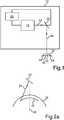
1 eine schematische Darstellung einer erfindungsgemäßen Behandlungsvorrichtung;2a eine Prinzipdarstellung der Erzeugung einer ersten Grenzfläche gemäß einer ersten Ausführungsform des erfindungsgemäßen Verfahrens;2b eine Prinzipdarstellung der Erzeugung eines abzutrennenden Volumenkörpers gemäß der ersten Ausführungsform des erfindungsgemäßen Verfahrens; und3 eine Prinzipdarstellung der Erzeugung eines abzutrennenden Volumenkörpers gemäß einer zweiten Ausführungsform des erfindungsgemäßen Verfahrens.
1 a schematic representation of a treatment device according to the invention;2a a schematic diagram of the generation of a first interface according to a first embodiment of the method according to the invention;2 B a schematic representation of the generation of a volume to be separated according to the first embodiment of the method according to the invention; and3 a schematic representation of the generation of a volume body to be separated according to a second embodiment of the method according to the invention.
Des Weiteren erkennt man, dass der durch den Laser
Bei dem dargestellten Laser
Die Steuereinrichtung
Man erkennt, dass die Grenzfläche
Bezüglich der Erklärung der weiteren Merkmale der
ZITATE ENTHALTEN IN DER BESCHREIBUNG QUOTES INCLUDED IN THE DESCRIPTION
Diese Liste der vom Anmelder aufgeführten Dokumente wurde automatisiert erzeugt und ist ausschließlich zur besseren Information des Lesers aufgenommen. Die Liste ist nicht Bestandteil der deutschen Patent- bzw. Gebrauchsmusteranmeldung. Das DPMA übernimmt keinerlei Haftung für etwaige Fehler oder Auslassungen.This list of the documents listed by the applicant was generated automatically and is included solely for the better information of the reader. The list is not part of the German patent or utility model application. The DPMA assumes no liability for any errors or omissions.
Zitierte PatentliteraturPatent literature cited
- WO 02/22003 [0002]WO 02/22003 [0002]
- US 6551306 [0002]US 6551306 [0002]
- EP 2211803 B1 [0013]EP 2211803 B1 [0013]
Claims (17)
Translated fromGermanPriority Applications (4)
| Application Number | Priority Date | Filing Date | Title |
|---|---|---|---|
| DE102019103848.0ADE102019103848B4 (en) | 2019-02-15 | 2019-02-15 | Method for controlling an ophthalmic surgical laser and treatment device |
| EP20155996.0AEP3695817B1 (en) | 2019-02-15 | 2020-02-07 | Treatment device for controlling an ophthalmic surgical laser |
| CN202010093066.5ACN111568639B (en) | 2019-02-15 | 2020-02-14 | Control method and treatment device of ophthalmic surgical laser |
| US16/791,190US11877957B2 (en) | 2019-02-15 | 2020-02-14 | Method for controlling an eye surgical laser and treatment device |
Applications Claiming Priority (1)
| Application Number | Priority Date | Filing Date | Title |
|---|---|---|---|
| DE102019103848.0ADE102019103848B4 (en) | 2019-02-15 | 2019-02-15 | Method for controlling an ophthalmic surgical laser and treatment device |
Publications (2)
| Publication Number | Publication Date |
|---|---|
| DE102019103848A1true DE102019103848A1 (en) | 2020-08-20 |
| DE102019103848B4 DE102019103848B4 (en) | 2023-03-09 |
Family
ID=69593546
Family Applications (1)
| Application Number | Title | Priority Date | Filing Date |
|---|---|---|---|
| DE102019103848.0AActiveDE102019103848B4 (en) | 2019-02-15 | 2019-02-15 | Method for controlling an ophthalmic surgical laser and treatment device |
Country Status (4)
| Country | Link |
|---|---|
| US (1) | US11877957B2 (en) |
| EP (1) | EP3695817B1 (en) |
| CN (1) | CN111568639B (en) |
| DE (1) | DE102019103848B4 (en) |
Families Citing this family (4)
| Publication number | Priority date | Publication date | Assignee | Title |
|---|---|---|---|---|
| DE102020128625B4 (en)* | 2020-10-30 | 2025-09-04 | Schwind Eye-Tech-Solutions Gmbh | Method for providing control data for an ophthalmic surgical laser of a treatment device, control device and treatment device |
| DE102020133189B4 (en)* | 2020-12-11 | 2024-07-18 | Schwind Eye-Tech-Solutions Gmbh | Method for controlling an ophthalmic surgical laser and treatment device |
| DE102023118630A1 (en)* | 2023-07-13 | 2025-01-16 | Schwind Eye-Tech-Solutions Gmbh | Method for providing control data for an ophthalmological laser of a treatment device for hyperopia correction |
| DE102023118632A1 (en)* | 2023-07-13 | 2025-01-16 | Schwind Eye-Tech-Solutions Gmbh | Method for providing control data for an ophthalmological laser of a treatment device for myopia correction |
Citations (1)
| Publication number | Priority date | Publication date | Assignee | Title |
|---|---|---|---|---|
| US6325792B1 (en)* | 1991-11-06 | 2001-12-04 | Casimir A. Swinger | Ophthalmic surgical laser and method |
Family Cites Families (16)
| Publication number | Priority date | Publication date | Assignee | Title |
|---|---|---|---|---|
| US5984916A (en)* | 1993-04-20 | 1999-11-16 | Lai; Shui T. | Ophthalmic surgical laser and method |
| US7892226B2 (en) | 1995-03-20 | 2011-02-22 | Amo Development, Llc. | Method of corneal surgery by laser incising a contoured corneal flap |
| US6110166A (en)* | 1995-03-20 | 2000-08-29 | Escalon Medical Corporation | Method for corneal laser surgery |
| US6551306B1 (en) | 1999-04-13 | 2003-04-22 | Cesar C. Carriazo | Refractive laser ablation through topography |
| CA2331223C (en)* | 2000-03-27 | 2011-08-16 | Intralase Corp. | A method of corneal surgery by laser incising a contoured corneal flap |
| IT1318699B1 (en) | 2000-09-15 | 2003-08-27 | Ligi Tecnologie Medicali S R L | EQUIPMENT TO DETERMINE AND ABLATE THE VOLUME OF THE CORNEAL TISSUE NECESSARY TO CARRY OUT A CORNEA LAMELLAR TRANSPLANT. |
| ATE365511T1 (en)* | 2002-03-23 | 2007-07-15 | Intralase Corp | SYSTEM FOR IMPROVED MATERIAL PROCESSING USING A LASER BEAM |
| US7131968B2 (en)* | 2003-06-02 | 2006-11-07 | Carl Zeiss Meditec Ag | Apparatus and method for opthalmologic surgical procedures using a femtosecond fiber laser |
| US8685006B2 (en)* | 2006-11-10 | 2014-04-01 | Carl Zeiss Meditec Ag | Treatment apparatus for surgical correction of defective eyesight, method of generating control data therefore, and method for surgical correction of defective eyesight |
| DE102007019814B4 (en) | 2007-04-26 | 2024-11-07 | Carl Zeiss Meditec Ag | follow-up treatment for ophthalmic surgical refractive correction |
| DE102007053283B4 (en)* | 2007-11-08 | 2019-08-29 | Carl Zeiss Meditec Ag | Treatment device for operative vision correction of an eye and method for generating control data therefor |
| US20090187172A1 (en)* | 2008-01-18 | 2009-07-23 | Luis Antonio Ruiz | Method for harvesting corneal donor plugs for use in keratophakia procedures |
| US10080684B2 (en)* | 2008-03-13 | 2018-09-25 | Optimedica Corporation | System and method for laser corneal incisions for keratoplasty procedures |
| DE102011109058A1 (en)* | 2011-07-29 | 2013-01-31 | Carl Zeiss Meditec Ag | "Ophthalmic Laser Device and Method for the Prevention and Treatment of After-Star" |
| DE102015008127A1 (en)* | 2015-06-24 | 2016-12-29 | Wavelight Gmbh | Apparatus for laser eye surgery and method for performing a transepithelial photorefractive keratectomy |
| EP3427705B1 (en) | 2017-07-13 | 2020-04-01 | Ziemer Ophthalmic Systems AG | Device for treating eye tissue using a pulsed laser beam |
- 2019
- 2019-02-15DEDE102019103848.0Apatent/DE102019103848B4/enactiveActive
- 2020
- 2020-02-07EPEP20155996.0Apatent/EP3695817B1/enactiveActive
- 2020-02-14USUS16/791,190patent/US11877957B2/enactiveActive
- 2020-02-14CNCN202010093066.5Apatent/CN111568639B/enactiveActive
Patent Citations (1)
| Publication number | Priority date | Publication date | Assignee | Title |
|---|---|---|---|---|
| US6325792B1 (en)* | 1991-11-06 | 2001-12-04 | Casimir A. Swinger | Ophthalmic surgical laser and method |
Also Published As
| Publication number | Publication date |
|---|---|
| EP3695817B1 (en) | 2023-05-10 |
| US20200261272A1 (en) | 2020-08-20 |
| US11877957B2 (en) | 2024-01-23 |
| DE102019103848B4 (en) | 2023-03-09 |
| CN111568639B (en) | 2024-08-23 |
| CN111568639A (en) | 2020-08-25 |
| EP3695817A1 (en) | 2020-08-19 |
Similar Documents
| Publication | Publication Date | Title |
|---|---|---|
| EP3695817B1 (en) | Treatment device for controlling an ophthalmic surgical laser | |
| EP3912607B1 (en) | Method for providing control data for an ophthalmic surgical laser of a treatment device | |
| EP3695818B1 (en) | Computerprogram for controlling an ophthalmic surgical laser and treatment device | |
| DE102020112277A1 (en) | Method for providing control data for an ophthalmic surgical laser and method for controlling a treatment device | |
| DE102019107182B4 (en) | Control of an ophthalmic laser for the separation of a solid | |
| DE102019122166A1 (en) | Method for controlling an ophthalmic laser and treatment device | |
| DE102009009382A1 (en) | Control data producing device for controlling treatment device for corrective surgery of defective vision of eye of patient, determines control data such that volumes lying at edge of lamella are ablated or removed according to control data | |
| DE102020112583A1 (en) | Method for controlling an ophthalmic laser and treatment device | |
| DE102020104687B4 (en) | Method for controlling an eye surgical laser and treatment device | |
| EP2621428B1 (en) | Device for lasering the human eye | |
| DE102021111266B4 (en) | Treatment device and method for providing control data for controlling an ophthalmic laser with a space-filling curve | |
| DE102021116497A1 (en) | Method for controlling an ophthalmic surgical laser, treatment device, computer program and computer-readable medium | |
| DE102019122167A1 (en) | Method for controlling an ophthalmic laser and treatment device | |
| DE102019133428B3 (en) | Method for controlling an ophthalmic laser with a transition zone on the solid | |
| DE102019118315A1 (en) | Method for controlling an ophthalmic laser and treatment device | |
| EP3906903A1 (en) | Method for providing control data for an ophthalmic laser, method for controlling a treatment device, control device, treatment device, computer program, computer readable medium | |
| DE102021101119A1 (en) | Method for controlling an ophthalmic surgical laser and treatment device | |
| DE102019135607B4 (en) | Method for controlling an ophthalmic surgical laser and treatment device | |
| DE102020114791B4 (en) | Method for controlling an ophthalmic surgical laser and treatment device | |
| DE102019115495A1 (en) | Method for controlling an ophthalmic laser and treatment device | |
| DE102020123611B4 (en) | System for controlling an eye surgical laser and method for determining control data for controlling an eye surgical laser | |
| DE102021100509B4 (en) | Method for controlling an ophthalmic surgical laser, computer program product and treatment device | |
| DE102020104681B4 (en) | Treatment device for the detachment of a solid body from an eye, method, computer program and computer-readable medium | |
| DE102019134146B4 (en) | Method for controlling an ophthalmic laser and treatment device | |
| DE102020104683A1 (en) | Treatment device for the separation of a solid from an eye, method, computer program and computer-readable medium |
Legal Events
| Date | Code | Title | Description |
|---|---|---|---|
| R012 | Request for examination validly filed | ||
| R082 | Change of representative | Representative=s name:HOFSTETTER, SCHURACK & PARTNER PATENT- UND REC, DE Representative=s name:HOFSTETTER, SCHURACK & PARTNER - PATENT- UND R, DE | |
| R016 | Response to examination communication | ||
| R083 | Amendment of/additions to inventor(s) | ||
| R083 | Amendment of/additions to inventor(s) | ||
| R016 | Response to examination communication | ||
| R018 | Grant decision by examination section/examining division | ||
| R020 | Patent grant now final |