DE102010009295A1 - Method for displaying a region to be examined and / or treated - Google Patents
Method for displaying a region to be examined and / or treatedDownload PDFInfo
- Publication number
- DE102010009295A1 DE102010009295A1DE102010009295ADE102010009295ADE102010009295A1DE 102010009295 A1DE102010009295 A1DE 102010009295A1DE 102010009295 ADE102010009295 ADE 102010009295ADE 102010009295 ADE102010009295 ADE 102010009295ADE 102010009295 A1DE102010009295 A1DE 102010009295A1
- Authority
- DE
- Germany
- Prior art keywords
- image data
- image
- data set
- section
- modality
- Prior art date
- Legal status (The legal status is an assumption and is not a legal conclusion. Google has not performed a legal analysis and makes no representation as to the accuracy of the status listed.)
- Granted
Links
- 238000000034methodMethods0.000titleclaimsabstractdescription15
- 238000012545processingMethods0.000claimsabstractdescription40
- 238000002604ultrasonographyMethods0.000claimsdescription14
- 241000699666Mus <mouse, genus>Species0.000description8
- 230000004927fusionEffects0.000description6
- 210000000481breastAnatomy0.000description5
- 238000003384imaging methodMethods0.000description5
- 230000005855radiationEffects0.000description4
- 230000012447hatchingEffects0.000description3
- 206010028980NeoplasmDiseases0.000description2
- 210000000038chestAnatomy0.000description2
- 238000001514detection methodMethods0.000description2
- 238000011161developmentMethods0.000description2
- 230000018109developmental processEffects0.000description2
- 238000003745diagnosisMethods0.000description2
- 238000010586diagramMethods0.000description2
- 238000009877renderingMethods0.000description2
- 206010006187Breast cancerDiseases0.000description1
- 208000026310Breast neoplasmDiseases0.000description1
- 241000699670Mus sp.Species0.000description1
- 230000005856abnormalityEffects0.000description1
- 201000011510cancerDiseases0.000description1
- 238000004891communicationMethods0.000description1
- 230000000295complement effectEffects0.000description1
- 238000005562fadingMethods0.000description1
- 238000009607mammographyMethods0.000description1
- 239000003550markerSubstances0.000description1
- 230000002265preventionEffects0.000description1
- 238000001454recorded imageMethods0.000description1
- 238000012216screeningMethods0.000description1
- 239000013589supplementSubstances0.000description1
- 230000009466transformationEffects0.000description1
- 230000000007visual effectEffects0.000description1
Images

Classifications
- A—HUMAN NECESSITIES
- A61—MEDICAL OR VETERINARY SCIENCE; HYGIENE
- A61B—DIAGNOSIS; SURGERY; IDENTIFICATION
- A61B8/00—Diagnosis using ultrasonic, sonic or infrasonic waves
- A61B8/52—Devices using data or image processing specially adapted for diagnosis using ultrasonic, sonic or infrasonic waves
- A61B8/5215—Devices using data or image processing specially adapted for diagnosis using ultrasonic, sonic or infrasonic waves involving processing of medical diagnostic data
- A61B8/5238—Devices using data or image processing specially adapted for diagnosis using ultrasonic, sonic or infrasonic waves involving processing of medical diagnostic data for combining image data of patient, e.g. merging several images from different acquisition modes into one image
- A—HUMAN NECESSITIES
- A61—MEDICAL OR VETERINARY SCIENCE; HYGIENE
- A61B—DIAGNOSIS; SURGERY; IDENTIFICATION
- A61B6/00—Apparatus or devices for radiation diagnosis; Apparatus or devices for radiation diagnosis combined with radiation therapy equipment
- A61B6/02—Arrangements for diagnosis sequentially in different planes; Stereoscopic radiation diagnosis
- A61B6/025—Tomosynthesis
- A—HUMAN NECESSITIES
- A61—MEDICAL OR VETERINARY SCIENCE; HYGIENE
- A61B—DIAGNOSIS; SURGERY; IDENTIFICATION
- A61B6/00—Apparatus or devices for radiation diagnosis; Apparatus or devices for radiation diagnosis combined with radiation therapy equipment
- A61B6/44—Constructional features of apparatus for radiation diagnosis
- A61B6/4417—Constructional features of apparatus for radiation diagnosis related to combined acquisition of different diagnostic modalities
- A—HUMAN NECESSITIES
- A61—MEDICAL OR VETERINARY SCIENCE; HYGIENE
- A61B—DIAGNOSIS; SURGERY; IDENTIFICATION
- A61B6/00—Apparatus or devices for radiation diagnosis; Apparatus or devices for radiation diagnosis combined with radiation therapy equipment
- A61B6/46—Arrangements for interfacing with the operator or the patient
- A61B6/461—Displaying means of special interest
- A61B6/463—Displaying means of special interest characterised by displaying multiple images or images and diagnostic data on one display
- A—HUMAN NECESSITIES
- A61—MEDICAL OR VETERINARY SCIENCE; HYGIENE
- A61B—DIAGNOSIS; SURGERY; IDENTIFICATION
- A61B6/00—Apparatus or devices for radiation diagnosis; Apparatus or devices for radiation diagnosis combined with radiation therapy equipment
- A61B6/50—Apparatus or devices for radiation diagnosis; Apparatus or devices for radiation diagnosis combined with radiation therapy equipment specially adapted for specific body parts; specially adapted for specific clinical applications
- A61B6/502—Apparatus or devices for radiation diagnosis; Apparatus or devices for radiation diagnosis combined with radiation therapy equipment specially adapted for specific body parts; specially adapted for specific clinical applications for diagnosis of breast, i.e. mammography
- A—HUMAN NECESSITIES
- A61—MEDICAL OR VETERINARY SCIENCE; HYGIENE
- A61B—DIAGNOSIS; SURGERY; IDENTIFICATION
- A61B6/00—Apparatus or devices for radiation diagnosis; Apparatus or devices for radiation diagnosis combined with radiation therapy equipment
- A61B6/52—Devices using data or image processing specially adapted for radiation diagnosis
- A61B6/5211—Devices using data or image processing specially adapted for radiation diagnosis involving processing of medical diagnostic data
- A61B6/5229—Devices using data or image processing specially adapted for radiation diagnosis involving processing of medical diagnostic data combining image data of a patient, e.g. combining a functional image with an anatomical image
- A61B6/5247—Devices using data or image processing specially adapted for radiation diagnosis involving processing of medical diagnostic data combining image data of a patient, e.g. combining a functional image with an anatomical image combining images from an ionising-radiation diagnostic technique and a non-ionising radiation diagnostic technique, e.g. X-ray and ultrasound
- A—HUMAN NECESSITIES
- A61—MEDICAL OR VETERINARY SCIENCE; HYGIENE
- A61B—DIAGNOSIS; SURGERY; IDENTIFICATION
- A61B8/00—Diagnosis using ultrasonic, sonic or infrasonic waves
- A61B8/42—Details of probe positioning or probe attachment to the patient
- A61B8/4209—Details of probe positioning or probe attachment to the patient by using holders, e.g. positioning frames
- A61B8/4218—Details of probe positioning or probe attachment to the patient by using holders, e.g. positioning frames characterised by articulated arms
- A—HUMAN NECESSITIES
- A61—MEDICAL OR VETERINARY SCIENCE; HYGIENE
- A61B—DIAGNOSIS; SURGERY; IDENTIFICATION
- A61B8/00—Diagnosis using ultrasonic, sonic or infrasonic waves
- A61B8/44—Constructional features of the ultrasonic, sonic or infrasonic diagnostic device
- A61B8/4416—Constructional features of the ultrasonic, sonic or infrasonic diagnostic device related to combined acquisition of different diagnostic modalities, e.g. combination of ultrasound and X-ray acquisitions
- A—HUMAN NECESSITIES
- A61—MEDICAL OR VETERINARY SCIENCE; HYGIENE
- A61B—DIAGNOSIS; SURGERY; IDENTIFICATION
- A61B8/00—Diagnosis using ultrasonic, sonic or infrasonic waves
- A61B8/46—Ultrasonic, sonic or infrasonic diagnostic devices with special arrangements for interfacing with the operator or the patient
- A61B8/461—Displaying means of special interest
- A61B8/463—Displaying means of special interest characterised by displaying multiple images or images and diagnostic data on one display
- A—HUMAN NECESSITIES
- A61—MEDICAL OR VETERINARY SCIENCE; HYGIENE
- A61B—DIAGNOSIS; SURGERY; IDENTIFICATION
- A61B90/00—Instruments, implements or accessories specially adapted for surgery or diagnosis and not covered by any of the groups A61B1/00 - A61B50/00, e.g. for luxation treatment or for protecting wound edges
- A61B90/36—Image-producing devices or illumination devices not otherwise provided for
- A61B2090/364—Correlation of different images or relation of image positions in respect to the body
- A—HUMAN NECESSITIES
- A61—MEDICAL OR VETERINARY SCIENCE; HYGIENE
- A61B—DIAGNOSIS; SURGERY; IDENTIFICATION
- A61B90/00—Instruments, implements or accessories specially adapted for surgery or diagnosis and not covered by any of the groups A61B1/00 - A61B50/00, e.g. for luxation treatment or for protecting wound edges
- A61B90/36—Image-producing devices or illumination devices not otherwise provided for
- A61B90/37—Surgical systems with images on a monitor during operation
- A61B2090/376—Surgical systems with images on a monitor during operation using X-rays, e.g. fluoroscopy
- A61B2090/3762—Surgical systems with images on a monitor during operation using X-rays, e.g. fluoroscopy using computed tomography systems [CT]
- A—HUMAN NECESSITIES
- A61—MEDICAL OR VETERINARY SCIENCE; HYGIENE
- A61B—DIAGNOSIS; SURGERY; IDENTIFICATION
- A61B90/00—Instruments, implements or accessories specially adapted for surgery or diagnosis and not covered by any of the groups A61B1/00 - A61B50/00, e.g. for luxation treatment or for protecting wound edges
- A61B90/36—Image-producing devices or illumination devices not otherwise provided for
- A61B90/37—Surgical systems with images on a monitor during operation
- A61B2090/378—Surgical systems with images on a monitor during operation using ultrasound
- A—HUMAN NECESSITIES
- A61—MEDICAL OR VETERINARY SCIENCE; HYGIENE
- A61B—DIAGNOSIS; SURGERY; IDENTIFICATION
- A61B6/00—Apparatus or devices for radiation diagnosis; Apparatus or devices for radiation diagnosis combined with radiation therapy equipment
- A61B6/44—Constructional features of apparatus for radiation diagnosis
- A61B6/4429—Constructional features of apparatus for radiation diagnosis related to the mounting of source units and detector units
- A61B6/4458—Constructional features of apparatus for radiation diagnosis related to the mounting of source units and detector units the source unit or the detector unit being attached to robotic arms
- A—HUMAN NECESSITIES
- A61—MEDICAL OR VETERINARY SCIENCE; HYGIENE
- A61B—DIAGNOSIS; SURGERY; IDENTIFICATION
- A61B6/00—Apparatus or devices for radiation diagnosis; Apparatus or devices for radiation diagnosis combined with radiation therapy equipment
- A61B6/46—Arrangements for interfacing with the operator or the patient
- A61B6/467—Arrangements for interfacing with the operator or the patient characterised by special input means
- A61B6/469—Arrangements for interfacing with the operator or the patient characterised by special input means for selecting a region of interest [ROI]
- A—HUMAN NECESSITIES
- A61—MEDICAL OR VETERINARY SCIENCE; HYGIENE
- A61B—DIAGNOSIS; SURGERY; IDENTIFICATION
- A61B8/00—Diagnosis using ultrasonic, sonic or infrasonic waves
- A61B8/46—Ultrasonic, sonic or infrasonic diagnostic devices with special arrangements for interfacing with the operator or the patient
- A61B8/467—Ultrasonic, sonic or infrasonic diagnostic devices with special arrangements for interfacing with the operator or the patient characterised by special input means
- A61B8/469—Ultrasonic, sonic or infrasonic diagnostic devices with special arrangements for interfacing with the operator or the patient characterised by special input means for selection of a region of interest
Landscapes
- Health & Medical Sciences (AREA)
- Life Sciences & Earth Sciences (AREA)
- Engineering & Computer Science (AREA)
- Medical Informatics (AREA)
- Surgery (AREA)
- Molecular Biology (AREA)
- Animal Behavior & Ethology (AREA)
- Pathology (AREA)
- Veterinary Medicine (AREA)
- Biomedical Technology (AREA)
- Heart & Thoracic Surgery (AREA)
- Public Health (AREA)
- General Health & Medical Sciences (AREA)
- Nuclear Medicine, Radiotherapy & Molecular Imaging (AREA)
- Physics & Mathematics (AREA)
- Biophysics (AREA)
- Radiology & Medical Imaging (AREA)
- High Energy & Nuclear Physics (AREA)
- Optics & Photonics (AREA)
- Computer Vision & Pattern Recognition (AREA)
- Oral & Maxillofacial Surgery (AREA)
- Human Computer Interaction (AREA)
- Dentistry (AREA)
- Apparatus For Radiation Diagnosis (AREA)
- Ultra Sonic Daignosis Equipment (AREA)
Abstract
Translated fromGermanDescription
Translated fromGermanDie Erfindung betrifft ein Verfahren zur Darstellung eines zu untersuchenden und/oder behandelnden Bereichs, wobei wenigstens ein mit Bildaufnahmemitteln einer ersten Modalität aufgenommener erster Bilddatensatz des zu untersuchenden und/oder behandelnden Bereichs und wenigstens ein mit Bildaufnahmemitteln einer zweiten, von der ersten Modalität verschiedenen Modalität aufgenommener zweiter Bilddatensatz des zu untersuchenden und/oder behandelnden Bereichs von einer Verarbeitungseinrichtung registriert werden.The invention relates to a method for displaying a region to be examined and / or treated, wherein at least one first image data record of the area to be examined and / or treated recorded with image acquisition means of a first modality and at least one photograph taken with image acquisition means of a second modality different from the first modality second image data set of the area to be examined and / or treated are registered by a processing device.
Zu Zwecken der Visualisierung insbesondere von 3D-Bilddaten verschiedener Modalitäten, etwa Röntgen-Tomosynthese und Ultraschall, ist es üblich, die mit Bildaufnahmemitteln unterschiedlicher Modalitäten aufgenommenen Bilddatensätze entweder auf verschiedenen Bildschirmen oder auf dem gleichen Bildschirm, jedoch an verschiedenen Stellen beziehungsweise in verschiedenen Fenstern anzuzeigen. Ferner ist eine Datenfusion, das heißt die mit Bildaufnahmemitteln unterschiedlicher Modalitäten aufgenommenen Bilddatensätze werden über eine geeignete rechnerbasierte Verarbeitungseinrichtung zu einem gemeinsamen Bilddatensatz fusioniert, bekannt, welcher ermöglicht, ein aus beiden Bilddatensätzen kombiniertes Bild anzuzeigen. Die Datenfusion ist äußerst rechenintensiv und bei Bildaufnahmen eines deformierbaren Objekts, für das die beiden Einzeldatensätze in unterschiedlicher Geometrie erzeugt wurden, sehr schwierig. Ein Beispiel ist die Tomosynthese und der 3D-Ultraschall der weiblichen Brust.For the purpose of visualizing in particular 3D image data of various modalities, such as X-ray tomosynthesis and ultrasound, it is customary to display the image data sets recorded with imaging means of different modalities either on different screens or on the same screen but at different locations or in different windows. Furthermore, a data fusion, that is to say the image data sets recorded with image acquisition means of different modalities, are fused to a common image data record via a suitable computer-based processing device, which makes it possible to display an image combined from both image data sets. The data fusion is extremely computationally intensive and very difficult when taking pictures of a deformable object, for which the two individual data sets were created in different geometries. An example is the tomosynthesis and the 3D ultrasound of the female breast.
Somit liegt der Erfindung das Problem zugrunde, ein Verfahren mit einer verbesserten gemeinsamen Darstellbarkeit von mit Bildaufnahmemitteln unterschiedlicher Modalitäten aufgenommener Bilddatensätze anzugeben.Thus, the invention is based on the problem of specifying a method with an improved common representability of image data sets recorded with image acquisition means of different modalities.
Erfindungsgemäß wird das Problem durch ein Verfahren der eingangs genannten Art gelöst, welches sich dadurch auszeichnet, dass in der Darstellung des ersten Bilddatensatzes an einem Anzeigemittel ein Bildausschnitt ausgewählt wird und anschließend die Verarbeitungseinrichtung die Bilddaten des dem ausgewählten Bildausschnitt des ersten Bilddatensatzes entsprechenden Bildausschnitt des zweiten Bilddatensatzes erfasst und als Bilddarstellung an der ausgewählten Stelle des Bildausschnitts des ersten Bilddatensatzes, diesen überlagernd anzeigt.According to the invention, the problem is solved by a method of the type mentioned above, which is characterized in that in the representation of the first image data set on a display means a picture detail is selected and then the processing means the image data of the selected image section of the first image data corresponding image detail of the second image data set recorded and displayed as an image representation at the selected location of the image section of the first image data set, superimposed on this.
Das erfindungsgemäße Verfahren läuft sonach im Wesentlichen nach folgenden Schritten ab. Zunächst wird das zu untersuchende und/oder zu behandelnde Untersuchungsobjekt beziehungsweise der zu untersuchende und/oder behandelnde Bereich eines Untersuchungsobjekts mit Bildaufnahmemitteln zweier unterschiedlicher Modalitäten aufgenommen und die Bilddaten an die Verarbeitungseinrichtung weitergeleitet. Es liegen in der Verarbeitungseinrichtung entsprechend der verwendeten unterschiedlichen Bildaufnahmemodalitäten sonach zwei unterschiedliche Bilddatensätze vor. Die unterschiedlichen Bilddatensätze werden von der Verarbeitungseinrichtung miteinander geometrisch registriert, so dass ein Zusammenhang zwischen Bildpunkten des ersten Bilddatensatzes und Bildpunkten des zweiten Bilddatensatzes erhalten wird.The method according to the invention therefore proceeds essentially according to the following steps. First of all, the examination object to be examined and / or the area of an examination object to be examined and / or treated is recorded with image recording means of two different modalities and the image data forwarded to the processing device. Accordingly, two different image data sets are present in the processing device in accordance with the different image acquisition modalities used. The different image data sets are geometrically registered with each other by the processing device, so that a relationship between pixels of the first image data set and pixels of the second image data set is obtained.
An einem Anzeigemittel, das heißt einem Bildschirm oder dergleichen, wird zunächst nur der mit den Bildaufnahmemitteln der ersten Modalität aufgenommene erste Bilddatensatz angezeigt. Die Anzeige des ersten Bilddatensatzes erfolgt dabei bevorzugt über die volle Anzeigefläche des Anzeigemittels, so dass sich z. B. ein Benutzer einen guten Überblick über das zu untersuchende und/oder behandelnde Untersuchungsobjekt respektive einen Bereich dessen verschaffen kann.At a display means, that is a screen or the like, only the first image data record recorded with the image acquisition means of the first modality is initially displayed. The display of the first image data set is preferably carried out over the full display area of the display means, so that z. B. a user can provide a good overview of the examined and / or treated examination object or an area thereof.
Im Weiteren wird ein Bereich beziehungsweise ein Bildausschnitt von Interesse, in welchem sich z. B. eine Auffälligkeit befindet, innerhalb des angezeigten ersten Bilddatensatzes ausgewählt. Die Auswahl kann dabei beispielsweise über wenigstens einen Benutzer oder automatisch durch die Verarbeitungseinrichtung erfolgen. Im Falle der Auswahl über einen Benutzer wählt dieser einen Bildausschnitt aus der Anzeige des ersten Bilddatensatzes aus, wobei es sich bei dem ausgewählten Bildausschnitt beispielsweise um einen Bereich handeln kann, in welchem sich eine zu beobachtende Geschwulst oder sonst eine medizinische Auffälligkeit befindet. Gleichermaßen kann die Auswahl z. B. einen Bildausschnitt betreffen, welcher ein für den Benutzer unklares Bild darstellt. Gleiches gilt, wenn die Auswahl des Bildausschnitts automatisch durch die Verarbeitungseinrichtung erfolgt. Die von der Verarbeitungseinrichtung getroffene Auswahl kann sich an der klinischen Vorgeschichte des Untersuchungsobjekts orientieren bzw. können von Algorithmen zur Bilderkennung nicht eindeutig erkennbare oder zuordbare Bereiche oder sonstige Strukturen als Bildausschnitt gewählt werden.In addition, an area or an image section of interest, in which z. B. an abnormality is selected within the displayed first image data set. The selection can be made for example via at least one user or automatically by the processing device. In the case of selection via a user, the latter selects an image section from the display of the first image data record, wherein the selected image detail can be, for example, an area in which there is a tumor to be observed or otherwise a medical conspicuity. Similarly, the selection z. B. relate to a picture, which represents an unclear for the user image. The same applies if the selection of the image section is carried out automatically by the processing device. The selection made by the processing device can be based on the clinical history of the examination subject or it is possible to select areas or other structures that are not clearly recognizable or assignable by image recognition algorithms as an image detail.
Im Folgenden wird der dem ausgewählten Bildausschnitt des ersten Bilddatensatzes entsprechende Bildausschnitt des zweiten Bilddatensatzes von der Verarbeitungseinrichtung erfasst. Dies ist möglich, da die beiden Bilddatensätze eingangs von der Verarbeitungseinrichtung miteinander registriert, das heißt mittels geeigneter Algorithmen aufeinander abbildbar sind. Hierunter ist keine Datenfusion zu verstehen.The following is the one of the selected image section of the first Image data set corresponding image section of the second image data set detected by the processing device. This is possible since the two image data records are registered with one another at the beginning by the processing device, that is, they can be imaged onto one another by means of suitable algorithms. This is not to be understood as a data fusion.
Der von der Verarbeitungseinrichtung erfasste, dem ausgewählten Bildausschnitt des ersten Bilddatensatzes entsprechende Bildausschnitt des zweiten Bilddatensatzes wird im Weiteren als Bilddarstellung an der ausgewählten Stelle des ersten Bilddatensatzes anstelle des Bildausschnitts des ersten Bilddatensatzes als überlagertes Bild simultan angezeigt. An dieser Stelle ist folglich das mit den Bildaufnahmemitteln der zweiten Modalität aufgenommene Bild zu sehen. Es befindet sich ausschnittsweise eine Darstellung des zweiten Bilddatensatzes in der Anzeige des ersten Bilddatensatzes.The image section of the second image data record corresponding to the selected image section of the first image data set acquired by the processing device is displayed as an image representation at the selected location of the first image data set instead of the image section of the first image data set as a superimposed image. Consequently, the image taken with the image acquisition means of the second modality can be seen at this point. There is a section of a representation of the second image data set in the display of the first image data set.
Selbstverständlich können auch mehrere, das heißt mehr als zwei mit Bildaufnahmemitteln mehrerer, verschiedener Modalitäten aufgenommene Bilddatensätze vorliegen. Derart kann der im ersten Bilddatensatz ausgewählte Bildausschnitt wahlweise mit der Anzeige einer diesem entsprechenden Bilddarstellung eines mit Bildaufnahmemitteln einer zweiten oder einer weiteren Modalität aufgenommenen Bilddatensatzes überlagert werden. Für den ausgewählten Bildausschnitt des ersten Bilddatensatzes liegt sonach eine der Anzahl der eingesetzten Modalitäten entsprechende Anzahl an unterschiedlichen, diesen überlagerbaren Bilddarstellungen vor. Ferner kann auch mehr als ein Bereich ausgewählt und durch den anderen Bilddatensatz im Bild ersetzt werden.Of course, there may also be a plurality of image data records, ie more than two image data sets recorded with image acquisition means of a plurality of different modalities. In this way, the image detail selected in the first image data record can optionally be overlaid with the display of an image representation corresponding to it of an image data record taken with image acquisition means of a second or another modality. For the selected image section of the first image data set, therefore, there is a corresponding number of different image representations that can be superimposed on the number of modalities used. Furthermore, more than one area can also be selected and replaced by the other image data set in the image.
Bevorzugt erfolgt die manuelle Auswahl durch den Benutzer über ein Bedienelement, insbesondere eine Maus, eine Tastatur oder einen Trackball. Der Benutzer wählt anhand eines Cursors oder einer ähnlichen Eingabemarkierung, welchen bzw. welche er über das Bedienelement steuert, wenigstens einen Bildausschnitt aus dem ersten Bilddatensatz aus. Als Bedienelemente kommen vorteilhaft jedoch nicht in abschließender Aufzählung Mäuse, Tastaturen, Trackballs oder Grafik-Tabletts in Frage. Unter einem Bedienelement ist grundsätzlich jedes geeignete Mittel zu verstehen, mit dem ein Benutzer manuell eine Auswahl eines Bildausschnitts innerhalb der Darstellung eines Bilddatensatzes treffen kann.The manual selection by the user preferably takes place via an operating element, in particular a mouse, a keyboard or a trackball. The user selects at least one image detail from the first image data set by means of a cursor or a similar input marker, which he or she controls via the operating element. As controls are advantageous but not in exhaustive enumeration mice, keyboards, trackballs or graphics trays in question. Under a control element is basically any suitable means to understand with which a user can manually make a selection of an image section within the representation of an image data set.
Bei der automatischen Auswahl des Bildausschnitts durch die Verarbeitungseinrichtung können beispielsweise Algorithmen zur kanten- bzw. geometrischen Strukturerkennung in der Verarbeitungseinrichtung implementiert sein. Die Verarbeitungseinrichtung kann dabei spezielle Computer-Aided-Detection bzw. Diagnose-Systeme (CAD-Systeme) verwenden.In the automatic selection of the image section by the processing device, for example, algorithms for edge or geometric structure recognition can be implemented in the processing device. The processing device can use special computer-aided detection or diagnostic systems (CAD systems).
In Weiterbildung der Erfindung kann es sein, dass die Bildausschnitte des ersten und des zweiten Bilddatensatzes in gleichen oder verschiedenen Dimensionen dargestellt werden. Es kann sich sonach bei dem Bildausschnitt des ersten Bilddatensatzes um eine dreidimensionale Darstellung handeln, wobei der eingeblendete Bildausschnitt des zweiten Bilddatensatzes ebenfalls eine dreidimensionale Darstellung ist. Natürlich kann der Bildausschnitt des ersten und des zweiten Bilddatensatzes auch beide als zweidimensionale Darstellung vorliegen. Andernfalls kann es sich bei dem Bildausschnitt des ersten Bilddatensatzes um eine zweidimensionale Darstellung handeln, wobei entsprechend der zweite Bildausschnitt des zweiten Bilddatensatzes dreidimensional dargestellt wird. Umgekehrt dazu wäre der Bildausschnitt des ersten Bilddatensatzes eine dreidimensionale Darstellung und der Bildausschnitt des zweiten Bilddatensatzes lediglich zweidimensional.In a further development of the invention, it may be that the image sections of the first and the second image data set are displayed in the same or different dimensions. Accordingly, the image section of the first image data set can be a three-dimensional representation, with the displayed image section of the second image data set likewise being a three-dimensional representation. Of course, the image detail of the first and the second image data set can both be present as a two-dimensional representation. Otherwise, the image section of the first image data set may be a two-dimensional representation, wherein the second image section of the second image data set is correspondingly displayed in three dimensions. Conversely, the image section of the first image data set would be a three-dimensional representation and the image section of the second image data set would be only two-dimensional.
Vorteilhaft kann eine dreidimensionale Darstellung des zweiten Bilddatensatzes um ihren Bildmittelpunkt rotieren. Unter Bildmittelpunkt ist hierbei der Volumenmittelpunkt zu verstehen. Aus der Rotation der dreidimensionalen Darstellung ergibt sich eine verbesserte Übersichtlichkeit, gegebenenfalls können derart zunächst verdeckte Strukturen sichtbar gemacht werden. Hierbei können in der Verarbeitungseinrichtung implementierte Bildverarbeitungsalgorithmen wie etwa Volume-Rendering (VR), Maximum-Intensity-Projection (MIP), Surface-Shaded-Display (SSD) angewendet werden. Handelt es sich um einen dreidimensionalen Tomosynthese-Datensatz, erfolgt die Rotation im eingeschränkten Winkelbereich des Tomosyntheseabtastwinkels. Die Rotation kann grundsätzlich automatisch oder benutzergesteuert erfolgen.Advantageously, a three-dimensional representation of the second image data set can rotate about its image center. The center of the image is the volume center. From the rotation of the three-dimensional representation results in an improved clarity, if necessary, such initially hidden structures can be made visible. In this case, image processing algorithms implemented in the processing device, such as volume rendering (VR), maximum intensity projection (MIP), surface shaded display (SSD), can be used. If it is a three-dimensional tomosynthesis data set, the rotation takes place in the restricted angular range of the tomosynthesis scanning angle. The rotation can basically be automatic or user-controlled.
In Weiterbildung der Erfindung kann die Anzeige des Bildausschnitts des zweiten Bilddatensatzes ausgeschaltet werden. Dies ermöglicht ein schnelles Hin- und Her- bzw. Umschalten zwischen dem Bildausschnitt des ersten und dem diesen überlagernden Bildausschnitt des zweiten Bilddatensatzes. Das Umschalten kann z. B. mittels eines Bedienelements, z. B. über einen Mausklick, erfolgen. Daneben ist es möglich, dass in regelmäßigen, intervallartigen zeitlichen Abständen ein Umschalten zwischen dem Bildausschnitt des ersten und dem Bildausschnitt des zweiten Bilddatensatzes stattfindet. Durch das Umschalten lässt sich mitunter eine bessere visuelle Beziehung zwischen dem Bildausschnitt des ersten Bilddatensatzes und dem Bildausschnitt des zweiten Bilddatensatzes herstellen.In a development of the invention, the display of the image section of the second image data set can be switched off. This allows a quick toggling or switching between the image section of the first and the superimposed image section of the second image data set. The switching can z. B. by means of an operating element, for. B. via a mouse click, done. In addition, it is possible that at regular, interval-like time intervals switching between the image section of the first and the image section of the second image data set takes place. The switching can sometimes produce a better visual relationship between the image detail of the first image data set and the image detail of the second image data set.
Als erster Bilddatensatz kann ein Tomosynthese-Bilddatensatz und als zweiter Bilddatensatz ein Ultraschall-Bilddatensatz verwendet werden. Mit Tomosynthese-Verfahren, welche röntgenbasierte Schichtaufnahmen des zu untersuchenden Objekts bzw. Bereichs liefern, können beispielsweise Gewebeveränderungen etwa im Rahmen einer Krebsvorsorge besser erkannt und damit eine Diagnose präziser gestellt werden. Insbesondere bei der Brustkrebsvorsorge bzw. -erkennung weist die Tomosynthese im Vergleich zu herkömmlichen Mammographie-Verfahren Vorteile auf. Ultraschallbilddatensätze sind aus der Sonographie bekannt und ermöglichen eine räumliche (dreidimensionale) Darstellung des zu untersuchenden und/oder behandelnden Untersuchungsobjekts bzw. eines Bereichs dessen.The first image data set can be a tomosynthesis image data set and a second image data set can be an ultrasound image data set. With tomosynthesis methods, which provide x-ray-based tomograms of the object or area to be examined, it is possible, for example, to better identify tissue changes in the context of cancer screening and thus make a diagnosis more precise. Especially in the case of breast cancer prevention or recognition, tomosynthesis has advantages compared to conventional mammography methods. Ultrasound image data sets are known from ultrasonography and allow a spatial (three-dimensional) representation of the examination object to be examined and / or treated or a region thereof.
Selbstverständlich können auch andere Modalitäten eingesetzt werden bzw. kann der erste Bilddatensatz ein Ultraschall-Bilddatensatz und der zweite Bilddatensatz ein Tomosynthese-Bilddatensatz sein.Of course, other modalities may be used, or the first image data set may be an ultrasound image data set and the second image data set may be a tomosynthesis image data set.
Daneben betrifft die Erfindung eine medizinische Untersuchungs- und/oder Behandlungsvorrichtung ausgebildet zur Aufnahme und Darstellung von Bildern eines zu untersuchenden und/oder behandelnden Bereichs, umfassend wenigstens eine erste Modalität und eine von dieser verschiedene zweite Modalität, wobei wenigstens ein mit Bildaufnahmemitteln der ersten Modalität aufgenommener erster Bilddatensatz des zu untersuchenden und/oder behandelnden Bereichs und wenigstens ein mit Bildaufnahmemitteln der zweiten Modalität aufgenommener zweiter Bilddatensatz des zu untersuchenden und/oder behandelnden Bereichs von einer Verarbeitungseinrichtung registrierbar ist. Die medizinische Untersuchungs- und/oder Behandlungsvorrichtung zeichnet sich dadurch aus, dass in der Darstellung des ersten Bilddatensatzes an einem Anzeigemittel ein Bildausschnitt auswählbar ist und die Bilddaten des dem ausgewählten Bildausschnitt des ersten Bilddatensatzes entsprechenden Bildausschnitts des zweiten Bilddatensatzes von der Verarbeitungseinrichtung erfassbar sind und als Bilddarstellung an der ausgewählten Stelle des Bildausschnitts des ersten Bilddatensatzes, diesen überlagernd, anzeigbar sind.In addition, the invention relates to a medical examination and / or treatment device designed to record and display images of an area to be examined and / or treated, comprising at least a first modality and a second modality different from it, wherein at least one recorded with image acquisition means of the first modality The first image data set of the area to be examined and / or treated and at least one second image data set of the area to be examined and / or treated recorded by image acquisition means of the second modality can be registered by a processing device. The medical examination and / or treatment device is characterized in that in the representation of the first image data set on a display means, an image detail is selectable and the image data of the selected image section of the first image data set corresponding image detail of the second image data set can be detected by the processing device and as an image representation at the selected location of the image section of the first image data set, these superimposed, can be displayed.
Der medizinischen Untersuchungs- und/oder Behandlungsvorrichtung sind sonach wenigstens zwei verschiedene Modalitäten mit entsprechenden Bildaufnahmemitteln zugeordnet. Von einem zu untersuchenden und/oder zu behandelnden Untersuchungsobjekt respektive einem Bereich dessen werden mit den verschiedenen Bildaufnahmemitteln der unterschiedlichen Modalitäten den Modalitäten entsprechende Bilddatensätze erstellt und von der Verarbeitungseinrichtung registriert. Es wird im Weiteren von zwei unterschiedlichen Modalitäten ausgegangen, wenngleich selbstverständlich auch mehrere Modalitäten vorstellbar sind.The medical examination and / or treatment device are therefore associated with at least two different modalities with corresponding imaging means. From an examination subject to be examined and / or to be treated or an area thereof, image data sets corresponding to the modalities are created with the different imaging means of the different modalities and registered by the processing device. It is assumed in the following of two different modalities, although of course also several modalities are conceivable.
Durch die Registrierung der Bilddatensätze besteht über eine Transformationsvorschrift ein Zusammenhang zwischen Bildpunkten des ersten Bilddatensatzes und Bildpunkten des zweiten Bilddatensatzes. Der erste Bilddatensatz ist an einem Anzeigemittel, wie z. B. einem Monitor oder dergleichen, dargestellt. Aus der Darstellung des ersten Bilddatensatzes ist wenigstens ein Bildausschnitt auswählbar, wobei nach Auswahl des Bildausschnitts die Verarbeitungseinrichtung die den Bilddaten des dem ausgewählten Bildausschnitt des ersten Bilddatensatzes entsprechenden Bilddaten des zweiten Bilddatensatzes erfassen kann.By registering the image data sets, there is a relationship between pixels of the first image data set and pixels of the second image data set via a transformation rule. The first image data set is on a display means such. As a monitor or the like. At least one image section can be selected from the representation of the first image data set, wherein after selecting the image section the processing device can acquire the image data of the second image data set corresponding to the selected image section of the first image data set.
Die erfassten Bilddaten des zweiten Bilddatensatzes kann die Verarbeitungseinrichtung im Weiteren als Bilddarstellung an der Stelle der ausgewählten Bilddarstellung des ersten Bilddatensatzes, diese überlagernd, darstellen. An dieser Stelle ist demnach nur noch der der Auswahl des Bildausschnitts des ersten Bilddatensatzes entsprechende Bildausschnitt des zweiten Bilddatensatzes angezeigt. Es befindet sich sonach ausschnittsweise eine Bilddarstellung des zweiten Bilddatensatzes innerhalb der Bilddarstellung des ersten Bilddatensatzes. Eine Fusion des ersten mit dem zweiten Bilddatensatz ist dafür nicht notwendig.The captured image data of the second image data set may further represent the processing device as an image representation at the location of the selected image representation of the first image data set, overlaying the same. Accordingly, only the image section of the second image data record corresponding to the selection of the image section of the first image data set is displayed at this point. Accordingly, there is a partial representation of an image representation of the second image data record within the image representation of the first image data set. A fusion of the first with the second image data set is not necessary for this.
Der Bildausschnitt aus dem ersten Bilddatensatz kann manuell durch einen Benutzer oder automatisch durch die Verarbeitungseinrichtung auswählbar sein. Ein Benutzer kann hierzu beispielsweise ein Bedienelement, insbesondere eine Maus, eine Tastatur oder ein Trackball, verwenden, mittels welchem ein Cursor oder eine sonstige Eingabemarkierung an dem Anzeigemittel steuerbar und so ein Bildausschnitt aus der Anzeige des ersten Bilddatensatzes auswählbar ist. Die automatische Auswahl erfolgt bevorzugt über in der Verarbeitungseinrichtung implementierte Algorithmen z. B. ausgebildet zur Erkennung bzw. Detektion von Kanten oder sonstigen geometrischen Strukturen. In der Verarbeitungseinrichtung können hierfür spezielle computergestützte Programme (Computer-Aided-Detection/Diagnose-Programme, kurz CAD-Programme) implementiert sein.The image section from the first image data set can be selected manually by a user or automatically by the processing device. For this purpose, a user can use, for example, an operating element, in particular a mouse, a keyboard or a trackball, by means of which a cursor or other input mark can be controlled on the display means and thus an image detail from the display of the first image data set can be selected. The automatic selection preferably takes place via algorithms implemented in the processing device. B. designed to detect or detect edges or other geometric structures. For this purpose, special computer-aided programs (computer-aided-detection / diagnosis programs, in short CAD programs) can be implemented in the processing device.
Bevorzugt sind die Bildausschnitte des ersten und zweiten Bilddatensatzes in gleichen oder verschiedenen Dimensionen darstellbar. Hieraus ergibt sich, dass beide Bildausschnitte z. B. zwei- oder dreidimensional angezeigt werden können. Dreidimensionale Darstellungen insbesondere des zweiten Bilddatensatzes können z. B. unterstützt von bildgebenden Verfahren wie Volume-Rendering (VR), Maximum-Intensity-Projection (MIP) oder Surface-Shaded-Display (SSD) erfolgen. Gleichermaßen kann die Dimension des ersten Bilddatensatzes anders als die des zweiten Bilddatensatzes sein. Dies ist der Fall, wenn ein Bilddatensatz dreidimensional und der andere lediglich zweidimensional ist.Preferably, the image sections of the first and second image data sets can be displayed in the same or different dimensions. It follows that both image sections z. B. can be displayed two- or three-dimensional. Three-dimensional representations, in particular of the second image data set can be z. Supported by imaging techniques such as volume rendering (VR), maximum intensity projection (MIP) or surface-shaded display (SSD). Likewise, the dimension of the first image dataset may be different than that of the second image dataset. This is the case when one image data set is three-dimensional and the other is only two-dimensional.
Handelt es sich bei dem zweiten Bilddatensatz um eine dreidimensionale Darstellung, ist es bevorzugt möglich, dass diese um ihren Mittelpunkt rotierbar ist. Unter Mittelpunkt ist hierbei der Mittelpunkt des angezeigten Volumens zu verstehen. Derart sind dem entsprechenden dreidimensional dargestellten Bildausschnitt noch mehr Informationen entnehmbar bzw. aus diesem ableitbar. Basiert die dreidimensionale Darstellung des zweiten Bilddatensatzes auf mittels Tomosynthese gewonnenen Bilddaten, erfolgt die Rotation im Winkelbereich des Tomosyntheseabtastwinkels. Die Rotation ist automatisch oder manuell steuerbar.If the second image data set is a three-dimensional representation, it is preferably possible for it to be rotatable about its center. The term "center" is to be understood here as the center of the displayed volume. Such are the corresponding three-dimensional illustrated Image section even more information removable or derivable from this. If the three-dimensional representation of the second image data set is based on image data obtained by means of tomosynthesis, the rotation takes place in the angular range of the tomosynthesis scanning angle. The rotation can be controlled automatically or manually.
Vorteilhaft ist die Anzeige des Bildausschnitts des zweiten Bilddatensatzes zuschaltbar. Entsprechend kann auf einfache Weise zwischen den Bildausschnitten des ersten und des zweiten Bilddatensatzes hin- und hergeschaltet bzw. von dem einen in den anderen umgeschaltet werden. Das Umschalten kann beispielsweise über einen Mausklick oder einen Tastatur-Befehl erfolgen. Auch ein automatisches Umschalten, etwa in regelmäßigen zeitlichen Abständen, ist denkbar.Advantageously, the display of the image section of the second image data set is switchable. Accordingly, it is easy to switch back and forth between the image sections of the first and second image data sets or to switch over from one to the other. The switching can be done for example via a mouse click or a keyboard command. An automatic switching, such as at regular intervals, is conceivable.
Weitere Vorteile, Merkmale und Einzelheiten der Erfindung ergeben sich aus dem im Folgenden beschriebenen Ausführungsbeispiel sowie anhand der Zeichnungen. Dabei zeigen:Further advantages, features and details of the invention will become apparent from the embodiment described below and with reference to the drawings. Showing:
Daneben umfasst die medizinische Untersuchungs- und/oder Behandlungsvorrichtung
Auf einer Patientenliege
Von der Ultraschall-Vorrichtung
Mit dem Cursor
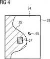
Anhand des aus der Darstellung des ersten Bilddatensatzes ausgewählten Bildausschnitts (vgl. Markierung
Die simultane Darstellung des ersten und diesen ausschnittsweise überlagernden zweiten Bilddatensatzes gemäß
Bei der Darstellung gemäß
Während
BezugszeichenlisteLIST OF REFERENCE NUMBERS
- 11
- medizinische Untersuchungs- und/oder Behandlungsvorrichtungmedical examination and / or treatment device
- 22
- RöntgenvorrichtungX-ray device
- 33
- Strahlungsquelleradiation source
- 44
- SteuerrungseinrichtungControl reasoner
- 55
- Roboterarmrobot arm
- 66
- Roboterarmrobot arm
- 77
- Gelenkjoint
- 88th
- Ultraschall-VorrichtungUltrasonic device
- 99
- Steuerungseinrichtungcontrol device
- 1010
- Ultraschall-KopfUltrasound head
- 1111
- Gelenkjoint
- 1212
- Gelenkjoint
- 1313
- Gelenkjoint
- 1414
- Gelenkjoint
- 1515
- Roboterarmrobot arm
- 1616
- Roboterarmrobot arm
- 1717
- Roboterarmrobot arm
- 1818
- Patientenliegepatient support
- 1919
- Patientinpatient
- 2020
- Verarbeitungseinrichtungprocessing device
- 2121
- Mausmouse
- 2222
- Tastaturkeyboard
- 2323
- Bildschirmscreen
- 2424
- Anzeigeflächedisplay area
- 2525
- Brustchest
- 2626
- Cursorcursor
- 2727
- Markierungmark
Claims (13)
Translated fromGermanPriority Applications (4)
| Application Number | Priority Date | Filing Date | Title |
|---|---|---|---|
| DE102010009295.9ADE102010009295B4 (en) | 2010-02-25 | 2010-02-25 | Method for displaying a region to be examined and / or treated |
| CN2011800106443ACN102770088A (en) | 2010-02-25 | 2011-02-15 | Method for displaying areas to be examined and/or treated |
| US13/576,867US20120293511A1 (en) | 2010-02-25 | 2011-02-15 | Method for displaying an area to be medically examined and/or treated |
| PCT/EP2011/052166WO2011104135A1 (en) | 2010-02-25 | 2011-02-15 | Method for displaying an area to be examined and/or treated |
Applications Claiming Priority (1)
| Application Number | Priority Date | Filing Date | Title |
|---|---|---|---|
| DE102010009295.9ADE102010009295B4 (en) | 2010-02-25 | 2010-02-25 | Method for displaying a region to be examined and / or treated |
Publications (2)
| Publication Number | Publication Date |
|---|---|
| DE102010009295A1true DE102010009295A1 (en) | 2011-08-25 |
| DE102010009295B4 DE102010009295B4 (en) | 2019-02-21 |
Family
ID=43982379
Family Applications (1)
| Application Number | Title | Priority Date | Filing Date |
|---|---|---|---|
| DE102010009295.9AExpired - Fee RelatedDE102010009295B4 (en) | 2010-02-25 | 2010-02-25 | Method for displaying a region to be examined and / or treated |
Country Status (4)
| Country | Link |
|---|---|
| US (1) | US20120293511A1 (en) |
| CN (1) | CN102770088A (en) |
| DE (1) | DE102010009295B4 (en) |
| WO (1) | WO2011104135A1 (en) |
Cited By (18)
| Publication number | Priority date | Publication date | Assignee | Title |
|---|---|---|---|---|
| EP2814396A4 (en)* | 2012-02-13 | 2015-11-04 | Hologic Inc | SYSTEM AND METHOD FOR NAVIGATING A TOMOSYNTHESIS STACK BY USING SYNTHETIZED IMAGE DATA |
| US10010302B2 (en) | 2002-11-27 | 2018-07-03 | Hologic, Inc. | System and method for generating a 2D image from a tomosynthesis data set |
| US10573276B2 (en) | 2011-11-27 | 2020-02-25 | Hologic, Inc. | System and method for generating a 2D image using mammography and/or tomosynthesis image data |
| US11403483B2 (en) | 2017-06-20 | 2022-08-02 | Hologic, Inc. | Dynamic self-learning medical image method and system |
| US11406332B2 (en) | 2011-03-08 | 2022-08-09 | Hologic, Inc. | System and method for dual energy and/or contrast enhanced breast imaging for screening, diagnosis and biopsy |
| US11419565B2 (en) | 2014-02-28 | 2022-08-23 | IIologic, Inc. | System and method for generating and displaying tomosynthesis image slabs |
| US11445993B2 (en) | 2017-03-30 | 2022-09-20 | Hologic, Inc. | System and method for targeted object enhancement to generate synthetic breast tissue images |
| US11455754B2 (en) | 2017-03-30 | 2022-09-27 | Hologic, Inc. | System and method for synthesizing low-dimensional image data from high-dimensional image data using an object grid enhancement |
| US11452486B2 (en) | 2006-02-15 | 2022-09-27 | Hologic, Inc. | Breast biopsy and needle localization using tomosynthesis systems |
| US11589944B2 (en) | 2013-03-15 | 2023-02-28 | Hologic, Inc. | Tomosynthesis-guided biopsy apparatus and method |
| US11701199B2 (en) | 2009-10-08 | 2023-07-18 | Hologic, Inc. | Needle breast biopsy system and method of use |
| US11775156B2 (en) | 2010-11-26 | 2023-10-03 | Hologic, Inc. | User interface for medical image review workstation |
| US11957497B2 (en) | 2017-03-30 | 2024-04-16 | Hologic, Inc | System and method for hierarchical multi-level feature image synthesis and representation |
| US12029602B2 (en) | 2013-10-24 | 2024-07-09 | Hologic, Inc. | System and method for navigating x-ray guided breast biopsy |
| US12211608B2 (en) | 2013-03-15 | 2025-01-28 | Hologic, Inc. | System and method for navigating a tomosynthesis stack including automatic focusing |
| US12236582B2 (en) | 2018-09-24 | 2025-02-25 | Hologic, Inc. | Breast mapping and abnormality localization |
| US12236597B2 (en) | 2021-11-29 | 2025-02-25 | Hologic, Inc. | Systems and methods for correlating objects of interest |
| US12254586B2 (en) | 2021-10-25 | 2025-03-18 | Hologic, Inc. | Auto-focus tool for multimodality image review |
Families Citing this family (8)
| Publication number | Priority date | Publication date | Assignee | Title |
|---|---|---|---|---|
| US10008184B2 (en) | 2005-11-10 | 2018-06-26 | Hologic, Inc. | System and method for generating a 2D image using mammography and/or tomosynthesis image data |
| CN103815926B (en)* | 2014-03-07 | 2016-04-27 | 杭州千思科技有限公司 | Breast cancer detection method and apparatus |
| EP3291735B1 (en)* | 2015-05-07 | 2024-10-09 | Koninklijke Philips N.V. | System and method for motion compensation in medical procedures |
| JP6772505B2 (en)* | 2016-03-25 | 2020-10-21 | ブラザー工業株式会社 | Programs and terminals |
| US10796430B2 (en)* | 2018-04-24 | 2020-10-06 | General Electric Company | Multimodality 2D to 3D imaging navigation |
| CN109770937A (en)* | 2019-03-06 | 2019-05-21 | 郑州大学第一附属医院 | A cardiac angiography surgical device that can be automatically adjusted for position in cardiology |
| US20220230345A1 (en)* | 2021-01-19 | 2022-07-21 | Home Depot Product Authority, Llc | Image based measurement estimation |
| CN116630680B (en)* | 2023-04-06 | 2024-02-06 | 南方医科大学南方医院 | Dual-mode image classification method and system combining X-ray photography and ultrasound |
Citations (3)
| Publication number | Priority date | Publication date | Assignee | Title |
|---|---|---|---|---|
| DE102006003609A1 (en)* | 2006-01-25 | 2007-08-09 | Siemens Ag | Computer-tomography system for scanning patient, has visual representation unit comprising addressing representation function for marking partial area of visual representation and representing partial area in high image resolution |
| US20080243142A1 (en)* | 2007-02-20 | 2008-10-02 | Gildenberg Philip L | Videotactic and audiotactic assisted surgical methods and procedures |
| DE102008037453A1 (en)* | 2007-10-15 | 2009-04-16 | General Electric Co. | Method and system for visualizing registered images |
Family Cites Families (6)
| Publication number | Priority date | Publication date | Assignee | Title |
|---|---|---|---|---|
| US7072501B2 (en)* | 2000-11-22 | 2006-07-04 | R2 Technology, Inc. | Graphical user interface for display of anatomical information |
| JP4509027B2 (en)* | 2003-06-13 | 2010-07-21 | パナソニック株式会社 | Ultrasonic diagnostic equipment |
| WO2005020147A1 (en)* | 2003-08-21 | 2005-03-03 | Philips Intellectual Property & Standards Gmbh | Device and method for combining two images |
| WO2007047782A2 (en)* | 2005-10-20 | 2007-04-26 | Intuitive Surgical, Inc | Auxiliary image display and manipulation on a computer display in a medical robotic system |
| CA2645539A1 (en)* | 2006-03-09 | 2007-09-13 | Imagnosis Inc. | Medical 3-dimensional image display control program and medical 3-dimensional image display method |
| CN107126182B (en) | 2007-01-19 | 2020-06-16 | 桑尼布鲁克健康科学中心 | Scanning mechanism for imaging probe |
- 2010
- 2010-02-25DEDE102010009295.9Apatent/DE102010009295B4/ennot_activeExpired - Fee Related
- 2011
- 2011-02-15CNCN2011800106443Apatent/CN102770088A/enactivePending
- 2011-02-15USUS13/576,867patent/US20120293511A1/ennot_activeAbandoned
- 2011-02-15WOPCT/EP2011/052166patent/WO2011104135A1/enactiveApplication Filing
Patent Citations (3)
| Publication number | Priority date | Publication date | Assignee | Title |
|---|---|---|---|---|
| DE102006003609A1 (en)* | 2006-01-25 | 2007-08-09 | Siemens Ag | Computer-tomography system for scanning patient, has visual representation unit comprising addressing representation function for marking partial area of visual representation and representing partial area in high image resolution |
| US20080243142A1 (en)* | 2007-02-20 | 2008-10-02 | Gildenberg Philip L | Videotactic and audiotactic assisted surgical methods and procedures |
| DE102008037453A1 (en)* | 2007-10-15 | 2009-04-16 | General Electric Co. | Method and system for visualizing registered images |
Cited By (37)
| Publication number | Priority date | Publication date | Assignee | Title |
|---|---|---|---|---|
| US10010302B2 (en) | 2002-11-27 | 2018-07-03 | Hologic, Inc. | System and method for generating a 2D image from a tomosynthesis data set |
| US10413263B2 (en) | 2002-11-27 | 2019-09-17 | Hologic, Inc. | System and method for generating a 2D image from a tomosynthesis data set |
| US12193853B2 (en) | 2006-02-15 | 2025-01-14 | Hologic, Inc. | Breast biopsy and needle localization using tomosynthesis systems |
| US11918389B2 (en) | 2006-02-15 | 2024-03-05 | Hologic, Inc. | Breast biopsy and needle localization using tomosynthesis systems |
| US11452486B2 (en) | 2006-02-15 | 2022-09-27 | Hologic, Inc. | Breast biopsy and needle localization using tomosynthesis systems |
| US12193886B2 (en) | 2009-10-08 | 2025-01-14 | Hologic, Inc. | Needle breast biopsy system and method of use |
| US11701199B2 (en) | 2009-10-08 | 2023-07-18 | Hologic, Inc. | Needle breast biopsy system and method of use |
| US11775156B2 (en) | 2010-11-26 | 2023-10-03 | Hologic, Inc. | User interface for medical image review workstation |
| US12239471B2 (en) | 2011-03-08 | 2025-03-04 | Hologic, Inc. | System and method for dual energy and/or contrast enhanced breast imaging for screening, diagnosis and biopsy |
| US11406332B2 (en) | 2011-03-08 | 2022-08-09 | Hologic, Inc. | System and method for dual energy and/or contrast enhanced breast imaging for screening, diagnosis and biopsy |
| US10978026B2 (en) | 2011-11-27 | 2021-04-13 | Hologic, Inc. | System and method for generating a 2D image using mammography and/or tomosynthesis image data |
| US12183309B2 (en) | 2011-11-27 | 2024-12-31 | Hologic, Inc. | System and method for generating a 2D image using mammography and/or tomosynthesis image data |
| US10573276B2 (en) | 2011-11-27 | 2020-02-25 | Hologic, Inc. | System and method for generating a 2D image using mammography and/or tomosynthesis image data |
| US11508340B2 (en) | 2011-11-27 | 2022-11-22 | Hologic, Inc. | System and method for generating a 2D image using mammography and/or tomosynthesis image data |
| US11837197B2 (en) | 2011-11-27 | 2023-12-05 | Hologic, Inc. | System and method for generating a 2D image using mammography and/or tomosynthesis image data |
| US10977863B2 (en) | 2012-02-13 | 2021-04-13 | Hologic, Inc. | System and method for navigating a tomosynthesis stack using synthesized image data |
| US11663780B2 (en) | 2012-02-13 | 2023-05-30 | Hologic Inc. | System and method for navigating a tomosynthesis stack using synthesized image data |
| US12307604B2 (en) | 2012-02-13 | 2025-05-20 | Hologic, Inc. | System and method for navigating a tomosynthesis stack using synthesized image data |
| EP2814396A4 (en)* | 2012-02-13 | 2015-11-04 | Hologic Inc | SYSTEM AND METHOD FOR NAVIGATING A TOMOSYNTHESIS STACK BY USING SYNTHETIZED IMAGE DATA |
| US10410417B2 (en) | 2012-02-13 | 2019-09-10 | Hologic, Inc. | System and method for navigating a tomosynthesis stack using synthesized image data |
| US12324707B2 (en) | 2013-03-15 | 2025-06-10 | Hologic, Inc. | Tomosynthesis-guided biopsy in prone |
| US12211608B2 (en) | 2013-03-15 | 2025-01-28 | Hologic, Inc. | System and method for navigating a tomosynthesis stack including automatic focusing |
| US11589944B2 (en) | 2013-03-15 | 2023-02-28 | Hologic, Inc. | Tomosynthesis-guided biopsy apparatus and method |
| US12064291B2 (en) | 2013-03-15 | 2024-08-20 | Hologic, Inc. | Tomosynthesis-guided biopsy in prone |
| US12029602B2 (en) | 2013-10-24 | 2024-07-09 | Hologic, Inc. | System and method for navigating x-ray guided breast biopsy |
| US11419565B2 (en) | 2014-02-28 | 2022-08-23 | IIologic, Inc. | System and method for generating and displaying tomosynthesis image slabs |
| US11801025B2 (en) | 2014-02-28 | 2023-10-31 | Hologic, Inc. | System and method for generating and displaying tomosynthesis image slabs |
| US12070349B2 (en) | 2017-03-30 | 2024-08-27 | Hologic, Inc. | System and method for targeted object enhancement to generate synthetic breast tissue images |
| US11983799B2 (en) | 2017-03-30 | 2024-05-14 | Hologic, Inc. | System and method for synthesizing low-dimensional image data from high-dimensional image data using an object grid enhancement |
| US11957497B2 (en) | 2017-03-30 | 2024-04-16 | Hologic, Inc | System and method for hierarchical multi-level feature image synthesis and representation |
| US12211124B2 (en) | 2017-03-30 | 2025-01-28 | Hologic, Inc. | System and method for synthesizing low-dimensional image data from high-dimensional image data using an object grid enhancement |
| US11455754B2 (en) | 2017-03-30 | 2022-09-27 | Hologic, Inc. | System and method for synthesizing low-dimensional image data from high-dimensional image data using an object grid enhancement |
| US11445993B2 (en) | 2017-03-30 | 2022-09-20 | Hologic, Inc. | System and method for targeted object enhancement to generate synthetic breast tissue images |
| US11403483B2 (en) | 2017-06-20 | 2022-08-02 | Hologic, Inc. | Dynamic self-learning medical image method and system |
| US12236582B2 (en) | 2018-09-24 | 2025-02-25 | Hologic, Inc. | Breast mapping and abnormality localization |
| US12254586B2 (en) | 2021-10-25 | 2025-03-18 | Hologic, Inc. | Auto-focus tool for multimodality image review |
| US12236597B2 (en) | 2021-11-29 | 2025-02-25 | Hologic, Inc. | Systems and methods for correlating objects of interest |
Also Published As
| Publication number | Publication date |
|---|---|
| WO2011104135A1 (en) | 2011-09-01 |
| US20120293511A1 (en) | 2012-11-22 |
| CN102770088A (en) | 2012-11-07 |
| DE102010009295B4 (en) | 2019-02-21 |
Similar Documents
| Publication | Publication Date | Title |
|---|---|---|
| DE102010009295B4 (en) | Method for displaying a region to be examined and / or treated | |
| DE102005059209B4 (en) | Method and device for visualizing a sequence of tomographic image data sets | |
| DE102005030646B4 (en) | A method of contour visualization of at least one region of interest in 2D fluoroscopic images | |
| DE102017221276B4 (en) | Medical image processing device, X-ray CT device and medical image processing method | |
| DE112013004898B4 (en) | A medical imaging system and method for providing an improved x-ray image | |
| DE102008037453A1 (en) | Method and system for visualizing registered images | |
| DE102011003135B4 (en) | An imaging method for rotating a tissue area | |
| DE102010036538A1 (en) | System and method for compensating for respiratory motion in acquired radiographic images | |
| EP2465096B1 (en) | Method and device for processing 3-d image data of a skull | |
| DE102017217599A1 (en) | Medical information processing apparatus, X-ray CT apparatus and medical information processing method | |
| DE202017107196U1 (en) | Medical image processing apparatus, X-ray CT apparatus and computer program product | |
| DE102016219887A1 (en) | Method and system for using measured data | |
| DE102007052857A1 (en) | Method and apparatus for synchronizing corresponding landmarks on a number of images | |
| DE102004022902A1 (en) | Medical imaging and processing method, computed tomography device, workstation and computer program product | |
| DE102005041602A1 (en) | Method for displaying a medical implant in an image and medical imaging system | |
| DE112012002671T5 (en) | View a variety of registered images | |
| EP3378401B1 (en) | Representation of an area of interest | |
| DE102014205313A1 (en) | Method for registering a near-infrared spectroscopy map and an anatomy image data set and x-ray device | |
| DE102005035430A1 (en) | Method for improved display of co-registered 2D-3D images in medical imaging | |
| DE102006008509A1 (en) | Conspicuousness detecting method, involves testing image with detection algorithm automatically, where image data sets are registered, in order to obtain transformations | |
| EP1914685A2 (en) | Graphical representation of three-dimensional data records | |
| DE102005005687A1 (en) | Magnetic resonance equipment for treating intra luminal pathologies of gastro intestinal tract, has image recording unit recording raw data containing perfusion information and/or angiographic information of identified abnormality | |
| DE102015210912A1 (en) | Reconstruction of a result image taking into account contour significance data | |
| DE102006058906B4 (en) | A method for displaying tomographic images and tomography system or Tomographiesystemverbund for performing this method | |
| DE102009048151B4 (en) | Method for controlling an imaging examination system and associated examination system |
Legal Events
| Date | Code | Title | Description |
|---|---|---|---|
| R016 | Response to examination communication | ||
| R079 | Amendment of ipc main class | Free format text:PREVIOUS MAIN CLASS: A61B0019000000 Ipc:A61B0034100000 | |
| R081 | Change of applicant/patentee | Owner name:SIEMENS HEALTHCARE GMBH, DE Free format text:FORMER OWNER: SIEMENS AKTIENGESELLSCHAFT, 80333 MUENCHEN, DE | |
| R018 | Grant decision by examination section/examining division | ||
| R020 | Patent grant now final | ||
| R119 | Application deemed withdrawn, or ip right lapsed, due to non-payment of renewal fee |