CN216257185U - Ultrasound Probes and Ultrasound Systems - Google Patents
Ultrasound Probes and Ultrasound SystemsDownload PDFInfo
- Publication number
- CN216257185U CN216257185UCN202122183683.7UCN202122183683UCN216257185UCN 216257185 UCN216257185 UCN 216257185UCN 202122183683 UCN202122183683 UCN 202122183683UCN 216257185 UCN216257185 UCN 216257185U
- Authority
- CN
- China
- Prior art keywords
- probe
- ultrasound
- articulating
- pressure
- pressure value
- Prior art date
- Legal status (The legal status is an assumption and is not a legal conclusion. Google has not performed a legal analysis and makes no representation as to the accuracy of the status listed.)
- Active
Links
Images


Classifications
- A—HUMAN NECESSITIES
- A61—MEDICAL OR VETERINARY SCIENCE; HYGIENE
- A61B—DIAGNOSIS; SURGERY; IDENTIFICATION
- A61B8/00—Diagnosis using ultrasonic, sonic or infrasonic waves
- A61B8/08—Clinical applications
- A61B8/0891—Clinical applications for diagnosis of blood vessels
- A—HUMAN NECESSITIES
- A61—MEDICAL OR VETERINARY SCIENCE; HYGIENE
- A61B—DIAGNOSIS; SURGERY; IDENTIFICATION
- A61B8/00—Diagnosis using ultrasonic, sonic or infrasonic waves
- A61B8/08—Clinical applications
- A61B8/0833—Clinical applications involving detecting or locating foreign bodies or organic structures
- A61B8/0841—Clinical applications involving detecting or locating foreign bodies or organic structures for locating instruments
- A—HUMAN NECESSITIES
- A61—MEDICAL OR VETERINARY SCIENCE; HYGIENE
- A61B—DIAGNOSIS; SURGERY; IDENTIFICATION
- A61B8/00—Diagnosis using ultrasonic, sonic or infrasonic waves
- A61B8/08—Clinical applications
- A61B8/0833—Clinical applications involving detecting or locating foreign bodies or organic structures
- A61B8/085—Clinical applications involving detecting or locating foreign bodies or organic structures for locating body or organic structures, e.g. tumours, calculi, blood vessels, nodules
- A—HUMAN NECESSITIES
- A61—MEDICAL OR VETERINARY SCIENCE; HYGIENE
- A61B—DIAGNOSIS; SURGERY; IDENTIFICATION
- A61B8/00—Diagnosis using ultrasonic, sonic or infrasonic waves
- A61B8/42—Details of probe positioning or probe attachment to the patient
- A61B8/4209—Details of probe positioning or probe attachment to the patient by using holders, e.g. positioning frames
- A61B8/4218—Details of probe positioning or probe attachment to the patient by using holders, e.g. positioning frames characterised by articulated arms
- A—HUMAN NECESSITIES
- A61—MEDICAL OR VETERINARY SCIENCE; HYGIENE
- A61B—DIAGNOSIS; SURGERY; IDENTIFICATION
- A61B8/00—Diagnosis using ultrasonic, sonic or infrasonic waves
- A61B8/42—Details of probe positioning or probe attachment to the patient
- A61B8/4245—Details of probe positioning or probe attachment to the patient involving determining the position of the probe, e.g. with respect to an external reference frame or to the patient
- A61B8/4254—Details of probe positioning or probe attachment to the patient involving determining the position of the probe, e.g. with respect to an external reference frame or to the patient using sensors mounted on the probe
- A—HUMAN NECESSITIES
- A61—MEDICAL OR VETERINARY SCIENCE; HYGIENE
- A61B—DIAGNOSIS; SURGERY; IDENTIFICATION
- A61B8/00—Diagnosis using ultrasonic, sonic or infrasonic waves
- A61B8/44—Constructional features of the ultrasonic, sonic or infrasonic diagnostic device
- A61B8/4444—Constructional features of the ultrasonic, sonic or infrasonic diagnostic device related to the probe
- A—HUMAN NECESSITIES
- A61—MEDICAL OR VETERINARY SCIENCE; HYGIENE
- A61B—DIAGNOSIS; SURGERY; IDENTIFICATION
- A61B8/00—Diagnosis using ultrasonic, sonic or infrasonic waves
- A61B8/46—Ultrasonic, sonic or infrasonic diagnostic devices with special arrangements for interfacing with the operator or the patient
- A61B8/461—Displaying means of special interest
- A61B8/463—Displaying means of special interest characterised by displaying multiple images or images and diagnostic data on one display
Landscapes
- Health & Medical Sciences (AREA)
- Life Sciences & Earth Sciences (AREA)
- Biomedical Technology (AREA)
- Biophysics (AREA)
- Nuclear Medicine, Radiotherapy & Molecular Imaging (AREA)
- Pathology (AREA)
- Radiology & Medical Imaging (AREA)
- Engineering & Computer Science (AREA)
- Physics & Mathematics (AREA)
- Heart & Thoracic Surgery (AREA)
- Medical Informatics (AREA)
- Molecular Biology (AREA)
- Surgery (AREA)
- Animal Behavior & Ethology (AREA)
- General Health & Medical Sciences (AREA)
- Public Health (AREA)
- Veterinary Medicine (AREA)
- Vascular Medicine (AREA)
- Ultra Sonic Daignosis Equipment (AREA)
Abstract
Translated fromChinese
Description
Translated fromChinese优先权priority
本申请要求2020年9月10日提交的美国临时申请号63/076,589 的优先权的权益,其通过引用整体并入本申请。This application claims the benefit of priority from US Provisional Application No. 63/076,589, filed September 10, 2020, which is incorporated herein by reference in its entirety.
技术领域technical field
本申请涉及医疗器械领域,更具体地涉及超声探测器和超声系统。The present application relates to the field of medical devices, and more particularly to ultrasound probes and ultrasound systems.
背景技术Background technique
目前存在多种现有的包括连接至显示器的有线或无线超声探测器的超声系统连接至探测器。这些系统可由临床医生用于评估诸如血管等用于放置包括导管的血管通路装置(“VAD”)的部位。这些系统还可以由临床医生用于评估VAD或导管在选定部位的放置。然而,患者的身体组织可能被明显压缩,同时通过简单地使用预期的超声探测器来评估这种部位。身体组织的压缩可能损害通过VAD或导管的血管接合(purchase),这又可能导致对患者健康有危险的导管外渗。现有的超声系统不提供用于测量在超声成像期间由超声探测器引起的身体组织压缩压力。There are currently a variety of existing ultrasound systems that include wired or wireless ultrasound probes connected to a display connected to the probe. These systems may be used by clinicians to assess sites such as blood vessels for placement of vascular access devices ("VADs") including catheters. These systems can also be used by clinicians to assess VAD or catheter placement at selected sites. However, the patient's body tissue may be significantly compressed while assessing such sites by simply using the intended ultrasound probe. Compression of body tissue can impair vascular purchase through the VAD or catheter, which in turn can lead to extravasation of the catheter that is dangerous to the patient's health. Existing ultrasound systems do not provide for measuring body tissue compression pressure induced by ultrasound probes during ultrasound imaging.
本文披露了具有压力测量能力用于在超声成像期间检测和确定身体组织是否被过度压缩的超声探测器、超声系统和超声方法。Disclosed herein are ultrasound probes, ultrasound systems, and ultrasound methods having pressure measurement capabilities for detecting and determining whether body tissue is overcompressed during ultrasound imaging.
实用新型内容Utility model content
本文披露了一种超声探测器,在一些实施方案中,超声探测器包括探测器主体;附接至探测器主体的铰接(articulating)探头;和压力感测装置,压力感测装置容纳在铰接探头与探测器主体之间的铰接区域中。Disclosed herein is an ultrasound probe that, in some embodiments, includes a probe body; an articulating probe attached to the probe body; and a pressure sensing device housed in the articulating probe in the hinged area with the detector body.
在一些实施方案中,超声探测器还包括保护罩(boot),保护罩在铰接区域中将铰接探头连接至探测器主体。保护罩配置为覆盖压力感测装置或将压力感测装置并入其中。In some embodiments, the ultrasound probe further includes a boot that connects the articulating probe to the probe body in the articulating region. The protective cover is configured to cover or incorporate the pressure sensing device therein.
在一些实施方案中,压力感测装置配置为检测保护罩的弹性材料中或周围的变形。变形是由将铰接探头压入患者中导致的。In some embodiments, the pressure sensing device is configured to detect deformation in or around the elastic material of the protective cover. Deformation is caused by pressing the articulating probe into the patient.
在一些实施方案中,压力感测装置通信地耦合至超声探测器的控制器。控制器配置为将对应于变形的电信号转换为测量压力值。In some embodiments, the pressure sensing device is communicatively coupled to the controller of the ultrasound probe. The controller is configured to convert the electrical signal corresponding to the deformation into a measured pressure value.
在一些实施方案中,超声探测器配置为将测量压力值提供给显示器以便向临床医生显示。In some embodiments, the ultrasound probe is configured to provide measured pressure values to a display for display to a clinician.
在一些实施方案中,超声探测器包括配置为将测量压力值与阈值压力值进行比较的逻辑。In some embodiments, the ultrasound probe includes logic configured to compare the measured pressure value to a threshold pressure value.
在一些实施方案中,超声探测器包括扬声器,扬声器配置为当测量压力值超过阈值压力值时发射音频信号以警告临床医生。In some embodiments, the ultrasound probe includes a speaker configured to emit an audio signal to alert the clinician when the measured pressure value exceeds a threshold pressure value.
在一些实施方案中,超声探测器包括发光二极管,发光二极管配置为当测量压力值超过阈值压力值时发射视觉信号以警告临床医生。In some embodiments, the ultrasound probe includes a light emitting diode configured to emit a visual signal to alert the clinician when the measured pressure value exceeds a threshold pressure value.
在一些实施方案中,压力感测装置是压力换能器。In some embodiments, the pressure sensing device is a pressure transducer.
在一些实施方案中,压力换能器是压阻式应变计压力换能器。压阻式应变计压力换能器包括结合至铰接探头与探测器主体之间的铰接区域中的柔性隔膜的应变计。隔膜的变形提供应变计电阻的相应可测量变化,变化指示通过将铰接探头压入患者中引起变形而导致的压力。In some embodiments, the pressure transducer is a piezoresistive strain gauge pressure transducer. Piezoresistive strain gauge pressure transducers include strain gauges coupled to a flexible diaphragm in the articulation region between the articulating probe and the probe body. Deformation of the diaphragm provides a corresponding measurable change in the resistance of the strain gauge, which is indicative of the pressure caused by the deformation caused by pressing the articulating probe into the patient.
在一些实施方案中,压力换能器是可变电容压力换能器。压力换能器包括在铰接探头与探测器主体之间的铰接区域中的隔膜电极和相对电极(opposing electrode)。柔性隔膜的变形影响隔膜电极与相对电极之间的距离,从而提供电容的相应可测量变化,变化指示通过将铰接探头压入患者中引起变形而导致的压力。In some embodiments, the pressure transducer is a variable capacitance pressure transducer. The pressure transducer includes a diaphragm electrode and an opposing electrode in the articulation region between the articulating probe and the probe body. Deformation of the flexible diaphragm affects the distance between the diaphragm electrode and the opposing electrode, providing a corresponding measurable change in capacitance indicative of the pressure caused by the deformation induced by pressing the articulating probe into the patient.
本文还公开了在一些实施方案中包括控制台和超声探测器的超声系统。控制台包括显示器,显示器配置为在显示器的显示屏上呈现超声图像。超声探测器包括探测器主体;附接至探测器主体的铰接探头;和压力感测装置,压力感测装置容纳在铰接探头与探测器主体之间的铰接区域中。Also disclosed herein are ultrasound systems that, in some embodiments, include a console and an ultrasound probe. The console includes a display configured to present an ultrasound image on a display screen of the display. The ultrasound probe includes a probe body; an articulating probe attached to the probe body; and a pressure sensing device housed in an articulation region between the articulating probe and the probe body.
在一些实施方案中,超声探测器还包括保护罩,保护罩在铰接区域中将铰接探头连接至探测器主体。保护罩配置为覆盖压力感测装置或将压力感测装置并入其中。In some embodiments, the ultrasound probe further includes a protective cover that connects the articulating probe to the probe body in the articulating region. The protective cover is configured to cover or incorporate the pressure sensing device therein.
在一些实施方案中,压力感测装置配置为检测保护罩的弹性材料中或周围的变形。变形是由将铰接探头压入患者中导致的。In some embodiments, the pressure sensing device is configured to detect deformation in or around the elastic material of the protective cover. Deformation is caused by pressing the articulating probe into the patient.
在一些实施方案中,压力感测装置通信地耦合至控制台的控制器。控制器配置为将对应于变形的电信号转换为测量压力值。In some embodiments, the pressure sensing device is communicatively coupled to a controller of the console. The controller is configured to convert the electrical signal corresponding to the deformation into a measured pressure value.
在一些实施方案中,超声探测器配置为将测量压力值提供给显示器以便向临床医生显示。In some embodiments, the ultrasound probe is configured to provide measured pressure values to a display for display to a clinician.
在一些实施方案中,控制台包括配置为将测量压力值与阈值压力值进行比较的逻辑。In some embodiments, the console includes logic configured to compare the measured pressure value to a threshold pressure value.
在一些实施方案中,控制台包括扬声器,扬声器配置为当测量压力值超过阈值压力值时发射音频信号以警告临床医生。In some embodiments, the console includes a speaker configured to emit an audio signal to alert the clinician when the measured pressure value exceeds a threshold pressure value.
在一些实施方案中,显示器配置为当测量压力值超过阈值压力值时发射视觉信号以警告临床医生。In some embodiments, the display is configured to emit a visual signal to alert the clinician when the measured pressure value exceeds a threshold pressure value.
在一些实施方案中,显示器配置为显示视觉反馈,视觉反馈包括目标静脉和放置在目标静脉中的导管的可视化。In some embodiments, the display is configured to display visual feedback including visualization of the target vein and the catheter placed in the target vein.
在一些实施方案中,压力感测装置是压阻式应变计压力换能器。压阻式应变计压力换能器包括结合至铰接探头与探测器主体之间的铰接区域中的柔性隔膜的应变计。隔膜的变形提供应变计电阻的相应可测量变化,变化指示通过将铰接探头压入患者中引起变形而导致的压力。In some embodiments, the pressure sensing device is a piezoresistive strain gauge pressure transducer. Piezoresistive strain gauge pressure transducers include strain gauges coupled to a flexible diaphragm in the articulation region between the articulating probe and the probe body. The deformation of the diaphragm provides a corresponding measurable change in the resistance of the strain gauge, the change being indicative of the pressure caused by the deformation caused by pressing the articulating probe into the patient.
在一些实施方案中,压力感测装置是可变电容压力换能器。压力换能器包括在铰接探头与探测器主体之间的铰接区域中的隔膜电极和相对电极。柔性隔膜的变形影响隔膜电极与相对电极之间的距离,从而提供电容的相应可测量变化,变化指示通过将铰接探头压入患者中引起变形而导致的压力。In some embodiments, the pressure sensing device is a variable capacitance pressure transducer. The pressure transducer includes a diaphragm electrode and a counter electrode in the articulation region between the articulating probe and the probe body. Deformation of the flexible diaphragm affects the distance between the diaphragm electrode and the opposing electrode, providing a corresponding measurable change in capacitance indicative of the pressure caused by the deformation induced by pressing the articulating probe into the patient.
本文还公开了一种超声系统的方法,方法在一些实施方案中包括超声探测器获取步骤、超声探测器放置步骤、超声探测器移动步骤和压力监测步骤。超声探测器获取步骤包括获取超声探测器。超声探测器包括探测器主体;附接至探测器主体的铰接探头;和压力感测装置,压力感测装置容纳在铰接探头与探测器主体之间的铰接区域中。超声探测器放置步骤包括将超声探测器的铰接探头放置在患者的皮肤表面上。超声探测器移动步骤包括在将超声信号从铰接探头发射到患者中用于超声成像的同时,使超声探测器的铰接探头在患者上方移动。压力监测步骤包括监测由超声探测器的铰接探头在患者上引起的超过阈值压力值的任何测量压力值。Also disclosed herein is a method of an ultrasound system comprising, in some embodiments, an ultrasound probe acquisition step, an ultrasound probe placement step, an ultrasound probe movement step, and a pressure monitoring step. The ultrasonic probe acquiring step includes acquiring an ultrasonic probe. The ultrasound probe includes a probe body; an articulating probe attached to the probe body; and a pressure sensing device housed in an articulation region between the articulating probe and the probe body. The ultrasound probe placement step includes placing the articulated probe of the ultrasound probe on the skin surface of the patient. The ultrasound probe moving step includes moving the articulating probe of the ultrasound probe over the patient while transmitting ultrasound signals from the articulating probe into the patient for ultrasound imaging. The pressure monitoring step includes monitoring any measured pressure values on the patient that exceed a threshold pressure value caused by the articulating probe of the ultrasound probe.
在一些实施方案中,压力监测步骤包括在显示器的显示屏上查看测量压力值。In some embodiments, the pressure monitoring step includes viewing the measured pressure value on a display screen of the display.
在一些实施方案中,压力监测步骤包括对显示器的显示屏上的视觉信号的监测,视觉信号在任何测量压力值超过阈值压力值时警告临床医生。In some embodiments, the pressure monitoring step includes monitoring a visual signal on the display screen of the display that alerts the clinician when any measured pressure value exceeds a threshold pressure value.
在一些实施方案中,压力监测步骤包括对在任何测量压力值超过阈值压力值时警告临床医生的音频信号的监测。In some embodiments, the pressure monitoring step includes monitoring of an audio signal that alerts the clinician when any measured pressure value exceeds a threshold pressure value.
在一些实施方案中,方法还包括导管放置调节步骤。导管放置调节步骤包括响应于超过阈值压力值的任何测量压力值来调整导管放置,以确保使导管外渗最小化的足够的血管接合。In some embodiments, the method further comprises a catheter placement adjustment step. The catheter placement adjustment step includes adjusting catheter placement in response to any measured pressure value exceeding a threshold pressure value to ensure adequate vessel engagement to minimize catheter extravasation.
鉴于更详细地描述本文提供的概念的具体实施方案的附图和以下描述,这些概念的这些和其他特征对于本领域技术人员将变得更加明显。These and other features of the concepts provided herein will become more apparent to those skilled in the art in view of the accompanying drawings and the following description, which describe in more detail specific embodiments of the concepts provided herein.
附图说明Description of drawings
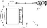
图1示出了根据一些实施方案的包括控制台和超声探测器的有线超声系统。1 illustrates a wired ultrasound system including a console and an ultrasound probe, according to some embodiments.
图2示出了根据一些实施方案的包括控制台和超声探测器的无线超声系统。2 illustrates a wireless ultrasound system including a console and an ultrasound probe, according to some embodiments.
图3示出了根据一些实施方案的图1的超声系统的框图。3 shows a block diagram of the ultrasound system of FIG. 1, according to some embodiments.
图4示出了根据一些实施方案的包括伴随装置和超声探测器的有线超声系统。4 illustrates a wired ultrasound system including a companion device and an ultrasound probe, according to some embodiments.
图5示出了根据一些实施方案的包括伴随装置和超声探测器的无线超声系统。5 illustrates a wireless ultrasound system including a companion device and an ultrasound probe, according to some embodiments.
图6示出了根据一些实施方案的图2、图4或5的超声系统的框图。Figure 6 shows a block diagram of the ultrasound system of Figures 2, 4, or 5, according to some embodiments.
图7示出了根据一些实施方案的包括压力感测装置的超声探测器的前视图。7 illustrates a front view of an ultrasound probe including a pressure sensing device, according to some embodiments.
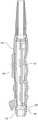
图8示出了根据一些实施方案的超声探测器的透视图。8 shows a perspective view of an ultrasound probe according to some embodiments.
图9示出了根据一些实施方案的超声探测器的横截面。9 shows a cross-section of an ultrasound probe according to some embodiments.
图10示出了根据一些实施方案的超声探测器的另一横截面。10 shows another cross-section of an ultrasound probe according to some embodiments.
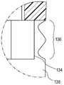
图11示出了包括根据一些实施方案的压力感测装置的超声探测器的铰接区域的详细视图。11 shows a detailed view of an articulation region of an ultrasound probe including a pressure sensing device according to some embodiments.
具体实施方式Detailed ways
在更详细地公开一些具体实施方案之前,应当理解,本文公开的具体实施方案不限制本文提供的概念的范围。还应当理解,本文公开的具体实施方案可具有可容易地与该具体实施方案分离并且任选地与本文公开的许多其他实施方案中的任何一个的特征组合,或其特征替代本文公开的许多其他实施方案中的任何一个的特征。Before some specific embodiments are disclosed in greater detail, it is to be understood that the specific embodiments disclosed herein do not limit the scope of the concepts presented herein. It is also to be understood that a particular embodiment disclosed herein may have features that are readily separable from that particular embodiment and optionally combined with, or substituted for, any of the many other embodiments disclosed herein. A feature of any of the embodiments.
关于本文使用的术语,还应当理解,这些术语是为了描述一些具体实施方案的目的,并且这些术语不限制本文提供的概念的范围。序数(例如第一、第二、第三等)通常用于区分或标识特征或步骤组中的不同特征或步骤,并且不提供系列或数字限制。例如“第一”、“第二”和“第三”特征或步骤不必按顺序出现,并且包括这些特征或步骤的具体实施方案不必局限于这三个特征或步骤。另外,前述特征或步骤中的任何一个可又包括一个或多个特征或步骤,除非另外指明。诸如“左”、“右”、“上”、“下”、“前”,“后”等的标签是为了方便而使用的,而并不旨在暗示例如任何特定的固定位置、取向或方向。相反,这样的标记用于反映例如相对位置、取向或方向。除非上下文另外明确指出,否则单数形式的“一个”,“一种”和“该”包括复数指代。With regard to the terms used herein, it is also to be understood that these terms are for the purpose of describing some specific embodiments and that these terms do not limit the scope of the concepts presented herein. Ordinal numbers (eg, first, second, third, etc.) are typically used to distinguish or identify different features or steps within a group of features or steps, and do not provide serial or numerical limitations. For example, "first," "second," and "third" features or steps do not necessarily have to occur in that order, and specific embodiments that include these features or steps are not necessarily limited to these three features or steps. Additionally, any of the aforementioned features or steps may further comprise one or more features or steps, unless otherwise indicated. Labels such as "left", "right", "upper", "lower", "front", "rear" etc. are used for convenience and are not intended to imply, for example, any particular fixed position, orientation or orientation . Rather, such indicia are used to reflect, for example, relative position, orientation, or direction. The singular forms "a," "an," and "the" include plural referents unless the context clearly dictates otherwise.
关于例如导管的“近侧”、“近侧部分”或“近端部分”包括当导管用于患者时旨在靠近临床医生的导管部分。同样地,例如导管的“近侧长度”包括当导管用于患者时旨在靠近临床医生的导管的长度。例如导管的“近端”包括当导管用于患者时旨在靠近临床医生的导管的端部。导管的近侧部分、近端部分或近侧长度可以包括导管的近端;然而,导管的近侧部分、近端部分或近侧长度不需要包括导管的近端。即,除非上下文另外暗示,导管的近侧部分,近端部分或近侧长度不是导管的末端部分或末端长度。"Proximal", "proximal portion" or "proximal portion" in reference to, for example, a catheter includes the portion of the catheter that is intended to be near the clinician when the catheter is used on a patient. Likewise, for example, the "proximal length" of a catheter includes the length of the catheter intended to be close to the clinician when the catheter is used in a patient. For example, the "proximal end" of a catheter includes the end of the catheter that is intended to be near the clinician when the catheter is used on a patient. The proximal portion, proximal portion or proximal length of the catheter may include the proximal end of the catheter; however, the proximal portion, proximal portion or proximal length of the catheter need not include the proximal end of the catheter. That is, unless the context otherwise implies, the proximal portion, proximal portion or proximal length of the catheter is not the distal portion or distal length of the catheter.
关于例如导管的“远侧”、“远侧部分”或“远端部分”包括当导管用于患者时旨在靠近或在患者中的导管部分。同样地,例如导管的“远侧长度”包括当导管用于患者时旨在靠近或在患者中的导管的长度。例如导管的“远端”包括当导管用于患者时旨在靠近或在患者中的导管的端部。导管的远侧部分、远端部分或远侧长度可以包括导管的远端;然而,导管的远侧部分、远端部分或远侧长度不需要包括导管的远端。即,除非上下文另外暗示,导管的远侧部分,远端部分或远侧长度不是导管的末端部分或末端长度。"Distal", "distal portion" or "distal portion" in reference to eg a catheter includes the portion of the catheter intended to be near or in the patient when the catheter is used in the patient. Likewise, for example, the "distal length" of a catheter includes the length of the catheter intended to be near or in a patient when the catheter is used in a patient. For example, the "distal end" of a catheter includes the end of the catheter that is intended to be near or in a patient when the catheter is used in a patient. The distal portion, distal portion or distal length of the catheter may include the distal end of the catheter; however, the distal portion, distal portion or distal length of the catheter need not include the distal end of the catheter. That is, unless the context otherwise implies, the distal portion, distal portion or distal length of the catheter is not the distal portion or distal length of the catheter.
最后,在以下描述中,本文所用的术语“或”和“和/或”应解释为包括性的或意指任何一个或任何组合。作为实例,“A、B或C”或“A、B和/或C”意指“以下中的任何一个:A;B;C;A和B; A和C;B和C;A、B和C”。该定义的例外仅仅在元件、部件、功能、步骤或动作的组合以某种方式固有地相互排斥时发生。Finally, in the following description, the terms "or" and "and/or" as used herein should be construed to be inclusive or to mean any one or any combination. As an example, "A, B or C" or "A, B and/or C" means "any of the following: A; B; C; A and B; A and C; B and C; A, B and C". Exceptions to this definition occur only when combinations of elements, components, functions, steps or acts are in some way inherently mutually exclusive.
除非另外定义,否则本文使用的所有技术和科学术语具有与本领域普通技术人员通常理解的相同的含义。Unless otherwise defined, all technical and scientific terms used herein have the same meaning as commonly understood by one of ordinary skill in the art.
如上所述,现有的超声系统不提供在超声成像期间测量由超声探测器引起的身体组织的压缩压力。本文公开了具有压力测量能力用于在超声成像期间检测和确定身体组织是否被过度压缩的超声探测器、超声系统和超声方法。As mentioned above, existing ultrasound systems do not provide for measuring the compressive pressure of body tissue induced by the ultrasound probe during ultrasound imaging. Disclosed herein are ultrasound probes, ultrasound systems, and ultrasound methods having pressure measurement capabilities for detecting and determining whether body tissue is overcompressed during ultrasound imaging.
超声系统Ultrasound system
图1示出了根据一些实施方案的有线超声系统100。图3示出了根据一些实施方案的有线超声系统100的框图。FIG. 1 shows a
如图所示,有线超声系统100包括控制台102、显示器104和有线超声探测器106。在有线超声系统100的操作期间,有线超声探测器106的铰接探头132抵靠患者的皮肤放置。产生超声波束,以对诸如患者的皮肤的表面下方的血管的目标的一部分进行超声波成像。血管的超声图像可以连同如下所述的测量压力值一起描绘在显示器104 的显示屏上。该有线超声系统100对于评估进入部位是有用的,例如在用针进行经皮穿刺以将VAD(例如导管)放置到血管中之前评估患者中的血管。有线超声系统100还可用于在放置VAD之后评估进入部位。然而,应当理解,有线超声系统100可用于除了导管插入之外的各种基于超声的医疗程序。例如可以进行使用针的经皮穿刺以对患者的器官的组织进行活检。As shown,
控制台102容纳有线超声系统100的各种部件,并且可以理解,控制台102可以采用各种形式中的任何一种。控制台102中包括处理器108和存储器110,例如随机存取存储器(“RAM”)或非易失性存储器(例如电可擦除可编程只读存储器[“EEPROM”]),用于控制有线超声系统100的各种功能,以及在有线超声系统100的操作期间根据存储在存储器110中用于由处理器108执行的可执行指令114 经由逻辑112执行各种逻辑操作或算法。例如控制台102配置为通过指令114例示一个或多个处理过程,以用于控制有线超声系统100的功能、将来自有线超声探测器106的超声换能器140的电信号处理成超声图像、将来自有线超声探测器106的压力感测装置的电信号处理成测量压力值等。数字控制器/模拟接口116也包括在控制台102中,并且与处理器108和其他系统部件二者通信,以管理有线超声探测器 106和这里阐述的其他系统部件之间的接口。Console 102 houses the various components of
任选地在控制台102的处理器108和存储器110之间实现的控制台102的控制器可通信地耦合至下面阐述的有线超声探测器106的压力感测装置134。该控制器配置为将与有线超声探测器106的保护罩 138的变形相对应的电信号转换成测量压力值,这些变形是通过将铰接探头132压入患者中而导致的、在保护罩138的弹性材料中或周围的那些变形探头而导致。特别地,控制台102的逻辑112配置为将每个测量压力值与阈值压力值进行比较,以在测量压力值超过阈值压力值时警告临床医生。例如控制台102可以包括扬声器,该扬声器配置为在测量压力值超过阈值压力值时发射音频信号以警告临床医生。在另一示例中,显示器104配置为在测量压力值超过阈值压力值时在显示屏上发射视觉信号以警告临床医生。The controller of the
有线超声系统100还包括用于与诸如包括打印机、存储介质、键盘等的可选部件等附加部件连接的端口118。端口118可以是通用串行总线(“USB”)端口,尽管其他类型的端口可用于此连接或本文示出或描述的任何其他连接。The
控制台102包括电源连接120以实现与外部电源122的可操作连接。内部电源124(例如电池)也可以与外部电源122一起使用或者不与外部电源122一起使用。电源管理电路126包括在控制台102的数字控制器/模拟接口116中以调节电源使用和分配。The
显示器104包括集成到控制台102中的显示屏,以提供图形用户界面(“GUI”),呈现由有线超声探测器106获得的目标(例如血管)的一个或多个超声图像,以及显示104任何相关信息,诸如当获得所述一个或多个超声图像时用于铰接探头132的测量压力值。此外,显示器104可以配置为显示视觉反馈,该视觉反馈包括目标(例如诸如静脉的血管)和诸如放置在目标中的导管的VAD的可视化。尽管如此,显示器104可以替代地与控制台102分离并且通信地与其耦合至。通过控制台102的控制台按钮接口128访问的控制按钮(参见图1)可用于立即将有线超声系统100的期望模式调用到显示屏,以辅助基于超声的医疗程序,例如评估前述目标或在其中放置VAD。
有线超声探测器106与诸如血管的目标的基于超声的可视化结合使用,以准备将诸如导管的VAD放置到目标中。这种可视化提供了实时超声引导,并且有助于减少通常与VAD放置相关的并发症,例如导管外渗。有线超声探测器106配置为向控制台102提供来自有线超声探测器106的超声换能器140的电信号、来自有线超声探测器106 的压力感测装置的电信号,或其组合,用于在VAD放置或其他医疗程序中的实时超声引导。The
图1和7-11示出了根据一些实施方案的有线超声探测器106的各种视图。1 and 7-11 illustrate various views of the
如图所示,有线超声探测器106包括探测器主体130、附接至探测器主体130的铰接探头132,以及容纳在铰接探头132与探测器主体130之间的铰接区域136中的压力感测装置134。有线超声探测器 106还包括保护罩138,保护罩138在铰接区域136中将铰接探头132连接至探测器主体130。该保护罩138配置为将该压力感测装置134 覆盖或并入其中。As shown, the
铰接探头132容纳超声换能器140的阵列,其中超声换能器140 是压电超声换能器或电容微加工超声换能器(“CMUT”)。该铰接探头132配置为用于抵靠患者的皮肤放置在预期VAD放置部位附近,在该预期VAD放置部位中该铰接探头132中的超声换能器140可以产生超声信号并且将产生的超声信号以多个脉冲发射到该患者中,通过由患者的身体反射所产生的超声脉冲来接收来自患者的反射超声信号或超声回波,并且通过控制台102将反射的超声信号转换成用于处理成超声图像的对应电信号。这样,临床医生可以使用有线超声系统100来确定合适的VAD放置部位并与其建立血管通路。The articulating
压力感测装置134配置成检测保护罩138的弹性材料中或周围的变形,该变形是由将铰接探头132压入患者中而导致的。压力感测装置134可以是压力换能器或多个压力换能器。例如压力换能器可以是压阻式应变计压力换能器。这种压力换能器包括结合至铰接探头132 与探测器主体130之间的铰接区域136中的柔性隔膜的应变计。隔膜的变形提供应变计电阻的相应可测量的变化,该变化指示通过将铰接探头132压入患者中引起变形而导致的压力。在另一示例中,压力换能器是可变电容压力换能器。例如压力换能器包括位于铰接探头132 与探测器主体130之间的铰接区域136中的隔膜电极和相对电极。柔性隔膜的变形影响隔膜电极与相对电极之间的距离,从而提供相应的可测量的电容变化,该电容变化指示通过将铰接探头132压入患者中引起变形而导致的压力。The
有线超声探测器106还包括用于在基于超声的医疗程序期间控制有线超声系统100的某些方面的控制按钮142,从而消除了临床医生到达患者周围的无菌区域之外以控制有线超声系统100的需要。例如包括在有线超声探测器106上的控制按钮142(参见图7)可以用于由临床医生立即向显示屏调用期望模式以帮助VAD放置或一些其他基于超声的医疗程序。The
图3示出了有线超声探测器106还包括用于管理按钮和超声探测器操作的按钮和存储器控制器144。按钮和存储器控制器144可以包括非易失性存储器(例如EEPROM)。该按钮和存储器控制器144 与控制台102的探测器接口146可操作地通信,该探测器接口146包括用于与超声换能器140接口的输入/输出(“I/O”)部件148以及用于与按钮和存储器控制器144接口的按钮和存储器I/O部件150。FIG. 3 shows that the
图2和5示出了根据一些实施方案的无线超声系统152。图4示出了根据除了上述实施方案之外的一些其他实施方案的有线超声系统100;实际上,图4所示的有线超声探测器106与无线超声探测器 154的相似之处在于,对来自有线超声探测器106的超声换能器140和压力感测装置134的电信号的处理是通过有线超声探测器106本身来进行的,以便通过有线连接而不是无线连接来显示在伴随装置156 上。图3示出了根据一些实施方案的无线超声系统152的框图。2 and 5 illustrate a
虽然下面阐述了对无线超声系统152的描述,但是应当理解,无线超声系统152包括与上面阐述的有线超声系统100类似的部件,尽管它们在无线超声系统152中以不同方式分布。例如无线超声探测器 154本身可以包括控制台102的处理器108、存储器110、指令114和逻辑112,用于控制无线超声探测器154的各种功能,将对应于无线超声探测器154的保护罩138内或周围的变形的电信号转换为测量压力值,将来自超声换能器140的电信号处理成超声图像数据或文件等。尽管如此,伴随装置156(例如图2的控制台102或图4和图5 的智能电话、平板手机或平板电脑)仍包括处理器、存储器、指令、逻辑等。然而,这种部件不需要配置为将来自超声换能器140的电信号处理成例如超声图像数据或文件。实际上,这种部件可以改为配置为显示对应于由无线超声探测器154提供的超声图像数据或文件的超声图像。Although a description of the
如图所示,无线超声系统152包括无线超声探测器154和伴随装置156,例如图4和图5的智能手机、平板手机或平板电脑,或者在一些实施方案中,图2的控制台102。伴随装置156包括显示器158 和无线模块160,其配置用于与无线超声探测器154和任选的远程电子健康记录(“EHR”)系统进行无线通信。在无线超声系统152的操作期间,无线超声探测器154的铰接探头132抵靠患者的皮肤放置。产生超声波束,以对诸如患者的皮肤的表面下方的血管的目标的一部分进行超声波成像。通过从无线超声探测器154向伴随装置156无线地提供与测量压力值相对应的数据,可以在伴随装置156的显示器 158的显示屏上描绘血管的超声图像以及测量压力值。无线超声系统 152用于在用针进行经皮穿刺以将诸如导管的VAD放置到血管中之前评估诸如患者体内的血管的目标。然而,应当理解,无线超声系统 152可以用于除了导管插入之外的各种基于超声的医疗程序。例如可以进行使用针的经皮穿刺以对患者的器官的组织进行活检。As shown, the
图7-11示出了根据一些实施方案的有线超声探测器106的各种视图;然而,有线超声探测器106和无线超声探测器154共享至少如下所述的特征。7-11 illustrate various views of the
如图所示,无线超声探测器154包括探测器主体130、附接至探测器主体130的铰接探头132,以及容纳在铰接探头132与探测器主体130之间的铰接区域136中的压力感测装置134。有线超声探测器 106还包括保护罩138,保护罩138在铰接区域136中将铰接探头132连接至探测器主体130。该保护罩138配置为将该压力感测装置134 覆盖或并入其中。具有铰接探头132的无线超声探测器154能够进行静脉和导管可视化。与上述有线超声探测器106类似,图7-11中描绘的无线超声探测器154可用于在放置VAD之前和之后评估进入部位。As shown, the
探测器主体130容纳印刷电路板组件(“PCBA”)162。PCBA 162 包括在其框图中示出的无线超声探测器154的多个电子部件。(参见图6。)PCBA 162通信地耦合至控制按钮142,该控制按钮142包括配置用于从电源163(例如内部电池)切换电源到无线超声探测器154 的电源按钮和用于操作无线超声探测器154的各种其他按钮探测器。The
类似于有线超声探测器106的铰接探头132,无线超声探测器154 的铰接探头132容纳超声换能器阵列140,其中超声换能器140是压电超声换能器或CMUT。再次地,铰接探头132配置为抵靠患者的皮肤放置在预期的VAD放置部位附近,在该预期的VAD放置部位处,铰接探头132中的超声换能器140可以产生超声信号并且将所产生的超声信号以多个脉冲发射到患者中,通过由患者身体反射所产生的超声脉冲来接收来自患者的反射的超声信号或超声回波,并将反射的超声信号转换成相应的电信号用于由无线超声探测器154处理成超声图像。Similar to the articulating
像有线超声探测器106一样,无线超声探测器154的压力感测装置134配置为检测保护罩138的弹性材料中或周围的变形,该变形是由将所述铰接探头132压入患者中导致的探头。压力感测装置134可以是设置在探测器主体130和铰接探头132之间的铰接区域136中的压力换能器或多个压力换能器。如上所述,压力换能器可以是压阻式应变计压力换能器,其包括结合至铰接探头132与探测器主体130之间的铰接区域136中的柔性隔膜的应变计。如上面进一步阐述的,压力换能器可以是可变电容压力换能器,其包括位于铰接探头132与探测器主体130之间的铰接区域136中的隔膜电极和相对电极。Like the
压力感测装置134通信地耦合至无线超声探测器154的控制器,该控制器任选地在无线超声探测器154的处理器166与存储器168之间实现。(参见图6)控制器配置为将对应于保护罩138的弹性材料中或周围的变形的电信号转换成测量压力值。无线超声探测器154配置为将测量压力值提供给伴随装置156以在伴随装置156的显示屏上显示给临床医生。The
特别地,无线超声探测器154包括逻辑164,该逻辑164配置为将测量压力值与阈值压力值进行比较。如果测量压力值超过阈值压力值,则无线超声探测器154可以向伴随装置156发送电信号,以在视觉上或听觉上警告临床医生所述测量压力值超过阈值。无线超声探测器154可以附加地或可选地包括扬声器,该扬声器配置为当测量压力值超过阈值压力值时发射音频信号以警告临床医生。附加地或可选地,无线超声探测器154可以包括发光二极管,该发光二极管配置为当测量压力值超过阈值压力值时发射视觉信号以警告临床医生。这样,无线超声探测器154可用于检测和确定在超声成像期间身体组织是否被过度压缩。特别地,如果患者拥有过多的脂肪组织,脂肪组织可以在由铰接探头132引起的压力下明显地压缩。这可以允许例如导管的更大部分被推进到血管中。然而,当去除由铰接探头132引起的压力时,脂肪组织可以回弹,导致一些导管被抽出,从而减少导管接合长度。这可导致导管外渗。一旦警告临床医生压力超过阈值,临床医生可以检查导管在血管内的正确放置以避免导管外渗。In particular, the
图6示出了根据一些实施方案的无线超声系统152的框图。FIG. 6 shows a block diagram of a
如图所示,无线超声探测器154包括用于通过使用通用操作系统 167来管理系统功能的处理器166、包括文件系统169的存储器168,以及可以存储在存储器168中并由处理器166执行的应用170。一些应用170可提供用户界面以允许临床医生监测由铰接探头132在患者上引起的压力。包括适当电路的波束形成工具172也由处理器166控制,以使得能够产生、接收和进一步处理信号。例如波束形成工具172 产生由铰接探头132中的超声换能器140接收的电信号。该铰接探头 132将对应于这些电信号的超声信号传递到患者的区域中并且接收来自该患者的反射的超声信号。反射的超声信号又由铰接探头132中的超声换能器140转换成相应的电信号,该电信号被提供给波束形成工具172以进一步处理成超声图像数据或文件以用于在伴随装置156上显示。注意,无线超声探测器154可以包括不同的部件,例如比本文阐述的那些部件更多或更少的部件,包括使得无线超声探测器154能够以无线方式与伴随装置156一起操作的那些部件,例如无线模块 174。As shown, the
具有集成的压力感测装置134的有线或无线超声系统100或152 为如上所述的VAD放置提供除静脉可视化之外的多功能性。具有有线或无线超声系统100或152,其不仅提供超声成像而且确保将有线或无线超声探测器106或154应用于患者皮肤不会导致由铰接探测器 132引起的过度压力,有利地降低了导管外渗的风险。Wired or
方法method
方法包括使用有线或无线超声系统100或152的方法。例如该方法包括从超声探测器获取步骤、超声探测器放置步骤、超声探测器移动步骤、压力监测步骤和导管放置调节步骤中选择的一个或多个步骤。Methods include methods using wired or
该超声探测器获取步骤包括获取有线或无线超声探测器106或 154。如上所述,有线和无线超声探测器106和154包括探测器主体 130、连接至探测器主体130的铰接探头132,以及容纳在铰接探头 132与探测器主体130之间的铰接区域136中的压力感测装置134。The ultrasound probe acquisition step includes acquiring a wired or
该超声探测器放置步骤包括将有线或无线超声探测器106或154 的铰接探头132放置在患者的皮肤表面上。The ultrasound probe placement step includes placing the articulated
该超声探测器移动步骤包括将有线或无线超声探测器106或154 的铰接探头132在患者上方移动,同时将超声信号从铰接探头132发射到患者中用于超声成像。The ultrasound probe moving step includes moving the articulating
该压力监测步骤包括监测由超声探测器的铰接探头132在患者上引起的超过阈值压力值的任何测量压力值。该监测可以包括查看控制台102的显示器104的显示屏上或伴随装置156的显示器158上的测量压力值。这种监测还可以包括监测显示器104或158的显示屏上的音频信号或视频信号。当任何测量压力值超过阈值压力值时,这种信号警告临床医生。This pressure monitoring step includes monitoring any measured pressure values on the patient caused by the articulating
该方法还包括导管放置调节步骤。该导管放置调节步骤包括响应于超过该阈值压力值的任何测量压力值来调节导管放置,以确保使导管外渗最小化的足够的血管接合。The method also includes a catheter placement adjustment step. The catheter placement adjustment step includes adjusting catheter placement in response to any measured pressure value exceeding the threshold pressure value to ensure adequate vessel engagement to minimize catheter extravasation.
虽然本文已经公开了一些实施方案,并且虽然已经详细公开了具体的实施方案,但是具体的实施方案并非意图限制本文提供的概念的范围。本领域的普通技术人员可以想到另外的调整或修改,并且在更广泛的方面,也包括这些调整或修改。因此,可在不脱离本文所提供的概念的范围的情况下偏离本文所揭示的具体实施方案。Although some embodiments have been disclosed herein, and although specific embodiments have been disclosed in detail, the specific embodiments are not intended to limit the scope of the concepts presented herein. Additional adjustments or modifications may occur to those of ordinary skill in the art, and are also included in broader aspects. Accordingly, departures may be made from the specific embodiments disclosed herein without departing from the scope of the concepts presented herein.
Claims (22)
Translated fromChineseApplications Claiming Priority (2)
| Application Number | Priority Date | Filing Date | Title |
|---|---|---|---|
| US202063076589P | 2020-09-10 | 2020-09-10 | |
| US63/076,589 | 2020-09-10 |
Publications (1)
| Publication Number | Publication Date |
|---|---|
| CN216257185Utrue CN216257185U (en) | 2022-04-12 |
Family
ID=78212616
Family Applications (2)
| Application Number | Title | Priority Date | Filing Date |
|---|---|---|---|
| CN202122183683.7UActiveCN216257185U (en) | 2020-09-10 | 2021-09-09 | Ultrasound Probes and Ultrasound Systems |
| CN202111058279.5APendingCN114159098A (en) | 2020-09-10 | 2021-09-09 | Ultrasonic probe with pressure measurement capability |
Family Applications After (1)
| Application Number | Title | Priority Date | Filing Date |
|---|---|---|---|
| CN202111058279.5APendingCN114159098A (en) | 2020-09-10 | 2021-09-09 | Ultrasonic probe with pressure measurement capability |
Country Status (4)
| Country | Link |
|---|---|
| US (2) | US12232910B2 (en) |
| EP (1) | EP4203799A1 (en) |
| CN (2) | CN216257185U (en) |
| WO (1) | WO2022056159A1 (en) |
Cited By (1)
| Publication number | Priority date | Publication date | Assignee | Title |
|---|---|---|---|---|
| CN114159098A (en)* | 2020-09-10 | 2022-03-11 | 巴德阿克塞斯系统股份有限公司 | Ultrasonic probe with pressure measurement capability |
Families Citing this family (14)
| Publication number | Priority date | Publication date | Assignee | Title |
|---|---|---|---|---|
| US11759166B2 (en) | 2019-09-20 | 2023-09-19 | Bard Access Systems, Inc. | Automatic vessel detection tools and methods |
| US11992363B2 (en) | 2020-09-08 | 2024-05-28 | Bard Access Systems, Inc. | Dynamically adjusting ultrasound-imaging systems and methods thereof |
| CN216933458U (en) | 2020-11-24 | 2022-07-12 | 巴德阿克塞斯系统股份有限公司 | Object recognition and needle guidance system |
| CN114569156A (en) | 2020-12-01 | 2022-06-03 | 巴德阿克塞斯系统股份有限公司 | Ultrasound imaging system and method for identifying one or more of a plurality of blood vessels |
| CN114569155A (en) | 2020-12-01 | 2022-06-03 | 巴德阿克塞斯系统股份有限公司 | Ultrasound imaging system and method for obtaining ultrasound image by the same |
| CN114601493A (en)* | 2020-12-09 | 2022-06-10 | 财团法人工业技术研究院 | Ultrasonic scanning operation guidance system and ultrasonic scanning operation guidance method |
| USD975738S1 (en)* | 2021-03-11 | 2023-01-17 | Bfly Operations, Inc. | Display panel or portion thereof with graphical user interface |
| USD975739S1 (en)* | 2021-03-11 | 2023-01-17 | Bfly Operations, Inc. | Display panel or portion thereof with graphical user interface |
| CN217960146U (en) | 2021-04-15 | 2022-12-06 | 巴德阿克塞斯系统股份有限公司 | Ultrasound imaging system |
| CN116058873A (en) | 2021-11-03 | 2023-05-05 | 巴德阿克塞斯系统股份有限公司 | Interoperation optimization function through Doppler and image-based vessel discrimination |
| US12433567B2 (en) | 2022-03-16 | 2025-10-07 | Bard Access Systems, Inc. | Ultrasound imaging system |
| JP2024060296A (en)* | 2022-10-19 | 2024-05-02 | 富士フイルム株式会社 | Ultrasonic diagnostic device and control method of ultrasonic diagnostic device |
| US20240197292A1 (en)* | 2022-12-20 | 2024-06-20 | Wuhan United Imaging Healthcare Co., Ltd. | Systems and methods for ultrasound examination |
| US20250255584A1 (en)* | 2024-02-08 | 2025-08-14 | Fujifilm Sonosite, Inc. | Enhancement of local features in ultrasound |
Family Cites Families (400)
| Publication number | Priority date | Publication date | Assignee | Title |
|---|---|---|---|---|
| US3697917A (en)* | 1971-08-02 | 1972-10-10 | Gen Electric | Semiconductor strain gage pressure transducer |
| US5148809A (en) | 1990-02-28 | 1992-09-22 | Asgard Medical Systems, Inc. | Method and apparatus for detecting blood vessels and displaying an enhanced video image from an ultrasound scan |
| FR2662813B1 (en) | 1990-05-29 | 1992-08-14 | Traitement Synthese Image | PROCESS FOR ACQUIRING ECHOGRAPHY IMAGES. |
| US5325293A (en) | 1992-02-18 | 1994-06-28 | Dorne Howard L | System and method for correlating medical procedures and medical billing codes |
| US5441052A (en) | 1992-12-28 | 1995-08-15 | Kabushiki Kaisha Toshiba | Color doppler-type ultrasonic diagnostic apparatus |
| US5349865A (en)* | 1993-08-30 | 1994-09-27 | Kavlico Corporation | Wide-pressure-range, adaptable, simplified pressure transducer |
| US5549554A (en) | 1994-04-01 | 1996-08-27 | Advanced Cardiovascular Systems, Inc. | Catheters having separable reusable components |
| US5573529A (en) | 1994-10-31 | 1996-11-12 | Haak; Benjamin A. | Color coded medical instruments |
| US6019724A (en) | 1995-02-22 | 2000-02-01 | Gronningsaeter; Aage | Method for ultrasound guidance during clinical procedures |
| JPH11508790A (en) | 1995-06-30 | 1999-08-03 | ボストン・サイエンティフィック・コーポレイション | Ultrasound projection catheter with cutting element |
| US6375615B1 (en) | 1995-10-13 | 2002-04-23 | Transvascular, Inc. | Tissue penetrating catheters having integral imaging transducers and their methods of use |
| US5908387A (en) | 1996-06-21 | 1999-06-01 | Quinton Instrument Company | Device and method for improved quantitative coronary artery analysis |
| US5775322A (en) | 1996-06-27 | 1998-07-07 | Lucent Medical Systems, Inc. | Tracheal tube and methods related thereto |
| US5758650A (en) | 1996-09-30 | 1998-06-02 | Siemens Medical Systems, Inc. | Universal needle guide for ultrasonic transducers |
| US6129668A (en) | 1997-05-08 | 2000-10-10 | Lucent Medical Systems, Inc. | System and method to determine the location and orientation of an indwelling medical device |
| US6263230B1 (en) | 1997-05-08 | 2001-07-17 | Lucent Medical Systems, Inc. | System and method to determine the location and orientation of an indwelling medical device |
| US5879297A (en) | 1997-05-08 | 1999-03-09 | Lucent Medical Systems, Inc. | System and method to determine the location and orientation of an indwelling medical device |
| CA2240757C (en) | 1997-07-14 | 2001-08-28 | Matsushita Electric Industrial Co., Ltd. | Blood vessel puncturing device |
| US5897503A (en)* | 1997-08-01 | 1999-04-27 | Acuson Corporation | Ultrasound transducer probe having case handle grip surfaces |
| CA2304735A1 (en) | 1997-10-01 | 1999-04-08 | Boston Scientific Limited | Preinsertion measurement of catheters |
| US5970119A (en) | 1997-11-18 | 1999-10-19 | Douglas Holtz (Part Interest) | Radiological scaling and alignment device |
| US20030135115A1 (en) | 1997-11-24 | 2003-07-17 | Burdette Everette C. | Method and apparatus for spatial registration and mapping of a biopsy needle during a tissue biopsy |
| KR100255730B1 (en) | 1997-12-15 | 2000-05-01 | 이민화 | Ultrasonic color doppler system for displaying artery and vein |
| US6231546B1 (en) | 1998-01-13 | 2001-05-15 | Lumend, Inc. | Methods and apparatus for crossing total occlusions in blood vessels |
| US7713190B2 (en) | 1998-02-24 | 2010-05-11 | Hansen Medical, Inc. | Flexible instrument |
| US6004270A (en) | 1998-06-24 | 1999-12-21 | Ecton, Inc. | Ultrasound system for contrast agent imaging and quantification in echocardiography using template image for image alignment |
| US6132379A (en) | 1998-11-04 | 2000-10-17 | Patacsil; Estelito G. | Method and apparatus for ultrasound guided intravenous cannulation |
| US6524249B2 (en) | 1998-11-11 | 2003-02-25 | Spentech, Inc. | Doppler ultrasound method and apparatus for monitoring blood flow and detecting emboli |
| IL127112A0 (en) | 1998-11-18 | 1999-09-22 | Biosonix Ltd | System for measuring flow and method therefor |
| JP2000271136A (en)* | 1999-03-25 | 2000-10-03 | Toshiba Corp | Ultrasound therapy device and ultrasound therapy device control method |
| US6233476B1 (en) | 1999-05-18 | 2001-05-15 | Mediguide Ltd. | Medical positioning system |
| US7534209B2 (en) | 2000-05-26 | 2009-05-19 | Physiosonics, Inc. | Device and method for mapping and tracking blood flow and determining parameters of blood flow |
| EP1185200B1 (en) | 1999-06-05 | 2008-01-09 | Wilson-Cook Medical Inc. | Indicia for an endoscopic medical device |
| US6687386B1 (en) | 1999-06-15 | 2004-02-03 | Hitachi Denshi Kabushiki Kaisha | Object tracking method and object tracking apparatus |
| WO2001010295A1 (en) | 1999-08-06 | 2001-02-15 | The Board Of Regents Of The University Of Texas System | Optoacoustic monitoring of blood oxygenation |
| US6251073B1 (en) | 1999-08-20 | 2001-06-26 | Novasonics, Inc. | Miniaturized ultrasound apparatus and method |
| DE60012305T2 (en) | 1999-12-07 | 2005-08-18 | Koninklijke Philips Electronics N.V. | ULTRASONIC IMAGE PROCESSING SYSTEM AND SYSTEM FOR PRESENTING A COMPOSIT BILTH SEQUENCE OF A TYPE OF ARTERY |
| JP2003517912A (en) | 1999-12-21 | 2003-06-03 | コーニンクレッカ フィリップス エレクトロニクス エヌ ヴィ | Ultrasound image processing method and inspection system for displaying ultrasonic composite image sequence of artery |
| WO2001047421A1 (en) | 1999-12-28 | 2001-07-05 | Koninklijke Philips Electronics N.V. | Ultrasonic image processing method and system for displaying an ultrasonic color-coded image sequence of an object having moving parts |
| US6612992B1 (en) | 2000-03-02 | 2003-09-02 | Acuson Corp | Medical diagnostic ultrasound catheter and method for position determination |
| US6554774B1 (en) | 2000-03-23 | 2003-04-29 | Tensys Medical, Inc. | Method and apparatus for assessing hemodynamic properties within the circulatory system of a living subject |
| US6640976B1 (en) | 2000-06-13 | 2003-11-04 | Careguide, Inc. | Male clean intermittent catheter system |
| US6524246B1 (en)* | 2000-10-13 | 2003-02-25 | Sonocine, Inc. | Ultrasonic cellular tissue screening tool |
| US7831449B2 (en) | 2001-02-02 | 2010-11-09 | Thompson Reuters (Healthcare) Inc. | Method and system for extracting medical information for presentation to medical providers on mobile terminals |
| US6592565B2 (en) | 2001-04-26 | 2003-07-15 | Zbylut J. Twardowski | Patient-tailored, central-vein catheters |
| ES2357099T3 (en) | 2001-05-23 | 2011-04-18 | Radi Medical Systems Ab | INTERACTIVE MEASUREMENT SYSTEM. |
| US7217266B2 (en) | 2001-05-30 | 2007-05-15 | Anderson R Rox | Apparatus and method for laser treatment with spectroscopic feedback |
| US6592520B1 (en) | 2001-07-31 | 2003-07-15 | Koninklijke Philips Electronics N.V. | Intravascular ultrasound imaging apparatus and method |
| WO2003022169A1 (en) | 2001-09-12 | 2003-03-20 | Scimed Life Systems, Inc. | System for identifying medical devices |
| US6543642B1 (en) | 2001-09-21 | 2003-04-08 | Daydots International, Inc. | Disposable glove dispenser system |
| US6733458B1 (en) | 2001-09-25 | 2004-05-11 | Acuson Corporation | Diagnostic medical ultrasound systems and methods using image based freehand needle guidance |
| JP3863414B2 (en) | 2001-11-22 | 2006-12-27 | 株式会社東芝 | Ultrasonic diagnostic equipment |
| US6689067B2 (en) | 2001-11-28 | 2004-02-10 | Siemens Corporate Research, Inc. | Method and apparatus for ultrasound guidance of needle biopsies |
| US6601705B2 (en) | 2001-12-07 | 2003-08-05 | The Procter & Gamble Company | Package containing a window and performance characteristic indicator |
| US6554771B1 (en) | 2001-12-18 | 2003-04-29 | Koninklijke Philips Electronics N.V. | Position sensor in ultrasound transducer probe |
| US6746402B2 (en) | 2002-01-02 | 2004-06-08 | E. Tuncay Ustuner | Ultrasound system and method |
| US6755789B2 (en) | 2002-02-05 | 2004-06-29 | Inceptio Medical Technologies, Llc | Ultrasonic vascular imaging system and method of blood vessel cannulation |
| JP4217023B2 (en) | 2002-02-25 | 2009-01-28 | 一郎 佐久間 | Vascular endothelial measuring device |
| US7734326B2 (en) | 2002-06-20 | 2010-06-08 | Brainlab Ag | Method and device for preparing a drainage |
| US7359554B2 (en) | 2002-08-26 | 2008-04-15 | Cleveland Clinic Foundation | System and method for identifying a vascular border |
| US7697972B2 (en) | 2002-11-19 | 2010-04-13 | Medtronic Navigation, Inc. | Navigation system for cardiac therapies |
| US7599730B2 (en) | 2002-11-19 | 2009-10-06 | Medtronic Navigation, Inc. | Navigation system for cardiac therapies |
| US7925327B2 (en) | 2002-12-04 | 2011-04-12 | Koninklijke Philips Electronics N.V. | Apparatus and method for assisting the navigation of a catheter in a vessel |
| US7074187B2 (en) | 2002-12-13 | 2006-07-11 | Selzer Robert H | System and method for improving ultrasound image acquisition and replication for repeatable measurements of vascular structures |
| US6979294B1 (en) | 2002-12-13 | 2005-12-27 | California Institute Of Technology | Split-screen display system and standardized methods for ultrasound image acquisition and processing for improved measurements of vascular structures |
| US7927278B2 (en) | 2002-12-13 | 2011-04-19 | California Institute Of Technology | Split-screen display system and standardized methods for ultrasound image acquisition and multi-frame data processing |
| JP2006510412A (en) | 2002-12-18 | 2006-03-30 | コーニンクレッカ フィリップス エレクトロニクス エヌ ヴィ | Ultrasound device for estimating arterial parameters |
| EP1578277A1 (en) | 2002-12-18 | 2005-09-28 | Koninklijke Philips Electronics N.V. | Ultrasonic doppler system for determining movement of artery walls |
| US6749569B1 (en) | 2003-01-07 | 2004-06-15 | Esaote S.P.A. | Method and apparatus for ultrasound imaging |
| EP1594551A2 (en) | 2003-02-19 | 2005-11-16 | Sicel Technologies, Inc. | In vivo fluorescence sensors, systems, and related methods operating in conjunction with fluorescent analytes |
| CA2433205A1 (en) | 2003-03-18 | 2004-09-18 | James Alexander Keenan | Drug delivery, bodily fluid drainage, and biopsy device with enhanced ultrasonic visibility |
| US7727153B2 (en) | 2003-04-07 | 2010-06-01 | Sonosite, Inc. | Ultrasonic blood vessel measurement apparatus and method |
| USD496596S1 (en) | 2003-04-30 | 2004-09-28 | Robert Dalrymple | Image french measuring adjunct |
| US7699779B2 (en) | 2003-05-19 | 2010-04-20 | Hitachi, Ltd. | Ultrasonic treatment equipment |
| US20050000975A1 (en) | 2003-05-28 | 2005-01-06 | Carco Darlene Marie | Sterile surgical glove dispenser |
| WO2005002446A1 (en) | 2003-07-03 | 2005-01-13 | Matsushita Electric Industrial Co., Ltd. | Ultrasonic diagnostic system |
| TWI221407B (en) | 2003-08-27 | 2004-10-01 | Micro Star Int Co Ltd | Device and method for detecting the location of vein by ultrasound |
| EP1665168A1 (en) | 2003-09-04 | 2006-06-07 | Philips Intellectual Property & Standards GmbH | Device and method for displaying ultrasound images of a vessel |
| NZ546204A (en) | 2003-09-12 | 2009-12-24 | Marinepolymer Tech Inc | Vascular access preservation in haemodialysis patients |
| US7244234B2 (en) | 2003-11-11 | 2007-07-17 | Soma Development Llc | Ultrasound guided probe device and method of using same |
| DE602005023833D1 (en) | 2004-01-20 | 2010-11-11 | Philips Intellectual Property | DEVICE AND METHOD FOR NAVIGATING A CATHETER |
| CN1933787A (en) | 2004-01-23 | 2007-03-21 | 特拉克斯医疗有限公司 | Methods and apparatus for performing procedures on target locations in the body |
| US9681925B2 (en) | 2004-04-21 | 2017-06-20 | Siemens Medical Solutions Usa, Inc. | Method for augmented reality instrument placement using an image based navigation system |
| DE602004013926D1 (en) | 2004-04-26 | 2008-07-03 | Brainlab Ag | Visualization of procedural guidelines for medical procedures |
| US8014848B2 (en) | 2004-04-26 | 2011-09-06 | Brainlab Ag | Visualization of procedural guidelines for a medical procedure |
| US20050267365A1 (en) | 2004-06-01 | 2005-12-01 | Alexander Sokulin | Method and apparatus for measuring anatomic structures |
| JP4648652B2 (en) | 2004-06-24 | 2011-03-09 | テルモ株式会社 | Ultrasonic diagnostic apparatus and method for operating ultrasonic diagnostic apparatus |
| US20060004290A1 (en)* | 2004-06-30 | 2006-01-05 | Smith Lowell S | Ultrasound transducer with additional sensors |
| US20060020204A1 (en) | 2004-07-01 | 2006-01-26 | Bracco Imaging, S.P.A. | System and method for three-dimensional space management and visualization of ultrasound data ("SonoDEX") |
| US20060013523A1 (en) | 2004-07-16 | 2006-01-19 | Luna Innovations Incorporated | Fiber optic position and shape sensing device and method relating thereto |
| US20060015039A1 (en) | 2004-07-19 | 2006-01-19 | Cassidy Kenneth T | Guidewire bearing markings simplifying catheter selection |
| US7426497B2 (en) | 2004-08-31 | 2008-09-16 | Microsoft Corporation | Method and apparatus for analysis and decomposition of classifier data anomalies |
| US11207548B2 (en) | 2004-10-07 | 2021-12-28 | Guided Therapy Systems, L.L.C. | Ultrasound probe for treating skin laxity |
| EP1815796A4 (en)* | 2004-11-17 | 2009-10-28 | Hitachi Medical Corp | ULTRASONIC UNIT AND METHOD FOR DISPLAYING AN ULTRASOUND IMAGE |
| US7720520B2 (en) | 2004-12-01 | 2010-05-18 | Boston Scientific Scimed, Inc. | Method and system for registering an image with a navigation reference catheter |
| US8047993B2 (en) | 2004-12-08 | 2011-11-01 | Industrial Technology Research Institute | Quantitative non-invasive method for detecting degree of malignancy in tumors and application thereof |
| US20060184029A1 (en) | 2005-01-13 | 2006-08-17 | Ronen Haim | Ultrasound guiding system and method for vascular access and operation mode |
| US7918787B2 (en) | 2005-02-02 | 2011-04-05 | Voyage Medical, Inc. | Tissue visualization and manipulation systems |
| US20090012399A1 (en) | 2005-02-07 | 2009-01-08 | Kazuhiro Sunagawa | Ultrasonic diagnostic apparatus |
| US7892177B2 (en) | 2005-02-28 | 2011-02-22 | Scimed Life Systems, Inc. | Systems and methods for estimating the length and position of a stent to be applied within a patient |
| JP5484674B2 (en) | 2005-03-04 | 2014-05-07 | シー・アール・バード・インコーポレーテッド | Access port and identification method |
| EP1866871A4 (en) | 2005-03-30 | 2012-01-04 | Worcester Polytech Inst | ULTRASONIC DIAGNOSTIC IMAGING IN THREE-DIMENSIONS HANDLING WITH POSITION AND ANGLE DETERMINATION SENSORS |
| US7680307B2 (en) | 2005-04-05 | 2010-03-16 | Scimed Life Systems, Inc. | Systems and methods for image segmentation with a multi-stage classifier |
| US20070016069A1 (en) | 2005-05-06 | 2007-01-18 | Sorin Grunwald | Ultrasound sensor |
| JP2008539908A (en) | 2005-05-12 | 2008-11-20 | コンピュメディクス メディカル イノベーション ピーティーワイ リミテッド | Ultrasound diagnostic and treatment equipment |
| WO2007002685A2 (en) | 2005-06-24 | 2007-01-04 | Volcano Corporation | Co-registration of graphical image data representing three-dimensional vascular features |
| US7681579B2 (en) | 2005-08-02 | 2010-03-23 | Biosense Webster, Inc. | Guided procedures for treating atrial fibrillation |
| US8147408B2 (en) | 2005-08-31 | 2012-04-03 | Sonosite, Inc. | Medical device guide locator |
| US8852111B2 (en) | 2005-09-02 | 2014-10-07 | Ultrasound Ventures, Llc | Ultrasound guidance system |
| EP1933715A4 (en) | 2005-10-14 | 2012-08-29 | Cleveland Clinic Foundation | System and method for characterizing vascular tissue |
| US8303505B2 (en) | 2005-12-02 | 2012-11-06 | Abbott Cardiovascular Systems Inc. | Methods and apparatuses for image guided medical procedures |
| JP5159041B2 (en) | 2006-01-30 | 2013-03-06 | 株式会社東芝 | Ultrasonic diagnostic apparatus and image processing program thereof |
| WO2007092054A2 (en) | 2006-02-06 | 2007-08-16 | Specht Donald F | Method and apparatus to visualize the coronary arteries using ultrasound |
| JP4812460B2 (en) | 2006-02-22 | 2011-11-09 | 株式会社ユネクス | Arterial blood vessel determination method and apparatus |
| US20070199848A1 (en) | 2006-02-28 | 2007-08-30 | Ellswood Mark R | Packaging with color-coded identification |
| EP2001363B1 (en) | 2006-03-31 | 2017-09-27 | Philips Electronics LTD | System and instrumentation for image guided prostate treatment |
| US8060181B2 (en) | 2006-04-07 | 2011-11-15 | Brainlab Ag | Risk assessment for planned trajectories |
| WO2007122698A1 (en) | 2006-04-18 | 2007-11-01 | Panasonic Corporation | Ultrasonograph |
| US8112292B2 (en) | 2006-04-21 | 2012-02-07 | Medtronic Navigation, Inc. | Method and apparatus for optimizing a therapy |
| ES2524303T3 (en) | 2006-05-08 | 2014-12-05 | C.R. Bard, Inc. | User interface and methods for an ultrasound presentation device |
| US20080021322A1 (en) | 2006-05-24 | 2008-01-24 | Michael Benjamin Stone | Ultrasonic imaging apparatus and method |
| US20080033759A1 (en) | 2006-08-02 | 2008-02-07 | Vastrac, Inc. | Information manager for a procedure-based medical practice |
| CN101500651B (en) | 2006-08-11 | 2012-08-08 | 皇家飞利浦电子股份有限公司 | Ultrasound system for brain blood flow imaging and microbubble-enhanced clot lysis |
| JP4886432B2 (en) | 2006-09-04 | 2012-02-29 | ジーイー・メディカル・システムズ・グローバル・テクノロジー・カンパニー・エルエルシー | Ultrasonic diagnostic equipment |
| US8478377B2 (en) | 2006-10-04 | 2013-07-02 | Dexcom, Inc. | Analyte sensor |
| US20080146915A1 (en) | 2006-10-19 | 2008-06-19 | Mcmorrow Gerald | Systems and methods for visualizing a cannula trajectory |
| US20080108930A1 (en) | 2006-11-03 | 2008-05-08 | The Regents Of The University Of Michigan | Methods and Systems for Determining Volume Flow in a Blood or Fluid Conduit, Motion, and Mechanical Properties of Structures Within the Body |
| US20080177186A1 (en) | 2007-01-18 | 2008-07-24 | Slater Charles R | Methods and Apparatus for Determining a Treatment Volume of a Fluid Treatment Agent for Treating The Interior of a Blood Vessel |
| US8790263B2 (en) | 2007-02-05 | 2014-07-29 | Siemens Medical Solutions Usa, Inc. | Automated movement detection with audio and visual information |
| US20110021924A1 (en) | 2007-02-09 | 2011-01-27 | Shriram Sethuraman | Intravascular photoacoustic and utrasound echo imaging |
| US11197651B2 (en) | 2007-03-08 | 2021-12-14 | Sync-Rx, Ltd. | Identification and presentation of device-to-vessel relative motion |
| US8781193B2 (en) | 2007-03-08 | 2014-07-15 | Sync-Rx, Ltd. | Automatic quantitative vessel analysis |
| US10433929B2 (en) | 2007-03-09 | 2019-10-08 | St. Jude Medical, Atrial Fibrillation Division, Inc. | System and method for local deformable registration of a catheter navigation system to image data or a model |
| JP4945300B2 (en) | 2007-04-25 | 2012-06-06 | 株式会社東芝 | Ultrasonic diagnostic equipment |
| US20090012394A1 (en)* | 2007-04-30 | 2009-01-08 | General Electric Company | User interface for ultrasound system |
| AU2008291763A1 (en) | 2007-05-23 | 2009-03-05 | Oscillon Ltd. | Apparatus and method for guided chronic total occlusion penetration |
| EP2164396A2 (en) | 2007-06-01 | 2010-03-24 | Koninklijke Philips Electronics N.V. | Light weight wireless ultrasound probe |
| US7976469B2 (en) | 2007-06-04 | 2011-07-12 | Medtronic, Inc. | Percutaneous needle guide |
| US8702609B2 (en) | 2007-07-27 | 2014-04-22 | Meridian Cardiovascular Systems, Inc. | Image-guided intravascular therapy catheters |
| US8073215B2 (en) | 2007-09-18 | 2011-12-06 | Siemens Medical Solutions Usa, Inc. | Automated detection of planes from three-dimensional echocardiographic data |
| US8818472B2 (en) | 2007-09-28 | 2014-08-26 | University Of Florida Research Foundation, Inc. | Methods and devices for noninvasive measurement of energy absorbers in blood |
| US10226234B2 (en) | 2011-12-01 | 2019-03-12 | Maui Imaging, Inc. | Motion detection using ping-based and multiple aperture doppler ultrasound |
| US8323202B2 (en) | 2007-11-16 | 2012-12-04 | Pneumrx, Inc. | Method and system for measuring pulmonary artery circulation information |
| JP5416900B2 (en) | 2007-11-22 | 2014-02-12 | 株式会社東芝 | Ultrasonic diagnostic apparatus and puncture support control program |
| US9636031B2 (en) | 2007-11-26 | 2017-05-02 | C.R. Bard, Inc. | Stylets for use with apparatus for intravascular placement of a catheter |
| US10449330B2 (en) | 2007-11-26 | 2019-10-22 | C. R. Bard, Inc. | Magnetic element-equipped needle assemblies |
| US10751509B2 (en) | 2007-11-26 | 2020-08-25 | C. R. Bard, Inc. | Iconic representations for guidance of an indwelling medical device |
| US9649048B2 (en) | 2007-11-26 | 2017-05-16 | C. R. Bard, Inc. | Systems and methods for breaching a sterile field for intravascular placement of a catheter |
| US8781555B2 (en) | 2007-11-26 | 2014-07-15 | C. R. Bard, Inc. | System for placement of a catheter including a signal-generating stylet |
| US10524691B2 (en) | 2007-11-26 | 2020-01-07 | C. R. Bard, Inc. | Needle assembly including an aligned magnetic element |
| US9521961B2 (en) | 2007-11-26 | 2016-12-20 | C. R. Bard, Inc. | Systems and methods for guiding a medical instrument |
| ES2465915T3 (en) | 2007-11-26 | 2014-06-09 | C.R. Bard, Inc. | Integrated system for intravascular catheter placement |
| US8849382B2 (en) | 2007-11-26 | 2014-09-30 | C. R. Bard, Inc. | Apparatus and display methods relating to intravascular placement of a catheter |
| US20090143672A1 (en) | 2007-12-04 | 2009-06-04 | Harms Steven E | Method for mapping image reference points to facilitate biopsy using magnetic resonance imaging |
| US8073529B2 (en) | 2007-12-04 | 2011-12-06 | Civco Medical Instruments Co., Inc. | Needle guide system for use with ultrasound transducers to effect shallow path needle entry and method of use |
| US20090281413A1 (en) | 2007-12-18 | 2009-11-12 | Searete Llc, A Limited Liability Corporation Of The State Of Delaware | Systems, devices, and methods for detecting occlusions in a biological subject |
| US8336536B1 (en) | 2008-06-23 | 2012-12-25 | The United States Of America As Represented By The Secretary Of The Navy | Active heating system for underwater diver |
| US8172753B2 (en) | 2008-07-11 | 2012-05-08 | General Electric Company | Systems and methods for visualization of an ultrasound probe relative to an object |
| US9022940B2 (en) | 2008-07-18 | 2015-05-05 | Joseph H. Meier | Handheld imaging devices and related methods |
| US20100063400A1 (en) | 2008-09-05 | 2010-03-11 | Anne Lindsay Hall | Method and apparatus for catheter guidance using a combination of ultrasound and x-ray imaging |
| WO2010029521A2 (en) | 2008-09-15 | 2010-03-18 | Moshe Ben Chorin | Vein locator and associated devices |
| US20120179044A1 (en)* | 2009-09-30 | 2012-07-12 | Alice Chiang | Ultrasound 3d imaging system |
| US8200313B1 (en) | 2008-10-01 | 2012-06-12 | Bioquantetics, Inc. | Application of image-based dynamic ultrasound spectrography in assisting three dimensional intra-body navigation of diagnostic and therapeutic devices |
| JP2010082337A (en)* | 2008-10-01 | 2010-04-15 | Hitachi Medical Corp | Ultrasonic diagnostic apparatus |
| US10863970B2 (en) | 2008-12-18 | 2020-12-15 | C. R. Bard, Inc. | Needle guide including enhanced visibility entrance |
| WO2010076808A1 (en) | 2008-12-31 | 2010-07-08 | Larsen & Tourbo Limited | Integrated ultrasound imaging device with pulse oximeter waveform display for application of regional anesthesia |
| US20100249598A1 (en)* | 2009-03-25 | 2010-09-30 | General Electric Company | Ultrasound probe with replaceable head portion |
| US8355554B2 (en) | 2009-04-14 | 2013-01-15 | Sonosite, Inc. | Systems and methods for adaptive volume imaging |
| US8781194B2 (en) | 2009-04-17 | 2014-07-15 | Tufts Medical Center, Inc. | Aneurysm detection |
| EP2429405B1 (en) | 2009-05-13 | 2018-07-18 | Koninklijke Philips N.V. | Ultrasonic blood flow doppler audio with pitch shifting |
| US8556815B2 (en) | 2009-05-20 | 2013-10-15 | Laurent Pelissier | Freehand ultrasound imaging systems and methods for guiding fine elongate instruments |
| US20100312121A1 (en) | 2009-06-09 | 2010-12-09 | Zhonghui Guan | Apparatus for a needle director for an ultrasound transducer probe |
| US20100324418A1 (en)* | 2009-06-23 | 2010-12-23 | Essa El-Aklouk | Ultrasound transducer |
| US20110002518A1 (en) | 2009-07-01 | 2011-01-06 | General Electric Company | Method and system for processing ultrasound data |
| KR101624846B1 (en) | 2009-07-16 | 2016-05-27 | 가부시키가이샤 유넥스 | Ultrasonic blood vessel examination apparatus |
| KR101121286B1 (en) | 2009-07-31 | 2012-03-23 | 한국과학기술원 | Ultrasound system and method for performing calibration of sensor |
| EP2742858B1 (en) | 2009-09-23 | 2024-06-05 | Light-Lab Imaging Inc. | Lumen morphology and vascular resistance measurements data collection systems, apparatus and methods |
| JP2011072585A (en)* | 2009-09-30 | 2011-04-14 | Fujifilm Corp | Ultrasonic probe |
| WO2011044421A1 (en)* | 2009-10-08 | 2011-04-14 | C. R. Bard, Inc. | Spacers for use with an ultrasound probe |
| US20140180116A1 (en)* | 2009-10-08 | 2014-06-26 | C. R. Bard, Inc. | Coupling Structures for an Ultrasound Probe |
| US9649037B2 (en) | 2009-12-03 | 2017-05-16 | Deltex Medical Limited | Method and apparatus for hemodynamic monitoring using combined blood flow and blood pressure measurement |
| US9445780B2 (en) | 2009-12-04 | 2016-09-20 | University Of Virginia Patent Foundation | Tracked ultrasound vessel imaging |
| WO2011074271A1 (en) | 2009-12-18 | 2011-06-23 | パナソニック株式会社 | Ultrasonic diagnostic device, and region-to-be-detected image display method and measurement method using same |
| JP2013516288A (en) | 2010-01-07 | 2013-05-13 | ベラソン インコーポレイテッド | Vascular access device, system and method |
| US9204858B2 (en) | 2010-02-05 | 2015-12-08 | Ultrasonix Medical Corporation | Ultrasound pulse-wave doppler measurement of blood flow velocity and/or turbulence |
| RU2589625C2 (en) | 2010-02-09 | 2016-07-10 | Конинклейке Филипс Электроникс Н.В. | Device, system and method for imaging and treatment using optical determination of position |
| WO2011099103A1 (en) | 2010-02-10 | 2011-08-18 | パナソニック株式会社 | Ultrasonic diagnostic device, and method for measuring intima-media complex thickness |
| US8961420B2 (en) | 2010-04-01 | 2015-02-24 | Siemens Medical Solutions Usa, Inc. | System for cardiac condition detection and characterization |
| EP4122385A1 (en) | 2010-05-28 | 2023-01-25 | C. R. Bard, Inc. | Insertion guidance system for needles and medical components |
| EP2912999B1 (en)* | 2010-05-28 | 2022-06-29 | C. R. Bard, Inc. | Apparatus for use with needle insertion guidance system |
| CN103228219B (en)* | 2010-08-09 | 2016-04-27 | C·R·巴德股份有限公司 | Support and Covering Structures for Ultrasound Probe Heads |
| US8315812B2 (en) | 2010-08-12 | 2012-11-20 | Heartflow, Inc. | Method and system for patient-specific modeling of blood flow |
| US8553954B2 (en) | 2010-08-24 | 2013-10-08 | Siemens Medical Solutions Usa, Inc. | Automated system for anatomical vessel characteristic determination |
| US8622913B2 (en) | 2010-09-28 | 2014-01-07 | General Electric Company | Method and system for non-invasive monitoring of patient parameters |
| US9084610B2 (en) | 2010-10-21 | 2015-07-21 | Medtronic Ardian Luxembourg S.A.R.L. | Catheter apparatuses, systems, and methods for renal neuromodulation |
| EP2637568B1 (en) | 2010-11-08 | 2017-04-12 | Vasonova, Inc. | Endovascular navigation system |
| JP5511641B2 (en) | 2010-11-30 | 2014-06-04 | ジーイー・メディカル・システムズ・グローバル・テクノロジー・カンパニー・エルエルシー | Ultrasonic probe, position display device, and ultrasonic diagnostic device |
| CN102551812B (en) | 2010-12-09 | 2015-11-25 | Ge医疗系统环球技术有限公司 | Ultrasound volume probe navigation and vehicle controL method and apparatus and ultrasonic device |
| US9364171B2 (en) | 2010-12-22 | 2016-06-14 | Veebot Systems, Inc. | Systems and methods for autonomous intravenous needle insertion |
| US9974516B2 (en)* | 2010-12-22 | 2018-05-22 | C. R. Bard, Inc. | Selectable angle needle guide |
| US20130023912A1 (en) | 2010-12-31 | 2013-01-24 | Volcano Corporation | Multiple Sclerosis Therapeutic Methods Using Therapeutic Cutting Devices and Systems |
| US20120179038A1 (en) | 2011-01-07 | 2012-07-12 | General Electric Company | Ultrasound based freehand invasive device positioning system and method |
| US10485513B2 (en) | 2011-01-31 | 2019-11-26 | Analogic Corporation | Ultrasound imaging apparatus |
| US9318032B2 (en) | 2011-02-04 | 2016-04-19 | University of Pittsburgh—of the Commonwealth System of Higher Education | Hybrid physical-virtual reality simulation for clinical training capable of providing feedback to a physical anatomic model |
| US8951195B2 (en) | 2011-04-05 | 2015-02-10 | Houston Medical Robotics, Inc. | Motorized systems and methods for accessing the lumen of a vessel |
| AU2012242639B2 (en) | 2011-04-14 | 2016-09-01 | Regents Of The University Of Minnesota | Vascular characterization using ultrasound imaging |
| US20120277576A1 (en) | 2011-04-26 | 2012-11-01 | Chun Kee Lui | Echogenic infusion port catheter |
| JP5788229B2 (en) | 2011-06-06 | 2015-09-30 | 株式会社東芝 | Ultrasonic diagnostic equipment |
| JP2013031651A (en)* | 2011-07-04 | 2013-02-14 | Toshiba Corp | Ultrasonic diagnostic device and control method for ultrasonic probe |
| RU2609203C2 (en) | 2011-07-06 | 2017-01-30 | Си.Ар. Бард, Инк. | Determination and calibration of needle length for needle guidance system |
| CN102871645A (en) | 2011-07-11 | 2013-01-16 | 浙江大学 | Near-infrared imaging ultrasonic vascular therapeutic apparatus |
| US9199082B1 (en) | 2011-07-27 | 2015-12-01 | Cvrx, Inc. | Devices and methods for improved placement of implantable medical devices |
| US20130041250A1 (en) | 2011-08-09 | 2013-02-14 | Ultrasonix Medical Corporation | Methods and apparatus for locating arteries and veins using ultrasound |
| US9295447B2 (en) | 2011-08-17 | 2016-03-29 | Volcano Corporation | Systems and methods for identifying vascular borders |
| US20150127031A1 (en) | 2011-08-26 | 2015-05-07 | EBM Corporation | Bloodstream simulation system for simulating blood vessel treatment effect, method therefor, and computer software program |
| US8744211B2 (en) | 2011-08-31 | 2014-06-03 | Analogic Corporation | Multi-modality image acquisition |
| CN103028185B (en) | 2011-09-30 | 2017-04-12 | Ge医疗系统环球技术有限公司 | Automatic vessel intervention device, system and method based on real-time volume ultrasonic waves |
| WO2013055707A1 (en) | 2011-10-09 | 2013-04-18 | Clear Guide Medical, Llc | Interventional in-situ image-guidance by fusing ultrasound video |
| JP6252480B2 (en) | 2011-10-21 | 2017-12-27 | 日産化学工業株式会社 | Improved synthesis of conjugated polymers by oxidative polymerization and related compositions |
| JP6373758B2 (en) | 2011-11-16 | 2018-08-15 | ボルケーノ コーポレイション | Medical measurement system and method |
| WO2013075112A1 (en) | 2011-11-18 | 2013-05-23 | Verathon, Inc. | Blood vessel access system and device |
| CN103997972B (en) | 2011-12-16 | 2017-05-03 | 皇家飞利浦有限公司 | Automatic blood vessel identification by name |
| EP2803321A4 (en) | 2012-01-10 | 2015-11-25 | Konica Minolta Inc | Ultrasonic diagnosis device, and blood vessel detection method |
| US8764663B2 (en) | 2012-03-14 | 2014-07-01 | Jeffrey Smok | Method and apparatus for locating and distinguishing blood vessel |
| WO2013170053A1 (en) | 2012-05-09 | 2013-11-14 | The Regents Of The University Of Michigan | Linear magnetic drive transducer for ultrasound imaging |
| US8548778B1 (en) | 2012-05-14 | 2013-10-01 | Heartflow, Inc. | Method and system for providing information from a patient-specific model of blood flow |
| CN104411249B (en) | 2012-05-31 | 2017-07-28 | 皇家飞利浦有限公司 | Ultrasonic image-forming system and method for image boot flow |
| EP2861153A4 (en)* | 2012-06-15 | 2016-10-19 | Bard Inc C R | Apparatus and methods for detection of a removable cap on an ultrasound probe |
| JP6065421B2 (en)* | 2012-06-15 | 2017-01-25 | セイコーエプソン株式会社 | Ultrasonic probe and ultrasonic inspection device |
| CN103505288B (en) | 2012-06-29 | 2017-11-17 | 通用电气公司 | Ultrasonic imaging method and supersonic imaging apparatus |
| US10499878B2 (en)* | 2012-07-26 | 2019-12-10 | Interson Corporation | Portable ultrasonic imaging probe including a transducer array |
| JP6051693B2 (en)* | 2012-09-03 | 2016-12-27 | セイコーエプソン株式会社 | Ultrasonic probe, electronic device and ultrasonic diagnostic apparatus |
| US10433740B2 (en) | 2012-09-12 | 2019-10-08 | Heartflow, Inc. | Systems and methods for estimating ischemia and blood flow characteristics from vessel geometry and physiology |
| US11272845B2 (en) | 2012-10-05 | 2022-03-15 | Philips Image Guided Therapy Corporation | System and method for instant and automatic border detection |
| US9814433B2 (en) | 2012-10-24 | 2017-11-14 | Cathworks Ltd. | Creating a vascular tree model |
| MY178704A (en) | 2012-12-14 | 2020-10-20 | Sec Dep Of Biotechnology | Devices and methods for biopsy |
| US9870721B2 (en) | 2012-12-18 | 2018-01-16 | Eric Savitsky | System and method for teaching basic ultrasound skills |
| US10588597B2 (en) | 2012-12-31 | 2020-03-17 | Intuitive Surgical Operations, Inc. | Systems and methods for interventional procedure planning |
| CN104936546B (en) | 2013-01-17 | 2016-09-14 | 皇家飞利浦有限公司 | In the medical of ultrasonic guidance, regulate the method for focal zone and use the system of described method |
| US20150359520A1 (en)* | 2013-01-22 | 2015-12-17 | Koninklijke Philips N.V. | Ultrasound probe and ultrasound imaging system |
| AU2014208379A1 (en) | 2013-01-24 | 2015-07-23 | Tylerton International Holdings Inc. | Body structure imaging |
| CN103961135B (en) | 2013-02-04 | 2017-04-12 | 通用电气公司 | System and method for detecting guide pipe position in three-dimensional ultrasonic image |
| JP2014150928A (en) | 2013-02-07 | 2014-08-25 | Hitachi Aloka Medical Ltd | Ultrasonic diagnostic device |
| GB201303917D0 (en) | 2013-03-05 | 2013-04-17 | Ezono Ag | System for image guided procedure |
| US20140276059A1 (en) | 2013-03-12 | 2014-09-18 | Volcano Corporation | Externally imaging a body structure within a patient |
| US10555719B2 (en) | 2013-03-12 | 2020-02-11 | St. Jude Medical Puerto Rico Llc | Ultrasound assisted needle puncture mechanism |
| US9057600B2 (en) | 2013-03-13 | 2015-06-16 | Hansen Medical, Inc. | Reducing incremental measurement sensor error |
| WO2014164992A1 (en) | 2013-03-13 | 2014-10-09 | Miller David G | Coregistered intravascular and angiographic images |
| US10758308B2 (en) | 2013-03-14 | 2020-09-01 | The Spectranetics Corporation | Controller to select optical channel parameters in a catheter |
| US20140276069A1 (en)* | 2013-03-15 | 2014-09-18 | EagIEyeMed | Ultrasound probe |
| WO2014152260A1 (en) | 2013-03-15 | 2014-09-25 | Nilus Medical, Llc | Hemodynamic monitoring device and methods of using same |
| US20140296694A1 (en) | 2013-04-02 | 2014-10-02 | General Electric Company | Method and system for ultrasound needle guidance |
| CN104302232B (en) | 2013-04-03 | 2016-08-31 | 株式会社日立制作所 | Diagnostic ultrasound equipment and photoelastic evaluation method |
| GB201307551D0 (en) | 2013-04-26 | 2013-06-12 | Ucl Business Plc | A method and apparatus for determining the location of a medical instrument with respect to ultrasound imaging and a medical instrument |
| WO2014188430A2 (en) | 2013-05-23 | 2014-11-27 | CardioSonic Ltd. | Devices and methods for renal denervation and assessment thereof |
| KR20140140331A (en)* | 2013-05-29 | 2014-12-09 | 삼성메디슨 주식회사 | Ultrasound system and method for detecting pressure applied to living body |
| JP2014233522A (en) | 2013-06-04 | 2014-12-15 | セイコーエプソン株式会社 | Ultrasonic measurement apparatus and ultrasonic measurement method |
| US11229490B2 (en) | 2013-06-26 | 2022-01-25 | Corindus, Inc. | System and method for monitoring of guide catheter seating |
| KR20150005052A (en) | 2013-07-04 | 2015-01-14 | 삼성메디슨 주식회사 | Ultrasound system and method for providing target object information |
| US11266466B2 (en) | 2013-07-29 | 2022-03-08 | Intuitive Surgical Operations, Inc. | Shape sensor systems with redundant sensing |
| US20150065916A1 (en) | 2013-08-29 | 2015-03-05 | Vasculogic, Llc | Fully automated vascular imaging and access system |
| EP3043717B1 (en) | 2013-09-11 | 2019-03-13 | Boston Scientific Scimed, Inc. | Systems for selection and displaying of images using an intravascular ultrasound imaging system |
| US20150257735A1 (en) | 2013-10-24 | 2015-09-17 | Evena Medical, Inc. | Systems and methods for displaying medical images |
| US20150190111A1 (en) | 2014-01-03 | 2015-07-09 | William R. Fry | Ultrasound-guided non-invasive blood pressure measurement apparatus and methods |
| WO2015108941A1 (en) | 2014-01-14 | 2015-07-23 | Volcano Corporation | Devices and methods for forming vascular access |
| US20150297097A1 (en) | 2014-01-14 | 2015-10-22 | Volcano Corporation | Vascular access evaluation and treatment |
| US10130329B2 (en) | 2014-01-28 | 2018-11-20 | General Electric Company | Distinct needle display in ultrasonic image |
| CN106102593B (en) | 2014-01-29 | 2019-10-15 | 贝克顿·迪金森公司 | System and method for collection confirmation and sample tracking in clinical use point |
| ES2662410T3 (en) | 2014-01-29 | 2018-04-06 | Becton, Dickinson And Company | Portable electronic device to enhance visualization during the insertion of an invasive device |
| JP5771297B1 (en) | 2014-02-24 | 2015-08-26 | 日立アロカメディカル株式会社 | Ultrasonic diagnostic equipment |
| JP2015160108A (en) | 2014-02-28 | 2015-09-07 | セイコーエプソン株式会社 | Ultrasonic measuring apparatus and ultrasonic measuring method |
| US9785746B2 (en) | 2014-03-31 | 2017-10-10 | Heartflow, Inc. | Systems and methods for determining blood flow characteristics using flow ratio |
| US10987088B2 (en) | 2014-07-01 | 2021-04-27 | Augusta University Research Institute, Inc. | Systems and methods for detecting intracranial pressure and volume |
| US20160000399A1 (en) | 2014-07-02 | 2016-01-07 | General Electric Company | Method and apparatus for ultrasound needle guidance |
| US9320493B2 (en) | 2014-07-08 | 2016-04-26 | Nadarasa Visveshwara | System and method for measuring fluidics in arteries |
| US20160026894A1 (en)* | 2014-07-28 | 2016-01-28 | Daniel Nagase | Ultrasound Computed Tomography |
| US11497406B2 (en) | 2014-07-31 | 2022-11-15 | Samsung Electronics Co., Ltd | Apparatus and method for enhancing accuracy of a contactless body temperature measurement |
| KR101705120B1 (en) | 2014-08-28 | 2017-02-09 | 삼성전자 주식회사 | Untrasound dianognosis apparatus and operating method thereof for self-diagnosis and remote-diagnosis |
| US9918701B2 (en) | 2014-09-03 | 2018-03-20 | Contextvision Ab | Methods and systems for automatic control of subjective image quality in imaging of objects |
| US10043272B2 (en) | 2014-09-16 | 2018-08-07 | Esaote S.P.A. | Method and apparatus for acquiring and fusing ultrasound images with pre-acquired images |
| KR20160050395A (en) | 2014-10-29 | 2016-05-11 | 삼성메디슨 주식회사 | Apparatus and method for displaying medical image |
| US20160120607A1 (en) | 2014-11-03 | 2016-05-05 | Michael Sorotzkin | Ultrasonic imaging device for examining superficial skin structures during surgical and dermatological procedures |
| US10905396B2 (en) | 2014-11-18 | 2021-02-02 | C. R. Bard, Inc. | Ultrasound imaging system having automatic image presentation |
| KR20160066927A (en) | 2014-12-03 | 2016-06-13 | 삼성전자주식회사 | Apparatus and method for supporting computer aided diagnosis |
| JP6751092B2 (en) | 2014-12-10 | 2020-09-02 | コーニンクレッカ フィリップス エヌ ヴェKoninklijke Philips N.V. | Device, system and method for predicting in-stent restenosis |
| US10973584B2 (en) | 2015-01-19 | 2021-04-13 | Bard Access Systems, Inc. | Device and method for vascular access |
| KR20160089647A (en) | 2015-01-20 | 2016-07-28 | 삼성전자주식회사 | X-ray imaging apparatus and control method for the same |
| US10136915B2 (en) | 2015-01-26 | 2018-11-27 | Loving Heart Medical Technology Inc. | Ultrasound needle guide apparatus |
| US10987010B2 (en) | 2015-02-02 | 2021-04-27 | Heartflow, Inc. | Systems and methods for vascular diagnosis using blood flow magnitude and/or direction |
| US10217022B2 (en) | 2015-03-06 | 2019-02-26 | Ricoh Company, Ltd. | Image acquisition and management |
| US10835210B2 (en) | 2015-03-30 | 2020-11-17 | Siemens Medical Solutions Usa, Inc. | Three-dimensional volume of interest in ultrasound imaging |
| WO2016172696A1 (en) | 2015-04-24 | 2016-10-27 | Us Government As Represented By The Secretary Of The Army | Vascular targeting system |
| EP3307353A4 (en) | 2015-06-15 | 2019-03-13 | The University Of Sydney | SYSTEM AND METHOD FOR INSERTION |
| US10792011B2 (en) | 2015-06-23 | 2020-10-06 | Hemonitor Medical Ltd. | Systems and methods for hand-free continuous ultrasonic monitoring |
| WO2016209398A1 (en) | 2015-06-25 | 2016-12-29 | Rivanna Medical Llc | Ultrasonic guidance of a probe with respect to anatomical features |
| CN105107067B (en) | 2015-07-16 | 2018-05-08 | 执鼎医疗科技(杭州)有限公司 | A kind of venipuncture system of infrared guiding localization by ultrasonic |
| US20180199914A1 (en) | 2015-07-22 | 2018-07-19 | Koninklijke Philips N.V. | Fiber-optic realshape sensor for enhanced dopper measurement display |
| CN108024699B (en) | 2015-08-14 | 2020-11-03 | 直观外科手术操作公司 | Registration system and method for image-guided surgery |
| US11185311B2 (en) | 2015-09-17 | 2021-11-30 | Koninklijke Philips N.V. | Distinguishing lung sliding from external motion |
| US20170086785A1 (en) | 2015-09-30 | 2017-03-30 | General Electric Company | System and method for providing tactile feedback via a probe of a medical imaging system |
| KR102531117B1 (en) | 2015-10-07 | 2023-05-10 | 삼성메디슨 주식회사 | Method and apparatus for displaying an image which indicates an object |
| CA3005790C (en) | 2015-12-10 | 2023-10-10 | 1929803 Ontario Corp. D/B/A Ke2 Technologies | Systems and methods for automated fluid response measurement |
| JP6547612B2 (en) | 2015-12-14 | 2019-07-24 | コニカミノルタ株式会社 | IMAGE PROCESSING APPARATUS, IMAGE PROCESSING METHOD, AND ULTRASONIC DIAGNOSTIC APPARATUS PROVIDED WITH IMAGE PROCESSING APPARATUS |
| JP2019500190A (en) | 2015-12-16 | 2019-01-10 | グロ−ティップ, エルエルシー | Method and apparatus for transducer array needle tracking |
| US10231789B2 (en)* | 2015-12-18 | 2019-03-19 | Biosense Webster (Israel) Ltd. | Using force sensor to give angle of ultrasound beam |
| KR102577752B1 (en) | 2016-02-02 | 2023-09-12 | 삼성메디슨 주식회사 | Method of outputting a velocity of object and ultrasound apparatus thereof |
| EP3422952A4 (en) | 2016-03-01 | 2019-09-11 | Echonous, Inc. | ULTRASOUND SYSTEM WITH RECEPTION STATION AND ANCHORABLE ULTRASONIC PROBE |
| DE102016108986A1 (en)* | 2016-05-13 | 2017-11-16 | Krohne Messtechnik Gmbh | Method for the detection of pipe vibrations and measuring device |
| WO2017214428A1 (en) | 2016-06-08 | 2017-12-14 | The United States Of America, As Represented By The Secretary, Department Of Health And Human Services | Tissue characterization with acoustic wave tomosynthesis |
| US20170367678A1 (en) | 2016-06-22 | 2017-12-28 | Cesare Sirtori | Ultrasound automated method for measuring the thickness of the walls of the left anterior descending, right and circumflex coronary arteries |
| US11020563B2 (en) | 2016-07-14 | 2021-06-01 | C. R. Bard, Inc. | Automated catheter-to-vessel size comparison tool and related methods |
| WO2018026878A1 (en) | 2016-08-02 | 2018-02-08 | Avent, Inc. | Motor-assisted needle guide assembly for ultrasound needle placement |
| US20210373602A1 (en) | 2016-09-22 | 2021-12-02 | Sang Kyu MIN | Foldable virtual reality device |
| CN108882916B (en) | 2016-09-30 | 2022-06-10 | 深圳迈瑞生物医疗电子股份有限公司 | Parameter display method of ultrasonic blood flow and ultrasonic imaging system thereof |
| US10231784B2 (en) | 2016-10-28 | 2019-03-19 | Medtronic Ardian Luxembourg S.A.R.L. | Methods and systems for optimizing perivascular neuromodulation therapy using computational fluid dynamics |
| JP6494591B2 (en) | 2016-12-08 | 2019-04-03 | キヤノン株式会社 | Ultrasonic probe and ultrasonic image acquisition apparatus |
| KR102107581B1 (en) | 2016-12-19 | 2020-05-07 | 지멘스 메디컬 솔루션즈 유에스에이, 인크. | Method of providing annotation information of ultrasound probe and ultrasound system |
| IT201700006088A1 (en) | 2017-01-20 | 2018-07-20 | Torino Politecnico | METHOD AND EQUIPMENT FOR NON-INVASIVE DETECTION OF BLOOD VESSEL CONDITIONS |
| US20210295048A1 (en) | 2017-01-24 | 2021-09-23 | Tienovix, Llc | System and method for augmented reality guidance for use of equipment systems |
| US10952700B2 (en) | 2017-01-27 | 2021-03-23 | Wayne State University | Ultrasound and photoacoustic systems and methods for fetal brain assessment during delivery |
| WO2018156196A1 (en) | 2017-02-22 | 2018-08-30 | Elkadi Hisham | Adjustable surgical device with ultrasound guidance and related techniques |
| EP3606592B1 (en) | 2017-04-07 | 2025-01-08 | Bard Access Systems, Inc. | Optical fiber-based medical device tracking and monitoring system |
| JP6880963B2 (en) | 2017-04-17 | 2021-06-02 | ニプロ株式会社 | Puncture guide and ultrasonic diagnostic equipment with puncture guide |
| US11369410B2 (en) | 2017-04-27 | 2022-06-28 | Bard Access Systems, Inc. | Magnetizing system for needle assemblies including orientation key system for positioning needle tray in magnetizer |
| US10945705B2 (en) | 2017-05-16 | 2021-03-16 | Sm Instrument Co., Ltd. | Portable ultrasonic facilities diagnosis device |
| EP3420914A1 (en) | 2017-06-30 | 2019-01-02 | Koninklijke Philips N.V. | Ultrasound system and method |
| US11259879B2 (en) | 2017-08-01 | 2022-03-01 | Inneroptic Technology, Inc. | Selective transparency to assist medical device navigation |
| US11950869B2 (en) | 2017-08-30 | 2024-04-09 | Intuitive Surgical Operations, Inc. | System and method for providing on-demand functionality during a medical procedure |
| US11191524B2 (en) | 2017-09-28 | 2021-12-07 | Canon Medical Systems Corporation | Ultrasonic diagnostic apparatus and non-transitory computer readable medium |
| US11000206B2 (en) | 2017-10-26 | 2021-05-11 | Biosense Webster (Israel) Ltd. | Esophageal probe with transmitting coils |
| JP7229240B2 (en) | 2017-11-14 | 2023-02-27 | コーニンクレッカ フィリップス エヌ ヴェ | Ultrasound vascular navigation device, system and method |
| US12144675B2 (en) | 2017-12-04 | 2024-11-19 | Bard Access Systems, Inc. | Systems and methods for visualizing anatomy, locating medical devices, or placing medical devices |
| US20190239850A1 (en) | 2018-02-06 | 2019-08-08 | Steven Philip Dalvin | Augmented/mixed reality system and method for the guidance of a medical exam |
| US11771399B2 (en) | 2018-02-07 | 2023-10-03 | Atherosys, Inc. | Apparatus and method to guide ultrasound acquisition of the peripheral arteries in the transverse plane |
| EP3773232A1 (en) | 2018-04-06 | 2021-02-17 | Medtronic, Inc. | Image-based navigation system and method of using same |
| JP7047556B2 (en) | 2018-04-10 | 2022-04-05 | コニカミノルタ株式会社 | Ultrasonic diagnostic device and puncture needle deviation angle calculation method |
| SG11202011278RA (en)* | 2018-05-22 | 2020-12-30 | Univ Leland Stanford Junior | Combined frequency and angle compounding for speckle reduction in ultrasound imaging |
| WO2019232427A1 (en) | 2018-05-31 | 2019-12-05 | Matt Mcgrath Design & Co., Llc | Integrated medical imaging apparatus including multi-dimensional user interface |
| AU2019283733B2 (en) | 2018-06-07 | 2025-03-06 | Healthcare Technology Innovation Centre | Multi-modal ultrasound probe for calibration-free cuff-less evaluation of blood pressure |
| WO2020002620A1 (en) | 2018-06-29 | 2020-01-02 | Koninklijke Philips N.V. | Biopsy prediction and guidance with ultrasound imaging and associated devices, systems, and methods |
| EP3590436A1 (en) | 2018-07-06 | 2020-01-08 | Koninklijke Philips N.V. | Identifying an optimal image from a number of ultrasound images |
| JP2021530314A (en) | 2018-07-18 | 2021-11-11 | コーニンクレッカ フィリップス エヌ ヴェKoninklijke Philips N.V. | Automatic imaging on handheld medical scanning devices |
| WO2020044769A1 (en) | 2018-08-27 | 2020-03-05 | 富士フイルム株式会社 | Ultrasound diagnosis device and ultrasound diagnosis device control method |
| US10603479B2 (en) | 2018-08-28 | 2020-03-31 | Access For Life Inc. | Vascular access device and method |
| EP3856056A1 (en) | 2018-09-27 | 2021-08-04 | UMC Utrecht Holding B.V. | Vascoscope |
| CN113164117B (en) | 2018-10-09 | 2024-12-20 | 新泽西州立罗格斯大学 | Ultrasound-guided alignment and insertion of percutaneous catheter instruments |
| US20220110603A1 (en) | 2018-10-22 | 2022-04-14 | Koninklijke Philips N.V. | Methods and systems for deriving parameter relating to flow from a blood vessel |
| WO2020083660A1 (en) | 2018-10-22 | 2020-04-30 | Koninklijke Philips N.V. | Methods and systems for deriving a parameter relating to flow from a blood vessel |
| US20200163654A1 (en)* | 2018-11-28 | 2020-05-28 | Butterfly Network, Inc. | Method and apparatus to calibrate ultrasound transducers |
| JP7042928B2 (en) | 2018-11-30 | 2022-03-28 | 富士フイルム株式会社 | Control method of ultrasonic diagnostic equipment and ultrasonic diagnostic equipment |
| EP3669786A1 (en) | 2018-12-17 | 2020-06-24 | Koninklijke Philips N.V. | Systems and methods for guided ultrasound data acquisition |
| JP7157649B2 (en) | 2018-12-19 | 2022-10-20 | 富士フイルムヘルスケア株式会社 | ULTRASOUND IMAGING DEVICE AND CONTROL METHOD THEREOF |
| US12178982B2 (en) | 2019-01-18 | 2024-12-31 | Becton, Dickinson And Company | Intravenous therapy system for blood vessel detection and vascular access device placement |
| US20200229795A1 (en) | 2019-01-22 | 2020-07-23 | General Electric Company | Method and systems for color flow imaging of arteries and veins |
| US20200237403A1 (en) | 2019-01-30 | 2020-07-30 | Bard Access Systems, Inc. | Systems And Methods For Tracking Medical Devices |
| US20200281561A1 (en) | 2019-03-05 | 2020-09-10 | Ethos Medical, Inc. | Systems, Methods, and Devices for Instrument Guidance |
| WO2020186198A1 (en) | 2019-03-13 | 2020-09-17 | University Of Florida Research Foundation | Guidance and tracking system for templated and targeted biopsy and treatment |
| EP3975867B1 (en) | 2019-05-31 | 2022-11-16 | Koninklijke Philips N.V. | Methods and systems for guiding the acquisition of cranial ultrasound data |
| US11730443B2 (en) | 2019-06-13 | 2023-08-22 | Fujifilm Sonosite, Inc. | On-screen markers for out-of-plane needle guidance |
| KR102176196B1 (en) | 2019-07-09 | 2020-11-09 | (주)메디센텍 | Real-time detection device and method of superficial vein |
| US11564861B1 (en)* | 2019-07-11 | 2023-01-31 | Richard Gaines | Method for treating erectile and sexual dysfunction with dual extracorporeal shockwave therapy |
| CN114206224B (en) | 2019-07-23 | 2023-09-08 | 富士胶片株式会社 | Ultrasonic diagnostic device and control method of ultrasonic diagnostic device |
| EP4022558A1 (en) | 2019-08-28 | 2022-07-06 | Intuitive Surgical Operations, Inc. | Systems and methods for registering imaging data from different imaging modalities based on subsurface image scanning |
| US11497467B2 (en)* | 2019-08-30 | 2022-11-15 | Bard Access Systems, Inc. | Probe head-cover applicator and method thereof |
| US11759166B2 (en) | 2019-09-20 | 2023-09-19 | Bard Access Systems, Inc. | Automatic vessel detection tools and methods |
| JP7379120B2 (en) | 2019-11-28 | 2023-11-14 | キヤノン株式会社 | Ultrasonic diagnostic equipment, medical imaging equipment, learning equipment, ultrasound image display method and program |
| JP2023506896A (en) | 2019-12-18 | 2023-02-20 | インサイテック・リミテッド | Systems and methods for providing tissue information in anatomical target regions using acoustic reflectors |
| JP7456151B2 (en) | 2019-12-24 | 2024-03-27 | コニカミノルタ株式会社 | Ultrasonic diagnostic device, method of controlling the ultrasonic diagnostic device, and control program for the ultrasonic diagnostic device |
| EP3878368A1 (en) | 2020-03-10 | 2021-09-15 | piur imaging GmbH | Method and apparatus for generating a combined three-dimensional ultrasound image |
| EP3888559A1 (en) | 2020-04-02 | 2021-10-06 | Koninklijke Philips N.V. | Ultrasound probe, user console, system and method |
| US11883229B2 (en) | 2020-04-10 | 2024-01-30 | GE Precision Healthcare LLC | Methods and systems for detecting abnormal flow in doppler ultrasound imaging |
| WO2021213927A1 (en) | 2020-04-21 | 2021-10-28 | Philips Image Guided Therapy Corporation | Automated control of intraluminal data acquisition and associated devices, systems, and methods |
| US20220019313A1 (en) | 2020-07-17 | 2022-01-20 | Shenzhen GOODIX Technology Co., Ltd. | Under-display ultrasound blood dynamic performance sensing |
| US11877810B2 (en) | 2020-07-21 | 2024-01-23 | Bard Access Systems, Inc. | System, method and apparatus for magnetic tracking of ultrasound probe and generation of 3D visualization thereof |
| WO2022035760A1 (en) | 2020-08-10 | 2022-02-17 | Bard Access Systems, Inc. | System and method for generating vessel representations in mixed reality/virtual reality |
| US11992363B2 (en) | 2020-09-08 | 2024-05-28 | Bard Access Systems, Inc. | Dynamically adjusting ultrasound-imaging systems and methods thereof |
| CN216257185U (en)* | 2020-09-10 | 2022-04-12 | 巴德阿克塞斯系统股份有限公司 | Ultrasound Probes and Ultrasound Systems |
| US20230371928A1 (en) | 2020-09-24 | 2023-11-23 | Koninklijke Philips N.V. | Appearance control for medical images |
| WO2022067101A1 (en) | 2020-09-25 | 2022-03-31 | Bard Access Systems, Inc. | Minimum catheter length tool |
| WO2022072727A2 (en) | 2020-10-02 | 2022-04-07 | Bard Access Systems, Inc. | Ultrasound systems and methods for sustained spatial attention |
| EP4228516A1 (en) | 2020-10-15 | 2023-08-23 | Bard Access Systems, Inc. | Ultrasound imaging system for generation of a three-dimensional ultrasound image |
| CN216933458U (en) | 2020-11-24 | 2022-07-12 | 巴德阿克塞斯系统股份有限公司 | Object recognition and needle guidance system |
| CN114569156A (en) | 2020-12-01 | 2022-06-03 | 巴德阿克塞斯系统股份有限公司 | Ultrasound imaging system and method for identifying one or more of a plurality of blood vessels |
| CN114569155A (en) | 2020-12-01 | 2022-06-03 | 巴德阿克塞斯系统股份有限公司 | Ultrasound imaging system and method for obtaining ultrasound image by the same |
| CN115137394A (en) | 2021-03-29 | 2022-10-04 | 巴德阿克塞斯系统股份有限公司 | System and method for vessel assessment tool |
| CN217960146U (en) | 2021-04-15 | 2022-12-06 | 巴德阿克塞斯系统股份有限公司 | Ultrasound imaging system |
| CN115211899A (en) | 2021-04-15 | 2022-10-21 | 巴德阿克塞斯系统股份有限公司 | Medical device system and method configured to detect one or more blood vessels within a target region |
| US11900593B2 (en) | 2021-04-23 | 2024-02-13 | Fujifilm Sonosite, Inc. | Identifying blood vessels in ultrasound images |
| US12357266B2 (en) | 2021-04-23 | 2025-07-15 | Fujifilm Sonosite, Inc. | Displaying blood vessels in ultrasound images |
| US20230048327A1 (en) | 2021-08-06 | 2023-02-16 | Zoll Medical Corporation | Systems and methods of use for a wearable ultrasound blood flow sensor |
| CN116421215A (en) | 2021-10-04 | 2023-07-14 | 巴德阿克塞斯系统股份有限公司 | Non-uniform ultrasound image modification of target subregions |
| EP4419011A1 (en) | 2021-10-25 | 2024-08-28 | Bard Access Systems, Inc. | High fidelity doppler ultrasound using vessel detection for relative orientation |
| CN116058872A (en) | 2021-11-03 | 2023-05-05 | 巴德阿克塞斯系统股份有限公司 | Doppler-based vein-artery detection for vessel assessment |
| CN116058873A (en) | 2021-11-03 | 2023-05-05 | 巴德阿克塞斯系统股份有限公司 | Interoperation optimization function through Doppler and image-based vessel discrimination |
| EP4426187A1 (en) | 2021-11-16 | 2024-09-11 | Bard Access Systems, Inc. | Temperature monitoring for vessel detection |
| EP4452100A1 (en) | 2021-12-23 | 2024-10-30 | Bard Access Systems, Inc. | U" guide for scalpel tip |
| US20230277154A1 (en) | 2022-03-01 | 2023-09-07 | Bard Access Systems, Inc. | Ultrasound Imaging System |
| US20230277153A1 (en) | 2022-03-01 | 2023-09-07 | Bard Access Systems, Inc. | Ultrasound Imaging System |
| US12433567B2 (en) | 2022-03-16 | 2025-10-07 | Bard Access Systems, Inc. | Ultrasound imaging system |
| US20240065673A1 (en) | 2022-08-24 | 2024-02-29 | Bard Access Systems, Inc. | Ultrasound Smart Port Accessory |
- 2021
- 2021-09-09CNCN202122183683.7Upatent/CN216257185U/enactiveActive
- 2021-09-09EPEP21794039.4Apatent/EP4203799A1/enactivePending
- 2021-09-09USUS17/471,015patent/US12232910B2/enactiveActive
- 2021-09-09CNCN202111058279.5Apatent/CN114159098A/enactivePending
- 2021-09-09WOPCT/US2021/049712patent/WO2022056159A1/ennot_activeCeased
- 2025
- 2025-02-20USUS19/058,331patent/US20250186026A1/enactivePending
Cited By (1)
| Publication number | Priority date | Publication date | Assignee | Title |
|---|---|---|---|---|
| CN114159098A (en)* | 2020-09-10 | 2022-03-11 | 巴德阿克塞斯系统股份有限公司 | Ultrasonic probe with pressure measurement capability |
Also Published As
| Publication number | Publication date |
|---|---|
| WO2022056159A1 (en) | 2022-03-17 |
| CN114159098A (en) | 2022-03-11 |
| US12232910B2 (en) | 2025-02-25 |
| EP4203799A1 (en) | 2023-07-05 |
| US20250186026A1 (en) | 2025-06-12 |
| US20220071593A1 (en) | 2022-03-10 |
Similar Documents
| Publication | Publication Date | Title |
|---|---|---|
| CN216257185U (en) | Ultrasound Probes and Ultrasound Systems | |
| US12165315B2 (en) | Ultrasound system with pressure and flow determination capability | |
| JP5400919B2 (en) | Method of operating ultrasonic diagnostic apparatus and ultrasonic diagnostic apparatus | |
| US8753278B2 (en) | Pressure control in medical diagnostic ultrasound imaging | |
| JP5587774B2 (en) | How to monitor fetal heart rate | |
| US12089994B2 (en) | Ultrasound elasticity measuring devices and elasticity comparative measuring methods | |
| US5423332A (en) | Device and method for determining the mass or volume of a body part | |
| CN116058873A (en) | Interoperation optimization function through Doppler and image-based vessel discrimination | |
| EP3272293B1 (en) | Ultrasound imaging apparatus and control method for the same | |
| EP3266380B1 (en) | Ascertaining tissue thickness | |
| US20240138810A1 (en) | Method and apparatus for evaluating contact state of ultrasound probe on basis of soft tissue morphology | |
| KR102361612B1 (en) | Untrasound dianognosis apparatus and operating method thereof | |
| WO2018191650A1 (en) | Non-invasive assessment of anatomic vessels | |
| KR102577601B1 (en) | Ultrasound diagnosis system method for measuring power using the same | |
| CN221750545U (en) | A color ultrasound probe with pressure detection function | |
| US20230240653A1 (en) | An interventional device with an ultrasound transceiver | |
| EP3944820A1 (en) | Interventional device with an ultrasound transceiver | |
| CN113317816A (en) | Wireless portable handheld ultrasonic processing equipment and method supporting real-time state display | |
| EP4620403A1 (en) | Attachment for an ultrasound probe |
Legal Events
| Date | Code | Title | Description |
|---|---|---|---|
| GR01 | Patent grant | ||
| GR01 | Patent grant |