CN211985342U - Soft lens leading-in device - Google Patents
Soft lens leading-in deviceDownload PDFInfo
- Publication number
- CN211985342U CN211985342UCN201922216656.8UCN201922216656UCN211985342UCN 211985342 UCN211985342 UCN 211985342UCN 201922216656 UCN201922216656 UCN 201922216656UCN 211985342 UCN211985342 UCN 211985342U
- Authority
- CN
- China
- Prior art keywords
- sheath
- flexible
- introduction
- guide
- channel
- Prior art date
- Legal status (The legal status is an assumption and is not a legal conclusion. Google has not performed a legal analysis and makes no representation as to the accuracy of the status listed.)
- Active
Links
Images


Classifications
- A—HUMAN NECESSITIES
- A61—MEDICAL OR VETERINARY SCIENCE; HYGIENE
- A61B—DIAGNOSIS; SURGERY; IDENTIFICATION
- A61B1/00—Instruments for performing medical examinations of the interior of cavities or tubes of the body by visual or photographical inspection, e.g. endoscopes; Illuminating arrangements therefor
- A61B1/00064—Constructional details of the endoscope body
- A—HUMAN NECESSITIES
- A61—MEDICAL OR VETERINARY SCIENCE; HYGIENE
- A61B—DIAGNOSIS; SURGERY; IDENTIFICATION
- A61B1/00—Instruments for performing medical examinations of the interior of cavities or tubes of the body by visual or photographical inspection, e.g. endoscopes; Illuminating arrangements therefor
- A61B1/005—Flexible endoscopes
- A61B1/0051—Flexible endoscopes with controlled bending of insertion part
- A61B1/0055—Constructional details of insertion parts, e.g. vertebral elements
Landscapes
- Health & Medical Sciences (AREA)
- Life Sciences & Earth Sciences (AREA)
- Surgery (AREA)
- Biomedical Technology (AREA)
- Medical Informatics (AREA)
- Optics & Photonics (AREA)
- Pathology (AREA)
- Radiology & Medical Imaging (AREA)
- Biophysics (AREA)
- Engineering & Computer Science (AREA)
- Physics & Mathematics (AREA)
- Heart & Thoracic Surgery (AREA)
- Nuclear Medicine, Radiotherapy & Molecular Imaging (AREA)
- Molecular Biology (AREA)
- Animal Behavior & Ethology (AREA)
- General Health & Medical Sciences (AREA)
- Public Health (AREA)
- Veterinary Medicine (AREA)
- Endoscopes (AREA)
- Instruments For Viewing The Inside Of Hollow Bodies (AREA)
- Surgical Instruments (AREA)
- Mechanical Control Devices (AREA)
- Prostheses (AREA)
Abstract
Translated fromChinese
Description
Translated fromChinese技术领域technical field
本实用新型涉及医疗器械技术领域,具体涉及软镜导入装置。The utility model relates to the technical field of medical instruments, in particular to a flexible mirror introduction device.
背景技术Background technique
随着微创手术的普及,头部可弯曲的各类软性镜在各类手术中使用,为患者减少痛苦。大多数软镜可以一步到位插到相应检查或手术位置,但输尿管软镜、膀胱软镜、肾盂软镜、肠道软镜、消化系统软镜等,还是会出现难以插入到体内预定位置的问题。例如,由于输尿管软镜、膀胱软镜、肾盂软镜等均需要由人体尿道口插入,而由于尿道的狭窄和敏感,必须经过多个步骤的操作才能将软镜放置到预定体内位置,这样增了医生的操作步骤,工作效率低,手术成本高,手术时间长,患者身心痛苦。With the popularization of minimally invasive surgery, various types of flexible endoscopes with flexible heads are used in various surgeries to reduce pain for patients. Most flexible scopes can be inserted into the corresponding examination or operation position in one step, but flexible ureteroscopes, flexible cystoscopes, flexible pyeloscopes, flexible intestinal scopes, flexible scopes of the digestive system, etc., still have problems that it is difficult to insert into the predetermined position in the body . For example, since flexible ureteroscopes, flexible cystoscopes, and flexible pyeloscopes all need to be inserted through the human urethral orifice, and due to the narrowness and sensitivity of the urethra, multiple steps must be taken before the flexible scope can be placed in a predetermined position in the body, which increases the number of The operation steps of the doctor are reduced, the work efficiency is low, the operation cost is high, the operation time is long, and the patient suffers both physically and mentally.
具体的,例如,现有的输尿管肾盂镜从人体尿道进入到体内需要多次重复操作才能将软镜放置到位:第一步将带镜头的输尿管硬镜从人体尿道插入通过通道找到预定位置,第二步将斑马导丝通过输尿管软硬镜的工作通道插入到肾盂位置(可以通过显示屏观察到),第三步将输尿管硬镜退出,但斑马导丝保留在肾盂位置,待输尿管硬镜完全退出,第四步将带有细小圆头的输尿管导入鞘组件顺着斑马导丝推进到肾盂位置(这时没有镜头监视,属于盲插),第五步将输尿管导入鞘组件内部的引导鞘拔出,同时将斑马导丝也拔出,将输尿管导入鞘保留在人体内,第六步将软镜顺着输尿管导入鞘插入到肾盂部位。这样大大增加了医生的操作步骤和患者机体收损伤的程度,手术时间长,操作复杂,工作效率低,手术稳定性和可靠性低,且手术器械繁多,手术成本高。Specifically, for example, the existing ureteroscope needs to be repeated many times to place the flexible scope in place from the human urethra into the body: the first step is to insert the rigid ureteroscope with lens from the human urethra through the channel to find the predetermined position, and the third step The second step is to insert the zebra guide wire into the renal pelvis through the working channel of the rigid ureteroscope (which can be observed on the display screen), and the third step is to withdraw the rigid ureteroscope, but the zebra guide wire remains in the renal pelvis until the rigid ureteroscope is completely Exit, the fourth step is to advance the ureteral introduction sheath assembly with the small round head along the zebra guide wire to the position of the renal pelvis (there is no camera monitoring at this time, it is a blind insertion), and the fifth step is to introduce the ureter into the sheath assembly. At the same time, the zebra guide wire is also pulled out, and the ureteral introduction sheath is kept in the human body. In the sixth step, the flexible scope is inserted into the renal pelvis along the ureteral introduction sheath. This greatly increases the doctor's operation steps and the degree of damage to the patient's body. The operation time is long, the operation is complicated, the work efficiency is low, the operation stability and reliability are low, and there are many surgical instruments and high operation costs.
实用新型内容Utility model content
为了克服现有技术的不足,本实用新型的目的在于提供一种软镜导入装置,减少手术的操作步骤,使用方便,提升工作效率,减小患者的损伤和降低手术成本。In order to overcome the deficiencies of the prior art, the purpose of the present invention is to provide a flexible endoscope introducing device, which reduces the operation steps of the operation, is convenient to use, improves the work efficiency, reduces the injury of the patient and reduces the operation cost.
为解决上述问题,本实用新型所采用的技术方案如下:In order to solve the above-mentioned problems, the technical scheme adopted by the utility model is as follows:
一种软镜导入装置,其为硬性导入鞘组件,包括导入鞘,所述导入鞘用于搭载软镜,使所述软镜具有硬镜的功能,由人体入口插入至人体内需要被医疗操作的部位或接近该部位;所述导入鞘为与所述软镜匹配的长条型管体,供所述软镜插入,直到所述软镜的头端与所述导入鞘的头端相平齐或者略突出于所述导入鞘的头端。可选的,所述人体入口为人体天然开口或手术形成的开口。A soft scope introduction device, which is a rigid introduction sheath assembly, including an introduction sheath, the introduction sheath is used to carry a soft scope, so that the soft scope has the function of a hard scope, and is inserted into the human body through a human body entrance and needs to be medically operated. the position of or close to the position; the introduction sheath is a long tube body matched with the flexible scope, for the flexible scope to be inserted until the tip end of the flexible scope is flush with the tip end of the introduction sheath flush with or slightly protrude beyond the tip of the introducer sheath. Optionally, the human body entrance is a natural opening of the human body or an opening formed by surgery.
作为优选,所述硬性导入鞘组件包括依次连接的所述导入鞘、导鞘主体和用于固定所述软镜的固定装置;所述导鞘主体用于引导所述软镜插入至所述导入鞘中,直到所述软镜的头端与所述导入鞘的头端相平齐或者略突出于所述导入鞘的头端,所述固定装置用于供所述软镜的尾端可拆卸地安装固定。Preferably, the rigid introducer sheath assembly comprises the introducer sheath, the introducer sheath body and the fixing device for fixing the flexible endoscope connected in sequence; the guide sheath body is used for guiding the insertion of the flexible endoscope into the introduction in the sheath until the head end of the flexible lens is flush with the head end of the introduction sheath or slightly protrudes from the head end of the introduction sheath, and the fixing device is used for the tail end of the flexible lens to be detachable fixed to the ground.
作为优选,所述导入鞘的外径为2-8mm。Preferably, the outer diameter of the introduction sheath is 2-8 mm.
作为优选,所述固定装置为固定支架。Preferably, the fixing device is a fixing bracket.
作为优选,所述导鞘主体靠近所述导入鞘的一端内设置有导鞘通道,另一端设置有用于供所述软镜穿过的开槽,所述开槽与所述导鞘通道连通,所述导鞘通道和所述导入鞘连通。Preferably, one end of the main body of the guide sheath close to the introduction sheath is provided with a guide sheath channel, and the other end is provided with a slot for the flexible endoscope to pass through, and the slot is communicated with the guide sheath channel, The introduction sheath channel communicates with the introduction sheath.
作为优选,所述导鞘通道靠近所述开槽的位置上设置有用于对所述软镜和所述导鞘通道进行密封的导鞘密封件,所述导鞘密封件与所述导鞘通道过盈配合。Preferably, a guide sheath seal for sealing the flexible endoscope and the guide sheath channel is provided at a position of the guide sheath channel close to the slot, and the guide sheath seal is connected to the guide sheath channel. interference fit.
作为优选,所述导鞘主体还设有出水通道,所述出水通道与所述导鞘通道连通。Preferably, the guide sheath body is further provided with a water outlet channel, and the water outlet channel communicates with the guide sheath channel.
作为优选,所述导鞘主体靠近所述开槽的端部上设置有固定筋,所述固定支架的对应的端部上设置有与所述固定筋相匹配的固定扣,所述固定支架通过所述固定筋与固定扣的扣合与所述导鞘主体可拆卸连接。Preferably, a fixing rib is provided on the end of the guide sheath body close to the slot, and a corresponding end of the fixing bracket is provided with a fixing buckle matching the fixing rib, and the fixing bracket passes through The fastening of the fixing rib and the fixing buckle is detachably connected to the main body of the guide sheath.
作为优选,所述导鞘主体的侧面设置有导鞘手柄。Preferably, a guide sheath handle is provided on the side of the guide sheath body.
作为优选,所述软镜的头端设为可弯曲壳转动的镜头组件,所述镜头组件的端部和所述导入鞘头端相平齐或者略突出于所述导入鞘的头端。Preferably, the head end of the soft lens is a lens assembly that can be rotated by a flexible shell, and the end of the lens assembly is flush with the head end of the introduction sheath or slightly protrudes from the head end of the introduction sheath.
相比现有技术,本实用新型的有益效果在于:Compared with the prior art, the beneficial effects of the present utility model are:
本实用新型提供一种用于搭载软镜进入到人体内(例如:肺部或腹腔内器官内)的硬性导入鞘组件,通过将软镜固定在硬性导入鞘组件上,使软镜具有硬镜的硬度和功能,顺利推进到需要诊治的器官入口处或者器官内;同时,利用软镜前端的摄像头等镜头组件进行实时观察,使插入过程在内窥下进行,可进一步确保操作的准确性和可靠性;整个过程中,医生的操作步骤只有一步,其间无需更换手术器械,手术简单快捷,工作效率提升,使用设备少,消毒步骤少,损耗少,手术成本大大降低,减少手术时间,从而减少了患者身体的损伤程度以及发生交叉感染的概率。The utility model provides a rigid introduction sheath assembly for carrying a soft scope into the human body (for example: in the lungs or the organs in the abdominal cavity). By fixing the flexible scope on the rigid introduction sheath component, the flexible scope has a rigid scope The hardness and function of the flexible endoscope can be smoothly advanced to the entrance of the organ that needs diagnosis and treatment or into the organ; at the same time, the camera and other lens components at the front end of the flexible endoscope can be used for real-time observation, so that the insertion process is carried out under the endoscope, which can further ensure the accuracy of the operation and Reliability; in the whole process, the doctor has only one operation step, and there is no need to replace the surgical instruments. The operation is simple and fast, the work efficiency is improved, the equipment is used less, the disinfection steps are less, the loss is less, the operation cost is greatly reduced, and the operation time is greatly reduced. The degree of damage to the patient's body and the probability of cross-infection.
附图说明Description of drawings
图1为本实用新型实施例所述的软镜导入装置与软镜分离结构示意图;1 is a schematic diagram of the separation structure of the flexible mirror introduction device and the flexible mirror according to the embodiment of the present utility model;
图2为本实用新型实施例所述的软镜导入装置与软镜相结合的剖面结构示意图;2 is a schematic cross-sectional structural diagram of the combination of the flexible mirror introduction device and the flexible mirror according to the embodiment of the present utility model;
图3为本实用新型实施例所述的软镜的结构示意图;3 is a schematic structural diagram of a flexible mirror according to an embodiment of the present invention;
图4为本实用新型实施例所述的镜头支架与软镜主体连接的结构示意图;4 is a schematic structural diagram of the connection between the lens holder and the main body of the flexible mirror according to the embodiment of the present utility model;
图5为图4的A部放大图;Fig. 5 is the enlarged view of A part of Fig. 4;
图6为镜头支架的结构示意图;6 is a schematic structural diagram of a lens holder;
图7为本实用新型实施例所述的硬性导入鞘组件的分解结构示意图;7 is a schematic diagram of an exploded structure of the rigid introduction sheath assembly according to the embodiment of the present invention;
图8为本实用新型实施例所述的硬性导入鞘组件的剖面结构示意图;8 is a schematic cross-sectional structural diagram of a rigid introducing sheath assembly according to an embodiment of the present invention;
图9为本实用新型实施例所述的导鞘主体的结构示意图。FIG. 9 is a schematic structural diagram of the guide sheath body according to the embodiment of the present invention.
具体实施方式Detailed ways
下面结合附图和具体实施方式对本实用新型作进一步详细说明。The present utility model will be described in further detail below with reference to the accompanying drawings and specific embodiments.
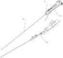
参考图1和图2,本实施例提供一种软镜导入装置,其为硬性导入鞘组件20,本实施例之软镜为输尿管软镜,硬性导入鞘组件20用于搭载输尿管软镜10到达肾盂口。Referring to FIGS. 1 and 2 , the present embodiment provides a flexible endoscope introducing device, which is a rigid introducing
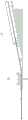
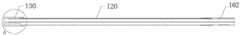
具体的,如图3所示,输尿管软镜10包括软镜主体100和与软镜主体100连接的软镜手柄110,软镜主体100包括顺序连接的软管102、弯曲件120和镜头支架130。Specifically, as shown in FIG. 3 , the
如图4及图5所示,输尿管软镜10的结构如下:软镜主体100内设置有与软镜主体100相匹配的工作通道101;软镜主体100的一端即软管102的一端穿过软镜手柄110并与软镜手柄110固定连接;工作通道101由软镜手柄110的自由端插入软管102,由软管102内穿过后继续穿过弯曲件120后伸入到镜头支架130的内部。如图6所示,镜头支架130内部设置有用于与工作通道101连通的贯通孔131,以便于输入或输出灌注液。镜头支架130前方对应于贯通孔131的下方的位置上设置有摄像通道132,摄像通道132内设置有摄像头装置,用于实时监控手术过程。镜头支架130前方对应于贯通孔131的一侧的位置上设置有照明通道133,照明通道133内设置有照明装置,该照明装置通过导光束提供照明,与照明通道133相对的另一侧设有两个用于安装转向钢丝的盲孔134,所述转向钢丝用于与弯曲件120配合。As shown in FIG. 4 and FIG. 5 , the structure of the
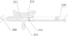
如图7和图8示,硬性导入鞘组件20主要包括依次连接的导入鞘200、导鞘主体21和用于固定软镜手柄110的固定支架220。导入鞘200的结构为长条管体,其结构与拉直的软镜主体100的结构相匹配,其材质为金属或者塑胶,刚性较强,同时具有一定的弹性,在外力作用下可发生弹性变形,导入鞘200的一端固定设置在导鞘主体210内。当输尿管软镜10与硬性导入鞘组件20结合使用时,软镜主体100经导鞘主体210插入导入鞘200中且软镜主体100的头端与导入鞘200的头端相平齐或者接近平齐,软镜手柄110可拆卸地设置在固定支架220上。所述接近平齐的含义是:软镜主体100的头端可以突出于导入鞘200的头部0-3mm,实现肉眼可见之平齐,而为了避免尿道和输尿管内壁损伤,导入鞘200的头端不突出于软镜主体100的头端。As shown in FIG. 7 and FIG. 8 , the rigid
具体的,导鞘主体210靠近导入鞘200的一端内设置有导鞘通道211,另一端设置有用于供软镜主体100穿过的开槽212,该开槽212与导鞘通道211连通。导入鞘200的端部密封地插接于导鞘通道211内,导鞘通道211远离导入鞘200的一端的内径往开槽212方向逐渐增大。导鞘通道211靠近开槽212的位置上设置有导鞘密封件213,该导鞘密封件213用于对软镜主体100和导鞘通道211进行密封,导鞘密封件213与导鞘通道211过盈配合,能够有效防止灌注液外泄。Specifically, one end of the
同时参看图9,导鞘主体210对应于导鞘通道211中部的位置上设置有出水通道214,出水通道214与导鞘通道211连通,用于引出灌注液,方便医生手术,有利于保持手术室环境的整洁。导鞘主体210对应于导鞘通道211上方的位置上设置有导鞘手柄215,方便医生抓握,同时也不用改变原有的使用习惯。导鞘主体210靠近开槽212的端部上设置有固定筋216,相应地,固定支架220的端部上设置有与固定筋216相匹配的固定扣221,固定支架220通过固定筋216与固定扣221的扣合与导鞘主体210可拆卸连接。固定支架220上设置软镜卡扣222,软镜手柄110卡接于软镜卡扣222上,对输尿管软镜10起到很好的固定和定位作用。同时参考图1及图2,将软镜主体100经由开槽212和导鞘通道211插入导入鞘200中,使软镜主体100的头部与导入鞘200的头部平齐,软镜手柄110卡接在固定支架220的软镜卡扣222上,从而使输尿管软镜10固定在硬性导入鞘组件20上。Referring to FIG. 9 at the same time, a
所述弯曲件120具有至少一个摆动自由度,使其可通过转向钢丝来调整其弯曲方向,以便于工作通道101的入口准确到达肾盂位置。The bending
固定好输尿管软镜10和硬性导入鞘组件20后,其使用方法如下:After the
1)将输尿管软镜安装入硬性导入鞘组件20内,开通灌注液,开启摄像头装置和照明装置,使与输尿管软镜10连接的显示屏实时显示镜头支架130前端的实时图像;1) the flexible ureteroscope is installed in the rigid
2)将硬性导入鞘组件20的导入鞘200经尿道置入,软镜主体100随导入鞘200同时置入体内:医生只需手握导鞘手柄215将导入鞘200和软镜主体100插入患者体内,在插入过程中,通过显示屏观察整个插入过程的情况,及时调整插入方向和速度;2) Insert the
3)在输尿管软镜10经尿道进入输尿管至肾盂口,硬性导入鞘组件20停止向前推进并预固定,取下固定支架220上的软镜手柄110,将软镜主体100继续沿导入鞘向前推进,进入肾盂内,即可进行检查或手术。3) After the
使用完毕后,先将输尿管软镜10经由硬性导入鞘组件20形成的通道抽出人体,然后再将导入鞘200拔出。After use, the
由此可见,本实用新型通过将输尿管软镜10固定在硬性导入鞘组件20上,使得输尿管软镜10可搭载在硬性导入鞘组件20上,具有硬镜的特性,与现有的软镜置入方法相比,仅需一步操作便可准确到达肾盂位置,操作简单快捷,大大提升了工作效率,不需要斑马导丝等设备,使用设备少,有效降低了手术成本和手术时间,有利于减少患者的损伤。同时,由于摄像头装置和照明装置始终设置在镜头支架130前端,使得整个插入过程和手术过程可视。It can be seen from this that the present invention fixes the
本实用新型之软镜导入装置的原理和结构亦可应用于其他内窥镜,如膀胱镜、肠镜、或宫腔镜等,适用范围较广。The principle and structure of the flexible endoscope introducing device of the present invention can also be applied to other endoscopes, such as cystoscopes, colonoscopes, or hysteroscopes, etc., with a wide range of applications.
上述实施方式仅为本实用新型的优选实施方式,不能以此来限定本实用新型保护的范围,本领域的技术人员在本实用新型的基础上所做的任何非实质性的变化及替换均属于本实用新型所要求保护的范围。The above-mentioned embodiments are only the preferred embodiments of the present invention, and the scope of protection of the present invention cannot be limited by this. Any insubstantial changes and replacements made by those skilled in the art on the basis of the present invention belong to the scope of the present invention. The scope of protection of the present utility model.
Claims (10)
Translated fromChineseApplications Claiming Priority (2)
| Application Number | Priority Date | Filing Date | Title |
|---|---|---|---|
| CN2019103539488 | 2019-04-29 | ||
| CN201910353948 | 2019-04-29 |
Publications (1)
| Publication Number | Publication Date |
|---|---|
| CN211985342Utrue CN211985342U (en) | 2020-11-24 |
Family
ID=69544577
Family Applications (16)
| Application Number | Title | Priority Date | Filing Date |
|---|---|---|---|
| CN201921010057.4UActiveCN210727707U (en) | 2019-04-29 | 2019-06-28 | Cold light source fixing structure easy to install |
| CN201910571802.0AActiveCN111839423B (en) | 2019-04-29 | 2019-06-28 | A micro snake bone component |
| CN201921009904.5UActiveCN210541480U (en) | 2019-04-29 | 2019-06-28 | A connection structure of a lens bracket and a snake bone component |
| CN201921010223.0UActiveCN210749119U (en) | 2019-04-29 | 2019-06-28 | A hose fixing structure |
| CN201921009859.3UActiveCN210749149U (en) | 2019-04-29 | 2019-06-28 | An anti-reverse knob structure |
| CN201921009902.6UActiveCN210541479U (en) | 2019-04-29 | 2019-06-28 | A steering wire fixing structure |
| CN201921009838.1UActiveCN210541477U (en) | 2019-04-29 | 2019-06-28 | A miniature snake bone component |
| CN201921010058.9UActiveCN210541478U (en) | 2019-04-29 | 2019-06-28 | A sealing structure of a curved part of an endoscope |
| CN201921009840.9UActiveCN210749127U (en) | 2019-04-29 | 2019-06-28 | An endoscope working channel connection structure |
| CN201921010219.4UActiveCN210643982U (en) | 2019-04-29 | 2019-06-28 | An endoscope tube structure |
| CN201922220114.8UActiveCN211985382U (en) | 2019-04-29 | 2019-12-11 | Ureter pyeloscope system |
| CN201922216656.8UActiveCN211985342U (en) | 2019-04-29 | 2019-12-11 | Soft lens leading-in device |
| CN201911270216.9APendingCN110811492A (en) | 2019-04-29 | 2019-12-11 | A soft mirror introduction device and method of using the same |
| CN201911263637.9APendingCN110811525A (en) | 2019-04-29 | 2019-12-11 | Ureter pyeloscope system and implantation method thereof |
| CN201922496421.9UActiveCN212261315U (en) | 2019-04-29 | 2019-12-31 | A linear working channel of a flexible lens and a flexible lens |
| CN202020641284.3UActiveCN213309607U (en) | 2019-04-29 | 2020-04-23 | Ureter pyeloscope system with water outlet channel |
Family Applications Before (11)
| Application Number | Title | Priority Date | Filing Date |
|---|---|---|---|
| CN201921010057.4UActiveCN210727707U (en) | 2019-04-29 | 2019-06-28 | Cold light source fixing structure easy to install |
| CN201910571802.0AActiveCN111839423B (en) | 2019-04-29 | 2019-06-28 | A micro snake bone component |
| CN201921009904.5UActiveCN210541480U (en) | 2019-04-29 | 2019-06-28 | A connection structure of a lens bracket and a snake bone component |
| CN201921010223.0UActiveCN210749119U (en) | 2019-04-29 | 2019-06-28 | A hose fixing structure |
| CN201921009859.3UActiveCN210749149U (en) | 2019-04-29 | 2019-06-28 | An anti-reverse knob structure |
| CN201921009902.6UActiveCN210541479U (en) | 2019-04-29 | 2019-06-28 | A steering wire fixing structure |
| CN201921009838.1UActiveCN210541477U (en) | 2019-04-29 | 2019-06-28 | A miniature snake bone component |
| CN201921010058.9UActiveCN210541478U (en) | 2019-04-29 | 2019-06-28 | A sealing structure of a curved part of an endoscope |
| CN201921009840.9UActiveCN210749127U (en) | 2019-04-29 | 2019-06-28 | An endoscope working channel connection structure |
| CN201921010219.4UActiveCN210643982U (en) | 2019-04-29 | 2019-06-28 | An endoscope tube structure |
| CN201922220114.8UActiveCN211985382U (en) | 2019-04-29 | 2019-12-11 | Ureter pyeloscope system |
Family Applications After (4)
| Application Number | Title | Priority Date | Filing Date |
|---|---|---|---|
| CN201911270216.9APendingCN110811492A (en) | 2019-04-29 | 2019-12-11 | A soft mirror introduction device and method of using the same |
| CN201911263637.9APendingCN110811525A (en) | 2019-04-29 | 2019-12-11 | Ureter pyeloscope system and implantation method thereof |
| CN201922496421.9UActiveCN212261315U (en) | 2019-04-29 | 2019-12-31 | A linear working channel of a flexible lens and a flexible lens |
| CN202020641284.3UActiveCN213309607U (en) | 2019-04-29 | 2020-04-23 | Ureter pyeloscope system with water outlet channel |
Country Status (1)
| Country | Link |
|---|---|
| CN (16) | CN210727707U (en) |
Cited By (1)
| Publication number | Priority date | Publication date | Assignee | Title |
|---|---|---|---|---|
| CN110811492A (en)* | 2019-04-29 | 2020-02-21 | 珠海市司迈科技有限公司 | A soft mirror introduction device and method of using the same |
Families Citing this family (16)
| Publication number | Priority date | Publication date | Assignee | Title |
|---|---|---|---|---|
| CN218651745U (en)* | 2019-04-29 | 2023-03-21 | 珠海市司迈科技有限公司 | Soft lens and ureter pyeloscope system adopting same |
| CN111714072A (en)* | 2020-07-27 | 2020-09-29 | 湖南省华芯医疗器械有限公司 | A kind of snake bone component and endoscope |
| CN111904541B (en)* | 2020-09-08 | 2024-07-23 | 北京成信盛达科技有限责任公司 | Capsule ureteroscope and operation method |
| CN112773303A (en)* | 2021-01-27 | 2021-05-11 | 杭州思康新医疗科技有限公司 | Endoscope snake bone and endoscope |
| CN113662501A (en)* | 2021-08-27 | 2021-11-19 | 温州市人民医院 | Bedside visual bladder soft lens |
| CN113966987B (en)* | 2021-09-14 | 2024-08-23 | 杭州好克光电仪器有限公司 | Electronic choledochoscope with changeable softness and hardness |
| CN113854935B (en)* | 2021-12-02 | 2022-02-18 | 极限人工智能有限公司 | Endoscope and surgical robot |
| CN114452011B (en)* | 2022-01-29 | 2024-04-26 | 上海璞跃医疗器械有限公司 | Renal pelvis internal pressure control system |
| CN114617521A (en)* | 2022-03-25 | 2022-06-14 | 浙江本书科技有限公司 | Spliced snake bone without riveting |
| CN115191912B (en)* | 2022-06-16 | 2025-01-21 | 上海工程技术大学 | Double-layer snake bone unit section, snake bone tube and endoscope |
| CN115054805B (en)* | 2022-07-05 | 2025-07-25 | 深圳市库珀科技发展有限公司 | Bendable guiding sheath tube and guiding device |
| CN115316915B (en)* | 2022-08-17 | 2025-05-27 | 浙江微度医疗器械有限公司 | Endoscope snake for easy bending |
| CN116211218A (en)* | 2022-12-21 | 2023-06-06 | 浙江优亿医疗器械股份有限公司 | Improved visualizer |
| CN116726345A (en)* | 2023-06-16 | 2023-09-12 | 东莞市正生瑞生物医学科技有限公司 | A kind of ureteral guiding sheath |
| CN119236282A (en)* | 2024-09-05 | 2025-01-03 | 深圳市人工智能与机器人研究院 | A catheter system for auxiliary medical treatment |
| CN118948194B (en)* | 2024-09-23 | 2025-04-18 | 江南大学 | A flexible ureteroscope with active tentacles |
Family Cites Families (6)
| Publication number | Priority date | Publication date | Assignee | Title |
|---|---|---|---|---|
| CN101019756A (en)* | 2006-08-10 | 2007-08-22 | 陈志强 | Soft uretero-renoscope with hard sheath |
| CN202505312U (en)* | 2012-04-09 | 2012-10-31 | 广州医学院第一附属医院 | Multi-purpose endoscope |
| CN103405261B (en)* | 2013-07-19 | 2016-02-03 | 孙颖浩 | The flexible ureterorenoscope of a kind of head end |
| WO2016054202A1 (en)* | 2014-10-02 | 2016-04-07 | Boston Scientific Scimed, Inc. | Attachment device for a ureteroscope |
| CN108670321B (en)* | 2018-06-01 | 2024-03-26 | 常州市久成电子设备有限公司 | Endoscopic surgical instrument and neck bending structure thereof |
| CN210727707U (en)* | 2019-04-29 | 2020-06-12 | 珠海市司迈科技有限公司 | Cold light source fixing structure easy to install |
- 2019
- 2019-06-28CNCN201921010057.4Upatent/CN210727707U/enactiveActive
- 2019-06-28CNCN201910571802.0Apatent/CN111839423B/enactiveActive
- 2019-06-28CNCN201921009904.5Upatent/CN210541480U/enactiveActive
- 2019-06-28CNCN201921010223.0Upatent/CN210749119U/enactiveActive
- 2019-06-28CNCN201921009859.3Upatent/CN210749149U/enactiveActive
- 2019-06-28CNCN201921009902.6Upatent/CN210541479U/enactiveActive
- 2019-06-28CNCN201921009838.1Upatent/CN210541477U/enactiveActive
- 2019-06-28CNCN201921010058.9Upatent/CN210541478U/enactiveActive
- 2019-06-28CNCN201921009840.9Upatent/CN210749127U/enactiveActive
- 2019-06-28CNCN201921010219.4Upatent/CN210643982U/enactiveActive
- 2019-12-11CNCN201922220114.8Upatent/CN211985382U/enactiveActive
- 2019-12-11CNCN201922216656.8Upatent/CN211985342U/enactiveActive
- 2019-12-11CNCN201911270216.9Apatent/CN110811492A/enactivePending
- 2019-12-11CNCN201911263637.9Apatent/CN110811525A/enactivePending
- 2019-12-31CNCN201922496421.9Upatent/CN212261315U/enactiveActive
- 2020
- 2020-04-23CNCN202020641284.3Upatent/CN213309607U/enactiveActive
Cited By (1)
| Publication number | Priority date | Publication date | Assignee | Title |
|---|---|---|---|---|
| CN110811492A (en)* | 2019-04-29 | 2020-02-21 | 珠海市司迈科技有限公司 | A soft mirror introduction device and method of using the same |
Also Published As
| Publication number | Publication date |
|---|---|
| CN111839423A (en) | 2020-10-30 |
| CN211985382U (en) | 2020-11-24 |
| CN210541477U (en) | 2020-05-19 |
| CN212261315U (en) | 2021-01-01 |
| CN110811525A (en) | 2020-02-21 |
| CN210643982U (en) | 2020-06-02 |
| CN210749119U (en) | 2020-06-16 |
| CN210727707U (en) | 2020-06-12 |
| CN210749127U (en) | 2020-06-16 |
| CN213309607U (en) | 2021-06-01 |
| CN111839423B (en) | 2025-06-13 |
| CN110811492A (en) | 2020-02-21 |
| CN210541480U (en) | 2020-05-19 |
| CN210541478U (en) | 2020-05-19 |
| CN210749149U (en) | 2020-06-16 |
| CN210541479U (en) | 2020-05-19 |
Similar Documents
| Publication | Publication Date | Title |
|---|---|---|
| CN211985342U (en) | Soft lens leading-in device | |
| EP1029414B1 (en) | Video rectoscope | |
| CN100558285C (en) | Endoscope insertion portion, endoscope, and endoscope system | |
| CN101732082B (en) | Soft and hard gallbladder and cholangioscopic system | |
| CN201701191U (en) | Pipeline endoscope | |
| CN113350653A (en) | Multifunctional catheter | |
| CN110575122A (en) | Endoscopic catheter, assembly and endoscopic visualization sinus balloon dilation system | |
| CN218651745U (en) | Soft lens and ureter pyeloscope system adopting same | |
| CN214912456U (en) | Visual catheter and have incision sword of this visual catheter | |
| KR20090087464A (en) | New bent neck for transesophageal echocardiography (TEE) probe | |
| JP3325103B2 (en) | Cover-type endoscope | |
| JP5400118B2 (en) | Endoscopy forceps plug | |
| CN212214357U (en) | Multifunctional catheter | |
| JP4847175B2 (en) | Endoscope system and endoscope insertion aid | |
| CN204445794U (en) | Endoscope and endoscope extend imaging device | |
| WO2023083206A1 (en) | Medical catheter probe, medical catheter, and medical device and system | |
| US20160029880A1 (en) | Cystoscopic device and methods for operating same | |
| JPH10309259A (en) | Endoscope | |
| CN221786197U (en) | Telescopic guiding endoscope suitable for narrow channel | |
| CN222398256U (en) | Comprehensive endoscope lens simultaneously applicable to ear, nose and throat examination | |
| CN213606253U (en) | Scope and have scope intubate structure of external pliers pipeline | |
| CN221813876U (en) | Disposable insertion device for an endoscope and corresponding combination endoscope | |
| US20230058772A1 (en) | Disposable endoscope sheath | |
| RU2806722C1 (en) | Videorectoscope | |
| JPH0817767B2 (en) | Endoscope device |
Legal Events
| Date | Code | Title | Description |
|---|---|---|---|
| GR01 | Patent grant | ||
| GR01 | Patent grant |