CN210842941U - Endoscope and endoscope system - Google Patents
Endoscope and endoscope systemDownload PDFInfo
- Publication number
- CN210842941U CN210842941UCN201920196433.7UCN201920196433UCN210842941UCN 210842941 UCN210842941 UCN 210842941UCN 201920196433 UCN201920196433 UCN 201920196433UCN 210842941 UCN210842941 UCN 210842941U
- Authority
- CN
- China
- Prior art keywords
- cannula
- handle portion
- endoscope
- handle
- relative
- Prior art date
- Legal status (The legal status is an assumption and is not a legal conclusion. Google has not performed a legal analysis and makes no representation as to the accuracy of the status listed.)
- Active
Links
Images


Classifications
- A—HUMAN NECESSITIES
- A61—MEDICAL OR VETERINARY SCIENCE; HYGIENE
- A61B—DIAGNOSIS; SURGERY; IDENTIFICATION
- A61B1/00—Instruments for performing medical examinations of the interior of cavities or tubes of the body by visual or photographical inspection, e.g. endoscopes; Illuminating arrangements therefor
- A61B1/00064—Constructional details of the endoscope body
- A61B1/00066—Proximal part of endoscope body, e.g. handles
- A—HUMAN NECESSITIES
- A61—MEDICAL OR VETERINARY SCIENCE; HYGIENE
- A61B—DIAGNOSIS; SURGERY; IDENTIFICATION
- A61B1/00—Instruments for performing medical examinations of the interior of cavities or tubes of the body by visual or photographical inspection, e.g. endoscopes; Illuminating arrangements therefor
- A61B1/00002—Operational features of endoscopes
- A61B1/00039—Operational features of endoscopes provided with input arrangements for the user
- A61B1/00042—Operational features of endoscopes provided with input arrangements for the user for mechanical operation
- A—HUMAN NECESSITIES
- A61—MEDICAL OR VETERINARY SCIENCE; HYGIENE
- A61B—DIAGNOSIS; SURGERY; IDENTIFICATION
- A61B1/00—Instruments for performing medical examinations of the interior of cavities or tubes of the body by visual or photographical inspection, e.g. endoscopes; Illuminating arrangements therefor
- A61B1/00002—Operational features of endoscopes
- A61B1/00004—Operational features of endoscopes characterised by electronic signal processing
- A61B1/00009—Operational features of endoscopes characterised by electronic signal processing of image signals during a use of endoscope
- A—HUMAN NECESSITIES
- A61—MEDICAL OR VETERINARY SCIENCE; HYGIENE
- A61B—DIAGNOSIS; SURGERY; IDENTIFICATION
- A61B1/00—Instruments for performing medical examinations of the interior of cavities or tubes of the body by visual or photographical inspection, e.g. endoscopes; Illuminating arrangements therefor
- A61B1/00002—Operational features of endoscopes
- A61B1/00043—Operational features of endoscopes provided with output arrangements
- A61B1/00045—Display arrangement
- A—HUMAN NECESSITIES
- A61—MEDICAL OR VETERINARY SCIENCE; HYGIENE
- A61B—DIAGNOSIS; SURGERY; IDENTIFICATION
- A61B1/00—Instruments for performing medical examinations of the interior of cavities or tubes of the body by visual or photographical inspection, e.g. endoscopes; Illuminating arrangements therefor
- A61B1/00064—Constructional details of the endoscope body
- A61B1/00071—Insertion part of the endoscope body
- A61B1/00078—Insertion part of the endoscope body with stiffening means
- A—HUMAN NECESSITIES
- A61—MEDICAL OR VETERINARY SCIENCE; HYGIENE
- A61B—DIAGNOSIS; SURGERY; IDENTIFICATION
- A61B1/00—Instruments for performing medical examinations of the interior of cavities or tubes of the body by visual or photographical inspection, e.g. endoscopes; Illuminating arrangements therefor
- A61B1/00064—Constructional details of the endoscope body
- A61B1/00071—Insertion part of the endoscope body
- A61B1/0008—Insertion part of the endoscope body characterised by distal tip features
- A61B1/00089—Hoods
- A—HUMAN NECESSITIES
- A61—MEDICAL OR VETERINARY SCIENCE; HYGIENE
- A61B—DIAGNOSIS; SURGERY; IDENTIFICATION
- A61B1/00—Instruments for performing medical examinations of the interior of cavities or tubes of the body by visual or photographical inspection, e.g. endoscopes; Illuminating arrangements therefor
- A61B1/00064—Constructional details of the endoscope body
- A61B1/00071—Insertion part of the endoscope body
- A61B1/0008—Insertion part of the endoscope body characterised by distal tip features
- A61B1/00101—Insertion part of the endoscope body characterised by distal tip features the distal tip features being detachable
- A—HUMAN NECESSITIES
- A61—MEDICAL OR VETERINARY SCIENCE; HYGIENE
- A61B—DIAGNOSIS; SURGERY; IDENTIFICATION
- A61B1/00—Instruments for performing medical examinations of the interior of cavities or tubes of the body by visual or photographical inspection, e.g. endoscopes; Illuminating arrangements therefor
- A61B1/00064—Constructional details of the endoscope body
- A61B1/00103—Constructional details of the endoscope body designed for single use
- A—HUMAN NECESSITIES
- A61—MEDICAL OR VETERINARY SCIENCE; HYGIENE
- A61B—DIAGNOSIS; SURGERY; IDENTIFICATION
- A61B1/00—Instruments for performing medical examinations of the interior of cavities or tubes of the body by visual or photographical inspection, e.g. endoscopes; Illuminating arrangements therefor
- A61B1/00064—Constructional details of the endoscope body
- A61B1/00105—Constructional details of the endoscope body characterised by modular construction
- A—HUMAN NECESSITIES
- A61—MEDICAL OR VETERINARY SCIENCE; HYGIENE
- A61B—DIAGNOSIS; SURGERY; IDENTIFICATION
- A61B1/00—Instruments for performing medical examinations of the interior of cavities or tubes of the body by visual or photographical inspection, e.g. endoscopes; Illuminating arrangements therefor
- A61B1/00112—Connection or coupling means
- A61B1/00121—Connectors, fasteners and adapters, e.g. on the endoscope handle
- A61B1/00124—Connectors, fasteners and adapters, e.g. on the endoscope handle electrical, e.g. electrical plug-and-socket connection
- A—HUMAN NECESSITIES
- A61—MEDICAL OR VETERINARY SCIENCE; HYGIENE
- A61B—DIAGNOSIS; SURGERY; IDENTIFICATION
- A61B1/00—Instruments for performing medical examinations of the interior of cavities or tubes of the body by visual or photographical inspection, e.g. endoscopes; Illuminating arrangements therefor
- A61B1/00131—Accessories for endoscopes
- A—HUMAN NECESSITIES
- A61—MEDICAL OR VETERINARY SCIENCE; HYGIENE
- A61B—DIAGNOSIS; SURGERY; IDENTIFICATION
- A61B1/00—Instruments for performing medical examinations of the interior of cavities or tubes of the body by visual or photographical inspection, e.g. endoscopes; Illuminating arrangements therefor
- A61B1/00142—Instruments for performing medical examinations of the interior of cavities or tubes of the body by visual or photographical inspection, e.g. endoscopes; Illuminating arrangements therefor with means for preventing contamination, e.g. by using a sanitary sheath
- A61B1/00144—Hygienic packaging
- A—HUMAN NECESSITIES
- A61—MEDICAL OR VETERINARY SCIENCE; HYGIENE
- A61B—DIAGNOSIS; SURGERY; IDENTIFICATION
- A61B1/00—Instruments for performing medical examinations of the interior of cavities or tubes of the body by visual or photographical inspection, e.g. endoscopes; Illuminating arrangements therefor
- A61B1/00163—Optical arrangements
- A61B1/00174—Optical arrangements characterised by the viewing angles
- A61B1/00179—Optical arrangements characterised by the viewing angles for off-axis viewing
- A—HUMAN NECESSITIES
- A61—MEDICAL OR VETERINARY SCIENCE; HYGIENE
- A61B—DIAGNOSIS; SURGERY; IDENTIFICATION
- A61B1/00—Instruments for performing medical examinations of the interior of cavities or tubes of the body by visual or photographical inspection, e.g. endoscopes; Illuminating arrangements therefor
- A61B1/005—Flexible endoscopes
- A61B1/0051—Flexible endoscopes with controlled bending of insertion part
- A61B1/0057—Constructional details of force transmission elements, e.g. control wires
- A—HUMAN NECESSITIES
- A61—MEDICAL OR VETERINARY SCIENCE; HYGIENE
- A61B—DIAGNOSIS; SURGERY; IDENTIFICATION
- A61B1/00—Instruments for performing medical examinations of the interior of cavities or tubes of the body by visual or photographical inspection, e.g. endoscopes; Illuminating arrangements therefor
- A61B1/012—Instruments for performing medical examinations of the interior of cavities or tubes of the body by visual or photographical inspection, e.g. endoscopes; Illuminating arrangements therefor characterised by internal passages or accessories therefor
- A61B1/018—Instruments for performing medical examinations of the interior of cavities or tubes of the body by visual or photographical inspection, e.g. endoscopes; Illuminating arrangements therefor characterised by internal passages or accessories therefor for receiving instruments
- A—HUMAN NECESSITIES
- A61—MEDICAL OR VETERINARY SCIENCE; HYGIENE
- A61B—DIAGNOSIS; SURGERY; IDENTIFICATION
- A61B1/00—Instruments for performing medical examinations of the interior of cavities or tubes of the body by visual or photographical inspection, e.g. endoscopes; Illuminating arrangements therefor
- A61B1/04—Instruments for performing medical examinations of the interior of cavities or tubes of the body by visual or photographical inspection, e.g. endoscopes; Illuminating arrangements therefor combined with photographic or television appliances
- A61B1/05—Instruments for performing medical examinations of the interior of cavities or tubes of the body by visual or photographical inspection, e.g. endoscopes; Illuminating arrangements therefor combined with photographic or television appliances characterised by the image sensor, e.g. camera, being in the distal end portion
- A—HUMAN NECESSITIES
- A61—MEDICAL OR VETERINARY SCIENCE; HYGIENE
- A61B—DIAGNOSIS; SURGERY; IDENTIFICATION
- A61B1/00—Instruments for performing medical examinations of the interior of cavities or tubes of the body by visual or photographical inspection, e.g. endoscopes; Illuminating arrangements therefor
- A61B1/06—Instruments for performing medical examinations of the interior of cavities or tubes of the body by visual or photographical inspection, e.g. endoscopes; Illuminating arrangements therefor with illuminating arrangements
- A61B1/0623—Instruments for performing medical examinations of the interior of cavities or tubes of the body by visual or photographical inspection, e.g. endoscopes; Illuminating arrangements therefor with illuminating arrangements for off-axis illumination
- A—HUMAN NECESSITIES
- A61—MEDICAL OR VETERINARY SCIENCE; HYGIENE
- A61B—DIAGNOSIS; SURGERY; IDENTIFICATION
- A61B1/00—Instruments for performing medical examinations of the interior of cavities or tubes of the body by visual or photographical inspection, e.g. endoscopes; Illuminating arrangements therefor
- A61B1/06—Instruments for performing medical examinations of the interior of cavities or tubes of the body by visual or photographical inspection, e.g. endoscopes; Illuminating arrangements therefor with illuminating arrangements
- A61B1/0661—Endoscope light sources
- A61B1/0676—Endoscope light sources at distal tip of an endoscope
- A—HUMAN NECESSITIES
- A61—MEDICAL OR VETERINARY SCIENCE; HYGIENE
- A61B—DIAGNOSIS; SURGERY; IDENTIFICATION
- A61B1/00—Instruments for performing medical examinations of the interior of cavities or tubes of the body by visual or photographical inspection, e.g. endoscopes; Illuminating arrangements therefor
- A61B1/307—Instruments for performing medical examinations of the interior of cavities or tubes of the body by visual or photographical inspection, e.g. endoscopes; Illuminating arrangements therefor for the urinary organs, e.g. urethroscopes, cystoscopes
- G—PHYSICS
- G02—OPTICS
- G02B—OPTICAL ELEMENTS, SYSTEMS OR APPARATUS
- G02B23/00—Telescopes, e.g. binoculars; Periscopes; Instruments for viewing the inside of hollow bodies; Viewfinders; Optical aiming or sighting devices
- G02B23/24—Instruments or systems for viewing the inside of hollow bodies, e.g. fibrescopes
- G02B23/2476—Non-optical details, e.g. housings, mountings, supports
- G02B23/2484—Arrangements in relation to a camera or imaging device
- G—PHYSICS
- G06—COMPUTING OR CALCULATING; COUNTING
- G06Q—INFORMATION AND COMMUNICATION TECHNOLOGY [ICT] SPECIALLY ADAPTED FOR ADMINISTRATIVE, COMMERCIAL, FINANCIAL, MANAGERIAL OR SUPERVISORY PURPOSES; SYSTEMS OR METHODS SPECIALLY ADAPTED FOR ADMINISTRATIVE, COMMERCIAL, FINANCIAL, MANAGERIAL OR SUPERVISORY PURPOSES, NOT OTHERWISE PROVIDED FOR
- G06Q30/00—Commerce
- G06Q30/018—Certifying business or products
- G06Q30/0185—Product, service or business identity fraud
- A—HUMAN NECESSITIES
- A61—MEDICAL OR VETERINARY SCIENCE; HYGIENE
- A61B—DIAGNOSIS; SURGERY; IDENTIFICATION
- A61B1/00—Instruments for performing medical examinations of the interior of cavities or tubes of the body by visual or photographical inspection, e.g. endoscopes; Illuminating arrangements therefor
- A61B1/00064—Constructional details of the endoscope body
- A61B1/00071—Insertion part of the endoscope body
- A61B1/0008—Insertion part of the endoscope body characterised by distal tip features
- A61B1/00096—Optical elements
- G—PHYSICS
- G02—OPTICS
- G02B—OPTICAL ELEMENTS, SYSTEMS OR APPARATUS
- G02B23/00—Telescopes, e.g. binoculars; Periscopes; Instruments for viewing the inside of hollow bodies; Viewfinders; Optical aiming or sighting devices
- G02B23/24—Instruments or systems for viewing the inside of hollow bodies, e.g. fibrescopes
- G02B23/2407—Optical details
- G02B23/2423—Optical details of the distal end
Landscapes
- Health & Medical Sciences (AREA)
- Life Sciences & Earth Sciences (AREA)
- Surgery (AREA)
- Physics & Mathematics (AREA)
- Engineering & Computer Science (AREA)
- Optics & Photonics (AREA)
- Biomedical Technology (AREA)
- General Health & Medical Sciences (AREA)
- Pathology (AREA)
- Nuclear Medicine, Radiotherapy & Molecular Imaging (AREA)
- Biophysics (AREA)
- Heart & Thoracic Surgery (AREA)
- Medical Informatics (AREA)
- Molecular Biology (AREA)
- Animal Behavior & Ethology (AREA)
- Radiology & Medical Imaging (AREA)
- Public Health (AREA)
- Veterinary Medicine (AREA)
- General Physics & Mathematics (AREA)
- Astronomy & Astrophysics (AREA)
- Signal Processing (AREA)
- Business, Economics & Management (AREA)
- Multimedia (AREA)
- Mechanical Engineering (AREA)
- Urology & Nephrology (AREA)
- Entrepreneurship & Innovation (AREA)
- Accounting & Taxation (AREA)
- Development Economics (AREA)
- Economics (AREA)
- Finance (AREA)
- Marketing (AREA)
- Strategic Management (AREA)
- General Business, Economics & Management (AREA)
- Theoretical Computer Science (AREA)
- Endoscopes (AREA)
- Instruments For Viewing The Inside Of Hollow Bodies (AREA)
Abstract
Translated fromChinese
Description
Translated fromChinese相关申请的引用Citations to Related Applications
本专利申请要求2018年2月14日提交的美国临时专利申请系列号62/630,718和2019年2月6日提交的美国正式专利申请系列号16/268,909的优先权,所述临时专利申请和正式专利申请通过引用的方式并入本申请。本说明书中提及的所有公布、专利和专利申请均以引用方式并入本文,所述引用的程度就如同已明确且个别地指示将各个别公布、专利或专利申请以引用的方式并入本文一般。This patent application claims priority to US Provisional Patent Application Serial No. 62/630,718, filed February 14, 2018, and US Official Patent Application Serial No. 16/268,909, filed February 6, 2019, which The patent application is incorporated herein by reference. All publications, patents and patent applications mentioned in this specification are incorporated herein by reference to the same extent as if each individual publication, patent or patent application was specifically and individually indicated to be incorporated by reference herein generally.
技术领域technical field
本专利说明书涉及用于组织检查和内窥镜外科手术的医疗装置,如用于宫腔镜和泌尿科。更具体地,一些实施例涉及内窥镜系统,其包括用后可弃的、单次使用的插管和远侧成像模块,以及可重复使用的、多次使用的手柄和显示塔。This patent specification relates to medical devices for tissue examination and endoscopic surgery, such as for hysteroscopy and urology. More specifically, some embodiments relate to an endoscopic system that includes a disposable, single-use cannula and distal imaging module, as well as a reusable, multiple-use handle and display tower.
背景技术Background technique
直视医疗流程(诸如内窥镜流程)用于检查身体的某些部位,包括内部解剖结构。例如,宫腔镜检查子宫,膀胱镜检查膀胱,胃镜检查食道、胃和/或小肠,支气管镜检查咽喉、喉头、气管和/或下呼吸道,乙状结肠镜检查直肠,结肠镜检查直肠和/或结肠,阴道镜检查子宫颈、阴道和/或外阴,鼻腔内窥镜检查鼻和鼻窦通道等。Direct vision medical procedures, such as endoscopic procedures, are used to examine certain parts of the body, including internal anatomy. For example, hysteroscopy to examine the uterus, cystoscopy to examine the bladder, gastroscopy to examine the esophagus, stomach and/or small intestine, bronchoscopy to examine the throat, larynx, trachea and/or lower airway, sigmoidoscopy to examine the rectum, colonoscopy to examine the rectum and/or colon , colposcopy to examine the cervix, vagina and/or vulva, nasal endoscopy to examine nasal and sinus passages, etc.
传统上,内窥镜规程是用昂贵的设备来执行的。这种设备可以包括膀胱镜、宫腔镜和各种其他设备。这种设备还可以包括显示塔,其包括相机控制单元和照明控制单元。现有的膀胱镜和宫腔镜通常是可重复使用的装置,其中金属轴内有金属插管和光学透镜。其他类型的膀胱镜和宫腔镜可以包括柔性的可重复使用的装置,所述装置是柔性插管,所述柔性插管的尖端的铰接通常由近端处的拉线和操纵杆控制。在每个规程之后,设备可能需要灭菌,所述灭菌可能成本高或难以操作,而且灭菌或消毒可能无效。最近,部分一次性内窥镜已经可用,例如美国专利8460182和9895048。Traditionally, endoscopic procedures have been performed with expensive equipment. Such devices may include cystoscopes, hysteroscopes, and various other devices. Such equipment may also include a display tower including a camera control unit and a lighting control unit. Existing cystoscopes and hysteroscopes are often reusable devices with metal cannulae and optical lenses within a metal shaft. Other types of cystoscopes and hysteroscopes may include a flexible reusable device, which is a flexible cannula, the articulation of the tip of which is typically controlled by a pull wire and joystick at the proximal end. After each procedure, equipment may require sterilization, which may be costly or difficult to perform, and which may be ineffective in sterilization or disinfection. More recently, partially disposable endoscopes have become available, such as US Pat. Nos. 8,460,182 and 9,895,048.
内窥镜是插入体腔中以检查所述体腔的细长管状结构。常规内窥镜包括在其远端处具有物镜的望远镜。望远镜包括图像转发系统。在刚性内窥镜中,它是一系列间隔开的透镜。在柔性内窥镜中,它是被相干地组装以转发图像的一束微小光纤。在通常为柔性镜的数字内窥镜中,成像传感器可以位于柔性插管的远侧。然而,当医生操纵患者体内的插管的具有相机(数字内窥镜)的可偏转尖端时,可能失去视图水平线,即医生可能失去取向。An endoscope is an elongated tubular structure that is inserted into a body cavity to examine the body cavity. A conventional endoscope includes a telescope with an objective lens at its distal end. The telescope includes an image forwarding system. In rigid endoscopes, it is a series of spaced lenses. In a flexible endoscope, it's a bundle of tiny optical fibers that are coherently assembled to forward images. In digital endoscopes, which are typically flexible scopes, the imaging sensor may be located distal to the flexible cannula. However, when the doctor manipulates the deflectable tip with a camera (digital endoscope) of the cannula inside the patient, the horizon of view may be lost, ie the doctor may lose orientation.
因此,期望克服这些挑战并以低成本提供具有用后可弃的插管部分的膀胱镜和/或宫腔镜。如果这样的装置或系统在降低成本的同时,可以捕获具有可控水平视图的图像,则将是合乎实用期望的。将通过本文描述的装置来满足这些目标。Therefore, it would be desirable to overcome these challenges and provide cystoscopes and/or hysteroscopes with disposable cannula portions at low cost. It would be practically desirable if such a device or system could capture images with a controllable horizontal view while reducing cost. These goals will be met by the apparatus described herein.
实用新型内容Utility model content
根据一些实施例,描述了一种内窥镜系统,其包括:计算机处理系统;具有至少一个高清或者VGA显示器,其与所述计算机处理系统电连接以接收和显示内窥镜图像;以及手持部分,其包括:多次使用的手柄部分,其具有用于接收单次使用部分的机械和电气联接器,和在所述多次使用的手柄部分和与所述计算机处理系统之间电连通的缆线;以及包括细长插管的单次使用部分,当手柄部分和单次使用部分组装到所述手柄部分时,所述近端被配置成与所述多次使用的手柄部分的所述耦合器可释放地耦合,所述细长插管具有安装在其远端上的成像模块并且具有近端上的机械和电气联接器,所述插管和成像模块被配置成相对于所述多次使用的手柄部分围绕所述插管的中心纵向轴线旋转;第一传感器,其被配置成检测所述插管和成像模块相对于所述多次使用部分围绕所述中心轴线的旋转移动或旋转位置;以及第二传感器,其被配置成检测所述多次使用的手柄部分相对于所述显示监视器围绕与所述中心轴线平行的轴线的旋转移动或旋转位置。According to some embodiments, an endoscopic system is described that includes: a computer processing system; having at least one high-definition or VGA display in electrical connection with the computer processing system to receive and display endoscopic images; and a handheld portion , comprising: a multiple-use handle portion having mechanical and electrical couplings for receiving a single-use portion, and a cable in electrical communication between the multiple-use handle portion and the computer processing system a wire; and a single-use portion including an elongated cannula, the proximal end being configured for the coupling of the multiple-use handle portion when the handle portion and the single-use portion are assembled to the handle portion The elongated cannula is releasably coupled with an imaging module having an imaging module mounted on its distal end and a mechanical and electrical coupling on its proximal end, the cannula and imaging module being configured relative to the multiple a handle portion of use rotates about a central longitudinal axis of the cannula; a first sensor configured to detect rotational movement or rotational position of the cannula and imaging module relative to the multiple-use portion about the central axis and a second sensor configured to detect rotational movement or rotational position of the multi-use handle portion relative to the display monitor about an axis parallel to the central axis.
根据一些实施例,描述了一种内窥镜,包括一组多次使用的手柄部分,两个或多个单次使用部分,其中一个单次使用部分包括供医疗器械通过的工作通道且被用于治疗用途,所述至少另一个单次使用部分中不具有工作通道,其中所述多次使用的手柄部分包括机械和电联接器,每个所述单次使用部分具有壳体,所述壳体具有近端和远端,所述插管从所述壳体向远处延伸,所述远端处具有照明和成像模块;每个所述单次使用部分的所述壳体具有机械和电连接器,其配置为与多次使用的手柄部分的机械和电气连接器相匹配,以将所述多次使用部分和选定的一个单次使用部分组装成组合内窥镜;每个所述单次使用部分的所述插管被安装为相对于所述多次使用的手柄部分运动;多次使用的手柄部分的手动控制器;运动转移装置,其耦合所述手动控制器和所述选定的单次使用部分的插管,以响应于所述手动控制器的操作而移动所述插管;传感器,响应所述选定的单次使用部分的所述插管的相对于所述多次使用的手柄部分的运动,被配置为提供所述运动的指示。According to some embodiments, an endoscope is described that includes a set of multiple-use handle portions, two or more single-use portions, wherein a single-use portion includes a working channel for the passage of a medical instrument and is used For therapeutic use, the at least one other single-use portion has no working channel therein, wherein the multiple-use handle portion includes mechanical and electrical couplings, each of the single-use portions has a housing, the housing The body has a proximal end and a distal end, the cannula extends distally from the housing, the distal end has an illumination and imaging module; the housing of each of the single-use portions has mechanical and electrical a connector configured to mate with the mechanical and electrical connectors of a multiple-use handle portion to assemble the multiple-use portion and a selected single-use portion into a combination endoscope; each of the The cannula of the single-use portion is mounted for movement relative to the multiple-use handle portion; a manual control of the multiple-use handle portion; a motion transfer device coupled to the manual control and the selector a cannula of a selected single-use portion to move the cannula in response to operation of the manual controller; a sensor responsive to movement of the cannula of the selected single-use portion relative to the multiple Movement of the second used handle portion is configured to provide an indication of said movement.
根据一些实施例,描述了一种方法,包括:提供一组多次使用的手柄部分,和两个或多个单次使用部分,其中一个单次使用部分包括具有供医疗器械通过的工作通道的插管,所述至少另一个单次使用部分中不具有工作通道,每一个所述单次使用部分位于独立的无菌包装中,其中选择其中一个所述单次使用部分,去除选定的单次使用部分的无菌包装,通过单次使用部分和多次使用的手柄部分的机械连接器和电连接器的相互配合,将所述选定的单次使用部分可释放地连接到所述多次使用的手柄部分,以形成组合内窥镜;将组合内窥镜的所述插管插入体腔内,并用位于所述插入的插管的远侧端部的成像模块对体腔进行成像;通过手动操作所述多次使用的手柄部分上的控制器,选择性地相对于所述体腔旋转插入的插管;用位于所述多次使用的手柄部分内的传感器感测所述组合内窥镜的所述插管的旋转;在远离所述多次使用的手柄部分的显示器上显示所述成像模块提供的图像;以及显示所述旋转的指示。According to some embodiments, a method is described comprising: providing a set of multiple-use handle portions, and two or more single-use portions, wherein one single-use portion includes a working channel having a working channel for passage of a medical device Cannulae, said at least one other single-use portion having no working channel therein, each of said single-use portions in a separate sterile package, wherein one of said single-use portions is selected, and the selected single-use portion is removed. Sterile packaging of the single-use portion releasably attaching the selected single-use portion to the multiple-use portion through the cooperation of the mechanical and electrical connectors of the single-use portion and the multiple-use handle portion. a second-use handle portion to form a combined endoscope; inserting the cannula of the combined endoscope into a body cavity and imaging the body cavity with an imaging module located at the distal end of the inserted cannula; manually operating a controller on the multiple-use handle portion to selectively rotate an inserted cannula relative to the body cavity; sensing the movement of the combination endoscope with a sensor located within the multiple-use handle portion rotation of the cannula; displaying an image provided by the imaging module on a display remote from the multi-use handle portion; and displaying an indication of the rotation.
附图说明Description of drawings
在所附权利要求中具体阐述本实用新型的新颖特征。通过参考阐述示意性实施例的以下详细描述和附图获得对本实用新型的特征和优点的更好理解,在所述示意性实施例中利用了本实用新型的原理,其中:The novel features of the invention are set forth with particularity in the appended claims. A better understanding of the features and advantages of the present invention can be obtained by reference to the following detailed description and accompanying drawings illustrating illustrative embodiments in which the principles of the present invention are utilized, wherein:
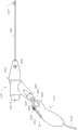
图1示出了根据一些实施例的内窥镜系统的示例;FIG. 1 shows an example of an endoscopic system according to some embodiments;
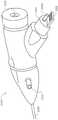
图2A和图2B示出了根据一些实施例的内窥镜系统的手持部分的侧视图;2A and 2B illustrate side views of a handheld portion of an endoscopic system according to some embodiments;
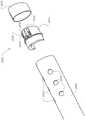
图3是根据一些实施例的具有水平视图管理的内窥镜检查的单次使用部分的透视图;3 is a perspective view of a single-use portion of an endoscopy with horizontal view management, according to some embodiments;
图4A示出根据一些实施例的具有水平视图管理的内窥镜系统的各方面的图示;4A shows an illustration of aspects of an endoscope system with horizontal view management, according to some embodiments;
图4B根据一些实施例的具有水平取向传感器的内窥镜系统的手持部分的截面图;4B is a cross-sectional view of a handheld portion of an endoscopic system with a horizontal orientation sensor, according to some embodiments;
图5示出根据一些实施例的用于治疗用途的内窥镜系统的手持部分的远侧尖端的进一步细节的透视图;5 shows a perspective view of further details of a distal tip of a hand-held portion of an endoscopic system for therapeutic use, according to some embodiments;
图6示出根据一些实施例的用于治疗用途的内窥镜系统的手持部分的远侧尖端的进一步细节的透视图;6 shows a perspective view of further details of a distal tip of a hand-held portion of an endoscopic system for therapeutic use, according to some embodiments;
图7A和图7B示出了根据一些实施例的用于诊断用途的内窥镜系统的手持部分的侧视图;7A and 7B illustrate side views of a hand-held portion of an endoscopic system for diagnostic use in accordance with some embodiments;
图8示出根据一些实施例的用于诊断用途的内窥镜系统的手持部分的远侧尖端的进一步细节的透视图;8 shows a perspective view of further details of a distal tip of a hand-held portion of an endoscopic system for diagnostic purposes, according to some embodiments;
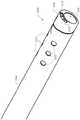
图9示出根据一些实施例的用于诊断用途的插管的一些内部结构的透视图;Figure 9 shows a perspective view of some internal structures of a cannula for diagnostic use, according to some embodiments;
图10根据一些实施例的具有水平取向传感器的用于诊断用途的内窥镜系统的手持部分的截面图;以及10 is a cross-sectional view of a handheld portion of an endoscopic system for diagnostic use with a horizontal orientation sensor, according to some embodiments; and
图11A和图11B示出根据一些实施例的与单次使用部分集成的医用盖布。11A and 11B illustrate a medical drape integrated with a single-use portion, according to some embodiments.
具体实施方式Detailed ways
以下提供了优选实施例的示例的详细描述。虽然描述了若干实施例,但应当理解,本专利说明书中描述的新主题不限于本文描述的任一个实施例或实施例的组合,而是包含许多替换、修改和等同物。此外,尽管在以下描述中阐述了许多具体细节以提供透彻理解,但可以在没有这些细节中的一些或全部的情况下实践一些实施例。此外,为了清楚起见,没有详细描述相关领域中已知的某些技术材料以便避免不必要地模糊本文所述的新主题。应当清楚的是,本文描述的一个或若干特定实施例的各个特征可以与其他描述的实施例的特征或其他特征组合使用。例外,各个附图中的相同参考数字和标号指示相同元件。Detailed descriptions of examples of preferred embodiments are provided below. While several embodiments have been described, it should be understood that the novel subject matter described in this patent specification is not limited to any one embodiment or combination of embodiments described herein, but encompasses many alternatives, modifications and equivalents. Furthermore, although numerous specific details are set forth in the following description to provide a thorough understanding, some embodiments may be practiced without some or all of these details. Furthermore, for the purpose of clarity, certain technical material that is known in the related art has not been described in detail in order to avoid unnecessarily obscuring the novel subject matter described herein. It should be clear that individual features of one or several of the specific embodiments described herein may be used in combination with features or other features of other described embodiments. Exceptionally, the same reference numerals and numerals in the various figures refer to the same elements.
虽然一些示例性实施例涉及膀胱镜和/或宫腔镜,但本领域技术人员将理解,这不是限制性的,并且本文描述的装置可以用于其他治疗或诊断规程以及患者身体的其他解剖区域。While some exemplary embodiments relate to cystoscopy and/or hysteroscopy, those skilled in the art will understand that this is not limiting and that the devices described herein may be used in other therapeutic or diagnostic procedures and other anatomical regions of a patient's body .
本文公开的实施例可能以多种方式中的一种或多种进行组合以向患者提供改善的诊断和治疗。所公开的实施例可以与现有的方法和设备组合以提供改善的治疗,诸如与已知的泌尿科或妇科诊断、外科手术以及例如其他组织和器官的外科手术的方法组合。应当理解的是,如本文所述的任何一个或多个结构和步骤可以与如本文所述的方法和设备的任何一个或多个附加结构和步骤组合,附图和支持文本提供根据实施例的描述。The embodiments disclosed herein may be combined in one or more of a variety of ways to provide improved diagnosis and treatment to patients. The disclosed embodiments can be combined with existing methods and devices to provide improved treatment, such as with known methods of urological or gynecological diagnosis, surgery, and, for example, surgery of other tissues and organs. It should be understood that any one or more of the structures and steps as described herein may be combined with any one or more of the additional structures and steps of the methods and apparatuses as described herein, and the accompanying figures and supporting text provide examples according to embodiments. describe.
虽然本文所述的治疗计划以及治疗方案和治疗量的确定是在泌尿科或妇科诊断或外科手术的背景下提出的,但本文所述的方法和设备可以用于治疗身体的任何组织以及身体的任何器官和血管,诸如脑、心脏、肺、肠、眼、皮肤、肾、肝、胰腺、胃、子宫、卵巢、睾丸、膀胱、耳、鼻、口、软组织(诸如骨髓、脂肪组织、肌肉、腺体和粘膜组织)、脊髓和神经组织、软骨、硬的生物组织(诸如牙齿、骨骼等)、以及体腔和通道(诸如鼻窦、输尿管、结肠、食道、肺通道、血管和咽喉)。While the treatment planning and determination of treatment regimens and amounts described herein are presented in the context of urological or gynecological diagnostics or surgery, the methods and devices described herein may be used to treat any tissue of the body as well as Any organ and blood vessel, such as brain, heart, lung, intestine, eye, skin, kidney, liver, pancreas, stomach, uterus, ovary, testis, bladder, ear, nose, mouth, soft tissue (such as bone marrow, adipose tissue, muscle, glands and mucosal tissue), spinal cord and nerve tissue, cartilage, hard biological tissue (such as teeth, bones, etc.), and body cavities and passages (such as sinuses, ureters, colon, esophagus, lung passages, blood vessels, and throat).
如本文所使用的,处理器包括一个或多个处理器,例如单个处理器,或者例如分布式处理系统的多个处理器。如本文描述的控制器或处理器通常包括有形介质以存储用于实现过程的步骤的指令,并且处理器可以包括例如中央处理单元、可编程阵列逻辑、门阵列逻辑、或现场可编程门阵列中的一个或多个。As used herein, a processor includes one or more processors, such as a single processor, or multiple processors such as a distributed processing system. A controller or processor as described herein typically includes tangible media to store instructions for implementing the steps of the process, and the processor may include, for example, a central processing unit, programmable array logic, gate array logic, or field programmable gate array in one or more of.
如本文所使用的,术语远侧和近侧是指从设备参考的位置,可以与解剖学参考相反。例如,探针的远侧位置可以对应于患者的细长构件的近侧位置,并且探针的近侧位置可以对应于患者的细长构件的远侧位置。As used herein, the terms distal and proximal refer to locations referenced from the device, which may be the opposite of anatomical reference. For example, the distal position of the probe may correspond to the proximal position of the elongated member of the patient, and the proximal position of the probe may correspond to the distal position of the elongated member of the patient.
根据各种实施例,装置包括直接插入体腔中的探测部分。使探测部分靠近待检查的组织和/或区域。如本文所使用的,探针包括插入受试者(诸如患者)中的物体。According to various embodiments, the device includes a detection portion that is inserted directly into the body cavity. Bring the probe part close to the tissue and/or area to be examined. As used herein, a probe includes an object inserted into a subject, such as a patient.
图1示出了根据一些实施例的内窥镜系统的示例。系统2100包括经由缆线2132 互连的手持部分2110和塔系统2112。手持部分2110包括单次使用的一次性的部分 2120和手柄部分2130。单次使用部分2120可从手柄部分2130拆卸,使得手柄部分2130可多次使用。根据一些实施例,可以使得不同版本类型的单次使用部分可用。在所示的示例中,单次使用部分2120用于治疗用途并且包括各种装置(诸如外科手术装置)可穿过的工作通道(未示出)。在一些实施例中,几个不同的单次使用部分可作为一组,例如,用于治疗目的的单次使用部分和用于诊断目的单次使用部分,或具有不同长度和/或套管直径或内部流明布置的一组单次使用部分。在图1中还示出了诊断用的单次使用部分2122,其主要用于诊断目的而非治疗目的,并且不具有工作通道。如下文将进一步详细描述的,治疗用的单次使用部分2120和诊断用的单次使用部分2122 都包括在其远侧尖端上的成像模块和LED照明模块以及用于运送流体的一个或多个内腔。塔系统2112包括安装到轮式基部2142的柱2140。塔系统2112还包括显示器 2150、键盘2160和鼠标2162、以及处理系统2170。根据一些实施例,显示器2150 可以是触敏的以用于接收用户输入,而且显示器2150是高分辨率的。根据一些实施例,显示器2150被配置成以1280×720、1920×1080、2048×1080、2560×1440、3840× 2160或更高的像素分辨率显示高清晰度图像。根据一些实施例,处理系统2170可以是合适的个人计算机或工作站,其包括一个或多个处理单元2174、输入/输出装置(诸如CD和/或DVD驱动器)、用于存储医学图像和相关数据库及其他信息的内部存储器 2142(诸如RAM、PROM、EPROM和磁类型存储介质,诸如一个或多个硬盘)、以及适合于在显示器2150上显示图像的图形处理器。根据一些实施例,塔系统2112由医疗级电源(未示出)供电。图1示出了包含用于治疗目的的单次使用部分2120的无菌包或袋2121,以及包含用于诊断目的的单次使用部分2122的无菌包或袋2123。FIG. 1 shows an example of an endoscopic system in accordance with some embodiments.
图2A和图2B示出了根据一些实施例的内窥镜系统的手持部分的侧视图。手持部分2110通常包括可重复使用的手柄部分2130和单次使用部分2120。根据一些实施例,单次使用部分2120可以在预先灭菌的包装中递送给医疗从业者并且旨在单次使用后丢弃,而手柄部分2130被设计成可重复使用多次。如上文所提及的,该示例中的单次使用部分2120是包括工作通道的治疗用的单次使用部分。治疗用的单次使用部分2120 包括具有远侧尖端2250的细长插管2240。远侧尖端2250包括成像模块2252、工作通道远侧端口2224和流体端口2232。工作通道2222以虚线轮廓示出并且从近侧端口 2220延伸到远侧端口2224。根据一些实施例,工作通道具有约3.2mm的内径,使得可以将标准手术装置设置在其中以执行各种外科手术规程。此类装置的示例包括:注射针、镊子、管、刀、勒除器、探针、凝固器装置、刷子、激光装置、微波装置(例如、用于消融)和光动力工具。2A and 2B illustrate side views of a handheld portion of an endoscopic system in accordance with some embodiments. The
插管2240可以是长的、薄的和半刚性的。根据一些实施例,插管2240的垂直于其主纵向轴线的截面基本上是圆形的。应当注意,截面可具有任何合适的形状,诸如椭圆形。插管的直径可以根据内窥镜的类型而不同,诸如从1mm到15mm。除了工作通道之外,插管2240可以具有用于支持各种功能的内部结构。例如,插管可以包括与各种流体端口流体连通的一个或多个流体通道。插管可以包括流入和流出共用的一个通道。可替代地,插管可以包括流入和流出分开的两个或更多个通道。根据一些实施例,插管2240还包括与工作通道流体隔离的流体腔。流体腔可以与远侧流体端口2232 以及近侧流体端口(诸如流体端口2230)流体连通。根据一些实施例,另一个近侧流体端口设置在端口2230的相对侧上。插管2240还被配置成容纳多个电导体,所述电导体用于向远侧尖端2250处的成像模块和照明模块提供电力、控制信号并且从所述成像模块接收视频和图像数据。在一些情况下,导体可以被隔绝并设置在插管2240内的单独腔内,在其他情况下,一些或所有导体可以设置在也用于其他目的腔(例如,流体和/或装置/工具通道)中。根据一些实施例,一个或多个光纤可以穿过插管2240以用于数据传输和/或向远侧尖端2250供应照明光的目的。
手柄部分2130被配置成多次使用并且适于重复接收单次使用部分。手柄部分2130包括主体,所述主体的尺寸和形状被设计成允许操作者的手进行安全且符合人体工程学的抓握。手柄部分2130还包括可以被配置成允许在使用期间执行常见任务的若干按钮,诸如按钮2212和2214。例如,按钮2212和2214可以被编程为控制LED照明水平(在远侧尖端2250处的LED,未示出),捕获静止图像,和/或开始和停止记录视频图像。The
根据一些实施例,插管2240可相对于手柄部分2130围绕其纵向轴线旋转。在此类情况下,手柄2130还可以包括圆柱形刻度盘2210,其被配置成沿虚线箭头旋转腔 2240(和远侧尖端2250)。根据一些实施例,壳体2241的远侧部分2242包围插管2240 的近侧部分2244,且与插管2240一起旋转,而壳体的近侧部分2244相对于手柄部分 2130保持固定。图2B示出了如何从多次使用的手柄部分2130安装和移除单次使用部分2120。具体地,手柄部分2130包括插座2260,其尺寸被设计成与从单次使用部分 2120突出的公配合部分2261联接。安装和拆卸的动作由虚线箭头2266表示。从配合部分2261突出的是电连接器2262和插管2264,其用于在刻度盘2210被启动提供插管2240的旋转。根据一些实施例,插管2264具有“D”形截面或能够提供插管2264 与手柄2130中的母插座(未示出)之间的可靠旋转联接的其他形状。According to some embodiments, the
根据一些实施例,手柄部分2130可以封装或包括被配置成处理图像数据、生成控制信号、提供电力、或与其他外部装置建立通信的部件。在一些情况下,通信可以是无线或有线通信。例如,无线通信可以包括Wi-Fi、无线电通信、蓝牙、IR通信或其他类型的直接通信。在一些实施例中,手柄部分可以是用于测量插管与手柄部分之间的相对位置的壳体传感器组件。在其他实施例中,传感器组件可以测量手柄相对于其环境的相对位置或取向。下面将进一步介绍此类传感器组件的示例。在一些情况下,手柄部分可以具有被作为用户输入装置的显示装置或具有任何类型的用户交互部件,诸如按钮、鼠标、操纵杆、轨迹球、触摸板、笔、图像捕获装置、动作捕捉装置、麦克风或触摸屏。According to some embodiments,
图3是根据一些实施例的具有水平视图管理的内窥镜系统的单次使用部分的透视图。在该近侧透视图中,示出了轴2264的“D”形截面和电连接器2262的细节。左侧的近侧流体端口3310和近侧工作通道端口2220也是可见的。3 is a perspective view of a single-use portion of an endoscopic system with horizontal view management, according to some embodiments. In this proximal perspective view, a "D" shaped cross-section of the
图4A是示出根据一些实施例的具有水平视图管理的内窥镜系统的各方面的图示。示出了简化的内窥镜系统,其在功能上类似于图1所示的系统2100。手持部分2110 被示为连接到集成的计算机系统3460。应当注意,计算机系统3460可以包括与塔系统2112类似或相同的功能。系统3460包括显示监视器3458,其与图1所示的显示器 2150类似或相同。根据一些实施例,提供了水平视图管理,其允许操作者在外部显示器3458(以及图1所示的塔式系统2112上的显示器2150)上查看适当取向的图像,尽管有安装在远侧尖端2250处的成像模块2252的相对旋转仍是如此。应当注意,远侧尖端处的摄像机可能以至少两种不同的方式相对于显示监视器旋转。首先,插管可以相对于手柄部分旋转,如用虚线箭头3452描绘的。其次,整个手持部分2110可相对于显示监视器旋转,如用虚线箭头3450描绘的。如果发生这些旋转中的任一者或两者,则在显示监视器上显示的未校正图像的方向可能是不正确的。可以相对于环境(例如,重力、患者解剖结构、床等)保持图像的水平面。这在图4A中示出,其中受试者3462由具有视线方向3468的成像模块捕获并在显示监视器3458上显示为圆形图像3464。根据一些实施例,可以借助于多个传感器来测量尖端2250中的成像模块的“侧倾”角(围绕插管轴线3454的旋转角度),使得系统得以自动调整图像数据以保持水平视图。可替代地,可以使用算法方法(例如,光流)而不使用传感器来保持或校正图像数据的水平视图。根据一些实施例,侧倾角指示器3456显示在图像圈3464的外周上以指示成像模块围绕轴线3454的相对角位置。在手柄部分2130内还示出了电连接器3412,其被配置成与单次使用部分2120上的电连接器2262电联接(参见图4B)。4A is a diagram illustrating aspects of an endoscopic system with horizontal view management in accordance with some embodiments. A simplified endoscopic system is shown that is similar in function to the
根据一些实施例,图4B描绘了具有水平取向传感器的内窥镜系统的手持部分的截面图。在该示例中,刻度盘2210由用户拨动以使插管2240围绕其轴线3454旋转。刻度盘2210与手柄部分2130内的一组齿轮3434啮合,使得当刻度盘2210旋转时,它使轴2264和齿轮3434两者旋转。齿轮3434附接到安装在印刷电路板3410上的旋转传感器3430。传感器3430可以是角位置或角旋转传感器。根据一些实施例,传感器 3430可以是编码器、电位计和/或霍尔传感器。轴2264通过齿轮3440来旋转齿轮3442。齿轮3442固定到壳体的旋转部分2242和插管2240,并且因此齿轮3442、旋转部分 2242、插管2240和成像模块2252都一起围绕轴线3454旋转。因此,旋转传感器3430 将测量插管和成像模块相对于手柄部分的旋转位置。根据一些实施例,可以通过位于单次使用部分2120(或图10中的2122)内的传感器3460来测量插管和成像模块相对于手柄部分的旋转位置。4B depicts a cross-sectional view of a handheld portion of an endoscopic system with a horizontal orientation sensor, according to some embodiments. In this example, dial 2210 is toggled by the user to rotate
根据一些实施例,用于测量成像装置或成像装置的光学元件的姿态或取向的一个或多个传感器可以包括安装在印刷电路板3410上的集成惯性测量单元(IMU)3420。通常,惯性测量传感器可以包括一个或多个陀螺仪、速度传感器、加速度计、磁力计、或一个或多个位置传感器。惯性传感器可以用于获得指示以下的数据:成像装置的空间布置(例如,位置、取向或角)和/或运动特性(例如,平移(线性)速度、角速度、平移(线性)加速度、角加速度)。惯性传感器可以在本文中用于指代运动传感器(例如,速度传感器、诸如加速度计的加速度传感器),取向传感器(例如,陀螺仪、倾斜计)、或者具有一个或多个集成运动传感器和/或一个或多个集成取向传感器(诸如IMU 3420)的IMU。根据一些实施例,IMU 3420被配置成提供相对于与插管轴线3454平行的单个运动轴线3470的感测数据。根据一些其他实施例,可以使用多个惯性传感器,其中每个惯性传感器提供沿不同运动轴线的测量。加速度计能够测量传感器在地球重力场中的取向。可以通过从地面参考坐标系到加速度传感器主体坐标系的旋转矩阵获得相对于地球/世界重力场的取向角。加速度计可以是单轴加速度计或三轴加速度计。三个角加速度计可以用于沿三个不同的运动轴线提供角加速度数据。三个运动方向可以是正交轴线。一个或多个角加速度计可以被配置成测量围绕旋转轴线的加速度。作为另一个示例,三个陀螺仪可以用于提供关于三个不同旋转轴线的取向数据。可替代地,至少一些或所有的惯性传感器可以提供相对于相同运动轴线的测量。例如,可以实现这种冗余以提高测量准确度。单个惯性传感器能够提供相对于多个轴线的感测数据。例如,IMU 3420可以包括多个加速度计和陀螺仪,其可用于生成相对于多达六个运动轴线的加速度数据和取向数据。在一些情况下,关于成像装置的姿态数据可以包括成像装置相对于多达三个旋转轴线的旋转角度。可以使用各种方法来导出成像装置的姿态数据,诸如卡尔曼滤波器、扩展卡尔曼滤波器的互补滤波器和各种其他传感器融合算法。根据一些其他实施例,一个或多个传感器可以位于插管上或位于除了图4B 所描绘的位置之外的其他位置。例如,一个或多个传感器可以封闭在远侧尖端2250 中。According to some embodiments, one or more sensors for measuring the pose or orientation of the imaging device or optical elements of the imaging device may include an integrated inertial measurement unit (IMU) 3420 mounted on a printed
由传感器3430和3420测量的取向或“侧倾”旋转角度用于处理成像数据以便保持水平视图。图像数据可以由手柄部分2130和/或处理系统2170(图1所示)或计算机系统3460(图4A所示)中的一个或多个处理器处理。在一些实施例中,一个或多个处理器(例如,图1中的处理器2174和/或图4A中的处理器3461)可以被配置成计算成像装置的姿态数据并基于姿态数据变换图像数据。在一些实施例中,一个或多个处理器可以是可编程处理器(例如,中央处理单元(CPU)或微控制器)、现场可编程门阵列(FPGA)和/或一个或多个高级RISC机器(ARM)处理器。在一些实施例中,一个或多个处理器可操作地联接到非暂时性计算机可读介质。非暂时性计算机可读介质可以存储可由一个或多个处理器单元执行以用于执行一个或多个步骤的逻辑、代码和/ 或程序指令。非暂时性计算机可读介质可以包括一个或多个存储器单元(例如,可移除的介质或外部存储装置,诸如SD卡或随机存取存储器(RAM))。在一些实施例中,可以在多次使用的手柄部分2130内执行部分或全部方向校正图像处理。例如,在图 4B中,计算和其他图像转换处理可以在通用处理器单元3480和/或图像信号处理器 3482上进行,通用处理器单元3480和图像信号处理器3482都安装在电路板3410上。The orientation or "roll" rotation angle measured by
根据一些其他实施例,可以在不使用传感器的情况下保持水平视角。例如,诸如光流的光学方法可以用于跟踪连续图像帧之间的运动矢量,使得可以跟踪视图的全局旋转。然后可以处理视频数据以保持水平视图。According to some other embodiments, the horizontal viewing angle can be maintained without the use of sensors. For example, optical methods such as optical flow can be used to track motion vectors between consecutive image frames so that the global rotation of the view can be tracked. The video data can then be processed to maintain the horizontal view.
在一些情况下,在图1中的监视器2150或图4A中的监视器3458上显示具有保持/校正的水平线的视图。根据一些实施例,提供多个水平视图控制选项以供用户选择。例如,可以通过软件配置打开或关闭具有保持/校正的水平线的视图以允许用户基于其需要进行调整。在另一个示例中,用户可以从包括以下的多个水平视图控制选项中进行选择:相对于地面参考的全自动水平控制、相对于手柄部分的水平控制、手动控制、相对于用户选定参考的水平控制、以及各种其他选项。在一些示例中,其中一个按钮 (例如图2A中的按钮2212或按钮2214)可编程以允许用户更改水平视图配置。In some cases, a view with a maintained/corrected horizon is displayed on
图5是示出根据一些实施例的被配置用于治疗用途的内窥镜系统的手持部分的远侧尖端的进一步细节的透视图。可见的是插管2240的远端和远侧尖端2250。远侧尖端2250包括成像模块2252和LED光源2310。还示出了流体端口2232和2332以及远侧工作通道端口2224。根据一些实施例,工作通道端口2224还被配置用于液体流入(使流体流出装置并流入患者),并且流体端口2232和2332被配置用于液体流出(使流体流入装置并流出患者)。5 is a perspective view showing further details of a distal tip of a hand-held portion of an endoscopic system configured for therapeutic use, according to some embodiments. Visible are the distal end of
图6是示出根据一些实施例的被配置用于治疗用途的内窥镜系统的手持部分的远侧尖端的进一步细节的透视图。在该分解视图中可见的是远侧尖端2250如何附接到插管2240。远侧尖端2250包括围绕远侧部分尖端模块2412的尖端壳体2410。模块2412 的近侧部分插入插管2240的远端并结合到该远端。模块2412包括其上安装有成像模块2252和LED2310的载体2414。应当注意,在该示例中,载体2414被配置成将成像模块保持在略微向下的视角。也就是说,成像模块2252向下指向,使得其视野朝向工作通道远侧端口2224偏置(如图5所示)。6 is a perspective view showing further details of a distal tip of a hand-held portion of an endoscopic system configured for therapeutic use, according to some embodiments. Visible in this exploded view is how the
图7A和图7B示出了根据一些实施例的被配置用于诊断用途的内窥镜系统的手持部分的侧视图。在这种情况下,手持部分2110包括可重复使用的手柄部分2130和诊断用的单次使用部分2122。应当注意,单次使用部分2122的许多部件与图2A、图2B、图3、图4A、图4B、图5和图6所示和相对于所述附图描述的单次使用部分2120的部件类似或相同。在这些图中示出并在此描述的用于单次使用部分2120的许多实施例同样适用于单次使用部分2122,并且为了清楚起见将不再重复。根据一些实施例,单次使用部分2122可以在预先灭菌的包装中递送给医疗从业者并且旨在单次使用后丢弃,而手柄部分2130被设计成可重复使用多次。如上文所提及的,该示例中的单次使用部分2122是不包括工作通道的诊断用的单次使用部分。诊断用的单次使用部分2122 包括具有远侧尖端2750的细长插管2740。远侧尖端2750包括成像模块2752和远侧流体端口2724。7A and 7B illustrate side views of a handheld portion of an endoscopic system configured for diagnostic use, according to some embodiments. In this case, the hand-held
插管2740可以是长的、薄的和半刚性的。根据一些实施例,插管2740的垂直于其主纵向轴线的截面可以是基本上圆形的。应当注意,截面可具有任何合适的形状,诸如椭圆形。插管的直径可以根据内窥镜的类型而不同,诸如从1mm到15mm。插管 2740可以具有用于支持各种功能的内部结构。例如,插管可以包括与各种流体端口流体连通的一个或多个流体通道。插管可以包括将由流入和流出共用的一个通道。可替代地,插管可以包括具有分开的流入和流出的两个或更多个通道。流体腔可以与远侧流体端口2724以及近侧流体端口(诸如流体端口2730)流体连通。插管2740还被配置成容纳多个电导体,所述电导体用于向远侧尖端2750处的成像模块和照明模块提供电力、控制信号并且从所述成像模块接收视频和图像数据。在一些情况下,导体可以被隔绝并设置在插管2740内的单独腔内,在其他情况下,一些或所有导体可以设置在也用于其他目的腔(例如,流体和/或装置/工具通道)中。根据一些实施例,一个或多个光纤可以穿过插管2740以用于数据传输和/或向远侧尖端2750供应照明光的目的。
根据一些实施例,插管2740可相对于手柄部分2130围绕其纵向轴线旋转。在此类情况下,手柄2130还可以包括圆柱形刻度盘2210,其被配置成沿虚线箭头旋转腔 2740(和远侧尖端2750)。根据一些实施例,壳体的远侧部分2742与插管2740一起旋转,而壳体的近侧部分2744相对于手柄部分2130保持固定。图7B示出了如何从多次使用的手柄部分2130安装和移除单次使用部分2122。具体地,手柄部分2130包括插座2260,其尺寸被设计成与从单次使用部分2120突出的公配合部分2760联接。安装和拆卸的动作由虚线箭头2766表示。从配合部分2760突出的是电连接器2762 和轴2764,其用于在刻度盘2710启动时提供插管2740的旋转。According to some embodiments, the
图8是示出根据一些实施例的被配置用于诊断用途的内窥镜系统的手持部分的远侧尖端的进一步细节的透视图。可见的是插管2740的远端和远侧尖端2750。远侧尖端2750包括成像模块2752和LED光源2820。还示出了两个流体端口2724。根据一些实施例,一个流体端口被配置用于流入(使流体流出装置并流入患者)并且另一个流体端口被配置用于流出(使流体流入装置并流出患者)。类似于远侧尖端2250(例如,图6所示),远侧尖端2750是附接到插管2740的单独组件。远侧尖端2750包括围绕远侧部分尖端模块(未示出)的尖端壳体2810。远侧尖端模块的近侧部分结合到插管2740的远端。载体2814保持成像模块2752和LED 2820。应当注意,在该示例中,载体2814被配置成将成像模块保持在略微向下的视角。也就是说,成像模块2752 是向下指向的,使得其视野朝向流体端口2724偏置。8 is a perspective view showing further details of a distal tip of a hand-held portion of an endoscopic system configured for diagnostic use, according to some embodiments. Visible are the distal end of
图9是示出根据一些实施例的用于诊断用途的插管的一些内部结构的透视图。在该视图中,可以看到三个腔。腔2910用于承载将成像模块和LED与电连接器2762连接的电线。腔2912和2914被配置成用于在近侧流体端口(例如,图7A和图7B中的 2730)与远侧流体端口2724之间运送流体。9 is a perspective view illustrating some of the internal structure of a cannula for diagnostic use, according to some embodiments. In this view, three cavities can be seen.
图10是根据一些实施例的具有水平取向传感器的被配置用于诊断用途的内窥镜系统的手持部分的截面图。在该示例中,刻度盘2210由用户致动以使插管2740围绕其轴线3054旋转。刻度盘2210与手柄部分2130内的一组齿轮3434啮合,使得当刻度盘2210旋转时,它使轴2764和齿轮3434两者旋转。齿轮3434附接到安装在印刷电路板3410上的旋转传感器3430。轴2764通过齿轮3040来旋转齿轮3042。齿轮3042 固定到壳体的旋转部分2742和插管2740,并且因此齿轮3042、部分2742、插管2740 和成像模块2752都一起围绕轴线3054旋转。旋转传感器3430测量插管和成像模块相对于手柄部分的旋转位置。10 is a cross-sectional view of a handheld portion of an endoscopic system with a horizontal orientation sensor configured for diagnostic use, according to some embodiments. In this example, dial 2210 is actuated by a user to rotate
可弯曲的插管。膀胱镜、宫腔镜、或者对子宫和膀胱内部直视的设备已经被证明可提高诊断准确度。然而,患者经常发现宫腔镜设备的插入和移除以及随后内窥镜设备进入患者的子宫腔是不舒服的。典型的宫腔镜装置具有远侧尖端,其尺寸被设计成有助于通过患者的子宫颈插入尖端并将其插入子宫中以获得用于诊断目的的组织样本。Bendable cannula. Cystoscopes, hysteroscopes, or devices that look directly into the inside of the uterus and bladder have been shown to improve diagnostic accuracy. However, patients often find the insertion and removal of the hysteroscopic device and subsequent entry of the endoscopic device into the patient's uterine cavity uncomfortable. A typical hysteroscopic device has a distal tip sized to facilitate insertion of the tip through the patient's cervix and into the uterus to obtain tissue samples for diagnostic purposes.
根据一些实施例,提供了一种可弯曲插管。参考图7A,插管2740可以包括由柔性材料(例如,塑料)制成的部分和由可弯曲以达到期望形状(如虚线轮廓2762所示) 的材料(例如,金属)制成的弯曲插入部2760。插管可以是一次性的。可替代地,插管可以被灭菌和重复使用。插管可以在使用时变形或弯曲以帮助到达子宫腔的凹陷部分。在所示的示例中,插管可以被制成可弯曲的,例如通过使用柔性材料(例如,塑料、尼龙),其包括沿着插管壳体的内部长度延伸的一个或多个插入部2760,其可以是可弯曲的金属线、管、扁平杆或类似物。插管可以由任何合适的材料制成,诸如 Provista共聚物、乙烯基(诸如聚氯乙烯)、尼龙(诸如vestamid、grillamid)、聚氨酯、聚乙烯、聚丙烯、聚碳酸酯、聚酯、硅弹性体、醋酸纤维等。According to some embodiments, a bendable cannula is provided. Referring to Figure 7A,
根据一些实施例,插管可以在使用时变形或弯曲以改变或调整远侧相机的视线方向(DOV)。常规插管可以具有光学棱镜,所述光学棱镜在远端附近以一定角度位于棒状透镜内以便适合于许多应用。例如,光学棱镜可以提供以各种角度(例如,30度、 70度、或0与180度之间的任何数值)的视线方向(DOV)以便扩大视场。然而,当成像传感器位于远侧尖端处时,这种光学棱镜可能导致远端处的非常大的包装或者极大地增加成像模块的成本。在一个实施例中,可弯曲或可变形的插管可以通过用户对不同的角度下插管的最佳弯曲度的手动变形来调整DOV,使得成像装置的光轴线在成像装置处与插管的纵向轴线相交。例如,弯曲插管可以被定位成使得成像装置的光轴线 (即,DOV)相对于插管轴线以下列角度倾斜:5度、10度、15度、20度、25度、 30度、35度、40度、45度、50度、55度、50度、55度、60度、65度、70度、75 度、90度、120度、150度、直到180度或其间的任何数值。According to some embodiments, the cannula may be deformed or bent in use to change or adjust the direction of sight (DOV) of the distal camera. Conventional cannulas may have optical prisms positioned within the rod lens at an angle near the distal end to suit many applications. For example, optical prisms may provide a direction of sight (DOV) at various angles (eg, 30 degrees, 70 degrees, or any value between 0 and 180 degrees) in order to expand the field of view. However, when the imaging sensor is located at the distal tip, such an optical prism can result in a very large package at the distal end or greatly increase the cost of the imaging module. In one embodiment, the bendable or deformable cannula can adjust the DOV by the user manually deforming the optimal curvature of the cannula at different angles such that the optical axis of the imaging device is at the imaging device and the cannula's The longitudinal axes intersect. For example, the curved cannula can be positioned such that the optical axis (ie, DOV) of the imaging device is inclined relative to the cannula axis at the following angles: 5 degrees, 10 degrees, 15 degrees, 20 degrees, 25 degrees, 30 degrees, 35 degrees , 40 degrees, 45 degrees, 50 degrees, 55 degrees, 50 degrees, 55 degrees, 60 degrees, 65 degrees, 70 degrees, 75 degrees, 90 degrees, 120 degrees, 150 degrees, up to 180 degrees or any value in between.
插管的配置或形状可以经由多个弯曲来形成。在一些情况下,插管的初始弯曲可能不完全令人满意,可以通过强制变形或弯曲的远侧部分来调整弯曲角度。由于内部可弯曲金属插入部2760可以克服外部柔性插管材料的刚度并因此保持形状,所以保持了更新的变形或弯曲形状。弯曲或变形的容易程度取决于内部金属加强件和外部柔性插管材料的相对刚度。The configuration or shape of the cannula may be formed via multiple bends. In some cases, the initial bend of the cannula may not be completely satisfactory, and the angle of bend can be adjusted by forcibly deforming or bending the distal portion. The updated deformed or curved shape is maintained since the inner
对一次性插管的单次使用的管理。根据一些实施例,提供了检测特征以确保插管或一次性部分的单次使用。例如,检测特征可以被配置成检测附接到手柄部分的已使用的插管。在检测到一次性部分的重复使用时,可以向用户通知违规或者可以禁止装置进行进一步的使用。可以采用各种方法来实现单次使用检测功能。例如,可以经由机械机构、电气机构、借助于传感器、以及上述任何组合来检测插管的重复使用。可以使用机械结构来实现检测特征。例如,机械保险丝可以位于单次使用部分的近端(例如,近侧接口)上。一旦将保险丝插入手柄部分并从手柄部分拔出,保险丝就被破坏。在另一个示例中,可以使用诸如电保险丝的电结构来实现检测特征。例如,可以在单次使用部分的近端处的电路上设置电保险丝。一旦将单次使用部分插入手柄部分中,手柄电子器件检测到其存在并烧毁保险丝。当相同的插管再次插入手柄时,手柄电子器件可以检测到开路的保险丝。在一些情况下,可以向用户呈现检测到的插管重复使用的通知。在各种其他实施例中,可以使用传感器来实现检测特征。诸如Dallas芯片、 EEPROM、RFID、条形码等的传感器可以用于跟踪插管的使用。Single-use administration of single-use cannulas. According to some embodiments, detection features are provided to ensure single use of the cannula or disposable portion. For example, the detection feature may be configured to detect a used cannula attached to the handle portion. Upon detection of reuse of the disposable portion, the user may be notified of the violation or the device may be prohibited from further use. Various methods can be used to implement the single-use detection function. For example, re-use of the cannula may be detected via mechanical mechanisms, electrical mechanisms, by means of sensors, and any combination of the foregoing. Detection features can be implemented using mechanical structures. For example, a mechanical fuse may be located on the proximal end of the single-use portion (eg, the proximal interface). Once the fuse is inserted into the handle portion and pulled out from the handle portion, the fuse is destroyed. In another example, the detection feature may be implemented using an electrical structure such as an electrical fuse. For example, an electrical fuse may be provided on the circuit at the proximal end of the single-use portion. Once the single-use portion is inserted into the handle portion, the handle electronics detects its presence and blows the fuse. When the same cannula is inserted into the handle again, the handle electronics can detect an open fuse. In some cases, a notification of detected cannula reuse may be presented to the user. In various other embodiments, the detection feature may be implemented using a sensor. Sensors such as Dallas chips, EEPROM, RFID, barcodes, etc. can be used to track the use of the cannula.
在一个示例中,使用Dallas芯片或EEPROM,插管上的传感器(Dallas芯片或EEPROM)由手柄内的电子器件读取。一旦检测到,手柄电子器件将对传感器和标签进行编程,如所使用的一样,其可以通过嵌入式软件编程。在另一个示例中,被配置成由读取器读取的条形码或RFID可以位于单次使用部分的近端上并且可以由位于手柄部分上的读取器读取。在一些情况下,一旦检测到重复使用,单次使用部分可以被配置成在由读取器第一次读取之后被禁用。In one example, using a Dallas chip or EEPROM, a sensor on the cannula (Dallas chip or EEPROM) is read by electronics within the handle. Once detected, the handle electronics will program the sensor and tag, as used, which can be programmed via embedded software. In another example, a barcode or RFID configured to be read by the reader can be located on the proximal end of the single-use portion and can be read by a reader located on the handle portion. In some cases, the single-use portion may be configured to be disabled after the first read by the reader once repeated use is detected.
条形码可以定义诸如条形码的版本、格式、位置、对准和定时的元素以使得能够读取和解码条形码。条形码的其余部分可能以任何类型的合适格式(诸如二进制或字母数字信息)对各种类型的信息进行编码。条形码可以是二维的,诸如PDF417、Aztec、 MaxiCode和QR码等。条形码可以是一维条形码,诸如Interleaved 2/5、Industrial 2/5、 Code 39、Code 39Extended、Codabar、Code 11、Code 128、Code 128Extended、EAN/UCC 128、UPC-E、UPC-A、EAN-8、EAN-13、Code 93、Code 93Extended、DataBar Omnidirectional(RSS-14)、DataBar Truncated(RSS-14Truncated)、DataBar Limited(RSS Limited)、DataBarStacked、DataBar Expanded、DataBar Expanded Stacked等。条形码可能以任何类型的合适格式(诸如二进制、字母数字、ASCII等)对各种类型的信息进行编码,并且代码可以基于任何标准。条形码可以由光学读取器、激光扫描仪或其他成像装置读取。A barcode can define elements such as the barcode's version, format, location, alignment, and timing to enable reading and decoding of the barcode. The remainder of the barcode may encode various types of information in any type of suitable format, such as binary or alphanumeric information. Barcodes can be two-dimensional, such as PDF417, Aztec, MaxiCode, and QR codes, among others. The barcode can be a one-dimensional barcode such as Interleaved 2/5, Industrial 2/5, Code 39, Code 39Extended, Codabar, Code 11, Code 128, Code 128Extended, EAN/UCC 128, UPC-E, UPC-A, EAN- 8. EAN-13, Code 93, Code 93Extended, DataBar Omnidirectional(RSS-14), DataBar Truncated(RSS-14Truncated), DataBar Limited(RSS Limited), DataBarStacked, DataBar Expanded, DataBar Expanded Stacked, etc. Bar codes may encode various types of information in any type of suitable format (such as binary, alphanumeric, ASCII, etc.), and the codes may be based on any standard. Barcodes can be read by optical readers, laser scanners, or other imaging devices.
集成医用盖布。根据一些实施例,可以提供具有集成医用盖布的装置。根据一些实施例,图11A和11B示出了与单次使用部分集成的医用盖布。医用盖布3100集成到次使用部分2120的外壳,该外壳正好高于公配合部分2261。根据其他实施例,盖布3100可以集成或附接到其他位置的单次使用部分2120,如靠近近侧插管2240或位于壳体的远侧从部分上。虽然该示例中示出的是治疗用的单次使用部分2120,盖布也可以类似地集成到诊断用的单次使用部分上。盖布3130可以容易地展开以覆盖手柄部分。在外科手术规程期间,多次使用的手柄部分可以由盖布覆盖,如图11B所示。当规程完成时,盖布可以容易地从手柄移除并从内向外折叠,并且可以用作用于容纳污染的一次性部分的容器。盖布3100可以包括被配置成将盖布联接到装置的联接器 3120。联接器3120可以是适配器环,其尺寸和形状被设计成集成/胶粘到插管的近侧部分或单次使用部分。联接器3120可以允许盖布组装到单次使用部分以变成具有盖布的集成插管。在一些情况下,盖布被直接胶粘、焊接或连接到插管而不用适配器。Integrated medical drape. According to some embodiments, a device with an integrated medical drape may be provided. 11A and 11B illustrate a medical drape integrated with a single-use portion, according to some embodiments. The
盖布可以包括有助于在外科手术规程之前展开盖布的特征。例如,盖布可以包括拉环3110,医师可以使用所述拉环3110将盖布拉离装置。拉环3110可以经由附件来附接到盖布的主体。非无菌人员可以接近环并展开盖布而不会污染盖布主体的无菌部分。根据一些实施例,拉环3110还被配置为作为拉绳来减小盖布的主开口3112的大小。例如,使用单次使用部分之后,当盖布内侧外翻(即回到图11A中所示的方向)时,这可能是有用的,因为拉绳可以用于进一步关闭主开口3112以更加充分地容纳使用过的受污染的单次使用部分。The drape may include features that facilitate deployment of the drape prior to surgical procedures. For example, the drape can include a
盖布可以由基本上是柔性的并且不透液体的材料片材构成。材料片材可以由医疗领域中使用的容易获得的塑料薄膜构成,例如乙烯基(诸如聚氯乙烯)、聚乙烯、聚丙烯、聚碳酸酯、聚酯、硅弹性体、乙酸酯等。The drape may be constructed from a sheet of material that is substantially flexible and liquid impermeable. The sheet of material may be constructed from readily available plastic films used in the medical field, such as vinyl (such as polyvinyl chloride), polyethylene, polypropylene, polycarbonate, polyester, silicone elastomers, acetate, and the like.
插管和手柄的认证。根据一些实施例,提供认证特征以确保单次使用插管来自经认证的来源。例如,单次使用部分可以不被来自其他未经认证的制造商的未经认证的插管替代。另外,认证特征可以帮助确保单次使用插管不用于来自其他未经认证的制造商的未经认证的手柄。这些规定可以为上述医疗装置提供确保的质量和安全性。例如,认证特征可以被配置成检测具有其固有身份(ID)的插管或手柄。在将ID从单次使用部分读取到手柄部分,可以向用户通知违规,或者可以禁止进一步使用装置,反之亦然。可以采用各种方法来实现认证。例如,单次使用部分或手柄部分可以被嵌入有 EEPROM、RFID或其他安全芯片以进行检测和读取。单次使用部分或手柄部分也可以在单次使用部分上具有由手柄或系统确认的独特电气或机械特征,反之亦然。可以使用上述任何组合来增强认证。在一个示例中,可以使用机械结构来实现认证特征。例如,机械特征可以位于单次使用部分的近端(例如,近侧接口)上,或者手柄部分的远端可以一旦被连接就进行检测且机械地验证。机械特征可以是特殊的连接器、结构或特殊材料。在另一个示例中,可以使用来自插管、手柄或系统的电气签名(诸如电阻值、阻抗值、电压值等)来实现认证特征。例如,电路可以设置在单次使用部分的近端或手柄的远端部分。一旦将单次使用部分插入手柄部分中,插管、手柄和系统上的电子电路可以相互检测并相互认证。当在系统中使用第三方插管或手柄时,系统其余部分中的电子器件可以检测错误匹配或未经认证的装置。在一些情况下,可以向用户呈现指示检测到的未经认证插管或手柄的通知。在各种其他实施例中,可以使用传感器来实现认证特征。诸如EEPROM、RFID、安全微芯片等的传感器可以用于跟踪单次使用插管或手柄。Certification of cannula and handle. According to some embodiments, an authentication feature is provided to ensure that the single-use cannula is from an authenticated source. For example, the single-use portion may not be replaced by uncertified cannulas from other uncertified manufacturers. Additionally, the certification feature can help ensure that single-use cannulas are not used with uncertified handles from other uncertified manufacturers. These regulations can provide assured quality and safety for the aforementioned medical devices. For example, the authentication feature can be configured to detect a cannula or handle with its inherent identity (ID). Upon reading the ID from the single use portion to the handle portion, the user may be notified of the violation, or further use of the device may be prohibited, and vice versa. Authentication can be achieved in various ways. For example, the single use portion or handle portion can be embedded with an EEPROM, RFID or other security chip for detection and reading. The single-use portion or handle portion may also have unique electrical or mechanical features on the single-use portion identified by the handle or system, and vice versa. Authentication can be enhanced using any combination of the above. In one example, the authentication feature may be implemented using a mechanical structure. For example, mechanical features can be located on the proximal end of the single-use portion (eg, the proximal interface), or the distal end of the handle portion can be detected and mechanically verified once connected. Mechanical features can be special connectors, structures, or special materials. In another example, the authentication feature may be implemented using electrical signatures (such as resistance values, impedance values, voltage values, etc.) from the cannula, handle, or system. For example, the circuit may be provided at the proximal end of the single use portion or at the distal end portion of the handle. Once the single-use portion is inserted into the handle portion, the electronic circuits on the cannula, handle, and system can detect and authenticate each other. When a third-party cannula or handle is used in the system, electronics in the rest of the system can detect mis-matched or unauthenticated devices. In some cases, the user may be presented with a notification indicating a detected unauthenticated cannula or handle. In various other embodiments, authentication features may be implemented using sensors. Sensors such as EEPROM, RFID, security microchips, etc. can be used to track single use cannulas or handles.
在一个示例中,被配置成由读取器读取的条形码或RFID可以位于单次使用部分的近端上并且可以由位于手柄部分上的读取器读取,反之亦然。在一些情况下,一旦检测到未经认证的单次使用部分或手柄,未经认证的部分可以被配置成从系统禁用。In one example, a barcode or RFID configured to be read by a reader may be located on the proximal end of the single-use portion and may be read by a reader located on the handle portion, and vice versa. In some cases, upon detection of an unauthenticated single-use portion or handle, the unauthenticated portion may be configured to be disabled from the system.
在一些实施例中,Dallas芯片、EEPROM、RFID、条形码、机械特征等也可以被配置成对与装置或外科手术相关的任何其他信息进行编码,所述信息例如是插管规格、建议的外科手术参数、和/或与单次使用插管或使用该装置的外科手术相关的各种其他信息。In some embodiments, Dallas chips, EEPROM, RFID, barcodes, mechanical features, etc. may also be configured to encode any other information related to the device or surgery, such as cannula specifications, recommended surgery parameters, and/or various other information related to a single-use cannula or surgical procedure using the device.
条形码可以定义诸如条形码的版本、格式、位置、对准和定时的元素以使得能够读取和解码条形码。条形码的其余部分可能以任何类型的合适格式(诸如二进制或字母数字信息)对各种类型的信息进行编码。条形码可以是二维的,诸如PDF417、Aztec、 MaxiCode和QR码等。条形码可以是一维条形码,诸如Interleaved 2/5、Industrial 2/5、 Code 39、Code 39Extended、Codabar、Code 11、Code 128、Code 128Extended、EAN/UCC 128、UPC-E、UPC-A、EAN-8、EAN-13、Code 93、Code 93Extended、DataBar Omnidirectional(RSS-14)、DataBar Truncated(RSS-14Truncated)、DataBar Limited(RSS Limited)、DataBarStacked、DataBar Expanded、DataBar Expanded Stacked等。条形码可能以任何类型的合适格式(诸如二进制、字母数字、ASCII等)对各种类型的信息进行编码,并且代码可以基于任何标准。条形码可以由光学读取器、激光扫描仪或其他成像装置读取。A barcode can define elements such as the barcode's version, format, location, alignment, and timing to enable reading and decoding of the barcode. The remainder of the barcode may encode various types of information in any type of suitable format, such as binary or alphanumeric information. Barcodes can be two-dimensional, such as PDF417, Aztec, MaxiCode, and QR codes, among others. The barcode can be a one-dimensional barcode such as Interleaved 2/5, Industrial 2/5, Code 39, Code 39Extended, Codabar, Code 11, Code 128, Code 128Extended, EAN/UCC 128, UPC-E, UPC-A, EAN- 8. EAN-13, Code 93, Code 93Extended, DataBar Omnidirectional(RSS-14), DataBar Truncated(RSS-14Truncated), DataBar Limited(RSS Limited), DataBarStacked, DataBar Expanded, DataBar Expanded Stacked, etc. Bar codes may encode various types of information in any type of suitable format (such as binary, alphanumeric, ASCII, etc.), and the codes may be based on any standard. Barcodes can be read by optical readers, laser scanners, or other imaging devices.
在一些实施例中,装置可以与外部计算系统(诸如图1中的塔系统2112和/或图 4A中的计算机系统3460)通信。外部计算系统可以被配置成例如处理从装置传输的图像数据以保持水平视图,使用从装置传输的认证数据来认证插管,检测插管的重复使用,以及根据本文描述的各种实施例的各种其他功能。外部计算系统可以任选地是移动装置,诸如手机、智能电话、手表、平板电脑、遥控器、膝上型电脑或其他装置。外部计算系统可以是固定装置,例如个人计算机、服务器计算机或其他结构。外部计算系统可以是或包括可穿戴装置,诸如头盔、帽子、眼镜、耳机、手表、腕带、臂带、或任何其他类型的可穿戴装置。In some embodiments, the device may communicate with an external computing system, such as
虽然本文已经示出和描述了本实用新型的优选实施例,但对于本领域技术人员显而易见的是,这些实施例仅以举例的方式提供。在不脱离本实用新型的情况下,本领域技术人员现在将想到许多变化、改变和替换。应当理解的是,本文所述的本实用新型实施例的各种替代方案可以用于实践本实用新型。以下权利要求旨在限定本实用新型的范围,并且由此覆盖这些权利要求及其等同物范围内的方法和结构。While preferred embodiments of the present invention have been shown and described herein, it will be obvious to those skilled in the art that these embodiments are provided by way of example only. Numerous variations, changes and substitutions will now occur to those skilled in the art without departing from the invention. It should be understood that various alternatives to the embodiments of the invention described herein may be employed in the practice of the invention. It is intended that the following claims define the scope of the invention and that methods and structures within the scope of these claims and their equivalents be covered thereby.
Claims (17)
Translated fromChineseApplications Claiming Priority (4)
| Application Number | Priority Date | Filing Date | Title |
|---|---|---|---|
| US201862630718P | 2018-02-14 | 2018-02-14 | |
| US62/630,718 | 2018-02-14 | ||
| US16/268,909 | 2019-02-06 | ||
| US16/268,909US11712149B2 (en) | 2018-02-14 | 2019-02-06 | Endoscopy devices and methods of use |
Publications (1)
| Publication Number | Publication Date |
|---|---|
| CN210842941Utrue CN210842941U (en) | 2020-06-26 |
Family
ID=67542407
Family Applications (4)
| Application Number | Title | Priority Date | Filing Date |
|---|---|---|---|
| CN201910114147.6AActiveCN110151100B (en) | 2018-02-14 | 2019-02-13 | Endoscope apparatus and method of use |
| CN201920196433.7UActiveCN210842941U (en) | 2018-02-14 | 2019-02-13 | Endoscope and endoscope system |
| CN201920200694.1UActiveCN210472105U (en) | 2018-02-14 | 2019-02-13 | Endoscopic systems and endoscopes with an off-center field of view |
| CN201910113035.9AActiveCN110151098B (en) | 2018-02-14 | 2019-02-13 | Endoscope system with off-center view |
Family Applications Before (1)
| Application Number | Title | Priority Date | Filing Date |
|---|---|---|---|
| CN201910114147.6AActiveCN110151100B (en) | 2018-02-14 | 2019-02-13 | Endoscope apparatus and method of use |
Family Applications After (2)
| Application Number | Title | Priority Date | Filing Date |
|---|---|---|---|
| CN201920200694.1UActiveCN210472105U (en) | 2018-02-14 | 2019-02-13 | Endoscopic systems and endoscopes with an off-center field of view |
| CN201910113035.9AActiveCN110151098B (en) | 2018-02-14 | 2019-02-13 | Endoscope system with off-center view |
Country Status (2)
| Country | Link |
|---|---|
| US (3) | US11712149B2 (en) |
| CN (4) | CN110151100B (en) |
Cited By (2)
| Publication number | Priority date | Publication date | Assignee | Title |
|---|---|---|---|---|
| CN110151100A (en)* | 2018-02-14 | 2019-08-23 | 苏州阿酷育医疗科技有限公司 | Endoscopic device and method of use |
| CN113180582A (en)* | 2021-05-18 | 2021-07-30 | 上海享帮检测技术有限公司 | Bronchoscope system |
Families Citing this family (79)
| Publication number | Priority date | Publication date | Assignee | Title |
|---|---|---|---|---|
| US10869592B2 (en) | 2015-02-23 | 2020-12-22 | Uroviu Corp. | Handheld surgical endoscope |
| US11684248B2 (en) | 2017-09-25 | 2023-06-27 | Micronvision Corp. | Endoscopy/stereo colposcopy medical instrument |
| US11832797B2 (en) | 2016-09-25 | 2023-12-05 | Micronvision Corp. | Endoscopic fluorescence imaging |
| WO2018098465A1 (en)* | 2016-11-28 | 2018-05-31 | Inventio, Inc. | Endoscope with separable, disposable shaft |
| US11771304B1 (en) | 2020-11-12 | 2023-10-03 | Micronvision Corp. | Minimally invasive endoscope |
| US12268358B2 (en) | 2019-12-05 | 2025-04-08 | Uroviu Corp. | Portable endoscope with side-mountable disposable portion |
| US11980342B2 (en)* | 2020-11-12 | 2024-05-14 | Micronvision Corp. | Minimally invasive endoscope |
| CN118177700A (en)* | 2018-01-05 | 2024-06-14 | 波士顿科学国际有限公司 | Fluorophore imaging device, system and method for endoscopic surgery |
| US12357164B2 (en)* | 2018-01-23 | 2025-07-15 | Clarus Medical, Llc | Medical device inspection scope |
| US12109081B2 (en) | 2018-03-16 | 2024-10-08 | Clarus Medical, Llc | Medical device inspection scope |
| EP3539445A1 (en) | 2018-03-14 | 2019-09-18 | Ambu A/S | Method for manufacturing a tip housing |
| EP3773128B1 (en)* | 2018-04-04 | 2023-07-19 | CooperSurgical, Inc. | Endoscopic devices and related methods |
| CN112261913B (en) | 2018-06-08 | 2025-01-10 | 波士顿科学医学有限公司 | Systems and methods for tissue coagulation |
| US10433717B1 (en) | 2018-06-28 | 2019-10-08 | Meditrina, Inc. | Endoscope having size-adjustable working channel |
| US11596298B2 (en)* | 2018-08-27 | 2023-03-07 | Meditrina, Inc. | Endoscope and method of use |
| US11013396B2 (en)* | 2018-09-10 | 2021-05-25 | Uroviu Corporation | Portable endoscope with disposable steerable cannula |
| CN113795187A (en)* | 2019-03-06 | 2021-12-14 | 诺亚医疗集团公司 | Single use endoscope, cannula and obturator with integrated vision and illumination |
| EP3708061A1 (en)* | 2019-03-14 | 2020-09-16 | Ambu A/S | A tip part for an endoscope |
| US10905309B2 (en)* | 2019-03-19 | 2021-02-02 | Reed Cam, Inc. | Method and apparatus for enforced provisioning and compliant usage authorization of endoscope applications |
| US12292564B2 (en) | 2019-04-08 | 2025-05-06 | Activ Surgical, Inc. | Systems and methods for medical imaging |
| WO2020214556A1 (en) | 2019-04-18 | 2020-10-22 | Meditrina, Inc. | Endoscope and method of use |
| US11134832B2 (en) | 2019-06-20 | 2021-10-05 | Cilag Gmbh International | Image rotation in an endoscopic hyperspectral, fluorescence, and laser mapping imaging system |
| US11877065B2 (en) | 2019-06-20 | 2024-01-16 | Cilag Gmbh International | Image rotation in an endoscopic hyperspectral imaging system |
| DE102019004433A1 (en)* | 2019-06-22 | 2020-12-24 | Karl Storz Se & Co. Kg | Video endoscope and handle for a video endoscope |
| CN110215180B (en)* | 2019-07-04 | 2024-06-07 | 上海安清医疗器械有限公司 | Rigid endoscope device |
| EP4003138A4 (en) | 2019-07-25 | 2023-08-30 | Uroviu Corp. | DISPOSABLE ENDOSCOPY NEEDLE WITH INTEGRATED GRIPPER |
| WO2021035094A1 (en)* | 2019-08-21 | 2021-02-25 | Activ Surgical, Inc. | Systems and methods for medical imaging |
| EP4017334A1 (en) | 2019-08-22 | 2022-06-29 | Boston Scientific Scimed, Inc. | Disposable endoscopic device |
| US11382489B2 (en)* | 2019-10-08 | 2022-07-12 | Medos International Sarl | Endoscope handpiece |
| US12232691B2 (en)* | 2019-10-31 | 2025-02-25 | Covidien Ag | User interface for steerable endoscope |
| US11607112B2 (en)* | 2019-11-08 | 2023-03-21 | Meditrina, Inc. | Endoscope and method of use |
| GB2605317B (en)* | 2019-11-08 | 2024-06-12 | Suzhou Acuvu Med Tech Co Ltd | Combined ultrasound and endoscopy |
| US20220395163A1 (en)* | 2019-11-15 | 2022-12-15 | Covidien Lp | Rotation assembly for endoscope lens |
| CN112656345B (en)* | 2019-12-05 | 2025-02-18 | 易诺威公司 | Portable endoscope with side-connected disposable components |
| CN120093197A (en)* | 2019-12-19 | 2025-06-06 | 诺亚医疗集团公司 | Modular endoscopy systems and methods |
| JP7335455B2 (en)* | 2020-03-04 | 2023-08-29 | 上海安清医療器械有限公司 | rigid endoscope |
| CN115426933A (en)* | 2020-03-12 | 2022-12-02 | 集成内镜公司 | Methods of endoscope design and manufacture |
| CN111387914A (en)* | 2020-03-25 | 2020-07-10 | 江苏健之缘医械科技有限公司 | Visual device for guiding stomach tube placement |
| JP6973542B2 (en)* | 2020-03-25 | 2021-12-01 | カシオ計算機株式会社 | Lighting control device, image pickup device, lighting control method and program |
| JP2023520853A (en)* | 2020-04-19 | 2023-05-22 | ゼットスクエア リミテッド | Endoscope with disposable portion and method of use thereof |
| CN116261417A (en)* | 2020-05-29 | 2023-06-13 | 诺亚医疗集团公司 | Method and system for disposable endoscopes |
| US11259695B2 (en)* | 2020-07-21 | 2022-03-01 | Meditrina, Inc. | Endoscope and method of use |
| US20220133138A1 (en)* | 2020-10-29 | 2022-05-05 | Clearmind Biomedical, Inc. | Dilator-less and obturator-less introducer for viewing and acting on internal passageways or tissue |
| US20220160220A1 (en)* | 2020-11-23 | 2022-05-26 | Medos International Sárl | Arthroscopic medical implements and assemblies |
| CN116568236A (en)* | 2020-11-24 | 2023-08-08 | 奥瑞斯健康公司 | Visual adjustment of instrument rollover |
| US11660473B2 (en) | 2020-12-30 | 2023-05-30 | Varian Medical Systems, Inc. | Radiotherapy methods, systems, and workflow-oriented graphical user interfaces |
| US11759656B2 (en) | 2020-12-30 | 2023-09-19 | Varian Medical Systems, Inc. | Radiotherapy methods, systems, and workflow-oriented graphical user interfaces |
| US11638840B2 (en) | 2020-12-30 | 2023-05-02 | Varian Medical Systems, Inc. | Radiotherapy methods, systems, and workflow-oriented graphical user interfaces |
| US11604564B2 (en) | 2020-12-30 | 2023-03-14 | Varian Medical Systems, Inc. | Radiotherapy methods, systems, and workflow-oriented graphical user interfaces |
| US11712587B2 (en) | 2020-12-30 | 2023-08-01 | Varian Medical Systems, Inc. | Radiotherapy methods, systems, and workflow-oriented graphical user interfaces |
| US11844962B2 (en) | 2020-12-30 | 2023-12-19 | Varian Medical Systems, Inc. | Radiotherapy methods, systems, and workflow-oriented graphical user interfaces |
| US11817210B2 (en) | 2020-12-30 | 2023-11-14 | Varian Medical Systems, Inc. | Radiotherapy methods, systems, and workflow-oriented graphical user interfaces |
| US11654303B2 (en) | 2020-12-30 | 2023-05-23 | Varian Medical Systems, Inc. | Radiotherapy methods, systems, and workflow-oriented graphical user interfaces |
| US11577095B2 (en) | 2020-12-30 | 2023-02-14 | Varian Medical Systems, Inc. | Radiotherapy methods, systems, and workflow-oriented graphical user interfaces |
| US11786757B2 (en) | 2020-12-30 | 2023-10-17 | Varian Medical Systems, Inc. | Radiotherapy methods, systems, and workflow-oriented graphical user interfaces |
| US11607563B2 (en) | 2020-12-30 | 2023-03-21 | Varian Medical Systems, Inc. | Radiotherapy methods, systems, and workflow-oriented graphical user interfaces |
| US11786756B2 (en) | 2020-12-30 | 2023-10-17 | Varian Medical Systems, Inc. | Radiotherapy methods, systems, and workflow-oriented graphical user interfaces |
| CN112806950A (en)* | 2021-01-29 | 2021-05-18 | 深圳世纪微创医疗科技有限公司 | Disposable portable hysteroscope |
| DE102021109865A1 (en)* | 2021-04-19 | 2022-10-20 | Karl Storz Se & Co. Kg | Method of completing a component of a medical instrument |
| EP4326135A4 (en)* | 2021-04-20 | 2025-02-26 | Malia Medical, LLC | STEERING ENDOSCOPES AND ASSOCIATED SYSTEMS AND METHODS |
| WO2022232789A1 (en)* | 2021-04-26 | 2022-11-03 | Stryker Corporation | Systems and methods for arthroscopic visualization |
| WO2022272290A1 (en)* | 2021-06-24 | 2022-12-29 | Stryker Corporation | Systems and methods for draping a medical device |
| CN216602813U (en)* | 2021-06-29 | 2022-05-27 | 美光视觉公司 | Endoscope |
| US20230380677A1 (en)* | 2021-07-08 | 2023-11-30 | Micronvision Corp. | Kits for minimally invasive percutaneous nephrolithotomy (pcnl) and sialendoscopy |
| US20230035590A1 (en)* | 2021-07-29 | 2023-02-02 | Altek Biotechnology Corporation | Light-guiding structure, endoscope tip, and method for manufacturing a light-guiding structure |
| EP4380422A1 (en) | 2021-08-03 | 2024-06-12 | Boston Scientific Scimed, Inc. | Medical device control mechanism and methods of use |
| US11839356B2 (en)* | 2021-08-19 | 2023-12-12 | Chia-Ling Wu | Endoscope decontamination sheath |
| US12064090B2 (en) | 2021-10-18 | 2024-08-20 | Omnivision Technologies, Inc. | Endoscope tip assembly using cavity interposer to allow coplanar camera and LEDs |
| US12251081B2 (en) | 2021-10-18 | 2025-03-18 | Omnivision Technologies, Inc. | Endoscope tip assembly using truncated trapezoid cavity interposer to allow coplanar camera and LEDs in small-diameter endoscopes |
| US20250057404A1 (en)* | 2022-01-14 | 2025-02-20 | Arizona Board Of Regents On Behalf Of The University Of Arizona | Miniature multispectral fluorescence imaging and cell collection probe |
| US11943525B2 (en)* | 2022-02-17 | 2024-03-26 | Omnivision Technologies, Inc. | Electronic camera module with integral LED and light-pipe illuminator |
| WO2024102873A1 (en)* | 2022-11-09 | 2024-05-16 | Intuitive Surgical Operations, Inc. | Horizontal image alignment in rotatable imaging system |
| US20240215812A1 (en)* | 2022-12-28 | 2024-07-04 | Scoperemote, Llc | Audiovisual teleconferencing with image data captured by a medical scope |
| DE102023103175A1 (en)* | 2023-02-09 | 2024-08-14 | Karl Storz Se & Co. Kg | Endoscope, base module and handling module for an endoscope, sterile single-use product, system and method for providing an endoscope |
| EP4413944A1 (en)* | 2023-02-10 | 2024-08-14 | Stryker European Operations Limited | Sterile handheld instrument assembly for use in a surgical environment |
| US12127804B1 (en)* | 2023-04-29 | 2024-10-29 | Syncrobotix, Inc. | Simplified highly maneuverable surgical catheter and bronchoscope |
| US12127805B1 (en)* | 2023-04-29 | 2024-10-29 | Syncrobotix, Inc. | Highly maneuverable surgical catheter and drive system |
| US12433693B2 (en)* | 2023-04-29 | 2025-10-07 | Syncrobotix, Inc. | Rotary and linear actuated robotic catheter steering system |
| US12310820B1 (en)* | 2024-04-29 | 2025-05-27 | Qinbin Chen | Image processing method and operation interface for ear cleaning arrangement |
Family Cites Families (51)
| Publication number | Priority date | Publication date | Assignee | Title |
|---|---|---|---|---|
| JPS4932484U (en)* | 1972-06-19 | 1974-03-20 | ||
| DE2428913C3 (en)* | 1973-06-19 | 1979-10-25 | Olympus Optical Co., Ltd., Tokio | Endoscope with movable body for aligning the area that can be detected by fiber optics |
| DE3740318A1 (en)* | 1986-11-29 | 1988-07-28 | Olympus Optical Co | IMAGING DEVICE AND AN ENDOSCOPE USING THIS DEVICE |
| US5792045A (en)* | 1994-10-03 | 1998-08-11 | Adair; Edwin L. | Sterile surgical coupler and drape |
| US6638216B1 (en)* | 2000-08-30 | 2003-10-28 | Durell & Gitelis, Inc. | Variable view arthroscope |
| US6695774B2 (en)* | 2001-01-19 | 2004-02-24 | Endactive, Inc. | Apparatus and method for controlling endoscopic instruments |
| US8562516B2 (en)* | 2004-04-14 | 2013-10-22 | Usgi Medical Inc. | Methods and apparatus for obtaining endoluminal access |
| JP4377745B2 (en)* | 2004-05-14 | 2009-12-02 | オリンパス株式会社 | Electronic endoscope |
| EP1769719A4 (en)* | 2004-07-05 | 2009-10-21 | Olympus Medical Systems Corp | ELECTRONIC ENDOSCOPE |
| US7479106B2 (en)* | 2004-09-30 | 2009-01-20 | Boston Scientific Scimed, Inc. | Automated control of irrigation and aspiration in a single-use endoscope |
| US20100145146A1 (en)* | 2005-12-28 | 2010-06-10 | Envisionier Medical Technologies, Inc. | Endoscopic digital recording system with removable screen and storage device |
| US8758251B2 (en)* | 2006-02-16 | 2014-06-24 | Fujifilm Corporation | Ultrasound endoscope |
| DE102010041857A1 (en)* | 2010-10-01 | 2012-04-05 | Olympus Winter & Ibe Gmbh | stereo endoscope |
| WO2012060932A2 (en)* | 2010-10-25 | 2012-05-10 | Endosee Corporation | Method and apparatus for hysteroscopy and endometrial biopsy |
| DE102010050011A1 (en)* | 2010-11-02 | 2012-05-03 | Karl Storz Gmbh & Co. Kg | Endoscope with adjustable viewing direction |
| CN102028507A (en) | 2010-12-30 | 2011-04-27 | 广州宝胆医疗器械科技有限公司 | Integrated color Doppler ultrasonic hysteroscope system |
| DE102011005259A1 (en)* | 2011-03-08 | 2012-09-13 | Olympus Winter & Ibe Gmbh | Method and system for displaying video endoscopic image data of a video endoscope |
| HK1198738A1 (en)* | 2011-05-03 | 2015-06-05 | Endosee股份有限公司 | Method and apparatus for hysteroscopy and endometrial biopsy |
| US8496331B2 (en) | 2011-08-12 | 2013-07-30 | Alcon Research, Ltd. | Portable pattern-generating ophthalmic probe |
| US20130046137A1 (en)* | 2011-08-15 | 2013-02-21 | Intuitive Surgical Operations, Inc. | Surgical instrument and method with multiple image capture sensors |
| DE102012202552B3 (en)* | 2012-02-20 | 2013-07-11 | Olympus Winter & Ibe Gmbh | Video endoscope with adjustable viewing direction |
| DE102012206413A1 (en)* | 2012-04-18 | 2013-10-24 | Karl Storz Gmbh & Co. Kg | Rotary device and method for rotating an endoscope |
| US9763563B2 (en)* | 2012-07-11 | 2017-09-19 | Karl Storz Imaging, Inc. | Endoscopic camera single-button mode activation |
| SG11201502034WA (en)* | 2012-09-19 | 2015-05-28 | Univ Nanyang Tech | Flexible master - slave robotic endoscopy system |
| US20170055813A1 (en) | 2013-01-16 | 2017-03-02 | Uvision 360, Inc. | Medical device introduction and imaging system, and associated method |
| US10616491B2 (en) | 2013-02-01 | 2020-04-07 | Deka Products Limited Partnership | Endoscope with pannable camera and related method |
| JP6375309B2 (en)* | 2013-02-01 | 2018-08-15 | デカ・プロダクツ・リミテッド・パートナーシップ | Endoscope with camera capable of panning |
| US9662466B2 (en)* | 2013-03-15 | 2017-05-30 | Sanovas, Inc. | Imaging stylet for intubation |
| CA2907171A1 (en)* | 2013-03-15 | 2014-09-18 | Olive Medical Corporation | Image rotation using software for endoscopic applications |
| WO2015142797A1 (en)* | 2014-03-17 | 2015-09-24 | Intuitive Surgical Operations, Inc. | Angled endoscope tip image capture unit |
| WO2015143067A1 (en)* | 2014-03-19 | 2015-09-24 | Intuitive Surgical Operations, Inc. | Medical devices, systems, and methods using eye gaze tracking |
| US20150305603A1 (en) | 2014-04-23 | 2015-10-29 | Calcula Technologies, Inc. | Integrated medical imaging system |
| EP3226799A4 (en)* | 2014-12-05 | 2018-07-25 | Camplex, Inc. | Surgical visualization systems and displays |
| EP3056145B1 (en)* | 2014-12-18 | 2023-12-06 | Karl Storz SE & Co. KG | Method for determining a position and an orientation of an endoscope within a cavity |
| KR101655653B1 (en) | 2015-02-16 | 2016-09-08 | 건국대학교 글로컬산학협력단 | Arthroscopy Camera for Adjusting Angle |
| US10278563B2 (en)* | 2015-02-23 | 2019-05-07 | Uroviu Corp. | Handheld surgical endoscope with detachable cannula |
| CN204562718U (en) | 2015-03-25 | 2015-08-19 | 何桂敏 | A kind of internal medicine nursing apparatus |
| EP3399899B1 (en)* | 2016-01-05 | 2021-03-31 | Uroviu Corp. | Handheld endoscope |
| WO2017161177A1 (en) | 2016-03-17 | 2017-09-21 | Trice Medical, Inc. | Clot evacuation and visualization devices and methods of use |
| US11589740B2 (en)* | 2016-04-13 | 2023-02-28 | Endopodium, Inc. | Endoscope with deployable tooltip camera and methods of use thereof |
| WO2017192960A1 (en) | 2016-05-06 | 2017-11-09 | North Carolina State University | Transapical delivery mirtal valve procedure devices |
| US10517470B2 (en)* | 2016-05-13 | 2019-12-31 | Karl Storz Endovision, Inc. | Optical instrument and articulating image sensing apparatus therefor |
| WO2018087226A1 (en) | 2016-11-09 | 2018-05-17 | Lina Medical International Operations Ag | A device for use in hysteroscopy |
| CN106943114B (en)* | 2017-04-27 | 2019-01-08 | 泗洪县正心医疗技术有限公司 | A kind of expandable endoscope in end |
| CN110868905A (en) | 2017-07-07 | 2020-03-06 | 皇家飞利浦有限公司 | Laparoscopic adapter, echocardiographic probe, and method for coupling the adapter to the probe |
| CN107638163A (en) | 2017-09-28 | 2018-01-30 | 深圳市华琥技术有限公司 | A kind of medical EUS |
| FR3075031B1 (en)* | 2017-12-18 | 2020-01-17 | Imv Technologies | VAGINAL PENETRATION ASSISTANCE APPARATUS FOR RECEIVING A WORKING TOOL |
| US11712149B2 (en) | 2018-02-14 | 2023-08-01 | Suzhou Acuvu Medical Technology Co, Ltd. | Endoscopy devices and methods of use |
| CN109077698B (en) | 2018-06-29 | 2021-03-30 | 华南师范大学 | A directional pre-scanning photoacoustic microscopic laparoscope |
| EP3829410A4 (en)* | 2018-08-01 | 2022-04-13 | 270 Surgical Ltd. | DISTAL TIP OF A MULTI-CAMERA MEDICAL IMAGING DEVICE |
| WO2020097361A1 (en)* | 2018-11-07 | 2020-05-14 | Meditrina, Inc. | Endoscope and method of use |
- 2019
- 2019-02-06USUS16/268,909patent/US11712149B2/enactiveActive
- 2019-02-06USUS16/268,819patent/US12279748B2/enactiveActive
- 2019-02-13CNCN201910114147.6Apatent/CN110151100B/enactiveActive
- 2019-02-13CNCN201920196433.7Upatent/CN210842941U/enactiveActive
- 2019-02-13CNCN201920200694.1Upatent/CN210472105U/enactiveActive
- 2019-02-13CNCN201910113035.9Apatent/CN110151098B/enactiveActive
- 2025
- 2025-04-11USUS19/176,809patent/US20250235080A1/enactivePending
Cited By (3)
| Publication number | Priority date | Publication date | Assignee | Title |
|---|---|---|---|---|
| CN110151100A (en)* | 2018-02-14 | 2019-08-23 | 苏州阿酷育医疗科技有限公司 | Endoscopic device and method of use |
| CN110151100B (en)* | 2018-02-14 | 2024-04-05 | 苏州阿酷育医疗科技有限公司 | Endoscope apparatus and method of use |
| CN113180582A (en)* | 2021-05-18 | 2021-07-30 | 上海享帮检测技术有限公司 | Bronchoscope system |
Also Published As
| Publication number | Publication date |
|---|---|
| CN110151100A (en) | 2019-08-23 |
| CN110151100B (en) | 2024-04-05 |
| US20190246873A1 (en) | 2019-08-15 |
| CN110151098B (en) | 2025-01-17 |
| US20250235080A1 (en) | 2025-07-24 |
| US20190246884A1 (en) | 2019-08-15 |
| CN210472105U (en) | 2020-05-08 |
| US12279748B2 (en) | 2025-04-22 |
| US11712149B2 (en) | 2023-08-01 |
| CN110151098A (en) | 2019-08-23 |
Similar Documents
| Publication | Publication Date | Title |
|---|---|---|
| CN210842941U (en) | Endoscope and endoscope system | |
| CN110769740B (en) | General device and method for integrating diagnostic tests into real-time therapy | |
| TW589170B (en) | Endoscopic device | |
| EP3094231B1 (en) | Fully integrated, disposable tissue visualization device | |
| KR101569781B1 (en) | Disposable endoscopic access device and portable display | |
| US8928746B1 (en) | Endoscope having disposable illumination and camera module | |
| CN115348831B (en) | Modular endoscopy systems and methods | |
| CN120477941A (en) | System and method for robotic bronchoscopy | |
| US20230255459A1 (en) | Fully integrated, disposable tissue visualization device | |
| CN107252298A (en) | Portable laparoscope system | |
| CN100367904C (en) | endoscope device | |
| US20220240760A1 (en) | Single use endoscopes, cannulas, and obturators with integrated vision and illumination | |
| JP2019517846A (en) | Endoscope type device having a sensor for providing position information | |
| US20230277049A1 (en) | Endoscope Systems with Detachable Scopes | |
| JP5895295B2 (en) | Electronic endoscope, endoscope attachment, and endoscope apparatus | |
| US20170127909A1 (en) | Apparatus for Clinical Use | |
| US20090198099A1 (en) | In vivo imaging system | |
| JP5419333B2 (en) | In-vivo imaging device for observing the lumen of a human body | |
| KR101962533B1 (en) | Management Device For Endoscope | |
| CN110584573A (en) | Miniature endoscope and instrument with image stabilization function | |
| CN114052625A (en) | Handheld Magnetron | |
| CN112450995B (en) | Situation simulation endoscope system | |
| US20230329539A1 (en) | Throat examination apparatus | |
| KR20250038204A (en) | Compact angle-view endoscope with image orientation correction | |
| JP2008080116A (en) | Endoscope camera and endoscope camera system |
Legal Events
| Date | Code | Title | Description |
|---|---|---|---|
| GR01 | Patent grant | ||
| GR01 | Patent grant |