CN201510322U - Bladder pyelonetomy mirror with outlet hole on the wall of mirror sheath - Google Patents
Bladder pyelonetomy mirror with outlet hole on the wall of mirror sheathDownload PDFInfo
- Publication number
- CN201510322U CN201510322UCN2009200727097UCN200920072709UCN201510322UCN 201510322 UCN201510322 UCN 201510322UCN 2009200727097 UCN2009200727097 UCN 2009200727097UCN 200920072709 UCN200920072709 UCN 200920072709UCN 201510322 UCN201510322 UCN 201510322U
- Authority
- CN
- China
- Prior art keywords
- mirror
- sheath
- channel
- mirror sheath
- channel tube
- Prior art date
- Legal status (The legal status is an assumption and is not a legal conclusion. Google has not performed a legal analysis and makes no representation as to the accuracy of the status listed.)
- Expired - Fee Related
Links
- 239000004575stoneSubstances0.000claimsabstractdescription85
- XLYOFNOQVPJJNP-UHFFFAOYSA-NwaterSubstancesOXLYOFNOQVPJJNP-UHFFFAOYSA-N0.000claimsabstractdescription35
- 238000000605extractionMethods0.000claimsabstractdescription30
- 210000000244kidney pelvisAnatomy0.000claimsabstractdescription21
- 238000003384imaging methodMethods0.000claimsabstractdescription18
- 239000013307optical fiberSubstances0.000claimsdescription20
- 238000002504lithotomyMethods0.000claims9
- 210000000078clawAnatomy0.000claims8
- 238000004826seamingMethods0.000claims1
- 239000000835fiberSubstances0.000abstractdescription18
- 238000011010flushing procedureMethods0.000abstractdescription11
- 210000000080chela (arthropods)Anatomy0.000description13
- 238000010586diagramMethods0.000description8
- 238000005516engineering processMethods0.000description5
- 238000000034methodMethods0.000description5
- 210000004197pelvisAnatomy0.000description4
- 238000003745diagnosisMethods0.000description3
- 208000006568Urinary Bladder CalculiDiseases0.000description2
- 230000007547defectEffects0.000description2
- 238000007689inspectionMethods0.000description2
- 239000007788liquidSubstances0.000description2
- 208000006784Cutaneous FistulaDiseases0.000description1
- 230000000694effectsEffects0.000description1
- 238000001125extrusionMethods0.000description1
- 230000037406food intakeEffects0.000description1
- 238000002324minimally invasive surgeryMethods0.000description1
- 210000004877mucosaAnatomy0.000description1
- 210000000056organAnatomy0.000description1
- 239000011435rockSubstances0.000description1
- 210000003708urethraAnatomy0.000description1
Images


Landscapes
- Surgical Instruments (AREA)
- Endoscopes (AREA)
Abstract
Translated fromChinese
Description
Translated fromChinese技术领域technical field
本实用新型涉及一种治疗体内结石的器械,具体涉及一种膀胱肾盂取石镜。The utility model relates to an apparatus for treating calculi in the body, in particular to a bladder and kidney pelvis stone-taking mirror.
背景技术Background technique
体内结石,如膀胱肾盂结石,是一种常见的疾病。临床上多采用腔内微创技术进行膀胱、肾盂结石的碎石治疗。Stones in the body, such as bladder and renal pelvis stones, are a common condition. Clinically, intracavitary minimally invasive techniques are often used for lithotripsy of bladder and renal pelvis stones.
目前,现有膀胱碎石镜采用机械力学原理,通过碎石钳咬合力的机械挤压将结石粉碎。但是由于操作时很难将结石粉碎,结石粉碎后也很难将碎石取净,并且在粉碎结石的过程中容易损伤膀胱、尿道黏膜。同时,目前的膀胱碎石镜也不具有固定结石、提供碎石通道、冲吸碎石、摄取碎石和连续冲洗的功能,故临床上已很少使用。At present, the existing bladder lithotripsy adopts the principle of mechanical mechanics, and crushes the stones through the mechanical extrusion of the occlusal force of the lithotripsy forceps. However, it is difficult to crush the stones during operation, and it is also difficult to remove the stones after crushing the stones, and it is easy to damage the bladder and urethral mucosa in the process of crushing the stones. At the same time, the current bladder lithotripsy does not have the functions of fixing stones, providing a channel for crushing stones, flushing crushed stones, ingesting crushed stones, and continuously flushing, so it is rarely used clinically.
现有的各种输尿管镜和肾镜在用于治疗膀胱结石和肾盂结石时,可为气压弹道碎石技术或激光碎石技术粉碎结石提供操作通道,但是结石粉碎后却缺乏有效方法将碎石取净,术中也不能连续冲洗。Various existing ureteroscopes and nephroscopes can provide operation channels for crushing stones by pneumatic ballistic lithotripsy or laser lithotripsy when they are used to treat bladder stones and renal pelvis stones, but there is no effective way to crush the stones after the stones are crushed Take it clean, and do not rinse continuously during the operation.
现有的各种膀胱、输尿管异物钳主要用于摄取异物,也可用于术中取石。但是由于膀胱、输尿管异物钳设计结构上的缺陷,作用单一,只能摄取体积较小的异物和碎石。不能用于术中固定结石,不能进行碎石操作,不能冲吸碎石,因此在腔内微创碎石手术中使用有限。Existing various bladder and ureteral foreign body forceps are mainly used for ingesting foreign bodies, and can also be used for intraoperative stone removal. However, due to the defects in the design and structure of bladder and ureteral foreign body forceps, they have a single function and can only ingest smaller foreign bodies and broken stones. It cannot be used for intraoperative fixation of stones, lithotripsy, and lithotripsy, so its use in minimally invasive intracavitary lithotripsy is limited.
采用现有的碎石技术治疗膀胱、肾盂结石时,术中无法固定结石,结石容易在膀胱、肾盂内滑动,给碎石操作带来困难,碎石后又缺乏有效手段将碎石及时取出。这些问题严重影响了腔内微创手术治疗膀胱、肾盂结石的疗效。因此临床上需要一种用于腔内微创碎石术中能将膀胱、肾盂结石固定后再进行碎石,结石粉碎后又能冲吸碎石,摄取碎石的手术器械。When the existing lithotripsy technology is used to treat bladder and renal pelvis stones, the stones cannot be fixed during the operation, and the stones are easy to slide in the bladder and renal pelvis, which brings difficulties to the lithotripsy operation. After the lithotripsy, there is no effective means to remove the stones in time. These problems have seriously affected the curative effect of minimally invasive surgery in the treatment of bladder and renal pelvis stones. Therefore clinically need a kind ofly be used in intracavity minimally invasive lithotripsy and can carry out lithotripsy after bladder, renal pelvis calculus are fixed, after calculus is pulverized, can flush rubble again, absorb the surgical instrument of rubble.
发明内容Contents of the invention
本实用新型的目的是提供一种镜鞘鞘壁上设有出水口的膀胱肾盂取石镜,以克服现有技术存在的上述缺陷。The purpose of this utility model is to provide a bladder and pelvis stone removal mirror with a water outlet on the wall of the mirror sheath to overcome the above-mentioned defects in the prior art.
本实用新型的膀胱肾盂取石镜,包括两端开放的直管状镜鞘和镜体,所说的镜体与镜鞘的一端相连接,其特征在于,还包括取石钳夹组件、取石钳夹通道管、碎石通道管、光纤通道管、进水通道管和内窥成像组件,所述的镜鞘鞘壁上设有出水孔;The stone-removing mirror for bladder and renal pelvis of the present utility model comprises a straight tubular mirror sheath and a mirror body with both ends open, and said mirror body is connected with one end of the mirror sheath, and is characterized in that it also includes a stone-removing forceps assembly and a stone-removing forceps channel Tube, gravel channel tube, fiber optic channel tube, water inlet channel tube and endoscopic imaging assembly, the mirror sheath wall is provided with water outlet holes;
所说的取石钳夹通道管、碎石通道管、进水通道管和光纤通道管的一端分别插在镜体中,另一端插在镜鞘中并延伸至镜鞘的端部,所说的碎石通道管延伸至镜体外,取石钳夹通道管延伸至镜体上的滑槽处;One end of said stone extraction forceps channel tube, gravel channel tube, water inlet channel tube and fiber channel tube are respectively inserted in the mirror body, and the other end is inserted in the mirror sheath and extends to the end of the mirror sheath, said The gravel channel tube extends to the outside of the mirror, and the stone-taking forceps channel tube extends to the chute on the mirror body;
所述进水通道管与镜体上的进水口相连接,镜鞘中的空隙为出水通道,出水通道与设置在镜鞘端部的出水阀相连接;The water inlet channel pipe is connected with the water inlet on the mirror body, the gap in the mirror sheath is the water outlet channel, and the water outlet channel is connected with the water outlet valve arranged at the end of the mirror sheath;
所说的取石钳夹组件包括钳夹和与钳夹相连接的拉杆,所说的拉杆的另一端穿过取石钳夹通道在滑槽处与设置在镜体外的手柄相连接,钳夹设置在远离镜体的取石钳夹通道管的一端的端部外;The stone extraction jaw assembly includes a jaw and a pull rod connected to the jaw, and the other end of the pull rod passes through the stone extraction jaw channel and is connected to a handle arranged outside the mirror at the chute, and the jaw is arranged on Outside the end of one end of the stone extraction forceps channel tube away from the mirror body;
所说的内窥成像组件穿过光纤通道管,一端与镜体相连接,另一端设置在光纤通道管的端部;Said endoscopic imaging component passes through the fiber channel tube, one end is connected with the mirror body, and the other end is arranged at the end of the fiber channel tube;
术中可利用进出水通道进行连续冲洗,以保持视野清晰,所述的镜鞘还可与冲吸器相连接,进行冲吸结石。During the operation, the water inlet and outlet channels can be used for continuous flushing to keep the field of view clear, and the mirror sheath can also be connected with a flushing device to flush and suck stones.
本实用新型的膀胱肾盂取石镜,可经尿道进行膀胱检查诊断和经皮肤瘘口进行肾盂检查诊断,并可用取石钳夹组件固定结石,便于碎石操作的进行,碎石通道可允许采用气压弹道碎石技术、各种激光碎石技术等碎石技术进行碎石操作,结石粉碎后将镜鞘与冲洗器连接,可冲吸碎石,进一步可用取石钳夹组件摄取碎石,术中可进行连续冲洗,并保持视野清晰,此外还可用于摄取其他各种膀胱异物,而从所述的镜鞘鞘壁上设有的出水孔中流出的水,可在膀胱内形成回流,将膀胱内的结石或者其他各种膀胱异物带到镜鞘端部处,从而为结石或者其他各种膀胱异物的摄取提供了极大的方便。The bladder and pelvis stone extraction mirror of the utility model can carry out bladder inspection and diagnosis through the urethra and renal pelvis inspection and diagnosis through the skin fistula, and can fix stones with the stone removal forceps clamp assembly, which is convenient for stone crushing operations, and the stone crushing channel can allow the use of air pressure ballistics Lithotripsy technology, various laser lithotripsy techniques and other lithotripsy techniques are used for lithotripsy operations. After the stones are crushed, the mirror sheath is connected to the irrigator, which can flush and suck the crushed stones. Continuous flushing, and keep the field of vision clear, in addition can also be used to ingest other various bladder foreign bodies, and the water flowing out from the water outlet hole that is provided with on the mirror sheath wall can form backflow in the bladder, and the bladder in the bladder Stones or other various bladder foreign bodies are brought to the end of the mirror sheath, thereby providing great convenience for the intake of stones or other various bladder foreign bodies.
本实用新型为临床医生提供了一种集合检查诊断、固定结石、冲吸碎石、摄取碎石和连续冲洗等功能为一体的多功能微创内镜,具有十分重要的临床应用价值。The utility model provides clinicians with a multi-functional minimally invasive endoscope that integrates the functions of examination and diagnosis, fixation of stones, suction of broken stones, intake of broken stones and continuous flushing, and has very important clinical application value.
附图说明Description of drawings
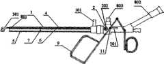
图1为膀胱肾盂取石镜结构示意图。Figure 1 is a schematic diagram of the structure of the stone mirror for bladder and pelvis extraction.
图2为放大的镜鞘结构示意图。Fig. 2 is an enlarged structural schematic diagram of the mirror sheath.
图3为放大的镜鞘横截面示意图。Fig. 3 is an enlarged schematic cross-sectional view of the mirror sheath.
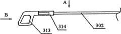
图4为取石钳夹组件结构示意图。Fig. 4 is a schematic diagram of the structure of the stone extraction jaw assembly.
图5为图4中的A向示意图。Fig. 5 is a schematic diagram of the direction A in Fig. 4 .
图6为图4中的B相示意图。FIG. 6 is a schematic diagram of phase B in FIG. 4 .
图7为拉杆与手柄的配合示意图。Fig. 7 is a schematic diagram of cooperation between the pull rod and the handle.
图8为内芯结构示意图。Figure 8 is a schematic diagram of the inner core structure.
图9为回旋流主动收集碎石的工作原理图。Figure 9 is a schematic diagram of the working principle of the swirl flow actively collecting gravel.
具体实施方式Detailed ways
参见图1、图2和图3,本实用新型的膀胱肾盂取石镜,包括两端开放的直管状镜鞘1和镜体2,所说的镜体2与镜鞘1的一端相连接;Referring to Fig. 1, Fig. 2 and Fig. 3, the bladder and pelvis stone extraction mirror of the present utility model comprises a straight
其特征在于,还包括取石钳夹组件、取石钳夹通道管4、碎石通道管5、光纤通道管6、进水通道管7和内窥成像组件,所述的镜鞘鞘壁上设有出水孔100;It is characterized in that it also includes stone extraction forceps assembly, stone extraction
所说的取石钳夹通道管4、碎石通道管5、进水通道管7和光纤通道管6的一端分别插在镜体2中,另一端插在镜鞘1中并延伸至镜鞘1的端部,所说的碎石通道管5延伸至镜体2外,取石钳夹通道管4延伸至镜体2上的滑槽201处;One end of the stone extraction
进水通道管7与镜体2上的进水口202相连接,镜鞘1中的空隙为出水通道,出水通道与设置在镜鞘1端部的出水阀101相连接;The water
优选的,所述出水孔100设置在所述镜鞘1的下部,并设有1~14排,所述出水孔100与所述镜鞘1端部之间的间距为1~3cm;Preferably, the
参见图1和图4,所说的取石钳夹组件包括钳夹301和与钳夹301相连接的拉杆302,所说的拉杆302的另一端穿过取石钳夹通道2在滑槽206处与设置在镜体2外的手柄9相连接,钳夹301设置在远离镜体2的取石钳夹通道管4的一端的端部外;Referring to Fig. 1 and Fig. 4, said stone extraction jaw assembly includes a
所说的内窥成像组件穿过光纤通道管6,一端与镜体2相连接,另一端设置在光纤通道管6的端部;Said endoscopic imaging assembly passes through the
结合图1、图2、图3、图4、图5、图6和图6,所说的镜体2上设有滑槽201,所说的钳夹301包括框架状的上钳体311、上钳杆312、框架状的下钳体313、下钳杆314和连接杆315,所说的上钳体311和下钳体313分别向外膨起,构成环状,如图5,便于固定结石或摄取其他各种体内异物,上钳杆312的一端与上钳体311的一边相连接,下钳杆314的一端与下钳体311的一边相连接,上钳杆312与下钳杆314的中部相互交叉,通过销钉活动连接,上钳杆312与下钳杆314的另一端通过销钉与连接杆315活动连接,连接杆315的另一端通过销钉与拉杆302相连接,拉杆302插入取石钳夹通道管4,并延伸至镜体2上的滑槽201处,与手柄9通过固定销钉11相连接,当所说的手柄9向水平运动时,即可带动拉杆302运动,从而使上上钳体311和下钳体313张开或闭合,用于固定结石或摄取其他各种体内异物;With reference to Fig. 1, Fig. 2, Fig. 3, Fig. 4, Fig. 5, Fig. 6 and Fig. 6, said mirror body 2 is provided with a
进一步,所述上钳体311和下钳体313的前缘和下缘内侧设有结石固定构件,如齿纹等,以增加阻力和摩擦力;Further, stone fixing components, such as tooth lines, etc., are provided on the inner sides of the front edge and the lower edge of the
进一步,参见图1,内窥成像组件包括物镜801、目镜802、光源803、光源光纤和成像返回光纤,所说的物镜801固定在光纤通道管7的端部,所说的目镜802和光源803分别设置在镜体2上,成像返回光纤的一端与目镜802相连接,另一端穿过光纤通道管6与物镜801相连接,光源光纤的一端与光源803相连接,另一端穿过光纤通道管6并延伸至光纤通道管6的端部;该内窥成像组件的结构和连接关系为常规的技术,本发明不再赘述;Further, referring to Fig. 1, the endoscopic imaging assembly includes an
进一步,镜体2与镜鞘1通过咬口固定连接。Further, the mirror body 2 is fixedly connected with the
进一步,本实用新型还包括与镜鞘1配合的、一端伸出镜鞘1且端部为圆弧状的内芯10,如图8,将镜鞘1插入人体前,先将内芯10插入镜鞘1,以防止镜鞘1对人体器官的损伤,插入人体后,拔出内芯10,再与镜体相连接,进行固定结石、粉碎结石和摄取碎石操作。Further, the utility model also includes an inner core 10 that cooperates with the
优选的如图3,取石钳夹通道管4位于上排,进水通道管7和光纤通道管6位于中间,碎石通道管5位于下排,进一步优选的,所述进水通道管7有两根,分别设置在碎石通道管5两侧的上方,光纤通道管6位于中间,便于视野观察,成像返回光纤和光源光纤可获得有效保护,不易损坏,取石钳夹通道管4位于上排,便于取石操作。Preferably, as shown in Figure 3, the rock extraction
本实用新型是这样使用的:The utility model uses like this:
将内芯10插入镜鞘1,插入人体后,拔出内芯10,然后再与镜体相连接,利用内窥成像组件寻找结石或异物,通过取石钳夹组件固定结石,然后采用常规的手段,如气压弹道碎石技术、激光碎石技术,将碎石杆、或激光光纤通过碎石通道管5碎石,进行粉碎结石操作,术中通过进水通道管6,送入冲洗液体,并且通过镜鞘1壁上的出水孔100,通过镜鞘1中的空隙和出水阀101排出液体,进行连续冲洗,利用回旋流主动收集碎石,其工作原理见图9,然后通过取石钳夹组件摄取结石。进一步,如将镜体连同取石钳夹组件、取石钳夹通道管4、碎石通道管5、光纤通道管6、进水通道管7和内窥成像组件拔出,可直连接冲吸器冲吸结石。Insert the inner core 10 into the
Claims (10)
Priority Applications (1)
| Application Number | Priority Date | Filing Date | Title |
|---|---|---|---|
| CN2009200727097UCN201510322U (en) | 2009-05-21 | 2009-05-21 | Bladder pyelonetomy mirror with outlet hole on the wall of mirror sheath |
Applications Claiming Priority (1)
| Application Number | Priority Date | Filing Date | Title |
|---|---|---|---|
| CN2009200727097UCN201510322U (en) | 2009-05-21 | 2009-05-21 | Bladder pyelonetomy mirror with outlet hole on the wall of mirror sheath |
Publications (1)
| Publication Number | Publication Date |
|---|---|
| CN201510322Utrue CN201510322U (en) | 2010-06-23 |
Family
ID=42481717
Family Applications (1)
| Application Number | Title | Priority Date | Filing Date |
|---|---|---|---|
| CN2009200727097UExpired - Fee RelatedCN201510322U (en) | 2009-05-21 | 2009-05-21 | Bladder pyelonetomy mirror with outlet hole on the wall of mirror sheath |
Country Status (1)
| Country | Link |
|---|---|
| CN (1) | CN201510322U (en) |
Cited By (8)
| Publication number | Priority date | Publication date | Assignee | Title |
|---|---|---|---|---|
| WO2015058329A1 (en)* | 2013-10-21 | 2015-04-30 | 龙刚 | Endoscope with continuous luminal perfusion and reflux functions |
| JP2015181939A (en)* | 2014-03-21 | 2015-10-22 | テルモ株式会社 | Calculus recovery device |
| CN105581821A (en)* | 2014-10-21 | 2016-05-18 | 沈阳沈大内窥镜有限公司 | Kidney stone clearing operation sheath |
| CN106551788A (en)* | 2015-09-24 | 2017-04-05 | 曾效参 | Optical Needle with Light Guide Groove |
| CN108403211A (en)* | 2018-03-23 | 2018-08-17 | 上海市杨浦区中心医院(同济大学附属杨浦医院) | Multifunctional bladder renal pelvis stone-taking lens |
| CN108451600A (en)* | 2018-02-13 | 2018-08-28 | 广州乔铁医疗科技有限公司 | A kind of visualization percutaneous transhepatic choledochoscope system and puncture sheath guard system |
| CN108888326A (en)* | 2018-06-29 | 2018-11-27 | 广州乔铁医疗科技有限公司 | A kind of hysteroscope system visualizing suction of foreign body |
| CN110101354A (en)* | 2019-06-20 | 2019-08-09 | 天津博朗科技发展有限公司 | A kind of rigid integral type hysteroscope applied in gynecological surgery |
- 2009
- 2009-05-21CNCN2009200727097Upatent/CN201510322U/ennot_activeExpired - Fee Related
Cited By (10)
| Publication number | Priority date | Publication date | Assignee | Title |
|---|---|---|---|---|
| WO2015058329A1 (en)* | 2013-10-21 | 2015-04-30 | 龙刚 | Endoscope with continuous luminal perfusion and reflux functions |
| CN105848556A (en)* | 2013-10-21 | 2016-08-10 | 武汉佑康科技有限公司 | A kind of endoscope with cavity continuous perfusion and reflux function |
| CN105848556B (en)* | 2013-10-21 | 2018-06-08 | 武汉佑康科技有限公司 | A kind of endoscope with cavity continuous perfusion and reflux function |
| JP2015181939A (en)* | 2014-03-21 | 2015-10-22 | テルモ株式会社 | Calculus recovery device |
| CN105581821A (en)* | 2014-10-21 | 2016-05-18 | 沈阳沈大内窥镜有限公司 | Kidney stone clearing operation sheath |
| CN106551788A (en)* | 2015-09-24 | 2017-04-05 | 曾效参 | Optical Needle with Light Guide Groove |
| CN108451600A (en)* | 2018-02-13 | 2018-08-28 | 广州乔铁医疗科技有限公司 | A kind of visualization percutaneous transhepatic choledochoscope system and puncture sheath guard system |
| CN108403211A (en)* | 2018-03-23 | 2018-08-17 | 上海市杨浦区中心医院(同济大学附属杨浦医院) | Multifunctional bladder renal pelvis stone-taking lens |
| CN108888326A (en)* | 2018-06-29 | 2018-11-27 | 广州乔铁医疗科技有限公司 | A kind of hysteroscope system visualizing suction of foreign body |
| CN110101354A (en)* | 2019-06-20 | 2019-08-09 | 天津博朗科技发展有限公司 | A kind of rigid integral type hysteroscope applied in gynecological surgery |
Similar Documents
| Publication | Publication Date | Title |
|---|---|---|
| CN101214163B (en) | Bladder renal pelvis stones extraction lens | |
| CN201510322U (en) | Bladder pyelonetomy mirror with outlet hole on the wall of mirror sheath | |
| CN106551672B (en) | Hysteroscopic system | |
| CN108403211A (en) | Multifunctional bladder renal pelvis stone-taking lens | |
| CN109875681B (en) | Attractive ureteroscope | |
| CN101773380B (en) | Confocal microscopy hard cholecystoscope system | |
| CN102670156A (en) | Purifying medical endoscope | |
| CN204890922U (en) | Percutaneous kidney fistulization wicresoft expansion drainage external member | |
| CN106037836A (en) | Urological endoscope surgical instrument | |
| CN206026368U (en) | Uropoiesis laparoscopic surgery ware | |
| CN113893439A (en) | A urethral protective sheath for treating urinary calculi | |
| CN201286738Y (en) | Improved type bladder pelvis-renal stone-exclusion lens of flushing structure | |
| CN208756160U (en) | Multifunctional bladder renal pelvis stone-taking lens | |
| CN111528783A (en) | Uretero-nephroscope | |
| CN208958078U (en) | side view lateral opening uretero-renoscope | |
| CN117257401A (en) | Urolithiasis negative pressure capturing basket and urolithiasis operation system | |
| CN201469347U (en) | Hard-lens gallbladder-reserved gall-stone sheath | |
| CN202010140U (en) | Purification type medical endoscope | |
| CN204468033U (en) | A kind of vermiform appendix primary and secondary mirror | |
| CN211583417U (en) | Direct-viewing abortion uterine curettage device and system with flushing mechanism | |
| CN202409041U (en) | Mirror bridge for urinary bladder and pelvis lithotomy mirror | |
| CN102309306A (en) | Choledochoscope for removing calculi | |
| CN201182626Y (en) | Clamps for vesico-renal lithotomy | |
| CN207356094U (en) | A kind of equipment for separating liquid from solid for endoscopic catheters | |
| CN2698266Y (en) | Straight type hysteroscope |
Legal Events
| Date | Code | Title | Description |
|---|---|---|---|
| DD01 | Delivery of document by public notice | Addressee:Luo Dachen Document name:Notification to Go Through Formalities of Registration | |
| C14 | Grant of patent or utility model | ||
| GR01 | Patent grant | ||
| CF01 | Termination of patent right due to non-payment of annual fee | Granted publication date:20100623 Termination date:20160521 |