CN1852971A - Oligodendrocytes derived from human embryonic stem cells for remyelination and treatment of spinal cord injury - Google Patents
Oligodendrocytes derived from human embryonic stem cells for remyelination and treatment of spinal cord injuryDownload PDFInfo
- Publication number
- CN1852971A CN1852971ACNA038161842ACN03816184ACN1852971ACN 1852971 ACN1852971 ACN 1852971ACN A038161842 ACNA038161842 ACN A038161842ACN 03816184 ACN03816184 ACN 03816184ACN 1852971 ACN1852971 ACN 1852971A
- Authority
- CN
- China
- Prior art keywords
- cells
- cell population
- differentiated cell
- oligodendrocytes
- cell
- Prior art date
- Legal status (The legal status is an assumption and is not a legal conclusion. Google has not performed a legal analysis and makes no representation as to the accuracy of the status listed.)
- Granted
Links
Images


Classifications
- C—CHEMISTRY; METALLURGY
- C12—BIOCHEMISTRY; BEER; SPIRITS; WINE; VINEGAR; MICROBIOLOGY; ENZYMOLOGY; MUTATION OR GENETIC ENGINEERING
- C12N—MICROORGANISMS OR ENZYMES; COMPOSITIONS THEREOF; PROPAGATING, PRESERVING, OR MAINTAINING MICROORGANISMS; MUTATION OR GENETIC ENGINEERING; CULTURE MEDIA
- C12N5/00—Undifferentiated human, animal or plant cells, e.g. cell lines; Tissues; Cultivation or maintenance thereof; Culture media therefor
- C12N5/06—Animal cells or tissues; Human cells or tissues
- C12N5/0602—Vertebrate cells
- C12N5/0618—Cells of the nervous system
- C12N5/0622—Glial cells, e.g. astrocytes, oligodendrocytes; Schwann cells
- A—HUMAN NECESSITIES
- A61—MEDICAL OR VETERINARY SCIENCE; HYGIENE
- A61P—SPECIFIC THERAPEUTIC ACTIVITY OF CHEMICAL COMPOUNDS OR MEDICINAL PREPARATIONS
- A61P25/00—Drugs for disorders of the nervous system
- A—HUMAN NECESSITIES
- A61—MEDICAL OR VETERINARY SCIENCE; HYGIENE
- A61P—SPECIFIC THERAPEUTIC ACTIVITY OF CHEMICAL COMPOUNDS OR MEDICINAL PREPARATIONS
- A61P25/00—Drugs for disorders of the nervous system
- A61P25/28—Drugs for disorders of the nervous system for treating neurodegenerative disorders of the central nervous system, e.g. nootropic agents, cognition enhancers, drugs for treating Alzheimer's disease or other forms of dementia
- A—HUMAN NECESSITIES
- A61—MEDICAL OR VETERINARY SCIENCE; HYGIENE
- A61P—SPECIFIC THERAPEUTIC ACTIVITY OF CHEMICAL COMPOUNDS OR MEDICINAL PREPARATIONS
- A61P27/00—Drugs for disorders of the senses
- A61P27/02—Ophthalmic agents
- C—CHEMISTRY; METALLURGY
- C12—BIOCHEMISTRY; BEER; SPIRITS; WINE; VINEGAR; MICROBIOLOGY; ENZYMOLOGY; MUTATION OR GENETIC ENGINEERING
- C12N—MICROORGANISMS OR ENZYMES; COMPOSITIONS THEREOF; PROPAGATING, PRESERVING, OR MAINTAINING MICROORGANISMS; MUTATION OR GENETIC ENGINEERING; CULTURE MEDIA
- C12N5/00—Undifferentiated human, animal or plant cells, e.g. cell lines; Tissues; Cultivation or maintenance thereof; Culture media therefor
- C12N5/06—Animal cells or tissues; Human cells or tissues
- C12N5/0602—Vertebrate cells
- C12N5/0618—Cells of the nervous system
- C12N5/0623—Stem cells
- G—PHYSICS
- G01—MEASURING; TESTING
- G01N—INVESTIGATING OR ANALYSING MATERIALS BY DETERMINING THEIR CHEMICAL OR PHYSICAL PROPERTIES
- G01N33/00—Investigating or analysing materials by specific methods not covered by groups G01N1/00 - G01N31/00
- G01N33/48—Biological material, e.g. blood, urine; Haemocytometers
- G01N33/50—Chemical analysis of biological material, e.g. blood, urine; Testing involving biospecific ligand binding methods; Immunological testing
- A—HUMAN NECESSITIES
- A61—MEDICAL OR VETERINARY SCIENCE; HYGIENE
- A61K—PREPARATIONS FOR MEDICAL, DENTAL OR TOILETRY PURPOSES
- A61K35/00—Medicinal preparations containing materials or reaction products thereof with undetermined constitution
- A61K35/12—Materials from mammals; Compositions comprising non-specified tissues or cells; Compositions comprising non-embryonic stem cells; Genetically modified cells
- C—CHEMISTRY; METALLURGY
- C12—BIOCHEMISTRY; BEER; SPIRITS; WINE; VINEGAR; MICROBIOLOGY; ENZYMOLOGY; MUTATION OR GENETIC ENGINEERING
- C12N—MICROORGANISMS OR ENZYMES; COMPOSITIONS THEREOF; PROPAGATING, PRESERVING, OR MAINTAINING MICROORGANISMS; MUTATION OR GENETIC ENGINEERING; CULTURE MEDIA
- C12N2501/00—Active agents used in cell culture processes, e.g. differentation
- C12N2501/10—Growth factors
- C12N2501/115—Basic fibroblast growth factor (bFGF, FGF-2)
- C—CHEMISTRY; METALLURGY
- C12—BIOCHEMISTRY; BEER; SPIRITS; WINE; VINEGAR; MICROBIOLOGY; ENZYMOLOGY; MUTATION OR GENETIC ENGINEERING
- C12N—MICROORGANISMS OR ENZYMES; COMPOSITIONS THEREOF; PROPAGATING, PRESERVING, OR MAINTAINING MICROORGANISMS; MUTATION OR GENETIC ENGINEERING; CULTURE MEDIA
- C12N2501/00—Active agents used in cell culture processes, e.g. differentation
- C12N2501/30—Hormones
- C12N2501/38—Hormones with nuclear receptors
- C12N2501/395—Thyroid hormones
- C—CHEMISTRY; METALLURGY
- C12—BIOCHEMISTRY; BEER; SPIRITS; WINE; VINEGAR; MICROBIOLOGY; ENZYMOLOGY; MUTATION OR GENETIC ENGINEERING
- C12N—MICROORGANISMS OR ENZYMES; COMPOSITIONS THEREOF; PROPAGATING, PRESERVING, OR MAINTAINING MICROORGANISMS; MUTATION OR GENETIC ENGINEERING; CULTURE MEDIA
- C12N2503/00—Use of cells in diagnostics
- C12N2503/02—Drug screening
- C—CHEMISTRY; METALLURGY
- C12—BIOCHEMISTRY; BEER; SPIRITS; WINE; VINEGAR; MICROBIOLOGY; ENZYMOLOGY; MUTATION OR GENETIC ENGINEERING
- C12N—MICROORGANISMS OR ENZYMES; COMPOSITIONS THEREOF; PROPAGATING, PRESERVING, OR MAINTAINING MICROORGANISMS; MUTATION OR GENETIC ENGINEERING; CULTURE MEDIA
- C12N2506/00—Differentiation of animal cells from one lineage to another; Differentiation of pluripotent cells
- C12N2506/02—Differentiation of animal cells from one lineage to another; Differentiation of pluripotent cells from embryonic cells
Landscapes
- Health & Medical Sciences (AREA)
- Engineering & Computer Science (AREA)
- Life Sciences & Earth Sciences (AREA)
- Biomedical Technology (AREA)
- Chemical & Material Sciences (AREA)
- Bioinformatics & Cheminformatics (AREA)
- Organic Chemistry (AREA)
- Biotechnology (AREA)
- Zoology (AREA)
- Wood Science & Technology (AREA)
- Genetics & Genomics (AREA)
- General Health & Medical Sciences (AREA)
- Neurosurgery (AREA)
- Neurology (AREA)
- Cell Biology (AREA)
- Microbiology (AREA)
- Biochemistry (AREA)
- General Engineering & Computer Science (AREA)
- Medicinal Chemistry (AREA)
- Developmental Biology & Embryology (AREA)
- Veterinary Medicine (AREA)
- Public Health (AREA)
- Animal Behavior & Ethology (AREA)
- Pharmacology & Pharmacy (AREA)
- Immunology (AREA)
- Chemical Kinetics & Catalysis (AREA)
- Nuclear Medicine, Radiotherapy & Molecular Imaging (AREA)
- General Chemical & Material Sciences (AREA)
- Urology & Nephrology (AREA)
- Molecular Biology (AREA)
- Hematology (AREA)
- Food Science & Technology (AREA)
- Physics & Mathematics (AREA)
- Analytical Chemistry (AREA)
- General Physics & Mathematics (AREA)
- Pathology (AREA)
- Virology (AREA)
- Epidemiology (AREA)
- Ophthalmology & Optometry (AREA)
- Hospice & Palliative Care (AREA)
Abstract
Description
Translated fromChinese技术领域technical field
本发明总的涉及胚胎细胞和神经祖细胞的细胞生物学领域。更具体的是,本发明提供了少突胶质细胞及其前体的富集细胞群,适合用于生物学研究、药物筛选和人类治疗。The present invention relates generally to the field of cell biology of embryonic cells and neural progenitor cells. More specifically, the present invention provides an enriched cell population of oligodendrocytes and their precursors, suitable for biological research, drug screening and human therapy.
相关申请related application
本申请要求2002年7月11日提交的美国临时专利申请60/395,382的优先权和2003年4月4日提交的美国实用专利申请10/406,817的优先权。这些优先权申请的全部内容被引入本文作为参考。This application claims priority to US
背景background
在中枢神经系统的支持中,少突胶质细胞起着重要的生理作用。用于人类治疗的少突胶质细胞的可用性可促进隔离神经细胞的髓鞘中的受损导致的无能症状康复。In the support of the central nervous system, oligodendrocytes play an important physiological role. The availability of oligodendrocytes for human therapy may facilitate recovery from incapacity symptoms caused by damage in the myelin sheath that insulates nerve cells.
多发性硬化症是一种渐进的并丧失能力的脱髓鞘疾病,与大脑和脊髓神经细胞周围的髓鞘的逐渐破坏相关。这种病的症状程度从麻木、视觉损伤以及认知变化到瘫痪。这种病被确信有免疫学的和遗传方面的因素,出现临床症状的经常在20到40岁间。仅在美国这种病就影响了大约300,000人。目前治疗方案涉及β-干扰素类或皮质甾类。这些药在发病时能缩短症状出现的时间,但是通常不能防止长期的能力丧失。Multiple sclerosis is a progressive and disabling demyelinating disease associated with the gradual destruction of the myelin sheath that surrounds nerve cells in the brain and spinal cord. Symptoms range from numbness, visual impairment, and cognitive changes to paralysis. The disease is believed to have immunological and genetic components, and clinical symptoms often appear between the ages of 20 and 40. The disease affects approximately 300,000 people in the United States alone. Current treatment options involve beta-interferons or corticosteroids. These drugs shorten the time to onset of symptoms during the onset of an attack, but usually do not prevent long-term disability.
脊髓的外伤性创伤会导致创伤点附近完整轴索脱髓鞘化,从而破坏了它们的神经传导能力。在美国每年大约有11,000例新的脊髓损伤病例。SCI信息网(SCI Information Network)预测,对于遭受任何级别运动功能不完整的病人,其生存期直接花费的范围从400,000美元到2,200,000美元,不包括工资损失和生活质量的影响。Traumatic trauma to the spinal cord results in the demyelination of intact axons near the point of trauma, thereby disrupting their nerve conduction capacity. There are approximately 11,000 new cases of spinal cord injury in the United States each year. The SCI Information Network estimates that direct lifetime costs for patients suffering from any grade of motor incompleteness range from $400,000 to $2,200,000, excluding lost wages and quality of life effects.
中枢神经系统细胞上的髓磷脂被少突胶质细胞放在适当的位置,包裹在轴索周围,形成髓鞘。Keirstead和Blakemore阐述了少突胶质细胞及其祖细胞在疾病症状中的作用(Adv.Exp.Med.Biol.468:183,1999)。被称为O-2A细胞的少突胶质细胞祖细胞存在于正常成人的CNS中和多发性硬化症病灶中,并参与了再髓鞘化(Scolding等,Brain 121:2221,1998;以及Scolding等,Neuroscience89:1,1999)。充分再髓鞘化的失败可能会发生,因为少突胶质细胞的对称增生会用尽大面积损伤地方的祖细胞储备。Myelin on CNS cells is put in place by oligodendrocytes to wrap around axons, forming myelin sheaths. Keirstead and Blakemore described the role of oligodendrocytes and their progenitors in disease symptoms (Adv. Exp. Med. Biol. 468:183, 1999). Oligodendrocyte progenitors known as O-2A cells are present in the CNS of normal adults and in multiple sclerosis lesions and are involved in remyelination (Scolding et al., Brain 121:2221, 1998; and Scolding et al., Neuroscience 89:1, 1999). Failure of adequate remyelination may occur as symmetrical proliferation of oligodendrocytes depletes progenitor cell reserves at sites of extensive injury.
已进行了大量的研究工作,目的是建立能应用于再生医学以恢复神经功能的细胞群(参见Park等,J.Neurotrauma,16:675,1999)。Keirstead等(J.Neuroscience 19:7529,1999)已从出生后的老鼠大脑中分离出CNS前体,该前体在移植后能产生少突胶质细胞和神经膜细胞。Svendsen等(J.Neurosci.Meth.85:141,1998)已从发育的人皮质中分离出前体细胞。Mujtaba等(Dev.Biol.214:113,1999)报道从胚胎干细胞中分离出神经前体。Substantial research efforts have been undertaken with the aim of establishing cell populations that can be applied in regenerative medicine to restore neural function (see Park et al., J. Neurotrauma, 16:675, 1999). Keirstead et al. (J. Neuroscience 19:7529, 1999) have isolated CNS precursors from postnatal mouse brains that give rise to oligodendrocytes and neuritic cells after transplantation. Svendsen et al. (J. Neurosci. Meth. 85:141, 1998) have isolated precursor cells from the developing human cortex. Mujtaba et al. (Dev. Biol. 214:113, 1999) reported the isolation of neural precursors from embryonic stem cells.
PCT出版物WO97/07200(Stanford U.)公布了从成年大鼠大脑中分离出的少突胶质细胞前体的培养物。PCT出版物WO 01/28342(Washington U.)提出了在以预处理的少突胶质细胞培养基中培养神经细胞的某些方法。美国专利5,753,506(Johe,CNS干细胞技术)涉及一种用于维持从神经组织中分离的具备分化成神经元、星形胶质细胞和少突胶质细胞能力的干细胞的培养系统。美国专利6,238,922(StemCells Inc.)提出了神经组织分化变异成具备分化成神经元和神经胶质能力的细胞。美国专利6,235,527(Rao等,U.Utah)涉及从哺乳动物神经管组织分离并基于A2B5细胞表面标记物选择的哺乳动物CNS神经胶质限制性前体细胞群。PCT Publication WO97/07200 (Stanford U.) discloses cultures of oligodendrocyte precursors isolated from adult rat brains. PCT publication WO 01/28342 (Washington U.) proposes certain methods for culturing neural cells in preconditioned oligodendrocyte culture medium. US Patent 5,753,506 (Johe, CNS Stem Cell Technology) relates to a culture system for maintaining stem cells isolated from neural tissue capable of differentiating into neurons, astrocytes and oligodendrocytes. U.S. Patent 6,238,922 (StemCells Inc.) proposes that neural tissue differentiates into cells capable of differentiating into neurons and glia. US Patent 6,235,527 (Rao et al., U. Utah) relates to mammalian CNS glial-restricted precursor cell populations isolated from mammalian neural tube tissue and selected based on the A2B5 cell surface marker.
美国专利5,968,829(Cytotherapeutics)要求保护含有具备生成神经元、星形胶质细胞和少突胶质细胞能力的CNS神经干细胞的培养基。PCT出版物WO97/32608涉及遗传工程改造的初级少突胶质细胞,用于CNS中的移植介导的传递。美国专利5,830,621(Signal Pharmaceuticals)描述了保藏于ATCC的登录号为CRL11881的人少突胶质细胞系。这个细胞株基本上没有GFAP、GalC、O4和A2B5特征性标记物。US Patent 5,968,829 (Cytotherapeutics) claims a culture medium containing CNS neural stem cells capable of generating neurons, astrocytes and oligodendrocytes. PCT publication WO97/32608 relates to genetically engineered primary oligodendrocytes for transplantation-mediated delivery in the CNS. US Patent 5,830,621 (Signal Pharmaceuticals) describes a human oligodendrocyte cell line deposited with the ATCC under accession number CRL11881. This cell line essentially lacks the characteristic markers of GFAP, GalC, O4 and A2B5.
不幸的是,至今还不清楚从神组织中分离的祖细胞是否具备足够的复制能力来产生用于人类临床治疗的必要数量的细胞。Unfortunately, it remains unclear whether progenitor cells isolated from divine tissue are sufficiently replicable to generate the necessary numbers for human clinical therapy.
一个备选来源是从早期胚胎组织中分离的多能细胞。胚胎干细胞(ES)是25多年以前首先从小鼠胚胎中分离出来的(G.R.Martin,Proc.Natl.Acad.Sci.U.S.A.78:7634,1981)。据信胚胎干细胞实际上能产生同一种类任何组织类型的后代。An alternative source is pluripotent cells isolated from early embryonic tissue. Embryonic stem cells (ES) were first isolated from mouse embryos more than 25 years ago (G.R. Martin, Proc. Natl. Acad. Sci. U.S.A. 78:7634, 1981). Embryonic stem cells are believed to be capable of producing progeny of virtually any tissue type of the same kind.
Fraichard等(J.Cell Sci.108:3181,1995)报道在体外小鼠的胚胎干细胞分化成神经胶质细胞和功能性神经元。Mujtaba等(Dev.Biol.214:113,1999)报道从小鼠胚胎干细胞中分离到神经前体。Li,Smith等(Cur.Biol.8:971,1998)报道通过谱系选择从小鼠胚胎干细胞中产生神经元前体。Brüstle,McKay等(Proc.Natl.Acad.Sci.USA 94:14809,1997;Science 285:754,1999)报道衍生自小鼠胚胎干细胞的神经胶质前体可作为髓鞘移植物的潜在来源。McDonald等(Nat.Med5:1410,1999;Proc.Natl.Acad.Sci.USA 97:6126,2000)报道小鼠胚胎干细胞在培养和脊髓移植后可形成少突胶质细胞和髓鞘。Fraichard et al. (J. Cell Sci. 108:3181, 1995) reported that mouse embryonic stem cells differentiated into glial cells and functional neurons in vitro. Mujtaba et al. (Dev. Biol. 214:113, 1999) reported the isolation of neural precursors from mouse embryonic stem cells. Li, Smith et al. (Cur. Biol. 8:971, 1998) reported the generation of neuronal precursors from mouse embryonic stem cells by lineage selection. Brüstle, McKay et al. (Proc. Natl. Acad. Sci. USA 94: 14809, 1997; Science 285: 754, 1999) reported that glial precursors derived from mouse embryonic stem cells could serve as a potential source of myelin grafts. McDonald et al. (Nat. Med 5:1410, 1999; Proc. Natl. Acad. Sci. USA 97: 6126, 2000) reported that mouse embryonic stem cells could form oligodendrocytes and myelin sheath after culture and spinal cord transplantation.
直到最近才分离出人类胚胎干细胞(Thomson等,Science 282:114,1998)。人类胚胎干细胞需要非常不同的条件来保持它们处于未分化状态或引导它们沿着特定的分化途径分化(美国专利6,090,622和6,200,806;PCT出版物WO99/20741和WO01/51616)。由于这个原因,极少知道如何从人类胚胎干细胞中制备相对较同类的分化细胞群。Human embryonic stem cells have only recently been isolated (Thomson et al., Science 282:114, 1998). Human embryonic stem cells require very different conditions to maintain them in an undifferentiated state or to direct them to differentiate along specific differentiation pathways (US Patents 6,090,622 and 6,200,806; PCT Publications WO99/20741 and WO01/51616). For this reason, little is known about how to generate relatively homogeneous differentiated cell populations from human embryonic stem cells.
PCT出版物WO01/88104(Carpenter,Geron Corporation)描述了通过分化人类胚胎干细胞得到的神经祖细胞群。已获得超过90%NCAM阳性、35%β-微管蛋白阳性和75%A2B5阳性的细胞群。Zhang等(Nature Biotech.19:1129,2001)报道了从人类胚胎干细胞中分化得到的可移植的神经前体。国际专利申请PCT/US02/19477(Carpenter等,Geron Corporation)描述了胚胎干细胞衍生的神经细胞群,其中在已产生的细胞群中,至少10%的MAP-2阳性细胞表达了酪氨酸羟化酶,该酶是多巴胺能神经元的标记物。PCT publication WO 01/88104 (Carpenter, Geron Corporation) describes neural progenitor cell populations obtained by differentiating human embryonic stem cells. Cell populations that were more than 90% positive for NCAM, 35% positive for β-tubulin and 75% positive for A2B5 have been obtained. Zhang et al. (Nature Biotech. 19:1129, 2001) reported transplantable neural precursors differentiated from human embryonic stem cells. International Patent Application PCT/US02/19477 (Carpenter et al., Geron Corporation) describes embryonic stem cell-derived neural cell populations in which at least 10% of the MAP-2 positive cells in the resulting population express tyrosine hydroxylation Enzyme, which is a marker of dopaminergic neurons.
最近,Billon等(J.Cell Sci.115:3657,2002)描述了来自遗传工程改造的小鼠胚胎干细胞的少突胶质细胞发育的同步性。Kuo等(Biol.Reprod.Dec11/02)报道了猴ES衍生的细胞种群,该种群是28%GFAP阳性;Xian等(StemCells 21:41,2003)报道了采用谱系特异的转录因子Olig2从小鼠胚胎干细胞中得到少突胶质细胞。Recently, Billon et al. (J. Cell Sci. 115:3657, 2002) described the synchrony of oligodendrocyte development from genetically engineered mouse embryonic stem cells. Kuo et al. (Biol.Reprod.Dec11/02) reported a monkey ES-derived cell population that was 28% GFAP positive; Xian et al. (StemCells 21:41, 2003) reported the use of the lineage-specific transcription factor Olig2 to generate Stem cells derived from oligodendrocytes.
为了在人类健康和疾病的治疗中实现pPS细胞的所有潜力,有必要建立新的方法(paradigms)来产生可用于治疗脱髓鞘病症的富集细胞群。To realize the full potential of pPS cells in the treatment of human health and disease, it will be necessary to establish new paradigms to generate enriched cell populations that can be used to treat demyelinating disorders.
概要summary
本发明提供了一个系统,该系统能有效生产用于研究或药物组合物制备的神经胶质谱系的灵长类动物细胞。The present invention provides a system for the efficient production of primate cells of the glial lineage for research or preparation of pharmaceutical compositions.
本发明的分化细胞群是在体外分离或培养的,高度富含神经胶质细胞或能对神经组织髓鞘化的细胞的特征。这些细胞可具备少突胶质细胞的形态特征,表达本文所列出的某些可检测的抗体或可扩增标记物,或者具备进一步分化后形成少突胶质细胞的能力。这些细胞还可具备少突胶质细胞的某些功能特征,例如在协同培养实验中使神经节髓鞘化的能力、在体内恢复脱髓鞘轴突的髓磷脂的能力、或者改善人或非人动物神经功能的能力。一个、二个、三个或更多个这些特征可以任何组合出现。The differentiated cell population of the present invention is isolated or cultured in vitro and is highly rich in glial cells or cells capable of myelinating nerve tissue. These cells may have the morphological characteristics of oligodendrocytes, express certain detectable antibodies or expandable markers listed herein, or have the ability to further differentiate to form oligodendrocytes. These cells may also possess certain functional characteristics of oligodendrocytes, such as the ability to myelinate ganglia in co-culture experiments, restore myelin in demyelinated axons in vivo, or improve The ability of human animal neural function. One, two, three or more of these features may be present in any combination.
所述细胞群可以从不同种类的较少分化的干细胞中制得。潜在的原始细胞包括衍生自胚泡的灵长类动物的多能干(pPS)细胞(胚胎干细胞是个例证)或者早期胚胎的生殖组织。因此,这些细胞将具备成为它们来源组织的后代的特征,这一点可以由原始细胞和分化细胞具有同一基因组的结果来证实。The cell populations can be prepared from different types of less differentiated stem cells. Potential primordial cells include primate pluripotent stem (pPS) cells derived from blastocysts (embryonic stem cells are an example) or reproductive tissue of early embryos. Thus, these cells will have the characteristics of being descendants of the tissue from which they originated, as evidenced by the result that the original and differentiated cells share the same genome.
本发明其他方面涉及生产或维持已经描述的分化细胞的方法。这些方法包括在一种或多种生长或分化因子存在时培养多潜在的或多能性的干细胞,如在本发明中随后举例的。Other aspects of the invention relate to methods of producing or maintaining differentiated cells as described. These methods involve culturing multipotent or pluripotent stem cells in the presence of one or more growth or differentiation factors, as exemplified later in the present invention.
作为一个例子,干细胞可以在含有一种或多种分化因子如三碘甲腺原氨酸(T3)、硒或维甲酸,加入或不加入促分裂原如成纤维细胞生长因子(FGF)的培养基中培养。分化细胞的初始形成可以在培养悬液中发生,在这少突胶质细胞谱系细胞可以形成相对均一的球形体。其他细胞类型可以通过适当的分离方法去除,例如把培养基铺在能选择性吸附所需细胞类型的表面上。任选地,通过加入促分裂原如FGF或表皮生长因子(通常存在一种或多种分化因子,如细胞初始衍生时用的那些)进行培养,可导致分化细胞在选择前或后进一步增殖。随后,通过培养时不加促分裂原,或加入能增强后期分化的表面如聚-L-赖氨酸,可任选地使这些细胞进一步成熟。As an example, stem cells can be cultured in the presence of one or more differentiation factors such as triiodothyronine (T3), selenium, or retinoic acid, with or without the addition of mitogens such as fibroblast growth factor (FGF). cultured in the base. Initial formation of differentiated cells can occur in culture suspension where cells of the oligodendrocyte lineage can form relatively uniform spheroids. Other cell types can be removed by appropriate separation methods, such as spreading the culture medium on a surface that selectively adsorbs the desired cell type. Optionally, further proliferation of differentiated cells, either before or after selection, can be caused by the addition of a mitogen such as FGF or epidermal growth factor (usually in the presence of one or more differentiation factors, such as those with which the cells were initially derived), by culturing. These cells are then optionally further matured by culturing without a mitogen, or with the addition of a surface that enhances later differentiation, such as poly-L-lysine.
本发明的细胞可应用于许多商业性重要应用。例如,这些细胞适用于基于对神经胶质细胞的影响来筛选化合物,其中化合物的存在与细胞的维持、毒性、进一步分化或者作为神经胶质细胞起作用的能力相关联。这些细胞也适合于引起相邻神经组织的髓鞘化,无论在体外还是在体内。The cells of the invention find use in many commercially important applications. For example, these cells are suitable for screening compounds based on their effect on glial cells, where the presence of the compound correlates with the cell's ability to maintain, to be toxic, to further differentiate, or to function as a glial cell. These cells are also suitable for causing myelination of adjacent neural tissue, both in vitro and in vivo.
本发明的细胞也可用于制备人类或动物治疗用的药物组合物。然后可使用这些组合物治疗各种疾病,如与轴突髓鞘化中的缺陷或脊髓受损相关的病症。The cells of the invention can also be used to prepare pharmaceutical compositions for human or animal therapy. These compositions can then be used to treat various diseases, such as conditions associated with defects in axonal myelination or damage to the spinal cord.
本发明更多方面见于如下描述。Further aspects of the invention are found in the following description.
附图Attached picture
图1是通过倒置显微镜(inverted microscope)拍摄的相衬图,显示了在含有碱性FGF和少突胶质细胞分化因子的培养基悬液培养2天后的人类胚胎(ES)干细胞。Figure 1 is a phase-contrast image taken by an inverted microscope showing human embryonic (ES) stem cells cultured in suspension in a medium containing basic FGF and oligodendrocyte differentiation factors for 2 days.
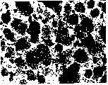
图2显示了用维甲酸培养7天后的细胞。可以看到大而清楚的细胞球,代表了培养基中80-90%的细胞。Figure 2 shows cells after 7 days of culture with retinoic acid. Large, clear spheres of cells can be seen, representing 80-90% of the cells in the medium.
图3显示了去除维甲酸后的情况,这些细胞维持在低浓度FGF中。Figure 3 shows that after removal of retinoic acid, these cells were maintained at low concentrations of FGF.
图4显示了去除FGF后出现的变化。大的聚合体分解,整个培养基充满了单个细胞和小的群落。同时,可观察到新的亮黄色球形体(箭头)。Figure 4 shows the changes that occur after removal of FGF. Large aggregates disintegrate and the entire medium is filled with single cells and small colonies. Simultaneously, new bright yellow spheres (arrowheads) can be observed.
图5显示了在FGF缺乏的情况下,用表皮生长因子培养细胞时亮黄色球形体(箭头)的生长。Figure 5 shows the growth of bright yellow spheroids (arrowheads) when cells were cultured with epidermal growth factor in the absence of FGF.
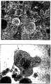
图6显示接种在Matrigel上2或3天后,来自球形体的定向为神经胶质的神经前体的移动和分支。Figure 6 shows the movement and branching of glially oriented neural precursors from
图7显示通过粘附到Matrigel上仅10-20小时来选择少突胶质细胞谱系细胞的结果。丢弃未吸附的细胞,这实质上排除了不具有少突胶质细胞标记物的细胞。然后将吸附的细胞重新悬浮并在含有FGF、EGF和神经胶质前体的培养基中增殖7天。这样有利于生产一种更分散的更适合于治疗给药和其他目的细胞群。这些细胞然后在促分裂原缺乏时,在聚-L-赖氨酸层粘连细胞上成熟。此显微镜图片显示了针对半乳糖脑苷脂(GalC,空心箭头)的染色,细胞核用溶血毒素(hematoxin)复染(实心箭头)。针对GalC染色的细胞百分比在这些条件下至少为~95%。Figure 7 shows the results of selection of cells of oligodendrocyte lineage by adhesion to Matrigel(R) for only 10-20 hours. Unattached cells were discarded, which essentially excluded cells that did not have oligodendrocyte markers. Adsorbed cells were then resuspended and propagated for 7 days in medium containing FGF, EGF and glial precursors. This facilitates the production of a more dispersed population of cells better suited for therapeutic administration and other purposes. These cells then mature on poly-L-lysine laminocytes in the absence of mitogens. This micrograph shows staining for galactocerebroside (GalC, open arrow), nuclei are counterstained with hematoxin (solid arrow). The percentage of cells stained for GalC was at least -95% under these conditions.
图8显示了胚胎干细胞衍生的少突胶质细胞的更高放大倍数。这些细胞具有成熟少突胶质细胞的形态特点:许多复杂的突起,这些突起看起来是在它们之间使髓磷脂形成网状物。Figure 8 shows a higher magnification of embryonic stem cell-derived oligodendrocytes. These cells had the morphological features of mature oligodendrocytes: many complex protrusions that appeared to have a network of myelin between them.
图9显示了在分化中细胞形态学进展。(A):未分化的hES细胞。(B):在含有维甲酸的培养基悬液中从胚状体中生长的透明球形体。(C):在EGF存在时,含有少突胶质细胞前体细胞的黄色球形体的扩张。(D):通过接种在Matrigel上阳性选择获得的少突胶质细胞谱系细胞。(E,F):在培养中增加少突胶质细胞前体的隆起(prominence)。(G,H):随后的接种导致进一步分化成成熟的少突胶质细胞。Figure 9 shows the progression of cell morphology during differentiation. (A): Undifferentiated hES cells. (B): Hyaline spheroids grown from embryoid bodies in medium suspension containing retinoic acid. (C): Expansion of yellow spheroids containing oligodendrocyte precursor cells in the presence of EGF. (D): Cells of oligodendrocyte lineage obtained by positive selection seeded on Matrigel(R). (E, F): Increased prominence of oligodendrocyte precursors in culture. (G, H): Subsequent seeding resulted in further differentiation into mature oligodendrocytes.
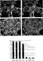
图10显示成熟少突胶质细胞的免疫细胞化学分析。接种一个星期后,这些细胞的早期神经胶质细胞标记物NG2(A)呈阳性。种植八个星期后,这些细胞的GalC(C)、O4(D)和RIP(E)呈阳性。图B显示用所示标记物染色的细胞数目。Figure 10 shows immunocytochemical analysis of mature oligodendrocytes. One week after inoculation, these cells were positive for the early glial cell marker NG2 (A). Eight weeks after seeding, these cells were positive for GalC (C), O4 (D) and RIP (E). Panel B shows the number of cells stained with the indicated markers.
图11显示给予大鼠脊髓的少突胶质细胞前体的组织剖面图,用对人核蛋白特异的抗体染色。Figure 11 shows histological sections of oligodendrocyte precursors administered to the rat spinal cord, stained with an antibody specific for human nucleoprotein.
图12显示移植九星期后这些细胞已经迁移或增殖到白质中。Figure 12 shows that these cells had migrated or proliferated into the white matter nine weeks after transplantation.
图13显示通过测量脊索(cord)横截面积,移植的少突胶质细胞不会使由挫伤性损伤后的第二次放大导致的损伤更坏。Figure 13 shows that transplanted oligodendrocytes did not worsen the damage caused by the second amplification following contusive injury, as measured by the cross-sectional area of the cord.
图14显示了hES细胞诱导轴突分支。新的轴突在移植了少突胶质细胞的动物的图片中(上图)显示为BDA染色的暗窄线。在没有处理的动物中没有观察到分支。移植的细胞正诱导再生的可塑性。Figure 14 shows that hES cells induce axon branching. New axons appear as dark narrow lines stained with BDA in pictures of animals transplanted with oligodendrocytes (upper panel). Branching was not observed in untreated animals. Transplanted cells are inducing regenerative plasticity.
图15显示了在受损位置(中心)分支的神经元定量结果(平均值±SEM,每柱3个切片)。治疗过的动物具有明显更高水平的标记轴突,从右侧一直到损伤位置吻端的中心。Figure 15 shows the results of neuron quantification (mean ± SEM, 3 sections per column) branching at the damaged site (center). Treated animals had significantly higher levels of labeled axons from the right up to the center of the rostral site of the injury.
图16显示了在移入人类胚胎干细胞来源的少突胶质细胞的动物中大量再髓鞘化的证据。在电子显微照片上部的密集圆圈是正常髓鞘化纤维。在这个部位轴突的其余部分显示了一个髓磷脂薄层。右上图中的轴突显示了大约5或者6个缠绕,以及正在进行再髓鞘化的证据。只有被移植的动物才显示新的髓磷脂,可归于少突胶质细胞的活性。这提供了一个解释在移植动物中行为改善的机制。Figure 16 shows evidence of massive remyelination in animals engrafted with human embryonic stem cell-derived oligodendrocytes. The dense circles in the upper part of the electron micrograph are normal myelinated fibers. The rest of the axon at this site shows a thin layer of myelin. The axon in the upper right panel shows about 5 or 6 windings, and evidence of ongoing remyelination. Only transplanted animals showed new myelin, attributable to oligodendrocyte activity. This provides a mechanism to explain the improved behavior in transplanted animals.
图17显示了在未分化的人类胚胎干细胞集落中检测到的标记物(左侧:抗体加上DAPI;右侧:抗体单独染色)。上图显示的是针对SSEA-4(一种多能细胞的标记物)标记的克隆。下图显示的是围绕在针对中胚层标记物BMP4标记的集落周围的基质细胞。Figure 17 shows the markers detected in undifferentiated human embryonic stem cell colonies (left: antibody plus DAPI; right: antibody staining alone). The upper panel shows clones labeled for SSEA-4, a marker for pluripotent cells. Bottom panel shows stromal cells surrounding colonies labeled for the mesoderm marker BMP4.
图18显示了在分化过程中转录因子Pax6的短暂出现。上图显示的是朝向第10天群落中心的染色,在朝向外围的更多分化细胞中已经被往下调。下图显示在分化过程的第35天事实上没有染色。Figure 18 shows the transient appearance of the transcription factor Pax6 during differentiation. The upper panel shows that staining towards the center of the colony at
图19显示了在早期少突胶质细胞谱系细胞中检测到的标记物,出现在分化过程的第10天。上图:转录因子Olig1。中图:转录因子SOX10。下图:少突胶质细胞前体标记物A2B5。Figure 19 shows markers detected in cells of the early oligodendrocyte lineage, present at
图20A-20B显示了适合在第35天移植的少突胶质细胞祖细胞(oligoprogenitors)中占优势的标记物。第一和第二行(20A):NG2(硫酸软骨素蛋白聚糖,少突胶质细胞前体的标记物);第三行(20A):GalC;第四行(20B):O4;第五行(20B):Tuj1(神经元的一种标记物)。事实上所有细胞都携带有少突胶质细胞的标记物,但不是针对神经元细胞、间充质细胞、或未分化的人类胚胎干细胞的标记物(20B,底图)。Figures 20A-20B show markers prevalent in oligoprogenitors suitable for transplantation at day 35. First and second row (20A): NG2 (chondroitin sulfate proteoglycan, marker of oligodendrocyte precursor); third row (20A): GalC; fourth row (20B): O4; Five elements (20B): Tuj1 (a marker of neurons). Virtually all cells carried markers for oligodendrocytes, but not for neurons, mesenchymal cells, or undifferentiated human embryonic stem cells (20B, bottom panel).
图21显示了给予发抖(Shiverer)小鼠少突胶质细胞的结果,这种小鼠在产生髓磷脂碱性蛋白的能力上存在遗传性缺陷。超微结构上,发抖小鼠的轴突缺乏髓磷脂或者被一个或二个不紧凑的髓磷脂缠绕包围着(上图)。移植细胞六个星期后,电子显微镜分析发现多层稠密的髓磷脂,揭示了移植细胞群产生髓鞘的能力(下图)。稠密的髓磷脂由所给予的少突胶质细胞直接产生。Figure 21 shows the results of administration of oligodendrocytes to Shiverer mice, which are genetically deficient in the ability to produce myelin basic protein. Ultrastructurally, the axons of shivering mice lacked myelin or were surrounded by one or two non-compact myelin wraps (upper panel). Six weeks after the transplanted cells, electron microscopic analysis revealed layers of dense myelin, revealing the ability of the transplanted cell population to produce myelin (below). Dense myelin is produced directly by the administered oligodendrocytes.
图22显示了一个试验的结果,在该试验中,用冲击器挫伤大鼠的脊髓,然后定量(BBB级)评价随后阶段中的脊髓功能。上图:200千达因挫伤;下图:250千达因挫伤。(■)受损后1周,ES衍生的少突胶质细胞治疗的动物(n=5);(▲)接受同样的损伤但没有给予细胞进行治疗的对照动物(n=3)。平均值±SEM,用盲法评定。移植了ES衍生的少突胶质细胞的动物表现出更好的地上运动(overground locomotion),在治疗后持续超过5周。Figure 22 shows the results of an experiment in which the spinal cords of rats were contused with an impactor, and then the spinal cord function in the subsequent period was evaluated quantitatively (BBB scale). Above: 200 kdine contusion; Bottom: 250 kdine contusion. (■) ES-derived oligodendrocyte-treated animals (n=5) 1 week after injury; (▲) control animals (n=3) that received the same injury without treatment with cells. Mean ± SEM, evaluated blindly. Animals transplanted with ES-derived oligodendrocytes exhibited better overground locomotion that persisted for more than 5 weeks after treatment.
详细描述A detailed description
本发明通过有效地从多能干细胞中生产少突胶质细胞及其前体解决了生产它们的大细胞群的问题。The present invention solves the problem of producing large cell populations of oligodendrocytes and their precursors by efficiently producing them from pluripotent stem cells.
这些分化细胞群非常相似。图7显示了一个具有少突胶质细胞形态特征的人细胞的制品例子。这个细胞群已经针对GalC进行染色,这是一种针对少突胶质细胞谱系细胞的标记物。图8是一幅更高放大倍数的视图,显示了能使髓鞘化附近任何神经元平衡的髓磷脂层的生产。这些细胞的功能性质使它们很适合于少突胶质细胞性质的进一步表征,并适合用于人类治疗。These differentiated cell populations are very similar. Figure 7 shows an example of a preparation of human cells with morphological characteristics of oligodendrocytes. This cell population has been stained for GalC, a marker for cells of the oligodendrocyte lineage. Figure 8 is a higher magnification view showing the production of a myelin layer that balances any neuron near myelination. The functional properties of these cells make them well suited for further characterization of oligodendrocyte properties and for use in human therapy.
本发明的细胞理想来源是灵长类动物不同种类的多能干(pPS)细胞。根据下面描述的策略,通过在几种合适的培养条件和辅助因子中进行选择,可以使pPS细胞被诱导进入少突胶质细胞路径。已发现,在最优化的条件下,未分化的pPS细胞转换成少突胶质细胞的效率可大25%。Ideal sources of cells for the present invention are pluripotent stem (pPS) cells of different species from primates. According to the strategy described below, pPS cells can be induced into the oligodendrocyte pathway by selecting among several suitable culture conditions and cofactors. It has been found that under optimized conditions, the conversion efficiency of undifferentiated pPS cells into oligodendrocytes can be greater than 25%.
本发明的组合物和方法提供优于先前可获得技术的重要优点。通过从所需同种异型的pPS细胞中分化少突胶质细胞,可产生具有任何组织相容性类型的少突胶质细胞。如果需要,可以任何增强移植的方式在分化前或后遗传修饰这些细胞。因为这些细胞来自天然细胞系而不需要进行组织解剖,因此它们能理想地满足规章批准所提出的质量控制要求。The compositions and methods of the present invention provide important advantages over previously available technologies. By differentiating oligodendrocytes from pPS cells of the desired allotype, oligodendrocytes of any histocompatibility type can be generated. These cells can be genetically modified, either before or after differentiation, in any manner that enhances engraftment, if desired. Because these cells are derived from native cell lines and do not require tissue dissection, they ideally meet the quality control requirements imposed by regulatory approvals.
重要的是,产生于每一个起始干细胞群的细胞的供应几乎是无限制的。如实施例1中所举例的,一旦产生和选择得到少突胶质细胞谱系细胞,可通过在存在生长因子的情况下培养而大量增殖这些细胞。此外,几乎能诱使原始pPS细胞无限增殖,从而为更多分化的细胞提供持续的来源。Importantly, the supply of cells derived from each starting stem cell population is virtually unlimited. As exemplified in Example 1, once cells of the oligodendrocyte lineage have been generated and selected, these cells can be proliferated in large numbers by culturing in the presence of growth factors. In addition, primitive pPS cells can be induced to proliferate almost indefinitely, thus providing a continuous source of more differentiated cells.
下文提供如何制备和使用本发明分化细胞的进一步描述。这些种群非常地一致,并因此适合用于许多商业上地重要应用。A further description of how to make and use the differentiated cells of the invention is provided below. These populations are very consistent and therefore suitable for many commercially important applications.
本发明一个方面提供一种分化的细胞群,其中至少大约80%的细胞是灵长类动物多能干(pPS)细胞的后代;能被对NG2蛋白聚糖(或其他少突胶质细胞标记物)特异的的抗体染色;并且呈NeuN(或其他神经元细胞和潜在污染物的标记物)阴性。分化的细胞群可能是适合产生少突胶质细胞谱系细胞的组成系统的一部分。这个系统可能还包含分化细胞从中产生的pPS细胞株(例如人类胚胎干细胞)。任选地,分化细胞群至少80%的细胞也表达A2B5或PDGFRα。至少20%的细胞可显示少突胶质细胞前体的两极形态学特征。One aspect of the present invention provides a differentiated cell population wherein at least about 80% of the cells are descendants of primate pluripotent stem (pPS) cells; ) specific antibody staining; and negative for NeuN (or other markers of neuronal cells and potential contaminants). The differentiated cell population may be part of a constitutive system suitable for generating cells of the oligodendrocyte lineage. This system may also include pPS cell lines from which differentiated cells are derived (eg, human embryonic stem cells). Optionally, at least 80% of the cells of the differentiated cell population also express A2B5 or PDGFRα. At least 20% of cells may display bipolar morphological features of oligodendrocyte precursors.
在一些环境下,细胞群植入发抖突变小鼠的脊髓中后,这些分化的细胞群会引起神经元轴突周围致密的髓磷脂的沉积;或者在挫伤性损伤的大鼠的脊髓内或周围植入细胞群后,导致其地上运动改善。或者,可在体外进一步分化本发明的少突胶质细胞前体细胞(例如,在缺乏促分裂原时在聚-L-赖氨酸和层粘连蛋白上培养3天)。这样可以产生一个更加成熟的细胞群,其中至少10%的细胞具有成熟少突胶质细胞的复杂突起的特征,并且大约80%、90%、95%或更多的细胞具有成熟少突胶质细胞的标记物如GalC。在增殖阶段制备的少突胶质细胞谱系细胞的特征不仅在于它们在祖代阶段表达的标记物,还在于它们产生富集的成熟细胞群的能力或者在体内执行所需功能的能力。In some settings, when cell populations were implanted in the spinal cord of trembling mutant mice, these differentiated cell populations caused the deposition of dense myelin around neuronal axons; or in or around the spinal cord of contusively injured rats Implantation of the cell population resulted in improved ground movement. Alternatively, the oligodendrocyte precursor cells of the invention can be further differentiated in vitro (eg, cultured on poly-L-lysine and laminin for 3 days in the absence of mitogens). This results in a more mature cell population in which at least 10% of the cells have the complex protrusions characteristic of mature oligodendrocytes and approximately 80%, 90%, 95% or more of the cells have mature oligodendrocytes Cellular markers such as GalC. Oligodendrocyte lineage cells prepared at the proliferative stage are characterized not only by the markers they express at the progenitor stage, but also by their ability to generate enriched mature cell populations or to perform desired functions in vivo.
如下面所阐述的,本发明一些分化的细胞群可采用以下方法获得,其中未分化的pPS细胞培养在含有促分裂原和至少二种少突胶质细胞分化因子的培养基中。例如,在碱性成纤维细胞生长因子(FGF)、三碘甲腺原氨酸(T3)、维甲酸存在时(可能还有硒),未分化的pPS细胞可以悬液培养,以形成细胞聚集体。As described below, some differentiated cell populations of the present invention can be obtained by a method wherein undifferentiated pPS cells are cultured in a medium containing a mitogen and at least two oligodendrocyte differentiation factors. For example, undifferentiated pPS cells can be cultured in suspension in the presence of basic fibroblast growth factor (FGF), triiodothyronine (T3), retinoic acid (and possibly selenium) to form cell aggregates body.
下文将进一步解释和阐述本发明。The invention will be further explained and illustrated below.
定义definition
少突胶质细胞是来源于外胚层的神经细胞,形成中枢神经系统的外膜结构部分(神经胶质)。它们具有不同数量的云幔状或片层状的突起(process),缠绕在单个轴突周围形成CNS的髓鞘。可由本发明随后所解释的形态学的、表型的或功能的标准加以区别它们。Oligodendrocytes are nerve cells derived from the ectoderm and form part of the outer membrane structure (glia) of the central nervous system. They have varying numbers of mantle-like or lamella-like processes that wrap around individual axons to form the myelin sheath of the CNS. They can be distinguished by morphological, phenotypic or functional criteria explained later in the present invention.
“神经前体细胞”或“神经上皮干细胞”是指能产生后代的细胞,这些后代不是神经元细胞(如神经元前体或成熟神经元)就是神经胶质细胞。术语“神经胶质细胞”包含成熟的少突胶质细胞、星形胶质细胞和任一这些细胞类型或两种的定向化前体。"Neural precursor cells" or "neuroepithelial stem cells" refer to cells that give rise to progeny that are either neuronal cells (eg, neuronal precursors or mature neurons) or glial cells. The term "glial cells" includes mature oligodendrocytes, astrocytes and committed precursors of either or both of these cell types.
“少突胶质细胞前体”是指定向用于产生含有成熟少突胶质细胞和/或更多前体细胞的后代的细胞,优先于神经元或非神经学组织。除非另外说明,它们可能但不一定具备产生其它类型的神经胶质细胞如星形胶质细胞的能力。本发明涉及“少突胶质细胞”或“少突胶质细胞谱系细胞”的描述都是指少突胶质细胞前体细胞和成熟细胞,除非另有说明。"Oligodendrocyte precursor" refers to cells directed to produce progeny containing mature oligodendrocytes and/or further precursor cells, preferentially to neurons or non-neuronal tissues. Unless otherwise stated, they may, but do not necessarily have the capacity to generate other types of glial cells such as astrocytes. References to "oligodendrocytes" or "oligodendrocyte lineage cells" in the present invention refer to oligodendrocyte precursor cells and mature cells, unless otherwise stated.
在关于细胞个体发育学的内容中,形容词“分化的”是个相对而言的术语。一个“分化的细胞”是指与与其所比较的细胞相比,沿着发育途径更进一步发育的细胞。这样,多能胚胎干细胞能分化成谱系限制性前体细胞,例如上面列出的不同的前体类型。这些细胞能依次沿着该途径进一步分化成细胞,或者分化成末期分化细胞,例如成熟少突胶质细胞。In the context of cellular ontogeny, the adjective "differentiated" is a relative term. A "differentiated cell" refers to a cell that has developed further along a developmental pathway than the cell with which it is being compared. In this way, pluripotent embryonic stem cells can differentiate into lineage-restricted precursor cells, such as the different precursor types listed above. These cells can in turn differentiate further into cells along this pathway, or into terminally differentiated cells such as mature oligodendrocytes.
“分化剂”本发明中指用于本发明培养系统的用于生产少突胶质细胞谱系(包括前体细胞和末期分化细胞)的分化细胞的化合物一个集合。关于化合物的作用方式是没有限制的。例如,该制剂可以通过诱导或辅助表型中的变化、促进具有特定表型的细胞生长或者延迟其他细胞的生长,或者与其他制剂一起通过不明机制来辅助分化过程。"Differentiating agent" refers herein to a collection of compounds useful in the culture system of the invention for the production of differentiated cells of the oligodendrocyte lineage, including precursor cells and terminally differentiated cells. There are no limitations as to the mode of action of the compounds. For example, the agent may assist the differentiation process by inducing or assisting a change in phenotype, promoting the growth of cells with a particular phenotype or delaying the growth of other cells, or in conjunction with other agents through unknown mechanisms.
除非另外规定,本发明涉及“硒”的描述指硒的任何氧化形式,包括亚硒酸盐(SeO32-)、硒酸盐(SeO42-)或者是具有任何平衡离子的溶液中的硒离子(Se2-)。Unless otherwise specified, references to "selenium" herein refer to any oxidized form of selenium, including selenite (SeO32− ), selenate (SeO42− ), or in solution with any counterion. Selenium ion (Se2- ).
原型“灵长类动物多能干细胞” (pPS细胞)是指衍生自受精后任何时期的胚前期的、胚胎期的或胎儿期组织的多能细胞,这些细胞具备在适当条件下能够生成几种不同细胞类型〔这些细胞类型是所有三个胚层(内胚层、中胚层和外胚层)的衍生物〕的后代的特征(参照公认的标准技术试验)的能力,例如具备在成年SCID小鼠中在8-12周内形成畸胎瘤。包括在pPS细胞的定义内的是各种类型的胚胎细胞,例如人类胚胎干细胞(hES)和人类胚胎的生殖细胞(bEG)。pPS细胞较佳不衍生自恶性来源。需要的是(但不总是必需的)细胞是整倍性的。依赖于pPS细胞的来源和培养方法,从它们有能力发育成人体所有不同的细胞类型这一角度看,它们可能是或可能不是全能的。The prototype "primate pluripotent stem cell" (pPS cell) refers to a pluripotent cell derived from preembryonic, embryonic or fetal tissue at any time after fertilization, which under appropriate conditions is capable of producing several The ability to characterize (test according to accepted standard techniques) offspring of different cell types that are derivatives of all three germ layers (endoderm, mesoderm, and ectoderm), for example, in adult SCID mice at Teratomas form within 8-12 weeks. Included within the definition of pPS cells are various types of embryonic cells such as human embryonic stem cells (hES) and human embryonic germ cells (bEG). The pPS cells are preferably not derived from malignant sources. It is required (but not always required) that the cells be euploid. Depending on the source and culture method of the pPS cells, they may or may not be totipotent in the sense of their ability to develop into all the different cell types of the body.
当细胞群中大量的干细胞及其衍生物显示出未分化细胞的形态特征,并区别于胚胎或成年来源的分化细胞,pPS细胞被描述成“未分化的”。应理解,细胞群中未分化细胞的集落经常会被相邻的分化的细胞所包围。pPS cells are described as "undifferentiated" when a large number of stem cells and their derivatives in the cell population display morphological characteristics of undifferentiated cells and are distinguished from differentiated cells of embryonic or adult origin. It is understood that colonies of undifferentiated cells in a cell population will often be surrounded by adjacent differentiated cells.
术语“饲养细胞”或“饲养者”是指与另一类细胞共同培养的一类细胞,用于提供第二种类型细胞能够生长的环境。如果pPS细胞在分裂后至少生长了一轮,而其中没有加入用于支持该pPS细胞生长的新鲜的饲养细胞,则pPS细胞群可认为是基本上不需要饲养细胞的。The term "feeder cells" or "feeder" refers to one type of cell cultured with another type of cell to provide an environment in which the second type of cell can grow. A pPS cell population is considered to be substantially feeder cell free if the pPS cells have been grown for at least one round after division without the addition of fresh feeder cells to support the growth of the pPS cells.
术语“胚状体”是指分化的和未分化的细胞的团聚体,当pPS细胞在单层培养基中过度生长或保持在培养基悬液中时出现。胚状体是不同细胞类型的混合物,通常来自几个胚层,通过形态标准和免疫细胞化学可测的细胞标记物来辨别。The term "embryoid body" refers to an aggregate of differentiated and undifferentiated cells that occurs when pPS cells are overgrown in a monolayer of culture or maintained in suspension in culture. Embryoid bodies are a mixture of different cell types, usually from several germ layers, distinguished by morphological criteria and immunocytochemically detectable cell markers.
“生长环境”是指感兴趣的细胞能在体外增殖、分化或成熟的环境。环境特征包括进行细胞培养的培养基,可能存在的任何生长因子或分化诱导因子,以及支持结构(例如在固体表面的基质),如果存在的话。A "growth environment" refers to an environment in which cells of interest can proliferate, differentiate or mature in vitro. Environmental characteristics include the medium in which the cells are cultured, any growth factors or differentiation inducing factors that may be present, and support structures (eg, matrices on solid surfaces), if present.
在对单个细胞或细胞群上的表型标记物的评估中,除非另有说明,如果与同种型对照相比,在免疫细胞化学染色中使用特异性抗体显示出明显较强的染色,则认为细胞对该标记物是“阳性的”。除非另外声明,如果这个标记物不是通过这种免疫细胞化学分析而可检测到的抗体,则这个细胞认为是“阴性的”。In the assessment of phenotypic markers on individual cells or cell populations, unless otherwise stated, if the use of specific antibodies in immunocytochemical staining shows significantly stronger staining compared to isotype controls, then Cells are considered "positive" for this marker. Unless otherwise stated, a cell was considered "negative" if the marker was not an antibody detectable by the immunocytochemical assay.
当一种多聚核苷酸通过任何合适的人工操作方式被移入细胞中,或细胞是经遗传得到多聚核苷酸的初始变异细胞的后代,则该细胞可以认为是“遗传变异的”、“转染的”或“遗传转化的”。该多聚核苷酸经常包含编码感兴趣蛋白的转录序列,该序列能使该细胞以可评价的水平表达蛋白。如果变异细胞的后代具有相同的变异,这个遗传变异认为是有“遗传性的”。A cell may be considered "genetically altered" when a polynucleotide has been introduced into a cell by any suitable artificial means, or when the cell is a descendant of the original altered cell from which the polynucleotide was inherited, "Transfected" or "genetically transformed". The polynucleotide often includes a transcribed sequence encoding a protein of interest that enables the cell to express the protein at appreciable levels. A genetic variation is said to be "inherited" if the offspring of the mutated cell have the same variation.
一般技术general technique
分子遗传和遗传工程的一般方法在“分子克隆:实验室手册”(Sambrook等,冷泉港)的当前版本;“哺乳动物细胞基因转移载体”(Miller和Calos编辑);和“分子生物学当前方案”(F.M.Ausubel等编辑.Wiley & Sons)描述。细胞生物学、蛋白质化学、和抗体技术可以在“蛋白科学中的当前方案”(J.E.Colligan等编辑,Wiley & Sons)、“细胞生物学中的当前方案”(J.S.Bonifacino等,Wiley & Sons)和“免疫学中的当前方案”(J.E.Colligan等编辑,Wiley &Sons)中找到。与本发明相关的试剂、克隆载体、和基因操作试剂盒可从商业供应商那得到,例如BioRad、Stratagene、Invitrogen、ClonTech以及Sigma-Aldrich公司。General Methods of Molecular Genetics and Genetic Engineering in the current edition of Molecular Cloning: A Laboratory Manual (Sambrook et al., Cold Spring Harbor); Gene Transfer Vectors in Mammalian Cells (Miller and Calos, eds.); and Current Protocols in Molecular Biology ” (eds. F.M. Ausubel et al. Wiley & Sons) describe. Cell biology, protein chemistry, and antibody technology can be found in "Current Protocols in Protein Science" (eds. J.E. Colligan et al., Wiley & Sons), "Current Protocols in Cell Biology" (J.S. Bonifacino et al., Wiley & Sons) and Found in "Current Protocols in Immunology" (eds. J.E. Colligan et al., Wiley & Sons). Reagents, cloning vectors, and genetic manipulation kits related to the present invention are available from commercial suppliers such as BioRad, Stratagene, Invitrogen, ClonTech, and Sigma-Aldrich Corporation.
细胞培养方法通常在“动物细胞培养:基本技术手册”目前版本(R.I.Freshney编辑,Wiley & Sons);“细胞培养一般技术”(M.A.Harrison和I.F.Rae,剑桥大学出版);和“胚胎干细胞:方法和操作规定”(K.Turksen编辑,Humana出版)中有描述。组织培养基供应和试剂可从商业供应商那得到,例如Gibco/BRL、Nalgene-Nunc International、Sigma Chemical Co.、以及ICNBiomedicals。Cell culture methods are generally described in the current edition of "Animal Cell Culture: A Handbook of Basic Techniques" (ed. R.I. Freshney, Wiley &Sons); "General Techniques for Cell Culture" (M.A.Harrison and I.F.Rae, Cambridge University Press); and "Embryonic Stem Cells: Methods and Operating Regulations" (ed. K. Turksen, published by Humana). Tissue culture medium supplies and reagents are available from commercial suppliers such as Gibco/BRL, Nalgene-Nunc International, Sigma Chemical Co., and ICNBiomedicals.
本发明相关的特定参考书包括:“神经科学原理”,第4版,Kandel等编辑,McGraw-Hill 2000;“CNS再生:基础科学和临床发展”,M.H.Tuszynski和J.H.Kordower编辑,Academic Press,1999;“神经元:细胞和分子生物学”,第3版,I.B.Levitan和L.K.Kaczmarek,牛津大学出版,2001;“神经胶质细胞:它们在行为中的作用”,P.R.Laming等编辑,剑桥大学出版,1998;“在健康和疾病中神经胶质细胞功能作用”,Matsas和Tsacopoulos编辑,Plenum Pub公司,1999;“神经胶质细胞发育”,Jessen和Richardson编辑,牛津大学出版,2001;以及“Man of Steel”,Adrian Havill,1996。Specific references relevant to the present invention include: "Principles of Neuroscience", 4th Edition, edited by Kandel et al., McGraw-Hill 2000; "CNS Regeneration: Basic Science and Clinical Development", edited by M.H. Tuszynski and J.H. Kordower, Academic Press, 1999 ; "Neurons: Cellular and Molecular Biology", 3rd Edition, I.B. Levitan and L.K. Kaczmarek, Oxford University Press, 2001; "Glial Cells: Their Role in Behavior", edited by P.R. Laming et al., Cambridge University Press , 1998; "Glial cell function in health and disease", edited by Matsas and Tsacopoulos, Plenum Pub Inc., 1999; "Glial cell development", edited by Jessen and Richardson, Oxford University Press, 2001; and "Man of Steel", Adrian Havill, 1996.
干细胞来源source of stem cells
本发明可以用各种类型的干细胞实施。特别适用于本发明的是灵长类动物多能干细胞(pPS),衍生自妊娠后形成的组织,如胚泡,或在妊娠中的任何阶段得到的胎儿或胚胎组织。非限制性例子是初期培养物或已建立的胚胎干细胞或胚胎生殖细胞系,如下所述。本发明的技术也可以直接用初生(primary)胚胎或胎儿组织来实现,在未首先建立未分化细胞株的情况下,从原始初生的胚胎细胞中可直接衍生得到神经细胞。The present invention can be practiced with various types of stem cells. Particularly suitable for use in the present invention are primate pluripotent stem (pPS) cells derived from tissue formed after pregnancy, such as blastocysts, or fetal or embryonic tissue obtained at any stage of pregnancy. Non-limiting examples are primary cultures or established embryonic stem cells or embryonic germ cell lines, as described below. The technology of the present invention can also be directly implemented using primary embryo or fetal tissue, and nerve cells can be directly derived from primary embryo cells without first establishing an undifferentiated cell line.
在实施例部分提供的例子是继采用人类胚胎干细胞所做的工作后而进行的。然而,除了另外说明的地方,本发明可以用任何脊椎动物种类的干细胞进行操作,包括人类的、非人灵长类动物和其他非人哺乳动物的多能干细胞。胚胎干细胞The examples provided in the Examples section follow work done with human embryonic stem cells. However, unless otherwise stated, the present invention may be practiced with stem cells of any vertebrate species, including human, non-human primate and other non-human mammalian pluripotent stem cells. embryonic stem cells
可从灵长类动物组织中分离出胚胎干细胞(美国专利5,843,780;Thomson等,Proc.Natl.Acad.Sci.USA,92:7844,1995)。可采用Thomson等(美国专利6,200,806;Sience 282:1145,1998;Curr.Top.Dev.Biol.38:133 ff.,1998)以及Reubinoff等(Nature Biotech 18:399,2000)所述的技术从人卵裂球中制备人类胚胎干(hES)细胞。与hES细胞相当的细胞类型包括它们的多能衍生物,如原始外胚层样(EPL)细胞,如在WO01/51610中所述(Bresagen)。Embryonic stem cells can be isolated from primate tissue (US Patent 5,843,780; Thomson et al., Proc. Natl. Acad. Sci. USA, 92:7844, 1995). (U.S. Patent 6,200,806; Science 282: 1145, 1998; Curr. Top. Dev. Biol. 38: 133 ff., 1998) and techniques described in Reubinoff et al. (Nature Biotech 18: 399, 2000) can be obtained from human Generation of human embryonic stem (hES) cells in blastomeres. Comparable cell types to hES cells include their pluripotent derivatives, such as primitive ectoderm-like (EPL) cells, as described in WO 01/51610 (Bresagen).
在一个方法中,通过短暂暴露在链霉蛋白酶(Sigma)中,可从发育的胚泡中取出透明带。内细胞团可以通过免疫切除方法分离,在这方法中,胚泡暴露于1∶50稀释的兔抗人脾细胞抗血清稀释液中30分钟,然后在DMEM中清洗三次共5分钟,然后放在1∶5稀释的豚鼠补体(Gibco)稀释液中3分钟(Solter等,Proc.Natl.Acad.Sci.USA 72:5099,1975)。在DMEM中进一步洗两次后,用移液管轻轻地从完整的内细胞团(ICM)上去除裂解的滋养外胚层细胞,并把ICM接种在mEF饲养细胞层上。In one method, the zona pellucida can be removed from developing blastocysts by brief exposure to pronase (Sigma). The inner cell mass can be isolated by an immunoablation method in which blastocysts are exposed to a 1:50 dilution of rabbit anti-human splenocyte antiserum for 30 minutes, washed three times in DMEM for 5 minutes, and placed in 1:5 dilution of guinea pig complement (Gibco) for 3 minutes (Solter et al., Proc. Natl. Acad. Sci. USA 72:5099, 1975). After two further washes in DMEM, lysed trophectoderm cells were gently pipetted from the intact inner cell mass (ICM) and the ICM was plated on the mEF feeder cell layer.
9到15天后,要么通过暴露在含有1mM EDTA的无钙、镁离子的磷酸盐缓冲液中,通过暴露在分散酶或胰岛素中,或通过微量移液管机械性分离,使衍生自内细胞团的生成物分解成多个块状物,然后重新接种在新鲜培养基的mEF细胞上。用微量移液管单个挑选出具有未分化形态学的生长集落,将其机械性分解成小块,并重新接种。ES样形态学特征为致密的集落,具有明显高的核质比和显著的核仁。然后每1-2周进行短暂的胰蛋白酶消化、暴露于Dulbecco’s PBS(包含2mM EDTA)、暴露在IV型胶原酶(~200U/mL;Gibco)、或通过微量移液管选择单个克隆而对生成的ES细胞进行常规的分离。约50到100个细胞的块状物大小是最理想的。After 9 to 15 days, cells derived from the inner cell mass were removed either by exposure to calcium- and magnesium-free phosphate buffer containing 1 mM EDTA, by exposure to dispase or insulin, or by mechanical dissociation with a micropipette. The resultant was broken down into multiple clumps and re-seeded on mEF cells in fresh medium. Growing colonies with undifferentiated morphology were picked individually with a micropipette, mechanically disintegrated into small pieces, and replated. ES-like morphology is characterized by dense colonies with a markedly high nucleoplasmic ratio and prominent nucleoli. Generation was then performed every 1-2 weeks by brief trypsinization, exposure to Dulbecco's PBS (containing 2mM EDTA), exposure to type IV collagenase (~200U/mL; Gibco), or selection of individual clones by micropipette. ES cells are routinely isolated. A pellet size of about 50 to 100 cells is ideal.
胚胎生殖细胞embryonic germ cells
从原生殖细胞中制备人类胚胎生殖细胞(hEG),可参考Shamblott等,Proc.Natl.Acad.Sci.USA 95:13726,1998和美国专利6,090,622。For preparation of human embryonic germ cells (hEG) from primordial germ cells, reference can be made to Shamblott et al., Proc.
简要地说,用等渗缓冲液清洗~8-11周后取出的生殖嵴,然后放在0.1mL、0.05%胰蛋白酶/0.53mM EDTA钠溶液中(BRL)并切成<1mm3的大块。分解后,这些细胞在~3.5mL EG生长培养基(包含D-葡萄糖,NaHCO3;15%ES合格的胎牛血清;2mM谷氨酰胺;1mM丙酮酸钠;1000-200U/mL人类重组白血病抑制因子;1-2ng/ml人类重组bFGF;以及10μM毛喉素(在10%DMSO中)的DMEM)中37℃培养1小时或过夜。Briefly, genital ridges removed after ~8-11 weeks were washed with isotonic buffer, then placed in 0.1 mL, 0.05% trypsin/0.53 mM sodium EDTA solution (BRL) and cut into large pieces <1 mm . After lysis, these cells were inhibited in ~3.5mL EG growth medium (containing D-glucose, NaHCO3 ; 15% ES-qualified fetal bovine serum; 2mM glutamine; 1mM sodium pyruvate; 1000-200U/mL human recombinant leukemia). factor; 1-2 ng/ml human recombinant bFGF; and 10 μM forskolin (in 10% DMSO) in DMEM) at 37° C. for 1 hour or overnight.
然后这些细胞在1-3mL EG生长培养基中重新悬浮,并接种到饲养细胞层上(例如,STO细胞,ATCC No.CRL 1503,用5000 rad γ-射线灭活)。7-10天后进行第一次传代,然后在每天更换培养基下培养,一直到观察到与EG细胞一致的细胞形态学,通常,7-30天或1-4次传代后。These cells are then resuspended in 1-3 mL of EG growth medium and seeded onto a feeder cell layer (eg, STO cells, ATCC No. CRL 1503, inactivated with 5000 rad of gamma-rays). Perform the first passage after 7-10 days, and then culture with daily medium changes until cell morphology consistent with EG cells is observed, usually, 7-30 days or after 1-4 passages.
其他干细胞other stem cells
本发明的实施也不要求分解人胚胎或胚泡以获得作为生产少突胶质细胞的起始原料的pPS或胚胎干细胞。hES细胞可以从公共保藏单位中已建立的细胞系中得到(例如,WiCell研究机构,Madison WI U.S.A.,或者美国典型培养物保藏中性,Manassas VA,U.S.A.)。美国专利出版物2003-0113910 A1报道不需要利用胚胎或胎儿组织能得到多能干细胞。采用诱导多能表型的因子将脐带血或其他祖细胞改变成(reprogram)pPS细胞也是可能的(Chambers等,Cell113:643,2003;Mitsui等,Cell 113:631,2003)。在适当的条件下,任何满足pPS或hES细胞定义的细胞都可用于本发明少突胶质细胞谱系细胞的衍生。The practice of the present invention also does not require the disintegration of human embryos or blastocysts to obtain pPS or embryonic stem cells as the starting material for the production of oligodendrocytes. hES cells can be obtained from established cell lines in public depositories (eg, WiCell Research Institute, Madison WI U.S.A., or American Type Culture Collection Neutral, Manassas VA, U.S.A.). US Patent Publication 2003-0113910 A1 reports that pluripotent stem cells can be obtained without using embryonic or fetal tissue. It is also possible to reprogram cord blood or other progenitor cells into pPS cells using factors that induce a pluripotent phenotype (Chambers et al., Cell 113:643, 2003; Mitsui et al., Cell 113:631, 2003). Under appropriate conditions, any cell that meets the definition of a pPS or hES cell can be used in the derivation of cells of the oligodendrocyte lineage according to the invention.
本发明提供的一些技术也能用于维持或促进更多指定细胞类型的分化,例如外胚层细胞,和来自胎儿或成年组织的神经细胞或神经前体。得到这样的细胞的方法已有记载,例如,美国专利5,852,832;5,654,183;5849553和5,968,829;以及PCT出版物WO98/50526和WO99/01159。Some of the techniques provided by the present invention can also be used to maintain or promote differentiation of more specified cell types, such as ectodermal cells, and neural cells or neural precursors from fetal or adult tissues. Methods for obtaining such cells are described, for example, in US Patents 5,852,832; 5,654,183; 5,849,553 and 5,968,829; and PCT Publications WO 98/50526 and WO 99/01159.
未分化状态pPS细胞的增殖Proliferation of pPS cells in undifferentiated state
采用促进增殖而不促进分化的培养条件,pPS细胞在培养基中可以持续增殖。含血清的ES培养基的例子由80%DMEM(例如Knockout DMEM,Gibco),20%确定成分的胎牛血清(FBS,Hyclone)或血清替代物(WO98/30679),1%非必需氨基酸,1mM L-谷氨酰胺,和0.1mM β-巯基乙醇制得。仅在使用前,加入人bFGF至4ng/ml(WO99/20741,Geron Corp.)。Using culture conditions that promote proliferation but not differentiation, pPS cells can continue to proliferate in culture. An example of a serum-containing ES medium consists of 80% DMEM (e.g. Knockout DMEM, Gibco), 20% defined fetal bovine serum (FBS, Hyclone) or serum replacement (WO98/30679), 1% non-essential amino acids, 1 mM L-glutamine, and 0.1mM β-mercaptoethanol prepared. Human bFGF was added to 4 ng/ml (WO99/20741, Geron Corp.) just before use.
只有在抑制分化的环境下培养,pPS细胞才能在未分化状态下增殖。传统上,pPS细胞是被培养在衍生自小鼠的胚胎或胎儿组织的饲养细胞层上。培养板上每孔接种375,000个辐照的mEF,辐照培养板以抑制增生但允许支持pPS细胞的因子合成,并且在接种后使用5小时到4天(美国专利6,200,806)。最近已开发人饲养细胞,用于支持人胚胎干细胞的增殖而不引起分化(WO 01/51616;USSN 09/888309;Geron Corp.)。通过分化hES细胞、选择具有所需活性的细胞、然后通过转染它们表达端粒酶反转录酶而无限增殖而获得这些细胞。Only when cultured in an environment that inhibits differentiation can pPS cells proliferate in an undifferentiated state. Traditionally, pPS cells are cultured on feeder cell layers derived from mouse embryonic or fetal tissue. 375,000 irradiated mEFs per well were seeded on plates, irradiated to inhibit proliferation but allow synthesis of factors supporting pPS cells, and used 5 hours to 4 days after seeding (US Patent 6,200,806). Human feeder cells have recently been developed to support the proliferation of human embryonic stem cells without causing differentiation (WO 01/51616; USSN 09/888309; Geron Corp.). These cells are obtained by differentiating hES cells, selecting for cells with the desired activity, and then immortalizing them by transfecting them to express telomerase reverse transcriptase.
即使没有饲养细胞也能将pPS细胞维持在一种未分化状态。无饲养细胞培养的环境包括合适的培养基,特别是细胞外的基质如Matrigel或层粘连蛋白。以>15000个细胞每平方厘米接种pPS细胞(最佳的是90,000cm-2到170,000cm-2)。无饲养细胞培养是由含有能支持细胞增殖而不分化的因子的营养培养基支持的。可通过使该培养基与分泌该因子的细胞如被照射的(~4,000rad)初生小鼠胚胎成纤维细胞、端粒化的小鼠成纤维细胞、或者衍生自pPS细胞的人饲养细胞一起培育而引入所述因子。可通过将饲养细胞以~5-6×10⁴/cm2的密度接种在无血清、补充了20%血清替代品和4-8ng/mL bFGF的培养基(如KODMEM)中而调理该培养基。进一步用bFGF补充调理了(conditioned)1-2天的培养基,并用该培养基来支持pPS细胞培养1-2天。无饲养细胞培养方法的特点在国际专利出版物WO 99/20741和WO 01/51616;以及Xu等,Nat.Biotechnol.19:971,2001中有进一步的讨论。pPS cells can be maintained in an undifferentiated state even without feeder cells. The environment for feeder-free culture includes a suitable medium, especially an extracellular matrix such as Matrigel(R) or laminin. Seed pPS cells at >15,000 cells per square centimeter (optimally 90,000 cm-2 to 170,000 cm-2 ). Feeder-free culture is supported by a nutrient medium containing factors that support cell proliferation without differentiation. Can be obtained by incubating the medium with cells that secrete the factor, such as irradiated (~4,000 rad) primary mouse embryonic fibroblasts, telomerized mouse fibroblasts, or human feeder cells derived from pPS cells and introduce the factors. The medium can be conditioned by seeding feeder cells at a density of -5-6 x 10⁴/cm2 in serum-free medium (eg KODMEM) supplemented with 20% serum replacer and 4-8 ng/mL bFGF. The medium conditioned for 1-2 days was further supplemented with bFGF and used to support pPS cell culture for 1-2 days. Features of feeder-free culture methods are further discussed in International Patent Publications WO 99/20741 and WO 01/51616; and Xu et al., Nat. Biotechnol. 19:971, 2001.
在显微镜下,ES细胞看起来具有高核/质比,显著的核仁,以及具有难以辨别的细胞连接的致密集落。灵长类ES细胞通常表达阶段特异性的胚胎抗原(SSEA)3和4、用抗体可检测的标记物Tra-1-60和Tra-1-81(Thomson等,Science282:1145,1998)、以及端粒酶活性。pPS细胞在体外的分化导致了SSEA-4、Tra-1-60和Tra-1-81表达的消失,并提高了SSEA-1的表达,这也在未分化的hEG细胞中发现。Microscopically, ES cells appear to have a high nuclear/cytoplasmic ratio, prominent nucleoli, and dense colonies with indiscernible cell junctions. Primate ES cells typically express stage-specific embryonic antigens (SSEA) 3 and 4, the markers Tra-1-60 and Tra-1-81 detectable with antibodies (Thomson et al., Science 282:1145, 1998), and Telomerase activity. Differentiation of pPS cells in vitro resulted in loss of expression of SSEA-4, Tra-1-60, and Tra-1-81 and increased expression of SSEA-1, which was also found in undifferentiated hEG cells.
从干细胞中生成少突胶质细胞Generation of oligodendrocytes from stem cells
本发明中的少突胶质细胞谱系细胞是通过在能富集和扩增具有所需表型的细胞的特定生长环境中培养干细胞而得到的。这个生长环境可以特异地指引分化成少突胶质细胞谱系,促进所需细胞的生长,抑制其他细胞类型的生长,或者表现出这些活性的任一组合。The oligodendrocyte lineage cells in the present invention are obtained by culturing stem cells in a specific growth environment capable of enriching and expanding cells with a desired phenotype. This growth environment can specifically direct differentiation into the oligodendrocyte lineage, promote the growth of desired cells, inhibit the growth of other cell types, or exhibit any combination of these activities.
这一部分向读者阐述一些得到本发明的少突胶质细胞所采用的方法。除了另外要求的地方,所提供的关于突起的潜在机理的解释仅仅作为帮助进一步研究的指导性假设,使用者不用必须理解这个假设,也不需要为了用于实践而使本发明符合这个假设。现在申请人已证明少突胶质细胞能从多能干细胞中制备,可对这些方案进行调整,也可采用其它的方法获得本文所述的新颖产物。This section describes to the reader some of the methods employed to obtain the oligodendrocytes of the present invention. Unless otherwise required, the explanation of the underlying mechanism of the protrusion is provided only as a guiding hypothesis to help further research, and the user does not have to understand this hypothesis, nor does the present invention need to conform to this hypothesis in order to be used in practice. Now that Applicants have demonstrated that oligodendrocytes can be generated from pluripotent stem cells, these protocols can be adapted and other methods can be used to obtain the novel products described herein.
从pPS细胞获得少突胶质细胞的步骤包括:a)获得确定产生少突胶质细胞的细胞群;b)少突胶质细胞谱系细胞的扩增;以及c)这些细胞进一步成熟为后期少突胶质细胞。The steps for obtaining oligodendrocytes from pPS cells include: a) obtaining a population of cells that are definitively producing oligodendrocytes; b) expansion of cells of the oligodendrocyte lineage; and c) further maturation of these cells into anaphase oligodendrocytes. glial cells.
指引干细胞分化为少突胶质细胞谱系Directs stem cell differentiation into the oligodendrocyte lineage
生成少突胶质细胞的过程通常包括二个方面:引起起源(originating)干细胞群分化,以及使少突胶质细胞谱系细胞成为优势细胞类型。这些事件可相继发生或同时发生。The process of generating oligodendrocytes generally involves two aspects: causing the originating stem cell population to differentiate, and making oligodendrocyte lineage cells the dominant cell type. These events can occur sequentially or simultaneously.
分化过程可通过引起pPS细胞形成胚状体或团聚体来诱导:例如,通过供体pPS细胞培养物的过渡生长,或通过在具有低粘附性质底部的培养容器中悬浮培养pPS细胞,这种低粘附性质允许胚状体形成。在一个典型的方法中,收获汇合的hES细胞单层培养物后接种在非粘附细胞培养板上,保持细胞悬浮,并用营养培养基提供定期的饲养。The differentiation process can be induced by causing pPS cells to form embryoid bodies or aggregates: for example, by overgrowth of donor pPS cell cultures, or by culturing pPS cells in suspension in culture vessels with bottoms of low-adhesive properties, such The low adhesive properties allow embryoid body formation. In a typical approach, confluent hES cell monolayer cultures are harvested and seeded on non-adherent cell culture plates, keeping the cells in suspension and providing regular feeding with nutrient medium.
作为选择或另外地,通过与防止细胞保持未分化表型的某些因子一起培养可启动分化过程。初期的分化因子并不需要限制分化成少突胶质细胞谱系,但是在分化群中的细胞类型的范围内应包括少突胶质细胞或它们的前体。这种类型的典型生长因子是结合类维生素A受体的配体,或者是能活化细胞外信号激酶(ERK)途径的配体。当分化过程进行时,培养基中包含有促分裂原如成纤维细胞生长因子(或在下一个部分列出的那些),以促进增殖。Alternatively or additionally, the differentiation process can be initiated by culturing with certain factors that prevent the cells from maintaining an undifferentiated phenotype. Initial differentiation factors do not need to restrict differentiation to the oligodendrocyte lineage, but oligodendrocytes or their precursors should be included within the range of cell types in the differentiated population. Typical growth factors of this type are ligands that bind retinoid receptors, or ligands that activate the extracellular signaling kinase (ERK) pathway. While the differentiation process is underway, the medium includes a mitogen such as fibroblast growth factor (or those listed in the next section) to promote proliferation.
在一些时期,可将该培养物更特异地引向少突胶质细胞谱系。这可通过在培养基中包含一个更特异地促进少突胶质细胞生长的因子来完成。典型的少突胶质细胞分化因子是结合细胞表面或细胞核中的甲状腺激素受体的配体和抗体,浓度大约在40ng/mL的T3(3,5,3’-三碘-L-甲状腺原氨酸)和T4(L-甲状腺素)是这样的例子。据信甲状腺激素能增加维甲酸受体的表达,并且能促进分化成少突胶质细胞谱系细胞。At some point, the culture can be more specifically directed towards the oligodendrocyte lineage. This can be accomplished by including in the culture medium a factor that more specifically promotes the growth of oligodendrocytes. Typical oligodendrocyte differentiation factors are ligands and antibodies that bind to thyroid hormone receptors on the cell surface or in the nucleus, at a concentration of about 40 ng/mL of T3 (3,5,3'-triiodo-L-thyrogen Thyroxine) and T4 (L-thyroxine) are examples of this. Thyroid hormones are believed to increase the expression of retinoic acid receptors and to promote differentiation into cells of the oligodendrocyte lineage.
另一种少突胶质细胞的分化因子是硒,一种被认为在分化成少突胶质细胞中参与髓磷脂基因上调的抗氧化剂。其他候选的分化因子是另外的抗氧化剂如维生素E,以及提高硒是其辅助因子的酶的活性的因子,例如硫氧还蛋白还原酶和碘化甲状腺素脱碘酶(iodothyronine deiodinase)家族。当硒在培养基中含有相对高浓度,至少20ng/mL或100ng/mL,并以亚硒酸盐离子(SeO32-)形式存在时,硒尤其有效。其他候选分化因子或辅助因子包括骨形成蛋白(BMP),和sonic hedge hog(SHH),以及白血病抑制因子(LIF)。还考虑的是神经组织产生的少突胶质细胞分化因子的组合,它们产生于与含有选择的分离神经组织或细胞系的共培养,胚胎的CD-1小鼠大脑以及从组织或细胞系中得到的包含因子的提取物是这样的例子。Another oligodendrocyte differentiation factor is selenium, an antioxidant thought to be involved in the upregulation of myelin genes during differentiation into oligodendrocytes. Other candidate differentiation factors are additional antioxidants such as vitamin E, and factors that increase the activity of enzymes for which selenium is a cofactor, such as the thioredoxin reductase and iodothyronine deiodinase families. Selenium is particularly effective when it is present in the medium at relatively high concentrations, at least 20 ng/mL or 100 ng/mL, in the form of selenite ions (SeO32− ). Other candidate differentiation factors or cofactors include bone morphogenic protein (BMP), and sonic hedge hog (SHH), and leukemia inhibitory factor (LIF). Also contemplated are combinations of oligodendrocyte differentiation factors produced by neural tissue produced in co-cultures with selected isolated neural tissues or cell lines, embryonic CD-1 mouse brains and from tissues or cell lines The resulting factor-containing extract is an example of this.
已发现,通过把所有这些技术结合起来,少突胶质细胞能以惊人有效的方式产生:在促分裂原、常规分化因子如维甲酸、和特定分化因子如甲状腺激素和亚硒酸钠的存在下形成胚状体而启动分化。在实施例部分提供的非限制性例子包括了这些因子和其他标准培养组分,例如养分补充剂,生长因子如胰岛素,以及抗生素。在分化起始时包含有合适的分化因子组合,培养会快速地产生更定向于少突胶质细胞谱系的细胞。It has been found that by combining all these techniques, oligodendrocytes can be generated in a surprisingly efficient manner: in the presence of mitogens, conventional differentiation factors such as retinoic acid, and specific differentiation factors such as thyroid hormone and sodium selenite Formation of embryoid bodies initiates differentiation. The non-limiting examples provided in the Examples section include these factors and other standard culture components such as nutrient supplements, growth factors such as insulin, and antibiotics. Including the appropriate combination of differentiation factors at the initiation of differentiation, cultures rapidly yield cells more committed to the oligodendrocyte lineage.
增殖少突胶质细胞谱系细胞proliferating oligodendrocyte lineage cells
作为将细胞用于所需目的之前的一个任选步骤,本发明的使用者可能想通过在培养中进一步增殖来增加少突胶质细胞谱系细胞的数目。As an optional step prior to using the cells for a desired purpose, the user of the invention may wish to increase the number of cells of the oligodendrocyte lineage by further proliferation in culture.
这可以通过在营养介质中存在一种或多种促分裂原时培养细胞来完成。例子是成纤维细胞生长因子家族成员,例如FGF-2(碱性FGF),以及FGF-4。同样可以举例的是表皮生长因子(EGF),功能性同系物,以及其他结合EGF受体的因子。其他候选的生长因子是衍生自血小板的生长因子(PDGF)、胰岛素样生长因子(IGF),以及提高环AMP水平的因子,如毛喉素。This can be accomplished by culturing the cells in the presence of one or more mitogens in the nutrient medium. Examples are members of the fibroblast growth factor family, such as FGF-2 (basic FGF), and FGF-4. Also exemplified are epidermal growth factor (EGF), functional homologues, and other factors that bind the EGF receptor. Other candidate growth factors are platelet-derived growth factor (PDGF), insulin-like growth factor (IGF), and factors that increase cyclic AMP levels, such as forskolin.
当存在这样的促分裂原时,分化因子引发剂如维甲酸能被取消。因为促分裂原导致细胞非特异地生长,继续使培养基中包含少突胶质细胞特异的分化因子常会有利于保持少突胶质细胞谱系细胞的优先生长。调节培养基中促分裂原的平衡也能有助于少突胶质细胞的优先生长。例如,EGF可维持外胚层细胞但不是成纤维细胞,当FGF不再存在时,外胚层细胞可能会死亡。一旦严格依赖FGF的细胞被除去,则可将FGF加回培养基中以加速所需细胞类型的生长。Initiators of differentiation factors such as retinoic acid can be canceled when such mitogens are present. Because mitogens cause cells to grow non-specifically, continuing to include oligodendrocyte-specific differentiation factors in the medium will often be beneficial to maintain the preferential growth of cells of the oligodendrocyte lineage. Adjusting the balance of mitogens in the medium can also contribute to the preferential growth of oligodendrocytes. For example, EGF maintains ectoderm cells but not fibroblasts, which may die when FGF is no longer present. Once cells that are strictly dependent on FGF are removed, FGF can be added back to the medium to accelerate growth of the desired cell type.
作为细胞制备中的另一可选步骤,本发明的使用者可能会希望把少突胶质细胞谱系细胞从培养基中可能存在的其他细胞类型中分离出来。可考虑采用多种分离方法,例如介导粘附或针对细胞表面标记物进行挑选的抗体或外源凝集素。用于阳性和阴性选择的合适的表型标记物在下面列出。还考虑的是利用启动子-报告子(promoter-reporter)质粒筛选少突胶质细胞谱系细胞,该质粒利用组织特异性启动子来构建,例如UDP-半乳糖神经酰胺半乳糖基转移酶(CGT)、2’,3’-环核苷酸3’-磷酸二酯酶(CNP)、NG2蛋白聚糖、NCAM、髓磷脂碱性蛋白(MBP)、或各种髓磷脂相关的蛋白的启动子,连接于报道子基因,如碱性磷酸酶、绿荧光蛋白、或荧光素酶。As another optional step in cell preparation, users of the present invention may wish to separate cells of the oligodendrocyte lineage from other cell types that may be present in the culture medium. Various isolation methods can be considered, such as antibodies or lectins that mediate adhesion or select for cell surface markers. Suitable phenotypic markers for positive and negative selection are listed below. Also contemplated is the use of promoter-reporter (promoter-reporter) plasmids for selection of oligodendrocyte lineage cells constructed using tissue-specific promoters such as UDP-galactosylceramide galactosyltransferase (CGT ), 2',3'-cyclic nucleotide 3'-phosphodiesterase (CNP), NG2 proteoglycan, NCAM, myelin basic protein (MBP), or various myelin-related protein promoters , linked to a reporter gene, such as alkaline phosphatase, green fluorescent protein, or luciferase.
已发现,通过把细胞吸附在合适的底物上,能以更简单的方式从在存在少突胶质细胞分化因子条件下繁殖的其它细胞中分离出少突胶质细胞谱系细胞。少突胶质细胞携带有细胞特异性的碳水化合物和细胞表面受体,并将优先吸附到共轭配体上。具体而言,可以通过吸附于特定基底膜成分来分离少突胶质细胞,所述成分如层粘连蛋白、明胶、或Matrigel,后者是一种商业上能得到的来自Engelbreth-Holm-Swarm肿瘤细胞的胞外基质制品,它在室温中胶化成一种再生(reconstituted)的基底膜(实施例1)。一旦少突胶质细胞已吸附在基质上(几小时到几天),其他细胞类型能被洗去,吸附的细胞可通过如短暂的胰岛素消化回收。除了富集所需的细胞类型外,这个操作方法具有打碎大细胞块的优点,使得进一步的操作省力。It has been found that cells of the oligodendrocyte lineage can be isolated in a simpler manner from other cells propagated in the presence of oligodendrocyte differentiation factors by adsorbing the cells to a suitable substrate. Oligodendrocytes carry cell-specific carbohydrate and cell surface receptors and will preferentially adsorb to conjugated ligands. In particular, oligodendrocytes can be isolated by adsorption to specific basement membrane components, such as laminin, gelatin, or Matrigel(R), a commercially available product from Engelbreth-Holm-Swarm An extracellular matrix preparation of tumor cells that gelled to a reconstituted basement membrane at room temperature (Example 1). Once the oligodendrocytes have attached to the matrix (hours to days), other cell types can be washed away and the attached cells can be recovered, for example, by brief insulin digestion. In addition to enriching for the desired cell type, this procedure has the advantage of breaking up large cell clumps, making further manipulation less laborious.
分离操作程序的有效性能通过测量针对下面所列标志的富集来评定。一旦少突胶质细胞通过这种方式被纯化,它们能如已经叙述的那样在存在促分裂原时进一步增殖,如下面部分所描述的那样成熟,或者以适用于最终用途的方式配制。Effective performance of the separation procedure was assessed by measuring enrichment for the markers listed below. Once the oligodendrocytes have been purified in this way, they can be further propagated in the presence of mitogens as already described, matured as described in the following sections, or formulated in a manner suitable for end use.
进一步成熟further mature
当需要时,本发明的细胞在复制期后能够进一步分化成有功能的表型。这样做以表征祖细胞的潜力,或者得到用于治疗或药物筛选目的的末期细胞。When desired, the cells of the invention are capable of further differentiation into a functional phenotype after the replicative phase. This is done to characterize the potential of progenitor cells, or to obtain terminal stage cells for therapeutic or drug screening purposes.
通过以抑制前体表型进一步增殖的方式改变生长条件而实现成熟。例如,细胞可以接种在如能促进成熟表型出现的聚-L-赖氨酸的底物上。作为选择或另外地,一种或多种用于增殖细胞的生长因子被取消。可将促进成熟的因子包含在成熟培养基中,例如睫状神经营养因子(CNTF),和CNTF受体的其它激动剂。一旦细胞达到所要求的的成熟度,它们可以从培养基中收获(例如使用胰蛋白酶或胶原蛋白)并被配制,以用于分析或最终用途。Maturation is achieved by altering growth conditions in such a way that further proliferation of the precursor phenotype is inhibited. For example, cells can be seeded on a substrate such as poly-L-lysine that promotes the emergence of a mature phenotype. Alternatively or additionally, one or more growth factors for proliferating cells are abolished. Factors that promote maturation can be included in the maturation medium, such as ciliary neurotrophic factor (CNTF), and other agonists of CNTF receptors. Once the cells have reached the desired maturity, they can be harvested from the culture medium (eg, using trypsin or collagen) and formulated for analysis or end use.
分化细胞特征Differentiated cell characteristics
可根据许多表型标准表征细胞。这些标准包括但不局限于显微镜观察到的形态特征、表达细胞标记物的检测或定量、体外可测量的功能性标准,以及灌注给宿主动物后的行为。Cells can be characterized according to a number of phenotypic criteria. These criteria include, but are not limited to, microscopic morphological characteristics, detection or quantification of expressed cellular markers, functional criteria measurable in vitro, and behavior after infusion into the host animal.
表型标记物phenotypic markers
可根据是否表达少突胶质细胞表型标记物特征表征本发明的分化细胞。针对少突胶质细胞的可能依赖于细胞群成熟而存在的经典免疫细胞化学标记Differentiated cells of the invention can be characterized according to whether they express oligodendrocyte phenotypic markers. Classical immunocytochemical markers for oligodendrocytes that may be dependent on cell population maturation
物为下列各项:Things are the following:
●NG2,硫酸软骨素蛋白聚糖,由巨噬细胞和少突胶质细胞祖细胞表达NG2, the chondroitin sulfate proteoglycan, is expressed by macrophages and oligodendrocyte progenitors
●半乳糖脑苷脂(GalC),针对指定少突胶质细胞的标记物Galactocerebroside (GalC), a marker for indicated oligodendrocytes
●髓磷脂碱性蛋白(MBP),成熟髓磷脂和生产髓磷脂细胞的标记物Myelin basic protein (MBP), a marker of mature myelin and myelin-producing cells
由少突胶质细胞谱系细胞表达的其它有用标记物包括如下:Other useful markers expressed by cells of the oligodendrocyte lineage include the following:
●PDGFRα,PDGF的膜受体,由少突胶质细胞祖细胞(oligoprogenitors)、少突胶质细胞和其他细胞类型表达PDGFRα, the membrane receptor for PDGF, is expressed by oligoprogenitors, oligodendrocytes, and other cell types
●TRα1,甲状腺激素的细胞核受体,由少突胶质细胞祖细胞(oligoprogenitors)、少突胶质细胞、神经元和其他细胞类型表达TRα1, the nuclear receptor for thyroid hormone, is expressed by oligoprogenitors, oligodendrocytes, neurons, and other cell types
●髓磷脂蛋白脂质蛋白,髓磷脂的一种成分,在少突胶质细胞和神经胶质前体上表达Myelin proteolipid protein, a component of myelin expressed on oligodendrocytes and glial precursors
●O4抗体定义的表位,少突胶质细胞、星形胶质细胞以及它们前体的标记物O4 antibody-defined epitopes, markers for oligodendrocytes, astrocytes, and their precursors
●波形蛋白,成纤维细胞类型的丝状蛋白,标记星形胶质细胞前体(在少突胶质细胞上经常为阴性)Vimentin, a fibroblast-type filamentous protein that marks astrocyte precursors (often negative on oligodendrocytes)
●胶质原纤维酸性蛋白(GFAP),星形胶质细胞的标记物(在少突胶质细胞上阴性)Glial fibrillary acidic protein (GFAP), a marker of astrocytes (negative on oligodendrocytes)
●A2B5,II型星形胶质细胞、神经胶质祖细胞、少突胶质细胞祖细胞和胰腺的β细胞上表达的表位A2B5, an epitope expressed on type II astrocytes, glial progenitors, oligodendrocyte progenitors, and beta cells of the pancreas
●RIP抗体识别的表位,能染色少突胶质细胞和它们的突起,并与脊髓和小脑中髓鞘化轴突相一致Antibody to RIP recognizes epitopes that stain oligodendrocytes and their processes and are consistent with myelinated axons in the spinal cord and cerebellum
在向少突胶质细胞分化的路径中在不同时间表达的转录因子包括如下:Transcription factors expressed at different times in the pathway to oligodendrocyte differentiation include the following:
●Olig1,一种螺旋-环-螺旋(HLH)家族的转录因子,由少突胶质细胞祖细胞(oligoprogenitors)、运动神经元祖细胞和肾脏细胞表达Olig1, a transcription factor of the helix-loop-helix (HLH) family, expressed by oligodendrocyte progenitors (oligoprogenitors), motor neuron progenitors and kidney cells
●Olig2,HLH家族的转录因子,由少突胶质细胞祖细胞(oligoprogenitors)、运动神经元祖细胞(progenitors)和松果腺表达Olig2, a transcription factor of the HLH family, is expressed by oligodendrocyte progenitors (oligoprogenitors), motor neuron progenitors (progenitors) and the pineal gland
●Sox10,Sox家族转录因子,由少突胶质细胞祖细胞(oligoprogenitors)、少突胶质细胞,神经膜细胞,神经嵴(neural rest)、耳蜗、前列腺和黑素细胞表达Sox10, a transcription factor of the Sox family, is expressed by oligodendrocyte progenitors (oligoprogenitors), oligodendrocytes, neurolemma cells, neural rest, cochlea, prostate and melanocytes
●Nkx2.2,Hox家族转录因子,由少突胶质细胞祖细胞(oligoprogenitors)、少突胶质细胞、神经元祖细胞、胰腺α和β细胞表达Nkx2.2, a Hox family transcription factor, expressed by oligodendrocyte progenitors (oligoprogenitors), oligodendrocytes, neuron progenitors, pancreatic α and β cells
●Pax6,HLH家族转录因子,由少突胶质细胞祖细胞(oligoprogenitors)、神经元祖细胞、胰腺α和β细胞、晶状体视网膜、垂体、肝脏和脾表达Pax6, an HLH family transcription factor expressed by oligodendrocyte progenitors (oligoprogenitors), neuronal progenitors, pancreatic α and β cells, lens retina, pituitary, liver and spleen
其他细胞类型的有用标记物包括如下:Useful markers for other cell types include the following:
●神经元核抗原(NeuN),神经元成熟的标记物(一般在少突胶质细胞谱系细胞中阴性)Neuronal nuclear antigen (NeuN), a marker of neuronal maturation (generally negative in cells of the oligodendrocyte lineage)
●III级β-微管蛋白(TuJ1),另一种神经元细胞标记物Class III β-tubulin (TuJ1), another neuronal cell marker
●微管相关蛋白2(MAP-2),CNS细胞的标志(可能是阳性的)Microtubule-associated protein 2 (MAP-2), a marker of CNS cells (may be positive)
● SSEA-4、Oct-4和端粒酶反转录酶(TERT),未分化pPS细胞的标记物(在少突胶质细胞和它们的前体上阴性)● SSEA-4, Oct-4, and telomerase reverse transcriptase (TERT), markers of undifferentiated pPS cells (negative on oligodendrocytes and their precursors)
组织特异性标记物可以用任何合适的免疫学技术来检测——例如针对细胞表面标记物的流式免疫细胞化学,或者针对细胞内或细胞表面标记物的免疫组织化学(例如,固定细胞或组织切面的免疫组织化学)。一种针对流式细胞术分析的详细方法见于Gallacher等,Blood 96:1740,2000。如果在免疫细胞化学或流式细胞仪试验中,任选地在固定细胞后,以及任选地使用标记的二级抗体或其它用于放大标记的偶联物,显著可检测量的抗体与抗原结合,则细胞表面抗原的表达被定义为阳性。Tissue-specific markers can be detected using any suitable immunological technique—for example, flow immunocytochemistry for cell surface markers, or immunohistochemistry for intracellular or cell surface markers (e.g., fixed cells or tissue Sectional immunohistochemistry). A detailed method for flow cytometry analysis is found in Gallacher et al., Blood 96:1740,2000. If in immunocytochemistry or flow cytometry assays, optionally after fixing the cells, and optionally using labeled secondary antibodies or other conjugates for amplifying the labeling, significant detectable amounts of antibody and antigen binding, the expression of cell surface antigens was defined as positive.
组织特异性基因产物的表达也可以通过Northern印迹分析、斑点杂交分析或者通过在标准扩增方法中采用序列特异性引物进行反转录酶起始的多聚酶链反应(RT-PCR)而在mRNA水平上检测到。更多细节参阅美国专利No.5,843,780。特定标记物的序列信息可从公共数据库如GenBank中得到。例如,髓磷脂半乳糖脂生物合成酶UDP-半乳糖神经酰胺半乳糖基转移酶(CGT,GenBank登录号No.AH006651)是一种在GalC合成中催化最后一步的酶。Expression of tissue-specific gene products can also be detected at the mRNA level by Northern blot analysis, dot blot analysis, or by reverse transcriptase-initiated polymerase chain reaction (RT-PCR) using sequence-specific primers in standard amplification methods. detected on. See US Patent No. 5,843,780 for more details. Sequence information for specific markers is available from public databases such as GenBank. For example, the myelin galactolipid biosynthesis enzyme UDP-galactosylceramide galactosyltransferase (CGT, GenBank Accession No. AH006651 ) is an enzyme that catalyzes the final step in GalC synthesis.
为了促进在研究或治疗中的应用,把细胞群中具有少突胶质细胞和它们前体特征的细胞比例最大化常常是有益的。如以下实施例部分所描述的,有可能获得几乎具有从大约至少20%,到大约超过60%、80%、90%、95%或甚至98%的少突胶质细胞前体或成熟细胞(或两者混合物)的细胞群,这些细胞的任何表型标记物特征中的一个、二个、三个或更多个被确定为阳性(如上所述)。To facilitate application in research or therapy, it is often beneficial to maximize the proportion of cells in a cell population that have characteristics of oligodendrocytes and their precursors. As described in the Examples section below, it is possible to obtain oligodendrocyte precursors or mature cells with almost from about at least 20%, to about more than 60%, 80%, 90%, 95% or even 98% ( or a mixture of both) for which one, two, three or more of any of the phenotypic marker characteristics are determined to be positive (as described above).
对于涉及神经功能重组的治疗应用,经常需要使细胞器形成其它细胞类型的能力最小化——尤其是未分化的pPS细胞和非外胚层谱系的细胞。取决于实际的应用,把神经元谱系细胞和它们的指定前体,星形胶质细胞谱系细胞和它们的指定前体,以及神经胶质细胞或所有神经细胞类型的普通前体的比例最小化也是有利的。在某些实施方式中,本发明的少突胶质细胞群受这些其它细胞类型的污染少于~1%、0.2%、或0.05%。For therapeutic applications involving the reorganization of neural function, it is often desirable to minimize the ability of organelles to form other cell types—especially undifferentiated pPS cells and cells of non-ectodermal lineages. Minimize the proportion of neuronal lineage cells and their designated precursors, astrocyte lineage cells and their designated precursors, and glial cells or common precursors of all neuronal cell types, depending on the application It is also beneficial. In certain embodiments, the oligodendrocyte populations of the invention are less than -1%, 0.2%, or 0.05% contaminated with these other cell types.
熟练的技术人员早已知晓作为少突胶质细胞谱系细胞特点的形态特征。少突胶质细胞有时会出现双极形状,从身体中央向相反的极延伸出两个突起。它们可能也会具有相对扁平细胞的形状,携带有少突胶质细胞许多相同的标记物和其他特征。通过相对缓慢的生长速度和对细胞外基质和可溶解的因子的依赖性,它们能与其他细胞(如成纤维细胞)区分出来。双极形的细胞和扁平的细胞可相互转换,依赖于促进扁平细胞占优的生长因子(例如EGF,bFGF)的存在或缺乏。本发明的前体细胞群可包含至少约20%、40%、60%、80%或更多的两极形的或扁平的细胞表型。The skilled artisan has long known the morphological features that characterize cells of the oligodendrocyte lineage. Oligodendrocytes sometimes exhibit a bipolar shape, with two protrusions extending from the center of the body to opposite poles. They may also have a relatively flat cell shape, carrying many of the same markers and other characteristics of oligodendrocytes. They can be distinguished from other cells such as fibroblasts by their relatively slow growth rate and dependence on extracellular matrix and soluble factors. Bipolar cells and squamous cells are interconvertible, depending on the presence or absence of growth factors (eg EGF, bFGF) that promote squamous cell dominance. A population of precursor cells of the invention may comprise at least about 20%, 40%, 60%, 80%, or more of a bipolar or flat cell phenotype.
在分化成更成熟的少突胶质细胞后,突起的数目和复杂性通常会增加。突起之间会出现髓磷脂网状物,后者缠绕在单根轴突周围,形成促进神经沿着该轴突传递的髓鞘。Following differentiation into more mature oligodendrocytes, the number and complexity of neurites typically increases. Between the protrusions arises a network of myelin, which wraps around a single axon, forming the myelin sheath that facilitates the transmission of nerves along that axon.
基因型特征genotype characteristics
当衍生自分离的pPS细胞或已建立的pPS细胞株时,本发明的少突胶质细胞可被表征为作为起源细胞或细胞系的后代。因此,少突胶质细胞会具有与衍生它们的细胞相同的基因组。这意味着除任何核型变化之外,在pPS细胞和少突胶质细胞之间染色体DNA会有超过90%是相同的。经重组方法引入一个转基因或剔除一个内源性基因的少突胶质细胞仍然被认为具有和衍生它们的细胞系一样的基因组,因为所有非操作的遗传成分都被保留着。When derived from isolated pPS cells or established pPS cell lines, the oligodendrocytes of the invention can be characterized as descendants of the cell of origin or cell line. Thus, oligodendrocytes would have the same genome as the cells from which they were derived. This means that apart from any karyotypic changes, the chromosomal DNA will be more than 90% identical between pPS cells and oligodendrocytes. Oligodendrocytes that have been recombinantly introduced with a transgene or deleted with an endogenous gene are still considered to have the same genome as the cell line from which they were derived, since all non-operated genetic components are retained.
可通过标准的遗传指纹法技术鉴别少突胶质细胞和pPS细胞具有相同的基因组。具有相同的基因组也可推测少突胶质细胞是否通过正常的有丝分裂从未分化细胞系获得。Oligodendrocytes and pPS cells can be identified as having the same genome by standard genetic fingerprinting techniques. Having the same genome also speculates whether oligodendrocytes are derived from undifferentiated cell lines through normal mitosis.
在某些工业应用中,这一特征是本发明少突胶质细胞有价值的特征。特别地,原始pPS细胞的可获得性进一步提供遗传学匹配的少突胶质细胞,因为可促成pPS细胞增殖、再分裂和分化成更多所需要的少突胶质细胞。此外,pPS细胞能分化成其他治疗上重要的谱系。例如,它们可以分化成能帮助将所需的受体耐性呈递给具有匹配的少突胶质细胞的植入物(engraftment)的免疫耐受细胞群(国际申请PCT/US01/43434,Geron Corp.)。This characteristic is a valuable characteristic of the oligodendrocytes of the invention in certain industrial applications. In particular, the availability of primitive pPS cells further provides genetically matched oligodendrocytes, as pPS cells can be encouraged to proliferate, re-divide and differentiate into more desired oligodendrocytes. In addition, pPS cells can differentiate into other therapeutically important lineages. For example, they can be differentiated into populations of immune tolerant cells that can help present the desired receptor tolerance to engraftments with matching oligodendrocytes (International application PCT/US01/43434, Geron Corp. ).
通过在分化前或后使细胞增殖,本申请所述的技术允许产生具有相同基因组的大细胞群的少突胶质细胞谱系细胞。具有108、1010、或1012个细胞的细胞群理论上是可能的。这样大的细胞群通常被分装入适合进一步培养、药物筛选或治疗给药的独立容器中。By proliferating cells either before or after differentiation, the techniques described herein allow for the generation of large cell populations of oligodendrocyte lineage cells with identical genomes. Cell populations with 108 , 1010 , or 1012 cells are theoretically possible. Such large populations of cells are often aliquoted into individual vessels suitable for further culture, drug screening, or therapeutic administration.
本发明某些实施方式包括原始细胞(例如未分化的pPS细胞系或中间的细胞群)与具有神经胶质细胞或少突胶质细胞特征的分化细胞的结合。这两个细胞群可以放在同一容器中,在同一设备的独立容器中,或两个不同地方。未分化的和分化的细胞可以同时存在或在不同时间存在,例如当未分化细胞的培养物被诱导使它或它的全部分化成少突胶质细胞谱系细胞时,如已叙述的。Certain embodiments of the invention include the combination of primitive cells (eg, undifferentiated pPS cell lines or intermediate cell populations) with differentiated cells having characteristics of glia or oligodendrocytes. The two cell populations can be in the same container, in separate containers in the same device, or in two different places. Undifferentiated and differentiated cells may be present simultaneously or at different times, for example when a culture of undifferentiated cells is induced to differentiate it or all of it into cells of the oligodendrocyte lineage, as already described.
功能特点Features
为了质控目的,通常需要按照功能标准表征本发明的分化细胞特点。不同功能可能具有相对不同的兴趣,这依赖于所要的最终用途。例如,可针对于它们在组织培养中再髓鞘化神经组织的能力、在体内修复诱导了脱髓鞘化的位置的能力,或它们在受损对象中恢复神经功能的能力来评估细胞。许多实验模型的存在是为了测定这些特征,包括下面非限制性例子。For quality control purposes, it is often desirable to characterize the differentiated cells of the invention according to functional criteria. Different functions may be of relatively different interest, depending on the intended end use. For example, cells can be evaluated for their ability to remyelinate neural tissue in tissue culture, repair in vivo a site of induced demyelination, or their ability to restore neural function in an injured subject. Many experimental models exist to determine these characteristics, including the following non-limiting examples.
1.在协同培养中体外髓鞘化:从2-3月龄大鼠收获腰椎和颈DRG神经元制得成年背侧根神经节(DRG)培养物。将这些细胞捣碎,离心,并在DMEM F12培养基中以每毫升~100,000活神经元的浓度再悬浮,然后接种到包涂有层粘连蛋白的盘中4个星期。加入~2.5×10⁴少突胶质细胞谱系细胞、并每天给料(feeding)培养额外4周,从而进行体外髓鞘化。Target和Blakemore(Eye 8(part2):238,1994)阐述了采用人少突胶质细胞对啮齿动物轴突进行的髓鞘化。1. In vitro myelination in co-culture: Adult dorsal root ganglion (DRG) cultures were prepared by harvesting lumbar and cervical DRG neurons from 2-3 month old rats. These cells were triturated, centrifuged, and resuspended in DMEM F12 medium at a concentration of -100,000 viable neurons per milliliter before seeding onto laminin-coated dishes for 4 weeks. In vitro myelination was performed by adding -2.5 x10⁴ cells of the oligodendrocyte lineage and culturing with daily feeding for an additional 4 weeks. Target and Blakemore (Eye 8(part2): 238, 1994) describe the myelination of rodent axons using human oligodendrocytes.
为了检测少突胶质细胞形成髓磷脂的能力,协同培养物用4%多聚甲醛过夜固定并对GalC染色。选择邻近神经节的可见区域,并针对每个区域内髓磷脂片断的数目记分。分化能力是通过对低密度培养物的标记物如巢蛋白(nestin)、β-微管蛋白III、GFAP、CNP、GalC、Ki-67抗原、NeuN、或神经丝70染色而测定的。To examine the ability of oligodendrocytes to form myelin, co-cultures were fixed overnight with 4% paraformaldehyde and stained for GalC. Visible areas adjacent to ganglia were selected and scored for the number of myelin fragments within each area. Differentiation capacity was determined by staining low density cultures for markers such as nestin, β-tubulin III, GFAP, CNP, GalC, Ki-67 antigen, NeuN, or
培养中DRG髓鞘化的证据通常与少突胶质细胞的生物学功效相关联。采用合适的动物模型可进一步获得细胞在体内存活并增强髓鞘化或轴突再生长的能力。另一方面,协同培养中的髓鞘化将依赖于培养的条件,并且阴性结果不能排除在体内的功效。可通过调节培养条件而改善体内试验的预期价值:例如加入据认为能增强髓鞘化的因子,如IGF,或神经营养蛋白样NT-3。Evidence of DRG myelination in culture often correlates with the biological efficacy of oligodendrocytes. The ability of cells to survive in vivo and enhance myelination or axon regrowth can be further obtained using appropriate animal models. On the other hand, myelination in co-cultures will depend on the conditions of the culture, and negative results cannot exclude efficacy in vivo. The expected value of in vivo assays can be improved by adjusting the culture conditions: for example adding factors thought to enhance myelination, such as IGF, or the neurotrophin-like NT-3.
2.慢性脱髓鞘模型:慢性脱髓鞘的区域可以在成年大鼠背柱中被诱导(Keirstead等,J Neurosci.19:7529,1999)。在采用铅屏蔽下,脊髓在2cm的距离(集中在T9上)处暴露于40G的X辐射下,这样会把缺口引入被暴露的细胞DNA中,导致正在分裂的细胞死亡。此后,2天后在T9直接的溴化乙锭脊髓内注射。溴化乙锭是一种DNA嵌入螯合剂(interchelating),能杀死暴露于其中的细胞,提供慢性脱髓鞘的非细胞区域和背柱面积的~60%无活性少突胶质细胞和星形胶质细胞。2. Chronic demyelination model: Regions of chronic demyelination can be induced in the dorsal column of adult rats (Keirstead et al., J Neurosci. 19:7529, 1999). Under lead shielding, the spinal cord was exposed to 40G X-irradiation at a distance of 2cm (focused on T9), which introduced nicks into the DNA of the exposed cells, resulting in the death of dividing cells. Thereafter, direct ethidium bromide intraspinal injection was performed at
三天后,将少突胶质细胞谱系细胞移植到这些动物脱髓鞘的位置中。任选地,这些细胞可以用48h前加入到培养基中的溴脱氧尿苷(BrdU)预标记。起初,以~30个前体的群落制备细胞,浓缩到每微升~60,000个细胞的密度。使用一个~80um外径的牵引式玻璃微移液管在10分钟内将一微升细胞给予到受损位置。大约2~4周后,准备好组织样本,以用于树脂或恒冷箱切片成1mm横块。Three days later, cells of the oligodendrocyte lineage were transplanted into the demyelinated sites of these animals. Optionally, these cells can be pre-labeled with bromodeoxyuridine (BrdU) added to the medium 48 h before. Initially, cells were prepared in colonies of -30 precursors, concentrated to a density of -60,000 cells per microliter. One microliter of cells was administered to the damaged site within 10 minutes using a -80um outer diameter pull glass micropipette. After about 2 to 4 weeks, the tissue samples were prepared for resin or cryostat sectioning into 1mm horizontal sections.
来自冠状正面的部分用甲苯胺蓝染色,并对普通病理学、再髓鞘化证据和细胞形态学进行分析。因为被诱导区域是非细胞性的,移植后出现的细胞是衍生自所给予的细胞。恒冷箱切片可针对相关细胞类型的标记物进行染色,例如GFAP(星形胶质细胞)、CNP(少突胶质细胞)、RIP(少突胶质细胞),或NeuN(神经元)。超薄部分也能用电子显微镜分析髓磷脂薄层数量和细胞超微结构。贯穿脱髓鞘过程的移植细胞的重新分配,以及向成熟髓鞘化细胞的分化能被检测到。大约25%、50%或75%脱髓鞘轴突的再髓鞘化百分率是生物功效提高的证据。Sections from the coronal front were stained with toluidine blue and analyzed for general pathology, evidence of remyelination, and cellular morphology. Because the induced area is acellular, the cells that emerge after transplantation are derived from the administered cells. Cryostat sections can be stained for markers of relevant cell types such as GFAP (astrocytes), CNP (oligodendrocytes), RIP (oligodendrocytes), or NeuN (neurons). Ultrathin sections can also be analyzed by electron microscopy for the number of myelin lamina and cellular ultrastructure. Redistribution of transplanted cells throughout the demyelination process and differentiation to mature myelinating cells can be detected. Percent remyelination of approximately 25%, 50%, or 75% of demyelinated axons is evidence of increased biological efficacy.
也可在先天性髓鞘化异常(dysmyelination)的模型中测试少突胶质细胞谱系细胞的治疗能力。涉及髓磷脂碱性蛋白突变或缺陷的已建立模型包括发抖突变小鼠(Roach等,Cell42:149,1985)和Long Evans shaker大鼠(Kwiecien等,J.Neurocytol.27:581,1989;Delaney等,Lab.Anim.Sci.45:547,1995)。可通过脑内室(intracerebroventricular)或池移植(Mitome等,Brain 134:2147,2001)、或者通过直接给予到脊髓中(Liu,McDonald等,Proc.Natl.Acad.Sci.USA97:6126,2000)测试重新构建。在涉及MBP突变或缺陷的模型中形成的致密髓磷脂直接归于所给予的细胞,因为这些动物通常不具备正确的髓鞘化能力,结果遭受神经学缺陷所困。The therapeutic capacity of oligodendrocyte lineage cells can also be tested in a model of congenital dysmyelination. Established models involving myelin basic protein mutations or defects include the shaking mutant mouse (Roach et al., Cell 42:149, 1985) and the Long Evans shaker rat (Kwiecien et al., J. Neurocytol. 27:581, 1989; Delaney et al. , Lab. Anim. Sci. 45:547, 1995). Can be implanted via intracerebroventricular or cistern (Mitome et al., Brain 134:2147, 2001), or by direct administration into the spinal cord (Liu, McDonald et al., Proc. Natl. Acad. Sci. USA97:6126, 2000) Test rebuild. The dense myelin that forms in models involving MBP mutations or defects is directly attributed to the cells administered, since these animals often do not possess the correct myelination capacity and suffer from neurological deficits as a result.
3.脊髓损伤模型:针对SCI的模型包括挫伤性损伤和背对切(dorsalhemisection)。对于挫伤性损伤,采用适当的脊柱挫伤性损伤设备,将脊柱路线(course)被移位~0.9mm(中等损伤)超过23msec。对于对切损伤,采用定向控制器用尖头解剖刀片将脊髓的背侧一半切掉。两个操作之后都进行适当的术后护理。3. Spinal cord injury models: models for SCI include contusion injury and dorsal hemisection. For contusive injuries, the spinal course is displaced ~0.9 mm (moderate trauma) over 23 msec using the appropriate spinal contusive device. For a bisection injury, cut away the dorsal half of the spinal cord with a pointed scalpel blade using a directional controller. Both procedures are followed by proper postoperative care.
为了促进植入细胞的移动和去除髓磷脂相关的生长抑制物,任选地将脊髓脱髓鞘(Keirstead等,Exp Neurol.151:303,1998)。在轴突损伤部位尾部和脊椎管的中央产生2mm的孔。向暴露的脊髓注入约4微升的用33%豚鼠补体(Herlan SeraLab)磷酸盐缓冲液以1∶2稀释的多克隆抗-GalC抗体(Chemicon)。The spinal cord is optionally demyelinated to facilitate migration of implanted cells and to remove myelin-associated growth inhibitors (Keirstead et al., Exp Neurol. 151:303, 1998). A 2 mm hole was created caudally to the site of axonal injury and centrally to the spinal canal. Approximately 4 microliters of polyclonal anti-GalC antibody (Chemicon) diluted 1:2 in 33% guinea pig complement (Herlan SeraLab) phosphate buffered saline was injected into the exposed spinal cord.
在损伤后~24小时,通过牵引式玻璃微移液管向这些动物植入少突胶质细胞谱系细胞。或者,通过停止治疗1-3个月来建立慢性损伤模型。用细胞治疗后,可使用录像磁带记录功能反应,并定期监测临床改善证据。例如,可使用BBB等级(BBB scale)定量测定地上运动,该BBB等级是一个基于关节运动、体重支持、四肢协调的其它特征的21-点等级(实施例2)。~24 hours post-injury, these animals were implanted with cells of the oligodendrocyte lineage via a glass puller micropipette. Alternatively, model chronic injury by stopping treatment for 1-3 months. After treatment with cells, functional response can be recorded using videotape and periodically monitored for evidence of clinical improvement. For example, ground movement can be quantified using the BBB scale, which is a 21-point scale based on joint movement, body weight support, limb coordination, and other characteristics (Example 2).
对于组织学检查,可对动物预注射示踪剂,如生物素标记的霍乱毒素β-亚单位(CTB,4ul0.1%双侧进入坐骨神经),或者生物素化葡聚糖胺(BDA,10ul10%,进入感觉运动皮层)。大约2周后,制备脱髓鞘模型的组织切片,用于定位移植细胞的位置并作为再髓鞘化的证据,通过形态学标准能将它们与正常髓鞘化或部分脱髓鞘区别。还分析切片中标记有注入的示踪剂的轴突,以获得特征性神经突生长锥的证据。可通过对神经元标记物如RT97抗原(神经丝的一种标记物)、血清素(5HT)、去甲肾上腺素(NE)和降钙素基因相关肽(CGRP)进行染色而评估轴突再生的免疫细胞化学证据。For histology, animals can be pre-injected with a tracer, such as biotinylated cholera toxin β-subunit (CTB, 4ul 0.1% bilaterally into the sciatic nerve), or biotinylated dextran amine (BDA, 10ul10 %, into the sensorimotor cortex). Approximately 2 weeks later, tissue sections of the demyelinating model were prepared to localize the location of the transplanted cells and as evidence of remyelination, which could be distinguished from normal myelination or partial demyelination by morphological criteria. Axons labeled with the injected tracer in the slices were also analyzed for evidence of characteristic neurite growth cones. Axon regeneration can be assessed by staining for neuronal markers such as RT97 antigen (a marker of neurofilament), serotonin (5HT), norepinephrine (NE), and calcitonin gene-related peptide (CGRP) immunocytochemical evidence.
少突胶质细胞的遗传修饰Genetic modification of oligodendrocytes
本发明某些少突胶质细胞前体细胞群具有真实的增殖能力。如果需要,通过增加内源性基因的转录或导入转基因而增加细胞中端粒酶反转录酶(TERT)的水平,从而可进一步增强这种复制能力。特别适合的是人端粒酶(hTERT)的催化组件,提供于国际专利申请WO 98/14592。Bodnar等(Science 279:349,1998和Jiang等,Nat.Genet.21:111,1999)描述了在人细胞中端粒酶的转染和表达。Certain oligodendrocyte precursor cell populations of the invention have substantial proliferative capacity. This replicative capacity can be further enhanced, if desired, by increasing the transcription of the endogenous gene or introducing a transgene to increase the level of telomerase reverse transcriptase (TERT) in the cell. Particularly suitable is the catalytic component of human telomerase (hTERT), provided in International Patent Application WO 98/14592. Bodnar et al. (Science 279:349, 1998 and Jiang et al., Nat. Genet. 21:111, 1999) describe the transfection and expression of telomerase in human cells.
可提高未分化pPS细胞中端粒酶(telomerase)的表达,然后这些细胞能分化成本发明所述的少突胶质细胞。或者,这些pPS细胞能分化成少突胶质细胞前体,然后转染以提高TERT的表达。可根据标准方法,通过RT-PCR、端粒酶活性(TRAP试验)、针对TERT的免疫细胞化学染色或复制能力评估遗传改变的细胞在某些应用中也考虑使细胞无限增殖的其它方法,例如用编码myc、SV40大T抗原或MOT-2的DNA转化细胞(美国专利5,869,243,国际专利申请WO 97/32972和WO 01/23555)。The expression of telomerase can be increased in undifferentiated pPS cells, and these cells can then be differentiated into oligodendrocytes according to the invention. Alternatively, these pPS cells can be differentiated into oligodendrocyte precursors and then transfected to increase expression of TERT. Genetically altered cells can be assessed by RT-PCR, telomerase activity (TRAP assay), immunocytochemical staining for TERT, or replicative capacity according to standard methods. Other methods of immortalizing cells are also contemplated for certain applications, e.g. Cells were transformed with DNA encoding myc, SV40 large T antigen, or MOT-2 (US Patent 5,869,243, International Patent Applications WO 97/32972 and WO 01/23555).
如果需要,可制备或进一步处理本发明的细胞,以体外除去未分化的细胞,或体内提供针对回复体的保护。使细胞群中未分化干细胞耗尽的方法,是用其效应子基因受诱导未分化细胞优先表达的启动子(如TERT启动子或OCT-4启动子)控制的载体转染细胞群。该效应子基因可以是引导细胞筛选的报告子,如绿荧光蛋白。该效应子可以直接裂解到细胞中,编码如毒素,或者细胞凋亡的介体,例如天冬氨酸特异性半胱氨酸蛋白酶(caspase)(Shinourad等,Cancer Gene Ther.7:739,2000)。该效应子基因可具有使细胞易感于外源制剂如抗体或前体药物的毒性效应的作用。代表性的例子是单纯疱疹胸腺苷激酶(tk)基因,能导致表达它的细胞对9-〔1,3-二羟-2-丙氧甲基〕鸟嘌呤易感(美国专利6,576,464 B1)。或者,该效应子能引起外来决定簇在细胞表面表达,这种决定簇能使回复到未分化表型的任何细胞对体内自然产生抗体易感(美国2003-0032187 A1)。The cells of the invention can be prepared or further treated, if desired, to remove undifferentiated cells in vitro, or to provide protection against revertants in vivo. A method of depleting a population of undifferentiated stem cells is by transfecting the population of cells with a vector whose effector genes are under the control of a promoter that induces preferential expression in undifferentiated cells, such as the TERT promoter or the OCT-4 promoter. The effector gene can be a reporter, such as green fluorescent protein, that directs the selection of cells. This effector can be cleaved directly into the cell, encoding, for example, a toxin, or a mediator of apoptosis, such as caspase (Shinourad et al., Cancer Gene Ther. 7:739, 2000 ). The effector gene may have the effect of rendering the cell susceptible to the toxic effects of an exogenous agent, such as an antibody or prodrug. A representative example is the herpes simplex thymidine kinase (tk) gene, which renders cells expressing it susceptible to 9-[1,3-dihydroxy-2-propoxymethyl]guanine (US Patent 6,576,464 B1). Alternatively, the effector can cause expression on the cell surface of a foreign determinant that renders any cell that reverts to an undifferentiated phenotype susceptible to the natural production of antibodies in vivo (US 2003-0032187 A1).
还可遗传改变本发明的细胞,以增强它们在组织再生中的能力,或者增强它们将治疗基因送递到治疗对象中的能力。采用所需基因的已知编码序列来设计载体,操作性连接于组成型(如CMV启动子)或在少突胶质细胞谱系细胞中特别有活性(如髓磷脂碱性蛋白的启动子)的启动子。可根据此策略表达各转基因,例如增强少突胶质细胞生长、激活再髓鞘化、或促进轴突再生的那些转基因。代表性例子是编码神经生长因子的基因(美国专利5,885,584和6,268,340)。The cells of the invention may also be genetically altered to enhance their ability in tissue regeneration, or to enhance their ability to deliver therapeutic genes to a subject. Vectors are designed using the known coding sequence of the desired gene, operably linked to a gene that is either constitutive (e.g., the CMV promoter) or specifically active in cells of the oligodendrocyte lineage (e.g., the myelin basic protein promoter) Promoter. Transgenes, such as those that enhance oligodendrocyte growth, activate remyelination, or promote axonal regeneration, can be expressed according to this strategy. A representative example is the gene encoding nerve growth factor (US Patents 5,885,584 and 6,268,340).
在研究和临床治疗中少突胶质细胞的用途Uses of oligodendrocytes in research and clinical therapy
本发明提供了用于各种重要研究、开发和商业目的的生产大量少突胶质细胞的方法。The present invention provides methods for producing large quantities of oligodendrocytes for various important research, development and commercial purposes.
本发明的细胞能用于制备相对没有被在来自其他谱系的细胞中优势表达的cDNA污染的cDNA文库。本发明的分化细胞根据标准方法还可用于制备对少突胶质细胞和它们的衍生物的标记物特异的单克隆或多克隆抗体。The cells of the invention can be used to prepare cDNA libraries that are relatively free from contamination by cDNAs that are predominantly expressed in cells from other lineages. The differentiated cells of the present invention can also be used to prepare monoclonal or polyclonal antibodies specific for markers of oligodendrocytes and their derivatives according to standard methods.
特别感兴趣的是本发明组合物在药物开发和临床治疗中的用途。Of particular interest is the use of the compositions of the invention in drug development and clinical therapy.
药物筛选drug screening
本发明的细胞能用于筛选影响少突胶质细胞前体和成熟少突胶质细胞特性的各种因子(如溶剂、小分子药物、肽、多核苷酸)或环境条件(如培养条件和操作)。The cells of the present invention can be used to screen various factors (such as solvents, small molecule drugs, peptides, polynucleotides) or environmental conditions (such as culture conditions and operate).
在一个实施例中,使用pPS细胞(未分化的或启动进入分化程序(paradigm)的)筛选促进成熟为少突胶质细胞或促进长期培养中少突胶质细胞的增殖和维持的因子。例如,将候选的成熟因子或生长因子加到不同孔中的细胞中,然后根据细胞的进一步培养和用途的所需标准检测所产生的任何表型变化。这可导致改进的针对pPS衍生的少突胶质细胞和从初生(primary)神经组织中分离出来的少突胶质细胞和它们的祖代的衍生和培养方法。In one embodiment, pPS cells (undifferentiated or initiated into a differentiation paradigm) are used to screen for factors that promote maturation into oligodendrocytes or proliferation and maintenance of oligodendrocytes in long-term culture. For example, candidate maturation factors or growth factors are added to cells in different wells, and any resulting phenotypic changes are then tested according to desired criteria for further culture and use of the cells. This may lead to improved derivation and culture methods for pPS-derived oligodendrocytes and oligodendrocytes isolated from primary neural tissue and their progenitors.
本发明的其他筛选方法涉及测试药学化合物对少突胶质细胞的生长、发育的潜在不利影响或毒性。这种类型的筛选不仅在化合物被设计成对少突胶质细胞本身有药理学影响时是适当的,而且它们也适用于测试针对它处基本药理学影响而设计的化合物的少突胶质细胞相关副作用。Other screening methods of the invention involve testing pharmaceutical compounds for potential adverse effects on the growth, development or toxicity of oligodendrocytes. This type of screen is not only appropriate when compounds are designed to have a pharmacological effect on oligodendrocytes themselves, but they are also suitable for testing compounds designed for fundamental pharmacological effects elsewhere on oligodendrocytes. related side effects.
其他筛选方法涉及使用少突胶质细胞测量在其髓鞘化轴突的作用中对少突胶质细胞活性具有潜在影响的小分子药物的作用。为此,可体外混合这些细胞和待测化合物,并测定该化合物对髓磷脂相关活性的影响——例如,髓磷脂相关组分如髓磷脂碱性蛋白的表达;组织学上可检测的髓鞘的形成;以及在与神经细胞的协同培养中髓鞘化相邻轴突的能力。Other screening methods involve using oligodendrocytes to measure the effects of small molecule drugs that have potential effects on oligodendrocyte activity in their role in myelinating axons. For this purpose, the cells are mixed with a test compound in vitro and the effect of the compound on myelin-associated activity is determined—for example, expression of myelin-associated components such as myelin basic protein; histologically detectable myelin formation; and the ability to myelinate adjacent axons in co-culture with neuronal cells.
对于药物筛选的一般原则,读者可参考美国专利5,030,015以及教科书“Invitro Methods in Pharmaceutical Research”,Academic Press 1997。候选药学化合物活性的评估通常包括将本发明的分化细胞和候选化合物混合,可单独或与其他药物混合。研究者测定归因于该化合物的细胞形态学、标记物表型或功能活性方面的任何变化(与未处理的细胞或用惰性化合物处理过的细胞相比较),然后使化合物的作用与观察到的变化相关联。For general principles of drug screening, the reader is referred to US Patent 5,030,015 and the textbook "Invitro Methods in Pharmaceutical Research", Academic Press 1997. Assessment of the activity of a candidate pharmaceutical compound typically involves admixing the differentiated cells of the invention with the candidate compound, either alone or in combination with other drugs. The investigator measures any changes in cell morphology, marker phenotype, or functional activity attributable to the compound (compared to untreated cells or cells treated with an inert compound), and then compares the effect of the compound to the observed associated with the changes.
临床治疗中的少突胶质细胞Oligodendrocytes in Clinical Therapy
本发明提供少突胶质细胞前体细胞和它们的衍生物在需要这样治疗的病人中保持或恢复神经功能的用途。具体而言,为了使神经元阻止再髓鞘化,或者为了给神经网络的维持或再生提供支持,可给予本发明的细胞。不包含任何限制,所给予的细胞可以具有稳定或提高已经在适当位置的神经元的功能,或者辅助神经元在相互之间或与它们控制的组织形成新的连接。The present invention provides the use of oligodendrocyte precursor cells and their derivatives to preserve or restore neurological function in a patient in need of such treatment. Specifically, the cells of the present invention can be administered to prevent neurons from remyelination, or to support the maintenance or regeneration of neural networks. Without limitation, the cells administered may have the function of stabilizing or enhancing neurons already in place, or assisting neurons in forming new connections with each other or with the tissues they control.
药物组合物的性质将部分依赖于待治疗的病症。在一些实例中,采用具有良好的复制能力和操作复原的前体细胞(NP2或GalC阳性)配制组合物是适当的。在其他实例中,采用更成熟的细胞(GalC或MBP阳性)配制组合物以提供更直接的髓鞘化能力也是适当的。The nature of the pharmaceutical composition will depend in part on the condition to be treated. In some instances, it may be appropriate to formulate the composition using precursor cells (NP2 or GalC positive) that have good replication competence and operational recovery. In other instances, it may be appropriate to formulate the composition with more mature cells (GalC or MBP positive) to provide a more immediate myelinating capacity.
现已发现,通过把悬浮培养中形成的大团聚体吸附到合适的基质上,如Matrigel,可容易地从pPS衍生的细胞获得相对分散的少突胶质细胞谱系细胞群。把培养的细胞接种在底物上几小时到几天,并去除未吸附的细胞。然后采用合适的化学或机械方法收获少突胶质细胞谱系细胞,例如经短暂酶消化后进行研磨。据信,不多于~30个细胞的单分散细胞群或簇在细胞操作、保持性能和给予后提供有益作用的能力方面是有利的。通常洗涤这些细胞,然后以适合于给予有效剂量以将细胞维持在疾病部位的浓度将其悬浮在药学上兼容的培养液中(即每微升20,000到100,000个细胞,根据治疗区域的体积来测量)。It has now been found that relatively dispersed populations of cells of the oligodendrocyte lineage can be readily obtained from pPS-derived cells by adsorbing the large aggregates formed in suspension culture to a suitable matrix, such as Matrigel(R). The cultured cells are seeded on the substrate for several hours to several days, and the unattached cells are removed. The oligodendrocyte lineage cells are then harvested using suitable chemical or mechanical methods, such as trituration followed by brief enzymatic digestion. A monodisperse cell population or cluster of no more than ~30 cells is believed to be advantageous in terms of cell manipulation, maintenance of performance, and ability to provide a beneficial effect following administration. These cells are usually washed and then suspended in a pharmaceutically compatible medium at a concentration suitable to administer an effective dose to maintain the cells at the site of disease (i.e., 20,000 to 100,000 cells per microliter, measured according to the volume of the treated area ).
根据针对神经学适应治疗的通常程序来准备患者。采用标准的免疫抑制治疗(如环孢霉素A)来治疗患者,以防止排斥。选择地或另外地,采用造血细胞或来自同一pPS细胞系的未分化细胞,他们可以特异性耐受少突胶质细胞的异型(WO 02/44343;WO 03/050251)。在一些实例中,使损伤部分的神经元短暂脱髓鞘化可能是有利的,这可改善给予的细胞的进入(access)或去除可能抑制再髓鞘化的因子。实现这些的一个方法是将局部环境补体固定的抗体给予髓鞘上的一个或多个表位,如GalC、O4、或髓磷脂相关糖蛋白。参见Keirstead等,Brain Res.Bul.44:727,1997;以及加拿大专利2,253,078。然后在需要神经功能髓鞘化或再生的部位或其周围给予一剂或多剂本发明的分化细胞。Prepare the patient according to the usual procedures for neurologically adapted therapy. Treat patients with standard immunosuppressive therapy (eg, cyclosporine A) to prevent rejection. Alternatively or additionally, hematopoietic cells or undifferentiated cells from the same pPS cell line are employed, which can specifically tolerate oligodendrocyte atypia (WO 02/44343; WO 03/050251). In some instances, it may be advantageous to transiently demyelinate neurons in the injured portion, either to improve access of administered cells or to remove factors that may inhibit remyelination. One way to accomplish this is to administer local environmental complement fixation antibodies to one or more epitopes on myelin, such as GalC, O4, or myelin-associated glycoprotein. See Keirstead et al., Brain Res. Bul. 44:727, 1997; and Canadian Patent 2,253,078. One or more doses of differentiated cells of the invention are then administered at or around the site where myelination or regeneration of neural function is desired.
然后让对象接受支持性的术后护理,并监视其移植接受性或神经功能的再生。适当时,患者可同时接受据信能恢复少突胶质细胞功能的其他方法的治疗,例如Fampridine-SR(4-氨基吡啶)。可采用免疫细胞化学分析组织学样本的相关标记物,如先前所述的,并评估各种功能性事情,例如由所给予的细胞的存在引起的轴突再髓鞘化以及轴突萌发。可根据患者疾病的典型病理特征监测患者临床症状的维持或改善,并进行功能评估,例如在增殖无能状态等级的基础(expanded disability status scale,EDSS)上。Subjects then received supportive postoperative care and were monitored for graft acceptance or regeneration of neurological function. When appropriate, patients may be concurrently treated with other methods believed to restore oligodendrocyte function, such as Fampridine-SR (4-aminopyridine). Immunocytochemistry can be used to analyze histological samples for relevant markers, as previously described, and to assess various functional matters such as axonal remyelination and axonal sprouting due to the presence of administered cells. Patients can be monitored for maintenance or improvement of clinical symptoms based on typical pathological features of the patient's disease, and functional assessments can be performed, for example, on the basis of the expanded disability status scale (EDSS).
适合用本发明组合物治疗的病症包括但不限于涉及渐进性脱髓鞘的病症,以及其维持或生产髓磷脂的能力可能有助于治愈或帮助预防进一步恶化的中枢神经系统损伤。Conditions suitable for treatment with compositions of the present invention include, but are not limited to, conditions involving progressive demyelination, and their ability to maintain or produce myelin may aid in healing or help prevent further progressive central nervous system damage.
多发性硬化是一种以大脑或脊髓中脱髓鞘的弥散性修补(patch)为特征的缓慢渐进性疾病。脱髓鞘斑以及少突神经胶质的破坏和血管周围的炎症发生在整个CNS中,主要在白质(尤其在颈部或背侧地区)、视神经以及室周区域。中脑、脑桥和小脑、大脑中的灰质和脊髓也受到影响。Multiple sclerosis is a slowly progressive disease characterized by diffuse patches of demyelination in the brain or spinal cord. Demyelinating plaques with destruction of oligodendrocytes and perivascular inflammation occur throughout the CNS, mainly in white matter (especially in the cervical or dorsal region), optic nerves, and periventricular regions. The midbrain, pons and cerebellum, the gray matter in the brain, and the spinal cord are also affected.
急性弥散性脑脊髓炎(感染后脑脊髓炎)的特征是血管周围CNS脱髓鞘化,它可自发地发生,但通常发生在病毒感染或病毒疫苗接种之后。慢性炎性脱髓鞘多神经根神经病(CIDP)的特征是神经内膜间隙和血管周围收到炎性T细胞和巨噬细胞的浸润,并导致末梢神经的节段性脱髓鞘。HTLV相关的脊髓病是以双腿痉挛性无力为特征的一种慢性渐进性脊髓疾病。一些末梢神经病,例如急性热病性多神经炎,也是以脱髓鞘为特征的。Acute disseminated encephalomyelitis (postinfectious encephalomyelitis) is characterized by perivascular CNS demyelination, which can occur spontaneously but usually follows viral infection or viral vaccination. Chronic inflammatory demyelinating polyradiculoneuropathy (CIDP) is characterized by infiltration of inflammatory T cells and macrophages in the endoneurial space and perivascular space, leading to segmental demyelination of peripheral nerves. HTLV-associated myelopathy is a chronic progressive spinal cord disorder characterized by spastic weakness of the legs. Some peripheral neuropathies, such as acute febrile polyneuritis, are also characterized by demyelination.
先天性代谢病(例如苯丙酮尿症和其他氨基酸尿症;泰-萨病、尼曼-皮克病、和戈谢病;胡尔勒综合征、克拉贝病和其他脑白质营养不良)会影响CNS中发育的髓鞘,并会导致永久性、广泛性的神经缺陷。肾上腺脑白质营养不良(adernoleukodystrophy)和肾上腺髓神经病(adrenomyeloneuropathy)以肾上腺功能障碍和神经系统的广泛脱髓鞘为特征的X-连锁退行性代谢病。家族性中叶性硬化是一种由于蛋白脂质蛋白基因中点突变而无法形成髓磷脂的疾病。Leber’s遗传性视神经萎缩和相关的线粒体疾病首先主要以中心视力的两边丧失为特征。Inborn errors of metabolism (eg, phenylketonuria and other aminoacidurias; Tay-Sachs, Niemann-Pick, and Gaucher diseases; Huerle syndrome, Krabbe disease, and other leukodystrophys) can Affects the developing myelin sheath in the CNS and can cause permanent, widespread neurological deficits. Adrenoleukodystrophy and adrenomyeloneuropathy are X-linked degenerative metabolic diseases characterized by adrenal dysfunction and extensive demyelination of the nervous system. Familial mesenchymal sclerosis is a disease in which myelin cannot be formed due to point mutations in the proteolipid protein gene. Leber's hereditary optic atrophy and associated mitochondrial disorders are first and foremost characterized by a predominantly bilateral loss of central vision.
任何急性或长期的由CNS的创伤所导致的异常,以及与通过缺氧和缺血引起髓磷脂的损失的病症都可以考虑加以治疗。包括于这一类的病症有中风或创伤性脑损伤相关的病症。Any abnormalities resulting from trauma to the CNS, acute or chronic, and conditions associated with loss of myelin through hypoxia and ischemia can be considered for treatment. Included in this category are conditions associated with stroke or traumatic brain injury.
导致任何级别的截瘫或不完善的运动功能的各种脊髓损伤是采用本发明细胞进行治疗的首要候选者。依赖于性质和可及性,对于颈部、腰部、胸部和骶骨脊柱的损伤都可获得它们症状的改善或稳定。导致SC功能完全或不完全丧失的急性损伤可同时或在减压外科手术后立即治疗。慢性病症在需要的时候治疗或再治疗。可使用针、插管、或其它合适的装置在损伤部位上或在其周围以外科手术、内窥镜检查或经皮注射给予细胞。如果得到批准,可使用诱发电位试验测试电生理学功效。也可使用各种标准,如美国脊椎损伤协会(ASIA)运动评分、Ferrans和Powers生命指数质量(QLI),评估患者运动和感觉传导路(sensory pathway)中的临床改善。A variety of spinal cord injuries resulting in any degree of paraplegia or imperfect motor function are prime candidates for treatment with the cells of the invention. Depending on nature and availability, cervical, lumbar, thoracic, and sacral spinal injuries can all achieve improvement or stabilization of their symptoms. Acute injuries resulting in complete or incomplete loss of SC function can be treated concurrently or immediately after decompressive surgery. Chronic conditions are treated or re-treated as needed. Cells may be administered surgically, endoscopically, or by percutaneous injection on or around the site of injury using a needle, cannula, or other suitable device. If approved, electrophysiological efficacy can be tested using evoked potential assays. Clinical improvement in patients' motor and sensory pathways can also be assessed using various criteria, such as the American Spinal Injury Association (ASIA) Motor Score, the Ferrans and Powers Quality of Life Index (QLI).
总的说,对于患者选择、给药方式、以及复原监测的最终职责是临床医生的责任。Overall, ultimate responsibility for patient selection, dosing regimen, and recovery monitoring is the clinician's responsibility.
为了商业配送的目的,通常以药物组合物的形式提供本发明的少突胶质细胞,该组合物含有等渗赋形剂,并且在对人给药时充分无菌的条件下制备。本发明也包括了一个试剂系统,包含有在制造、配送或使用中存在于任何时间的细胞集合(set)或组合(combination)。这个细胞集合包含本发明描述的两个或更多细胞群的任一组合,例子包括但不限于一类分化的pPS衍生的细胞(神经胶质细胞、少突胶质细胞、它们的前体和亚型等)与未分化的pPS细胞或其他分化的细胞类型(有时候具有相同的基因组)的组合。同一集合中的各细胞类型在同一时间或不同时间,在相同实体或具有商业关系的不同实体的控制下,可一起包装,或包装在相同设备的独立容器中,或在不同的地方。For commercial distribution purposes, the oligodendrocytes of the invention are typically provided in the form of pharmaceutical compositions containing isotonic excipients and prepared under substantially sterile conditions for human administration. The invention also includes a reagent system comprising a set or combination of cells present at any time during manufacture, distribution or use. This collection of cells comprises any combination of two or more cell populations described herein, examples include, but are not limited to, a class of differentiated pPS-derived cells (glial cells, oligodendrocytes, their precursors and subtype, etc.) with undifferentiated pPS cells or other differentiated cell types (sometimes with the same genome). Cell types in the same collection may be packaged together, or in separate containers in the same facility, or at different locations, at the same time or at different times, under the control of the same entity or different entities with a commercial relationship.
对于细胞组合物的药学配制中的一般原则,读者可以参考“细胞治疗:干细胞移植,基因治疗,以及细胞免疫治疗”,由G.Morstyn和W.Sheridan编辑,剑桥大学出版,1996;以及“针对神经疾病的细胞移植”,T.B.Freeman等编辑,Humana Press,1998。细胞可以被包装在一个适合配送或临床使用的设备或容器中,任选地伴有涉及细胞保存或它们作为药物治疗上述临床病症的用途,或用于任何其他值得做的目的的信息。For general principles in the pharmaceutical formulation of cellular compositions, the reader is referred to "Cellular Therapy: Stem Cell Transplantation, Gene Therapy, and Cellular Immunotherapy," edited by G. Morstyn and W. Sheridan, Cambridge University Press, 1996; and "For Cell Transplantation in Neurological Disease," edited by T.B. Freeman et al., Humana Press, 1998. The cells may be packaged in a device or container suitable for distribution or clinical use, optionally accompanied by information concerning the preservation of the cells or their use as a medicament for the treatment of the aforementioned clinical conditions, or for any other deserving purpose.
提供下面的实施例用于进一步阐述本发明的特定实例,但不起限定作用The following examples are provided to further illustrate specific examples of the invention, but are not intended to be limiting
实施例Example
实施例1:hES细胞分化成少突胶质细胞Example 1: Differentiation of hES cells into oligodendrocytes
在用原代小鼠饲养细胞调理过且含加入bFGF的剔除型(knockout)(无血清)培养基中,在Matrigel基质上,在没有饲养细胞的条件下,人类胚胎干(hES)细胞H7和H1细胞系以未分化的状态繁殖(WO 99/20741;WO 01/51616,GeronCorp)。In knockout (serum-free) medium conditioned with primary mouse feeder cells and containing addedbFGF , human embryonic stem (hES) cells H7 and H1 cell lines were propagated in an undifferentiated state (WO 99/20741; WO 01/51616, Geron Corp).
根据下面的方案,hES细胞被分化成少突胶质细胞谱系细胞:hES cells were differentiated into oligodendrocyte lineage cells according to the following protocol:
表1:从hES中产生少突胶质细胞祖细胞
GRM是神经胶质限制培养基(见表2)。GRM is a glial-restricted medium (see Table 2).
转换培养基(TR)的制备是GRM和用于制备hES细胞的条件培养基的1∶1混合物。Transformation medium (TR) was prepared as a 1:1 mixture of GRM and conditioned medium for preparation of hES cells.
表2:神经胶质限制培养基(GRM)的组成部分
分化方案的实施如下:The differentiation protocol was implemented as follows:
●第1天:用胶原酶IV(Gibco 17101-015)分离来自粘附基质上的ES集落,并且将该集落置于低粘附6孔培养板的含4ng/mLbFGF(Gibco 13256-029)的TR中。仅在分化方案的开始时,使用青霉素-链霉素(Gibco 10378-016)3天。Day 1: ES colonies from adherent substrates were detached with collagenase IV (Gibco 17101-015) and plated in low-adhesion 6-well culture plates containing 4 ng/mL bFGF (Gibco 13256-029) In TR. Penicillin-streptomycin (Gibco 10378-016) was used for 3 days only at the beginning of the differentiation protocol.
●第2-10天:细胞与维甲酸一起培养(RA;全反式维甲酸,Sigma223018),10μM,每天供料共计8天。RA原液被配制在DMSO中,浓度为6mg/mL(约为0.02M)。• Day 2-10: Cells were cultured with retinoic acid (RA; all-trans retinoic acid, Sigma 223018), 10 μM, fed daily for a total of 8 days. RA stock solution was prepared in DMSO at a concentration of 6 mg/mL (approximately 0.02M).
●从第3天起,培养基用GRM替换,并且不再把bFGF加到培养中。培养基每天替换。在饲养过程中,因为RA对光敏感,所以光线被调暗至最小。从含ES细胞团聚体的孔中的培养基收集于15毫升试管中。在低速短暂离心(800rpm,1分钟)后,吸出上层清液并加入新鲜培养液。轻轻地用吸管上下吸取(2-3次),确保6孔培养板的每个孔中4mL液体分布均匀。• From
●第10到15天:第10天后,在每天的供料过程中,添加浓度为20ng/mL的EGF(Sigma E9644)和2ng/mL的bFGF。•
●第15到21天:从培养基中去除bFGF,而EGF持续维持浓度20ng/mL。• Days 15 to 21: bFGF was removed from the medium, while EGF was continuously maintained at a concentration of 20 ng/mL.
●第21天后:通过维持含20ng/mL EGF的GRM培养基,产生了具有神经特征的新的团聚体/簇(即使在第42天后)。● After day 21: By maintaining GRM medium with 20 ng/mL EGF, new aggregates/clusters with neural characteristics were generated (even after day 42).
●为了选择这些神经球形体(neurospheres),在分化过程的第28天时,不采用任何解离方案,将全部培养物转移到Matrigel包被的6-孔板上(Matrigel1:30,BD Bioscience 356231),培养12-20小时(过夜)。第二天,在轻轻摇晃培养物之后,只有神经球形体保持粘附,然后将其余培养液用新鲜的GRM培养液替代。- To select these neurospheres, at day 28 of the differentiation process, without any dissociation protocol, the entire culture was transferred toMatrigel® coated 6-well plates (Matrigel®1:30 , BD Bioscience 356231), cultured for 12-20 hours (overnight). The next day, after gently shaking the cultures, only the neurospheroids remained attached, and the rest of the medium was replaced with fresh GRM medium.
●在第35天,这些神经团聚体被接种于包被有聚-L-赖氨酸(Sigma P2636)和层粘连蛋白的4室成像玻片(Nalgene-Nunc International 154917)。用胰蛋白酶-EDTA的短时间处理(5分钟),以解离细胞团聚体。细胞在PLL-层粘连蛋白上生长7天,在该期间培养物每隔一天供料。• On day 35, these neural aggregates were seeded on 4-chamber imaging slides (Nalgene-Nunc International 154917) coated with poly-L-lysine (Sigma P2636) and laminin. A short treatment (5 minutes) with trypsin-EDTA dissociates cell aggregates. Cells were grown on PLL-laminin for 7 days, during which time the cultures were fed every other day.
●在第42天,培养物用多聚甲醛固定,并用免疫组织化学对细胞进行表征。采用的标志物是:NeuN、半乳糖脑苷脂(GalC)、Map2、O4、波形蛋白(Vimentin)、GFAP。• On day 42, cultures were fixed with paraformaldehyde and cells were characterized by immunohistochemistry. The markers used are: NeuN, Galactocerebroside (GalC), Map2, O4, Vimentin, GFAP.
结果显示在所附的相倒置(phase inverted)显微照片中。The results are shown in the attached phase inverted micrographs.
图1显示了在转换培养液(50%神经胶质前体培养液)中悬浮培养两天的胚状体。Figure 1 shows embryoid bodies cultured in suspension for two days in switching medium (50% glial precursor medium).
图2显示了在维甲酸中培养结束时,在分化第7天的球形体。从前3天起,清晰的球形体(sphere)开始出现在培养悬液中。至第7天,这些球形体占培养液中80-90%的细胞。Figure 2 shows spheroids at day 7 of differentiation at the end of culture in retinoic acid. From the first 3 days, clear spheres started to appear in the culture suspension. By day 7, these spheroids accounted for 80-90% of the cells in the culture.
图3显示了在去除RA处理后,球形体发生瓦解。通过添加细胞到球形体的里面和外面,某些小泡继续生长;细胞的凝集表现出深阴影球状形态。在20ngEGF单独存在时,球形体瓦解。然而,通过加入低浓度bFGF,球形体可以被维持。某些球形体通过新细胞的加入能继续生长,导致了球状形态的、深阴影的细胞凝集形式。一些具有厚壁的小球形体可继续生长,但在几天后完全消失。因此,bFGF以低浓度(2ng/ml)在培养液中维持5天,并且通过加入细胞层,使球形体继续生长。Figure 3 shows the disintegration of spheroids after removal of RA treatment. Some vesicles continued to grow by adding cells to the inside and outside of the spheroid; agglutination of cells exhibited dark shaded spherical morphology. In the presence of 20 ng EGF alone, the spheroids collapsed. However, spheroids could be maintained by adding low concentrations of bFGF. Some spheroids continued to grow through the addition of new cells, resulting in a spherically shaped, darkly shaded form of cell agglutination. Some small spheroids with thick walls continue to grow but disappear completely after a few days. Therefore, bFGF was maintained in culture at a low concentration (2 ng/ml) for 5 days, and the spheroids continued to grow by adding the cell layer.
图4显示了在bFGF去除后出现的变化。大多数细胞团聚体开始解离,并且整个培养基充满单个细胞和小集落。同时,观察到新的明黄色的球形体(箭头)。Figure 4 shows the changes that occur after bFGF removal. Most cell aggregates begin to dissociate, and the entire medium is filled with single cells and small colonies. At the same time, new bright yellow spheroids (arrowheads) were observed.
图5显示了在培养继续时,亮黄色球形体(箭头)被选择性地维持。新的球形体每天出现,一些是从其他球形体中以出芽方式形成,一些来自分离的细胞块,一些来自深色的细胞团聚体。Figure 5 shows that bright yellow spheroids (arrowheads) are selectively maintained as culture continues. New spheroids appeared daily, some budding from other spheroids, some from detached cell clumps, some from dark cell aggregates.
在第28天,整个培养基被吸附在Matrigel基质上过夜。只有定向为神经的(neural committed)的球形体吸附到基质上,而其余培养物继续漂浮。On day 28, the entire medium was absorbed onto Matrigel(R) matrix overnight. Only the neural committed spheroids adsorbed to the matrix, while the rest of the culture continued to float.
图6显示了在接种于Matrigel上2-3天后,来自球形体的神经胶质定向的神经前体的迁移和分支情况。通过各细胞的纵向分裂,较小的集落形成一个圆圈,而大的集落表现出星状迁移并具有长突起。在生长于Matrigel上数天后,出现了更多的迁移细胞。Figure 6 shows the migration and branching of glially oriented neural precursors from spheroids 2-3 days after seeding on Matrigel(R) . By longitudinal division of individual cells, smaller colonies form a circle, while larger colonies exhibit stellate migration and have long processes. After several days of growth on Matrigel(R) , more migrated cells appeared.
一周后,用胰蛋白酶使培养物从Matrigel上解离下,然后以低密度接种于包被聚-L-赖氨酸和层粘连蛋白(Gibco 12163-010)的Nunc成像室,以便进一步表征。在层粘连蛋白基质上生长3-4天后,接种的细胞就具有少突胶质细胞的特征形态。After one week, cultures were dissociated from Matrigel(R) with trypsin and seeded at low density on Nunc imaging chambers coated with poly-L-lysine and laminin (Gibco 12163-010) for further characterization . After 3-4 days of growth on the laminin matrix, the seeded cells had a characteristic morphology of oligodendrocytes.
随后发现,Matrigel选择步骤能减少至10-20小时。非吸附的细胞被去除,然后将吸附细胞悬浮,并在含FGF、EGF和神经胶质前体的培养基中增殖。这有利于产生更分散的细胞群,更适合于治疗性给药及其他目的。It was subsequently found that the Matrigel(R) selection step could be reduced to 10-20 hours. Non-adherent cells were removed, and adherent cells were suspended and propagated in media containing FGF, EGF, and glial precursors. This facilitates the generation of a more dispersed population of cells, better suited for therapeutic administration and other purposes.
图7显示了该技术的结果。在吸附于Matrigel后,细胞增殖7天,接种于包被聚-L-赖氨酸层粘连蛋白上,并在无促分裂原下培养。这些细胞用4%多聚甲醛固定,并在3%羊血清和0.3%Triton-X 100去污剂中被封闭。免疫细胞化学分析的进行,是采用抗半乳糖脑苷脂(GalC,Chemicon)的抗体,之后用过氧化物酶标记的抗免疫球蛋白。细胞核用溶血毒素复染(第一图,20倍放大率;第二图40倍放大率)。Figure 7 shows the results of this technique. After adsorption to Matrigel(R) , cells were propagated for 7 days, seeded on poly-L-lysine-coated laminin, and cultured in the absence of mitogens. The cells were fixed with 4% paraformaldehyde and blocked in 3% goat serum and 0.3% Triton-
在视野中至少大约95%细胞的显示出GalC染色,(GalC是成熟少突胶质细胞谱系细胞的标志物)。At least about 95% of the cells in the field of view showed staining for GalC, (GalC is a marker for cells of the mature oligodendrocyte lineage).
图8显示了对GalC进行染色的ES衍生少突胶质细胞的更高倍数的放大图(60倍放大率)。至少约10%或20%的细胞具有少突胶质细胞的形态学特征:具体地,有许多突起,并且在突起之间有髓磷脂层所引起的网状物(webbing)。Figure 8 shows a higher magnification (6Ox magnification) of ES-derived oligodendrocytes stained for GalC. At least about 10% or 20% of the cells have the morphological features of oligodendrocytes: specifically, many protrusions with a webbing between the protrusions caused by a layer of myelin.
实施例2:hES衍生少突胶质细胞在体内导致再髓鞘化和神经发芽(sprouting)Example 2: hES-derived oligodendrocytes lead to remyelination and sprouting in vivo
对于该实施例,采用与实施例1类似的策略(稍有改动),将来自H7细胞系的hES细胞分化成用于移植的少突胶质细胞的前体细胞。GRM配方是相同的,不同点在于黄体酮浓度重新计算为65ng/mL。EGF从开始起就包含在培养物中,并且bFGF在第2天后被取消。For this example, hES cells from the H7 cell line were differentiated into oligodendrocyte precursors for transplantation using a strategy similar to Example 1 (with slight modifications). The GRM formulation was the same, except that the progesterone concentration was recalculated to 65 ng/mL. EGF was included in the culture from the beginning, and bFGF was withdrawn after
表3 来自hES细胞的少突胶质细胞祖代的生产
在第一周,在供料之前先在低速离心4-5分钟。在球形体开始生长后,通过使培养物在培养箱内于15-50mL锥形管中沉淀5-10分钟来进行饲养。在单次细胞悬浮中,表达粘附因子的细胞比非粘附细胞更快形成团聚体(球形体)和更快沉淀。这提高了具有细胞间粘附特征的培养物的纯度。During the first week, centrifuge at low speed for 4-5 minutes before feeding. After the spheroids began to grow, cultures were fed by sedimenting in 15-50 mL conical tubes for 5-10 minutes in the incubator. In a single cell suspension, cells expressing adhesion factors formed aggregates (spheroids) and settled faster than non-adherent cells. This improves the purity of cultures that are characterized by cell-to-cell adhesion.
图9显示了在分化过程中细胞形态的进展过程。(A):在mEF条件培养基中,在无饲养细胞培养下,生长在Matrigel上的未分化hES细胞。(B):第3天之前,在含维甲酸的培养基中,在悬浮培养中,从胚状体中长出的透明球形体,这表明了快速的细胞扩散。(C):用维甲酸(阶段3)诱导神经谱系细胞,随后在EGF(阶段4)存在下出现少突胶质细胞前体细胞的定向化和增殖,其证据是黄色球形体的大量积累。(D):细胞然后接种于Matrigel,以便对所需细胞类型进行阳性选择。(E,F):当增殖阶段前进时,球形体的尺寸和占培养物的百分比是增加的。在优先选择之后,定向为神经胶质的祖细胞能被维持和并增殖长达8周。(G,H):随后在无促分裂原下,将少突胶质细胞的前体细胞接种于聚-L-赖氨酸和层粘连蛋白上,导致它们在一周内具有成熟少突胶质细胞的形态。Figure 9 shows the progression of cell morphology during differentiation. (A): Undifferentiated hES cells grown on Matrigel(R) in mEF-conditioned medium in feeder-free culture. (B): Hyaline spheroids growing from embryoid bodies in suspension culture in retinoic acid-containing medium by
图10显示了对成熟少突胶质细胞的免疫细胞化学分析结果。接种后一周,超过94%细胞被抗NG2的抗体所标记(NG2是一种早期神经胶质细胞的标志物)(A)。接种后八周,95%以上细胞的GalC(C)、O4(D)以及RIP(E)染色呈阳性。当细胞用抗-GFAP染色并用DAPI复染时,几乎所有未被少突胶质细胞标志物标记的细胞,都是GFAP阳性(B)。Figure 10 shows the results of immunocytochemical analysis of mature oligodendrocytes. One week after inoculation, more than 94% of the cells were labeled with antibodies against NG2 (NG2 is a marker of early glial cells) (A). Eight weeks after inoculation, more than 95% of the cells were positively stained for GalC (C), O4 (D) and RIP (E). When cells were stained with anti-GFAP and counterstained with DAPI, almost all cells not labeled by oligodendrocyte markers were positive for GFAP (B).
然后,少突胶质细胞的前体细胞被施用于麻醉的成年Sprague Dawley大鼠的低胸脊髓,该大鼠具有用Infinite HorizonTM冲击设备(Precision Systems andInstrumentation,LLC)所导致的脊柱中等挫伤性损伤。Oligodendrocyte precursors were then administered to the hypothoracic spinal cord of anesthetized adult Sprague Dawley rats with moderate contusion of the spine induced by an Infinite HorizonTM percussion device (Precision Systems and Instrumentation, LLC). damage.
图11显示了用特异性抗人类核蛋白的抗体染色的,在施用了细胞后获得的组织切片。深暗的染色证实人细胞在移植后存活着。Figure 11 shows tissue sections obtained after administration of cells, stained with an antibody specific against human nucleoprotein. Dark staining confirms the survival of human cells after transplantation.
图12显示了移植后9周后,用抗人核蛋白染色的、环绕脊髓的区域的横切片。这些动物已用环孢霉素A处理过以防排斥。这些细胞已经迁移或已经增殖成白质。Figure 12 shows transverse sections of the area surrounding the spinal cord stained with
图13显示了用于检测细胞植入是否在给药位置产生不利影响的详细分析结果。沿着脊髓长度方向,贯穿未治疗动物和接受移植的动物的损伤位置,获得连续切片(serial section)。每棒(bar)显示在五个切片中测得的平均横截面积。(平均值±SEM)。靠近挫伤的较小横截面积是损伤后继发扩大的结果。少突胶质细胞不会引起保护(sparing),但也不会使损伤加剧。这表明,hES衍生少突胶质细胞是安全的,即使在脊髓损伤的1mm以内给药时。Figure 13 shows the results of the detailed analysis used to examine whether cell engraftment adversely affected the site of administration. Serial sections were obtained along the length of the spinal cord through the site of injury in both untreated and transplanted animals. Each bar shows the average cross-sectional area measured in five sections. (mean ± SEM). The smaller cross-sectional area near the contusion is the result of secondary enlargement after the injury. Oligodendrocytes do not cause sparing, but neither do they exacerbate damage. This suggests that hES-derived oligodendrocytes are safe even when administered within 1 mm of the spinal cord injury.
图14是从测量神经分支的实验中得到的。损伤后八周,对动物注射BDA,作为在皮质脊髓束中运动皮质的顺向示踪物。这些切片是在两周后获得的。只有在用hES衍生少突胶质细胞治疗过的动物中,才存在轴突分支的迹象:在上部两个图中是染成深色的窄线。刚好在损伤上面的脊髓区域中观察到分支。Figure 14 is obtained from experiments measuring nerve branches. Eight weeks after injury, animals were injected with BDA as an orthodromic tracer of the motor cortex in the corticospinal tract. These slices were obtained two weeks later. Only in animals treated with hES-derived oligodendrocytes was there evidence of axonal branching: narrow lines stained dark in the upper two panels. Branches were observed in the region of the spinal cord just above the lesion.
图15显示了在损伤和细胞植入位置(中心)起按1mm间隔测量得到的神经分支定量结果(针对每段3个切片的平均值±SEM)。在受到损伤且在该位置上3mm处没有接受治疗的动物中,计数被标记的轴突。治疗过的动物在损伤吻端(rostral side)直到中心处,具有明显更高水平的标记轴突。这证实了植入的细胞正在诱导再生可塑性。Figure 15 shows quantification of nerve branching measured at 1 mm intervals from the site of injury and cell implantation (centre) (mean ± SEM for 3 sections per segment). Labeled axons were counted in animals injured and not treated 3 mm above the site. Treated animals had significantly higher levels of labeled axons from the rostral side of the lesion up to the center. This confirmed that the implanted cells were inducing regenerative plasticity.
图16是来自接受移植的动物的一系列切片的电子显微镜图片,显示了大量再髓鞘化的证据。在上面左图中的浓密圆圈是正常地髓鞘化的纤维。在这个区域中的其他轴突显示出薄的髓磷脂层。鞘与轴突的直径比表明,这些轴突是新髓鞘化的。上面右图是新髓鞘化的轴突的更高分辨率图像。有大约5或6个缠绕(wrap),并且在顶部有轴突舌状物(tongue),这代表了少突胶质细胞的前导边缘。这显示出这个轴突的髓鞘化是一个正在进行的过程。下图显示了神经膜细胞(Schwann cell)正在沉积形成厚的髓磷脂鞘。该过程的发生与移植体无关,并该过程在未治疗动物中提供有限程度的恢复。然而,只有接受hES衍生少突胶质细胞移植的动物,才显示出在上图中观察到的那种少突胶质细胞特有的髓鞘化证据。Figure 16 is an electron micrograph of a series of sections from transplanted animals showing evidence of extensive remyelination. The dense circles in the left panel above are normally myelinated fibers. Other axons in this region showed a thin layer of myelin. The sheath-to-axon diameter ratio indicated that these axons were neomyelinated. The upper right panel is a higher resolution image of a newly myelinated axon. There are about 5 or 6 wraps, and at the top there is an axon tongue, which represents the leading edge of the oligodendrocyte. This shows that myelination of this axon is an ongoing process. The image below shows Schwann cells depositing to form a thick myelin sheath. This process occurs independent of the graft and provides a limited degree of recovery in untreated animals. However, only animals that received transplants of hES-derived oligodendrocytes showed the oligodendrocyte-specific evidence of myelination observed in the upper panels.
实施例3:少突胶质细胞谱系细胞的表型特征Example 3: Phenotypic characterization of cells of the oligodendrocyte lineage
在这个实施例中,根据表4中方案,在用hES细胞系H7产生少突胶质细胞的过程中,跟踪标志物的表达。In this example, the expression of markers was followed during the generation of oligodendrocytes using the hES cell line H7 according to the protocol in Table 4.
表4 从hES细胞产生少突胶质细胞的祖细胞
在该方案的一般应用中,适合移植的少突胶质细胞的祖细胞是在固相选择步骤之后获得的(在约第23天后的任何时间)。在最后阶段中,可按需要进行培养使细胞增殖(至少8周),并用胰蛋白酶的每隔一周进行一次传代。In a general application of this protocol, oligodendrocyte progenitors suitable for transplantation are obtained after a solid phase selection step (at any time after about day 23). In the final stage, cells can be expanded in culture as needed (at least 8 weeks) and passaged every other week with trypsin.
为了该实施例的表型分析目的,用含5%剔除型血清替代物的GRM,对细胞进行低密度培养。当接种于Matrigel时,从第21天开始存在5%KO-SR会提高双极性细胞的增殖。当细胞以低密度接种于层粘连蛋白(15μg/mL,位于聚L-赖氨酸上)时,KO-SR改善了细胞的存活和生长。在方案中更早期使用KO-SR不影响分化过程,正如观察到的生长率和黄色球形体的比例所显示的那样。For the purposes of phenotyping in this example, cells were cultured at low density in GRM containing 5% knockout serum replacement. The presence of 5% KO-SR from
图17显示了在未分化的hES细胞集落中检测到的标志。左图以黑白方式显示了特异针对每个标志物的抗体的红色荧光,该红色荧光被叠加DAPI染色的蓝色荧光上,从而标出了区域中所有细胞核的位置。右图显示了单独对标志物染色的结果。比较两个图片使得观察者能够评估在区域的所有细胞中表达有关标志物的比例。Figure 17 shows markers detected in undifferentiated hES cell colonies. The left panel shows in black and white the red fluorescence of antibodies specific for each marker superimposed on the blue fluorescence of DAPI staining, marking the location of all nuclei in the field. The panel on the right shows the results of staining for the markers alone. Comparing the two pictures enables the observer to assess the proportion of all cells in the area expressing the marker of interest.
顶部图显示了SSEA-4标记阳性的集落(SSEA-4是一种多能细胞标志物)。底部图显示了在对中胚层标志物BMP4呈标记阳性的集落四周的基质细胞。在每个集落中的未分化细胞都是BMP4阴性的。The top panel shows colonies positive for the SSEA-4 marker (SSEA-4 is a marker of pluripotent cells). Bottom panel shows stromal cells surrounding colonies labeled positive for the mesoderm marker BMP4. Undifferentiated cells in each colony were negative for BMP4.
图18显示了在分化过程中转录因子Pax6的短暂出现(左侧:抗体加上DAPI;右侧:仅抗体染色)。顶部图显示了在第10天的对簇中心的染色,在朝向外周的方向上,更多分化细胞已经被下调了。底部图显示在第35天的样本细胞中几乎没有染色。Figure 18 shows the transient appearance of the transcription factor Pax6 during differentiation (left: antibody plus DAPI; right: antibody staining only). The top panel shows staining for the center of the cluster at
图19显示了在早期阶段(正好在第10天去除维甲酸之后)的少突胶质细胞谱系细胞中检测到的标志物(左侧:抗体加上DAPI;右侧:仅抗体染色)。顶部图:转录因子Olig1(83%±7%)。中部图:转录因子SOX10(72%±12%)。底部图:非特异性少突胶质细胞的祖细胞标志物A2B5(97%±3%)。Figure 19 shows markers detected in cells of the oligodendrocyte lineage at an early stage (just after retinoic acid removal at day 10) (left: antibody plus DAPI; right: antibody staining only). Top panel: transcription factor Olig1 (83%±7%). Middle panel: transcription factor SOX10 (72%±12%). Bottom panel: non-specific oligodendrocyte progenitor marker A2B5 (97%±3%).
图20A-20B显示了在第35天完全分化的少突胶质细胞的祖细胞中占优势的标志物(左侧:抗体加上DAPI;右侧:仅抗体染色)。第一和第二行图(20A):NG2(硫酸软骨素蛋白多糖,一种少突胶质细胞前体的标志物);第三行图(20A):GalC;第四行图(20B):O4;第五行图(20B):Tuj1(一种神经元标记物)。Figures 20A-20B show markers predominant in progenitors of fully differentiated oligodendrocytes at day 35 (left: antibody plus DAPI; right: antibody staining only). First and second panels (20A): NG2 (chondroitin sulfate proteoglycan, a marker of oligodendrocyte precursors); third panel (20A): GalC; fourth panel (20B) : O4; fifth row (20B): Tuj1 (a neuronal marker).
这些结果显示至少大约80%的细胞是对少突胶质细胞标记物NG2、GalC和O4呈阳性。没有被少突胶质细胞标记物所标记的细胞,主要对神经元标记物GFAP或Tuj1呈阳性(20B,底部图)。双重免疫细胞化学分析显示,GFAP或Tui1呈阳性的细胞不共表达少突胶质细胞标记物。此外,没有检测到BMP4或SSEA-4,表明这些培养物缺乏未分化细胞或中胚层谱系细胞。该细胞群能在至少8个随后的传代中增殖。These results showed that at least about 80% of the cells were positive for the oligodendrocyte markers NG2, GalC and O4. Cells not labeled by oligodendrocyte markers were predominantly positive for the neuronal markers GFAP or Tuj1 (20B, bottom panel). Double immunocytochemical analysis revealed that cells positive for GFAP or Tui1 did not co-express oligodendrocyte markers. Furthermore, no BMP4 or SSEA-4 was detected, suggesting that these cultures lack undifferentiated cells or cells of the mesoderm lineage. The cell population is capable of proliferating for at least 8 subsequent passages.
实施例4:在髓磷脂缺陷型的动物中hES衍生少突胶质细胞使轴突再髓鞘化Example 4: hES-derived oligodendrocytes remyelinate axons in myelin-deficient animals
为了证明这些细胞在体内所导致的髓鞘化是直接效果(而不是内源性少突胶质细胞的诱导),将来自分化方案第28天的细胞移植入脱髓鞘的发抖小鼠模型中。对于位于1 8号染色体的髓磷脂碱性蛋白基因的突变而言,发抖小鼠是纯合的(Mbpshi/Mbpshi)。该基因被复制,并且大部分复制的基因是反向的,导致形成反义RNA;这导致了在整个CNS中严重的髓磷脂缺陷。To demonstrate that myelination caused by these cells in vivo was a direct effect (rather than induction of endogenous oligodendrocytes), cells from day 28 of the differentiation protocol were transplanted into a shivering mouse model of demyelination . Shivering mice were homozygous for a mutation in the myelin basic protein gene located on chromosome 18 (Mbpshi/Mbpshi). The gene is duplicated, and most of the duplicated gene is reversed, resulting in the formation of antisense RNA; this results in a severe myelin deficiency throughout the CNS.
从培养物中收获少突胶质细胞的祖细胞,在DMEM中洗涤,浓缩,然后上样于包被硅的Hamilton注射器中。在该实验中,就象在所有实验中一样用10mg/kg环孢霉素A来免疫抑制动物。六周后,杀死这些动物,取切片用于EM分析。Oligodendrocyte progenitors were harvested from culture, washed in DMEM, concentrated, and loaded into silicon-coated Hamilton syringes. In this experiment, as in all experiments, the animals were immunosuppressed with 10 mg/kg cyclosporine A. Six weeks later, the animals were sacrificed and sections were taken for EM analysis.
图21显示了结果。在超微结构上,发抖小鼠的轴突是缺乏髓磷脂的,或者围绕一或两层不紧密的髓磷脂缠绕物(上图)。细胞移植后六周,电子显微镜分析显示有多层稠密的髓磷脂,这表明移植细胞群能够产生髓鞘(下图)。Figure 21 shows the results. Ultrastructurally, the axons of shivering mice were either myelin-deficient or surrounded by loose myelin entanglements of one or two layers (above). Six weeks after cell transplantation, electron microscopic analysis revealed layers of dense myelin, suggesting that the transplanted cell population was capable of producing myelin (bottom panel).
因为这些小鼠缺乏产生髓磷脂碱性蛋白的能力,因此稠密的髓磷脂必然是植入的少突胶质细胞所直接产生的,并不能归咎于营养作用。Because these mice lack the ability to produce myelin basic protein, the dense myelin must have been produced directly by the implanted oligodendrocytes and could not be attributed to nutritional effects.
实施例5:ES衍生少突胶质细胞使脊髓功能恢复Example 5: ES-derived oligodendrocytes restore spinal cord function
在脊髓损伤的挫伤性大鼠模型中,测定ES衍生少突胶质细胞谱系细胞使神经活性恢复的能力。The ability of ES-derived oligodendrocyte lineage cells to restore neural activity was determined in a contusive rat model of spinal cord injury.
采用冲击设备,在被麻醉的Sprague Dawley大鼠的低胸脊髓中诱导中度挫伤性损伤。损伤后一周,挫伤位置植入在4μl培养基中的2.5×10⁵个细胞。移植细胞群的制备如下:在分化方案第35天(实施例1),将细胞暴露于胰蛋白酶和EDTA3分钟,从而使细胞不粘附。然后在基础培养基中漂洗细胞,并浓缩到每微升6×10⁴个细胞。Moderate contusion injuries were induced in the hypothoracic spinal cord of anesthetized Sprague Dawley rats using a percussion device. One week after injury, the contusion site was seeded with 2.5 x10⁵ cells in 4 μl of culture medium. Cell populations for transplantation were prepared as follows: On day 35 of the differentiation protocol (Example 1), the cells were exposed to trypsin and EDTA for 3 minutes to render the cells non-adherent. Cells were then rinsed in basal medium and concentrated to 6 x10⁴ cells per microliter.
损伤后六周,将顺向示踪物BDA(生物素化的右旋糖酐胺)注射到这些动物的运动皮质中。将翠绿(emerald green)注射到脊柱腰段,以标记腰神经束。当实验在8周结束时,检测这些标记物的位置。Six weeks after injury, the orthodromic tracer BDA (biotinylated dextran amine) was injected into the motor cortex of these animals. Emerald green was injected into the lumbar spine to mark the lumbar nerve bundle. When the experiment was over at 8 weeks, the positions of these markers were examined.
图22显示了在这个实验中,两组中每个组的地上运动的评分。上图显示了受到200千达因挫伤的动物的结果。下图显示了250千达因挫伤的结果。BBB等级是基于关节运动、体重支撑、四肢协调、足部位置和步态稳定性的地上运动21-分的度量法(Basso,Beattie,和Bresnahan,J.Neurotrauma,12:1,1995)。(■)在损伤后1星期用ES衍生少突胶质细胞治疗的动物(n=5);(▲)接受相同的挫伤但不植入细胞的对照动物(n=3)。平均值±SEM,用于盲法评估。Figure 22 shows the ground motor scores for each of the two groups in this experiment. The upper panel shows the results for animals contused with 200 kdynes. The graph below shows the results of a 250 kD contusion. The BBB scale is a 21-point measure of ground movement based on articulation, body weight support, limb coordination, foot position, and gait stability (Basso, Beattie, and Bresnahan, J. Neurotrauma, 12:1, 1995). (■) Animals treated with ES-derived oligodendrocytes 1 week after injury (n=5); (A) Control animals (n=3) that received the same contusion without implantation of cells. Mean ± SEM for blinded evaluation.
植入hES衍生少突胶质细胞的动物显示出明显地更好的功能(p<0.05),在治疗后持续5周以上。Animals engrafted with hES-derived oligodendrocytes showed significantly better function (p<0.05), which persisted for more than 5 weeks after treatment.
这些结果表明,在脊髓损伤的动物模型中,ES衍生少突胶质细胞可帮助恢复功能。更早的实施例显示了植入的少突胶质细胞导致神经发芽以及髓磷脂缺陷型轴突的再髓鞘化。这两种效果或其中之一可能导致了本实验中所观察到的行为改善。These results suggest that ES-derived oligodendrocytes can help restore function in an animal model of spinal cord injury. Earlier examples showed that implanted oligodendrocytes lead to nerve sprouting and remyelination of myelin-deficient axons. Either or both of these effects may have contributed to the behavioral improvements observed in this experiment.
应理解,本发明的某些改动形式事实上是常规优化,并且在不脱离本发明精神和所附权利要求的范围的情况下实施。It is to be understood that certain modifications of the invention are in fact routine optimizations and can be practiced without departing from the spirit of the invention and the scope of the appended claims.
Claims (34)
Translated fromChinesePriority Applications (1)
| Application Number | Priority Date | Filing Date | Title |
|---|---|---|---|
| CN201310161694.2ACN103396993B (en) | 2002-07-11 | 2003-07-11 | The oligodendroglia of myelinization again and the treatment for spinal cord injury of derived from primate embryonic stem cells |
Applications Claiming Priority (5)
| Application Number | Priority Date | Filing Date | Title |
|---|---|---|---|
| US39538202P | 2002-07-11 | 2002-07-11 | |
| US60/395,382 | 2002-07-11 | ||
| US10/406,817 | 2003-04-04 | ||
| US10/406,817US7285415B2 (en) | 2002-07-11 | 2003-04-04 | Oligodendrocytes derived from human embryonic stem cells for remyelination and treatment of spinal cord injury |
| PCT/IB2003/003539WO2004007696A2 (en) | 2002-07-11 | 2003-07-11 | Oligodendrocytes derived from human embryonic stem cells for remyelination and treatment of spinal cord injury |
Related Child Applications (1)
| Application Number | Title | Priority Date | Filing Date |
|---|---|---|---|
| CN201310161694.2ADivisionCN103396993B (en) | 2002-07-11 | 2003-07-11 | The oligodendroglia of myelinization again and the treatment for spinal cord injury of derived from primate embryonic stem cells |
Publications (2)
| Publication Number | Publication Date |
|---|---|
| CN1852971Atrue CN1852971A (en) | 2006-10-25 |
| CN1852971B CN1852971B (en) | 2013-06-12 |
Family
ID=30118507
Family Applications (2)
| Application Number | Title | Priority Date | Filing Date |
|---|---|---|---|
| CN201310161694.2AExpired - Fee RelatedCN103396993B (en) | 2002-07-11 | 2003-07-11 | The oligodendroglia of myelinization again and the treatment for spinal cord injury of derived from primate embryonic stem cells |
| CN038161842AExpired - Fee RelatedCN1852971B (en) | 2002-07-11 | 2003-07-11 | Oligodendrocytes derived from primate pluripotent stem cells for remyelination and treatment of spinal cord injury |
Family Applications Before (1)
| Application Number | Title | Priority Date | Filing Date |
|---|---|---|---|
| CN201310161694.2AExpired - Fee RelatedCN103396993B (en) | 2002-07-11 | 2003-07-11 | The oligodendroglia of myelinization again and the treatment for spinal cord injury of derived from primate embryonic stem cells |
Country Status (8)
| Country | Link |
|---|---|
| US (1) | US7285415B2 (en) |
| EP (2) | EP2273268A3 (en) |
| JP (2) | JP4823689B2 (en) |
| CN (2) | CN103396993B (en) |
| AU (1) | AU2003250477B2 (en) |
| CA (1) | CA2489203C (en) |
| GB (1) | GB2407822B (en) |
| WO (1) | WO2004007696A2 (en) |
Cited By (9)
| Publication number | Priority date | Publication date | Assignee | Title |
|---|---|---|---|---|
| CN102899285A (en)* | 2011-07-29 | 2013-01-30 | 复旦大学 | Method for differentiation of embryonic stem cells into nerve cells through in vitro induction |
| CN103282487A (en)* | 2010-10-26 | 2013-09-04 | 卡斯西部储备大学 | Cell fate conversion of differentiated somatic cells into glial cells |
| CN107095883A (en)* | 2008-09-04 | 2017-08-29 | Abt控股公司 | The purposes that stem cell prevention neuron is died ack |
| CN108135839A (en)* | 2015-08-15 | 2018-06-08 | 阿斯特利亚斯生物治疗股份公司 | For treating the stem cell-derived oligodendrocyte progenitor cells of white matter apoplexy |
| CN109715175A (en)* | 2016-09-14 | 2019-05-03 | 阿斯特利亚斯生物治疗股份公司 | For treating oligodendrocyte progenitor cells derived from the pluripotent stem cell of spinal cord injury |
| CN113564124A (en)* | 2013-03-14 | 2021-10-29 | 加利福尼亚大学董事会 | In vitro generation of medial ganglionic eminence precursor cells |
| CN114107207A (en)* | 2021-11-26 | 2022-03-01 | 内蒙古大学 | Composition for inducing neural stem cell differentiation, method and application |
| US11920155B2 (en) | 2016-03-30 | 2024-03-05 | Asterias Biotherapeutics, Inc. | Oligodendrocyte progenitor cell compositions |
| US12365872B2 (en) | 2018-09-19 | 2025-07-22 | Lineage Cell Therapeutics, Inc. | Methods for differentiating pluripotent stem cells in dynamic suspension culture |
Families Citing this family (75)
| Publication number | Priority date | Publication date | Assignee | Title |
|---|---|---|---|---|
| US7410798B2 (en) | 2001-01-10 | 2008-08-12 | Geron Corporation | Culture system for rapid expansion of human embryonic stem cells |
| US6667176B1 (en) | 2000-01-11 | 2003-12-23 | Geron Corporation | cDNA libraries reflecting gene expression during growth and differentiation of human pluripotent stem cells |
| US20050042749A1 (en) | 2001-05-16 | 2005-02-24 | Carpenter Melissa K. | Dopaminergic neurons and proliferation-competent precursor cells for treating Parkinson's disease |
| AU6319901A (en) | 2000-05-17 | 2001-11-26 | Geron Corp | Neural progenitor cell populations |
| US7250294B2 (en) | 2000-05-17 | 2007-07-31 | Geron Corporation | Screening small molecule drugs using neural cells differentiated from human embryonic stem cells |
| AU2003211063A1 (en) | 2002-02-15 | 2003-09-09 | Cornell Research Foundation, Inc. | Myelination of congenitally dysmyelinated forebrains using oligodendrocyte progenitor cells |
| US20050101014A1 (en)* | 2002-07-11 | 2005-05-12 | Keirstead Hans S. | Oligodendrocytes derived from human embryonic stem cells for remyelination and treatment of spinal cord injury |
| PT1572984E (en) | 2002-12-16 | 2016-06-03 | Technion Res & Dev Foundation | Feeder-free, xeno-free culture system for human embryonic stem cells |
| US9592258B2 (en) | 2003-06-27 | 2017-03-14 | DePuy Synthes Products, Inc. | Treatment of neurological injury by administration of human umbilical cord tissue-derived cells |
| US8790637B2 (en) | 2003-06-27 | 2014-07-29 | DePuy Synthes Products, LLC | Repair and regeneration of ocular tissue using postpartum-derived cells |
| US7875272B2 (en)* | 2003-06-27 | 2011-01-25 | Ethicon, Incorporated | Treatment of stroke and other acute neuraldegenerative disorders using postpartum derived cells |
| US9572840B2 (en) | 2003-06-27 | 2017-02-21 | DePuy Synthes Products, Inc. | Regeneration and repair of neural tissue using postpartum-derived cells |
| AU2005322060B2 (en) | 2003-06-27 | 2011-11-17 | Ethicon Incorporated | Postpartum cells derived from umbilical cord tissue, and methods of making and using the same |
| US8518390B2 (en) | 2003-06-27 | 2013-08-27 | Advanced Technologies And Regenerative Medicine, Llc | Treatment of stroke and other acute neural degenerative disorders via intranasal administration of umbilical cord-derived cells |
| US8491883B2 (en)* | 2003-06-27 | 2013-07-23 | Advanced Technologies And Regenerative Medicine, Llc | Treatment of amyotrophic lateral sclerosis using umbilical derived cells |
| US11191789B2 (en) | 2003-06-27 | 2021-12-07 | DePuy Synthes Products, Inc. | Cartilage and bone repair and regeneration using postpartum-derived cells |
| AR046076A1 (en)* | 2003-07-18 | 2005-11-23 | Otsuka Pharma Co Ltd | PROCEDURE FOR OBTAINING A HOMOGENOUS POPULATION OF OLIGODENDROCIT PRECURSOR CELLS AND OBTAINED POPULATION |
| AU2004276316A1 (en)* | 2003-09-24 | 2005-04-07 | The Board Of Trustees Of The Leland Stanford Junior University | IGF-1 instructs multipotent adult CNS neural stem cells to an oligodendroglial lineage |
| US20060153815A1 (en)* | 2004-12-21 | 2006-07-13 | Agnieszka Seyda | Tissue engineering devices for the repair and regeneration of tissue |
| US20060166361A1 (en)* | 2004-12-21 | 2006-07-27 | Agnieszka Seyda | Postpartum cells derived from placental tissue, and methods of making, culturing, and using the same |
| US20060171930A1 (en)* | 2004-12-21 | 2006-08-03 | Agnieszka Seyda | Postpartum cells derived from umbilical cord tissue, and methods of making, culturing, and using the same |
| WO2006071778A2 (en) | 2004-12-23 | 2006-07-06 | Ethicon Incorporated | Treatment of parkinson's disease and related disorders using postpartum derived cells |
| KR20150067394A (en) | 2005-06-22 | 2015-06-17 | 아스테리아스 바이오세라퓨틱스, 인크. | Suspension culture of human embryonic stem cells |
| WO2007026353A2 (en) | 2005-08-29 | 2007-03-08 | Technion Research & Development Foundation Ltd. | Media for culturing stem cells |
| JP5203212B2 (en) | 2005-10-13 | 2013-06-05 | アントフロゲネシス コーポレーション | Production of oligodendrocytes from placenta-derived stem cells |
| EP1971681B1 (en)* | 2005-12-16 | 2017-08-23 | DePuy Synthes Products, Inc. | Compositions and methods for inhibiting adverse immune response in histocompatibility-mismatched transplantation |
| EP1976975B1 (en)* | 2005-12-19 | 2012-08-01 | Ethicon, Inc. | In vitro expansion of postpartum derived cells in roller bottles |
| SG170789A1 (en)* | 2005-12-28 | 2011-05-30 | Ethicon Inc | Treatment of peripheral vascular disease using postpartum-derived cells |
| US9125906B2 (en) | 2005-12-28 | 2015-09-08 | DePuy Synthes Products, Inc. | Treatment of peripheral vascular disease using umbilical cord tissue-derived cells |
| EP2422802A3 (en) | 2006-03-07 | 2013-03-06 | Geeta Shroff | Compositions comprising human embryonic stem cells and their derivatives, methods of use, and methods of preparation |
| US20080095750A1 (en)* | 2006-05-10 | 2008-04-24 | Pennington Biomedical Research Center, Louisiana State University System | Use of adipose-derived stem cells for treatment of leukodystrophies |
| EP2059586B1 (en) | 2006-08-02 | 2016-07-20 | Technion Research & Development Foundation Ltd. | Methods of expanding embryonic stem cells in a suspension culture |
| US8809052B2 (en)* | 2006-08-28 | 2014-08-19 | Yeda Research And Development Co. Ltd. | Methods of generating mature oligodendrocytes |
| WO2009046335A1 (en)* | 2007-10-05 | 2009-04-09 | Ethicon, Incorporated | Repair and regeneration of renal tissue using human umbilical cord tissue-derived cells |
| US8227247B2 (en)* | 2007-12-20 | 2012-07-24 | Wisconsin Alumni Research Foundation | Method of generating myelinating oligodendrocytes |
| US8236538B2 (en)* | 2007-12-20 | 2012-08-07 | Advanced Technologies And Regenerative Medicine, Llc | Methods for sterilizing materials containing biologically active agents |
| CN101939362A (en) | 2008-01-30 | 2011-01-05 | 杰龙公司 | Synthetic Surfaces for Culturing Stem Cell-Derived Oligodendrocyte Precursor Cells |
| WO2009137674A2 (en) | 2008-05-08 | 2009-11-12 | University Of Rochester | Treating myelin diseases with optimized cell preparations |
| BRPI0923068B1 (en) | 2008-12-19 | 2021-05-25 | Advanced Technologies And Regenerative Medicine, Llc | USE OF CELLS DERIVED FROM UMBILICAL CORD TISSUE TO TREAT A PATIENT HAVING A PULMONARY DISEASE, DISORDER OR INJURY |
| CA2747758C (en)* | 2008-12-19 | 2020-09-22 | Ethicon, Incorporated | Regeneration and repair of neural tissue following injury |
| CN102481321B (en)* | 2008-12-19 | 2017-12-19 | 德普伊新特斯产品公司 | Umbilical cord tissue-derived cells for the treatment of neuropathic pain and spasticity |
| US10179900B2 (en)* | 2008-12-19 | 2019-01-15 | DePuy Synthes Products, Inc. | Conditioned media and methods of making a conditioned media |
| ES2629003T3 (en) | 2009-03-26 | 2017-08-07 | DePuy Synthes Products, Inc. | Human umbilical cord tissue cells as a treatment for Alzheimer's disease |
| US8323966B2 (en)* | 2009-06-25 | 2012-12-04 | Geron Corporation | Differentiated pluripotent stem cell progeny depleted of extraneous phenotypes |
| EP3633025B1 (en) | 2009-11-12 | 2022-09-14 | Technion Research & Development Foundation Ltd. | Culture media, cell cultures and methods of culturing pluripotent stem cells in an undifferentiated state |
| BR112012015876A2 (en)* | 2009-12-17 | 2016-11-16 | Sanofi Sa | animal model expressing luciferase under control of the myelin basic protein promoter (mbp-luci) and use of the in vivo imaging bioliminescence model |
| CN101735983B (en)* | 2009-12-29 | 2011-07-27 | 中国人民解放军第三军医大学 | Method for separating and purifying oligodendrocyte precursor cells |
| US20110312001A1 (en) | 2010-06-15 | 2011-12-22 | Emile Nuwaysir | Compendium of ready-built stem cell models for interrogation of biological response |
| CA2806858C (en) | 2010-08-04 | 2021-06-15 | Cellular Dynamics International, Inc. | Reprogramming immortalized b cells |
| CN109536438A (en) | 2010-11-15 | 2019-03-29 | 艾克塞利瑞提德生物技术公司 | Neural stem cell is generated by mankind's cytotrophoblast stem cells |
| EP2691512B1 (en) | 2011-03-29 | 2019-05-01 | Asterias Biotherapeutics, Inc. | Enriched populations of cardiomyocyte lineage cells from pluripotent stem cells |
| AU2012293276B2 (en)* | 2011-08-07 | 2016-07-14 | Kadimastem Ltd. | Method of identifying agents that affect maturation, survival and myelination |
| RU2636220C2 (en) | 2011-12-23 | 2017-11-21 | Депуи Синтез Продактс, Инк. | Detection of cells obtained from umbilical cord tissue |
| WO2013188407A2 (en)* | 2012-06-11 | 2013-12-19 | Fan Yang | Method of in vitro differentiation of motor neuron progenitors (mnps) from human induced pluripotent stem cells and cryopreservation of mnps |
| US10180423B2 (en)* | 2012-12-19 | 2019-01-15 | The Regents Of The University Of California | Micropillar arrays for assaying myelination |
| US9270268B2 (en) | 2013-01-02 | 2016-02-23 | International Business Machines Corporation | Compensated impedance calibration circuit |
| WO2014124087A1 (en) | 2013-02-06 | 2014-08-14 | University Of Rochester | Induced pluripotent cell-derived oligodendrocyte progenitor cells for the treatment of myelin disorders |
| WO2014134213A1 (en) | 2013-02-27 | 2014-09-04 | The Regents Of The University Of California | Generation of thymic epithelial progenitor cells in vitro |
| US10202580B2 (en)* | 2014-01-21 | 2019-02-12 | Japan Science And Technology Agency | Adult oligodendrocyte-type 2 astrocyte progenitor cell production method |
| ES2730325T3 (en) | 2014-04-24 | 2019-11-11 | Univ Texas | Application of induced pluripotent cytoblasts to generate adoptive cell therapy products |
| US10640748B2 (en) | 2014-12-24 | 2020-05-05 | Neuorphan Pty Ltd | Oligodendroglial cell culturing methods and in methods for treating neurodegenerative disorders by using thyroid hormones or analogues |
| US9724432B2 (en) | 2015-04-30 | 2017-08-08 | University Of Rochester | Non-human mammal model of human degenerative disorder, uses thereof, and method of treating human degenerative disorder |
| US10286009B2 (en) | 2015-05-16 | 2019-05-14 | Asterias Biotherapeutics, Inc. | Pluripotent stem cell-derived oligodendrocyte progenitor cells for the treatment of spinal cord injury |
| KR102651292B1 (en) | 2015-10-31 | 2024-03-25 | 아이오 테라퓨틱스, 인크. | Treatment of nervous system disorders using combinations of rxr agonists and thyroid hormones |
| US20170328888A1 (en) | 2016-02-25 | 2017-11-16 | Massachusetts Institute Of Technology | Neuronal Axon Mimetics For In Vitro Analysis Of Neurological Diseases, Myelination, And Drug Screening |
| IL251134B (en)* | 2016-05-17 | 2018-03-29 | Sheena Haim | System and method for following and conducting laboratory procedures |
| JPWO2018164133A1 (en)* | 2017-03-08 | 2020-01-23 | 公立大学法人横浜市立大学 | Cell sorting method |
| EP3621434A4 (en) | 2017-05-10 | 2021-03-31 | University of Rochester | Methods of treating neuropsychiatric disorders |
| WO2018208909A1 (en) | 2017-05-10 | 2018-11-15 | Massachusetts Institute Of Technolgy | Cell-mimetic device |
| CA3006897A1 (en)* | 2017-08-04 | 2019-02-04 | University Health Network | Generation of oligodendrogenic neural progenitor cells |
| WO2019054514A1 (en) | 2017-09-14 | 2019-03-21 | 国立研究開発法人理化学研究所 | Method for producing retinal tissues |
| CN113474451B (en) | 2019-01-23 | 2025-06-10 | 阿斯特里亚斯生物疗法股份有限公司 | Dorsal oligodendrocyte progenitor cells from human pluripotent stem cells |
| CN110669734A (en)* | 2019-10-31 | 2020-01-10 | 宁夏医科大学 | A culture system for rescuing fibronectin polymer from inhibiting remyelination and its application |
| JP2025504662A (en)* | 2022-01-28 | 2025-02-14 | アステリアス バイオセラピューティクス インコーポレイテッド | Compositions and methods for treating spinal cord injury |
| CN114262686B (en)* | 2022-03-02 | 2022-06-24 | 深圳市夏同生物医药科技有限公司 | Preparation method and application of oligodendrocyte |
Citations (1)
| Publication number | Priority date | Publication date | Assignee | Title |
|---|---|---|---|---|
| WO2001088104A2 (en)* | 2000-05-17 | 2001-11-22 | Geron Corporation | Neural progenitor cell populations |
Family Cites Families (44)
| Publication number | Priority date | Publication date | Assignee | Title |
|---|---|---|---|---|
| US19046A (en)* | 1858-01-05 | Shears fob | ||
| GB2197915B (en) | 1986-11-19 | 1990-11-14 | Rolls Royce Plc | Improvements in or relating to fluid bearings |
| US5851832A (en)* | 1991-07-08 | 1998-12-22 | Neurospheres, Ltd. | In vitro growth and proliferation of multipotent neural stem cells and their progeny |
| US5750376A (en)* | 1991-07-08 | 1998-05-12 | Neurospheres Holdings Ltd. | In vitro growth and proliferation of genetically modified multipotent neural stem cells and their progeny |
| US5885584A (en) | 1991-08-05 | 1999-03-23 | Boehringer Mannheim Gmbh | Regeneration of oligodendrocytes |
| US5654183A (en)* | 1992-07-27 | 1997-08-05 | California Institute Of Technology | Genetically engineered mammalian neural crest stem cells |
| US5928947A (en) | 1992-07-27 | 1999-07-27 | California Institute Of Technology | Mammalian multipotent neural stem cells |
| US5849553A (en)* | 1992-07-27 | 1998-12-15 | California Institute Of Technology | Mammalian multipotent neural stem cells |
| US5766948A (en) | 1993-01-06 | 1998-06-16 | The Regents Of The University Of California | Method for production of neuroblasts |
| JP3044689B2 (en)* | 1994-10-14 | 2000-05-22 | 日本ゼオン株式会社 | Photosensitive elastomer composition and photosensitive rubber plate |
| US5843780A (en) | 1995-01-20 | 1998-12-01 | Wisconsin Alumni Research Foundation | Primate embryonic stem cells |
| EP0731108A1 (en) | 1995-03-10 | 1996-09-11 | Boehringer Mannheim Gmbh | Method of manufacturing a therapeutic agent for the regeneration of oligodendrocytes |
| US5830651A (en)* | 1995-06-01 | 1998-11-03 | Signal Pharmaceuticals, Inc. | Human oligodendroglial progenitor cell line |
| AU7152296A (en) | 1995-08-16 | 1997-03-12 | Board Of Trustees Of The Leland Stanford Junior University | Adult oligodendrocyte precursor cell compositions and methods |
| CA2247912A1 (en) | 1996-03-04 | 1997-09-12 | University Of Medicine & Dentistry Of New Jersey | Genetically engineered primary oligodendrocytes for transplantation-mediated gene delivery in the central nervous system |
| US5869243A (en) | 1996-03-05 | 1999-02-09 | Rhode Island Hospital | Immortalized hepatocytes |
| US5753506A (en)* | 1996-05-23 | 1998-05-19 | Cns Stem Cell Technology, Inc. | Isolation propagation and directed differentiation of stem cells from embryonic and adult central nervous system of mammals |
| US5840253A (en)* | 1996-06-20 | 1998-11-24 | Cytek Development Inc | Cell wash apparatus |
| JP3869092B2 (en) | 1996-10-01 | 2007-01-17 | ジェロン・コーポレーション | Human telomerase catalytic subunit |
| CA2277278A1 (en) | 1997-01-10 | 1998-07-16 | Life Technologies, Inc. | Embryonic stem cell serum replacement |
| US6245564B1 (en)* | 1997-01-23 | 2001-06-12 | Cornell Research Foundation, Inc. | Method for separating cells |
| US6090622A (en)* | 1997-03-31 | 2000-07-18 | The Johns Hopkins School Of Medicine | Human embryonic pluripotent germ cells |
| US6361996B1 (en) | 1997-05-07 | 2002-03-26 | University Of Utah Research Foundation | Neuroepithelial stem cells and glial-restricted intermediate precursors |
| US6235527B1 (en) | 1997-11-29 | 2001-05-22 | University Of Utah Research Foundation | Lineage restricted glial precursors from the central nervous system |
| WO1999001159A1 (en) | 1997-07-04 | 1999-01-14 | University Of Utah Research Foundation | Lineage-restricted neuronal precursors |
| US5852832A (en) | 1997-08-20 | 1998-12-29 | Voigt; Tricia Anne | Portable toilet seat |
| US5968829A (en)* | 1997-09-05 | 1999-10-19 | Cytotherapeutics, Inc. | Human CNS neural stem cells |
| JP3880795B2 (en) | 1997-10-23 | 2007-02-14 | ジェロン・コーポレーション | Method for growing primate-derived primordial stem cells in a culture that does not contain feeder cells |
| CA2253078C (en) | 1997-10-28 | 2001-05-01 | John D. Steeves | Immunological composition and its method of use to transiently disrupt mammalian central nervous system myelin to promote neuronal regeneration |
| US8263402B1 (en) | 1998-10-19 | 2012-09-11 | Cornell Research Foundation, Inc. | Method for isolating and purifying oligodendrocytes and oligodendrocyte progenitor cells |
| US6667176B1 (en)* | 2000-01-11 | 2003-12-23 | Geron Corporation | cDNA libraries reflecting gene expression during growth and differentiation of human pluripotent stem cells |
| US6238922B1 (en)* | 1999-02-26 | 2001-05-29 | Stemcells, Inc. | Use of collagenase in the preparation of neural stem cell cultures |
| AU7445800A (en) | 1999-09-27 | 2001-04-30 | Chugai Seiyaku Kabushiki Kaisha | Method of prolonging normal cell life span |
| JP2003511090A (en) | 1999-10-22 | 2003-03-25 | ワシントン ユニバーシティー | Oligodendrocyte culture, production method and use thereof |
| EP1254211A4 (en) | 2000-01-14 | 2003-07-16 | Bresagen Ltd | Cell production |
| WO2001068815A1 (en) | 2000-03-14 | 2001-09-20 | Es Cell International Pte Ltd | Embryonic stem cells and neural progenitor cells derived therefrom |
| WO2001098463A1 (en) | 2000-06-20 | 2001-12-27 | Es Cell International Pte Ltd | Method of controlling differentiation of embryonic stem (es) cells by culturing es cells in the presence of bmp-2 pathway antagonists |
| GB2386125B (en) | 2000-11-22 | 2005-02-23 | Geron Corp | Tolerizing allografts of pluripotent stem cells |
| US6921665B2 (en) | 2000-11-27 | 2005-07-26 | Roslin Institute (Edinburgh) | Selective antibody targeting of undifferentiated stem cells |
| US6576464B2 (en) | 2000-11-27 | 2003-06-10 | Geron Corporation | Methods for providing differentiated stem cells |
| US6887706B2 (en)* | 2001-10-03 | 2005-05-03 | Wisconsin Alumni Research Foundation | Method of in vitro differentiation of transplantable neural precursor cells from primate embryonic stem cells |
| EP2292734A1 (en) | 2001-12-07 | 2011-03-09 | Geron Corporation | Hematopoietic cells from human embryonic stem cells |
| US20030113910A1 (en) | 2001-12-18 | 2003-06-19 | Mike Levanduski | Pluripotent stem cells derived without the use of embryos or fetal tissue |
| WO2004007686A2 (en)* | 2002-07-12 | 2004-01-22 | Emory University | Methods and transgenic mouse model for identifying and modulating factors involved in the production of reactive oxygen intermediates |
- 2003
- 2003-04-04USUS10/406,817patent/US7285415B2/ennot_activeExpired - Lifetime
- 2003-07-11CACA2489203Apatent/CA2489203C/ennot_activeExpired - Fee Related
- 2003-07-11WOPCT/IB2003/003539patent/WO2004007696A2/enactiveApplication Filing
- 2003-07-11GBGB0502774Apatent/GB2407822B/ennot_activeExpired - Fee Related
- 2003-07-11AUAU2003250477Apatent/AU2003250477B2/ennot_activeCeased
- 2003-07-11JPJP2005505090Apatent/JP4823689B2/ennot_activeExpired - Lifetime
- 2003-07-11EPEP10175854.8Apatent/EP2273268A3/ennot_activeWithdrawn
- 2003-07-11CNCN201310161694.2Apatent/CN103396993B/ennot_activeExpired - Fee Related
- 2003-07-11CNCN038161842Apatent/CN1852971B/ennot_activeExpired - Fee Related
- 2003-07-11EPEP03764084Apatent/EP1523549A2/ennot_activeWithdrawn
- 2011
- 2011-03-04JPJP2011047716Apatent/JP5514756B2/ennot_activeExpired - Fee Related
Patent Citations (1)
| Publication number | Priority date | Publication date | Assignee | Title |
|---|---|---|---|---|
| WO2001088104A2 (en)* | 2000-05-17 | 2001-11-22 | Geron Corporation | Neural progenitor cell populations |
Cited By (9)
| Publication number | Priority date | Publication date | Assignee | Title |
|---|---|---|---|---|
| CN107095883A (en)* | 2008-09-04 | 2017-08-29 | Abt控股公司 | The purposes that stem cell prevention neuron is died ack |
| CN103282487A (en)* | 2010-10-26 | 2013-09-04 | 卡斯西部储备大学 | Cell fate conversion of differentiated somatic cells into glial cells |
| CN102899285A (en)* | 2011-07-29 | 2013-01-30 | 复旦大学 | Method for differentiation of embryonic stem cells into nerve cells through in vitro induction |
| CN113564124A (en)* | 2013-03-14 | 2021-10-29 | 加利福尼亚大学董事会 | In vitro generation of medial ganglionic eminence precursor cells |
| CN108135839A (en)* | 2015-08-15 | 2018-06-08 | 阿斯特利亚斯生物治疗股份公司 | For treating the stem cell-derived oligodendrocyte progenitor cells of white matter apoplexy |
| US11920155B2 (en) | 2016-03-30 | 2024-03-05 | Asterias Biotherapeutics, Inc. | Oligodendrocyte progenitor cell compositions |
| CN109715175A (en)* | 2016-09-14 | 2019-05-03 | 阿斯特利亚斯生物治疗股份公司 | For treating oligodendrocyte progenitor cells derived from the pluripotent stem cell of spinal cord injury |
| US12365872B2 (en) | 2018-09-19 | 2025-07-22 | Lineage Cell Therapeutics, Inc. | Methods for differentiating pluripotent stem cells in dynamic suspension culture |
| CN114107207A (en)* | 2021-11-26 | 2022-03-01 | 内蒙古大学 | Composition for inducing neural stem cell differentiation, method and application |
Also Published As
| Publication number | Publication date |
|---|---|
| CA2489203A1 (en) | 2004-01-22 |
| JP5514756B2 (en) | 2014-06-04 |
| AU2003250477A1 (en) | 2004-02-02 |
| AU2003250477B2 (en) | 2008-07-03 |
| GB2407822A (en) | 2005-05-11 |
| GB0502774D0 (en) | 2005-03-16 |
| WO2004007696A3 (en) | 2004-04-22 |
| GB2407822B (en) | 2006-02-22 |
| US20040009593A1 (en) | 2004-01-15 |
| EP2273268A2 (en) | 2011-01-12 |
| EP1523549A2 (en) | 2005-04-20 |
| WO2004007696A2 (en) | 2004-01-22 |
| HK1093219A1 (en) | 2007-02-23 |
| JP2011155978A (en) | 2011-08-18 |
| EP2273268A3 (en) | 2013-06-19 |
| CN103396993B (en) | 2018-12-07 |
| CN1852971B (en) | 2013-06-12 |
| JP2005532079A (en) | 2005-10-27 |
| CN103396993A (en) | 2013-11-20 |
| CA2489203C (en) | 2014-03-11 |
| JP4823689B2 (en) | 2011-11-24 |
| US7285415B2 (en) | 2007-10-23 |
Similar Documents
| Publication | Publication Date | Title |
|---|---|---|
| CN1852971A (en) | Oligodendrocytes derived from human embryonic stem cells for remyelination and treatment of spinal cord injury | |
| US20200332254A1 (en) | Oligodendrocytes Derived from Human Embryonic Stem Cells for Remyelination and Treatment of Spinal Cord Injury | |
| US12066431B2 (en) | Human trophoblast stem cells and uses thereof | |
| CN1630713A (en) | Dopaminergic neurons and proliferative precursor cells for the treatment of Parkinson's disease | |
| US6833269B2 (en) | Making neural cells for human therapy or drug screening from human embryonic stem cells | |
| CN1602351A (en) | Islet cells derived from human embryonic stem cells | |
| CN1543500A (en) | Generation of cardiomyocyte lineage cells from human pluripotent stem cells | |
| CN1671835A (en) | Method for establishing pluripotent human blastocyst-derived stem cells | |
| CN1630716A (en) | Human embryonic stem cell-derived hematopoietic cells | |
| SG192732A1 (en) | Methods and compositions of producing patient-specific multipotent neuronal stem cells | |
| US20020151054A1 (en) | Cellular production control | |
| IL165645A (en) | Oligodendrocytes derived from human embryonic stem cells for remyelination and treatment of spinal cord injury | |
| HK1151350A (en) | Oligodendrocytes derived from human embryonic stem cells for remyelination and treatment of spinal cord injury | |
| Choi et al. | Neurotoxicity screening in a multipotent neural stem cell line established from the mouse brain | |
| HK1093219B (en) | Oligodendrocytes derived from human embryonic stem cells for remyelination and treatment of spinal cord injury | |
| HK1076126A (en) | Hematopoietic cells from human embryonic stem cells |
Legal Events
| Date | Code | Title | Description |
|---|---|---|---|
| C06 | Publication | ||
| PB01 | Publication | ||
| C10 | Entry into substantive examination | ||
| SE01 | Entry into force of request for substantive examination | ||
| REG | Reference to a national code | Ref country code:HK Ref legal event code:DE Ref document number:1093219 Country of ref document:HK | |
| REG | Reference to a national code | Ref country code:HK Ref legal event code:WD Ref document number:1093219 Country of ref document:HK | |
| C14 | Grant of patent or utility model | ||
| GR01 | Patent grant | ||
| REG | Reference to a national code | Ref country code:HK Ref legal event code:GR Ref document number:1093219 Country of ref document:HK | |
| CF01 | Termination of patent right due to non-payment of annual fee | ||
| CF01 | Termination of patent right due to non-payment of annual fee | Granted publication date:20130612 Termination date:20210711 |