CN1784497A - Nucleic acid mismatch detection - Google Patents
Nucleic acid mismatch detectionDownload PDFInfo
- Publication number
- CN1784497A CN1784497ACNA2004800121335ACN200480012133ACN1784497ACN 1784497 ACN1784497 ACN 1784497ACN A2004800121335 ACNA2004800121335 ACN A2004800121335ACN 200480012133 ACN200480012133 ACN 200480012133ACN 1784497 ACN1784497 ACN 1784497A
- Authority
- CN
- China
- Prior art keywords
- nucleic acid
- dna
- circuit
- double chain
- impedance
- Prior art date
- Legal status (The legal status is an assumption and is not a legal conclusion. Google has not performed a legal analysis and makes no representation as to the accuracy of the status listed.)
- Pending
Links
Images


Classifications
- C—CHEMISTRY; METALLURGY
- C12—BIOCHEMISTRY; BEER; SPIRITS; WINE; VINEGAR; MICROBIOLOGY; ENZYMOLOGY; MUTATION OR GENETIC ENGINEERING
- C12Q—MEASURING OR TESTING PROCESSES INVOLVING ENZYMES, NUCLEIC ACIDS OR MICROORGANISMS; COMPOSITIONS OR TEST PAPERS THEREFOR; PROCESSES OF PREPARING SUCH COMPOSITIONS; CONDITION-RESPONSIVE CONTROL IN MICROBIOLOGICAL OR ENZYMOLOGICAL PROCESSES
- C12Q1/00—Measuring or testing processes involving enzymes, nucleic acids or microorganisms; Compositions therefor; Processes of preparing such compositions
- C12Q1/68—Measuring or testing processes involving enzymes, nucleic acids or microorganisms; Compositions therefor; Processes of preparing such compositions involving nucleic acids
- C12Q1/6813—Hybridisation assays
- C12Q1/6816—Hybridisation assays characterised by the detection means
- C12Q1/6825—Nucleic acid detection involving sensors
- B—PERFORMING OPERATIONS; TRANSPORTING
- B82—NANOTECHNOLOGY
- B82Y—SPECIFIC USES OR APPLICATIONS OF NANOSTRUCTURES; MEASUREMENT OR ANALYSIS OF NANOSTRUCTURES; MANUFACTURE OR TREATMENT OF NANOSTRUCTURES
- B82Y15/00—Nanotechnology for interacting, sensing or actuating, e.g. quantum dots as markers in protein assays or molecular motors
- G—PHYSICS
- G01—MEASURING; TESTING
- G01N—INVESTIGATING OR ANALYSING MATERIALS BY DETERMINING THEIR CHEMICAL OR PHYSICAL PROPERTIES
- G01N27/00—Investigating or analysing materials by the use of electric, electrochemical, or magnetic means
- G01N27/26—Investigating or analysing materials by the use of electric, electrochemical, or magnetic means by investigating electrochemical variables; by using electrolysis or electrophoresis
- G01N27/28—Electrolytic cell components
- G01N27/30—Electrodes, e.g. test electrodes; Half-cells
- G01N27/327—Biochemical electrodes, e.g. electrical or mechanical details for in vitro measurements
- G01N27/3275—Sensing specific biomolecules, e.g. nucleic acid strands, based on an electrode surface reaction
- G01N27/3277—Sensing specific biomolecules, e.g. nucleic acid strands, based on an electrode surface reaction being a redox reaction, e.g. detection by cyclic voltammetry
Landscapes
- Chemical & Material Sciences (AREA)
- Life Sciences & Earth Sciences (AREA)
- Health & Medical Sciences (AREA)
- Molecular Biology (AREA)
- Engineering & Computer Science (AREA)
- Analytical Chemistry (AREA)
- Physics & Mathematics (AREA)
- General Health & Medical Sciences (AREA)
- Organic Chemistry (AREA)
- Zoology (AREA)
- Biochemistry (AREA)
- Immunology (AREA)
- Nanotechnology (AREA)
- Wood Science & Technology (AREA)
- Proteomics, Peptides & Aminoacids (AREA)
- Spectroscopy & Molecular Physics (AREA)
- Electrochemistry (AREA)
- General Engineering & Computer Science (AREA)
- Biophysics (AREA)
- Genetics & Genomics (AREA)
- Microbiology (AREA)
- Chemical Kinetics & Catalysis (AREA)
- Bioinformatics & Cheminformatics (AREA)
- General Physics & Mathematics (AREA)
- Pathology (AREA)
- Crystallography & Structural Chemistry (AREA)
- Biotechnology (AREA)
- Investigating Or Analyzing Materials By The Use Of Electric Means (AREA)
- Measuring Or Testing Involving Enzymes Or Micro-Organisms (AREA)
- Apparatus Associated With Microorganisms And Enzymes (AREA)
Abstract
Description
Translated fromChinese发明领域field of invention
[1]本发明属于核酸化学领域,尤其是进行核酸分析的电化学技术领域。[1] The present invention belongs to the field of nucleic acid chemistry, especially the field of electrochemical technology for nucleic acid analysis.
发明背景Background of the invention
[2]DNA的导电性可用于发展DNA生物传感器,即所谓的“DNA芯片”(Bixon等,1999;Schena等,1996;Fodor等,1993)。DNA芯片的形式之一由以阵列形式附着于表面上的单链DNA探针构成。靶DNA可采用荧光标记物进行标记,并成功地与可利用荧光测定法进行检测的单个探针杂交。相反,在电化学检测方法中则可直接读出测得的信号(Takagi,2001;Kelly等,1999)。电化学技术包括电势步进计时安培分析法(potentialstep chronoamperometry)、整流循环伏安法(dc cyclic voltammetry)以及电化学阻抗法(electrochemical impedance)(Bard和Faulkner,2001)。电化学DNA传感器可采用具有电化学活性的DNA结合药物作为检测标记物,如金属配位络合物Ru(bpy)32+(Carter和Bard,1987;Millan等,1994)、电活性染料(Hashimoto等,1994)、醌类(Kertesz等,2000;Ambroise和Maiya,2000)以及甲基蓝(Tani等,2001;Kelley等,1997)等。在另外的情况下,也在溶液中使用简单的氧化还原探针Fe(CN)63-/4-(Patolsky等,2001)。在某些这类技术中,靶DNA不需要事先标记。[2] The conductivity of DNA can be used to develop DNA biosensors, so-called "DNA chips" (Bixon et al., 1999; Schena et al., 1996; Fodor et al., 1993). One form of DNA chip consists of single-stranded DNA probes attached to a surface in an array. Target DNA can be labeled with a fluorescent marker and successfully hybridized to a single probe that can be detected using a fluorometric assay. In contrast, in electrochemical detection methods the measured signal can be read directly (Takagi, 2001; Kelly et al., 1999). Electrochemical techniques include potential step chronoamperometry, dc cyclic voltammetry, and electrochemical impedance (Bard and Faulkner, 2001). Electrochemical DNA sensors can use electrochemically active DNA-binding drugs as detection labels, such as metal coordination complexes Ru(bpy)32+ (Carter and Bard, 1987; Millan et al., 1994), electroactive dyes ( Hashimoto et al., 1994), quinones (Kertesz et al., 2000; Ambroise and Maiya, 2000) and methylene blue (Tani et al., 2001; Kelley et al., 1997), etc. In another case, a simple redox probe Fe(CN)63-/4- was also used in solution (Patolsky et al., 2001). In some of these techniques, the target DNA does not need to be previously labeled.
[3]表面修饰的电极的电学特性可采用阻抗谱法(impedancespectroscopy)和通过等效电路模型化的数据进行检测(Macdonald,1987)。例如,在美国专利US 6,556,001中公开了一种可供选择的电化学阻抗谱学方法(这里引作参考)。近年来,对通过金属表面自组装的链烷硫醇单层的电子传递进行了深入研究(Ulman,1996)。经过自组装单层进行不均一性电子传递后,电极的阻抗通常在由Randles(Randles,1947)建立的模型的基础上进行描述。[3] The electrical properties of surface-modified electrodes can be detected using impedance spectroscopy (impedancespectroscopy) and data modeled by equivalent circuits (Macdonald, 1987). An alternative electrochemical impedance spectroscopy method is disclosed, for example, in U.S. Patent No. 6,556,001 (herein incorporated by reference). Electron transport through self-assembled alkanethiol monolayers on metal surfaces has been intensively studied in recent years (Ulman, 1996). After heterogeneous electron transfer through a self-assembled monolayer, the impedance of the electrode is usually described on the basis of a model developed by Randles (Randles, 1947).
[4]双链DNA包括一个堆叠的π键系统,天然DNA(B-DNA)的导电性曾引起了激烈的争论。近来对其进行的直接测定显示,B-DNA是一种带隙(band gap)非常宽的半导体(Storm等,2001);(Rakitin等,2001);(Porath等,2000);(Murphy等,1993)。将银原子沿着DNA的长度方向上沉积可提高其导电性,但该过程基本上是不可逆的(Braun等,1998)。另一种可能性在于,在高于pH 8.5时通过添加二价金属离子(Zn2+、Co2+以及Ni2+),将B-DNA转化成M-DNA(Lee等,1993)(Aich等,1999)。据称,在M-DNA中金属离子置换了每一碱基对中鸟嘌呤及胸腺嘧啶的亚胺质子,但通过将金属离子与EDTA螯合,或者降低pH,其结构可被重新转换成B-DNA。通过M-DNA的电子传递可通过在相反末端标记有供体及受体发色团的双链的荧光光谱来进行监测。当形成M-DNA时,而且仅仅当受体位于同一个DNA分子上时,供体的荧光才淬灭(Aich等,1999;Aich等,2002)。近来进行的直接监测使人们确信,M-DNA显示了类似金属的导电性,并且当长度达到500个碱基对时,就可在双链中观察到电子传递(Rakitin等,2001)。因此,通过使DNA的状态可直接进行电子阅读,M-DNA在生物传感器应用中是十分有用的。[4] Double-stranded DNA includes a stacked π-bond system, and the electrical conductivity of natural DNA (B-DNA) has been hotly debated. Recent direct measurements have shown that B-DNA is a semiconductor with a very wide band gap (Storm et al., 2001); (Rakitin et al., 2001); (Porath et al., 2000); (Murphy et al., 1993). Depositing silver atoms along the length of DNA increases its conductivity, but the process is largely irreversible (Braun et al., 1998). Another possibility is to convert B-DNA to M-DNA by adding divalent metal ions (Zn2+ , Co2+ and Ni2+ ) above pH 8.5 (Lee et al., 1993) (Aich et al., 1999). It is claimed that in M-DNA the metal ion replaces the imine protons of guanine and thymine in each base pair, but by chelating the metal ion with EDTA, or lowering the pH, its structure can be reconverted to B -DNA. Electron transport through M-DNA can be monitored by fluorescence spectroscopy of duplexes labeled with donor and acceptor chromophores at opposite ends. The fluorescence of the donor is quenched when M-DNA is formed, and only when the acceptor is located on the same DNA molecule (Aich et al., 1999; Aich et al., 2002). Recent direct monitoring has confirmed that M-DNA exhibits metal-like conductivity and that electron transfer can be observed in the duplex up to 500 base pairs in length (Rakitin et al., 2001). Therefore, M-DNA is very useful in biosensor applications by making the state of DNA directly electronically readable.
发明概述Summary of the invention
[5]本发明在多个方面提供了电化学核酸分析的方法及装置。[5] The present invention provides methods and devices for electrochemical nucleic acid analysis in various aspects.
[6]一方面,本发明提供了用于阻抗谱学系统的硬件及软件,其特征在于通过测定不同频率的阻抗来描述核酸等聚合物。例如,硬件可提供以不同频率输入到样品中的电压及电流,并测定产生的阻抗。软件可存储多个样品的等效电路(equivalent circuit)参数、控制硬件对样品的输入、显示测定数据、显示结果并在结果超出预设限度时通知操作员。[6] In one aspect, the present invention provides hardware and software for an impedance spectroscopy system, characterized in that polymers such as nucleic acids are described by measuring impedance at different frequencies. For example, the hardware can provide voltage and current input to the sample at different frequencies and measure the resulting impedance. The software stores equivalent circuit parameters for multiple samples, controls hardware input to the samples, displays assay data, displays results and notifies the operator when results exceed preset limits.
[7]在多个方面,本发明提供了检测附着于(tethered to)电化学电路中的电极上的核酸双链中碱基对错配的方法。例如,许多核酸可在电极上形成核酸双链单层。核酸可包括自然存在的单体,例如DNA和RNA,也可带有合成的取代基,包括范围广泛的可作为选择的单体单元。[7] In various aspects, the present invention provides methods for detecting base pair mismatches in nucleic acid duplexes tethered to electrodes in electrochemical circuits. For example, many nucleic acids can form a nucleic acid double-stranded monolayer on the electrode. Nucleic acids can include naturally occurring monomers, such as DNA and RNA, and can carry synthetic substituents, including a wide variety of alternative monomeric units.
[8]本发明的方法可包括下列步骤:a)将电能施加到电化学电路中的电极上;b)收集与电路中电极上的核酸双链阻抗有关的电化学电路数据;以及c)将电化学电路数据与电路模型进行拟合,以得出指示核酸双链中碱基对错配的电路性能信息(circuit performance information)。[8] The method of the present invention may include the following steps: a) applying electrical energy to electrodes in the electrochemical circuit; b) collecting electrochemical circuit data related to the impedance of the nucleic acid double strands on the electrodes in the circuit; and c) applying The electrochemical circuit data is fitted to the circuit model to derive circuit performance information indicative of base pair mismatches in the nucleic acid duplex.
[9]一个作为选择的方面,本发明提供了检测碱基对错配的系统。例如,该系统可包括:a)为电化学电路中的电极提供电能的装置,如电源;b)用于收集与电路中的电极上的核酸双链阻抗有关的电化学电路数据的装置,如控制器;以及c)将电化学电路数据与电路模型拟合以获得指示核酸双链中碱基对错配的电路性能信息的装置,如分析仪。该系统还可包括显示器或者用于显示电路性能信息的装置;和/或记录仪或者用于记录电路性能信息的装置。例如,电路性能信息可以在尼奎斯特图(Nyquist plot)上绘出。[9] In an optional aspect, the present invention provides a system for detecting base pair mismatches. For example, the system may include: a) means for providing electrical energy to the electrodes in the electrochemical circuit, such as a power supply; b) means for collecting electrochemical circuit data related to the impedance of nucleic acid duplexes on the electrodes in the circuit, such as a controller; and c) means, such as an analyzer, for fitting the electrochemical circuit data to the circuit model to obtain circuit performance information indicative of base pair mismatches in the nucleic acid duplex. The system may also include a display or means for displaying circuit performance information; and/or a recorder or means for recording circuit performance information. For example, circuit performance information can be plotted on a Nyquist plot.
[10]在一个备选实施方案中,收集电化学电路数据可包括测定阻抗谱(impedance spectra),例如在频域(frequency domain)中测得的阻抗谱。各种电化学电路参数提供了与核酸双链阻抗有关的数据。例如,核酸或者单层的实际阻抗及虚拟阻抗与电化学参数有关,如华伯氏阻抗(Warburgimpedance)、单层的电容、电荷转移阻抗以及电子传递率。这些参数还可用于从全部双链DNA样品中鉴别出错配的DNA样品。[10] In an alternative embodiment, collecting electrochemical circuit data may include determining an impedance spectrum, eg, an impedance spectrum measured in a frequency domain. Various electrochemical circuit parameters provide data related to the impedance of the nucleic acid duplex. For example, the actual and virtual impedances of nucleic acids or monolayers are related to electrochemical parameters such as Warburg impedance, capacitance of the monolayer, charge transfer resistance, and electron transport rate. These parameters can also be used to identify mismatched DNA samples from total double-stranded DNA samples.
[11]本发明的电化学电路数据可包括复阻抗(complex impedance)的测定值。在一些实施方案中,可将电能施加到阻抗谱学系统中,并且阻抗谱学系统可涉及在不连续的时段内(discrete period)以恒定频率及恒定振幅施加正弦信号。在一些选择的实施方案中,电路模型可包括一些电路元件,例如:[11] The electrochemical circuit data of the present invention may include measured values of complex impedance. In some implementations, electrical energy can be applied to the impedance spectroscopy system, and the impedance spectroscopy system can involve applying a sinusoidal signal at a constant frequency and constant amplitude over discrete periods. In some selected embodiments, the circuit model may include circuit elements such as:
溶液电阻Rs;Solution resistance Rs;
电荷转移电阻RCT;Charge transfer resistance RCT;
恒定相元件CPE;Constant phase element CPE;
传质元件W(华伯氏阻抗);以及Mass transfer element W (Wahburg impedance); and
并联电阻Rx;Parallel resistance Rx;
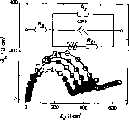
其中电路元件的排列如图1所示。The arrangement of the circuit components is shown in Figure 1.
[12]在一些实施方案中,核酸可以是脱氧核糖核酸(DNA),并且核酸双链可以是双螺旋。在一些实施例中,核酸可包括含有金属的核酸双链M-DNA,其具有第一链核酸和第二链核酸,第一及第二核酸链包括多个通过主链共价连接的含氮芳香族碱基,第一核酸链的含氮芳香族碱基通过氢键与第二核酸链的含氮芳香族碱基结合,第一及第二核酸链上的含氮芳香族碱基形成氢键结合的碱基对,其沿着导电性的含金属的核酸双链的长度方向堆叠排列,氢键结合的碱基对包括一个内部螯合(interchelated)的二价金属阳离子,其与芳香族的含氮芳香族碱基之一中的氮原子形成配位键。[12] In some embodiments, the nucleic acid may be deoxyribonucleic acid (DNA), and the nucleic acid duplex may be a double helix. In some embodiments, the nucleic acid may comprise a metal-containing nucleic acid double-stranded M-DNA having a first-strand nucleic acid and a second-strand nucleic acid, the first and second nucleic acid strands comprising a plurality of nitrogen-containing Aromatic bases, the nitrogen-containing aromatic bases of the first nucleic acid chain combine with the nitrogen-containing aromatic bases of the second nucleic acid chain through hydrogen bonds, and the nitrogen-containing aromatic bases on the first and second nucleic acid chains form hydrogen Bonded base pairs that are stacked along the length of a conductive metal-containing nucleic acid duplex, hydrogen bonded base pairs that include an interchelated divalent metal cation that interacts with an aromatic The nitrogen atom in one of the nitrogen-containing aromatic bases forms a coordination bond.
[13]本发明可涉及将第一核酸双链的电路性能信息与第二核酸双链的电路性能信息相比较。例如,第一核酸双链可以是B-DNA,第二核酸双链可以是含有金属的核酸双链M-DNA。[13] The present invention may involve comparing circuit performance information of a first nucleic acid duplex with circuit performance information of a second nucleic acid duplex. For example, the first nucleic acid duplex may be B-DNA, and the second nucleic acid duplex may be metal-containing nucleic acid duplex M-DNA.
[14]例如,电化学电路可包括电解质水溶液(aqueous electrolyte),并且核酸可以限制在并且溶剂化于电解质水溶液中。在该水溶液中可提供氧化还原探针。[14] For example, an electrochemical circuit may include an aqueous electrolyte, and the nucleic acid may be confined and solvated in the aqueous electrolyte. A redox probe can be provided in the aqueous solution.
附图简述Brief description of the drawings
[15]图1图示了B-DNA及M-DNA的等效电路模式。虚线框中的电路为标准Randles电路。Rs:溶液阻抗;Rx:通过DNA的电阻;Rct:电荷转移阻抗;CPE:恒定相元件,W:Warburg阻抗。[15] Figure 1 illustrates the equivalent circuit models of B-DNA and M-DNA. The circuit in the dotted box is a standard Randles circuit.Rs : solution impedance;Rx : resistance through DNA;Rct : charge transfer impedance; CPE: constant phase element, W: Warburg impedance.
[16]图2是金电极表面上的天然DNA(B-DNA)及金属DNA(M-DNA)的模式图。如图所示,Zn2+离子可被认为是与M-DNA的外侧结合以及插入到DNA螺旋中。[16] FIG. 2 is a schematic diagram of natural DNA (B-DNA) and metal DNA (M-DNA) on the surface of a gold electrode. As shown, Zn2+ ions can be considered to bind to the outside of M-DNA and insert into the DNA helix.
[17]图3显示了(a)裸露的金及(b)装配于金电极上的20个碱基对的双链B-DNA在4mM K3[Fe(CN)6]/K4[Fe(CN)6](1∶1)、20mM NaClO4及20mM Tris-ClO4缓冲液(pH 8.6)中的伏安图(voltammogram),扫描速率为50mV/s。[17] Figure 3 shows (a) bare gold and (b) double-stranded B-DNA of 20 base pairs assembled on a gold electrode at 4 mM K3 [Fe(CN)6 ]/K4 [Fe (CN)6 ] (1:1), 20mM NaClO4 and 20mM Tris-ClO4 buffer (pH 8.6) in the voltammogram (voltammogram), the scan rate is 50mV/s.
[18]图4显示了(a)裸露的金、(b)装配于金电极上的20个碱基对的双链B-DNA以及(c)装配于金电极上的20个碱基对的双链M-DNA的XPS光谱。[18] Figure 4 shows (a) bare gold, (b) 20 bp double-stranded B-DNA assembled on a gold electrode, and (c) 20 bp double-stranded B-DNA assembled on a gold electrode. XPS spectrum of double-stranded M-DNA.
[19]图5显示了以4mM Fe(CN)63-/4-(1∶1)混合物作为氧化还原探针,在20mM Tris-ClO4及20mM NaClO4溶液中,以Ag/AgCl为参比施加0.250V电压的Nyquist图(Zim vs Zre)。在所有情况下,在含有0.4mM Zn2+、pH 7.0(■)或者0.4mM Mg2+、pH8.6(□)时,测得的数据点均显示为0,计算的与Randles电路-----或者修正的Randles电路——的拟合曲线为:(A)裸露金电极,(B)装配于金电极上的20个碱基对的双链B-DNA,(C)装配于金电极上的20个碱基对的双链M-DNA,以及(D)装配于金电极上的20个碱基对的双链B-DNA。[19] Figure 5 shows that 4mM Fe(CN)63-/4- (1:1) mixture was used as redox probe in 20mM Tris-ClO4 and 20mM NaClO4 solution, with Ag/AgCl as reference Nyquist plot (Zim vs Zre ) for an applied voltage of 0.250 V. In all cases, the data points measured at 0.4 mM Zn2+ , pH 7.0 (■) or 0.4 mM Mg2+ , pH 8.6 (□),
[20]图6显示了不存在氧化还原探针时,(A)装配于金电极表面上的20个碱基对的双链B-DNA(□),以及(B)装配于金电极表面上的20个碱基对的双链M-DNA(■)的Nyquist图。其中将实验数据与所示等效电路进行了拟合。[20] Figure 6 shows (A) 20 base pair double-stranded B-DNA (□) assembled on the gold electrode surface in the absence of the redox probe, and (B) assembled on the gold electrode surface Nyquist plot of 20 base pair double-stranded M-DNA (■). where the experimental data are fitted to the equivalent circuit shown.
[21]图7显示了以Fe(CN)63-/4-作为氧化还原探针,15个碱基对的双链单层为B-DNA(□)或者M-DNA(■)、20个碱基对的双链单层为B-DNA(○)或者M-DNA(●)、以及30个碱基对的双链单层为B-DNA(△)或者M-DNA(▲)的Nyquist图。如文中所述,将实验数据与修正的等效电路进行了拟合。[21] Figure 7 shows that with Fe(CN)63-/4- as the redox probe, the double-stranded monolayer of 15 base pairs is B-DNA (□) or M-DNA (■), 20 The double-stranded monolayer of 30 base pairs is B-DNA (○) or M-DNA (●), and the double-stranded monolayer of 30 base pairs is B-DNA (△) or M-DNA (▲) Nyquist diagram. The experimental data were fitted with a revised equivalent circuit as described in the text.
[22]图8显示了B-DNA和M-DNA修饰的金电极在5mM Ru(NH3)3+/2+,20mM Tris-ClO4缓冲液(pH8.6)中,以Ag/AgCl为参比施加电势为-0.10V时阻抗测定的Nyquist图。[22] Figure 8 shows B-DNA and M-DNA modified gold electrodes in 5mM Ru(NH3 )3+/2+ , 20mM Tris-ClO4 buffer (pH8.6), with Ag/AgCl as Nyquist plot for impedance measurements at a reference applied potential of -0.10V.
[23]图9是显示如实施例2中所探讨的附着于电极表面上的双链中DNA错配的示意图。[23] FIG. 9 is a schematic diagram showing DNA mismatches in the duplex attached to the electrode surface as discussed in Example 2.
[24]图10是显示如实施例2中所探讨的在B-DNA与M-DNA状态下,正确配对的双链以及中间有错配的双链的阻抗谱的图表。[24] FIG. 10 is a graph showing the impedance spectra of a correctly paired double strand and a mismatched double strand in the state of B-DNA and M-DNA as discussed in Example 2.
[25]图11显示了分别将20个碱基对的互补B-DNA(○)、中间存在错配的B-DNA(□)、互补M-DNA(●)以及中间存在错配的M-DNA(■)装配于金电极上,以4mM[Fe(CN)6]3-/4-混合物(1∶1)作为氧化还原探针,在20mM Tris-ClO4和20mM NaClO4溶液中的Nyquist图(-Zim vs Zre),以Ag/AgCl为参比施加电势为250mV,[ZnII]=0.4,pH 8.6。在所有情况下,测得的数据点均以符号显示,计算的与等效电路的拟合曲线显示为实线。注意:实验数据与等效电路进行了拟合。Rs:溶液电阻,Rx:单层小孔/缺损电阻,RCT:电荷转移电阻,CPE:恒定相元件,W:Warburg阻抗。[25] Figure 11 shows that 20 base pairs of complementary B-DNA (○), B-DNA with a mismatch in the middle (□), complementary M-DNA (●) and M-DNA with a mismatch in the middle DNA (■) was assembled on a gold electrode, with 4mM [Fe(CN)6 ]3-/4- mixture (1:1) as redox probe, Nyquist in 20mM Tris-ClO4 and 20mM NaClO4 solution Figure (-Zim vs Zre ), using Ag/AgCl as a reference, the applied potential is 250mV, [ZnII ]=0.4, pH 8.6. In all cases, the measured data points are shown as symbols and the calculated fit curves to the equivalent circuits are shown as solid lines. NOTE: Experimental data were fitted to equivalent circuits. Rs: solution resistance, Rx: monolayer pore/defect resistance,RCT : charge transfer resistance, CPE: constant phase element, W: Warburg impedance.
[26]图12中,a)为杂交-解链过程。i)水浸渍:60℃ EtOH(60∶40)温浴10分钟,然后室温下用20mM Tris-ClO4缓冲液漂洗10分钟。ii)将靶ss-DNA加入到SSC缓冲液中,37℃下10分钟,然后室温3小时使其形成双链。b)为按1∶2构成的全部杂交的“理想”单层(○)、杂交步骤后的1倍ss-DNA单层(□)以及重新杂交步骤之后按1∶2构成的复性的ds-DNA膜(●)的Nyquist图。与公认的指示不完全杂交结果的单层不均一性的理想的“1∶2”膜的阻抗谱相比,复性DNA膜的阻抗谱存在差别。[26] In Figure 12, a) is the hybridization-melting process. i) Water immersion: 60° C. EtOH (60:40) incubation for 10 minutes, then rinsed with 20 mM Tris-ClO4 buffer for 10 minutes at room temperature. ii) The target ss-DNA was added to the SSC buffer, at 37°C for 10 minutes, and then at room temperature for 3 hours to form a double strand. b) is an "ideal" monolayer of all hybridizations constituted 1:2 (○), a 1x ss-DNA monolayer after the hybridization step (□), and an annealed ds constituted 1:2 after the rehybridization step - Nyquist plot of DNA membrane (•). There is a difference in the impedance spectrum of renatured DNA membranes compared to the accepted impedance spectrum of the ideal "1:2" membrane indicative of monolayer heterogeneity that results in incomplete hybridization.
[27]图13显示了分别将复性的20个碱基对的互补B-DNA(○)、中间存在错配的B-DNA(□)、互补M-DNA(●)以及中间存在错配的M-DNA(■)装配于金电极上,以4mM Fe(CN)63-/4-混合物(1∶1)作为氧化还原探针,在20mM Tris-ClO4和20mM NaClO4溶液中的Nyquist图(-Zim vs Zre)。以Ag/AgCl为参比,施加的电势为250mV,[ZnII]=0.4mM;pH 8.6。在所有情况下,测得的数据点以符号显示,计算的等效电路的拟和曲线以实线表示。[27] Figure 13 shows the annealed 20 base pair complementary B-DNA (○), B-DNA with a mismatch in the middle (□), complementary M-DNA (●) and a mismatch in the middle The M-DNA(■) was assembled on a gold electrode, with 4mM Fe(CN)63-/4- mixture (1:1) as redox probe, in 20mM Tris-ClO4 and 20mM NaClO4 solution Nyquist plot (-Zim vs Zre ). With Ag/AgCl as reference, the applied potential is 250 mV, [ZnII ]=0.4 mM; pH 8.6. In all cases, the measured data points are shown as symbols and the fitted curves of the calculated equivalent circuits are shown as solid lines.
[28]图14显示了通过监测B-DNA与M-DNA之间RCT中的变化作为目标单链DNA浓度的函数来测定检测范围。互补DNA链以(□)表示,中间错配的DNA链以(○)表示。误差范围(error bar)最少由5个以上电极确定。[28] Figure 14 shows the determination of detection range by monitoring the change in RCT between B-DNA and M-DNA as a function of target single-stranded DNA concentration. Complementary DNA strands are indicated by (□), and mismatched DNA strands in the middle are indicated by (◯). The error range (error bar) is determined by at least 5 electrodes.
发明详述Detailed description of the invention
[29]在本发明的一个方面,阻抗谱用于探测在金电极表面上自组装单层的B-DNA及M-DNA的电学特性。[29] In one aspect of the invention, impedance spectroscopy was used to probe the electrical properties of self-assembled monolayers of B-DNA and M-DNA on the surface of gold electrodes.
[30]通过背景技术介绍,图1解释了模拟受到通过自组装单层进行非均一性电子传递的电极阻抗的电路模型,其通常在由Randles(Randles,1947)建立的模型的基础上进行描述。等效电路(图1的虚框中)由电阻元件和电容元件组成。Rs为溶液的电阻,Rct为电荷转移阻抗,C为双层电容(double-layer capacitance),W为由于质子传递到电极所产生的Warburg阻抗。通常,Randles电路提供了链烷硫醇(alkanethiol)单层行为的很好的模型。然而,如下观察结果引起了相当大的兴趣:即由Randles电路不足以描述含有共轭π键系统的HMB(4’-羟基-4-巯基联苯)单层,但如果增加一个另外的并联电阻(图1中的Rx),就可以很好的对其谱图进行拟合(Janek等,1998)。[30] By way of background, Figure 1 illustrates the circuit model for simulating the impedance of electrodes subjected to inhomogeneous electron transport through self-assembled monolayers, which is usually described on the basis of the model developed by Randles (Randles, 1947) . The equivalent circuit (in the dashed box in Figure 1) consists of resistive and capacitive elements. Rs is the resistance of the solution, Rct is the charge transfer impedance, C is the double-layer capacitance (double-layer capacitance), and W is the Warburg impedance due to the transfer of protons to the electrodes. In general, Randles circuits provide a good model of the behavior of alkanethiol monolayers. However, considerable interest has been generated by the observation that HMB (4'-hydroxy-4-mercaptobiphenyl) monolayers containing conjugated π-bond systems are not adequately described by the Randles circuit, but if an additional parallel resistance (Rx in Figure 1), it can fit its spectrum very well (Janek et al., 1998).
[31]如图2中所示,当加入Zn2+形成M-DNA时,离子插入DNA螺旋中并与螺旋外侧的磷酸酯主链结合。B-DNA转换成M-DNA导致观察到的15、20及30碱基对双链的阻抗谱(impedance spectra)的特征发生变化。研究发现,包括并联电阻Rx的修正Randles电路可用于给出与实验数据较好的拟合(图1)。在这些情况下,M-DNA表现为使Rx和Rct都减小,并使通过单层的电子传递增加。[31] As shown in Figure 2, when Zn2+ is added to form M-DNA, the ions intercalate into the DNA helix and bind to the phosphate backbone on the outside of the helix. The conversion of B-DNA to M-DNA results in a change in the characteristics observed in the impedance spectra of the 15, 20 and 30 base pair duplexes. It was found that a modified Randles circuit including the shunt resistor Rx could be used to give a good fit to the experimental data (Figure 1). In these cases, M-DNA appears to decrease bothRx andRct and increase electron transport through the monolayer.
[32]本发明的多个方面涉及M-DNA,即一种含金属的导电寡核苷酸双链。在本发明的一些可供选择的方面,含金属的导电寡核苷酸双链可包括第一核酸链和第二核酸链,第一及第二核酸链分别包括多个由主链共价连接的含氮芳香碱基。第一核酸链的含氮芳香碱基可通过氢键与第二核酸链的含氮芳香碱基结合。第一核酸链与第二核酸链的含氮芳香碱基可形成沿着含金属导电寡核苷酸双链长度方向堆叠排列的氢键结合的碱基对。氢键结合的碱基对可包括内部螯合(interchelated)的金属阳离子,其与含氮芳香碱基之一中的氮原子配位结合。[32] Aspects of the invention relate to M-DNA, a metal-containing conductive oligonucleotide duplex. In some alternative aspects of the invention, the metal-containing conductive oligonucleotide duplex may comprise a first nucleic acid strand and a second nucleic acid strand, each comprising a plurality of nitrogenous aromatic bases. The nitrogen-containing aromatic base of the first nucleic acid strand can combine with the nitrogen-containing aromatic base of the second nucleic acid strand through hydrogen bonding. The nitrogen-containing aromatic bases of the first nucleic acid strand and the second nucleic acid strand can form hydrogen-bonded base pairs stacked along the length direction of the metal-containing conductive oligonucleotide double strand. A hydrogen-bonded base pair may include an interchelated metal cation coordinating to a nitrogen atom in one of the nitrogen-containing aromatic bases.
[33]内部螯合的金属阳离子可包括内部螯合的二价金属阳离子,二价金属阳离子可选自由锌、钴及镍组成的组。作为选择,金属阳离子可选自由下列阳离子组成的组:Li,Be,Na,Mg,Al,K,Ca,Sc,Ti,V,Cr,Mn,Fe,Co,Ni,Cu,Zn,Ga,Ge,As,Rb,Sr,Y,Zr,Nb,Mo,Tc,Ru,Rh,Pd,Ag,Cd,In,Sn,Sb,Cs,Ba,La,Ce,Pr,Nd,Pm,Sm,Eu,Gd,Tb,Dy,Ho,Er,Tm,Yb,Lu,Hf,Ta,W,Re,Os,Ir,Pt,Au,Hg,TI,Pb,Bi,Po,Fr,Ra,Ac,Th,Pa,U,Np以及Pu。[33] The internally chelated metal cations may include internally chelated divalent metal cations selected from the group consisting of zinc, cobalt, and nickel. Alternatively, the metal cation may be selected from the group consisting of the following cations: Li, Be, Na, Mg, Al, K, Ca, Sc, Ti, V, Cr, Mn, Fe, Co, Ni, Cu, Zn, Ga, Ge, As, Rb, Sr, Y, Zr, Nb, Mo, Tc, Ru, Rh, Pd, Ag, Cd, In, Sn, Sb, Cs, Ba, La, Ce, Pr, Nd, Pm, Sm, Eu, Gd, Tb, Dy, Ho, Er, Tm, Yb, Lu, Hf, Ta, W, Re, Os, Ir, Pt, Au, Hg, TI, Pb, Bi, Po, Fr, Ra, Ac, Th, Pa, U, Np and Pu.
[34]第一及第二核酸链可包括脱氧核糖核酸,含氮芳香族碱基可选自由腺嘌呤、胸腺嘧啶、鸟嘌呤及胞嘧啶组成的组。二价金属阳离子可取代含氮芳香族碱基的亚胺质子,该含氮芳香碱基可选自由胸腺嘧啶及鸟嘌呤组成的组。含氮芳香碱基中的至少一个可包括具有N3氮原子的胸腺嘧啶,并且二价金属阳离子可与N3氮原子配位结合。作为选择,含氮芳香碱基中的至少一个可包括具有N1氮原子的鸟嘌呤,并且二价金属阳离子可与该N1氮原子配位结合。[34] The first and second nucleic acid strands may include deoxyribonucleic acid, and the nitrogen-containing aromatic base may be selected from the group consisting of adenine, thymine, guanine, and cytosine. The divalent metal cation can replace the imine proton of the nitrogen-containing aromatic base, and the nitrogen-containing aromatic base can be selected from the group consisting of thymine and guanine. At least one of the nitrogen-containing aromatic bases may include thymine having an N3 nitrogen atom, and a divalent metal cation may coordinately bind to the N3 nitrogen atom. Alternatively, at least one of the nitrogen-containing aromatic bases may include guanine having a N1 nitrogen atom, and a divalent metal cation may be coordinated to the N1 nitrogen atom.
[35]在本发明的各个方面,如在下列实施例中所公开的,DNA单层可装配于金电极表面,并由循环伏安法(CV)或者X-射线光电子光谱(XPS)来估算。如实施例中所示,CV光谱可提供对Fe(CN)63-/4-封闭良好的密集组装的单层的很好证据。通过XPS,膜的厚度可基于Au 4f信号的指数衰减来估算,在实施例中算出为45_(Pressprich等,1989)。20个碱基对的双链可预期具有大约70_的长度,所以作为例子的测得的45_的厚度与DNA以约50°角从电极表面突出相一致。通常,与也可通过碱基连接的单链DNA不同的是,双链DNA通过连接物(linker)进行连接(Heme及Tarlov,1997)。在实施例中,S2p的峰值162.4eV与以前报道的指示DNA通过S-Au键与电极表面相互作用的烷硫醇(alkanethiol)非常一致(Ishida等,1999)。[35] In various aspects of the invention, as disclosed in the following examples, DNA monolayers can be assembled on the surface of gold electrodes and evaluated by cyclic voltammetry (CV) or X-ray photoelectron spectroscopy (XPS). . As shown in the examples, the CV spectra can provide good evidence of a densely packed monolayer that is well closed3-/4- fe(CN)6 . By XPS, the thickness of the film can be estimated based on the exponential decay of the Au 4f signal, calculated to be 45 Å in the examples (Pressprich et al., 1989). A duplex of 20 base pairs would be expected to have a length of about 70 mm, so the measured thickness of 45 mm as an example is consistent with DNA protruding from the electrode surface at an angle of about 50°. Typically, double-stranded DNA is joined by a linker, unlike single-stranded DNA, which can also be joined by bases (Heme and Tarlov, 1997). In the Examples, the peak of S2p at 162.4 eV is in good agreement with previously reported alkanethiols indicating that DNA interacts with electrode surfaces through S-Au bonds (Ishida et al., 1999).
[36]AC阻抗波谱法是已知的探测并模拟电极的界面特征的方法(Bard与Faulkner,2001)。例如,数据可以Nyquist图(Zim vs Zre)的方式显示,在图可以很容易观察并解释其特征变化。复阻抗可以分别主要来自于测定的电化学系统的电阻与电容的实际值Zre(ω)与假想值Zim(ω)分量之和来显示。如这里所示的例子,裸露电极的Nyquist图为Zre轴上方的一个半圆形区域与随后的一段直线组成。推定在频率较高下测定的半圆部分与直接的电子传递限制过程相应,相反,推定观察到的在频率较低下测定的直线部分则表示扩散控制的电子传递过程。与裸露的金属电极相比,用有机分子层对金属表面进行修饰可减小双层之间的电容,并降低了界面的电子传递率(Finklea等,1993;Kharitonov等,2000)。[36] AC impedance spectroscopy is a known method to probe and model the interface characteristics of electrodes (Bard and Faulkner, 2001). For example, data can be displayed in the form of a Nyquist plot (Zim vs Zre ), where characteristic variations can be easily observed and interpreted. The complex impedance can be displayed as the sum of the components of the actual value Zre (ω) and the imaginary value Zim (ω) mainly from the resistance and capacitance of the measured electrochemical system, respectively. As in the example shown here, the Nyquist plot for a bare electrode consists of a semicircular region above the Zre axis followed by a straight line. The semicircular portion measured at higher frequencies is putatively corresponding to a direct electron transport limited process, whereas the straight line portion observed at lower frequency is putatively indicative of a diffusion controlled electron transport process. Modification of metal surfaces with layers of organic molecules reduces the capacitance between bilayers and lowers the electron transport rate at the interface compared to bare metal electrodes (Finklea et al., 1993; Kharitonov et al., 2000).
[37]在一些实施例中,数据分析可需要使用由电学元件构成的等效电路对电极动力学进行模拟。对于许多单层来说,通常可接受的等效电路是基于如图1中所示的Randles模型。然而,为了获得与这里所公开的例子的数据的很好拟合,在等效电路中增加了并联的界面电阻Rx,其名义上与通过DNA的电子传递相应。例如,可通过在不存在氧化还原活性探针Fe(CN)63-/4-的情况下测定的阻抗来提供并联的界面电阻证据(见图6)。[37] In some embodiments, data analysis may require simulation of electrode dynamics using equivalent circuits made up of electrical components. A generally acceptable equivalent circuit for many monolayers is based on the Randles model as shown in Figure 1. However, to obtain a good fit with the data of the examples disclosed here, a parallel interfacial resistanceRx , which nominally corresponds to electron transport through DNA, is added to the equivalent circuit. Evidence for interfacial resistance in parallel can be provided, for example, by impedance measurements in the absence of the redox-active probe Fe(CN)63-/4- (see Figure 6).
[38]对于许多不带电荷的单层来说,不同的氧化还原探针可给出定量上类似的结果,这大概是由于探针与单层之间的相互作用为非静电型的缘故(Boubour及Lennox,2000;Finklea,1996;Finklea等,1993)。然而,DNA带负电荷,因此,Ru(NH3)63+/2+等带正电荷的探针可进入单层,而Fe(CN)63-/4-等带负电荷的探针则不能进入单层。例如,这些差别可反映在这里的实施例所显示的结果中,其中带有Ru(NH3)63+/2+的Rct大约为1KΩ(图8),与裸露的电极类似,而带有Fe(CN)63-/4以及B-DNA的相应值接近20KΩ。因此,由于电荷转移可以基本上绕过单层,Ru(NH3)63+/2+不适于用作DNA的探针。[38] For many uncharged monolayers, different redox probes give quantitatively similar results, presumably because the interaction between the probes and the monolayer is non-electrostatic ( Boubour and Lennox, 2000; Finklea, 1996; Finklea et al., 1993). However, DNA is negatively charged, so positively charged probes such as Ru(NH3 )63+/2+ can enter the monolayer, while negatively charged probes such as Fe(CN)63-/4- You cannot enter the single layer. These differences are reflected, for example, in the results shown in the examples herein, where the Rct with Ru(NH3 )63+/2+ is about 1 KΩ (Fig. 8), similar to the bare electrode, while with The corresponding value of Fe(CN)63-/4 and B-DNA is close to 20KΩ. Therefore, Ru(NH3 )63+/2+ is not suitable as a probe for DNA since the charge transfer can essentially bypass the monolayer.
[39]这里实施例所公开的结果表明,在某些情况下,由于M-DNA的Rct及Rx都比较小,其可成为比B-DNA更好的导体。在这些例子中,随着长度的增加,B-DNA与M-DNA的Rct之差趋于增大,而随着DNA双链的长度增加,Rx之差趋于减小。在这些例子中,DNA不直接与电极相连,使Rct及Rx都具备了串联的条件,来进行来自通过连接物与电极连接的DNA的电子传递。在作为选择的实施方案中,DNA可直接与电极相连,也可通过可改变长度的连接物与电极相连,来解决连接物的影响问题。在一些实施方案中,B-DNA与M-DNA的相互转换可形成系统,通过改变金属离子或pH,其中的Rx及Rct都可受到调节。[39] The results disclosed in the examples here show that, in some cases, M-DNA can be a better conductor than B-DNA due to its smaller Rct and Rx . In these examples, as the length increases, the difference in Rct between B-DNA and M-DNA tends to increase, while the difference in Rx tends to decrease as the length of the DNA double strand increases. In these examples, the DNA is not directly connected to the electrode, so that both Rct and Rx have the condition of being connected in series to carry out the electron transfer from the DNA connected to the electrode through the linker. In an alternative embodiment, the DNA can be directly connected to the electrode, or can be connected to the electrode through a linker whose length can be changed, so as to solve the problem of the influence of the linker. In some embodiments, the interconversion of B-DNA and M-DNA can form a system in which both Rx and Rct can be adjusted by changing metal ions or pH.
实施例1Example 1
[40]在本发明的一些方面的实施例中,下面将更为详细地描述装配于金电极上具有15、20及30个碱基对的硫醇标记的DNA双链单层。通过以Fe(CN)63-/4-作为氧化还原探针的电化学阻抗谱学来研究电子传递。以Nyquist图形式表示的谱图由包括增加了用于测定通过DNA的电阻的并联元件Rx的修正Randles电路来进行分析。天然B-DNA的电荷转移电阻Rx及Rct都随着长度的增加而增大。通过在pH 8.6下添加Zn2+形成M-DNA,并引起在Nyquist图中的特征变化,而在添加Mg2+或者在pH 7.0下则观察不到这些变化。M-DNA的Rx及Rct也都随着双链长度的增加而增大,但与B-DNA相比其增加的幅度都明显较小。因此,某些金属离子可调节DNA单层的电化学特性,而且通过金属DNA膜的电子传递也比天然的DNA膜要快。[40] In an example of some aspects of the invention, the assembly of thiol-labeled double-stranded DNA monolayers of 15, 20, and 30 base pairs on gold electrodes is described in more detail below. Electron transport was studied by electrochemical impedance spectroscopy with Fe(CN)63-/4- as redox probe. Spectra, represented as Nyquist plots, were analyzed by a modified Randles circuit including the addition of a parallel elementRx for measuring the resistance through the DNA. The charge transfer resistances Rx and Rct of native B-DNA both increase with the increase of length. M-DNA was formed by the addition of Zn2+ at pH 8.6 and caused characteristic changes in the Nyquist plot, whereas these changes were not observed with the addition of Mg2+ or at pH 7.0. Rx and Rct of M-DNA also increased with the increase of double-strand length, but compared with B-DNA, the magnitude of the increase was significantly smaller. Thus, certain metal ions can modulate the electrochemical properties of DNA monolayers, and electron transport through metallic DNA membranes is faster than native DNA membranes.
化学试剂chemical reagent
[41]六铁氰化钾、六亚铁氰化钾、六胺基钌(III)、氯化六胺基钌(II)氯化物购自Aldrich,为ACS试剂级;Zn(ClO4)2、Mg(ClO4)2以及Tris-ClO4购自Fluka Co;标准缓冲液为pH 8.7或7.0的20mM Tris-ClO4,其它化学试剂为分析级,所有溶液均采用Millipore过滤水配制。[41] Potassium hexaferrocyanide, potassium hexaferrocyanide, hexaamine ruthenium(III), and hexaamine ruthenium(II) chloride were purchased from Aldrich, which were ACS reagent grade; Zn(ClO4 )2 , Mg(ClO4 )2 and Tris-ClO4 were purchased from Fluka Co; the standard buffer solution was 20mM Tris-ClO4 at pH 8.7 or 7.0, other chemical reagents were of analytical grade, and all solutions were prepared with Millipore filtered water.
DNAdna
[42]探针DNA采用标准的DNA合成方法在Saskatoon植物生物技术研究所(Plant Biotechnology Institute,Saskatoon)合成并纯化。寡聚核苷酸碱基序列为:15-mer DNA,5′-AAC TAC TGG GCCATC-(CH2)3-S-S-(CH2)3-OH-3′,靶互补序列为5′-GAT GGC CCA GTAGTT-3′;20-mer DNA,5′-AAC TAC TGG GCC ATC GTGAC-(CH2)3-S-S-(CH2)3-OH-3′,靶互补序列为5′-GTC ACG ATG GCCCAG TAG TT-3′,30mer DNA,5′-GTG GCT AAC TAC GCA TTC CACGAC CAA ATG-(CH2)3-S-S-(CH2)3-OH-3′,靶互补序列为5′-CAT TTGGTC GTG GAA TGC GTA GTT AGC CAC-3′。[42] Probe DNA was synthesized and purified at the Plant Biotechnology Institute, Saskatoon, using standard DNA synthesis methods. The oligonucleotide base sequence is: 15-mer DNA, 5′-AAC TAC TGG GCCATC-(CH2 )3 -SS-(CH2 )3 -OH-3′, the target complementary sequence is 5′-GAT GGC CCA GTAGTT-3′; 20-mer DNA, 5′-AAC TAC TGG GCC ATC GTGAC-(CH2 )3 -SS-(CH2 )3 -OH-3′, the target complementary sequence is 5′-GTC ACG ATG GCCCAG TAG TT-3′, 30mer DNA, 5′-GTG GCT AAC TAC GCA TTC CACGAC CAA ATG-(CH2 )3 -SS-(CH2 )3 -OH-3′, the target complementary sequence is 5′- CAT TTGGTC GTG GAA TGC GTA GTT AGC CAC-3'.
电极制备Electrode preparation
[43]金盘电极(几何表面积为0.02cm2)与Ag/AgCl参比电极购自Bioanalytical Systems。使用前,用0.05μm铝土浆液仔细抛光,然后在0.01M KOH溶液中清洗几分钟,接着在Millipore H2O中清洗两次。电极用波谱法仔细检查,以确保不存在明显缺陷。最后,在下述池中进行电化学处理,在0.5M H2SO4溶液中以Ag/AgCl为参比,电压为-0.1到+1.25V进行循环扫描,直到在以Ag/AgCl为参比,电压为1.1V时获得稳定的金氧化峰(Finklea,1996)。[43] A gold disk electrode (geometric surface area of 0.02 cm2 ) and an Ag/AgCl reference electrode were purchased from Bioanalytical Systems. Before use, it was carefully polished with a 0.05 μm alumina slurry, followed by a few minutes of cleaning in 0.01 M KOH solution, followed by two cleanings in MilliporeH2O . Electrodes are carefully inspected spectroscopically to ensure that there are no obvious defects. Finally, electrochemical treatment was carried out in the following cell, using Ag/AgCl as reference in 0.5M H2 SO4 solution, and the voltage was -0.1 to +1.25V to carry out cyclic scanning until Ag/AgCl was used as reference, the voltage A stable gold oxidation peak was obtained at 1.1V (Finklea, 1996).
DNA修饰金电极的制备Preparation of DNA-modified gold electrodes
[44]将10nmol二硫化物标记的DNA链加入到溶解于含有20mMNaClO4的50μl 20mM、pH 8.7的Tris-ClO4缓冲液的10nmol互补链溶液中,20℃放置2小时制备DNA双链。双链DNA终浓度约为100μM。新制备的金电极与DNA双链在密封容器中温育5天。将电极用缓冲液(20mM Tris-ClO4与20mM NaClO4)彻底漂洗并将其固定于电化学池中。加入0.4mM ZnClO4,pH 8.7下放置2小时,将B-DNA转化成M-DNA。[44] Add 10 nmol of disulfide-labeled DNA strand to 10 nmol complementary strand solution dissolved in 50 μl of 20 mM Tris-ClO4 buffer solution containing 20 mM NaClO4 , pH 8.7, and place at 20°C for 2 hours to prepare DNA double strands. The final concentration of double-stranded DNA is approximately 100 μM. Freshly prepared gold electrodes were incubated with DNA double strands in a sealed container for 5 days. The electrodes were rinsed thoroughly with buffer (20 mM Tris-ClO4 and 20 mM NaClO4 ) and fixed in the electrochemical cell. 0.4 mM ZnClO4 was added and left at pH 8.7 for 2 hours to convert B-DNA to M-DNA.
X射线光电子波谱法X-ray photoelectron spectroscopy
[45]采用带有Al-Ka放射源(1486.6eV)的Leybold MAX200光电子分光计来收集光发射光谱。测定过程中分析室中的基本压强保持低于10-9mbar,起飞角(take-off angle)为60°。常规仪器校准标准为Au 4f7/2峰(结合能为84.0eV)。[45] employed a Leybold MAX200 photoelectron spectrometer with an Al-Ka radiation source (1486.6 eV) to collect light emission spectra. The base pressure in the analysis chamber was kept below 10−9 mbar during the measurement and the take-off angle was 60°. The conventional instrument calibration standard is the Au 4f7/2 peak (binding energy 84.0 eV).
电化学electrochemical
[46]采用常用的三电极池。所有实验均在室温下进行。池用接地Faraday隔离罩封闭。采用含有电解液的Luggin毛细管使参比电极始终与池分离。为了避免Cl-离子从标准Ag/AgCl参比电极中泄露到测定系统中,采用盐桥参比电极。反电极为铂导线。采用由GPIB在PC上运行鲍尔程序组(Power Suite)(Princeton Applied Research)的与EG&G 283恒电位器/恒流器连接的1025频率响应分析仪(FRA)测定阻抗谱。在以Ag/AgCl为参比电势为250mV时测定阻抗,并叠加到调幅为±5mV的正弦电势上。用于阻抗测定的频率范围可以从100kHz到100mHz。裸露金电极、B-DNA及M-DNA修饰金电极的阻抗数据采用ZSimpWin软件(PrincetonApplied Research)进行分析。在所有阻抗谱中,符号表示未处理的实验数据,实线表示拟合曲线。[46] employed a commonly used three-electrode cell. All experiments were performed at room temperature. The pool is enclosed with a grounded Faraday enclosure. The reference electrode is always separated from the cell using a Luggin capillary containing the electrolyte. To avoid leakage of Cl- ions from the standard Ag/AgCl reference electrode into the measurement system, a salt bridge reference electrode is used. The counter electrode is a platinum wire. Impedance spectra were measured using a 1025 Frequency Response Analyzer (FRA) connected to an EG&G 283 potentiostat/galvanostat running Power Suite (Princeton Applied Research) on a PC via GPIB. Impedance was measured when Ag/AgCl was used as a reference potential of 250mV, and superimposed on a sinusoidal potential with an amplitude modulation of ±5mV. The frequency range for impedance measurements can be from 100kHz to 100mHz. The impedance data of bare gold electrodes, B-DNA and M-DNA modified gold electrodes were analyzed by ZSimpWin software (Princeton Applied Research). In all impedance spectra, symbols represent unprocessed experimental data and solid lines represent fitted curves.
结果result
单层的组装single layer assembly
[47]如在材料及方法中所述,将天然双链B-DNA组装于金电极表面。单层以4mM K3[Fe(CN)6]/K4[Fe(CN)6](1∶1)混合物作为氧化还原探针时的循环电压电流(cycle voltammetery)为特征。典型的扫描显示于图3中,裸露金电极显示了准可逆氧化还原循环的特征,峰值间距为158mV。对于装配了20个碱基对双链的电极来说,峰值电流下降超过95%,并且氧化与还原峰值的间距增大,表明电极表面上DNA的存在,而且溶液与表面之间的电子传递能力降低。[47] Native double-stranded B-DNA was assembled on the gold electrode surface as described in Materials and Methods. The monolayer was characterized by cycle voltammetry when a 4 mMK3 [Fe(CN)6 ]/K4 [Fe(CN)6 ] (1:1) mixture was used as a redox probe. A typical scan is shown in Fig. 3, with the bare gold electrode showing the signature of a quasi-reversible redox cycle with a peak separation of 158 mV. For electrodes assembled with 20 base pair duplexes, the peak current drops by more than 95%, and the separation between the oxidation and reduction peaks increases, indicating the presence of DNA on the electrode surface and the ability of electron transfer between the solution and the surface reduce.
[48]金电极表面还可用X光电子波谱法(XPS)进行分析。如图4所示,如所期望的修饰表面那样,当连接有DNA(B-DNA或M-DNA)时,Au4f的峰值密度下降(Kondo等,1998;Ishida等,1999)。在B-DNA与M-DNA光谱中S2p(162.4eV)、P2p(133eV)以及N1s(400eV)处的峰值很明显,但在裸露金电极的光谱中这些峰值则没有出现,这给出了二硫化物连接的DNA与表面发生结合的很好证据。特别有意义的是,观察到B-DNA与M-DNA的Nls及Ols(在pH 8.7下加入Zn2+之后)光谱是不同的。这与锌离子与DNA双螺旋的相互作用一致,尤其是与碱基对中的氮原子及氧原子相互作用相一致(Lee等,1993;Aich等,1999)。[48] The gold electrode surface can also be analyzed by X-ray photoelectron spectroscopy (XPS). As shown in Figure 4, the peak density of Au4f decreased when DNA (B-DNA or M-DNA) was attached, as expected for a modified surface (Kondo et al., 1998; Ishida et al., 1999). The peaks at S2p (162.4eV), P2p (133eV) and N1s (400eV) are obvious in the B-DNA and M-DNA spectra, but these peaks do not appear in the spectrum of the bare gold electrode, which gives There is good evidence that disulfide-linked DNA binds to surfaces. Of particular interest is the observation that the N1s and O1s (after addition of Zn2+ at pH 8.7) spectra are different for B-DNA and M-DNA. This is consistent with the interaction of zinc ions with the DNA double helix, especially with the nitrogen and oxygen atoms in the base pairs (Lee et al., 1993; Aich et al., 1999).
B-DNA的阻抗分光检测Impedance Spectroscopic Detection of B-DNA
[49]在存在4mM K3[Fe(CN)6]/K4[Fe(CN)6](1∶1)混合物作为氧化还原探针时进行阻抗测定。图5A显示了裸露金电极的Nyquist图,其在高频率下可以描述为原点附近的半圆形,随后为具有相同斜率的线性尾巴。其它电极可用相同曲线描述,并且数据可用图1中的Randles电路充分拟合。半圆的直径用电荷转移电阻Rct度量。对于20-mer的B-DNA来说(图5B),与裸露的金电极相比,由于传递到电极的电子减少,Rct显著增加。然而,低频区域不再呈线性,而且不能用简单的Randles电路充分拟合。当不存在氧化还原探针时,B-DNA(以及M-DNA,见下文)的阻抗测定证明了对简单绝缘体来说不期望出现的非线性行为(图6)。然而,图6的曲线可与由电容器与电阻并联所构成的简单电路拟合。这种结果表明增加的与Randles电路并联的额外界面电阻Rx的存在(图1)。如图5B所示,修正电路在低频区与高频区都能与实验数据拟合得非常好。[49] Impedance measurements were performed in the presence of 4 mMK3 [Fe(CN)6 ]/K4 [Fe(CN)6 ] (1:1) mixture as redox probe. Figure 5A shows the Nyquist plot of a bare gold electrode, which at high frequencies can be described as a semicircle near the origin, followed by a linear tail with the same slope. Other electrodes can be described by the same curve, and the data can be adequately fitted by the Randles circuit in Figure 1 . The diameter of the semicircle is measured by the charge transfer resistanceRct . For the 20-mer B-DNA (Fig. 5B), theRct increased significantly compared to the bare gold electrode due to the reduction of electrons delivered to the electrode. However, the low frequency region is no longer linear and cannot be adequately fitted by a simple Randles circuit. Impedance measurements of B-DNA (and M-DNA, see below) in the absence of redox probes demonstrated nonlinear behavior that is not expected for simple insulators (Fig. 6). However, the curve of Figure 6 can be fitted to a simple circuit consisting of a capacitor connected in parallel with a resistor. This result indicates the presence of an additional interfacial resistanceRx added in parallel with the Randles circuit (Fig. 1). As shown in FIG. 5B , the correction circuit can fit the experimental data very well in both the low frequency region and the high frequency region.
M-DNA的形成Formation of M-DNA
[50]在pH 8.7时将Zn2+加入到20-mer B-DNA修饰的金电极形成M-DNA时,阻抗谱的特征模式发生改变,在高频区与低频区Zim与Zre都减小(图5C)。对比实验表明,M-DNA的形成在两小时内完成。修正的Randles电路再次给出了与实验数据的很好拟合。在图5D中也显示了在存在和不存在Zn2+的pH 7.0的缓冲液中以及在存在Mg2+的pH 8.7的缓冲液中DNA修饰电极的阻抗谱。在这些条件下,不会形成M-DNA,观察到的阻抗谱也仅仅发生了很小的变化。Rs、Rx、Rct、C以及W的计算值在表1中列出。显然,在M-DNA形成时Rx与Rct显著下降,这既不是在加入Mg2+的情况下,也不是在pH 7.0时加入Zn2+的情况下发现。[50] When Zn2+ was added to the 20-mer B-DNA modified gold electrode to form M-DNA at pH 8.7, the characteristic mode of the impedance spectrum changed, and both Zim and Zre in the high frequency and low frequency regions decrease (Fig. 5C). Comparative experiments showed that the formation of M-DNA was completed within two hours. The modified Randles circuit again gave a good fit to the experimental data. Impedance spectra of the DNA-modified electrode in pH 7.0 buffer in the presence and absence of Zn2+ and in pH 8.7 buffer in the presence of Mg2+ are also shown in Fig. 5D. Under these conditions, no M-DNA was formed and only minor changes in the observed impedance spectrum occurred. The calculated values of Rs , Rx , Rct , C and W are listed in Table 1. Apparently, a significant decrease in Rx and Rct upon M-DNA formation was found neither with the addition of Mg2+ nor with the addition of Zn2+ at pH 7.0.
表1:pH及离子型aDNA
(a)除裸露金电极的数据是与未修正的Randles电路进行拟合外,其它数值均来自修正的Randles电路。(a) Except that the data of the bare gold electrode is fitted with the unmodified Randles circuit, other values are from the modified Randles circuit.
DNA序列长度DNA sequence length
[51]为了提供与所建议的模型中的元件有关的更多信息,采用15、20及30个碱基对的DNA双链来修饰金电极的表面。如图7所示,所有的阻抗谱均具有相同的特征形状,并能与修正的Randles电路拟合得非常好。Rs、Rx、Rct、C以及W的计算值在表2中列出。这些值具有两种截然不同的趋势。第一,随着B-DNA与M-DNA长度的增加,Rx与Rct都增大;第二,对于任何长度的双链M-DNA,其Rx与Rct都小于B-DNA的相应值。代表传递到电极的质子的Warburg阻抗W的可变性更大,但在所有情况下M-DNA双链均更高。如同期望的那样,溶液电阻Rs与双链的长度无关,而随着双链长度的增加,双层电容C减小。[51] In order to provide more information about the elements in the proposed model, DNA duplexes of 15, 20 and 30 base pairs were used to modify the surface of gold electrodes. As shown in Figure 7, all impedance spectra have the same characteristic shape and can be fitted very well with the modified Randles circuit. The calculated values of Rs , Rx , Rct , C and W are listed in Table 2. The values have two distinct trends. First, as the length of B-DNA and M-DNA increases, both Rx and Rct increase; second, for any length of double-stranded M-DNA, its Rx and Rct are smaller than those of B-DNA corresponding value. The Warburg impedance W, representing protons delivered to the electrodes, was more variable, but higher in all cases for the M-DNA duplex. As expected, the solution resistance Rs is independent of the duplex length, while the double-layer capacitance C decreases with the increase of the duplex length.
表2:不同长度的DNA序列
Ru(NH3)3+/2+氧化还原电极Ru(NH3 )3+/2+ redox electrode
[52]在该实施例中以Fe(CN)63-/4-作为氧化还原探针,其带负电荷,因此可被DNA的磷酸酯骨架所排斥。而相反,Ru(NH3)3+/2+则可期望能够穿过单层。采用Ru(NH3)3+/2+作为20个碱基对的B-DNA及M-DNA双链的氧化还原探针进行了阻抗谱测定(图8)。如在插页中所示,Rct相当小,因而B-DNA与M-DNA的光谱之差也非常小。[52] In this example Fe(CN)63-/4- was used as the redox probe, which is negatively charged and thus repelled by the phosphate backbone of DNA. In contrast, Ru(NH3 )3+/2+ can be expected to pass through the monolayer. Impedance spectroscopy was performed using Ru(NH3 )3+/2+ as a redox probe of 20 base pairs of B-DNA and M-DNA double strands ( FIG. 8 ). As shown in the insert, Rct is quite small and thus the spectral difference between B-DNA and M-DNA is also very small.
实施例2Example 2
[53]在前一实施例中,描述了B-DNA及M-DNA的自组装单层(SAMs)的阻抗谱,并显示了依赖于DNA长度和离子浓度的每个电阻(R)及电容(C)特征值。本实施例说明了DNA中的单碱基对错配也引起阻抗谱的界限分明的的变化,从而在一定条件下将其与正确配对的双链准确地区分开来。[53] In the previous example, the impedance spectra of self-assembled monolayers (SAMs) of B-DNA and M-DNA were described, and the resistance (R) and capacitance of each depending on DNA length and ion concentration were shown. (C) Eigenvalues. This example demonstrates that single base pair mismatches in DNA also cause well-defined changes in the impedance spectrum, thereby accurately distinguishing them from correctly paired duplexes under certain conditions.
[54]DNA序列及错配的位置在图9及表3中显示。[54] The DNA sequences and the positions of the mismatches are shown in Figure 9 and Table 3.
表3
[55]除非特别指出,在本实施例中所采用的方法与实施例1中相同。[55] Unless otherwise specified, the method used in this example was the same as that in Example 1.
[56]在B-DNA与M-DNA条件下,正确配对双链的阻抗谱和一个带有中间错配的双链显示于图10中。每个点表示在特定AC频率下测得的Zi及Zr值。例如,在0.1Hz和49Hz的点高亮显示,并可看出B-DNA与M-DNA的正确配对的双链与中间错配的双链的相应Zi及Zr值明显不同。[56] Impedance spectra of a correctly paired duplex and a duplex with an intermediate mismatch are shown in Fig. 10 under B-DNA and M-DNA conditions. Each point represents the measured Zi and Zr values at a specific AC frequency. For example, the points at 0.1 Hz and 49 Hz are highlighted, and it can be seen that the corresponding Zi and Zr values of the correctly paired duplex of B-DNA and M-DNA are significantly different from those of the mismatched duplex in the middle.
[57]从图10显示的阻抗谱中,可以精确计算出R及C值(如实施例1中所述),并采用这些值来鉴别正确配对的双链和错配的双链。[57] From the impedance spectrum shown in Figure 10, R and C values (as described in Example 1) can be accurately calculated and used to identify correctly paired duplexes from mismatched duplexes.
[58]在一些实施例中,不同的电极可给出不同的R和C值。因此,在一些实施例中,可采用匹配的成套电极来进行错配检测。在作为选择的实施方案中,由于B-DNA与M-DNA的Z值之间的差别更为一致,而且对电极及实验条件的依赖性较小,可在两种频率下测定B-DNA与M-DNA的Z值。从这些测定数据就能够分辨正确配对的双链与错配的双链。例如可设Li为在低频率(0.1Hz)时测得的B-DNA与M-DNA的Zi之差,Hi为在高频率(49Hz)时测得的B-DNA与M-DNA的Zr之差,设Y因子为Y=Li×Lr×Hi×Hr。在一些实施方案中,例如,测定的正确配对的双链的Y因子可以为大约1000,而错配双链则为大约1到约40。[58] In some embodiments, different electrodes may give different R and C values. Thus, in some embodiments, matched sets of electrodes can be used for mismatch detection. In an alternative embodiment, since the difference between the Z values of B-DNA and M-DNA is more consistent and less dependent on electrodes and experimental conditions, B-DNA and M-DNA can be measured at two frequencies. Z value of M-DNA. From these assay data, correctly paired duplexes can be distinguished from mismatched duplexes. For example, it can be set that Li is thedifference between B-DNA and M-DNA measured at low frequency (0.1Hz), and Hiis the difference between B-DNA and M-DNA measured at high frequency (49Hz). The difference between Zr and Y factor is Y=Li ×Lr ×Hi ×Hr . In some embodiments, for example, the Y-factor can be determined to be about 1000 for a correctly paired duplex and about 1 to about 40 for a mismatched duplex.
[59]在一个实施方案中,提供了一种可用于测定Y因子等的装置。这种装置包括每个电极可单独寻址的电极阵列。探针,例如20-mer的双链探针可通过硫醇盐键与每个电极连接,然后双链变性,仅保留一个与电极连接的单链探针。与单链直接与电极连接相比,该方法可提供更为一致的电极表面。然后靶核酸可与电极上的探针杂交,并在两种频率下测定其阻抗。然后可对电极进行处理,以转化形成M-DNA,例如,可用0.2mMZnClO4对电极进行处理,并重复测定阻抗。在该实施例中,如果测得的Y因子在大约100以下就可作为错配的指示;反之,如果测定值高于大约100则可作为配对正确的双链的指示。[59] In one embodiment, there is provided a device useful for determining Y factor and the like. Such a device includes an array of electrodes, each electrode being individually addressable. Probes, such as 20-mer double-stranded probes, can be linked to each electrode through thiolate bonds, and then the double strands will be denatured, leaving only a single-stranded probe linked to the electrodes. This method provides a more consistent electrode surface than direct attachment of single strands to the electrode. The target nucleic acid can then hybridize to the probe on the electrode and its impedance measured at both frequencies. The electrodes can then be treated for conversion to M-DNA, for example, with 0.2 mMZnClO4 , and impedance measurements repeated. In this example, a measured Y factor below about 100 is indicative of a mismatch; conversely, a measured Y factor above about 100 is indicative of a correctly paired duplex.
[60]在一些实施方案中,如果测定仔细,则可检测出错配的位置,例如可将错配定位于双链的顶部、中部或底部。在一些实施方案中,例如单核苷酸多态性(SNP)检测等,来自杂合体的样品可给出Y值的中间值等。[60] In some embodiments, if measured carefully, the location of the mismatch can be detected, eg, the mismatch can be localized to the top, middle or bottom of the duplex. In some embodiments, such as single nucleotide polymorphism (SNP) detection and the like, samples from heterozygotes may give median Y values and the like.
[61]在一些实施方案中,可采用多晶金电极(polycrystalline goldelectrode)。作为替代,也可采用单晶金电极(monocrystalline goldelectrode),其可提高鉴别率并增加了系统的灵敏度。[61] In some embodiments, polycrystalline gold electrodes may be used. As an alternative, monocrystalline gold electrodes (monocrystalline gold electrodes) can also be used, which can improve the discrimination rate and increase the sensitivity of the system.
[62]在一些作为替代的实施例中,可以理解,本发明的系统也可作为数据存储及读取装置来使用,信息可以电极上的核酸分子构型的形式储存。[62] In some alternative embodiments, it can be understood that the system of the present invention can also be used as a data storage and reading device, and information can be stored in the form of nucleic acid molecule configurations on the electrodes.
实施例3Example 3
[63]在该实施例中,采用电化学法来测定未标记的双链DNA中的单核苷酸错配。采用阻抗谱来描述正确配对的双链单层以及三种错配位置不同的DNA单层的特征。单层以B-DNA的形式检测,然后将其转化成M-DNA。阻抗数据与等效电路的模拟提供了鉴别四种单层构型的有用参数。对于所有类型的双链来说,当转化成M-DNA后,电荷转移电阻RCT都很低。令人惊异的是,发现含有错配的双链的RCT也同样减小了,从而发现RCT可用于错配检测诊断。特别地,B-DNA与M-DNA的RCT之差(RCT)由正确配对双链的190(22)Ωcm2分别减小到顶部位置错配的双链为95(20)Ωcm2,中间错配为30(20)Ωcm2,底部错配为85(20)Ω·cm2。[63] In this example, an electrochemical method was used to detect single nucleotide mismatches in unlabeled double-stranded DNA. Impedance spectroscopy was used to characterize correctly paired double-stranded monolayers as well as three DNA monolayers with different positions of mismatches. The monolayer is detected as B-DNA, which is then converted to M-DNA. Impedance data and simulation of equivalent circuits provided useful parameters to identify the four monolayer configurations. For all types of double strands, the charge transfer resistance RCT is low when converted to M-DNA. Surprisingly, it was found that the RCT of the duplex containing mismatches was also reduced, thus finding that RCT can be used for mismatch detection diagnosis. In particular, the RCT difference (RCT ) between B-DNA and M-DNA is reduced from 190(22)Ωcm2 for the correct paired double strands to 95(20)Ωcm2 for the mismatched double strands at the top position, respectively, The middle mismatch is 30(20)Ωcm2 and the bottom mismatch is 85(20)Ω·cm2 .
[64]在此作为例证的本发明的一个可供选择的方面,采用将变性后能够复性形成靶链的双链变性形成松散堆叠的单链(ss)-DNA单层的方法。复性的效率为40-70%的范围。在不完全杂交情况下,在B-DNA状态下正确配对与错配的双链的RCT是相同的。然而在不完全杂交情况下,B-DNA与M-DNA之间的RCT仍然很显著。正确配对的双链的RCT为76(12)Ωcm2,而在序列中间有错配的双链产生的RCT值为30(15)Ωcm2。对检测范围进行了测定,并且阻抗方法学上可靠检测的单碱基对DNA错配浓度低至100pM。[64] An alternative aspect of the invention exemplified herein employs a method of denaturing double strands capable of annealing to form target strands after denaturation to form loosely packed single-stranded (ss)-DNA monolayers. The efficiency of renaturation is in the range of 40-70%. In the case of incomplete hybridization, the RCT of the correctly paired and mismatched double strands in the B-DNA state is the same. However, in the case of incomplete hybridization, the RCT between B-DNA and M-DNA is still significant. The RCT of a correctly paired duplex is 76(12)Ωcm2 , while a duplex with a mismatch in the middle of the sequence yields an RCT value of 30(15)Ωcm2 . The detection range was determined, and the impedance method reliably detected single base pair DNA mismatch concentrations as low as 100 pM.
材料Material
[65]采用全自动DNA合成仪,通过标准磷酰胺化固相DNA合成法(phosphoamidate solid-phase DNA synthesis)来合成5’-二硫化物标记以及未标记的寡聚核苷酸链,采用反向HPLC纯化,然后采用电喷射电离质谱分析(electrospray ionization mass spectrometry)来描述其特征。DNA序列及错配的位置在表4中显示。[65] used a fully automatic DNA synthesizer to synthesize 5'-disulfide-labeled and unlabeled oligonucleotide chains through the standard phosphoramidate solid-phase DNA synthesis method (phosphoamidate solid-phase DNA synthesis). Purified by HPLC and characterized by electrospray ionization mass spectrometry. The DNA sequences and the positions of the mismatches are shown in Table 4.
表4:列出了用于在实施例3中制备单层膜的DNA序列。序列中的错配碱基对以加粗字体表示。
单层制备monolayer preparation
[66]将新洗净的金电极(BAS,直径1.6mm)在20mM Tris-ClO4缓冲液(pH8.6)中与0.05mM单链或双链B-DNA温育5天。然后用Tris-ClO4缓冲液清洗电极并将其固定于电化学池中。可通过将电极浸渍于热水(60℃):EtOH(60∶40)中变性10分钟然后在室温下用20mM Tris-ClO4缓冲液漂洗来实现单链探针电极的变性和再生。发现了对于在不同电极上重复测定的可重复性质。通过将单链DNA自组装单层暴露于SSC缓冲液(300mM NaCl,30mM柠檬酸钠,pH7.0)中,在靶DNA存在下37℃加热10分钟,然后冷却到室温后再放置3小时复性。在pH 8.6下加入0.4mMZn(ClO4)2·6H2O,放置2小时使B-DNA转化成M-DNA。[66] Freshly washed gold electrodes (BAS, 1.6 mm in diameter) were incubated with 0.05 mM single- or double-stranded B-DNA in 20 mM Tris-ClO4 buffer (pH 8.6) for 5 days. The electrodes were then washed with Tris-ClO4 buffer and fixed in an electrochemical cell. Denaturation and regeneration of single-stranded probe electrodes can be achieved by immersing the electrodes in hot water (60°C):EtOH (60:40) for 10 minutes followed by rinsing with 20 mM Tris-ClO4 buffer at room temperature. A reproducible property was found for repeated assays on different electrodes. By exposing the single-stranded DNA self-assembled monolayer to SSC buffer (300 mM NaCl, 30 mM sodium citrate, pH 7.0), heating at 37 °C for 10 min in the presence of target DNA, and then cooling to room temperature for another 3 h. sex. 0.4 mM Zn(ClO4 )2 ·6H2 O was added at pH 8.6 and left for 2 hours to convert B-DNA into M-DNA.
[67]通过Fe(CN)63-/4-、X射线光电子波谱法(XPS)及EIS技术,采用标准封闭研究来评估单层的形成。封闭研究显示了有助于减少氧化还原探针向金表面扩散的峰值电流的减小。XPS数据显示了Au-硫醇键(Au-thiolate bond)的存在,并且1∶2单层的厚度为44_。[67] employed standard confinement studies to assess monolayer formation by means of Fe(CN)63-/4- , X-ray photoelectron spectroscopy (XPS) and EIS techniques. Enclosure studies showed a reduction in the peak current that contributes to the reduction of redox probe diffusion to the gold surface. XPS data showed the presence of Au-thiolate bonds and the 1:2 monolayer was 44 mm thick.
电化学测定Electrochemical determination
[68]采用常规的三电极池。所有实验均在室温下(22℃)进行。电极池用接地法拉第氏罩(Faraday cage)封闭。用3M KCl将Ag/AgCl导线密封于玻璃管中,用维克尖头(Vycor tip)封盖构成参比电极。反电极为铂导线。阻抗谱采用与EG& G283恒电位器/恒流器相连的EG&G 1025频率响应分析仪测定。交流电压振幅为5mV,用于EIS测定的电压频率范围为100kHz到100mHz。以Ag/AgCl为参比施加的电势为250mV(氧化还原探针[Fe(CN)6]3-/4-的表观电位E0)。所有测定最少在不同电极上重复5次,以得到统计学上有意义的结果。[68] employed a conventional three-electrode cell. All experiments were performed at room temperature (22°C). The electrode cell was enclosed with a grounded Faraday cage. The Ag/AgCl wire was sealed in a glass tube with 3M KCl and covered with a Vycor tip to form a reference electrode. The counter electrode is a platinum wire. Impedance spectrum was measured by EG&G 1025 frequency response analyzer connected with EG&G283 potentiostat/galvanostat. The AC voltage amplitude is 5mV, and the voltage frequency range for EIS measurement is 100kHz to 100mHz. The applied potential was 250 mV (apparent potential E0 of the redox probe [Fe(CN)6 ]3-/4- ) with reference to Ag/AgCl. All measurements were repeated at least 5 times on different electrodes to obtain statistically significant results.
结果result
[69]金电极表面上完全配对的B-DNA单层由寡聚核苷酸1及其完全配对的互补链2制备。为了通过EIS法估计错配的影响,制备了三种错配的单层,每种在互补链上带有单个嘧啶-嘧啶错配。互补的错配链3的顶部第二碱基对为错配碱基对,产生的错配远离电极表面。互补的错配链4在11位由T代替了G,形成的单层在双链中间带有错配碱基。互补的错配链5在19位由C代替了A,产生的错配与电极表面邻近。以类似方式制备了1∶3、1∶4及1∶5的错配B-DNA单层。在存在4mM[Fe(CN)6]3-/4-混合物(1∶1)作为氧化还原探针的情况下,在20mM Tris-ClO4缓冲液(pH8.6)中测定了所有单层的阻抗。在此如其它任何段落所述相同,然后在pH8.6下加入0.4mM ZnII将B-DNA转化为M-DNA。在M-DNA的情况下,对所有4种单层重复进行阻抗测定。在图11中以Nyquist图的形式显示了完全配对双链(1∶2)和双螺旋中间有错配碱基的双链(1∶4)的B-DNA和M-DNA单层的典型阻抗谱。每个点代表在特定交流频率时测得的Zim和Zre值。如事先观察所预期的那样,该光谱显示M-DNA的阻抗比B-DNA低40-44。更重要的是,DNA双链中错配的存在使B-DNA的阻抗减小,但使M-DNA的阻抗增加。为了解释这种行为的原理,采用修正的Randles等效电路对所有DNA膜的阻抗谱进行了分析,图11中显示了电路图。采用同样的模型与在该实施例中描述的所有单层进行拟合。等效电路与实验值的拟合以实线表示。这种处理使得可以根据电路元件对阻抗数据进行解释,本实施例的所有单层的阻抗数据在表5中列出。等效电路包括如下所述五个元件。[69] A perfectly paired B-DNA monolayer on the surface of a gold electrode was prepared from oligonucleotide 1 and its perfectly paired
表5.本实施例的互补DNA单层以及一系列错配DNA单层的电路元件值。括号中的值表示来自多个电极(n=5)测定结果的标准偏差,而不是非线性曲线的拟合误差。
*CPE以及相关元件解释为改性指数>0.9的电容器。* CPE and related components are interpreted as capacitors with modification index > 0.9.
[70]在支持电解液浓度和温度相同的条件下,溶液电阻Rs保持稳定在5-6Ωcm2。电路包括模拟为非理想电容器的稳定相元件(CPE),以计算电极表面的不均一性。CPE可解释为在改性指数大于0.9的情况下的电容器。这就是所有在本实施例中显示的单层的情况。单层的组成和厚度是影响CPE的因素。1∶2配对双链以及两种1∶3和1∶5的顶部和底部错配双链膜的CPE大小范围为10-25μF cm2。然而,对1∶4的带有中间错配碱基的B-DNA与M-DNA膜来说,观察到的电容非常高,约为40(2)μF·cm2。[70] Under the same conditions of supporting electrolyte concentration and temperature, the solution resistance Rs remained stable at 5-6Ωcm2 . The circuit includes a stable phase element (CPE) modeled as a non-ideal capacitor to account for the inhomogeneity of the electrode surface. CPE can be interpreted as a capacitor with a modification index greater than 0.9. This is the case for all the single layers shown in this example. The composition and thickness of the monolayer are factors that affect the CPE. The CPE size range for the 1:2 paired duplex and the two 1:3 and 1:5 top and bottom mismatched duplex films ranged from 10-25 μF cm2 . However, for a 1:4 B-DNA and M-DNA membrane with an intermediate mismatched base, the observed capacitance is very high, about 40(2) μF·cm2 .
[71]等效电路的Rx分量可推定为与单层结构中的小孔有关。每个B-DNA的Rx值都类似,表明单层之间的气孔数目及大小均没发生变化。然而,当转化成M-DNA时,Rx趋向于减小。Warburg阻抗元件W依赖于[Fe(CN)6]3-/4-氧化还原探针的扩散速率。在B-DNA构象中配对双链的Warburg阻抗最小,表明其为最有序的单层,体现了通过这种DNA单层的溶液电泳的路径最短。[71] The Rx component of the equivalent circuit can be deduced to be related to the small hole in the monolayer structure. The Rx values for each B-DNA were similar, indicating that the number and size of stomata did not change between monolayers. However, Rx tends to decrease when converted to M-DNA. The Warburg impedance element W is dependent on the diffusion rate of the [Fe(CN)6 ]3-/4- redox probe. The Warburg impedance of paired double strands in the B-DNA conformation is the smallest, indicating that it is the most ordered monolayer, reflecting the shortest path through the solution electrophoresis of this DNA monolayer.
[72]电荷转移电阻率(resistance terms)RCT可视为包括了(a)从[Fe(CN)6]3-/4-氧化还原探针传递到DNA单层的电子传递电阻,(b)DNA双螺旋的碱基对之间的电荷转移电阻,以及(c)从双螺旋到金电极表面的电子传递产生的电阻率。对所有的单层来说,M-DNA的RCT都比B-DNA的低。RCT使得单核苷酸错配与完全配对的DNA膜能够鉴别。在该实施例中,错配的存在导致所有带有错配碱基的膜的RCT都减小。就错配检测而言,给定膜的B-DNA与M-DNA之间的电荷转移电阻之差RCT提供了完全配对双链与顶部或中间位置带有单碱基错配的双链之间的鉴别方法。表5中列出了所有膜的RCT。1∶2的完全配对的双链膜的RCT为190(22)Ωcm2,而错配膜的RCT则相当小。有趣地是,1∶3的顶部错配的膜以及底部错配的膜(1∶5)的RCT相近(1∶3的为95(19)Ωcm2,1∶5的为85(20)Ωcm2)。带有中间错配碱基的双链的RCT非常低(1∶4的为30(18)Ωcm2)。例如,使用RCT在作为选择的实施方案中是很有利的,因为在B-DNA与M-DNA之间的相对阻抗测定可重复的情况下,不同电极形态可产生不同的阻抗。[72] The charge transfer resistivity (resistance terms) RCT can be considered to include (a) electron transfer resistance from [Fe(CN)6 ]3-/4- redox probe to DNA monolayer, (b ) the charge transfer resistance between the base pairs of the DNA double helix, and (c) the resistivity resulting from electron transfer from the double helix to the gold electrode surface. For all monolayers, the RCT of M-DNA was lower than that of B-DNA.RCT enables the discrimination of single nucleotide mismatches from perfectly paired DNA membranes. In this example, the presence of mismatches resulted in a decrease inRCT for all membranes with mismatched bases. For mismatch detection, the difference in charge transfer resistance RCT between B-DNA and M-DNA of a given membrane provides the difference between a perfectly paired duplex and a duplex with a single base mismatch at the top or middle position. method of identification. RCT for all membranes is listed in Table 5. The RCT of a 1:2 fully paired duplex membrane is 190(22)Ωcm2 , while the RCT of a mismatched membrane is quite small. Interestingly, the RCT of the 1:3 top mismatched membrane and the bottom mismatched membrane (1:5) are similar (95(19) Ωcm2 for 1:3 and 85(20) for 1:5 Ωcm2 ). The RCT of the duplex with an intermediate mismatched base is very low (30(18)Ωcm2 for 1:4). For example, the use ofRCT is advantageous in an alternative embodiment because different electrode morphologies can produce different impedances while relative impedance measurements between B-DNA and M-DNA are reproducible.
[73]为了解释在非理想状态下的阻抗测定,这里以复性(rehybridization)的影响为例。在该形式中,DNA探针序列穿过单链DNA单层洗涤(这将导致杂交的不同)。直接形成单链DNA单层可产生DNA链密集堆叠的膜,并干扰了与互补链的结合。因此,在本实施例中,先形成双链DNA膜,然后使其变性形成堆叠更松散的单链DNA单层。在这种方法中,在一些实施方案中靶DNA的复性率可达到40-70%。图12a以图解的形式说明了杂交-变性过程。用热水(60℃):EtOH(60∶40)洗涤双链DNA膜,然后在室温下用Tris-ClO4缓冲液漂洗至变性并形成由DNA链1组成的单链DNA膜。然后将该膜暴露于互补靶ss-DNA溶液中并杂交3小时。在一些实施方案中,加热可对单层造成有害影响,然而,如在该实施例中所述,当Rx分量保持实质相同或者有所增加时,推定其显示没有产生新的小孔或缺陷位点,这种影响是可以消除的。在本实施例中,如图12b所示,当在变性-复性过程后采用2或4重新形成双链DNA膜时,阻抗信号没有恢复到完全配对的双链DNA的值,表明形成的膜由双链DNA和单链DNA组成。重要地是,尽管明显没有完全复性,仍可检测到错配的存在,如图13中的阻抗谱所示。含有互补链2以及中间带有单碱基错配的链4的单链DNA膜复性后,所有的光谱均与如上所述的相同等效电路进行拟合,电路参数显示于表6中。[73] In order to explain the impedance measurement under non-ideal conditions, the effect of rehybridization is taken as an example. In this format, DNA probe sequences are washed across a monolayer of single-stranded DNA (which will result in differences in hybridization). Direct formation of single-stranded DNA monolayers produces densely packed membranes of DNA strands and interferes with binding to complementary strands. Therefore, in this example, a double-stranded DNA film was first formed and then denatured to form a more loosely packed single-stranded DNA monolayer. In this method, the renaturation rate of target DNA can reach 40-70% in some embodiments. Figure 12a illustrates the hybridization-denaturation process in schematic form. The double-stranded DNA membrane was washed with hot water (60 °C): EtOH (60:40), and then rinsed with Tris-ClO buffer at room temperature until denatured and a single-stranded DNA film composed of DNA strand 1 was formed. The membrane was then exposed to a solution of complementary target ss-DNA and hybridized for 3 hours. In some embodiments, heating can have a detrimental effect on the monolayer, however, as described in this example, when the Rx component remains substantially the same or increases, it is presumed to show that no new pinholes or defect sites are created point, this effect can be eliminated. In this example, as shown in Figure 12b, when the double-stranded DNA film was re-formed by 2 or 4 after the denaturation-refolding process, the impedance signal did not return to the value of the fully paired double-stranded DNA, indicating that the formed film Composed of double-stranded DNA and single-stranded DNA. Importantly, despite the apparent lack of complete renaturation, the presence of mismatches was still detectable, as shown by the impedance spectrum in FIG. 13 . After renaturation of single-stranded DNA membranes containing
表6:复性的互补DNA单层以及复性的中间错配DNA单层的拟合阻抗值。括号中的值表示来自多个电极测定结果的标准偏差(n=5),而不是非线性曲线的拟合误差。
*CPE以及相关元件解释为改性指数>0.9的电容器。* CPE and related components are interpreted as capacitors with modification index > 0.9.
[74]由图13及表6中的实施方案可以看出,由配对及错配的靶DNA杂交形成的B-DNA膜在一些条件下是可区分的。然而,在该实施方案中,在M-DNA情况下膜显示出了明显的不同。此外,RCT可用于鉴别配对与错配的DNA膜。B-DNA与M-DNA的RCT的差别在作为例证的实施方案(76(12)Ωcm2)十分一致,正确配对的双链都大于RCT/Ωcm2减小到29(15)Ω·cm2的错配膜。[74] As can be seen from Figure 13 and the embodiments in Table 6, the B-DNA films formed by the hybridization of paired and mismatched target DNA are distinguishable under some conditions. In this embodiment, however, the membranes show a marked difference in the case of M-DNA. In addition,RCT can be used to identify paired versus mismatched DNA membranes. The difference between the RCT of B-DNA and M-DNA is very consistent in the exemplary embodiment (76(12)Ωcm2 ), and the correct paired double strands are all greater than RCT /Ωcm2 and reduced to 29(15)Ω· cm2 of mismatched membranes.
[75]复性实施例采用各种浓度的靶互补链作为例证,解释了测定需要将配对膜从错配DNA膜中鉴别出来的最小浓度的靶单链DNA。每次测定记录B-DNA与M-DNA膜的阻抗谱,并将其与等效电路拟合。如图14中所示,当靶单链DNA浓度下降到100pM时,RCT也保持相当恒定。如这里的例证,通过B-DNA与M-DNA之间的RCT之差,可清楚地鉴别出配对DNA与错配的DNA。[75] The refolding example uses various concentrations of target complementary strands as an illustration to explain the determination of the minimum concentration of target single-stranded DNA required to discriminate paired membranes from mismatched DNA membranes. Impedance spectra of B-DNA and M-DNA membranes were recorded for each measurement and fitted to equivalent circuits. As shown in Figure 14, RCT also remained fairly constant when the target ssDNA concentration was decreased to 100 pM. As exemplified here, paired DNA can be clearly identified from mismatched DNA by the difference in RCT between B-DNA and M-DNA.
参考文献references
[76]下列文献通过引用而包含于本申请中:[76] The following documents are incorporated into this application by reference:
[77]Aboul-Ela,F.;Koh,D.;Tinoco,1.J.Nucleic Acids Res.1985,13,4811-4824。[77] Aboul-Ela, F.; Koh, D.; Tinoco, 1. J. Nucleic Acids Res. 1985, 13, 4811-4824.
[78]Aich,P.,L.Labiuk,W.Tari,L.J.T.Delbaere,W.J.Roesler,Falk K.J.,R.P.Steer,and J.S.Lee.1999.M-DNA:A complex betweendivalent metal ions and DNA which behaves as a molecular wire.J.Mol.Biol.294:477-483。[78]Aich, P., L.Labiuk, W.Tari, L.J.T.Delbaere, W.J.Roesler, Falk K.J., R.P.Steer, and J.S.Lee.1999. M-DNA: A complex between valent metal ions and DNA which behaves as a molecular wire. J. Mol. Biol. 294:477-483.
[79]Aich,P.,R.J.S.Skinner,S.D.Wettig,R.P.Steer,and J.S.Lee.2002.Long range molecular wire behaviour in a metal complex of DNA.J.Biomol.Struct.& Dyn.20:1-6。[79] Aich, P., R.J.S. Skinner, S.D. Wettig, R.P. Steer, and J.S. Lee. 2002. Long range molecular wire behavior in a metal complex of DNA. J. Biomol. Struct. & Dyn. 20: 1-6.
[80]Aich,P.;Labiuk,S.L.;Tari,L.W.;Delbaere,L.J.T.;Roesler,W.J.;Falk,K.J.;Steer,R.P.;Lee,J.S.J.Mol.Biol.1999,294,477-485。[80] Aich, P.; Labiuk, S.L.; Tari, L.W.; Delbaere, L.J.T.; Roesler, W.J.;
[81]Ambroise,A.and B.G.Maiya.2000.Ruthenium(II)complexes ofredox-related,modified dipyridophenazine ligands:Synthesis,characterization,and DNA interaction.Inorg.Chem.39:4256-4263。[81] Ambroise, A. and B.G. Maiya. 2000. Ruthenium (II) complexes ofredox-related, modified dipyridophenazine ligands: Synthesis, characterization, and DNA interaction. Inorg. Chem. 39: 4256-4263.
[82]Aoki,H.;Buhimann,P.;Umezawa,Y.Electroanalysis 2000,12,1272-1276。[82] Aoki, H.; Buhimann, P.; Umezawa, Y.
[83]Bard,A.J.and L.R.Faulkner.2001.Electrochemical Methods:fundamentals and applications.John Wlley & Sons,Inc.,blew York。[83] Bard, A.J. and L.R.Faulkner. 2001. Electrochemical Methods: fundamentals and applications. John Wlley & Sons, Inc., blew York.
[84]Bemacchi,S.;Mely,Y.Nucleic Acids Res.2001,29,e62/61-e62/68。[84] Bemacchi, S.; Mely, Y. Nucleic Acids Res. 2001, 29, e62/61-e62/68.
[85]Bhalla,V.;Bajpai,R.P.;Bharadwaj,L.M.EMBO Reports 2003,4,442-445。[85] Bhalla, V.; Bajpai, R.P.; Bharadwaj, L.M. EMBO Reports 2003, 4, 442-445.
[86]Bixon,M.,B.Giese,S.Wessely,T.Langenbacher,M.E.Michel-Beyerle,and J.Jortner.1999.Long-range charge hopping inDNA.Proc.Nat.Acad.Sci.USA96:11713-11716。[86] Bixon, M., B. Giese, S. Wessely, T. Langenbacher, M. E. Michel-Beyerle, and J. Jortner. 1999. Long-range charge hopping in DNA. Proc. Nat. Acad. Sci. USA96: 11713 -11716.
[87]Bontidean,I.;Kumar,A.;Csoeregi,E.;Galaev,I.Y.;Mattiasson,B.Angew.Chem.,Int.Ed.Engl.2001,40,2676-2678。[87] Bontidean, I.; Kumar, A.; Csoeregi, E.; Galaev, I.Y.; Mattiasson, B. Angew. Chem., Int. Ed. Engl.
[88]Boon,E.M.,D.M.Ceres,T.G.Drummond,M.G.Hill,and J.K.Barton.2000.Mutation detection by electrocatalysis at DNA-modifiedelectrodes。Nat.Biotechnol.18:1096-1100。[88] Boon, E.M., D.M. Ceres, T.G. Drummond, M.G. Hill, and J.K. Barton. 2000. Mutation detection by electrocatalysis at DNA-modified electrolytes. Nat. Biotechnol. 18:1096-1100.
[89]Boon,E.M.;Kisko,J.L.;Barton,J.K.Methods Enzymol.2002,353,506-522。[89] Boon, E.M.; Kisko, J.L.; Barton, J.K. Methods Enzymol. 2002, 353, 506-522.
[90]Boubour,E.and R.B.Lennox.2000.Insulating Properties ofSelf-Assembled Monolayers Monitored by Impedance Spectroscopy.Langmuir16:4222-4228。[90] Boubour, E. and R.B. Lennox. 2000. Insulating Properties of Self-Assembled Monolayers Monitored by Impedance Spectroscopy. Langmuir16: 4222-4228.
[91]Braun,E.,Y.Eichen,U.Sivan,and G.Ben Yoseph.1998.DNA-template assembly and electrode attachment of a conducting silver wire.Nature 391:775-778。[91]Braun, E., Y.Eichen, U.Sivan, and G.Ben Yoseph.1998.DNA-template assembly and electrode attachment of a conducting silver wire.Nature 391:775-778.
[92]Brookes,A.Gene.1999,234,177-186。[92] Brookes, A. Gene. 1999, 234, 177-186.
[93]Carell,T.;Behrens,C.;Gierlich,J.Org.Biomol.Chem 2003,1,2221-2228。[93] Carell, T.; Behrens, C.; Gierlich, J. Org. Biomol. Chem 2003, 1, 2221-2228.
[94]Carter,M.T.and A.J.Bard.1987.Voltammetric studies of theinteraction of tris(1,10-phenanthroline)cobalt(III)with DNA.J.Am.Chem.Soc.109:7528-7530。[94] Carter, M.T. and A.J.Bard. 1987. Voltammetric studies of the interaction of tris (1, 10-phenanthroline) cobalt (III) with DNA. J. Am. Chem. Soc. 109: 7528-7530.
[95]Caruana,D.J.;Heller,A.J.Am.Chem.Soc.1999,121,769-774。[95] Caruana, D.J.; Heller, A.J. Am. Chem. Soc. 1999, 121, 769-774.
[96]Creager,S.E.;Wooster,T.T.Anal.Chem.1998,70,4257-4263。[96] Creager, S.E.; Wooster, T.T. Anal. Chem. 1998, 70, 4257-4263.
[97]DeWitt,N.Nat.Biotechnol.2000,18,1027。[97] DeWitt, N. Nat. Biotechnol. 2000, 18, 1027.
[98]Dijksma,M.;Boukamp,B.A.;Kamp,B.;van Bennekom,W.P.Langmuir 2002,18,3105-3112。[98] Dijksma, M.; Boukamp, B.A.; Kamp, B.; van Bennekom, W.P. Langmuir 2002, 18, 3105-3112.
[99]Drummond,T.G.;Hill,M.G.;Barton,J.K.Nat.Biotechnol.2003,21,1192-1199。[99] Drummond, T.G.; Hill, M.G.; Barton, J.K. Nat. Biotechnol. 2003, 21, 1192-1199.
[100]Fan,C.-H.;Plaxco,K.W.;Heeger,A.J.Proc.Natl.Acad.Sci.U.S.A.2003,100,9134-9137。[100] Fan, C.-H.; Plaxco, K.W.; Heeger, A.J.Proc.
[101]Ferguson,J.A.;Boles,T.C.;Adams,C.P.;Walt,D.R.Nat.Biotechnol.1996,14,1681-1684。[101] Ferguson, J.A.; Boles, T.C.; Adams, C.P.; Walt, D.R. Nat. Biotechnol. 1996, 14, 1681-1684.
[102]Finklea,H.O.1996.Electroanalytical Chemistry.MarcelDekkerInc.,New York。[102] Finklea, H.O. 1996. Electroanalytical Chemistry. Marcel Dekker Inc., New York.
[103]Finklea,H.O.,D.A.Snider,J.Fedyk,E.Sabatani,Y.Gafni,and1.Rubinstein.1993.Characterization of octadecanethiol-coated goldelectrodes as microarray electrodes by cyclic voltammetry and ac impedancespectroscopy.Languir 9:3660-3667。[103]Finklea, H.O., D.A.Snider, J.Fedyk, E.Sabatani, Y.Gafni, and1.Rubinstein.1993.Characterization of octadecanethiol-coated gold electrodes as microarray electrodes by cyclic voltammetry and ac impedancespectroscopy.Languir 9:6 .
[104]Fodor,S.P.A.,R.P.Rava,X.H.C.Huang,A.C.Pease,andC.P.Holmes.1993.Multiplexed biochemical assays with biological chips.Nature364:555-556。[104] Fodor, S.P.A., R.P. Rava, X.H.C. Huang, A.C. Pease, and C.P. Holmes. 1993. Multiplexed biochemical assays with biological chips. Nature 364: 555-556.
[105]Fritz,J.;Cooper,E.B.;Gaudet,S.;Sorger,P.K.;Manalis,S.R.Proc.Natl.Acad.Sci.U.S.A.2002,99,14142-14146。[105] Fritz, J.; Cooper, E.B.; Gaudet, S.; Sorger, P.K.; Manalis, S.R. Proc.
[106]Hacia,J.G.Nature Genet.1999,21,42-47。[106] Hacia, J.G. Nature Genet. 1999, 21, 42-47.
[107]Hanafi-Bagby,D.;Piunno,P.A.E.;Wust,C.C.;Krull,U.J.Anal.Chim.Acta 2000,411,19-30。[107] Hanafi-Bagby, D.; Piunno, P.A.E.; Wust, C.C.; Krull, U.J. Anal. Chim. Acta 2000, 411, 19-30.
[108]Hartwich,G.;Caruana,D.J.;deLumley-Woodyear,T.;Wu,Y.;Campbell,C.N.;Heller,A.J.Am.Chem.Soc.1999,121,10803-10812。[108] Hartwich, G.; Caruana, D.J.; deLumley-Woodyear, T.; Wu, Y.; Campbell, C.N.;
[109]Hashimoto,K.,K.Ito,and Y.Ishimori.1994.Sequence-specific genedetection with a gold electrode modified with DNA probes andanelectrochemically active dye.Anal.Chem.66:3830-3833。[109] Hashimoto, K., K. Ito, and Y. Ishimori. 1994. Sequence-specific gene detection with a gold electrode modified with DNA probes and an electrochemically active dye. Anal. Chem. 66: 3830-3833.
[110]Heaton,R.J.;Peterson,A.W.;Georgiadis,R.M.Proc.Nati.Acad.Sci.U.S.A.2001,98,3701-3704。[110] Heaton, R.J.; Peterson, A.W.; Georgiadis, R.M.Proc.Nati.Acad.Sci.U.S.A.2001, 98, 3701-3704.
[111]Herne,T.M.andM.J.Tarlov.1997.Characterization of DNA probesimmobilized on gold surface.J.Am.Chem.Soc.119:8916-8920。[111] Herne, T.M.andM.J.Tarlov.1997.Characterization of DNA probesimmobilized on gold surface.J.Am.Chem.Soc.119:8916-8920.
[112]Hu,X.In Chemisty;Duke University:Durham,NorthCarolina,U.S.A.,2001,p172。[112] Hu, X. In Chemistry; Duke University: Durham, North Carolina, U.S.A., 2001, p172.
[113]Huang,T.J.;Liu,M.;Knight,L.D.;Grody,W.W.;Miller,J.F.;Ho,C.-M.Nucleic Acids Res.2002,30,e55。[113] Huang, T.J.; Liu, M.; Knight, L.D.; Grody, W.W.; Miller, J.F.;
[114]Ikuta,S.;Takagi,K.;Wallace,R.B.;Itakura,K.Nucleic AcidsRes.1987,15,797-811。[114] Ikuta, S.; Takagi, K.; Wallace, R.B.; Itakura, K. Nucleic Acids Res. 1987, 15, 797-811.
[115)Ishida,T.,N.Choi,W.Mizutani,H.Tokumoto,1.Kojima,H.Azehara,H.Hokari,U.Akiba,and M.Fujihira.1999.High-resolution X-rayphotoelectron spectra of organosulfur monolayers on Au(111):S(2p)spectral dependence on molecular species.Languir 15:6799-6806。[115) Ishida, T., N.Choi, W.Mizutani, H.Tokumoto, 1.Kojima, H.Azehara, H.Hokari, U.Akiba, and M.Fujihira.1999.High-resolution X-rayphotoelectron spectrum of organosulfur monolayers on Au(111): S(2p)spectral dependence on molecular species. Languir 15:6799-6806.
[116]Janek,R.P.,W.R.Fawcett,and A.Ulman.1998.Impedancespectroscopy of self-assembled monolayers on Au(111):sodiumferrocyanide charge transfer at modified electrodes.Languir 14:3011-3018。[116]Janek, R.P., W.R.Fawcett, and A.Ulman.1998.Impedancespectroscopy of self-assembled monolayers on Au(111):sodiumferrocyanide charge transfer at modified electrodes.Languir 14:3011-3018.
[117]Kelley,S.O.,J.K.Barton,N.M.Jackson,and M.G.Hill.1997.Electrochemistry of methylene blue bound to a DNA-modified electrode.Bioconjugate Chem.8:31-37。[117] Kelley, S.O., J.K.Barton, N.M.Jackson, and M.G.Hill. 1997. Electrochemistry of methylene blue bound to a DNA-modified electrode. Bioconjugate Chem.8: 31-37.
[118]Kelley,S.O.;Boon,E.M.;Barton,J.K.;Jackson,N.M.;Hill,M.G.Nucleic Acids Res 1999,27,4830-4837。[118] Kelley, S.O.; Boon, E.M.; Barton, J.K.; Jackson, N.M.; Hill, M.G. Nucleic Acids Res 1999, 27, 4830-4837.
[119]Kelly,S.O.,N.M.Jackson,M.G.Hill,and J.K.Barton.1999.Long-range electron transfer through DNA films.Angew.Chem.Int.Ed.38:941-945。[119] Kelly, S.O., N.M.Jackson, M.G.Hill, and J.K.Barton.1999.Long-range electron transfer through DNA films.Angew.Chem.Int.Ed.38:941-945.
[120]Kertesz,V.,N.A.Whittemore,J.Q.Chambers,M.S.McKinney,and D.C.Baker.2000.Surface titration of DNA-modified gold electrodes witha thiol-tethered anthraquinone.J.Electroanal.Chem.493:28-36。[120] Kertesz, V., N.A. Whittemore, J.Q. Chambers, M.S. McKinney, and D.C. Baker. 2000. Surface titration of DNA-modified gold electrodes with thiol-tethered anthraquinone. J. Electroanal. Chem. 493: 28-36.
[121]Kharitonov,A.B.,L.Alfonta,E.Katz,and1.Wither.2000.Probingof bioaffinity interactions at interfaces using impedance spectroscopy andchronopotentiometry.J.Electroanal.Chem.4O7:133-141。[121] Kharitonov, A.B., L. Alfonta, E. Katz, and 1. Wither. 2000. Probing of bioaffinity interactions at interfaces using impedance spectroscopy and chronopotentiometry. J. Electroanal. Chem. 4O7: 133-141.
[122]Kondo,T.,M.Yanagida,K.Shimazu,andK.Uosaki.1998.Determination of thickness of a self-assembled monolayer of dodecanethiol onAu(111)by Angle-resolved X-ray photoelectron spectroscopy.Languir 14:5656-5658。[122] Kondo, T., M. Yanagida, K. Shimazu, and K. Uosaki. 1998. Determination of thickness of a self-assembled monolayer of dodecanethiol on Au(111) by Angle-resolved X-ray photoelectron spectroscopy. Languir 14: 5656-5658.
[123]Laviolette,J.P.;Ardlie,K.;Reich,D.E.;Robinson,E.;Sklar,P.;Shah,N.;Thomas,D.;Fan,J.B.;Gingeras,T.;Warrington,J.;Patil,N.;Hudson,T.J.;Lander,E.S.Nature Genet.2000,24,381-386。[123] Laviolette, J.P.; Ardlie, K.; Reich, D.E.; Robinson, E.; Sklar, P.; Shah, N.; Thomas, D.; Fan, J.B.; Gingeras, T.; Warrington, J.; Patil, N.; Hudson, T.J.; Lander, E.S. Nature Genet. 2000, 24, 381-386.
[124]Lee,J.S.,L.J.P.Latimer,and R.S.Reid.1993.A cooperativeconformational change in duplex DNA induced by Zn2+ and other divalentmetal ions.Biochem.Cell Biol.71:162-168。[124] Lee, J.S., L.J.P. Latimer, and R.S. Reid. 1993. A cooperative conformational change in duplex DNA induced by Zn2+ and other divalentmetal ions. Biochem. Cell Biol. 71: 162-168.
[125]Li,C.-Z.;Long,Y.-T.;Kraatz,H.-B.;Lee,J.S.J.Phys.Chem.B 2003,107,22912 296。[125] Li, C.-Z.; Long, Y.-T.; Kraatz, H.-B.; Lee, J. S. J. Phys.
[126]Lindblad-Toh,K.;Winchester,E.;Daly,M.J.;Wang,D.G.;Hirschhorn,J.N.;[126] Lindblad-Toh, K.; Winchester, E.; Daly, M.J.; Wang, D.G.; Hirschhorn, J.N.;
[127]Liu,X.;Tan,W.Anal.Chem.1999,71,5054-5059。[127] Liu, X.; Tan, W. Anal. Chem. 1999, 71, 5054-5059.
[128]Long,Y.-T.;Li,C.-Z.;Kraatz,H.-B.;Lee,J.S.Biophys.J.2003,84,3218-3225。[128] Long, Y.-T.; Li, C.-Z.; Kraatz, H.-B.; Lee, J.S. Biophys. J. 2003, 84, 3218-3225.
[129]Macdonald,J.R.1987.Impedance Spectroscopy.John Wiley &Sons,New York。[129] Macdonald, J.R. 1987. Impedance Spectroscopy. John Wiley & Sons, New York.
[130]Mao,Y.-D.;Luo,C.-X.;Ouyang,Q.Nucleic Acids Res.2003,13,DOI:10.1093/nar/gng 1108。[130] Mao, Y.-D.; Luo, C.-X.; Ouyang, Q. Nucleic Acids Res. 2003, 13, DOI: 10.1093/nar/gng 1108.
[131]McCarthy,J.J.;Hilfiker,R.Nat.Biotechnol 2000,18,505-508.[131] McCarthy, J.J.; Hilfiker, R. Nat. Biotechnol 2000, 18, 505-508.
[132]Millan,K.M.,A.Saraullo,and S.R.Mikkelsen.1994.Diagnosisof cystic fibrosis in PCR-amplified human DNA using voltammetric DNAsensors.Anal.Chem.66:2943-2948。[132]Millan, K.M., A.Saraullo, and S.R.Mikkelsen.1994.Diagnosis of cystic fibrosis in PCR-amplified human DNA using voltammetric DNAsensors.Anal.Chem.66:2943-2948.
[133]Murphy,C.J.,Arkin M.R.,Y.Engins,N.D.Ghatlia,BossmannS.H.,Turro N.J.,and J.K.Barton.1993.Long-range photoinducedelectron-transfer through a DNA helix.Science 262:1025-1029。[133] Murphy, C.J., Arkin M.R., Y. Engins, N.D. Ghatlia, Bossmann S.H., Turro N.J., and J.K.Barton. 1993. Long-range photoinduced electron-transfer through a DNA helix. Science 262:1025-1029.
[134]Palecek,E.;Fojta,M.;Jelen,F.;Vetterl,V.In Encyclopedia ofElectrochemistry;Bard,A.J.,Stratmann,M.,Eds.;Wiley-VCH VerlagGmbH & Co.KGaA:Weinheim,Germany,2002;Vol.9,pp 365-429。[134] Palecek, E.; Fojta, M.; Jelen, F.; Vetterl, V. In Encyclopedia of Electrochemistry; Bard, A.J., Stratmann, M., Eds.; Wiley-VCH Verlag GmbH & Co. KGaA: Weinheim, Germany , 2002; Vol.9, pp 365-429.
[135]Patolsky,F.,A.Lichtenstein,and1.Willner.2001.Electronictransduction of DNA sensing processes on surfaces:Amplification of DNAdetection and analysis of single-base mismatches by taggediiposomes.J.Am.Chem.Soc.123:5194-5205。[135]Patolsky, F., A.Lichtenstein, and 1.Willner.2001.Electronic transduction of DNA sensing processes on surfaces: Amplification of DNA detection and analysis of single-base mismatches by taggediiposomes.J.Am.Chem.Soc.123:5194 -5205.
[136]Patolsky,F.;Lichtenstein,A.;Willner,1.Nat.Biotechnol.2001,19,253-257。[136] Patolsky, F.; Lichtenstein, A.; Willner, 1. Nat. Biotechnol. 2001, 19, 253-257.
[137]Patolsky,F.;Weizmann,Y.;Katz,E.;Willner,1.Angew.Chem.,Int.Ed.Engl.2003,42,2372-2376。[137] Patolsky, F.; Weizmann, Y.; Katz, E.; Willner, 1. Angew. Chem., Int. Ed. Engl. 2003, 42, 2372-2376.
[138]Peterlinz,K.A.;Georgiadis.,R.M.J.Am.Chem.Soc.1997,119,3401-3402。[138] Peterlinz, K.A.; Georgiadis., R.M.J. Am. Chem. Soc. 1997, 119, 3401-3402.
[139]Peterson,A.W.;Wolf,L.K.;Georgiadis,R.M.J.Am.Chem.Soc.2002,124,14601-14607。[139] Peterson, A.W.; Wolf, L.K.; Georgiadis, R.M.J. Am. Chem. Soc. 2002, 124, 14601-14607.
[140]Porath,D.,A.Bezryadin,S.de Vries,and C.Dekker.2000.Directmeasurement of electrical transport through DNA molecules.Nature 403:635-638。[140] Porath, D., A. Bezryadin, S. de Vries, and C. Dekker. 2000. Direct measurement of electrical transport through DNA molecules. Nature 403: 635-638.
[141]Pressprich,K.A.,S.G.Maybury,R.E.Thomas,R.W.Linton,E.A.Irene,and R.W.Murray.1989.Molecular sieving by electropolymerizedporphyrin films only a few monolayers thick.J.Phys.Chem.93:5568-5574。[141] Pressprich, K.A., S.G.Maybury, R.E.Thomas, R.W.Linton, E.A.Irene, and R.W.Murray. 1989. Molecular sieving by electropolymerized porphyrin films only a few monolayers thick. J.Phys.Chem.93: 55468.5
[142]Rakitin,A.,P.Aich,C.Papaopoulos,Y.Kobzar,A.S.Vedeneev,J.S.Lee,and J.M.Xu.2001.Metallic conduction through engineered DNA:DNA nanoelectronic building blocks.Phys.Rev.Lett.86:3670-3673。[142]Rakitin, A., P.Aich, C.Papaopoulos, Y.Kobzar, A.S.Vedeneev, J.S.Lee, and J.M.Xu.2001.Metallic conduction through engineered DNA: DNA nanoelectronic building blocks.Phys.Rev.Lett.86 : 3670-3673.
[143]Randies,J.E.B.1947.Kinetics of rapid electrode reactions.Discussions Faraday Soc.1:11-19。[143] Randies, J.E.B. 1947. Kinetics of rapid electrode reactions. Discussions Faraday Soc. 1:11-19.
[144]Richter,J.Physica E 2003,16,157-173。[144] Richter,
[145]Robertson,N.;McGowan,C.A.Chem.Soc.Rev.2003,32,96-103。[145] Robertson, N.; McGowan, C. A. Chem. Soc. Rev. 2003, 32, 96-103.
[146]Schena,M.,D.Shalon,R.Heller,A.Chai,P.O.Brown,and R.W.Davis.1996.Parallel human genome analysis:Microarray-basedexpression monitoring of 1000 genes.Proc.Nat.Acad.Sci.USA 93:10614-10619。[146]Schena, M., D.Shalon, R.Heller, A.Chai, P.O.Brown, and R.W.Davis.1996.Parallel human genome analysis: Microarray-based expression monitoring of 1000 genes.Proc.Nat.Acad.Sci. USA 93: 10614-10619.
[147]Storm,A.J.,J.van Noort,S.de Vries,and C.Dekker.2001.Insulating behavior for DN A molecules between nanoelectrodes at the 100nmlength scale.Appl.Phys.Lett.79:3881-3883。[147] Storm, A.J., J.van Noort, S.de Vries, and C.Dekker.2001.Insulating behavior for DNA A molecules between nanoelectrodes at the 100nmlength scale.Appl.Phys.Lett.79:3881-3883.
[148]Takagi,M.2001.Threading intercalation to double-stranded DNAand the application to DNA sensing.Electrochemistry array technique.PureAppl.Chem.73:1573-1577。[148] Takagi, M. 2001. Threading intercalation to double-stranded DNA and the application to DNA sensing. Electrochemistry array technique. Pure Appl. Chem. 73: 1573-1577.
[149]Takenaka,S.;Yamashita,K.;Takagi,M.;Uto,Y.;Kondo,H.Anal.Chem.2000,72,1334-1341。[149] Takenaka, S.; Yamashita, K.; Takagi, M.; Uto, Y.;
[150]Tan,W.;Fang,X.;Li,J.;Liu,X.Chem.-Eur.J.2000,6,1107-1111。[150] Tan, W.; Fang, X.; Li, J.; Liu, X. Chem.-Eur. J. 2000, 6, 1107-1111.
[151]Tani,A.,A.J.Thomson,and J.N.Butt.2001.Methylene blue asan electrochemical discriminator of single-and double-strandedoligonucleotides immobilised on gold substrates.Analyst 126:1756-1759。[151] Tani, A., A.J. Thomson, and J.N. Butt. 2001. Methylene blue asan electrochemical discriminator of single- and double-stranded oligonucleotides immobilised on gold substrates. Analyst 126: 1756-1759.
[152]Tyagi,S.;Bratu,D.P.;Kramer,F.R.Nat.Biotechnol.1998,16,49-53。[152] Tyagi, S.; Bratu, D.P.; Kramer, F.R. Nat. Biotechnol. 1998, 16, 49-53.
[153]Ulman,A.1996.Formation and structure of self-assembledmonolayers.Chem.Rev.96:1533-1554。[153]Ulman, A.1996.Formation and structure of self-assembled monolayers.Chem.Rev.96:1533-1554.
[154]Umek,R.M.;Lin,S.W.;Vielmetter,J.;Terbrueggen,R.,H.;Irvine,B.;Yu,C.J.;Kayyem,J.F.;Yowanto,H.;Blackburn,G.F.;Farkas,D.H.;Chen,Y.-P.J.Mol.Diag.2001,3,74-84。[154] Umek, R.M.; Lin, S.W.; Vielmetter, J.; Terbrueggen, R., H.; Irvine, B.; Yu, C.J.; Chen, Y.-P.J. Mol. Diag. 2001, 3, 74-84.
[155]Venter,J.;Adams,M.;Sutton,G.;Kerlavage,A.;Smith,H.;Hunnkapiller,fVi.Science 1998,280,1540-1542。[155] Venter, J.; Adams, M.; Sutton, G.; Kerlavage, A.; Smith, H.;
[156]Wang,D.G.;Fan,J.B.;Siao,C.J.;Bemo,A.;Young,P.;Sapolsky,R.;Ghandour,G.;Perkins,N.;Winchester,E.;Spencer,J.;Kruglyak,L.;Stein,L.;Hsie,L.;Topaloglou,T.;Hubbell,E.;Robinson,E.;Mittmann,M.;Morris,M.S.;Shen,N.P.;Kilburn,D.;Rioux,J.;Nusbaum,C.;Rozen,S.;Hudson,T.J.;Lipshutz,R.;Chee,M.;Lander,E.S.Science 1998,280,1077-1082。[156] Wang, D.G.; Fan, J.B.; Siao, C.J.; Bemo, A.; Young, P.; Sapolsky, R.; Kruglyak, L.; Stein, L.; Hsie, L.; Topaloglou, T.; Hubbell, E.; Robinson, E.; Mittmann, M.; J.; Nusbaum, C.; Rozen, S.; Hudson, T.J.; Lipshutz, R.;
[157]Wang,J.Anal.Chim.Acta 2002,469,63-71。[157] Wang, J. Anal. Chim. Acta 2002, 469, 63-71.
[158]Wang,J.Chem.-Eur.J.1999,5,1681-1685。[158] Wang, J. Chem.-Eur. J. 1999, 5, 1681-1685.
[159]Willner,I.Science 2002,298,2407-2408。[159] Willner, I. Science 2002, 298, 2407-2408.
[160]Yamashita,K.;Takagi,M.;Kondo,H.;Takenaka,S.Anal.Biochem.2002,306,188-196。[160] Yamashita, K.; Takagi, M.; Kondo, H.; Takenaka, S. Anal. Biochem. 2002, 306, 188-196.
*[161]Yan,F.and O.A.Sadik.2001a.Enzyme-modulated cleavage ofdsDNA for studying interfacial biomolecular interactions.J.Am.Chem.Soc.123:11335-11340。* [161] Yan, F. and OASadik. 2001a. Enzyme-modulated cleavage of dsDNA for studying interfacial biomolecular interactions. J. Am. Chem. Soc. 123: 11335-11340.
[162]Yan,F.and O.A.Sadik.2001b.Enzyme-modulated cleavage ofdsDNA for supramolecular design of biosensors.Anal.Chem.73:5272-5280。[162] Yan, F. and O.A. Sadik. 2001b. Enzyme-modulated cleavage ofdsDNA for supramolecular design of biosensors. Anal. Chem.73: 5272-5280.
[163]Yang,M.-S.;Yau,H.C.M.;Chan,H.L.Langmuir 1998,14,6121-6129。[163] Yang, M.-S.; Yau, H.C.M.; Chan, H.L. Langmuir 1998, 14, 6121-6129.
[164]Yu,C.J.;Wan,Y.J.;Yowanto,H.;Li,J.;Tao,C.L.;James,M.D.;Tan,C.L.;Blackburn,G.F.;Meade,T.J.J.Am.Chem.Soc.2001,123,11155-11161。[164] Yu, C.J.; Wan, Y.J.; Yowanto, H.; Li, J.; 11155-11161.
结论in conclusion
[165]尽管在此描述了本发明的多个实施方案,本领域技术人员根据公知技术在本发明的范围内可对其进行多种变化与修改。这些修改包括采用公知的等同物来替代本发明的任一方面,以实质上相同的方式获得相同的结果。其中的数值范围包括限定该范围的数据。词语“包括”这里用作开放式术语,实质上与短语“包括但不限于”等同,单词“包括”也具有相应的意思。如这里所使用的,除非在上下文中明确指出以外,单数形式“a”、“an”及“the”也包括复数的指示对象。因此,例如,在提及“一件事情”时,包括了一件以上的这种事情。这里引用的参考文献并不是将这些参考文献作为本发明的现有技术的一种认可。在说明书中引用的所有出版物,包括但不限于专利和专利申请,均通过引用而包含于本申请中,如同特别并单独指出每个单个出版物在此均包含于本申请中那样,并如同对其进行了完整的阐述。本发明包括了此前参照例子与附图充分描述的所有的实施方案及其变化。[165] While various embodiments of the invention have been described herein, various changes and modifications within the scope of the invention will be apparent to those skilled in the art in accordance with known techniques. Such modifications include the substitution of known equivalents for any aspect of the invention in substantially the same manner to achieve the same result. Numerical ranges herein are inclusive of the data defining that range. The word "comprising" is used here as an open term and is substantially equivalent to the phrase "including but not limited to", and the word "including" has a corresponding meaning. As used herein, the singular forms "a," "an," and "the" include plural referents unless the context clearly dictates otherwise. Thus, for example, reference to "a thing" includes more than one of such things. Citation of references herein is not an admission that these references are prior art to the present invention. All publications, including but not limited to patents and patent applications, cited in the specification are hereby incorporated by reference as if each individual publication were specifically and individually indicated to be incorporated herein, and as if It is fully explained. The invention includes all the embodiments and variations thereto fully described with reference to the examples and drawings.
Claims (20)

Applications Claiming Priority (2)
| Application Number | Priority Date | Filing Date | Title |
|---|---|---|---|
| US45162703P | 2003-03-05 | 2003-03-05 | |
| US60/451,627 | 2003-03-05 |
Publications (1)
| Publication Number | Publication Date |
|---|---|
| CN1784497Atrue CN1784497A (en) | 2006-06-07 |
Family
ID=32962611
Family Applications (1)
| Application Number | Title | Priority Date | Filing Date |
|---|---|---|---|
| CNA2004800121335APendingCN1784497A (en) | 2003-03-05 | 2004-03-04 | Nucleic acid mismatch detection |
Country Status (6)
| Country | Link |
|---|---|
| US (1) | US20060282224A1 (en) |
| EP (1) | EP1608775A1 (en) |
| JP (1) | JP2006520198A (en) |
| CN (1) | CN1784497A (en) |
| CA (1) | CA2516647A1 (en) |
| WO (1) | WO2004079001A1 (en) |
Families Citing this family (7)
| Publication number | Priority date | Publication date | Assignee | Title |
|---|---|---|---|---|
| WO2005001122A2 (en)* | 2003-06-27 | 2005-01-06 | Adnavance Technologies, Inc. | Electrochemical detection of dna binding |
| US20120285829A1 (en)* | 2009-12-09 | 2012-11-15 | Iti Scotland Limited | Detecting analytes |
| WO2012076349A1 (en) | 2010-12-08 | 2012-06-14 | Imec | Heat-transfer resistance based analysis bioparticles |
| US10222391B2 (en) | 2011-12-07 | 2019-03-05 | The Johns Hopkins University | System and method for screening a library of samples |
| EP2772753B1 (en) | 2013-02-28 | 2018-10-03 | IMEC vzw | Heat-transfer resistance based analysis of bioparticles |
| US10139407B2 (en) | 2016-04-11 | 2018-11-27 | Universiteit Maastricht | Methods for detecting bacteria using polymer materials |
| US10324087B2 (en) | 2016-04-11 | 2019-06-18 | Universiteit Maastricht | Thermocouples comprising a polymer for detecting analytes and related methods |
Family Cites Families (4)
| Publication number | Priority date | Publication date | Assignee | Title |
|---|---|---|---|---|
| US6306584B1 (en)* | 1997-01-21 | 2001-10-23 | President And Fellows Of Harvard College | Electronic-property probing of biological molecules at surfaces |
| US6287776B1 (en)* | 1998-02-02 | 2001-09-11 | Signature Bioscience, Inc. | Method for detecting and classifying nucleic acid hybridization |
| WO2001027605A1 (en)* | 1999-10-12 | 2001-04-19 | Gerald Wiegand | Highly time resolved impedance spectroscopy |
| AU2907201A (en)* | 1999-12-09 | 2001-06-18 | Motorola, Inc. | Methods and compositions relating to electrical detection of nucleic acid reactions |
- 2004
- 2004-03-04CNCNA2004800121335Apatent/CN1784497A/enactivePending
- 2004-03-04USUS10/547,862patent/US20060282224A1/ennot_activeAbandoned
- 2004-03-04CACA002516647Apatent/CA2516647A1/ennot_activeAbandoned
- 2004-03-04EPEP04717000Apatent/EP1608775A1/ennot_activeWithdrawn
- 2004-03-04JPJP2006504068Apatent/JP2006520198A/enactivePending
- 2004-03-04WOPCT/CA2004/000332patent/WO2004079001A1/enactiveApplication Filing
Also Published As
| Publication number | Publication date |
|---|---|
| CA2516647A1 (en) | 2004-09-16 |
| WO2004079001A1 (en) | 2004-09-16 |
| JP2006520198A (en) | 2006-09-07 |
| US20060282224A1 (en) | 2006-12-14 |
| EP1608775A1 (en) | 2005-12-28 |
Similar Documents
| Publication | Publication Date | Title |
|---|---|---|
| Du et al. | Electrochemical DNA biosensor for the detection of DNA hybridization with the amplification of Au nanoparticles and CdS nanoparticles | |
| US7829275B2 (en) | Light addressable electrochemical detection of duplex structures | |
| Cai et al. | Direct electrical detection of hybridization at DNA-modified silicon surfaces | |
| Long et al. | AC impedance spectroscopy of native DNA and M-DNA | |
| Ensafi et al. | Sensitive DNA impedance biosensor for detection of cancer, chronic lymphocytic leukemia, based on gold nanoparticles/gold modified electrode | |
| Zhou et al. | Rational design of magnetic micronanoelectrodes for recognition and ultrasensitive quantification of cysteine enantiomers | |
| Yang et al. | A high-sensitive amperometric hydrogen peroxide biosensor based on the immobilization of hemoglobin on gold colloid/l-cysteine/gold colloid/nanoparticles Pt–chitosan composite film-modified platinum disk electrode | |
| Majd et al. | Fabrication of an Electrochemical L‐Cysteine Sensor Based on Graphene Nanosheets Decorated Manganese Oxide Nanocomposite Modified Glassy Carbon Electrode | |
| Moazampour et al. | Femtomolar determination of an ovarian cancer biomarker (miR-200a) in blood plasma using a label free electrochemical biosensor based on L-cysteine functionalized ZnS quantum dots | |
| Khoshfetrat et al. | Amplified electrochemical genotyping of single-nucleotide polymorphisms using a graphene–gold nanoparticles modified glassy carbon platform | |
| Wain et al. | Scanning electrochemical microscopy imaging of DNA microarrays using methylene blue as a redox-active intercalator | |
| Mazloum-Ardakani et al. | Sex determination based on amelogenin DNA by modified electrode with gold nanoparticle | |
| Liu et al. | Development of electrochemical DNA biosensor based on gold nanoparticle modified electrode by electroless deposition | |
| Shiddiky et al. | Highly selective and sensitive DNA assay based on electrocatalytic oxidation of ferrocene bearing zinc (ii)− cyclen complexes with diethylamine | |
| Hussain et al. | TiO2 nanotube array-modified electrodes for L-cysteine biosensing: Experimental and density-functional theory study | |
| Silva-Moraes et al. | Geometry-dependent DNA-TiO2 immobilization mechanism: A spectroscopic approach | |
| Zhan et al. | A subfemtomolar electrochemical DNA biosensor realized by in-situ grafting of gold nanoparticle/neutral red on the terminal of hairpin probe as the signal tag | |
| Malecka et al. | Electrochemical detection of avian influenza virus genotype using amino‐ssDNA probe modified gold electrodes | |
| CN1784497A (en) | Nucleic acid mismatch detection | |
| Ramírez-Segovia et al. | Glassy carbon electrodes sequentially modified by cysteamine-capped gold nanoparticles and poly (amidoamine) dendrimers generation 4.5 for detecting uric acid in human serum without ascorbic acid interference | |
| Hajihosseini et al. | An electrochemical DNA biosensor based on Oracet Blue as a label for detection of Helicobacter pylori | |
| US9157161B1 (en) | Multifunctional thin film surface | |
| Wang et al. | Development of an electrochemical DNA biosensor with the DNA immobilization based on in situ generation of dithiocarbamate ligands | |
| Pacios et al. | A simple approach for DNA detection on carbon nanotube microelectrode arrays | |
| Kakikura et al. | Supramolecular linear assemblies of cytochrome b 562 immobilized on a gold electrode |
Legal Events
| Date | Code | Title | Description |
|---|---|---|---|
| C06 | Publication | ||
| PB01 | Publication | ||
| C10 | Entry into substantive examination | ||
| SE01 | Entry into force of request for substantive examination | ||
| ASS | Succession or assignment of patent right | Owner name:UNIVERSITY OF SASKACHEWAN Free format text:FORMER OWNER: UNIV SASKATCHEWAN Effective date:20060721 | |
| C41 | Transfer of patent application or patent right or utility model | ||
| TA01 | Transfer of patent application right | Effective date of registration:20060721 Address after:Saskatchewan Applicant after:Univ. of Saskatchewan Address before:Saskatchewan Applicant before:Univ Saskatchewan | |
| C02 | Deemed withdrawal of patent application after publication (patent law 2001) | ||
| WD01 | Invention patent application deemed withdrawn after publication | Open date:20060607 |