CN1767789A - Functional image display method and device - Google Patents
Functional image display method and deviceDownload PDFInfo
- Publication number
- CN1767789A CN1767789ACN 200480008844CN200480008844ACN1767789ACN 1767789 ACN1767789 ACN 1767789ACN 200480008844CN200480008844CN 200480008844CN 200480008844 ACN200480008844 ACN 200480008844ACN 1767789 ACN1767789 ACN 1767789A
- Authority
- CN
- China
- Prior art keywords
- image
- function
- functional
- function image
- images
- Prior art date
- Legal status (The legal status is an assumption and is not a legal conclusion. Google has not performed a legal analysis and makes no representation as to the accuracy of the status listed.)
- Granted
Links
Images

Landscapes
- Image Processing (AREA)
Abstract
Description
Translated fromChinese技术领域technical field
本发明涉及在CT装置或MRI装置等图像诊断装置中根据从它们得到的断面图像进行活体功能信息的分析或评价。The present invention relates to analysis or evaluation of living body function information based on cross-sectional images obtained from CT apparatuses, MRI apparatuses, and other imaging diagnostic apparatuses.
背景技术Background technique
在分析活体功能信息时,有时综合性地考虑多个功能信息进行诊断。例如,在脑灌注功能信息的分析中,通常综合性地观察分别从脑血流量(Cerebral Blood Flow,即CBF)像、脑血液容量(Cerebral Blood Volume,即CBV)像、平均通过时间(Mean Transit Time,即MTT)像等多个功能图像得到的信息和从断面图像得到的信息(例如,早期CT征像、血管走行、组织的位置等在解剖学所见)后进行诊断。When analyzing functional information of a living body, a plurality of functional information may be comprehensively considered for diagnosis. For example, in the analysis of cerebral perfusion function information, it is usually observed comprehensively from cerebral blood flow (Cerebral Blood Flow, or CBF) image, cerebral blood volume (Cerebral Blood Volume, or CBV) image, mean transit time (Mean Transit Time (MTT) images and other functional images and information obtained from cross-sectional images (for example, early CT signs, blood vessel course, tissue location, etc. in anatomical findings) are used for diagnosis.
作为表示活体功能信息的图像的显示方法,如特开2002-282248号公报中所示,有整合一幅功能图像与断面图像而作为一幅合成图像显示的方法。通过该方法,将从断面图像获得的信息和从某幅功能图像获得的信息在一幅图像上显示出来。关于血流、血液容量、平均通过时间等当中的一个参数,把计测值的范围分割成多个,使用彩色绘图,向每个计测值的范围分配不同的色相。但是,能够显示的参数是一个,所以存在不能综合辨识活体功能异常、症状以及危险性是轻度还是重度(以下称作严重度)的问题。另外,因为整个断面图像均用彩色显示,存在信息过于繁琐而异常判断困难的问题。As a method of displaying an image representing functional information of a living body, as disclosed in Japanese Unexamined Patent Publication No. 2002-282248, there is a method of integrating one functional image and a cross-sectional image and displaying it as one composite image. With this method, information obtained from a cross-sectional image and information obtained from a certain functional image are displayed on one image. For one parameter among blood flow, blood volume, average transit time, etc., the range of measured values is divided into a plurality of ranges, and a different hue is assigned to each range of measured values using a color map. However, since only one parameter can be displayed, there is a problem that it is not possible to comprehensively identify whether the abnormality, symptom, and risk of a living body are mild or severe (hereinafter referred to as "severity"). In addition, since the entire cross-sectional image is displayed in color, there is a problem that the information is too cumbersome and abnormal judgment is difficult.
作为显示在多次检查中得到的多个功能图像而进行观察的方法,也有使SPECT(Single PHoton Emission Computed Tomography,单个正电子发射断面成像)图像存在差别并将显示显著变化的区域在标准脑MR图像上合成的称之为SISCOM(Subtracted Ictal SPECT Co-Registered to MRI)的方法。该方法特别是使对癫痫患者等在其发作时(ictal)和发作期间(interictal)分别摄像的SPECT图像有差别而获得功能图像的方法,此时再辅助性地并用电极式脑电图机(electroencephalogram(EEG))。上述的SISCOM只与SPECT相对应,而在由CT图像或MR图像作成的功能图像中却不适用。也就是说,与用单一图像诊断装置进行摄像相比,当重叠用不同的图像诊断装置所摄像的系列断面图像时,有必要使CT图像或MRI图像成为标准脑,为此的对位和形状对照比较困难。另外,存在SPECT图像和MR图像必须同时获得而限制患者的时间太长的问题。在整合SPECT图像和MR图像时,SPECT图像与标准脑图像对照有变形而与上述MR图像对位,因此有可能失去患者原来的脑的形状而失去重要的病情信息。特别是在患者头颅发生变形的时候,该病情信息的丧失是深刻的问题。As a method of observing multiple functional images obtained in multiple examinations, there is also a method of making differences in SPECT (Single PHoton Emission Computed Tomography) images and showing significant changes in standard brain MR The method of image synthesis is called SISCOM (Subtracted Ictal SPECT Co-Registered to MRI). In particular, this method is a method of obtaining functional images by making a difference in the SPECT images taken separately during seizures (ictal) and during seizures (interictal) of epileptic patients. At this time, an electrode-type electroencephalograph ( electroencephalogram (EEG)). The above-mentioned SISCOM only corresponds to SPECT, and is not applicable to functional images created from CT images or MR images. In other words, when superimposing a series of cross-sectional images captured by different imaging diagnostic equipment compared with imaging with a single imaging diagnostic equipment, it is necessary to make the CT image or MRI image a standard brain. Comparing is more difficult. In addition, there is a problem that the SPECT image and the MR image must be obtained simultaneously, which limits the patient's time to too long. When integrating SPECT images and MR images, SPECT images are deformed compared with standard brain images and aligned with the above MR images, so it is possible to lose the shape of the patient's original brain and lose important disease information. Especially when the patient's head is deformed, the loss of information about the condition is a serious problem.
发明内容Contents of the invention
本发明的目的在于,提供一种通过单一的图像诊断器件(医疗器械:modality)上将从断面图像获得的信息和从多个功能图像获得的信息汇集在一幅图像上而使对严重程度的判定容易化的图像诊断装置。本发明的目的还在于,提供只显示必要的多个功能图像信息中的必要部分的信息、防止复杂的信息导致的判断困扰而可以高效地判断严重程度的图像诊断装置和图像诊断方法。The object of the present invention is to provide a single image diagnosis device (medical device: modality) that integrates information obtained from a cross-sectional image and information obtained from a plurality of functional images on one image, which can accurately assess the degree of severity. An imaging diagnostic device that facilitates judgment. Another object of the present invention is to provide an image diagnostic device and an image diagnostic method that can efficiently determine the severity by displaying only necessary information of a plurality of necessary functional image information, preventing judgment confusion caused by complicated information.
另外,本发明的另一目的在于,提供通过在不失去检查部位原来的形状的情况下,从原始作成多次检查期间的CT图像或者MR图像的功能图像容易地掌握活体功能信息随时间的改变而可以进行活体功能信息的分析的图像诊断装置和图像诊断方法。In addition, another object of the present invention is to provide a method for easily grasping changes in biological function information over time by creating functional images of CT images or MR images during multiple inspections from the original without losing the original shape of the inspection site. An image diagnosis device and an image diagnosis method capable of analyzing living body function information.
进而,本发明的另一目的在于,提供即使在同一数据由不同的操作者进行分析的情况下也可以与操作者的偏好没有关系而进行对活体功能信息随时间的变化进行客观的评价和分析的图像诊断装置和图像诊断方法。Furthermore, another object of the present invention is to provide an objective evaluation and analysis of changes in biological function information over time regardless of operator preferences even when the same data is analyzed by different operators. An image diagnosis device and an image diagnosis method.
本发明的其他目的在于,提供在不使用多个医疗器械的情况下使用CT装置或者MR装置等任何医疗器械而可以掌握和分析活体功能信息随时间的变化的图像诊断装置和图像诊断方法。Another object of the present invention is to provide an image diagnosis device and an image diagnosis method that can grasp and analyze changes in biological function information over time by using any medical device such as a CT device or an MR device without using a plurality of medical devices.
即,根据本发明的第1技术特征,是一种图像显示装置,包括:对受检者的图像数据进行采集的机构,从上述图像数据作成断面图像的机构,从上述断面图像计算出至少一个活体功能信息的机构,以上述活体功能信息为基础作成至少一个功能图像的机构,作成对上述系列功能图像经演算后的演算后图像、上述系列功能图像的合成图像、对上述演算后图像或上述功能图像中的至少一种和上述断面图像进行合成的合成图像的机构,能够显示上述功能图像、上述演算后图像、上述断面图像以及上述合成图像的显示机构;其特征在于,在作成上述功能图像的机构和作成合成图像的机构中,上述功能图像以及上述演算后图像中的至少一部分区域用对应于上述活体功能信息的评价值的任意连续色标(gradation color scale)显示,上述功能图像及上述演算后图像中的其他区域用上述连续色标不含的任意颜色来显示或显示成透明。That is, according to the first technical feature of the present invention, it is an image display device including: a mechanism for collecting image data of a subject, a mechanism for creating a cross-sectional image from the image data, and calculating at least one A mechanism for creating at least one functional image based on the above-mentioned biological function information, creating a calculated image of the above-mentioned series of functional images, a composite image of the above-mentioned series of functional images, a combination of the above-mentioned calculated images or the above-mentioned A mechanism for synthesizing at least one of the functional images with the cross-sectional image, a display mechanism capable of displaying the functional image, the calculated image, the cross-sectional image, and the composite image; In the mechanism and the mechanism for creating a composite image, at least a part of the above-mentioned functional image and the above-mentioned calculated image are displayed with an arbitrary continuous color scale (gradation color scale) corresponding to the evaluation value of the above-mentioned living body function information, and the above-mentioned functional image and the above-mentioned Other areas in the calculated image are displayed with any color not included in the above-mentioned continuous color scale or displayed as transparent.
根据本发明的第2个技术特征,在上述1的图像显示装置中,上述合成图像可以通过重叠显示、并列显示或者部分显示中的任一种而被显示。According to the second technical feature of the present invention, in the image display device of the above-mentioned 1, the composite image may be displayed by any one of overlapping display, parallel display, or partial display.
根据本发明的第3个技术特征,在基于上述1或者2的特征的图像显示装置中,利用作成上述功能图像的机构使所述功能图像内的上述其他区域的上述功能图像的比率为0。According to a third technical feature of the present invention, in the image display device based on the above-mentioned
根据本发明的第4个技术特征,在基于上述1至3的特征的图像显示装置中,利用作成上述功能图像的机构,可以任意变更分配到上述活体功能信息的连续色标。According to the fourth technical feature of the present invention, in the image display device based on the above-mentioned features 1 to 3, the continuous color scale assigned to the biological function information can be arbitrarily changed by means for creating the functional image.
根据本发明的第5个技术特征,在基于上述1至4的特征的图像显示装置中,利用作成上述合成图像的机构,可以任意设定上述合成图像中的各个功能图像与上述断面图像的比率。According to the fifth technical feature of the present invention, in the image display device based on the above-mentioned features 1 to 4, the ratio of each functional image in the above-mentioned composite image to the above-mentioned cross-sectional image can be set arbitrarily by means of the mechanism for creating the above-mentioned composite image. .
根据本发明的第6个技术特征,在基于上述1至5的特征的图像显示装置中,利用作成上述功能图像的机构,通过使上述像素单元的图像数据值在规定范围之内或之外,对上述功能图像内的上述一部分区域进行特定。According to the sixth technical feature of the present invention, in the image display device based on the above-mentioned features 1 to 5, by using the mechanism for creating the functional image, by making the image data value of the pixel unit be within or outside a predetermined range, The above-mentioned partial area in the above-mentioned functional image is specified.
根据本发明的第7个技术特征,在基于上述1至6的特征的图像显示装置中,利用作成上述功能图像的机构,将上述功能图像内的任意感兴趣区作为上述功能图像中的上述一部分区域来决定。According to the seventh technical feature of the present invention, in the image display device based on the above-mentioned features 1 to 6, using the mechanism for creating the above-mentioned functional image, any region of interest in the above-mentioned functional image is used as the above-mentioned part of the above-mentioned functional image region to decide.
根据本发明的第8个技术特征,在基于上述1至7的特征的图像显示装置中,利用作成上述功能图像的机构,使根据作为图像数据的每个像素的值的像素值而位于规定的窗位(window level)和窗宽度中的值,与变换系数相对应,并以该变换系数为基础来确定上述连续色标。According to the eighth technical feature of the present invention, in the image display device based on the above-mentioned features 1 to 7, the mechanism for creating the above-mentioned functional image is used to make the pixel value according to the value of each pixel of the image data be located at a predetermined position. The values in window level and window width correspond to the transformation coefficient, and the above-mentioned continuous color scale is determined based on the transformation coefficient.
根据本发明的第9个技术特征,在基于上述1至8的特征的图像显示装置中,利用作成上述功能图像的机构,分配到上述功能图像的连续色标由如下所述的各种一览表来决定,其中所述的一览表是在每个RGB中使作为图像数据的每个像素的值的像素值与变换系数相对应。According to the ninth technical feature of the present invention, in the image display device based on the above-mentioned features 1 to 8, the continuous color code assigned to the above-mentioned functional image is determined by various lists as follows by using the mechanism for creating the above-mentioned functional image. It is determined that the list table is such that a pixel value that is a value of each pixel of image data is associated with a conversion coefficient for each RGB.
根据本发明的第10个技术特征,在基于上述1至9的特征的图像显示装置中,上述活体功能信息是以血流量,血液量和平均通过时间等为代表的血流功能信息当中的至少一种。According to the tenth technical feature of the present invention, in the image display device based on the above-mentioned features 1 to 9, the living body function information is at least one of blood flow function information represented by blood flow, blood volume, and average transit time. A sort of.
根据本发明的第11个技术特征,是一种图像显示方法,包括:对受检者的图像数据进行采集的步骤,从上述图像数据作成断面图像的步骤,从上述断面图像计算出至少一个活体功能信息的步骤,以上述活体功能信息为基础作成至少一个功能图像的步骤,作成对上述系列功能图像经演算后的演算后图像、上述系列功能图像的合成图像、对上述演算后图像或上述功能图像中的至少一种和上述断面图像进行合成的合成图像的步骤,能够显示上述功能图像、上述演算后图像、上述断面图像以及上述合成图像的显示步骤;其特征在于,在作成上述功能图像的步骤和作成合成图像的步骤中,上述功能图像以及上述演算后图像中的至少一部分区域用对应于上述活体功能信息的评价值的任意连续色标显示,上述功能图像及上述演算后图像中的其他区域用上述连续色标不含的任意颜色来显示或显示成透明。According to an eleventh technical feature of the present invention, it is an image display method, including the steps of collecting image data of a subject, creating a cross-sectional image from the image data, and calculating at least one living body from the cross-sectional image. The step of function information, the step of creating at least one functional image based on the above-mentioned living body function information, creating a calculated image of the above-mentioned series of functional images, a composite image of the above-mentioned series of functional images, a calculation of the above-mentioned calculated image or the above-mentioned functional image. The step of synthesizing at least one of the images with the cross-sectional image is a step of displaying the functional image, the calculated image, the cross-sectional image, and the synthetic image; In the step and the step of creating a composite image, at least a part of the functional image and the calculated image are displayed with an arbitrary continuous color scale corresponding to the evaluation value of the living body function information, and other areas in the functional image and the calculated image are displayed. Areas are displayed in any color not included in the above continuous color scale or displayed as transparent.
根据本发明的第12个技术特征,在第11个技术特征的在图像显示方法中,上述合成图像可以通过重叠显示、并列显示或部分显示中任一种而被显示。According to the twelfth technical feature of the present invention, in the image display method of the eleventh technical feature, the composite image may be displayed by any one of overlapping display, parallel display or partial display.
根据本发明的第13个技术特征,在基于第11和12个技术特征的图像显示方法中,利用作成上述功能图像的步骤,使上述功能图像内的其他区域中的上述功能图像的比率为0。According to the thirteenth technical feature of the present invention, in the image display method based on the eleventh and twelfth technical features, the ratio of the above-mentioned functional image in other regions within the above-mentioned functional image is set to 0 by using the step of creating the above-mentioned functional image. .
根据本发明的第14个技术特征,在基于第11至13个技术特征的图像显示方法中,利用作成上述功能图像的步骤,可以任意变更分配到上述活体功能信息图像的连续色标。According to the fourteenth technical feature of the present invention, in the image display method based on the eleventh to thirteenth technical features, the step of creating the functional image can arbitrarily change the continuous color scale assigned to the biological function information image.
根据本发明的第15个技术特征,在基于第11至14个技术特征的图像显示方法中,利用作成上述合成图像的步骤,可以任意设定上述合成图像中的各个功能图像与上述断面图像的比率。According to the 15th technical feature of the present invention, in the image display method based on the 11th to 14th technical features, the step of creating the composite image can arbitrarily set the relationship between each functional image in the composite image and the cross-sectional image. ratio.
根据本发明的第16个技术特征,在基于第11至15个技术特征的图像显示方法中,利用作成上述功能图像的步骤,通过使上述像素单元的图像数据值在规定范围之内或之外,对上述功能图像内的上述一部分区域进行特定。According to the 16th technical feature of the present invention, in the image display method based on the 11th to 15th technical features, by using the step of creating the functional image, by making the image data value of the pixel unit be within or outside the specified range , to specify the part of the region in the functional image.
根据本发明的第17个技术特征,在基于第11至16个技术特征的图像显示方法中,利用作成上述功能图像的步骤,将上述功能图像内的任意感兴趣区作为上述功能图像中的上述一部分区域来决定。According to the 17th technical feature of the present invention, in the image display method based on the 11th to 16th technical features, using the step of creating the above-mentioned functional image, any region of interest in the above-mentioned functional image is used as the above-mentioned part of the region to decide.
根据本发明的第18个技术特征,在基于第11至17个技术特征的图像显示方法中,利用作成上述功能图像的步骤,使根据作为图像数据的每个像素的值的像素值而位于规定的窗水平和窗宽度中的值,与变换系数相对应,并以该变换系数为基础来确定上述连续色标。According to the 18th technical feature of the present invention, in the image display method based on the 11th to 17th technical features, by using the step of creating the above-mentioned functional image, the pixel value according to the value of each pixel of the image data is positioned at a predetermined position. The values in Window Level and Window Width of , correspond to the transform coefficients upon which the continuous color scale described above is determined.
根据本发明的第19个技术特征,在基于第11至18个技术特征的图像显示方法中,利用作成上述功能图像的步骤,分配到上述功能图像的连续色标由如下所述的各种一览表来决定,其中所述的一览表是在每个RGB中使作为图像数据的每个像素的值的像素值与变换系数相对应。According to the 19th technical feature of the present invention, in the image display method based on the 11th to 18th technical features, using the step of creating the above-mentioned functional image, the continuous color patches assigned to the above-mentioned functional image are determined by various lists as follows In the above-mentioned list, a pixel value that is a value of each pixel of image data is associated with a transformation coefficient in each RGB.
根据本发明的第20个技术特征,在基于第11至19个技术特征的图像显示方法中,上述活体功能信息是以血流量,血液量和平均通过时间等为代表的血流功能信息当中的至少一种。According to the twentieth technical feature of the present invention, in the image display method based on the 11th to 19th technical features, the above-mentioned living body function information is blood flow function information represented by blood flow, blood volume and average transit time, etc. at least one.
附图说明Description of drawings
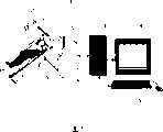
图1是本发明的功能图像的显示方法和装置的构成图。FIG. 1 is a block diagram of a method and device for displaying functional images according to the present invention.
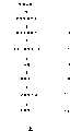
图2是从数据采集到合成图像显示的流程图。Figure 2 is a flow chart from data acquisition to composite image display.
图3是变换系数的算出方法的示意图。FIG. 3 is a schematic diagram of a method of calculating a transform coefficient.
图4是表示功能能图像用一览表的构成的图。FIG. 4 is a diagram showing the structure of a table for function image.
图5是功能图像用一览表的示意图。Fig. 5 is a schematic diagram of a list for functional images.
图6是表示断面成像用一览表的构成的图。FIG. 6 is a diagram showing the configuration of a list for cross-sectional imaging.
图7是混合功能图像的样本图像。Fig. 7 is a sample image of a hybrid function image.
图8是断面图像上投影混合功能图像的样本图像。Fig. 8 is a sample image of projecting a hybrid functional image on a cross-sectional image.
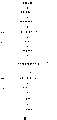
图9是本发明的实施例2中的从数据采集到合成图像显示的流程图。FIG. 9 is a flow chart from data acquisition to composite image display in
图10是说明本发明的实施例2中的一例的图像间演算时的ROI设定方法的一个例子的图。FIG. 10 is a diagram illustrating an example of an ROI setting method in an example of inter-image calculation in
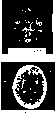
图11是本发明的实施例2中在治疗前的MTT功能图像。Fig. 11 is an image of MTT function before treatment in Example 2 of the present invention.
图12是本发明的实施例2中在治疗后的MTT功能图像。Fig. 12 is an MTT functional image after treatment in Example 2 of the present invention.
图13是本发明的实施例2中对治疗前后的MTT差别图像和CT断面图像进行合成的图像。Fig. 13 is an image obtained by synthesizing MTT difference images and CT cross-sectional images before and after treatment in Example 2 of the present invention.
图14是在治疗前的CBV功能图像。Figure 14 is a functional image of CBV before treatment.
图15是在治疗后的CBV功能图像。Figure 15 is a functional image of CBV after treatment.
图16是有关上述图11、12、14、15的对治疗前后的CBV以及MTT差别图像与CT断面图像进行合成的图像。FIG. 16 is an image obtained by combining CBV and MTT difference images before and after treatment with CT cross-sectional images related to the above-mentioned FIGS. 11 , 12 , 14 , and 15 .
具体实施方式Detailed ways
下面,用附图详细说明本发明的功能图像的显示方法以及装置的优选实施方式。Hereinafter, preferred embodiments of the method and device for displaying functional images of the present invention will be described in detail with reference to the drawings.
〔实施例1〕[Example 1]
图1是表示本发明的功能图像的显示方法以及装置的优选实施方式的图。本发明的功能图像显示方法以及装置,是指由X线衰减信号或磁共振发射的回波信号等的断面图像数据采集机构1,如CT装置或MRI装置。由进行采集机构1的控制和各种演算的计算机2、鼠标或键盘等操作台、和显示器等显示方法4构成。在计算机2中搭载有:控制采集机构1的程序、进行图像重构等作成断面图像的程序、进行活体功能信息的分析及绘图的程序、以及作成合成图像的程序。在构成本发明的功能图像的显示方法和装置时,上述各程序可以搭载于一个计算机内,也可以以每个运算种类分别搭载于多个计算机内。FIG. 1 is a diagram showing a preferred embodiment of a method and device for displaying a functional image of the present invention. The functional image display method and device of the present invention refer to a cross-sectional image data acquisition mechanism 1 such as a CT device or an MRI device, such as an X-ray attenuation signal or an echo signal emitted by magnetic resonance. It consists of a
图2是本发明的功能图像的显示方法和装置的从数据采集到合成图像的显示的流程图。这个流程是通过在图1的计算机2或未图示的外部计算机中内置的软件来实现的。Fig. 2 is a flow chart from data collection to display of composite images of the method and device for displaying functional images of the present invention. This flow is realized by software built into the
按照该流程图对本实施例加以说明。首先,通过由在计算机2中搭载的控制程序控制的采集机构1,采集X线衰减数据或磁化率信号强度数据(步骤201)。This embodiment will be described in accordance with this flow chart. First, X-ray attenuation data or magnetic susceptibility signal intensity data is collected by the collection mechanism 1 controlled by the control program installed in the computer 2 (step 201).
在步骤201中,通过由电脑2上的控制程序控制的采集机构1,采集由X线衰减信号或磁共振发射的回波信号。例如数据采集装置是CT装置,如果想要分析的活体功能信息是头部的灌注信息,在向患者5注入如碘系造影剂那样的对比增强物质之后,集中于该物质流入的特定脏器或部位,进行时时摄像(也就是动态摄像),由此可以采集进行活体功能信息的分析所必需的数据。In
在步骤202中,使用在计算机2上搭载的图像重构等程序,作成断面图像。In
在步骤203中,显示在步骤202中作成的断面图像。In
在步骤204中,例如使用在计算机2上搭载的活体功能信息的分析程序,计算出表示活体功能信息的参数即例如像素值P。作为这里所说的参数的代表性参数,有脑血流量图(Cerebral Blood Flow,即CBF)像、脑血液容量(Cerebral Blood Volume,即CBV)图像、平均通过时间(Mean TransitTime,即MTT)图像。In
参数的计算优选对断面图像的每个像素进行以防止分辨力下降,但在像急速进行活体功能信息的诊断时那样急迫需要结束演算的情况下,可以通过缩小图像进行演算,也可以多个像素进行演算。The calculation of the parameters is preferably performed for each pixel of the cross-sectional image to prevent a decrease in resolution. However, when the calculation needs to be terminated urgently, such as when the diagnosis of biological function information is urgently performed, the calculation can be performed by reducing the image, or multiple pixels can be used. Do the math.
在步骤205中,使用在计算机2中搭载的绘图程序,将在步骤204中得到的演算结果绘制成图,由此作成功能图像。In
在步骤206中,将在步骤205中作成的功能图像显示于显示机构4中。其中,在步骤206中,不仅显示功能图像,根据需要还可以将功能图像和断面图像一起显示。其中,当继续作成合成图像时,在这里可以不进行图像显示。In
在步骤207中,如后面所述,使用在计算机2中搭载的合成图像作成程序,作成合成图像。In
在步骤208中,将合成图像表示于显示机构4。其中,在步骤208中,不仅显示图像,根据需要还可以将合成图像、功能图像以及断面图像中的至少2个一起显示。In
关于图2的流程图,当X线衰减数据或磁化率信号强度数据已采集完毕时,在从内置或外接于计算机2上的硬盘等存储机构6读取了X线衰减数据或磁化率信号强度数据之后,可以执行步骤202以后的步骤。Regarding the flowchart of Fig. 2, when the X-ray attenuation data or the magnetic susceptibility signal strength data have been collected, the X-ray attenuation data or the magnetic susceptibility signal strength have been read from
进而,关于图2的流程图,当断面图像已作成完毕时,在从内置或外接于计算机2上的硬盘等存储机构5读取了断面图像之后,可以执行步骤203以后的步骤。Furthermore, with regard to the flowchart of FIG. 2 , when the cross-sectional image has been created, after the cross-sectional image is read from the
进而,关于图2的流程图,当功能图像已作成完毕时,在从内置或外接于计算机2上的硬盘等存储机构5读取了功能图像之后,另外根据需要还要读取功能图像和断层图像,然后可以执行步骤206以后的步骤。Furthermore, with regard to the flowchart of FIG. 2 , when the functional image has been created, after the functional image is read from a
接下来,对步骤207的合成图像的作成方法进行说明。在本实施方式中,将某个脏器中的活体功能信息的种类即功能图像的总数设为M幅进行说明。Next, a method of creating a composite image in
另外,灰度数是正整数,例如8比特(256灰度)、12比特(4096灰度)、16比特(65536灰度)、32比特(4294967296灰度)。In addition, the grayscale number is a positive integer, such as 8 bits (256 grayscales), 12 bits (4096 grayscales), 16 bits (65536 grayscales), and 32 bits (4294967296 grayscales).
下面,色调是指色相、彩度、亮度或者它们中的至少两个的组合。另外,连续色标是指将像素值的最大值和最小值的范围至少分割成一个阶段,向各阶段分别分配不同的色调,也就是说是色调的连续体。Hereinafter, hue refers to hue, chroma, lightness, or a combination of at least two of them. In addition, the continuous color scale refers to dividing the range of the maximum value and the minimum value of pixel values into at least one stage, and assigning different hues to each stage, that is, it is a continuum of hues.
(1)由像素到变换系数的变换(1) Transformation from pixels to transformation coefficients
对被称之为混合功能图像的图像进行说明。该混合功能图像是,把活体功能信息对应于连续色标而显示的功能图像重叠多个而进行合成的图像。这里,功能图像之间的使用色调是不同的。其中,在作为混合功能图像的原始图像的功能图像中,也包括用某连续色标只显示图像中的特定区域。此时,特定区域以外的区域可以用任意的特定色显示。在作成混合功能图像的时候,以功能图像的像素值P、显示窗位值WL以及显示窗宽值WW为基础,计算出变换系数C。变换系数C例如参照图3用下式1式求得。这里,WL代表窗位,WW代表窗宽。显示的浓度是以窗位为中心,在上下每WW/2的范围内分配。在该范围WW的外侧,没有浓度或者因为饱和而没有变化。即,根据显示窗宽WW,在像素值大的地方达到饱和而发白,与之相反,在像素值小的地方则趋向最暗。另外,PMAX代表像素最大值,CMAX代表变换系数的最大值。An image called a hybrid function image will be described. The mixed functional image is an image synthesized by superimposing a plurality of functional images displayed with biological function information corresponding to continuous color scales. Here, the used tone is different between function images. Wherein, in the functional image which is the original image of the mixed functional image, only a specific region in the image is displayed with a continuous color scale. In this case, the areas other than the specific area may be displayed in any specific color. When creating a mixed functional image, the conversion coefficient C is calculated based on the pixel value P of the functional image, the display window level value WL, and the display window width value WW. The conversion coefficient C is obtained, for example, by the following Equation 1 with reference to FIG. 3 . Here, WL represents the window level, and WW represents the window width. The concentration displayed is centered on the window level and distributed within the range of WW/2 up and down. Outside this range WW, there is no concentration or no change due to saturation. That is, according to the display window width WW, places where the pixel values are large become saturated and whitish, and on the contrary, places where the pixel values are small tend to be darkest. In addition, PMAX represents the maximum value of pixels, and CMAX represents the maximum value of transform coefficients.
其中,在图3所示的例子中,对自(WL-WW/2)到(WL+WW/2)的区间进行线性变换,根据需要也可以进行任意的非线性变换。另外,也可以直接将像素值P作为变换系数C。Wherein, in the example shown in FIG. 3 , linear transformation is performed on the interval from (WL-WW/2) to (WL+WW/2), and arbitrary nonlinear transformation may be performed as needed. Alternatively, the pixel value P may be directly used as the transform coefficient C.
图4是表示在作成混合功能图像中使用的一览表(以下称作LUT)。在本实施例中所说的LUT,是指上述的变换系数和显示颜色的各成分(如R成分,G成分,B成分)的对应表。适用于(WL-WW/2)以下的区域的像素的变换系数C,是连续色标中的一端的最暗的颜色(下端颜色)。下面,将用于颜色显示的R、G、B中各自的最暗颜色表示成R1、G1、B1。其中,当只改变色调中的色相而分配到上述连续色标的各阶段时,有时未必向最暗色部分配看起来最暗的颜色。不过在这样的情况下,为了方便而将其称作最暗色(下端色)。FIG. 4 shows a list (hereinafter referred to as LUT) used for creating a hybrid image. The LUT mentioned in this embodiment refers to the correspondence table between the above-mentioned transformation coefficient and each component of the displayed color (such as R component, G component, and B component). The conversion coefficient C applied to the pixels in the area below (WL-WW/2) is the darkest color (lower end color) at one end of the continuous color scale. In the following, the darkest colors among R, G, and B used for color display are represented as R1, G1, and B1. However, when assigning to each stage of the above-mentioned continuous color scale by changing only the hue in the color tone, it may not always be necessary to assign the darkest color to the darkest color part. However, in such a case, it is called the darkest color (lower end color) for convenience.
另一方面,变换系数C的值适合于(WL+WW/2)以上的区域的像素的变换系数C,在连续色标中的另一端为最明亮的最明色。下面,将用于颜色表示的R、G、B中各自的最明色表示成Rh,Gh,Bh。其中,与上述一样,当只改变色调中的色相而分配到上述连续色标的各阶段时,有时未必向最亮色部分配看起来最亮的颜色。不过在这样的情况下,为了方便而将其称作最亮色(上端色)。On the other hand, the value of the transformation coefficient C is suitable for the transformation coefficient C of the pixels in the region of (WL+WW/2), and the other end of the continuous color scale is the brightest brightest color. Hereinafter, the brightest colors among R, G, and B used for color representation are expressed as Rh, Gh, and Bh. However, when assigning to each step of the above-mentioned continuous color scale by changing only the hue in the hue as above, sometimes it is not necessary to assign the brightest color part to the brightest color part. However, in such a case, it is called the brightest color (upper end color) for convenience.
某个变换系数C中的LUT的R、G、B的各成分R(c)、G(c)、B(c),例如可以参照图5所示的功能图像用LUT,然后按照下式(2)来确定。其中,图5是每个R、G、B各色的LUT的一例,分配到每个功能图像的RGB的图表(table)的起始值和斜率各不相同是比较常见的。通过作为RGB的起始值的最暗色R1、G1、B1的组合规定显示活体功能信息的颜色的系统。The components R(c), G(c), and B(c) of the R, G, and B components of the LUT in a certain transform coefficient C, for example, can refer to the LUT for functional images shown in FIG. 5, and then use the following formula ( 2) to determine. Among them, FIG. 5 is an example of LUTs for each color of R, G, and B, and it is common that the RGB tables assigned to each functional image have different initial values and slopes. A color system for displaying biological function information is defined by a combination of the darkest colors R1, G1, and B1 as initial values of RGB.
在图5所示的例子中,将从最暗色到最亮色的各成分值连接成线性,但根据需要还可以连接成任意的非线性。如果各功能图像有M个,优选设定与各功能图像1、功能图像2、……、功能图像M相对应的M个一览表,即LUT1、LUT2、……、LUTM。但是,并不限于此,在多个功能图像之间可以使用同一个LUT。In the example shown in FIG. 5 , the component values from the darkest color to the brightest color are connected linearly, but they may be connected arbitrarily nonlinearly if necessary. If there are M functional images, it is preferable to set M lists corresponding to each functional image 1,
(2)混合功能图像的作成(2) Creation of mixed function image
对多幅功能图像重叠的部分的显示像素的处理进行说明。这里,当将支配混合功能图像中的某像素(i,j)的显示色的R、G、B的各成分表示成RF(i,j),GF(i,j),BF(i,j)时,它们可以下式3来决定。The processing of the display pixels in the portion where a plurality of functional images overlap will be described. Here, when each component of R, G, and B that dominates the display color of a certain pixel (i, j) in the blend function image is expressed as RF(i, j), GF(i, j), BF(i, j ), they can be determined by the following
在这里,Wk表示合成多幅功能图像的权重,Ck(i,j)表示像素(i,j)中的功能能图像k的变换系数。另外,Rk(Ck(i,j))、Gk(Ck(i,j))、Bk(Ck(i,j))表示用变换系数Ck(i,j)的LUTk规定的R、G、B的各成分的值,是将每个像素的变换系数Ck(i,j)输入到式2的变换系数C中经过计算获得的值。其中,如上所述,这里特别将功能图像的种类的数k作为1~M的整数。Here, Wk represents the weight for synthesizing a plurality of functional images, and Ck(i, j) represents the transformation coefficient of the functional image k in the pixel (i, j). In addition, Rk(Ck(i, j)), Gk(Ck(i, j)), and Bk(Ck(i, j)) represent R, G, and B defined by the LUTk of the transformation coefficient Ck(i, j). The value of each component of is calculated by inputting the transformation coefficient Ck(i, j) of each pixel into the transformation coefficient C of
用连续色标显示的区域可以是整个图像,也可以是图像的一部分。当用连续色标只显示图像的一部分即特定的区域时,可以通过控制台4根据阈值、范围合ROI等中的至少一个进行设定。这样的阈值、范围以及ROI可以在每种功能图像中设定一个或者多个(处理1)。The area displayed with a continuous color scale can be the entire image or a portion of the image. When using a continuous color scale to display only a part of the image, that is, a specific area, it can be set according to at least one of threshold, range, and ROI through the
当某功能图像k当中的某像素(i,j)正好表示活体功能时,即例如当该像素(i,j)在ROI内時,或者如果该像素值是在由相对功能图像k的阈值决定的范围内,按照如上述的图4所示的LUTk,决定各成分Rk(Ck(i,j))、Gk(Ck(i,j)),Bk(Ck(i,j)),如果像素值或者像素没有如上所述在阈值、范围以及ROI的任意范围内,将特定值分配到各成分Rk(Ck(i,j))、Gk(Ck(i,j)),Bk(Ck(i,j))以便用不妨碍关心的功能显示那样的特定颜色进行显示(处理2)。When a certain pixel (i, j) in a certain functional image k just represents the function of the living body, that is, for example, when the pixel (i, j) is within the ROI, or if the pixel value is determined by the threshold value of the relative functional image k Within the range, according to the LUTk shown in Figure 4 above, each component Rk(Ck(i,j)), Gk(Ck(i,j)), Bk(Ck(i,j)) is determined, if the pixel value or pixel is not within any range of threshold, range, and ROI as described above, assign a specific value to each component Rk(Ck(i,j)), Gk(Ck(i,j)), Bk(Ck(i , j)) to display in a specific color that does not interfere with the display of the function of interest (processing 2).
通过对所有像素进行处理1和处理2,在某幅功能图像中用连续色标只显示上述设定范围,其他范围可以用特定颜色来显示,处理1和处理2可以对所有的功能图像进行,也可以只对部分功能图像进行。By performing processing 1 and
对已实施处理1和处理2后的各功能图像的合成进行说明。如此合成的功能图像在下面被称作混合功能图像,混合功能图像的数据在每个像素中获得。按照式3由每个像素确定RF(i,j)、GF(i,j)、BF(i,j),此时用特定颜色显示的像素是将该功能图像的权重Wk设置为0而计算的。如果得到所有像素的数据RF(i,j)、GF(i,j)、BF(i,j),按照坐标(i,j)进行绘图,从而进行图像显示。如此,可以作成混合功能图像。即使在没有必要合成所有N幅功能图像的情况下,可以使没有必要合成的功能图像的权重为0而进行合成。其中,作为特定范围的设定,例示了阈值、范围和ROI,但根据需要还可以使用其他参数进行设定。Synthesis of each functional image after processing 1 and 2 will be described. The functional image thus synthesized is hereinafter referred to as a mixed functional image, and data of the mixed functional image is obtained in each pixel. Determine RF(i, j), GF(i, j), and BF(i, j) from each pixel according to
(3)断面图像上投影混合功能图像的作成(3) Creation of projected hybrid functional images on cross-sectional images
接着,说明断面图像与混合图像的重叠合成图像(下面,断面图像上的投影混合功能图像)的作成方法。这里,如果将在断面图像上投影混合功能图像中的某个像素(i,j)的显示颜色的各成分设为RTF(i,j)、GTF(i,j)、BTF(i,j),可以使用如下所示并通过式4来确定,即根据上式3求得的混合功能图像的每个像素的颜色成分RF(i,j)、GF(i,j)、BF(i,j)和在像素(i,j)中的断面图像的变化系数CC(i,j),使对应于用符号t辨别的多幅断面图像用一览表LUTT而求得的有关变换系数CC(P)的各颜色成分值RT(CC(i,j))、GT(CC(i,j))、BT(CC(i,j)),每幅混合功能图像与断面图像的权重WB和WT。Next, a method of creating a superimposed composite image of a cross-sectional image and a blended image (hereinafter, a projected blended functional image on a cross-sectional image) will be described. Here, if each component of the display color of a certain pixel (i, j) in the mixed function image projected on the cross-sectional image is RTF(i, j), GTF(i, j), BTF(i, j) , can be determined by
断面图像一般用灰标来显示,所以断面图像用一览表例如可以如图6所示那样设定。为了作成断面图像上投影混合功能图像,在是用连续色标显示的像素的情况下,按照式4确定RTF(i,j)、GTF(i,j)、BTF(i,j)。用特定颜色显示的像素在公4中是把权重WB设为0,从而确定RTF(i,j)、GTF(i,j)、BTF(i,j)。如果对所有像素进行这样的处理后绘图,就完成了断面图像上投影混合功能图像。Cross-sectional images are generally displayed in grayscale, so the cross-sectional image list can be set as shown in FIG. 6, for example. In order to project a hybrid functional image on a cross-sectional image, RTF(i,j), GTF(i,j), and BTF(i,j) are determined according to Eq. For pixels displayed in a specific color, the weight WB is set to 0 in FIG. 4 to determine RTF(i, j), GTF(i, j), and BTF(i, j). When all the pixels are drawn after such processing, the projection of the mixed function image on the cross-sectional image is completed.
在混合图像或断面图像上投影混合功能图像中,当想要变更某功能图像k的连续色标时,根据从控制台4输入的参数,采用上述的方法变更与该功能图像对应的一览表LUTk,由此可以变更上式3中的Rk(Ck(i,j))、Gk(Ck(i,j))、Bk(Ck(i,j))。其中,k为1~m的整数。When projecting a mixed functional image on a mixed image or a cross-sectional image, when it is desired to change the continuous color scale of a certain functional image k, according to the parameters input from the
在混合图像或断面图像上投影混合功能图像中,当变更从某功能图像k获得的信息的强调程度时,根据从控制台4输入的参数,可以变更上式3中的Wk。In projecting a mixed functional image on a mixed image or a cross-sectional image, when changing the emphasis degree of information obtained from a certain functional image k, Wk in
在混合图像或断面图像上投影混合功能图像中,当想要变更用连续色标显示的区域时,可以从控制台4输入阈值、范围、ROI等规定该区域的参数而进行变更。When projecting a hybrid functional image on a hybrid image or a cross-sectional image, if it is desired to change the region displayed by the continuous color scale, parameters defining the region, such as threshold, range, and ROI, can be input from the
在断面图像上投影混合功能图像中,当变更混合功能图像的强调程度时,根据从控制台4输入的参数,可以变更上式4中的WB或WT。In projecting the hybrid functional image on the cross-sectional image, when changing the degree of emphasis of the hybrid functional image, WB or WT in
图7和图8是表示将本发明的实施例应用于由CT图像作成的脑血流功能图像的例子。图7是根据这三种功能信息(脑血流量、脑血液容量、平均通过时间)作成的混合功能图像的样本图像,是脑血流量的异常区域31、脑血液容量的异常区域32、平均通过时间的异常区域33、和其它区域39的合成图像。图8是根据由三种功能信息(脑血流量、脑血液容量、平均通过时间)所作成的混合功能图像与CT图像30作成的断面图像上投影混合功能图像的样本图像。在这些样本图像中,用绿色系的连续色标显示脑血流量的异常区域31,用蓝色系的连续色标来显示脑血液容量的异常区域32,用红色系的连续色标来显示平均通过时间异常区域33。这些样本图像不仅将认为在脑血流量、脑血液容量、平均通过时间等各生理学参数方面异常的区域显示于一幅图像上,还可以通过配色的浓淡或者各种颜色的混杂程度显示异常的严重程度,由此可以明白本发明的效果。这些混合功能图像或断面图像上投影混合功能图像,不是一种而是选择多个存在的功能图像中的不同的几种并进行合成,由此可以作成不同的多幅混合功能图像或断面图像上投影混合功能图像。这些多幅混合功能图像或断面图像上投影混合功能图像可以同时在图像上显示。7 and 8 show an example of applying an embodiment of the present invention to a cerebral blood flow function image created from a CT image. Fig. 7 is a sample image of a mixed functional image created based on these three kinds of functional information (cerebral blood flow, cerebral blood volume, and average transit time), which are abnormal regions 31 of cerebral blood flow, abnormal regions 32 of cerebral blood volume, average transit Composite image of temporal anomalous region 33 and other regions 39 . 8 is a sample image in which a mixed functional image is projected on a cross-sectional image created from a mixed functional image created from three types of functional information (cerebral blood flow, cerebral blood volume, and mean transit time) and a CT image 30 . In these sample images, the abnormal area 31 of cerebral blood flow is displayed with a continuous color scale of green color, the abnormal area 32 of cerebral blood volume is displayed with a continuous color scale of blue, and the average Pass time anomaly area 33. These sample images not only display areas considered to be abnormal in various physiological parameters such as cerebral blood flow, cerebral blood volume, and average transit time on one image, but also show the severity of the abnormality through the shade of color matching or the degree of mixing of various colors. Degree, thus can understand the effect of the present invention. By projecting a mixed functional image on these mixed functional images or cross-sectional images, not one but different types of multiple existing functional images are selected and synthesized, so that a plurality of different mixed functional images or cross-sectional images can be created. Project the mixed function image. These multiple mixed function images or projected mixed function images on the cross-sectional images can be simultaneously displayed on the images.
综上,通过本实施例1,推测功能图像中表示阈值以上的值的像素为病变部位等特征部位而进行显示。通过将该功能图像的参数设成多个,能够增加判断的信息,通过就每个参数以不同的颜色显示这些特征位,哪个参数在哪个部位显示怎样的异常就一目了然,即使在对它们进行重叠而显示的部位出现多个参数的异常也可以一目了然。To sum up, according to the first embodiment, pixels showing a value equal to or greater than the threshold in the functional image are presumed to be characteristic sites such as lesion sites and displayed. By setting multiple parameters of the functional image, the information for judgment can be increased, and by displaying these flags in different colors for each parameter, it is clear at a glance which parameter shows which abnormality, even if they are overlapped And the abnormality of multiple parameters in the displayed part can also be seen at a glance.
进而,在上述已着色的特征部位内,可以根据像素值的大小变更颜色的浓度或配色,因而就可以判断异常的程度。Furthermore, in the above-mentioned colored characteristic part, the density or color matching of the color can be changed according to the size of the pixel value, so the degree of abnormality can be judged.
另外,可以变更这些特征部位的透明度,所以可以变更成操作者容易识别的画面状态。进而在图像上选择特征部位的外周或特别想诊断的部位,作为ROI可以如上所述只对该部分着色而可以进行显示,不会因没有用的信息妨碍诊断。In addition, since the transparency of these characteristic parts can be changed, it is possible to change to a screen state that can be easily recognized by the operator. Furthermore, by selecting the periphery of a characteristic part or a part particularly desired to be diagnosed on the image, only this part can be colored and displayed as an ROI as described above, and diagnosis will not be hindered by useless information.
另外,如图8所示,通过与断面图像40重叠后进行显示,可以容易地掌握与头颅等外部的位置关系,使诊断变得更容易。功能图像可以选择并列显示、重叠,部分重叠等任何配置,可以成为与时时诊断或使用者的意图一致的使用方法。另外,功能图像主要使用CBF、CBV、MTT,它们的计测值以能够同时在图像上确认。另外,通过记录作成上述的特征部位的ROI、阈值或配置,在任何时候都可以重复同一条件下的诊断,因而手术或者治疗前后的功能图像的比较变得容易,同时可以进行由操作者的差异等引起的无法恣意进入的手术或治疗的效果测量。In addition, as shown in FIG. 8 , by superimposing and displaying the
进而,也可以显示治疗前后的差别图像Furthermore, the difference images before and after treatment can also be displayed
实施例2Example 2
实施例2与实施例1一样也利用图1所示的构成。各构成要素如实施例1所述,所以省略对它们的说明。在实施例2中,图像处理装置2例如是电脑,搭载有对数据采集机构1进行控制的程序、进行图像重构等断面图像的作成的程序、进行活体功能信息分析及绘图的程序、和作成合成图像的程序。其中,上述各程序可以搭载于在一台计算机内,或者,也可以以每个运算种类分别搭载于多个计算机内。Example 2 also utilizes the configuration shown in FIG. 1 as in Example 1. The constituent elements are as described in Embodiment 1, and therefore their descriptions are omitted. In
图9是表示通过本实施例的图像诊断装置的程序的从数据采集到合成图像的显示的流程图。按照该流程图对通过本实施例的处理进行说明。在步骤301中,通过由搭载于电脑2上的控制程序控制的采集机构1(参照图1),采集由X线衰减信号或磁共振发射的回波信号。FIG. 9 is a flow chart showing the procedure from data acquisition to display of a synthesized image by the program of the image diagnostic apparatus of this embodiment. The processing by this embodiment will be described according to this flowchart. In
例如,采集装置是CT装置,如果想要分析的活体功能信息是头部的灌注信息,在向患者5注入如碘系造影剂那样的对比增强物质之后,进行动态摄像,由此可以采集进行活体功能信息的分析所必需的数据。For example, if the collection device is a CT device, if the biofunction information to be analyzed is the perfusion information of the head, after injecting a contrast-enhancing substance such as an iodine-based contrast agent into the
在步骤302中,使用在计算机2上搭载的图像重构用的程序等,作成断面图像。断面图像最好是横断面,冠状面以及矢状面等任意断面。在步骤303中,显示在步骤302中作成的断面图像。在步骤304中,使用在计算机2等上搭载的活体功能信息的分析程序,计算出表示活体功能信息的参数。从防止分辨力下降的观点来看,参数的计算优选对断面图像的每个像素进行,但在像急速进行活体功能信息的诊断时那样急迫需要在短时间内结束演算的情况下,可以缩小图像进行演算,也可以多个像素进行演算。在步骤305中,使用在计算机2等中搭载的绘图程序,将在步骤304中得到的演算结果绘制成图,由此作成功能图像。在步骤306中,将在步骤305中作成的功能图像显示于显示机构4中。其中,在步骤306中,不仅显示功能图像,根据需要还可以将功能图像和断面图像一起显示。In
在步骤307中,如后面所述,使用在计算机2中搭载的合成图像作成程序,作成合成图像。In
在步骤308中,由操作者选择是否有必要进行差别演算等图像间演算,如果不需要,则进入步骤309。当想要强调显示在多次检查中活体功能信息发生显著变化的区域等时,优选执行差别演算等图像间演算。In
在步骤307中,在选择有必要进行图像间演算的情况下,由操作者选择是否有必要进行定量值补正,如果不需要,则进入步骤308。例如在脑灌注图像中,根据摄像断面,由于存在跨越摄像层面的低CT值的物体,无法适当补正部分容积效应(Partial Volume Effect)的影响,其中所述的部分容积效应是指无法准确计算出在沿着该层面的断面图像中特别是在主动脉等高CT值的部分的CT值,有时导致定量值过大。在这种情况下,优选在补正定量值之后再进行图像间演算。在步骤308中,当需要补正定量值时,使用在计算机2中搭载的定量值补正程序进行定量值补正。在步骤308中,进而使用在计算机2中搭载的图像间演算程序作成演算后图像。其中,步骤308中的图像间演最好是差别演算等任意演算。在步骤309中,对演算后图像或功能图像设定与断面图像进行合成的区域的条件。In
不过,当将演算后图像或功能图像的全部区域直接在断面图像上重叠并合成时,这个步骤没有用。当只将演算后图像或功能图像的特定区域在断面图像上重叠并合成时,通过设定阈值或ROI,或只选择满足任意条件式的像素,由此指定重叠的区域。例如在演算后图像是多个检查中的脑灌注功能图像的差别图像的情况下,当想要只显示出表示在右半球有显著变化的区域时,将ROI指定在整个右半球,可以只将像素值P满足下式5的条件的像素在断面图像上重叠并合成。However, this step is not useful when the entire area of the calculated image or functional image is directly superimposed on the cross-sectional image and synthesized. When superimposing and synthesizing only a specific region of the calculated image or functional image on the cross-sectional image, specify the superimposed region by setting a threshold or ROI, or selecting only pixels satisfying an arbitrary conditional expression. For example, if the calculated image is a difference image of brain perfusion function images in multiple examinations, if you want to display only the region that shows a significant change in the right hemisphere, specify the ROI on the entire right hemisphere, and you can set only Pixels whose pixel value P satisfies the condition of
P=Mean+k·SD (5)P=Mean+k·SD
其中,在上式中,Mean为演算后图像的全体像素值的平均值,SD为标准差,k为任意实数。Wherein, in the above formula, Mean is the average value of all pixel values of the calculated image, SD is the standard deviation, and k is any real number.
另外,例如在演算后图像是多个检查中的脑灌注功能图像的情况下,当想显示异常区域随时间的变化时,只将像素值在阈值以上或阈值以下的像素在断面图像上重叠并合成。Also, for example, when the computed image is a brain perfusion function image under multiple examinations, and it is desired to display changes over time in an abnormal region, only pixels whose pixel values are greater than or less than the threshold are superimposed on the cross-sectional image and synthesis.
在步骤310中,使用在计算机2中搭载的合成图像作成程序,作成合成图像。该合成图像的作成的详细内容如后所述。在步骤311中,将合成图像显示于显示机构4。在步骤311中,不仅显示合成图像,根据需要还可以将合成图像与功能图像、演算后图像、断面图像等图像同时显示。另外,此时,通过结合在步骤309中设定的合成的区域中的像素数、平均值、标准差及柱状图进行显示,可以通过活体功能信息的分析提供有用的信息。In
在X线衰减数据或磁化率信号强度数据以采集完毕时,在从内置或外接于计算机2上的硬盘等存储机构5读取了X线衰减数据或磁化率信号强度数据之后,执行步骤302以后的步骤。另外,当断面图像已作成完毕时,内置或外接于计算机2上。当从硬盘等存储机构5读取了断面图像之后,执行步骤303以后的步骤。当功能图像已作成完毕时,在从内置或外接于计算机2上的硬盘等存储机构5读取了功能图像之后,另外根据需要还要读取功能图像和断层图像,然后执行步骤306以后的步骤。When the X-ray attenuation data or the magnetic susceptibility signal strength data have been collected, after reading the X-ray attenuation data or the magnetic susceptibility signal strength data from the
接下来,关于步骤308,详细说明在多次检查得到的多个系列功能图像经过演算而成的新的诊断用图像的作成。例如当看到在多次检查中活体功能信息发生变化时,可以使多次检查的功能图像有差别。演算的种类并不限于差别演算,也可以根据用途进行加、乘、除或者任意四则运算的混合运算。图像间演算可以在每个像素对全部像素进行演算。Next,
另外,如图10所示,根据需要例如对每个用任意设定的ROI8包围的区域进行计算,也可以在ROI区域中把平均值、中位数值、最大值、最小值等特征量作为演算对象。如此可以进行在易于诊断的尺寸中的视觉评价。In addition, as shown in FIG. 10 , as needed, for example, each area surrounded by an arbitrarily set ROI8 is calculated, and feature quantities such as the average value, median value, maximum value, and minimum value can also be used as calculations in the ROI area. object. This allows visual evaluation in a diagnostically easy size.
另外,通过引入等值线将功能图像分割成几个区域,可以在每个区域进行演算。例如在功能图像位脑灌注图像的情况下,通过引入等值线可以分割成白质、灰质、血管床等,进而通过指定ROI,可以分割成丘脑、豆状核、边缘带等各解剖学片段(segment)。在这样的每个解剖学片段进行图像间演算,这对活体功能信息的变化的评价也是有用的。In addition, by introducing isolines to divide the functional image into several regions, calculations can be performed in each region. For example, in the case of a functional image of a brain perfusion image, it can be segmented into white matter, gray matter, vascular bed, etc. by introducing contour lines, and then can be segmented into anatomical segments such as thalamus, lentiform nucleus, and marginal zone by specifying ROI ( segment). Calculations between images are performed for each of such anatomical segments, which is also useful for evaluating changes in living body function information.
接着,关于步骤308,说明定量值的补正方法。例如,在脑灌注图像中,以在上矢状静脉窦中的时间密度曲线的最大值或曲线下面积为基础,对如实施例1中所述的那样的部分容积效应的影响进行补正,得到定量的稳定性。但是,有时通过摄像断面在摄像断面中也不含有上矢状静脉窦。在这样的情况下无法适当补正部分容积效应的影响,定量值变得不准确。如果是可以判断成在多次检查中适当进行了部分容积效应的补正的图像,使用该检查中的补正参数(最大值或曲线下面积)对其他检查中的定量值进行补正。Next, with regard to step 308, a correction method of the quantitative value will be described. For example, in the cerebral perfusion image, based on the maximum value or the area under the curve of the time density curve in the superior sagittal sinus, the influence of the partial volume effect as described in Example 1 is corrected to obtain Quantitative stability. However, sometimes the superior sagittal sinus is not included in the imaging section through the imaging section. In such a case, the influence of the partial volume effect cannot be properly corrected, and the quantitative value becomes inaccurate. If the image can be judged to have been appropriately corrected for the partial volume effect in multiple examinations, the quantitative values in other examinations are corrected using the correction parameters (maximum value or area under the curve) in this examination.
作为其他的补正方法,有以健康区域中的平均值为基础进行补正的方法。这种方法是假设在同一受检查者的健康区域的活体功能信息不依靠检查时间而稳定的方法。将某个检查中的功能图像上的健康区域的定量值的平均值设为Mean1,将其他检查中功能图像上的健康区域的定量值的平均值设为Mean2。这里,为了使Mean1和Mean2一致,通过移动(shift)某个功能图像的像素值(定量值),可以补正定量值。As another correction method, there is a method of performing correction based on the average value in the healthy area. This method assumes that the biological function information in the healthy area of the same subject is stable regardless of the time of examination. The average value of the quantitative values of the healthy regions on the functional images in a certain examination is set to Mean1, and the average value of the quantitative values of the healthy regions on the functional images in the other examinations is set to Mean2. Here, the quantitative value can be corrected by shifting (shifting) the pixel value (quantitative value) of a certain functional image so that Mean1 and Mean2 match.
接下来,关于步骤310,说明演算后图像或者功能图像和断面图像的合成图像(下面称为断面图像上投影混合功能图像)的作成方法。在本实施例中,将色阶的灰度数设为M进行说明,灰度数是正整数,设成8比特(256灰度)、12比特(4096灰度)、16比特(65536灰度)、32比特(4294967296灰度)等任意的灰度数。灰度数越高则可以显示的灰度越丰富。一般在色阶中有色相、亮度、彩度的色阶,其种类也各种各样。Next, with regard to step 310, a method for creating a calculated image or a composite image of a functional image and a cross-sectional image (hereinafter referred to as projecting a mixed functional image on a cross-sectional image) will be described. In this embodiment, the number of gray levels of the color scale is set as M for illustration, and the number of gray levels is a positive integer, which is set to 8 bits (256 gray levels), 12 bits (4096 gray levels), and 16 bits (65536 gray levels). , 32 bits (4294967296 gray levels) and other arbitrary gray levels. The higher the number of gray levels, the richer the gray levels that can be displayed. Generally, in the color scale, there are hue, brightness, and chroma color scales, and the types are also various.
当作成合成图像时,以演算后图像或者功能图像的像素值P和显示窗位值WL、显示窗宽WW为基础,计算出变化系数C。变化系数C同实施例1一样,例如如式1及图3所示的那样来确定。在该例中,将WL-WW/2至WL+WW/2的区间变换成线性,但根据需要可以进行任意的非线性变换。另外,可以将像素值直接用作变换系数。When forming a composite image, the coefficient of variation C is calculated based on the pixel value P of the calculated image or functional image, the display window level value WL, and the display window width WW. The coefficient of variation C is determined as shown in Equation 1 and FIG. 3 , for example, as in the first embodiment. In this example, the interval from WL-WW/2 to WL+WW/2 is converted linearly, but any nonlinear conversion may be performed as necessary. Alternatively, pixel values can be directly used as transform coefficients.
图4是表示用于作成合成图像的一览表(LUT)。在本实施例中所述的LUT,是指上述的变化系数C和显示颜色的各成分(R成分、G成分、B成分)的对应表。将显示窗中最暗的像素即变换系数值在WL-WW/2以下的上述最暗色(下端色)的R、G、B各成分用R1、G1、B1表示,将适合显示窗中最明亮的像素即变换系数值在WL+WW/2以上的像素的最亮色的R、G、B各成分用Rh、Gh、Bh表示,此时,某变化系数C中的LUT的R、G、B各成分R(C)、G(C)、B(C),例如如式2和图5所示来确定。FIG. 4 shows a lookup table (LUT) for creating a composite image. The LUT described in this embodiment refers to a correspondence table between the above-mentioned coefficient of variation C and each component (R component, G component, B component) of the displayed color. The darkest pixel in the display window, that is, the R, G, and B components of the darkest color (lower end color) whose conversion coefficient value is below WL-WW/2 is represented by R1, G1, and B1, which will be suitable for the brightest pixel in the display window. The R, G, and B components of the brightest color of the pixel whose transformation coefficient value is above WL+WW/2 are represented by Rh, Gh, and Bh. At this time, the R, G, and B of the LUT in a certain variation coefficient C The components R(C), G(C), and B(C) are determined, for example, as shown in
在图5所示的例子中,将最暗色(下端色)至最亮色(上端色)的各成分值连接成线性,但可以根据需要连接成任意的非线性。即使在表示活体功能信息参数的存在多个且分别与该多个参数相对应的功能图像有多个的情况下,可以作成多个演算后图像。如果演算后图像、功能图像、或者演算后图像和功能图像存在M个,优选设定分别与演算后图像1(或功能图像1)、演算后图像2(或功能图像2)、…演算后图像M(或功能图像M)对应的M个一览表即LUT1、LUT2、…LUTM。但是,即使在多个演算后图像或者功能图像中共用同一个一览表也不会造成妨碍。In the example shown in FIG. 5 , the component values from the darkest color (lower end color) to the brightest color (upper end color) are connected linearly, but they may be connected arbitrarily nonlinearly if necessary. Even when there are a plurality of parameters indicating the biological function information and there are a plurality of functional images corresponding to the plurality of parameters, a plurality of calculated images can be created. If there are M post-calculated images, functional images, or post-calculated images and functional images, it is preferable to set the post-calculated image 1 (or functional image 1), post-calculated image 2 (or functional image 2), ... post-calculated image The M lists corresponding to M (or functional image M) are LUT1, LUT2, ... LUTM. However, even if the same table is shared among a plurality of calculated images or functional images, there is no hindrance.
接着,详细说明步骤311的合成图像显示。当将合成图像中的某个像素(i,j)的显示颜色的各成分设为RTF(i,j)、GTF(i,j)、BTF(i,j),它们由式4来确定。Next, the composite image display in
这里,WB表示演算后图像或者功能图像的权重,WT表示断面图像的权重,CC(i,j)表示像素(i,j)中的断面图像的变换系数,与上述的演算后图像或功能图像的变换系数的计算方法一样进行确定。Here, WB represents the weight of the calculated image or functional image, WT represents the weight of the cross-sectional image, CC(i, j) represents the transformation coefficient of the cross-sectional image in the pixel (i, j), and the above-mentioned calculated image or functional image The calculation method of the transformation coefficient is determined in the same way.
另外,RT(CC(i,j))、GT(CC(i,j))、BT(CC(i,j))表示变换系数CT(P)中的由断面图像用一览表LUTT规定的R、G、B各成分值。当灰标表示断面图像时,断面图像用一览表LUT例如可以如图6所示进行设定。当断面图像用颜色显示时,需要图5所示的RGB的图表。另外,RF(i,j)、GF(i,j)、BF(i,j)是根据合成各演算后图像或功能图像的比率来确定的参数,由式3进行确定。In addition, RT(CC(i,j)), GT(CC(i,j)), and BT(CC(i,j)) indicate R, which are defined by the cross-sectional image table LUTT among the transformation coefficients CT(P). The value of each component of G and B. When the gray scale represents the cross-sectional image, the cross-sectional image list LUT can be set as shown in FIG. 6 , for example. When the cross-sectional image is displayed in color, the RGB chart shown in FIG. 5 is required. In addition, RF(i, j), GF(i, j), and BF(i, j) are parameters determined according to the ratio of each calculated image or functional image combined, and are determined by
这里,Wk表示合成演算后图像k或者功能图像k的权重,Ck(i,j)表示像素(i,j)中的演算后图像k或者功能图像k的变换系数。Rk(Ck(i,j))、Gk(Ck(i,j))、Bk(Ck(i,j))表示变换系数Ck(i,j)中用LUTk规定的R、G、B各成分值。其中,k为1~M的整数,M与演算后图像或功能图像的幅数一致。其中,在演算后图像或功能图像的幅数为一幅的情况下,式4成为式6。Here, Wk represents the weight of the combined calculated image k or functional image k, and Ck(i, j) represents the transformation coefficient of the calculated image k or functional image k in the pixel (i, j). Rk(Ck(i,j)), Gk(Ck(i,j)), Bk(Ck(i,j)) represent the R, G, and B components specified by LUTk in the transformation coefficient Ck(i,j) value. Wherein, k is an integer from 1 to M, and M is consistent with the number of calculated images or functional images. However, when the number of calculated images or functional images is one,
这里,Ck(i,j)表示像素(i,j)中的演算后图像或者功能图像的变换系数。为了作成演算后图像或功能图像与断面图像的合成图像,当是用连续色标显示的像素时,按照式4或式6来确定RTF(i,j)、GTF(i,j)、BTF(i,j),用特定颜色显示的像素在式4或6中把权重WB设为0,然后确定RTF(i,j)、GTF(i,j)、BTF(i,j)。可以对全部像素进行该处理,然后按照RTF(i,j)、GTF(i,j)、BTF(i,j)进行绘图。Here, Ck(i, j) represents the conversion coefficient of the calculated image or the functional image in the pixel (i, j). In order to make a calculated image or a composite image of a functional image and a cross-sectional image, when it is a pixel displayed with a continuous color scale, RTF(i, j), GTF(i, j), BTF( i, j), a pixel displayed with a specific color sets the weight WB to 0 in
用连续色标显示的区域(特定的区域)可以根据通过控制台4设定的阈值、任意的条件式或ROI来确定。阈值、任意的条件式或ROI在每个演算后图像中设定一个或多个。某个演算后图像k或者功能图像k的某个像素(i,j)中的像素值,如果在通过针对演算后图像k或功能图像k的阈值或条件式所设定的范围内且像素(i,j)在用ROI设定的范围内,按照上述LUT的设定方法确定Rk(Ck(i,j))、Gk(Ck(i,j))、Bk(Ck(i,j))的各成分,如果在范围之外,则将任意的特定值分配给Rk(Ck(i,j))、Gk(Ck(i,j))、Bk(Ck(i,j))的各成分。The region (specific region) displayed with a continuous color scale can be determined based on a threshold value set in the
通过对全部像素进行以上的处理,在某演算后图像或者功能图像中用连续色标只显示特定的区域。另外,其他的区域是用特定颜色如图7~图8和图11~18中黑色或灰色显示的只为CT断面图像40的区域来表示。上述处理对所有的演算后图像或功能图像进行。即使在没有必要合成M幅演算后图像或功能图像的全部的情况下,可以将没有必要合成的演算后图像或者功能图像的权重设定为0后进行合成。By performing the above processing on all pixels, only a specific area can be displayed with a continuous color scale in a certain calculated image or functional image. In addition, other regions are represented by specific colors such as black or gray in FIGS. 7 to 8 and FIGS. 11 to 18 . The above processing is performed on all calculated images or functional images. Even when it is not necessary to synthesize all the M calculated images or functional images, the weights of the calculated images or functional images that do not need to be synthesized can be set to 0 and then synthesized.
在合成图像中,当对从某个演算后图像k或功能图像k得到的信息的强调程度进行变更时,可以根据从控制台4输入的参数来变更式5中的Wk。In the synthesized image, when changing the degree of emphasis of information obtained from a certain calculated image k or functional image k, Wk in
在合成图像中,在想要变更用连续色标显示的区域的情况下,可以根据从控制台4输入的参数来变更阈值、条件式或ROI。In the synthesized image, when it is desired to change the region displayed by the continuous color scale, the threshold value, the conditional expression, or the ROI can be changed according to the parameters input from the
在合成图像中,当对演算后图像或功能图像、断面图像的强调程度进行变更时,根据从控制台4输入的参数来变更式4或6中的WB或WB。In the composite image, when changing the degree of emphasis of the calculated image, functional image, or cross-sectional image, WB or WB in
图11~18表示将本发明应用于从CT图像作成的制作到脑灌注功能图的例子。图11~13是作成右颈内动脉狭窄的患者在治疗前后的、平均通过时间图像MTT的治疗前后的差别图像与CT图像的合成图像的样本图像。在该样本图像中,用虹彩色的连续色标只显示平均通过时间MTT有显著变化的区域,在变化有意义的区域将式5的WB设为0.8、WT设为0.2后进行合成,在其他区域将WB设为0、WT设为1来进行合成。在图13中,34表示MTT的变化比较大且用暖色系显示的部分,35表示MTT的变化比较小且用冷色系显示的区域。这里,颜色是根据图11和图12的差别的大小来分配的。11 to 18 show examples in which the present invention is applied from CT image creation to cerebral perfusion function map. FIGS. 11 to 13 are sample images for creating a composite image of a difference image of a mean transit time image MTT before and after treatment and a CT image of a patient with right internal carotid artery stenosis before and after treatment. In this sample image, only the region where the mean transit time MTT changes significantly is displayed with an iridescent continuous color scale, and in the region where the change is significant, the WB of
图11、12、图14至16是作成右颈内动脉狭窄的患者在治疗前后的、平均通过时间图像MTT的差别图像、脑血液容量图像CBV的差别图像、CT图像的合成图像的样本图像。在该样本图像中,用蓝色系的连续色标36显示脑血液容量图像CBV有显著变化的区域,用红色系的连续色标37显示平均通过时间MTT有显著变化的区域。另外,将式3的W1(即脑血液容量图像的差别图像的权重)设为0.75,W2(即平均通过时间图像的差别图像的权重)设为0.25,然后进行合成。进而,在脑血液容量CBV或平均通过时间MTT有显著变化的区域将式4中的WB设为0.8、WT设为0.2,然后进行合成。11 , 12 , and 14 to 16 are sample images for creating a composite image of a difference image of a mean transit time image MTT, a difference image of a cerebral blood volume image CBV, and a CT image of a patient with right internal carotid artery stenosis before and after treatment. In this sample image, the region where the cerebral blood volume image CBV changes significantly is displayed by the blue
图11~13表示演算后图像或功能图像为1幅时的应用例,图11、12、图14~16表示演算后图像或功能图像为两幅时的应用例,也同样可以应用于演算后图像或功能图像为3幅以上时的情况。通过这些样本图像,可以容易地掌握在活体功能信息中出现显著变化的区域或疾病区域随时间的变化,还可以减少因操作者的主观导致的ROI设定的不确定性,同时又不损坏检查部位原来的形状。如上所示,通过本实施例,具有如下效果,即可以减少因操作者的主观导致的ROI设定的不确定性,而且不坏检查部位原有的形状,可以容易地掌握在活体功能信息中出现显著变化的区域或疾病区域随时间的变化。Figures 11 to 13 show the application examples when there is one calculated image or functional image, and Figures 11, 12, and Figures 14 to 16 show the application examples when there are two calculated images or functional images, which can also be applied to the calculated When there are three or more images or functional images. Through these sample images, it is possible to easily grasp the time-dependent change of regions or diseased regions where significant changes have occurred in the functional information of the living body, and it is also possible to reduce the uncertainty of ROI setting caused by the operator's subjectivity without damaging the inspection. original shape of the part. As described above, according to this embodiment, there is an effect that the uncertainty of ROI setting caused by the operator's subjectivity can be reduced, and the original shape of the inspection site can be easily grasped in the living body function information. Changes in areas of significant change or disease areas over time.
如上所述,根据本说明,具有如下效果,即可以从一幅图像得到从断面图像获得的信息和分别从多个功能图像得到的信息,且活体功能异常的严重程度的判定变得容易。As described above, according to the present description, there is an effect that information obtained from a cross-sectional image and information obtained from a plurality of functional images can be obtained from one image, and the determination of the severity of an abnormal function of a living body becomes easy.
Claims (20)
Applications Claiming Priority (3)
| Application Number | Priority Date | Filing Date | Title |
|---|---|---|---|
| JP101284/2003 | 2003-04-04 | ||
| JP2003101284 | 2003-04-04 | ||
| JP345364/2003 | 2003-10-03 |
Publications (2)
| Publication Number | Publication Date |
|---|---|
| CN1767789Atrue CN1767789A (en) | 2006-05-03 |
| CN100577107C CN100577107C (en) | 2010-01-06 |
Family
ID=36743267
Family Applications (1)
| Application Number | Title | Priority Date | Filing Date |
|---|---|---|---|
| CN200480008844AExpired - LifetimeCN100577107C (en) | 2003-04-04 | 2004-04-02 | Method and device for displaying functional image |
Country Status (1)
| Country | Link |
|---|---|
| CN (1) | CN100577107C (en) |
Cited By (15)
| Publication number | Priority date | Publication date | Assignee | Title |
|---|---|---|---|---|
| CN101120874B (en)* | 2006-08-09 | 2010-07-14 | 株式会社东芝 | Medical image synthesis method and device thereof |
| CN111180039A (en)* | 2014-09-02 | 2020-05-19 | 苹果公司 | Body activity and fitness monitor |
| CN115187515A (en)* | 2022-06-16 | 2022-10-14 | 沈阳先进医疗设备技术孵化中心有限公司 | Image processing apparatus, method, device, and readable storage medium |
| US11896871B2 (en) | 2022-06-05 | 2024-02-13 | Apple Inc. | User interfaces for physical activity information |
| US11908343B2 (en) | 2015-08-20 | 2024-02-20 | Apple Inc. | Exercised-based watch face and complications |
| US11918857B2 (en) | 2016-06-11 | 2024-03-05 | Apple Inc. | Activity and workout updates |
| US11931625B2 (en) | 2021-05-15 | 2024-03-19 | Apple Inc. | User interfaces for group workouts |
| US11972853B2 (en) | 2019-05-06 | 2024-04-30 | Apple Inc. | Activity trends and workouts |
| US11977729B2 (en) | 2022-06-05 | 2024-05-07 | Apple Inc. | Physical activity information user interfaces |
| US11979467B2 (en) | 2019-06-01 | 2024-05-07 | Apple Inc. | Multi-modal activity tracking user interface |
| US11985506B2 (en) | 2020-02-14 | 2024-05-14 | Apple Inc. | User interfaces for workout content |
| US11996190B2 (en) | 2013-12-04 | 2024-05-28 | Apple Inc. | Wellness aggregator |
| US12039146B2 (en) | 2017-05-15 | 2024-07-16 | Apple Inc. | Displaying a scrollable list of affordances associated with physical activities |
| US12036018B2 (en) | 2016-09-22 | 2024-07-16 | Apple Inc. | Workout monitor interface |
| US12080421B2 (en) | 2013-12-04 | 2024-09-03 | Apple Inc. | Wellness aggregator |
Family Cites Families (4)
| Publication number | Priority date | Publication date | Assignee | Title |
|---|---|---|---|---|
| US5982953A (en)* | 1994-09-02 | 1999-11-09 | Konica Corporation | Image displaying apparatus of a processed image from temporally sequential images |
| JP2001212138A (en)* | 2000-02-02 | 2001-08-07 | Mitsubishi Plastics Ind Ltd | Image processing system |
| CN1331454A (en)* | 2000-06-24 | 2002-01-16 | 王建华 | Dynamic monitor method for digital medium image |
| JP2003070781A (en)* | 2001-09-04 | 2003-03-11 | Hitachi Medical Corp | Supporting unit for medical image diagnosis |
- 2004
- 2004-04-02CNCN200480008844Apatent/CN100577107C/ennot_activeExpired - Lifetime
Cited By (30)
| Publication number | Priority date | Publication date | Assignee | Title |
|---|---|---|---|---|
| CN101120874B (en)* | 2006-08-09 | 2010-07-14 | 株式会社东芝 | Medical image synthesis method and device thereof |
| US11996190B2 (en) | 2013-12-04 | 2024-05-28 | Apple Inc. | Wellness aggregator |
| US12394523B2 (en) | 2013-12-04 | 2025-08-19 | Apple Inc. | Wellness aggregator |
| US12094604B2 (en) | 2013-12-04 | 2024-09-17 | Apple Inc. | Wellness aggregator |
| US12080421B2 (en) | 2013-12-04 | 2024-09-03 | Apple Inc. | Wellness aggregator |
| CN111180039A (en)* | 2014-09-02 | 2020-05-19 | 苹果公司 | Body activity and fitness monitor |
| US11798672B2 (en) | 2014-09-02 | 2023-10-24 | Apple Inc. | Physical activity and workout monitor with a progress indicator |
| CN111180039B (en)* | 2014-09-02 | 2023-10-24 | 苹果公司 | Physical activity and fitness monitor |
| US12243444B2 (en) | 2015-08-20 | 2025-03-04 | Apple Inc. | Exercised-based watch face and complications |
| US11908343B2 (en) | 2015-08-20 | 2024-02-20 | Apple Inc. | Exercised-based watch face and complications |
| US11918857B2 (en) | 2016-06-11 | 2024-03-05 | Apple Inc. | Activity and workout updates |
| US12274918B2 (en) | 2016-06-11 | 2025-04-15 | Apple Inc. | Activity and workout updates |
| US12036018B2 (en) | 2016-09-22 | 2024-07-16 | Apple Inc. | Workout monitor interface |
| US12039146B2 (en) | 2017-05-15 | 2024-07-16 | Apple Inc. | Displaying a scrollable list of affordances associated with physical activities |
| US12224051B2 (en) | 2019-05-06 | 2025-02-11 | Apple Inc. | Activity trends and workouts |
| US11972853B2 (en) | 2019-05-06 | 2024-04-30 | Apple Inc. | Activity trends and workouts |
| US11979467B2 (en) | 2019-06-01 | 2024-05-07 | Apple Inc. | Multi-modal activity tracking user interface |
| US11985506B2 (en) | 2020-02-14 | 2024-05-14 | Apple Inc. | User interfaces for workout content |
| US12413981B2 (en) | 2020-02-14 | 2025-09-09 | Apple Inc. | User interfaces for workout content |
| US11938376B2 (en) | 2021-05-15 | 2024-03-26 | Apple Inc. | User interfaces for group workouts |
| US11931625B2 (en) | 2021-05-15 | 2024-03-19 | Apple Inc. | User interfaces for group workouts |
| US12239884B2 (en) | 2021-05-15 | 2025-03-04 | Apple Inc. | User interfaces for group workouts |
| US11992730B2 (en) | 2021-05-15 | 2024-05-28 | Apple Inc. | User interfaces for group workouts |
| US12186645B2 (en) | 2022-06-05 | 2025-01-07 | Apple Inc. | User interfaces for physical activity information |
| US12194366B2 (en) | 2022-06-05 | 2025-01-14 | Apple Inc. | User interfaces for physical activity information |
| US12197716B2 (en) | 2022-06-05 | 2025-01-14 | Apple Inc. | Physical activity information user interfaces |
| US11977729B2 (en) | 2022-06-05 | 2024-05-07 | Apple Inc. | Physical activity information user interfaces |
| US11896871B2 (en) | 2022-06-05 | 2024-02-13 | Apple Inc. | User interfaces for physical activity information |
| US12023567B2 (en) | 2022-06-05 | 2024-07-02 | Apple Inc. | User interfaces for physical activity information |
| CN115187515A (en)* | 2022-06-16 | 2022-10-14 | 沈阳先进医疗设备技术孵化中心有限公司 | Image processing apparatus, method, device, and readable storage medium |
Also Published As
| Publication number | Publication date |
|---|---|
| CN100577107C (en) | 2010-01-06 |
Similar Documents
| Publication | Publication Date | Title |
|---|---|---|
| JP4558645B2 (en) | Image display method and apparatus | |
| JP6968840B2 (en) | Medical image processing method | |
| US6198797B1 (en) | X-ray CT apparatus and image diagnostic apparatus | |
| CN1767789A (en) | Functional image display method and device | |
| US9615804B2 (en) | Method for image generation and image evaluation | |
| JP4980723B2 (en) | Image generation method and image generation apparatus | |
| JP5442530B2 (en) | Image processing apparatus, image display apparatus, program, and X-ray CT apparatus | |
| CN1976629A (en) | Medical Imaging System for Accurate Determination of Oriented Tumor Changes | |
| JP2006212430A (en) | Method and apparatus for controlling imaging modality | |
| CN1505490A (en) | Method and apparatus for non-invasive imaging of anatomical tissue structures | |
| US20230141302A1 (en) | Image analysis processing apparatus, endoscope system, operation method of image analysis processing apparatus, and non-transitory computer readable medium | |
| CN1284117C (en) | Method of displaying images in medical imaging | |
| CN108601570B (en) | Tomographic image processing apparatus and method, and recording medium relating to the method | |
| JP2018528826A (en) | Device for displaying medical image data of a body part | |
| WO2020235502A1 (en) | Image processing device | |
| Kocasarac et al. | Effectiveness of digital subtraction radiography in detecting artificially created osteophytes and erosions in the temporomandibular joint | |
| JP7257544B2 (en) | Information display system and information display method | |
| CN1947148A (en) | Method and system for automatically improving the usability of a medical picture | |
| CN1843298A (en) | X-ray CT device | |
| JP2005006832A (en) | X-ray ct system and control method therefor | |
| CN115192057B (en) | CT-based composite imaging method and device | |
| JP6721939B2 (en) | Fluorescence image analyzer | |
| CN120413069B (en) | Construction method of parotid tumor enhancement model for teaching based on clustering algorithm | |
| Mamman et al. | Defining Noise Index in Abdominal CT: A Comprehensive Analysis Using Liver and Kidney as Regions of Interests | |
| JP2025142490A (en) | Dynamic analysis device, dynamic analysis method, and program |
Legal Events
| Date | Code | Title | Description |
|---|---|---|---|
| C06 | Publication | ||
| PB01 | Publication | ||
| C10 | Entry into substantive examination | ||
| SE01 | Entry into force of request for substantive examination | ||
| C14 | Grant of patent or utility model | ||
| GR01 | Patent grant | ||
| TR01 | Transfer of patent right | Effective date of registration:20170322 Address after:Tokyo, Japan Patentee after:Hitachi, Ltd. Address before:Tokyo, Japan Patentee before:Hitachi Medical Corp. | |
| TR01 | Transfer of patent right | ||
| TR01 | Transfer of patent right | Effective date of registration:20211117 Address after:Chiba County, Japan Patentee after:Fujifilm medical health Co.,Ltd. Address before:Tokyo, Japan Patentee before:Hitachi, Ltd. | |
| TR01 | Transfer of patent right | ||
| CX01 | Expiry of patent term | Granted publication date:20100106 | |
| CX01 | Expiry of patent term |