CN1717412A - Method and apparatus for concentrating and purifying nucleic acids - Google Patents
Method and apparatus for concentrating and purifying nucleic acidsDownload PDFInfo
- Publication number
- CN1717412A CN1717412ACN 200380104540CN200380104540ACN1717412ACN 1717412 ACN1717412 ACN 1717412ACN 200380104540CN200380104540CN 200380104540CN 200380104540 ACN200380104540 ACN 200380104540ACN 1717412 ACN1717412 ACN 1717412A
- Authority
- CN
- China
- Prior art keywords
- nucleic acid
- sample
- electrophoresis
- concentrate
- concentrating
- Prior art date
- Legal status (The legal status is an assumption and is not a legal conclusion. Google has not performed a legal analysis and makes no representation as to the accuracy of the status listed.)
- Granted
Links
Images


Landscapes
- Measuring Or Testing Involving Enzymes Or Micro-Organisms (AREA)
- Apparatus Associated With Microorganisms And Enzymes (AREA)
Abstract
Description
Translated fromChinese技术领域technical field
本发明涉及一种分离核酸的方法。更详细地,本发明涉及一种利用电泳分离靶核酸的方法。The present invention relates to a method for isolating nucleic acids. In more detail, the present invention relates to a method for separating target nucleic acids using electrophoresis.
背景技术Background technique
由于近来对人类基因组的解读,生命过程和基因之间的不同关系已经被分析。因此,医学重点从病理学转移到病因学,从医学治疗转移到预防。因此,基因检测技术成为了重要基础。Due to the recent reading of the human genome, different relationships between life processes and genes have been analyzed. Consequently, the medical emphasis shifted from pathology to etiology, and from medical treatment to prevention. Therefore, genetic testing technology has become an important basis.
基因检测可以被用于在常规临床检查中很难完成的检测,例如,对培养困难的病原微生物的鉴定,在用抗生素进行治疗时或在感染早期检测病原微生物,在怀疑存在抗体转移的情况下检测抗原,调查病原微生物传染源,个人识别例如血统诊断,白血病和实体瘤的疾病类型的基因诊断,和遗传疾病的诊断。需要长时间培养的细菌可以有效地通过基因检测被发现,因为基因检测相对于培养细菌的方法而言所耗费的时间更短。此外,因为DNA的稳定依赖于良好的保藏条件,老样品,例如冷冻的活组织检测样品或骨也能被检测。Genetic testing can be used for detection that is difficult to perform in routine clinical examinations, for example, identification of pathogenic microorganisms that are difficult to culture, detection of pathogenic microorganisms during treatment with antibiotics or early in infection, in cases of suspected antibody transfer Detection of antigens, investigation of infectious sources of pathogenic microorganisms, identification of individuals such as blood lineage diagnosis, gene diagnosis of disease types of leukemia and solid tumors, and diagnosis of genetic diseases. Bacteria that require a long time to grow can be effectively detected by genetic testing, because genetic testing is less time-consuming than the method of culturing bacteria. Furthermore, since DNA stability is dependent on good storage conditions, older samples, such as frozen biopsies or bones, can also be tested.
基因检测吸引了公众的注意力,因为它能扩展对最近增加的性传播疾病的检测的几率。Genetic testing has attracted public attention because it could expand the chances of testing for a recently increased number of sexually transmitted diseases.
众所周知,常规的核酸纯化和浓缩的方法,包括使用苯酚、氯仿或乙醇的纯化方法,使用吸附核酸的柱子或过滤器的纯化方法,和使用磁性二氧化硅珠的纯化方法。Conventional nucleic acid purification and concentration methods are well known, including purification methods using phenol, chloroform, or ethanol, purification methods using nucleic acid-adsorbing columns or filters, and purification methods using magnetic silica beads.
此外,还有众所周知的从平板状凝胶电泳中回收核酸的常规方法,如同在日本实用新型公开平5-88296中所描述的那样,其中核酸在备好的凝胶上电泳,一回收装置被移到含有靶核酸的凝胶部分,然后靶核酸被进一步电泳回收。In addition, there is a well-known conventional method for recovering nucleic acid from slab gel electrophoresis, as described in Japanese Utility Model Laid-Open No. Hei 5-88296, in which nucleic acid is electrophoresed on a prepared gel, and a recovery device is Move to the portion of the gel containing the target nucleic acid, which is then recovered by further electrophoresis.
此外,有如日本特许公开平8-327595中所述那样的众所周知的常规方法,其中核酸在平板状凝胶中电泳以分离靶核酸,一段回收片(chip)插入到凝胶中靠近靶核酸带以回收靶核酸。In addition, there is a well-known conventional method as described in Japanese Patent Laid-Open No. Hei 8-327595, in which nucleic acids are electrophoresed in a slab gel to separate target nucleic acids, and a recovery chip is inserted into the gel near the target nucleic acid band to Target nucleic acid is recovered.
关于常规的核酸纯化和浓缩方法,使用苯酚、氯仿或乙醇的纯化方法仅可以在有限的环境下利用,因为这需要要求高级化学设备的有效药物。进一步地,很难使该纯化方法自动化,因为这需要有难度的操作和高速的离心步骤。也很难获得高精度。Regarding conventional nucleic acid purification and concentration methods, purification methods using phenol, chloroform, or ethanol can only be utilized in limited circumstances because they require effective drugs requiring advanced chemical equipment. Further, it is difficult to automate this purification method because it requires difficult handling and a high-speed centrifugation step. It is also difficult to obtain high precision.
应用柱子或过滤器来吸附核酸的纯化方法通过流动溶液而进行。因此,当一个样品包括许多杂质如垃圾时,柱子或过滤器往往被堵塞导致纯化效率降低。此外,很难使该方法自动化,因为它要求离心或吸出的步骤。A purification method using a column or a filter to adsorb nucleic acid is performed by flowing a solution. Therefore, when a sample includes many impurities such as garbage, the column or filter is often clogged resulting in a reduction in purification efficiency. Furthermore, it is difficult to automate this method because it requires centrifugation or aspiration steps.
应用磁性二氧化硅珠的回收方法很难得到核酸的高回收,因为不能被磁体回收或从磁性材料上落下的二氧化硅珠可能会保留在样品中。It is difficult to obtain high recovery of nucleic acids using magnetic silica bead recovery methods because silica beads that cannot be recovered by the magnet or fall off the magnetic material may remain in the sample.
常规的从平板状电泳凝胶回收核酸的方法要求平板状电泳凝胶和对包含靶核酸的凝胶部分处理之前核酸在平板状电泳凝胶中的电泳。Conventional methods of recovering nucleic acids from slab electrophoresis gels require slab electrophoresis gels and electrophoresis of the nucleic acids in the slab electrophoresis gels prior to processing the gel portion containing the target nucleic acid.
用于电泳的凝胶抗震荡的能力很弱,且根据其形成过程可能发生特征上的改变。因此,一般来说,靶核酸在电泳凝胶中的位置在电泳后通过紫外线进行分析,随后含有高浓度靶核酸的凝胶部分被处理。Gels used for electrophoresis are weakly resistant to shock and may change in character depending on their formation process. Therefore, in general, the position of the target nucleic acid in the electrophoresis gel is analyzed by ultraviolet light after electrophoresis, and then the portion of the gel containing a high concentration of the target nucleic acid is processed.
因此,应用该方法的基因检测的每一个检测都需要花费很长的时间。如果用于电泳的凝胶很大,由于凝胶的不均衡导致的核酸带印迹增大,因此减少了核酸的回收。此外,该大凝胶需要强的电力用于电泳。Therefore, each test of the genetic test to which this method is applied takes a long time. If the gel used for electrophoresis is very large, the nucleic acid band footprint will increase due to gel imbalance, thus reducing nucleic acid recovery. In addition, this large gel requires strong electric power for electrophoresis.
发明概述Summary of the invention
作为解决上述问题的各种实验的结果,发现了一种如下方法,即在样品中添加表面活性剂以吸附存在于样品中的杂质,从而使得杂质的移动不同于核酸的移动,并将杂质从核酸分离开。As a result of various experiments to solve the above-mentioned problems, a method was found in which a surfactant is added to a sample to adsorb impurities present in the sample so that the movement of the impurities is different from that of nucleic acids, and the removal of the impurities from Nucleic acids are separated.
此外,除了核酸外的杂质经阳离子表面活性剂和非离子型表面活性剂而带电,并被置于电场中,其中核酸从含有杂质的样品中分离和纯化以被浓缩或很容易被浓缩。In addition, impurities other than nucleic acids are charged by cationic surfactants and nonionic surfactants, and placed in an electric field in which nucleic acids are separated and purified from a sample containing impurities to be concentrated or easily concentrated.
图1:在表面活性剂存在的条件下,利用电泳浓缩核酸的模拟图。样品含有核酸1和杂质2。表面活性剂被加入到样品中。非离子型表面活性剂3和阳离子表面活性剂4被用做表面活性剂。与样品混合的表面活性剂吸附杂质2。被阳离子表面活性剂4吸附的杂质2带正电,从而与很少或不被表面活性剂吸附的核酸1分离开。相应地,杂质2能被容易地从样品中除去以浓缩核酸1。Figure 1: Schematic diagram of electrophoresis for the concentration of nucleic acids in the presence of surfactants. The sample contains Nucleic Acid 1 and Impurity 2. Surfactants are added to the samples. Nonionic Surfactant 3 and Cationic Surfactant 4 were used as surfactants. The surfactant mixed with the sample adsorbs the impurity 2. Impurity 2 adsorbed by cationic surfactant 4 is positively charged and thus separated from nucleic acid 1 which is adsorbed by little or no surfactant. Accordingly, the impurity 2 can be easily removed from the sample to concentrate the nucleic acid 1 .
下面,将解释一个浓缩核酸的方法。在该方法中,进行两次电泳以确保对核酸的浓缩。样品中过量的离子在第一次电泳中被除去,样品中的核酸在第二次电泳中被浓缩。首先,1%Triton(注册商标)X-100,用为非离子表面活性剂被添加到含有核酸的样品中并混合,然后在96℃加热10分钟。然后,100μL的0.2%DPC,用做阳离子表面活性剂被添加到样品中。或者,非离子表面活性剂和阳离子表面活性剂两者可以在加热处理前同时加入。即使核酸存在于大肠菌或类似菌的原核细胞中,细胞壁被添加到样品中用于预处理的表面活性剂所破坏。相应地,大肠菌或类似菌的培养液可以被用做样品,从而帮助对样品预处理的操作。如前述进行预处理,施加100V的直流电压进行电泳10分钟,从而从样品中移走过量的离子。随后,施加125V-150V的直流电压进行电泳120分钟从而在正极回收到核酸。Next, a method for concentrating nucleic acid will be explained. In this method, electrophoresis is performed twice to ensure the concentration of nucleic acids. Excess ions in the sample are removed in the first electrophoresis, and nucleic acids in the sample are concentrated in the second electrophoresis. First, 1% Triton (registered trademark) X-100, which is a nonionic surfactant, was added to a nucleic acid-containing sample and mixed, followed by heating at 96° C. for 10 minutes. Then, 100 μL of 0.2% DPC, used as a cationic surfactant, was added to the sample. Alternatively, both the nonionic surfactant and the cationic surfactant may be added simultaneously before heat treatment. Even if the nucleic acid is present in prokaryotic cells of coliform bacteria or the like, the cell wall is disrupted by the surfactant added to the sample for pretreatment. Accordingly, a culture solution of coliform bacteria or the like can be used as a sample, thereby facilitating the operation of sample pretreatment. Pretreatment was performed as before, applying a DC voltage of 100 V for 10 minutes to remove excess ions from the sample. Subsequently, electrophoresis was performed for 120 minutes by applying a DC voltage of 125V-150V to recover nucleic acid at the positive electrode.
以下将解释用于样品电泳的电泳槽的结构。对于第一次电泳,图2表示用于第一次电泳的电泳槽5的结构。电泳槽5中有一样品槽6和将电泳槽5区分为正极侧部分和负极侧部分的隔壁9。样品槽6贯穿隔壁9,且其一侧向正极凸出,一侧向负极凸出。样品槽6两端开口,且开口部分用凝胶8封闭。在样品槽6中产生了电位差以对核酸和被表面活性剂吸附的杂质进行电泳。非离子表面活性剂和阳离子表面活性剂两者都被添加到样品中,并加热样品。然后,样品被注入到样品槽6中。在电极间使用100V的电压以进行电泳10分钟。相应地,过量的离子从样品中除去。除去过量的离子后,核酸通过第二次电泳而浓缩。The structure of the electrophoresis tank used for sample electrophoresis will be explained below. For the first electrophoresis, FIG. 2 shows the structure of the
第二次电泳说明:含有在第一次电泳过程中被注入的样品的样品槽6被连接到一个回收槽并被放置于一电泳槽中进行第二次电泳。图3表示用于第二次电泳的电泳槽5的结构。电泳槽5中有样品槽6,回收槽7和将电泳槽5区分为正极侧部分和负极侧部分的隔壁9。样品槽6贯穿隔壁9,且向负极侧凸出。回收槽7插入隔壁9并向正极侧凸出。样品槽6和回收槽7在隔壁9处穿过凝胶8而相互连接起来。样品槽6两端开口,且开口部分分别由凝胶8封闭。回收槽7两端开口。位于负极侧的回收槽7的一端由凝胶8封闭,位于正极侧的回收槽7的一端用超滤膜11封闭。在如上述所构成的电泳槽,用120V的电压穿过电极进行电泳120分钟。Second electrophoresis description: The
通过在电泳槽中进行的第二次电泳,除去过量离子后的样品除核酸以外的部分被电泳,其中核酸可以被有效地回收到回收槽7中。在回收槽7的正极侧所提供的超滤膜11阻止核酸从回收槽7漏出。相应地,核酸的回收效率能提高。如果样品的状态适当,可以只进行第二次电泳而不进行第一次电泳。预处理过的样品被注入到连接在回收槽7上的样品槽6中进行电泳,其中核酸可以被很容易地浓缩。存在于样品中的过量离子能通过超滤膜,从而不影响核酸的回收。Through the second electrophoresis performed in the electrophoresis tank, the portion of the sample after removing excess ions other than the nucleic acid is electrophoresed, wherein the nucleic acid can be efficiently recovered into the recovery tank 7 . The ultrafiltration membrane 11 provided on the positive electrode side of the recovery tank 7 prevents nucleic acid from leaking out of the recovery tank 7 . Accordingly, the recovery efficiency of nucleic acid can be improved. If the state of the sample is appropriate, it is possible to perform the second electrophoresis instead of the first electrophoresis. The pretreated sample is injected into the
也就是说,根据本发明,一种利用电泳来浓缩和纯化核酸的方法的特征在于样品被放置于一电场中以在调节了存在于含有核酸的样品中的杂质的荷电量后浓缩和纯化核酸。进一步地,根据本发明,一种利用电泳来浓缩和纯化核酸的方法的特征在于在含有核酸的样品中添加了阳离子表面活性剂来调节样品中的杂质的荷电量,然后该样品被置于一电场中以利用电泳来浓缩和纯化核酸。阳离子表面活性剂和非离子表面活性剂被添加到含有核酸的样品中以调节存在于样品中的杂质的荷电量,然后,该样品被置于一电场中以利用电泳浓缩和纯化核酸。样品中除核酸外的其它物质的电荷通过其吸附到阳离子表面活性剂上进行调节,且这些物质对阳离子表面活性剂的吸附通过调节添加的非离子表面活性剂的量来调节。That is, according to the present invention, a method for concentrating and purifying nucleic acid using electrophoresis is characterized in that the sample is placed in an electric field to concentrate and purify nucleic acid after adjusting the charge amount of impurities present in the sample containing nucleic acid . Further, according to the present invention, a method for concentrating and purifying nucleic acid by electrophoresis is characterized in that a cationic surfactant is added to a sample containing nucleic acid to adjust the charged amount of impurities in the sample, and then the sample is placed in a Electrophoresis is used to concentrate and purify nucleic acids in an electric field. Cationic surfactants and nonionic surfactants are added to a nucleic acid-containing sample to adjust the charged amount of impurities present in the sample, and then the sample is placed in an electric field to concentrate and purify the nucleic acid by electrophoresis. The charges of substances other than nucleic acids in the sample are adjusted by their adsorption to the cationic surfactant, and the adsorption of these substances to the cationic surfactant is adjusted by adjusting the amount of nonionic surfactant added.
根据本发明,用于浓缩和纯化核酸的装置含有添加到样品中的阳离子表面活性剂和非离子表面活性剂,其中的样品被电泳以在正极侧浓缩和纯化核酸。进一步地,该装置含有一容器,其侧壁由绝缘体形成。在容器中含有一用于阻止扩散的导电分离体,从而将容器内部划分为样品注入室和核酸回收室。该容器的每端通过缓冲槽分别连接到电极上。According to the present invention, the device for concentrating and purifying nucleic acid contains a cationic surfactant and a nonionic surfactant added to a sample in which the sample is electrophoresed to concentrate and purify nucleic acid on the positive electrode side. Further, the device includes a container, the side walls of which are formed by an insulator. A conductive separator for preventing diffusion is contained in the container, thereby dividing the inside of the container into a sample injection chamber and a nucleic acid recovery chamber. Each end of the container is respectively connected to the electrodes through buffer tanks.
或者,也可以使用以下的方法。含有核酸的样品与由一种材料形成的分离介质接触,在该材料中,具有不同分子量的物质表现出不同迁移率,加上电压以将核酸与其他物质分离。然后,通过分离介质的核酸被一过滤器回收。该过滤器具有通过核酸的小孔,该小孔比靶核酸要小。Alternatively, the following methods can also be used. A sample containing nucleic acid is brought into contact with a separation medium formed of a material in which substances with different molecular weights exhibit different mobilities, and a voltage is applied to separate the nucleic acid from other substances. Then, the nucleic acids passing through the separation medium are recovered by a filter. The filter has pores through which the nucleic acid passes, the pores being smaller than the target nucleic acid.
例如,用于电泳的分离介质的一端配有用缓冲液填充的样品供给部分,在另一端配有用缓冲液填充的取样部分。通过对样品供给部分和取样部分施加电压,样品在分离介质中电泳迁移。由于各核酸的不同分子量,样品中的核酸在分离介质中以不同的速度迁移,从而导致具有不同分子量的核酸需要不同的时间来通过分离介质并被洗脱到取样部分中。考虑到这样的情形,在靶核酸被洗脱到取样部分结束后立刻停止电泳,以减少靶核酸以外的组分。取样部分的缓冲液可以根据靶核酸洗脱进取样部分的时间进行更换,以从取样部分仅取样靶核酸。靶核酸的浓度依赖于取样部分中的缓冲液的量。For example, a separation medium for electrophoresis is provided with a sample supply portion filled with a buffer at one end and a sampling portion filled with a buffer at the other end. By applying a voltage to the sample supply part and the sampling part, the sample electrophoretically migrates in the separation medium. Due to the different molecular weights of the individual nucleic acids, the nucleic acids in the sample migrate at different speeds in the separation medium, resulting in that nucleic acids with different molecular weights take different times to pass through the separation medium and to be eluted into the sampling portion. In consideration of such a situation, the electrophoresis is stopped immediately after the target nucleic acid is eluted to the end of the sampling portion to reduce components other than the target nucleic acid. The buffer of the sampling portion may be replaced according to the time when the target nucleic acid is eluted into the sampling portion, so that only the target nucleic acid is sampled from the sampling portion. The concentration of target nucleic acid depends on the amount of buffer in the sampled portion.
这样,上述操作可以很容易的自动化操作,因为它不要求离心或抽吸操作。核酸可以立刻从样品中大量分离出来。通过减少含有靶核酸的洗脱溶液,溶液被浓缩以增加靶核酸在样品中的浓度。进一步地,被纯化以易于加工的这样的核酸在电泳后被立即取样,从而由当前步骤平稳地过渡到下一步骤。Thus, the above operation can be easily automated since it does not require centrifugation or suction. Nucleic acids can be isolated in large quantities from samples immediately. By reducing the elution solution containing the target nucleic acid, the solution is concentrated to increase the concentration of the target nucleic acid in the sample. Further, such nucleic acid purified for easy processing is sampled immediately after electrophoresis, thereby smoothly transitioning from the current step to the next step.
相比较于其它方法而言,本发明方法可以获得更高的回收率和更高精度的靶,且不需要昂贵的试剂或昂贵的装置,从而降低了运行成本和操作成本。在这一点上,根据本发明的利用电泳来浓缩和纯化核酸的方法,含有核酸的样品中存在的杂质与一种分离介质接触,在该介质中具有不同分子量的被电泳的核酸根据分子量的差异以不同的迁移率迁移,然后,小于靶核酸的核酸穿过具有在迁移方向上减小的截面积的过滤器,而靶核酸由过滤器回收,从而从样品中分离出靶核酸。该方法可以在不进行离心和抽吸操作的情形下进行,并可以使用一个简单的装置来分离和浓缩核酸。进一步地,这样的简单结构可以很容易地实现自动化。进一步地,可以立刻得到大量样品。可以减少含有靶核酸的洗脱溶液以浓缩和增加样品中靶核酸的浓度。分离和浓缩的运行成本以及操作时间都被降低了。此外,用于鉴定最初不存在的外源基因的检测可以很容易地进行。Compared with other methods, the method of the present invention can obtain higher recovery rate and higher precision target, and does not require expensive reagents or expensive devices, thereby reducing operating costs and operating costs. In this regard, according to the method for concentrating and purifying nucleic acid using electrophoresis of the present invention, impurities present in a nucleic acid-containing sample are brought into contact with a separation medium in which the electrophoresed nucleic acids having different molecular weights according to the difference in molecular weight Migrate with different mobility, and then, nucleic acids smaller than the target nucleic acid pass through a filter having a decreasing cross-sectional area in the direction of migration, and the target nucleic acid is recovered by the filter, thereby separating the target nucleic acid from the sample. This method can be performed without centrifugation and suction operations, and can separate and concentrate nucleic acids using a simple device. Further, such a simple structure can be easily automated. Further, a large number of samples can be obtained at once. The elution solution containing the target nucleic acid can be reduced to concentrate and increase the concentration of the target nucleic acid in the sample. Operating costs and operating times for separation and concentration are reduced. In addition, assays for identifying foreign genes that were not originally present can be easily performed.
附图说明:Description of drawings:
图1是在表面活性剂存在的条件下,利用电泳浓缩核酸的模式图。Fig. 1 is a schematic view of nucleic acid concentrated by electrophoresis in the presence of a surfactant.
图2是用于第一次电泳的电泳槽的图示。Figure 2 is a diagram of the electrophoresis tank used for the first electrophoresis.
图3是用于第二次电泳的电泳槽的图示。Fig. 3 is a schematic diagram of an electrophoresis tank used for a second electrophoresis.
图4是第一电泳槽的图示。Fig. 4 is a diagram of the first electrophoresis tank.
图5是第二电泳槽的图示。Fig. 5 is a diagram of the second electrophoresis tank.
图6是取样单元的透视图。Fig. 6 is a perspective view of a sampling unit.
图7是取样单元的平面图。Fig. 7 is a plan view of a sampling unit.
图8是取样单元的侧面图。Fig. 8 is a side view of a sampling unit.
图9是取样单元的侧面截面图。Fig. 9 is a side sectional view of the sampling unit.
图10是连接部的透视图。Fig. 10 is a perspective view of a connection portion.
图11是连接部的侧面截面图。Fig. 11 is a side sectional view of the connecting portion.
图12是过滤部的侧面截面图。Fig. 12 is a side sectional view of a filter unit.
图13是分离单元的装配侧面图。Fig. 13 is an assembled side view of the separation unit.
图14图解的是被回收溶液的紫外线光谱。Figure 14 illustrates the ultraviolet spectrum of the recovered solution.
图15是核酸浓缩单元一部分剖面透视图。Fig. 15 is a partially cutaway perspective view of a nucleic acid concentrating unit.
图16是核酸浓缩单元的平面图。Fig. 16 is a plan view of a nucleic acid enrichment unit.
图17是核酸浓缩单元的侧面图。Fig. 17 is a side view of a nucleic acid concentrating unit.
图18图解的是靶核酸的浓缩过程。Fig. 18 illustrates the process of concentrating target nucleic acid.
图19是浓缩单元的开封过程的图示。Figure 19 is an illustration of the unsealing process of the concentration unit.
图20是浓缩单元的侧面截面图。Fig. 20 is a side sectional view of the enrichment unit.
本发明的最佳实施方式:The best mode of implementation of the present invention:
以下给出对本发明的实施方式的说明。首先,将描述用于电泳的电泳槽。图4图解的是一个第一电泳槽21。电泳槽21被隔壁24和25分为阴极侧槽22和正极侧槽23。隔壁24和25位于电泳槽21的中央,取样单元26附着于隔壁24和25上。取样单元26的一端凸出到负极侧槽中,其另一端凸出到正极侧槽23中。取样单元26的负极一侧部分填充有凝胶,其侧表面有一注入孔,通过该孔样品被注入到样品单元中。电泳时,用一个塞子注入将孔关闭。负极插入到负极侧槽22中,正极插入到正极侧槽23中,以对电泳槽21施加电压。A description is given below of embodiments of the present invention. First, an electrophoresis tank used for electrophoresis will be described. FIG. 4 illustrates a first electrophoresis tank 21 . The electrophoresis tank 21 is divided into a cathode side tank 22 and a positive electrode side tank 23 by partition walls 24 and 25 . The partition walls 24 and 25 are located at the center of the electrophoresis tank 21 , and the
以下描述了另一电泳槽。图5图解了一个第二电泳槽21。第二电泳槽21被隔壁24和25分为阴极侧槽22和正极侧槽23。隔壁24和25位于电泳槽21的中央,分离单元32附着于隔壁24和25上。分离单位32的一端凸出到负极侧槽22中,其另一端凸出到正极侧槽23中。负极插入到负极侧槽22中,正极插入到正极侧槽23中,以对电泳槽21施加电压。分离单元包括3个单元,即取样单元26、连接部33和过滤部34,它们相互连接在一起。O型环位于取样单元26与连接部33之间以及连接部33与过滤部34之间,以确保它们的连接并防止缓冲液流出来。取样单元26的负极一侧部分填充凝胶,连接部33的正极一侧部分也填充凝胶。一超滤膜附着于过滤部34。Another electrophoresis tank is described below. FIG. 5 illustrates a second electrophoresis tank 21 . The second electrophoresis tank 21 is divided into a cathode side tank 22 and a positive electrode side tank 23 by partition walls 24 and 25 . The partition walls 24 and 25 are located at the center of the electrophoresis tank 21 , and the separation unit 32 is attached to the partition walls 24 and 25 . One end of the separation unit 32 protrudes into the negative electrode side groove 22 , and the other end thereof protrudes into the positive electrode side groove 23 . The negative electrode is inserted into the negative electrode side tank 22 , and the positive electrode is inserted into the positive electrode side tank 23 to apply voltage to the electrophoresis tank 21 . The separation unit includes three units, namely the
核酸在这样的电泳槽中被浓缩。首先,对于第一次电泳,一种溶解样品通过注入孔被注入,然后将该孔关闭。然后,取样单元26被安置在电泳槽21中使得取样单元26的上表面稍从溶液上凸出。随后,施加100V的直流电压进行20分钟的电泳,从而从样品中除去过量的离子。缓冲液由1x TAE溶液、40mM Tris、40mM冰醋酸和1mM EDTA来制备,其pH被调节到8.0。Nucleic acids are concentrated in such electrophoresis tanks. First, for the first electrophoresis, a dissolved sample is injected through the injection hole, and then the hole is closed. Then, the
从第一电泳槽21除去过量的离子后,连接部33和过滤部34被连接到取样单元26。O型环位于每一连接处以防止溶液流出来。以6∶4比例混合100%乙醇和1x TAE所制备的溶液被添加到连接部33中,TE-1(10mM Tris-HCl,0.1mM EDTA,pH8.0)被添加到取样单元26中。After removing excess ions from the first electrophoresis tank 21 , the
接下来描述取样单元26、连接部33和过滤部34的结构。首先描述取样单元26的结构。图6是取样单元的透视图,图7是取样单元的平面图,图8是取样单元的侧面图,图9是取样单元的侧面截面图。取样单元包括一个用于容纳凝胶的容器。该容器是用Microcon(注册商标)YM-3离心容器单元(Millipore)加工的,这样超滤膜被从其上移走,并在其中开有一5mm直径的孔。或者,任何单元都可以用做取样单元26,只要它具有相同的功能。Next, the structures of the
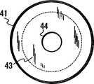
取样单元26包括一圆柱体41和一台座43。圆柱体41连接到台座43上,其中有一注入孔42用于注入样品。台座43被做成梯状柱体,且一垂直的孔44穿透台座43。凝胶48被置于圆柱体41中。凝胶48有几个毫米的厚度从而填充圆柱体41的开口。相应地,样品通过注入孔42被提供到凝胶48内。The
以下将解释连接部33。图10是连接部的透视图,图11是连接部的侧面截面视图。与取样单元26相似,Millipore的离心过滤单元被加工使得超过滤膜被移走,且单元中开有5mm直径的孔,从而构成了连接部33。连接部33包含一圆柱体41和一台座43。圆柱体41连接于台座43。台座43被做成梯状柱体,一垂直的孔44穿透台座43。具有几个毫米的厚度的凝胶48被置于圆柱体41中。在圆柱体41中,凝胶48被置于台座43的上表面内以防止液体从取样单元26中流出或流向取样单元26。The connecting
以下解释过滤部34。图12是过滤部的侧面截面图。过滤部34也通过加工Millipore的离心过滤单元构成。过滤部34包括一圆柱体41和一台座43。圆柱体41被连接到台座43上。该圆柱体41较取样单元26的圆柱体和连接部33的圆柱体41短约5mm。台座43被做成梯状柱体,一垂直孔44穿透台座43。在圆柱体41中,超滤膜49被置于台座43的上表面内以防止核酸流出,从而确保核酸的浓缩。The
接下来,解释核酸浓缩操作的实施方式。样品是大肠菌培养液,进行第一次和第二次电泳以浓缩核酸。回收的核酸的浓度通过吸光率测量法测定。100μL的大肠菌(Escherichia coli DH5α)的培养液被用做样品。100μL的1%Triton(注册商标)X-100被添加到样品中,且在96℃条件下加热10分钟。随后,100μL的0.2%DPC被添加到其中,从而制备用于电泳的样品。0.5x TAE用做电泳缓冲液。琼脂糖溶解在1x TAE中。此外,该1x TAE溶液从40mM Tris,40mM冰醋酸和1mM EDTA制备,其pH被调节到8.0.Next, an embodiment of the nucleic acid concentration operation is explained. The sample is a culture of coliform bacteria and undergoes first and second electrophoresis to concentrate nucleic acids. The concentration of recovered nucleic acid is determined by absorbance measurement. 100 µL of a culture solution of Escherichia coli DH5α was used as a sample. 100 μL of 1% Triton (registered trademark) X-100 was added to the sample, and heated at 96° C. for 10 minutes. Subsequently, 100 µL of 0.2% DPC was added thereto, thereby preparing a sample for electrophoresis. 0.5x TAE was used as electrophoresis buffer. Agarose was dissolved in 1x TAE. In addition, the 1x TAE solution was prepared from 40mM Tris, 40mM glacial acetic acid and 1mM EDTA, the pH of which was adjusted to 8.0.
连接部33被竖起来以使圆柱体41的开口翻转向上,1%琼脂糖凝胶(SeaKem Gold agarose:购自TaKaRa)通过该开口被添加到圆柱体41中使凝胶有几个毫米的厚度,然后使凝胶变硬。与连接部33相似,取样单元26被竖起来以使圆柱体41的开口向上,1%琼脂糖凝胶(SeaKem Gold agarose:购自TaKaRa)通过该开口被添加到圆柱体41中使得凝胶具有几个毫米的厚度。凝胶变硬后,取样单元26被倒置且凝胶被添加到圆柱体41的开口中。HU-6(AR BROWN制造)用做电泳槽,MPSU-200(AR BROWN制造)用做电源。The
制备用于电泳的样品通过注入孔42加入到取样单元26中,然后将孔关闭。电泳槽被封固(putty)分为负极侧部分和正极侧部分,0.5xTAE被加入到槽的两侧部分。取样单元26被放置于封固处以使取样单元26的上表面比缓冲液高。随后,施加100V的直流电压并进行第一次电泳20分钟。A sample prepared for electrophoresis is introduced into the
第一次电泳完成后,连接部33和过滤部34被连接到取样单元26上,以构建分离单元,并进行第二次电泳。After the first electrophoresis is completed, the
以下将给出对第二次电泳的操作的具体实施方式。以6∶4的比例混合100%乙醇和1x TAE所得溶液被加入连接部33中。TE-1(10mMTris-HCl,0.1mM EDTA,pH8.0)被添加入取样单元26中。A specific embodiment of the operation of the second electrophoresis will be given below. A solution obtained by mixing 100% ethanol and 1x TAE in a ratio of 6:4 was added to the
接下来,连接部33和过滤部34连接到取样单元26上以装配分离单元。图13是分离单元装配结构侧面图。分离单元从朝向同一方向的取样单元26、连接部33和过滤部34装配而来。O型环51被置于连接部33和过滤部34之间,以及连接部33和过滤部34之间,以防止溶液从分离单元中流出。Next, the
电泳槽被封固分为负极侧部分和正极侧部分,0.5x TAE被加入到槽的两侧。放入装配的分离单元以使得取样单元26被置于封固的负极侧,过滤部34被置于封固的正极侧。随后,施加100V的直流电压并进行第二次电泳240分钟。The electrophoresis tank was sealed and divided into a negative side part and a positive side part, and 0.5x TAE was added to both sides of the tank. The assembled separation unit was placed such that the
接下来,核酸溶液在过滤部34回收,其吸光率通过紫外线光谱测定以计算出回收核酸的浓度。图14图解的是回收溶液的紫外线光谱。计算出的核酸浓度为32.3ng(6.7×10⁶个拷贝/μL)。核酸的浓度如此计算,在260nm(A260)处的吸光率乘以核酸固有的系数,乘以比色皿的光程长度(mm),再除以10。Next, the nucleic acid solution is recovered in the
回收的核酸的纯度通过紫外线光谱计算。计算出来的回收核酸的纯度为1.91。纯度用260nm(A260)处的吸光率除以280nm(A280)处的吸光率来计算。当样品是纯度为100%的DNA时,计算值约为1.8。当样品是纯度为100%的RNA时,计算值约为2.0。混合在受测材料中的蛋白质和苯酚的量用A280的值来反应。当吸光率低于1.5时,可能混入了低分子物质,例如蛋白质。The purity of the recovered nucleic acid was calculated by UV spectroscopy. The calculated purity of the recovered nucleic acid was 1.91. Purity was calculated by dividing the absorbance at 260 nm (A260) by the absorbance at 280 nm (A280). When the sample is 100% pure DNA, the calculated value is about 1.8. When the sample is 100% pure RNA, the calculated value is about 2.0. The amount of protein and phenol mixed in the test material is reflected in the A280 value. When the absorbance is lower than 1.5, low-molecular substances such as proteins may be mixed.
根据本发明的利用电泳来浓缩和纯化核酸的方法,含有核酸的样品中的杂质的荷电量被调节,然后样品被置于电场中进行电泳以浓缩和纯化核酸。作为该方法的一个结果,电泳时核酸的迁移不同于杂质的迁移,这样可以有效地分离出核酸。杂质和核酸间的迁移差异可以通过荷电量的调节来调节,从而可以很容易的控制迁移。无需使用离心分离机或类似装置,从而用于浓缩核酸的装置可以很紧凑。According to the method for concentrating and purifying nucleic acid using electrophoresis of the present invention, the charged amount of impurities in a sample containing nucleic acid is adjusted, and then the sample is placed in an electric field for electrophoresis to concentrate and purify nucleic acid. As a result of this method, the migration of nucleic acids during electrophoresis differs from the migration of impurities, allowing efficient separation of nucleic acids. The difference in migration between impurities and nucleic acids can be adjusted by adjusting the amount of charge, so that the migration can be easily controlled. There is no need to use a centrifuge or the like, so that the apparatus for concentrating nucleic acid can be compact.
根据利用电泳来浓缩和纯化核酸的方法,阳离子表面活性剂被加入到含有核酸的样品中,以调节存在于样品中的杂质的荷电量,然后,该样品被放置于电场中进行电泳以浓缩和纯化核酸。归因于阳离子表面活性剂,该方法的操作很容易,且操作的安全性可以很容易地得到保证。通过增加对杂质的吸附,杂质和核酸间的迁移差异变大以有效地分离到核酸。杂质与核酸呈相反的方向迁移以防杂质降低核酸的纯度,从而可以容易地纯化核酸。According to the method of concentrating and purifying nucleic acid using electrophoresis, a cationic surfactant is added to a sample containing nucleic acid to adjust the charged amount of impurities present in the sample, and then the sample is placed in an electric field for electrophoresis to concentrate and Purify nucleic acids. Owing to the cationic surfactant, the method is easy to operate, and the safety of operation can be easily guaranteed. By increasing the adsorption of impurities, the migration difference between impurities and nucleic acids becomes larger to efficiently separate nucleic acids. The impurities migrate in the opposite direction to the nucleic acid to prevent impurities from reducing the purity of the nucleic acid, so that the nucleic acid can be easily purified.
根据本发明的利用电泳来浓缩和纯化核酸的方法,阳离子表面活性剂和非离子型表面活性剂被添加到含有核酸的样品中,以调节存在于样品中的杂质的荷电量,然后该样品被放置于电场中进行电泳以浓缩和纯化核酸。相应地,阳离子表面活性剂的吸附可以通过调节阳离子表面活性剂与非离子型表面活性剂两者间的比例来进行,从而可以容易地调节被电泳的杂质的迁移。非离子型表面活性剂吸附核酸从而防止阳离子表面活性剂吸附核酸。According to the method for concentrating and purifying nucleic acid using electrophoresis of the present invention, a cationic surfactant and a nonionic surfactant are added to a sample containing nucleic acid to adjust the charged amount of impurities present in the sample, and then the sample is Place in an electric field for electrophoresis to concentrate and purify nucleic acids. Accordingly, the adsorption of the cationic surfactant can be performed by adjusting the ratio between the cationic surfactant and the nonionic surfactant, so that the migration of the impurity to be electrophoresed can be easily adjusted. Nonionic surfactants adsorb nucleic acids thereby preventing cationic surfactants from adsorbing nucleic acids.
核酸以外的其它物质的荷电量通过其吸附到阳离子表面活性剂来进行调节,这种吸附则通过添加的非离子型表面活性剂的量来进行调节。相应地,杂质吸附到阳离子表面活性剂上的比例及非离子型表面活性剂可以容易地被控制。此外,杂质的荷电量可以通过简单的操作进行调节。非离子型表面活性剂吸附到核酸上以防止阳离子表面活性剂吸附核酸。The amount of charge of substances other than nucleic acids is regulated by their adsorption to cationic surfactants, which in turn is regulated by the amount of nonionic surfactants added. Accordingly, the ratio of impurities adsorbed to the cationic surfactant and the nonionic surfactant can be easily controlled. In addition, the charge amount of impurities can be adjusted by simple operations. Nonionic surfactants adsorb to nucleic acids to prevent cationic surfactants from adsorbing nucleic acids.
在装置中,阳离子表面活性剂和非离子型表面活性剂被添加到样品中,然后该样品被电泳以在正极侧浓缩和纯化核酸。相应地,该装置可以被简化来纯化样品中的核酸并调控核酸的迁移,从而在保证安全性的同时自身很紧凑。In the device, a cationic surfactant and a nonionic surfactant are added to a sample, and then the sample is subjected to electrophoresis to concentrate and purify nucleic acid on the positive electrode side. Accordingly, the device can be simplified to purify nucleic acids in samples and regulate the migration of nucleic acids, thereby being compact while ensuring safety.
利用电泳浓缩和纯化核酸的装置包括一容器,该容器的侧壁由绝缘体形成。该容器被用于防止扩散的导电性分离介质分为样品注入室和核酸回收室。该容器的两端通过缓冲液槽被分别连接到两个电极。因此,用于浓缩和纯化的装置可以简便地构成从而降低其生产成本。此外,该装置容易控制并具有高度的安全性。An apparatus for concentrating and purifying nucleic acid using electrophoresis includes a container whose side walls are formed of an insulator. The container is divided into a sample injection chamber and a nucleic acid recovery chamber by a conductive separation medium for diffusion prevention. Both ends of the container are respectively connected to two electrodes through buffer tanks. Therefore, the apparatus for concentration and purification can be constructed simply to reduce its production cost. In addition, the device is easy to control and has a high degree of safety.
接下来,以下将参照附图描述本发明的第二实施方案。Next, a second embodiment of the present invention will be described below with reference to the drawings.
图15是核酸浓缩单元一部分剖面透视图,图16是该单元的平面图,图17是该单元的侧面图。以下将描述图15-17所示的核酸浓缩单元。Fig. 15 is a partially cutaway perspective view of a nucleic acid concentrating unit, Fig. 16 is a plan view of the unit, and Fig. 17 is a side view of the unit. The nucleic acid enrichment unit shown in FIGS. 15-17 will be described below.
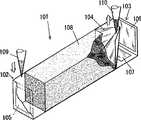
位于t-DNA检测器中的浓缩单元1分离并浓缩靶核酸。浓缩单元101包括注入室102,由分离介质形成的分离室108,和取样室103。含有靶核酸的样品通过注口109注入到注入室,对注入室102和取样室103施加电压以使核酸迁移进入取样室103中。已经通过分离室108的靶核酸流入到取样室103中,并通过注口110从取样室103中取样。生物样品,例如血液、尿、痰液或类似物,饮料或食物可以用做样品被注入到注入室102中。另外,基因组或质粒也能被注入。The concentration unit 1 located in the t-DNA detector separates and concentrates target nucleic acids. The
以下将详述浓缩单元101中的每一部分的结构。注入室102被连接到分离室108的一端,取样室103被连接到分离室108的另一端。电极105被置于注入室中,且电极106和107被置于取样室103中。注入室102和取样室103装满缓冲液用于电泳。取样室103中的电极106被置于面对注入室102中的电极,取样室103中的电极107被置于取样室103的底部。过滤器104被置于取样室103中以将取样室103分为分离室108和包括电极106的室。过滤器104允许小于靶核酸的物质通过,阻止不小于靶核酸的物质通过。过滤器104上有很多滤孔,它们具有例如阻止与靶核酸互相作用和阻止靶核酸通过这样的特性。The structure of each part in the
过滤器104被制成具有一底部开口和一侧面开口的四角锥形。锥体的底部开口朝向分离室108。锥体的侧面开口是一向上开口的水平面。相应地,过滤器104朝向分离室108的部分中的靶核酸可以通过注口110取样。随着靶核酸利用电泳向电极106迁移,靶核酸在过滤器104朝向电极106的部分中浓缩。The
接下来,将根据图18解释通过浓缩单元对靶核酸的浓缩过程。图18是靶核酸的浓缩过程的示意图。首先,图18(a)表示样品被注入到注入室102中的状态。为在此更清楚地进行说明,我们假设样品含有靶核酸112,大于靶核酸112(体积大或在分子量量上大)的核酸111,小于靶核酸的核酸112。Next, the process of concentrating the target nucleic acid by the concentrating unit will be explained based on FIG. 18 . Fig. 18 is a schematic diagram of a process for concentrating target nucleic acids. First, FIG. 18( a ) shows a state where a sample is injected into the
图18(a)表示大核酸111、靶核酸112和小核酸113被混合的状态。当电压施加在电极105和106上时,大核酸111,靶核酸112和小核酸113被引入到分离室108中,且在分离室108中以不同的速度迁移,如图18(b)所示。对于图18所示的结构,琼脂糖凝胶被用做分离介质填充分离室108,以使得小核酸113位于其它核酸之前。Fig. 18(a) shows a state where a large
在前的小核酸113通过分离室108到达取样室103。然后,小核酸113穿过过滤器104并迁移到取样室103中的电极106。随后,靶核酸112通过分离室108到达取样室103。因此,如图18(c)所示,过滤器104阻止靶核酸112进一步向电极106迁移。因为过滤器104被作成锥形,其底部开口朝向分离室108,其侧面开口向上,靶核酸112在过滤器104对着电极106的部分被浓缩。The preceding small
在这种状态下,停止通过电极105和106施加电压,电压通过电极107和106施加,如图18(d)所示。结果,电极106仍然捕获比靶核酸小的杂质。当通过两电极施加的电压被停止时,核酸从过滤器上被释放下来。这样,在靶核酸112从过滤器释放下来的过程中,防止了小的杂质和小核酸113的扩散,从而高精度浓缩靶核酸112。In this state, the voltage application through the
然后,靶核酸112通过分离室108的时间被计算出来以确定从样品注入到注入室2、从通过电极105和106到通过电极107和106的电压的改变的设定时间,从而自动地进行靶核酸的分离和浓缩。Then, the time for the target
任何用于电泳的凝胶,例如琼脂糖凝胶,可以被用做分离室108。也可以使用用作柱填充物的分离介质。例如,用于凝胶过滤的载体,如可使用Sephadex(注册商标)(Phatmacia)。还值得考虑的是用于电泳的凝胶与柱填充物在分离部8被结合起来并对之进行调节以使得靶核酸最先流出来。Any gel used for electrophoresis, such as agarose gel, can be used as the
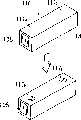
下面,将对根据图19和图20的浓缩单元的另一结构进行解释。图19是浓缩单元的开封过程的示意图,图20是浓缩单元的侧面截面图。浓缩单元被插入到一检测装置中以分离和浓缩靶核酸。Next, another structure of the concentrating unit according to FIGS. 19 and 20 will be explained. FIG. 19 is a schematic diagram of the unsealing process of the concentration unit, and FIG. 20 is a side sectional view of the concentration unit. The concentration unit is inserted into a detection device to separate and concentrate target nucleic acids.
浓缩单元116的上表面贴着膜117a和117b,电极105和106在浓缩单元116的侧面露出。电极7在其底面露出。电极105被连接到注入室,且电极106和107被连接到取样室。浓缩单元116中有分离室108,注入室和取样室填充有电泳缓冲液。取样室被过滤器分开。The upper surface of the
在这种状态下,浓缩单元116被密封且电压可以从外部施加于其上。相应地,浓缩单元可以容易的进行操作。当样品被注入到浓缩单元116中以对靶核酸取样时,膜117a和117b如图19所示被撕破,且样品被注入以对靶核酸取样。因为膜117a和117b被粘贴在浓缩单元的上表面上,在撕破膜117时没有对分离室108施加任何大的冲击,从而可以稳定地进行浓缩和分离。In this state, the concentrating
工业实用性:Industrial applicability:
如上述,本发明的方法简化了对核酸进行浓缩和纯化的操作,且根据本发明的用于核酸的浓缩和纯化的装置在结构上很简单,因此可用于自动检测装置以浓缩和检测核酸。As mentioned above, the method of the present invention simplifies the operation of concentrating and purifying nucleic acid, and the device for concentrating and purifying nucleic acid according to the present invention is simple in structure, so it can be used in an automatic detection device to concentrate and detect nucleic acid.
Claims (7)
Translated fromChineseApplications Claiming Priority (3)
| Application Number | Priority Date | Filing Date | Title |
|---|---|---|---|
| JP345210/2002 | 2002-11-28 | ||
| JP2002345210 | 2002-11-28 | ||
| JP379796/2003 | 2003-11-10 |
Related Child Applications (2)
| Application Number | Title | Priority Date | Filing Date |
|---|---|---|---|
| CNB200610141538XADivisionCN100491391C (en) | 2002-11-28 | 2003-11-27 | Method and apparatus for concentration and purification of nucleic acid |
| CN 200610141539DivisionCN1990498A (en) | 2002-11-28 | 2003-11-27 | Method and apparatus for concentration and purification of nucleic acid |
Publications (2)
| Publication Number | Publication Date |
|---|---|
| CN1717412Atrue CN1717412A (en) | 2006-01-04 |
| CN1332972C CN1332972C (en) | 2007-08-22 |
Family
ID=35822523
Family Applications (3)
| Application Number | Title | Priority Date | Filing Date |
|---|---|---|---|
| CN 200610141539PendingCN1990498A (en) | 2002-11-28 | 2003-11-27 | Method and apparatus for concentration and purification of nucleic acid |
| CNB200610141538XAExpired - Fee RelatedCN100491391C (en) | 2002-11-28 | 2003-11-27 | Method and apparatus for concentration and purification of nucleic acid |
| CNB2003801045404AExpired - Fee RelatedCN1332972C (en) | 2002-11-28 | 2003-11-27 | Method and apparatus for concentrating and purifying nucleic acids |
Family Applications Before (2)
| Application Number | Title | Priority Date | Filing Date |
|---|---|---|---|
| CN 200610141539PendingCN1990498A (en) | 2002-11-28 | 2003-11-27 | Method and apparatus for concentration and purification of nucleic acid |
| CNB200610141538XAExpired - Fee RelatedCN100491391C (en) | 2002-11-28 | 2003-11-27 | Method and apparatus for concentration and purification of nucleic acid |
Country Status (1)
| Country | Link |
|---|---|
| CN (3) | CN1990498A (en) |
Cited By (3)
| Publication number | Priority date | Publication date | Assignee | Title |
|---|---|---|---|---|
| CN102824854A (en)* | 2011-06-15 | 2012-12-19 | 杜权 | Electrophoresis apparatus and its application |
| WO2012171329A1 (en)* | 2011-06-15 | 2012-12-20 | Du Quan | Method for separating nucleic acid and use thereof |
| CN105873664A (en)* | 2013-12-23 | 2016-08-17 | 因斯布鲁克大学 | Electrochemical cell |
Families Citing this family (4)
| Publication number | Priority date | Publication date | Assignee | Title |
|---|---|---|---|---|
| CN101619289B (en)* | 2008-06-30 | 2012-04-25 | 广东出入境检验检疫局检验检疫技术中心 | Long fragment nucleic acid enrichment purification device |
| CN106770596B (en)* | 2016-12-02 | 2019-03-01 | 中国科学院生态环境研究中心 | Washing device for Protein Separation and the column gel electrophoresis apparatus using it |
| CN107058075B (en)* | 2017-06-20 | 2023-10-20 | 商丘师范学院 | Plant cell protoplast purification instrument and purification method |
| CN110656108B (en)* | 2019-10-31 | 2021-06-08 | 东莞市东阳光诊断产品有限公司 | Chip, nucleic acid extraction and purification device, and nucleic acid extraction and purification method |
Family Cites Families (6)
| Publication number | Priority date | Publication date | Assignee | Title |
|---|---|---|---|---|
| JPH0599899A (en)* | 1991-09-18 | 1993-04-23 | Hitachi Ltd | Method for separating and purifying biological materials |
| US6129828A (en)* | 1996-09-06 | 2000-10-10 | Nanogen, Inc. | Apparatus and methods for active biological sample preparation |
| US6146511A (en)* | 1998-01-30 | 2000-11-14 | The Perkin-Elmer Corporation | Electrophoretic nucleic acid purification method |
| US6699986B2 (en)* | 1998-08-04 | 2004-03-02 | Ortho-Clinical Diagnostics, Inc. | Electrophoretic separation of nucleic acids from proteins at low ph |
| JP2003500645A (en)* | 1999-05-19 | 2003-01-07 | ビラテック アクチェンゲゼルシャフト | Apparatus and method for isolating charged molecules |
| US7001724B1 (en)* | 2000-11-28 | 2006-02-21 | Applera Corporation | Compositions, methods, and kits for isolating nucleic acids using surfactants and proteases |
- 2003
- 2003-11-27CNCN 200610141539patent/CN1990498A/enactivePending
- 2003-11-27CNCNB200610141538XApatent/CN100491391C/ennot_activeExpired - Fee Related
- 2003-11-27CNCNB2003801045404Apatent/CN1332972C/ennot_activeExpired - Fee Related
Cited By (4)
| Publication number | Priority date | Publication date | Assignee | Title |
|---|---|---|---|---|
| CN102824854A (en)* | 2011-06-15 | 2012-12-19 | 杜权 | Electrophoresis apparatus and its application |
| WO2012171329A1 (en)* | 2011-06-15 | 2012-12-20 | Du Quan | Method for separating nucleic acid and use thereof |
| CN105873664A (en)* | 2013-12-23 | 2016-08-17 | 因斯布鲁克大学 | Electrochemical cell |
| CN105873664B (en)* | 2013-12-23 | 2019-12-06 | 因斯布鲁克大学 | electrochemical cell |
Also Published As
| Publication number | Publication date |
|---|---|
| CN1990498A (en) | 2007-07-04 |
| CN100491391C (en) | 2009-05-27 |
| CN1332972C (en) | 2007-08-22 |
| CN1990497A (en) | 2007-07-04 |
Similar Documents
| Publication | Publication Date | Title |
|---|---|---|
| CN106754292B (en) | The single-stranded separators of DNA and separation method for pyrosequencing | |
| KR101005924B1 (en) | Nucleic Acid Extraction Device | |
| AU772213B2 (en) | Biochemical purification devices with immobilized capture probes and their uses | |
| KR101006562B1 (en) | Nucleic Acid Extraction Method | |
| CN102888394A (en) | Nucleic-acid extraction method and nucleic-acid extraction cartridge | |
| EP1411340A2 (en) | Biochemical purification devices with immobilized capture probes and their uses | |
| CN1304589C (en) | Process for separating nucleic acid from biological particles by solid-phase carrier | |
| US20090000949A1 (en) | Method And Apparatus Of Concentration And Purification Of Nucleic Acid | |
| US8691559B2 (en) | Micro channel, device for recovering nucleic acid and method for recovering nucleic acid | |
| CN1717412A (en) | Method and apparatus for concentrating and purifying nucleic acids | |
| KR20150127917A (en) | Method of extracting and amplifying nucleic acids by using magnetic bead | |
| CN1681925A (en) | Method for the enrichment of prokaryotic DNA | |
| JPWO2005045023A1 (en) | Method and apparatus for concentration and purification of nucleic acid | |
| EP1568766B1 (en) | Device for pretreating specimen | |
| US20070197780A1 (en) | Method and apparatus for DNA purification | |
| US20140251809A1 (en) | Apparatus for preparing nucleic acids and method for preparing nucleic acids | |
| JP2004514548A (en) | A multi-step method for collecting or removing biological samples by floating from an underlying colloidal medium | |
| EP1462520A1 (en) | DNA isolation method | |
| JP2002345462A (en) | Adsorption buffer and method for purifying nucleic acid | |
| CN1796398A (en) | Method for extracting nucleic acid with amine surfactant |
Legal Events
| Date | Code | Title | Description |
|---|---|---|---|
| C06 | Publication | ||
| PB01 | Publication | ||
| C10 | Entry into substantive examination | ||
| SE01 | Entry into force of request for substantive examination | ||
| C14 | Grant of patent or utility model | ||
| GR01 | Patent grant | ||
| C17 | Cessation of patent right | ||
| CF01 | Termination of patent right due to non-payment of annual fee | Granted publication date:20070822 Termination date:20091228 |