CN1586404A - Method for detecting myocardial blood supply state based on myocardial edge tracking - Google Patents
Method for detecting myocardial blood supply state based on myocardial edge trackingDownload PDFInfo
- Publication number
- CN1586404A CN1586404ACN 200410062765CN200410062765ACN1586404ACN 1586404 ACN1586404 ACN 1586404ACN 200410062765CN200410062765CN 200410062765CN 200410062765 ACN200410062765 ACN 200410062765ACN 1586404 ACN1586404 ACN 1586404A
- Authority
- CN
- China
- Prior art keywords
- myocardial
- frame
- edge
- image
- particle
- Prior art date
- Legal status (The legal status is an assumption and is not a legal conclusion. Google has not performed a legal analysis and makes no representation as to the accuracy of the status listed.)
- Granted
Links
Images
Landscapes
- Ultra Sonic Daignosis Equipment (AREA)
Abstract
Translated fromChineseDescription
Translated fromChinese技术领域technical field
本发明涉及一种测定心肌供血状况的方法,尤其涉及对心肌背向散射积分起伏程度(以下简称CVIB)进行成像后测定心肌供血状况的方法,属于医学超声技术领域,特别用于心肌的超声组织定征,即心肌缺血的检测。The invention relates to a method for measuring myocardial blood supply status, in particular to a method for measuring myocardial blood supply status after imaging myocardial backscatter integral fluctuation (hereinafter referred to as CVIB). Characterization, the detection of myocardial ischemia.
背景技术Background technique
冠心病和心肌梗塞在人群中的发病率非常高,尤其在中老年人群中,是当今社会的主要致死病因。经临床研究发现,大多数冠心病或心肌梗塞患者,在患病初期,均有心肌缺血症状。因此,如果能够及时发现他们的心肌缺血症状,找出心肌缺血的位置,就可能更及时地采取治疗措施,对预防和救治冠心病或心肌梗塞的病人提供技术上的帮助。The incidence of coronary heart disease and myocardial infarction is very high in the population, especially among the middle-aged and elderly population, and they are the main causes of death in today's society. Clinical studies have found that most patients with coronary heart disease or myocardial infarction have symptoms of myocardial ischemia in the early stages of the disease. Therefore, if their symptoms of myocardial ischemia can be detected in time and the location of myocardial ischemia can be found out, treatment measures can be taken in a more timely manner, and technical help can be provided for the prevention and treatment of patients with coronary heart disease or myocardial infarction.
大量实验研究表明,心肌的超声背向散射积分(以下简称IB)可以成为一个定量诊断心肌缺血的新参数。在实验中可以观察到,IB随心动周期出现周期性的起伏,一般称这种周期性起伏的幅度为CVIB。一般来说,正常心肌的CVIB值较大,缺血心肌的CVIB值较小。如果能够得到心肌在整个心动周期的IB曲线,并计算CVIB值,就可以对心肌供血状况有一个基本了解。A large number of experimental studies have shown that the ultrasonic integral backscatter of the myocardium (hereinafter referred to as IB) can become a new parameter for the quantitative diagnosis of myocardial ischemia. It can be observed in experiments that IB fluctuates periodically with the cardiac cycle, and the amplitude of this periodic fluctuation is generally called CVIB. Generally speaking, the CVIB value of normal myocardium is larger and that of ischemic myocardium is smaller. If the IB curve of the myocardium in the whole cardiac cycle can be obtained and the CVIB value can be calculated, a basic understanding of the blood supply to the myocardium can be obtained.
中国专利ZL98203234.X公开了一种二维CVIB成像方法,其原理是:用颜色对不同的CVIB值进行编码(比如用从蓝色渐变到红色对应CVIB值从3dB变化到12dB);在B超图上选取关注区域(ROI),并计算出关注区域内每一心肌质点在一个心动周期内的CVIB值;然后选取对应心舒末期的一帧B超图像,对ROI内的心肌质点以与其对应的CVIB值为成像因子按预先设定的编码进行着色。颜色表示了心肌的供血状况,由于它是和以灰度表示的B超图像融合在一起的,B超图包含的是心脏的结构信息,这样心脏的供血信息和结构信息在一张图中有机地融合在一起,可以很直观地表现出关注区域在心脏中的相对位置及其供血功能情况。Chinese patent ZL98203234.X discloses a two-dimensional CVIB imaging method, the principle of which is: use color to encode different CVIB values (such as changing from blue to red corresponding to the CVIB value from 3dB to 12dB); Select the region of interest (ROI) on the map, and calculate the CVIB value of each myocardial particle in the region of interest within one cardiac cycle; then select a frame of B-ultrasound image corresponding to the end diastole, and correspond to the myocardial particle in the ROI The CVIB value of the imaging factor is colored according to a preset encoding. The color indicates the blood supply status of the myocardium. Since it is fused with the B-ultrasound image expressed in gray scale, the B-ultrasound image contains the structural information of the heart, so that the blood supply information and structural information of the heart are organically fused in one image. Together, the relative position of the region of interest in the heart and its blood supply function can be displayed intuitively.
上述方法从理论上讲,应该对关注区域内每一心肌质点分别进行追踪,计算各自的IB曲线和CVIB,最后成像。然而,由于追踪心肌质点比较困难,所以已有专利技术在成像时假定心肌的运动可以忽略,计算关注区域内某心肌质点A的IB曲线时,是找出各帧B超图像位置和A点重合的点所对应的射频信号,形成IB曲线,计算CVIB,这样的计算方式在某些情况下会产生误判。Theoretically speaking, the above method should track each myocardial particle in the region of interest, calculate the respective IB curve and CVIB, and finally image. However, due to the difficulty of tracking myocardial particles, the existing patented technology assumes that the movement of the myocardium can be ignored during imaging. When calculating the IB curve of a certain myocardial particle A in the area of interest, it is to find out that the position of the B-ultrasound image of each frame coincides with point A The radio frequency signal corresponding to the point of , forms the IB curve, and calculates the CVIB. This calculation method may cause misjudgment in some cases.
发明内容Contents of the invention
本发明的目的是提出一种测定心肌供血状况的方法,通过对关注区域内的心肌边缘进行自动追踪,在此基础上对关注区域内所有心肌质点在各帧B超图像上的位置进行追踪,以实现二维CVIB成像,提高CVIB成像的准确度,避免传统CVIB成像方法可能出现误判的情况。The purpose of the present invention is to propose a kind of method of measuring myocardial blood supply situation, by automatically tracking the myocardial edge in the area of interest, on this basis, the positions of all myocardial particles in the area of interest are tracked on each frame B-ultrasonic image, In order to realize two-dimensional CVIB imaging, improve the accuracy of CVIB imaging, and avoid the possible misjudgment of traditional CVIB imaging methods.
本发明提出的基于追踪心肌边缘测定心肌供血状况的方法,包括以下步骤:The method that the present invention proposes is based on tracing myocardial edge and measuring myocardial blood supply condition, comprises the following steps:
(1)建立背向散射积分起伏程度与颜色的映射表;(1) Establish a mapping table of backscattering integral fluctuations and colors;
(2)扫描被测对象心脏,连续采集超声射频信号;(2) scan the heart of the measured object, and continuously collect ultrasonic radio frequency signals;
(3)根据上述超声射频信号,重建反映扫描区域结构的B超图像序列;(3) Reconstructing a B-ultrasound image sequence reflecting the scanning region structure according to the above-mentioned ultrasonic radio frequency signal;
(4)在上述B超图像序列的第一帧中选取矩形关注区域;(4) Select a rectangular area of interest in the first frame of the above-mentioned B-ultrasonic image sequence;
(5)对上述关注区域内的心肌边缘在各帧的位置进行追踪;(5) Track the position of the myocardial edge in each frame in the above-mentioned attention region;
(6)根据上述追踪到的心肌边缘在各帧B超图像中的位置,对关注区域内所有心肌质点在各帧位置进行计算;(6) According to the position of the myocardial edge tracked above in each frame of the B-ultrasound image, calculate the positions of all myocardial particles in the region of interest at each frame position;
(7)根据上述关注区域内每一心肌质点在各帧图像中的位置,找出各心肌质点在不同时刻对应的射频信号段,根据射频信号段计算各心肌质点的背向散射积分曲线;(7) According to the position of each myocardial mass point in each frame image in the above-mentioned attention area, find out the radio frequency signal segment corresponding to each myocardial mass point at different times, calculate the backscatter integral curve of each myocardial mass point according to the radio frequency signal segment;
(8)根据关注区域内每个心肌质点的背向散射积分曲线,计算各心肌质点的背向散射积分起伏程度,根据上述映射表,得到与关注区域内每个心肌质点的供血状况相对应的颜色图像;(8) According to the backscatter integral curve of each myocardial particle in the attention area, calculate the backscatter integral fluctuation degree of each myocardial particle, and according to the above mapping table, obtain the blood supply corresponding to each myocardial particle in the attention area color image;
(9)将上述颜色图像与上述B超图像序列的第一帧相融合,得到关注区域内每个心肌质点的供血状况图。(9) The above color image is fused with the first frame of the above B-ultrasound image sequence to obtain the blood supply status map of each myocardial particle in the attention area.
上述方法中追踪心肌边缘在其余各帧B超图像上的位置的方法,其过程包括以下步骤:In the above method, the method for tracking the position of the edge of the myocardium on the B-ultrasound images of the remaining frames comprises the following steps:
(1)将B超图像序列上选取的矩形关注区域从原图像中分离,得到一组子图像;(1) Separate the rectangular area of interest selected on the B-ultrasonic image sequence from the original image to obtain a group of sub-images;
(2)从上述子图像的第一帧上勾画出心肌边缘的初始轮廓线;(2) delineate the initial contour line of the myocardial edge from the first frame of the above-mentioned sub-image;
(3)对子图像和初始轮廓线进行坐标变换、图像翻转和合并,形成封闭的心肌边缘和初始轮廓线;(3) Coordinate transformation, image flipping and merging are performed on the sub-image and the initial contour line to form a closed myocardial edge and initial contour line;
(4)用主动轮廓线模型法,根据上述封闭的初始轮廓线对相应的封闭心肌边缘进行边缘检测;(4) Using the active contour line model method, according to the above-mentioned closed initial contour line, the edge detection is carried out to the corresponding closed myocardial edge;
(5)对上述检测到的边缘,进行坐标变换、图像翻转和合并的逆变换,得到原始子图像中的心肌边缘;(5) For the above-mentioned detected edge, carry out the inverse transformation of coordinate transformation, image flipping and merging, and obtain the myocardial edge in the original sub-image;
(6)以本次检测到的心肌边缘作为下一帧的子图像的初始轮廓线,重复步骤(3)到(5),得到各帧B超图像上关注区域内的心肌边缘。(6) Using the detected myocardial edge as the initial contour line of the sub-image of the next frame, repeat steps (3) to (5) to obtain the myocardial edge in the region of interest on each frame of B-ultrasound image.
上述方法中,根据心肌边缘在各帧B超图像中的位置,对关注区域内所有心肌质点在各帧位置进行计算的方法,当扫描视图为左心室短轴时,其过程包括以下步骤:In the above method, according to the position of the myocardial edge in each frame of the B-ultrasound image, the method for calculating the positions of all myocardial particles in the region of interest in each frame, when the scan view is the short axis of the left ventricle, the process includes the following steps:
(1)根据追踪到的心肌边缘,拟合得到左心室在各帧B超图像的中心;(1) According to the traced myocardial edge, the center of the left ventricle in each frame of the B-ultrasound image is obtained by fitting;
(2)计算第一帧B超图像中,待追踪的心肌质点和左心室中心的连线与水平方向的夹角θ;(2) Calculate the included angle θ between the connection line between the myocardial particle to be tracked and the center of the left ventricle and the horizontal direction in the first frame of B-ultrasound image;
(3)计算各帧中以左心室中心为端点的与水平方向夹角为θ的射线与心内膜及心外膜的交点;(3) Calculate the intersection point of the ray with the center of the left ventricle as the endpoint and the angle θ with the horizontal direction and the endocardium and epicardium in each frame;
(4)设心肌在径向为均匀收缩,计算待追踪心肌质点在各帧B超图像上的位置。(4) Let the myocardium contract uniformly in the radial direction, and calculate the position of the myocardial particle to be tracked on each frame of the B-ultrasound image.
当扫描视图为左心室长轴时,其过程包括以下步骤:When the scan view is the long axis of the left ventricle, the procedure includes the following steps:
(1)根据追踪到的心肌边缘,拟合得到左心室在各帧B超图像的中心、心肌边缘与左心室中心构成的扇形角度以及扇形的两边界位置;(1) According to the traced myocardial edge, the center of the left ventricle in each frame of B-ultrasound images, the fan angle formed by the myocardial edge and the center of the left ventricle, and the positions of the two boundaries of the fan are obtained by fitting;
(2)计算第一帧B超图像中,待追踪的心肌质点和左心室中心之间的连线与扇形左边界的夹角α1;(2) Calculate the angle α1 between the line between the myocardial particle to be tracked and the center of the left ventricle and the fan-shaped left boundary in the first frame of B-ultrasound image;
(3)根据上述夹角α1,计算待追踪心肌质点和左心室中心的连线在其余各帧中与扇形左边界的夹角αi,i代表B超图像序列的帧序数;(3) According to the above-mentioned included angle α1 , calculate the included angle αi between the connection line between the myocardial particle to be tracked and the center of the left ventricle and the left boundary of the fan in the remaining frames, where i represents the frame number of the B-ultrasound image sequence;
(4)计算各帧中,以左心室的中心为端点的、和扇形左边界的夹角为αi的射线与心内膜及心外膜的交点;(4) In each frame, the intersection point of the ray with the center of the left ventricle as the end point and the angle αi with the fan-shaped left boundary and the endocardium and epicardium;
(5)设心肌在径向为均匀收缩,计算待追踪心肌质点在各帧B超图像上的位置。(5) Assuming that the myocardium contracted uniformly in the radial direction, calculate the position of the myocardial particle to be tracked on each frame of the B-ultrasound image.
利用本发明提出的测定心肌供血状况的方法,分别对正常人,心肌缺血病人,按照本发明所说CVIB成像方法进行CVIB成像。成像结果显示,对于正常人,选取任何位置的心肌作为关注区域,关注区域内心肌标注的颜色主要均是红色和绿色,表明心肌正常,而对心肌缺血的病人,成像结果显示,在缺血位置的心肌颜色被标注上了蓝色,表明缺血,其它位置以红色和绿色为主,表明正常。这说明本发明所提出方法能够有效地区分正常心肌和缺血心肌。另外,用本发明所提测定心肌供血状况的方法和已有专利的方法分别对缺血对象的心肌进行CVIB成像,结果表明,对已有专利报导方法可能导致误判的情况,用本发明所提方法能够有效避免误判。Using the method for measuring myocardial blood supply proposed by the present invention, CVIB imaging is performed on normal people and myocardial ischemia patients according to the CVIB imaging method of the present invention. The imaging results show that, for normal people, select the myocardium at any location as the region of interest, and the colors marked on the myocardium in the region of interest are mainly red and green, indicating that the myocardium is normal. The myocardium in the location is colored blue, indicating ischemia, and the other locations are dominated by red and green, indicating normal. This shows that the method proposed by the present invention can effectively distinguish between normal myocardium and ischemic myocardium. In addition, the method for measuring myocardial blood supply proposed by the present invention and the existing patented method were used to perform CVIB imaging on the myocardium of the ischemic subject respectively. The proposed method can effectively avoid misjudgment.
附图说明:Description of drawings:
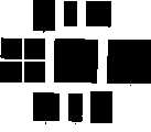
图1为本发明方法中重建的B超图像,矩形框代表所选取的关注区域,其中图1(a)为长轴视图,图1(b)短轴视图。Fig. 1 is the B-ultrasound image reconstructed in the method of the present invention, and the rectangular frame represents the selected attention area, and wherein Fig. 1 (a) is long-axis view, and Fig. 1 (b) short-axis view.
图2为用主动轮廓线模型法对非封闭边界进行检测的示意图。其中(a)为原始图像的一部分,矩形框为关注区域,曲线为初始轮廓;(b)为关注区域包含子图像经旋转后形成的正直矩形图像;(c)为从图(b)直角坐标转换到极坐标的结果;(d)为将图(c)翻转、合并示意图;(e)为合并结果;(f)用Snakes方法对图(e)追踪得到的边界(g)-(i)为(c)到(e)的逆变换;(i)图中曲线段为最后追踪到的心外膜。Fig. 2 is a schematic diagram of detecting non-closed boundaries with the active contour model method. Where (a) is a part of the original image, the rectangular frame is the region of interest, and the curve is the initial contour; (b) is the upright rectangular image formed by the region of interest including the sub-image after rotation; (c) is the Cartesian coordinates from (b) The result of converting to polar coordinates; (d) is the schematic diagram of flipping and merging the graph (c); (e) is the merged result; (f) the boundary (g)-(i) obtained by tracing the graph (e) with the Snakes method It is the inverse transformation from (c) to (e); the curve segment in (i) is the last tracked epicardium.
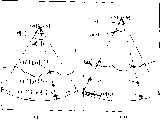
图3为左心室短轴视图中,利用心肌边缘在各帧B超图像位置对关注区域内心肌质点在各帧B超图像中位置进行追踪的示意图。3 is a schematic diagram of tracking the positions of myocardial particles in the region of interest in each frame of B-ultrasound images by using the edge of the myocardium in each frame of B-ultrasound images in the short-axis view of the left ventricle.
图4为左心室长轴视图中,利用心肌边缘在各帧B超图像位置对关注区域内心肌质点在各帧B超图像中位置进行追踪的示意图。Fig. 4 is a schematic diagram of tracking the positions of myocardial particles in the region of interest in each frame of B-ultrasound images by using the edge of the myocardium in each frame of B-ultrasound images in the long-axis view of the left ventricle.
具体实施方式Detailed ways
本发明提出的基于追踪心肌边缘测定心肌供血状况的方法,包括以下步骤:The method that the present invention proposes is based on tracing myocardial edge and measuring myocardial blood supply condition, comprises the following steps:
(1)建立背向散射积分起伏程度与颜色的映射表。用从蓝色渐变到绿色再渐变到红色来对应CVIB值由低到高的变化。即蓝色代表心肌缺血,红色代表心肌供血状况正常。(1) Establish a mapping table of backscattering integral fluctuations and colors. Use a gradient from blue to green and then to red to correspond to the change of CVIB value from low to high. That is, blue represents myocardial ischemia, and red represents normal myocardial blood supply.
(2)扫描被测对象心脏,连续采集超声射频信号。本发明一实施例中,用常用B超设备的探头对被测对象进行扫描,从B超设备内部电路提取波束合成后的射频信号,经过放大,用高速数据采集卡进行采集,一般要求采集时间超过1秒,采样率10M,本发明一实施例使用的是AD-LINK公司的PCI-9812数据采集卡。(2) Scanning the heart of the subject to be measured, and continuously collecting ultrasonic radio frequency signals. In one embodiment of the present invention, the probe of the B-ultrasound equipment commonly used is used to scan the measured object, and the radio frequency signal after the beam synthesis is extracted from the internal circuit of the B-ultrasound equipment, after being amplified, the high-speed data acquisition card is used for acquisition, and the acquisition time is generally required More than 1 second, the sampling rate is 10M, and what one embodiment of the present invention uses is the PCI-9812 data acquisition card of AD-LINK Company.
(3)根据上述超声射频信号,重建反映扫描区域结构的B超图像序列。重建B超图像序列的方法为:对射频信号去直流后,做HILBERT变换,然后按照B超系统中的DSC算法,即可形成最后图像。(3) Reconstruct the B-ultrasound image sequence reflecting the structure of the scanned area according to the above-mentioned ultrasonic radio frequency signal. The method of reconstructing the B-ultrasound image sequence is as follows: After removing the direct current from the radio frequency signal, perform HILBERT transformation, and then follow the DSC algorithm in the B-ultrasound system to form the final image.
(4)在上述B超图像序列的第一帧中选取关注区域,一般对短轴视图,选取关注区域应该包含整个左心室壁,对长轴视图,关注区域选取左心室后壁。有关关注区域选取位置见图1(a)和图1(b)的矩形框。(4) Select the region of interest in the first frame of the above-mentioned B-ultrasound image sequence. Generally, for the short-axis view, the selected region of interest should include the entire left ventricular wall, and for the long-axis view, the region of interest is selected for the posterior wall of the left ventricle. See the rectangular boxes in Figure 1(a) and Figure 1(b) for the location of the attention area.
(5)对上述关注区域内的心肌边缘在各帧的位置进行追踪。对关注区域内的心肌边缘用基于Snakes的方法进行追踪,对短轴视图封闭的心肌边缘,用传统Snakes方法进行边缘检测、追踪即可,对长轴视图,心肌边缘是非封闭的,不能直接使用Snakes方法,所以用本发明提出的针对非封闭边界的Snakes法进行追踪。(5) Track the position of the myocardial edge in each frame in the above-mentioned attention area. Use the Snakes-based method to track the myocardial edge in the area of interest. For the closed myocardial edge in the short-axis view, use the traditional Snakes method to detect and track the edge. For the long-axis view, the myocardial edge is not closed and cannot be used directly. Snakes method, so use the Snakes method proposed by the present invention for non-closed boundaries to track.
(6)根据上述追踪到的心肌边缘在各帧B超图像中的位置,对关注区域内所内所有心肌质点在各帧位置进行计算。(6) Calculate the positions of all myocardial particles in the region of interest in each frame according to the tracked positions of the myocardial edges in each frame of the B-ultrasound image.
(7)根据上述关注区域内每一心肌质点在各帧图像中的位置,找出各心肌质点在不同时刻对应的射频信号段,根据射频信号段计算各心肌质点的背向散射积分曲线。计算IB的公式为:(7) Find out the radio frequency signal segments corresponding to each myocardial mass point at different times according to the position of each myocardial mass point in each frame image in the above-mentioned attention area, and calculate the backscatter integral curve of each myocardial mass point according to the radio frequency signal segment. The formula for calculating IB is:
公式(1)中,x(t)是指射频信号。In formula (1), x(t) refers to the radio frequency signal.
(8)根据关注区域内每个心肌质点的背向散射积分曲线,计算各心肌质点的背向散射积分起伏程度,根据上述映射表,得到与关注区域内每个心肌质点的供血状况相对应的颜色图像。计算CVIB可以采取公开文献的任何一种方法,本实施例使用一阶傅立叶级数法,其过程为:(8) According to the backscatter integral curve of each myocardial particle in the attention area, calculate the backscatter integral fluctuation degree of each myocardial particle, and according to the above mapping table, obtain the blood supply corresponding to each myocardial particle in the attention area color image. Calculating CVIB can take any method in the open literature, and this embodiment uses the first-order Fourier series method, and its process is:
CVIB=4*max(|X(k)|)/N (2)CVIB=4*max(|X(k)|)/N (2)
其中,N是IB序列的长度,X(k)是长度为N的IB序列在频域10*N点的CZT变换。Wherein, N is the length of the IB sequence, and X(k) is the CZT transformation of the IB sequence with a length of N at 10*N points in the frequency domain.
(9)将上述颜色图像与上述B超图像序列的第一帧相融合,得到关注区域内每个心肌质点的供血状况图。融合方式为:在上述B超图像序列的第一帧所标注的点,以其CVIB值对应的颜色进行着色即可。(9) The above color image is fused with the first frame of the above B-ultrasound image sequence to obtain the blood supply status map of each myocardial particle in the attention area. The fusion method is as follows: the points marked in the first frame of the above-mentioned B-ultrasound image sequence are colored with the color corresponding to the CVIB value.
上述方法中追踪非封闭的心肌边缘在其余各帧B超图像上的位置的方法,其过程包括以下步骤:In the method for tracking the position of the non-closed myocardial edge on the remaining frames of B-ultrasound images, the process includes the following steps:
(1)将B超图像序列上选取的矩形关注区域从原图像中分离,得到一组子图像。即将图2(a)中红色矩形框中的部分单独分离出来,如果选择的非正直矩形区域,可以经过旋转形成正直矩形,如果图2(b)所示。(1) Separate the rectangular region of interest selected on the B-ultrasound image sequence from the original image to obtain a set of sub-images. That is to separate the part in the red rectangle frame in Figure 2(a), if the non-right rectangular area is selected, it can be rotated to form an upright rectangle, as shown in Figure 2(b).
(2)从上述子图像的第一帧上勾画出心肌边缘的初始轮廓线。一般可以利用常规计算机绘软件进行手工勾划。(2) Outline the initial contour line of the myocardial edge from the first frame of the above sub-image. Generally, conventional computer drawing software can be used for manual sketching.
(3)对子图像和初始轮廓线进行坐标变换、图像翻转和合并,形成封闭的心肌边缘和初始轮廓线。首先对矩形关注区域实施从直角坐标到极坐标的转换,在极坐标中图像的角度范围为90度(类似于扇扫B超中的DSC过程)。为了转换方便,首先将关注区域进行旋转,使倾斜的矩形变成竖直的矩形(图2(b)),直角坐标到极坐标的转换结果见图2(c)。把2(c)的图形分别进行上下、左右、180度翻转,得到四幅图(见图2(d)),将四幅图合并,得到图形转换最后结果(图2(e))。对于初始化轮廓,我们也进行了同样的操作,结果见图2(e)中的虚线。经过这一步,最后我们得到的图像上就有了封闭的边界和封闭的初始轮廓线。(3) Coordinate transformation, image flipping and merging are performed on the sub-image and the initial contour line to form a closed myocardial edge and the initial contour line. Firstly, the transformation from Cartesian coordinates to polar coordinates is implemented for the rectangular region of interest, and the angle range of the image in polar coordinates is 90 degrees (similar to the DSC process in sector-scan B-ultrasound). For the convenience of conversion, firstly, the focus area is rotated so that the oblique rectangle becomes a vertical rectangle (Fig. 2(b)). The conversion result from Cartesian coordinates to polar coordinates is shown in Fig. 2(c). Flip the graphics in 2(c) up and down, left and right, and 180 degrees to obtain four pictures (see Figure 2(d)), and combine the four pictures to obtain the final result of graphic conversion (Figure 2(e)). We do the same for the initialization contour, and the result is shown as the dashed line in Fig. 2(e). After this step, we finally have a closed boundary and a closed initial contour line on the image we get.
(4)用主动轮廓线模型法,根据上述封闭的初始轮廓线对相应的封闭心肌边缘进行边缘检测,结果见图2(f)。(4) Use the active contour line model method to perform edge detection on the corresponding closed myocardial edge according to the above closed initial contour line, and the results are shown in Fig. 2(f).
(5)对上述检测到的边缘,进行坐标变换、图像翻转和合并的逆变换,得到原始子图像中的心肌边缘。这个过程和由图2(a)到图2(e)的过程正好相反,每一步变换的结果参见图2(g),图2(h),图2(i),最终追踪到的关注区域内心外膜的位置如图2(i)所示。(5) Carry out inverse transformation of coordinate transformation, image flipping and merging on the above detected edge to obtain the myocardial edge in the original sub-image. This process is just the opposite of the process from Figure 2(a) to Figure 2(e). The results of each step of transformation can be seen in Figure 2(g), Figure 2(h), and Figure 2(i), and the final tracked area of interest The location of the inner and outer layers is shown in Figure 2(i).
(6)以本次检测到的心肌边缘作为下一帧的子图像的初始轮廓线,重复步骤(3)到(5),得到各帧B超图像上关注区域内的心肌边缘。(6) Using the detected myocardial edge as the initial contour line of the sub-image of the next frame, repeat steps (3) to (5) to obtain the myocardial edge in the region of interest on each frame of B-ultrasound image.
对左心长轴非封闭的心内膜和心外膜都可以使用以上方法,实行对心内膜和心外膜在不同帧的位置进行追踪。The above method can be used for both the non-closed endocardium and epicardium of the long axis of the left heart, and the positions of the endocardium and epicardium in different frames are tracked.
上述方法中,根据心肌边缘在各帧B超图像中的位置,对关注区域内所有心肌质点在各帧位置进行计算的方法,当扫描视图为左心室短轴时,参见图3,其过程包括以下步骤:In the above method, according to the position of the myocardial edge in each frame of the B-ultrasound image, the method of calculating the positions of all myocardial particles in the region of interest in each frame, when the scan view is the short axis of the left ventricle, see Figure 3, the process includes The following steps:
(1)根据追踪到的心肌边缘,拟合得到左心室在各帧B超图像的中心(Ox[i],Oy[i])。拟合方式为:找出和心肌边缘各点的距离方差最小的点即是中心点。图3中的(Ox[1],Oy[1])和(Ox[i],Oy[i])分别为第1帧和第i帧拟合得到的左心室中心。(1) According to the tracked myocardial edge, the center (Ox[i], Oy[i]) of the left ventricle in each frame of B-ultrasound images is obtained by fitting. The fitting method is: find out the point with the smallest distance variance from each point on the edge of the myocardium, which is the central point. (Ox[1], Oy[1]) and (Ox[i], Oy[i]) in Figure 3 are the left ventricle centers obtained by fitting the first frame and the i-th frame, respectively.
(2)计算第一帧B超图像中,待追踪的心肌质点和左心室中心的连线与水平方向的夹角θ,如图3所示,计算公式为:(2) Calculate the angle θ between the line connecting the myocardial particle to be tracked and the center of the left ventricle and the horizontal direction in the first frame of B-ultrasound image, as shown in Figure 3, the calculation formula is:
θ=atan2(y[1]-Oy[1],x[1]-Ox[1]) (1)θ=atan2(y[1]-Oy[1], x[1]-Ox[1])
式(1)中,x[1],y[1]是指待追踪心肌质点在第一帧的坐标,Ox[1],Oy[1]是左心室在第一帧的中心坐标,atan2是MATLAB函数,返回的是0~2π的角度。In formula (1), x[1], y[1] refer to the coordinates of the myocardial particle to be tracked in the first frame, Ox[1], Oy[1] are the center coordinates of the left ventricle in the first frame, and atan2 is The MATLAB function returns the angle from 0 to 2π.
(3)计算各帧中以左心室中心为端点的与水平方向夹角为θ的射线与心内膜及心外膜的交点(xi[i],yi[i])和(xo[i],yo[i])。,此处i为B超图像序列的帧序数。我们假设心肌整体是没有旋转的,所以待追踪的心肌质点在其余各帧中和左心室的连线与水平方向的夹角也是θ,因此,待追踪心肌质点应该在(xi[i],yi[i])和(xo[i],yo[i])连线上。参见图3(b)。(3) Calculate the intersection points (xi[i], yi[i]) and (xo[i]) of the ray with the center of the left ventricle as the endpoint and the angle θ with the horizontal direction and the endocardium and epicardium in each frame , yo[i]). , where i is the frame number of the B-ultrasound image sequence. We assume that the whole myocardium does not rotate, so the angle between the line connecting the left ventricle and the left ventricle of the myocardial particle to be tracked in the remaining frames is also θ. Therefore, the myocardial particle to be tracked should be in (xi[i], yi [i]) and (xo[i], yo[i]) are connected. See Figure 3(b).
(4)设心肌在径向为均匀收缩,计算待追踪心肌质点在各帧B超图像上的位置。计算公式为:(4) Let the myocardium contract uniformly in the radial direction, and calculate the position of the myocardial particle to be tracked on each frame of the B-ultrasound image. The calculation formula is:
x[i]=xi[i]+(xo[i]-xi[i])·(x[1]-xi[1])/(xo[1]-xi[1]) (2)x[i]=xi[i]+(xo[i]-xi[i])·(x[1]-xi[1])/(xo[1]-xi[1]) (2)
y[i]=yi[i]+(yo[i]-y[i])·(y[1]-yi[1])/(yo[1]-yi[1]) (3)y[i]=yi[i]+(yo[i]-y[i])·(y[1]-yi[1])/(yo[1]-yi[1]) (3)
以上以在第一帧坐标(x[1],y[1])的待追踪的心肌质点为例,描述了短轴视图中利用心肌边缘计算其在其余帧位置的过程。对关注区域内其余心肌质点实施同样过程,就完成了关注区域内所有心肌质点的追踪。Taking the myocardial particle to be tracked at the first frame coordinates (x[1], y[1]) as an example, the process of calculating its position in the remaining frames by using the myocardial edge in the short-axis view is described above. Carrying out the same process for the rest of the myocardial particles in the region of interest completes the tracking of all the myocardial particles in the region of interest.
当扫描视图为左心室长轴时,其过程包括以下步骤:When the scan view is the long axis of the left ventricle, the procedure includes the following steps:
(1)根据追踪到的心肌边缘,拟合得到左心室在各帧B超图像的中心、心肌边缘与左心室中心构成的扇形角度θ[i](i代表B超图像序列的帧序数)以及扇形的两边界位置。图4(a)和图4(b)显示了第1帧和第i帧左心室中心,心肌边缘和左心室中心构成的扇形。(1) According to the traced myocardial edge, fit the left ventricle at the center of each frame of B-ultrasound image, the fan angle θ[i] formed by the myocardial edge and the center of the left ventricle (i represents the frame number of the B-ultrasound image sequence) and The positions of the two boundaries of the sector. Figure 4(a) and Figure 4(b) show the sector formed by the center of the left ventricle, the edge of the myocardium and the center of the left ventricle in
(2)计算第一帧B超图像中,待追踪的心肌质点和左心室中心之间的连线与扇形左边界的夹角α1;(2) Calculate the angle α1 between the line between the myocardial particle to be tracked and the center of the left ventricle and the fan-shaped left boundary in the first frame of B-ultrasound images;
(3)根据上述夹角α1,计算待追踪心肌质点和左心室中心的连线在其余各帧中与扇形左边界的夹角αi,i代表B超图像序列的帧序数。假定扇形的弧度变化是均匀的,则:(3) According to the above angle α1 , calculate the angle αi between the line connecting the myocardial particle to be tracked and the center of the left ventricle and the left boundary of the fan in the remaining frames, where i represents the frame number of the B-ultrasound image sequence. Assuming that the radian change of the sector is uniform, then:
α[i]=α[1]*θ[1]/θ[i] (4)α[i]=α[1]*θ[1]/θ[i]
(4)计算各帧中,以左心室的中心为端点的、和扇形左边界的夹角为αi的射线与心内膜及心外膜的交点(xi[i],yi[i]),(xo[i],yo[i])。(4) Calculate the intersection point (xi[i], yi[i]) of the ray with the center of the left ventricle as the endpoint and the angle αi between the left boundary of the fan and the endocardium and epicardium in each frame , (xo[i], yo[i]).
(5)设心肌在径向为均匀收缩,计算得到待追踪心肌质点在各帧B超图像上的位置。计算公式按照公式(2)和公式(3)即可。(5) Assuming that the myocardium contracted uniformly in the radial direction, the position of the myocardial particle to be tracked on each frame of the B-ultrasound image was calculated. The calculation formula can be according to formula (2) and formula (3).
以上以在第一帧坐标(x[1],y[1])的待追踪的心肌质点为例,描述了长轴视图中利用心肌边缘计算其在其余帧位置的过程。对关注区域内其余心肌质点实施同样过程,就完成了关注区域内所有心肌质点的追踪。Taking the myocardial particle to be tracked at the first frame coordinates (x[1], y[1]) as an example, the process of calculating its position in the remaining frames by using the myocardial edge in the long-axis view is described above. Carrying out the same process for the rest of the myocardial particles in the region of interest completes the tracking of all the myocardial particles in the region of interest.
Claims (4)
Translated fromChinesePriority Applications (1)
| Application Number | Priority Date | Filing Date | Title |
|---|---|---|---|
| CNB2004100627654ACN1302751C (en) | 2004-07-09 | 2004-07-09 | Method for detecting myocardial blood supply state based on myocardial edge tracking |
Applications Claiming Priority (1)
| Application Number | Priority Date | Filing Date | Title |
|---|---|---|---|
| CNB2004100627654ACN1302751C (en) | 2004-07-09 | 2004-07-09 | Method for detecting myocardial blood supply state based on myocardial edge tracking |
Publications (2)
| Publication Number | Publication Date |
|---|---|
| CN1586404Atrue CN1586404A (en) | 2005-03-02 |
| CN1302751C CN1302751C (en) | 2007-03-07 |
Family
ID=34603776
Family Applications (1)
| Application Number | Title | Priority Date | Filing Date |
|---|---|---|---|
| CNB2004100627654AExpired - Fee RelatedCN1302751C (en) | 2004-07-09 | 2004-07-09 | Method for detecting myocardial blood supply state based on myocardial edge tracking |
Country Status (1)
| Country | Link |
|---|---|
| CN (1) | CN1302751C (en) |
Cited By (7)
| Publication number | Priority date | Publication date | Assignee | Title |
|---|---|---|---|---|
| CN100418478C (en)* | 2006-06-08 | 2008-09-17 | 上海交通大学 | Color mapping method of virtual endoscope surface based on blood flow imaging |
| CN101380238B (en)* | 2007-09-07 | 2011-05-25 | 株式会社东芝 | Ultrasonic diagnostic device, ultrasonic image processing device and method |
| CN101606850B (en)* | 2008-06-19 | 2012-01-04 | 株式会社东芝 | Ultrasonic diagnostic device, ultrasonic image processing device, and medical image processing device |
| CN101292266B (en)* | 2005-10-20 | 2012-05-30 | 皇家飞利浦电子股份有限公司 | Ultrasonic imaging system and method |
| CN101658433B (en)* | 2008-08-29 | 2013-10-02 | 株式会社东芝 | Ultrasonic diagnosis apparatus, image processing apparatus, and image processing method |
| CN107427279A (en)* | 2015-03-10 | 2017-12-01 | 皇家飞利浦有限公司 | Use the Ultrasonic Diagnosis of the cardiac function of the cardiac module chamber with user's control |
| CN107708570A (en)* | 2015-07-10 | 2018-02-16 | 深圳迈瑞生物医疗电子股份有限公司 | A monitoring system, method and device |
Family Cites Families (6)
| Publication number | Priority date | Publication date | Assignee | Title |
|---|---|---|---|---|
| DE69029211T2 (en)* | 1989-02-16 | 1997-03-27 | Fujitsu Ltd | Ultrasound diagnostic device for characterizing tissue by analyzing backscatter radiation |
| JPH08249450A (en)* | 1995-03-14 | 1996-09-27 | Fujitsu Ltd | Image tracking device |
| GB2319421B (en)* | 1996-11-15 | 1999-02-10 | Daewoo Electronics Co Ltd | Method and apparatus for target tracking |
| CN1135956C (en)* | 2000-06-02 | 2004-01-28 | 清华大学 | Myocardial Backscatter Integral Fluctuation B-ultrasound Fusion Imaging Method |
| US6445832B1 (en)* | 2000-10-10 | 2002-09-03 | Lockheed Martin Corporation | Balanced template tracker for tracking an object image sequence |
| US6592522B2 (en)* | 2001-06-12 | 2003-07-15 | Ge Medical Systems Global Technology Company, Llc | Ultrasound display of displacement |
- 2004
- 2004-07-09CNCNB2004100627654Apatent/CN1302751C/ennot_activeExpired - Fee Related
Cited By (8)
| Publication number | Priority date | Publication date | Assignee | Title |
|---|---|---|---|---|
| CN101292266B (en)* | 2005-10-20 | 2012-05-30 | 皇家飞利浦电子股份有限公司 | Ultrasonic imaging system and method |
| CN100418478C (en)* | 2006-06-08 | 2008-09-17 | 上海交通大学 | Color mapping method of virtual endoscope surface based on blood flow imaging |
| CN101380238B (en)* | 2007-09-07 | 2011-05-25 | 株式会社东芝 | Ultrasonic diagnostic device, ultrasonic image processing device and method |
| CN101606850B (en)* | 2008-06-19 | 2012-01-04 | 株式会社东芝 | Ultrasonic diagnostic device, ultrasonic image processing device, and medical image processing device |
| CN101658433B (en)* | 2008-08-29 | 2013-10-02 | 株式会社东芝 | Ultrasonic diagnosis apparatus, image processing apparatus, and image processing method |
| CN107427279A (en)* | 2015-03-10 | 2017-12-01 | 皇家飞利浦有限公司 | Use the Ultrasonic Diagnosis of the cardiac function of the cardiac module chamber with user's control |
| CN107708570A (en)* | 2015-07-10 | 2018-02-16 | 深圳迈瑞生物医疗电子股份有限公司 | A monitoring system, method and device |
| CN107708570B (en)* | 2015-07-10 | 2020-10-16 | 深圳迈瑞生物医疗电子股份有限公司 | Monitoring system, method and device |
Also Published As
| Publication number | Publication date |
|---|---|
| CN1302751C (en) | 2007-03-07 |
Similar Documents
| Publication | Publication Date | Title |
|---|---|---|
| JP5670324B2 (en) | Medical diagnostic imaging equipment | |
| US9717474B2 (en) | Image processing apparatus, ultrasound diagnosis apparatus, and image processing method | |
| Leung et al. | Automated border detection in three-dimensional echocardiography: principles and promises | |
| CN102068281B (en) | Processing method for space-occupying lesion ultrasonic images | |
| KR101625256B1 (en) | Automatic analysis of cardiac m-mode views | |
| JP6925824B2 (en) | Ultrasound diagnostic equipment, image processing equipment, and image processing programs | |
| CN102499701B (en) | Geometrical calibrating method for X-ray and fluorescent double-mode living body imaging system | |
| US12205293B2 (en) | System and methods for segmenting images | |
| CN1302751C (en) | Method for detecting myocardial blood supply state based on myocardial edge tracking | |
| CN106030657B (en) | Motion-adaptive visualization in medical 4D imaging | |
| CN103761767A (en) | Quick three-dimensional ultrasound image reconstruction method based on sparse data | |
| JP2022179433A (en) | Image processing device and image processing method | |
| CN117392109A (en) | A three-dimensional reconstruction method and system for breast lesions | |
| CN108024789B (en) | Inter-volume lesion detection and image preparation | |
| CN102512140A (en) | Locating optical projection coherence tomography imaging rotation center method | |
| JP4091318B2 (en) | X-ray CT system | |
| Grau et al. | Phase-based registration of multi-view real-time three-dimensional echocardiographic sequences | |
| CN113243935A (en) | Echocardiogram acquisition method and device, electronic equipment and storage medium | |
| JP6411183B2 (en) | Medical image diagnostic apparatus, image processing apparatus, and image processing program | |
| CN117297666A (en) | Automatic measurement method for carotid intima-media thickness | |
| CN114418981A (en) | Ultrasonic brain standard surface imaging and abnormal area automatic detection display method | |
| CN1270794A (en) | Ultrasonic fusion imaging method integrating cardiac muscle's backward scattering | |
| CN103767733B (en) | The method of estimation of RF volume data in freedom-arm, three-D ultrasonic elastograph imaging | |
| Deng et al. | Automated detection of fetal cardiac structure from first-trimester ultrasound sequences | |
| Oxborough | A practical approach to transthoracic echocardiography |
Legal Events
| Date | Code | Title | Description |
|---|---|---|---|
| C06 | Publication | ||
| PB01 | Publication | ||
| C10 | Entry into substantive examination | ||
| SE01 | Entry into force of request for substantive examination | ||
| C14 | Grant of patent or utility model | ||
| GR01 | Patent grant | ||
| C17 | Cessation of patent right | ||
| CF01 | Termination of patent right due to non-payment of annual fee | Granted publication date:20070307 Termination date:20100709 |