CN1310629C - Epithelial layering device for preparation of epithelial flaps on the cornea and placement of intraocular devices and lenses under the epithelial flaps - Google Patents
Epithelial layering device for preparation of epithelial flaps on the cornea and placement of intraocular devices and lenses under the epithelial flapsDownload PDFInfo
- Publication number
- CN1310629C CN1310629CCNB038063484ACN03806348ACN1310629CCN 1310629 CCN1310629 CCN 1310629CCN B038063484 ACNB038063484 ACN B038063484ACN 03806348 ACN03806348 ACN 03806348ACN 1310629 CCN1310629 CCN 1310629C
- Authority
- CN
- China
- Prior art keywords
- epithelium
- epithelial
- tinsel
- cornea
- delaminator
- Prior art date
- Legal status (The legal status is an assumption and is not a legal conclusion. Google has not performed a legal analysis and makes no representation as to the accuracy of the status listed.)
- Expired - Fee Related
Links
Images


Classifications
- A—HUMAN NECESSITIES
- A61—MEDICAL OR VETERINARY SCIENCE; HYGIENE
- A61F—FILTERS IMPLANTABLE INTO BLOOD VESSELS; PROSTHESES; DEVICES PROVIDING PATENCY TO, OR PREVENTING COLLAPSING OF, TUBULAR STRUCTURES OF THE BODY, e.g. STENTS; ORTHOPAEDIC, NURSING OR CONTRACEPTIVE DEVICES; FOMENTATION; TREATMENT OR PROTECTION OF EYES OR EARS; BANDAGES, DRESSINGS OR ABSORBENT PADS; FIRST-AID KITS
- A61F9/00—Methods or devices for treatment of the eyes; Devices for putting in contact-lenses; Devices to correct squinting; Apparatus to guide the blind; Protective devices for the eyes, carried on the body or in the hand
- A61F9/007—Methods or devices for eye surgery
- A61F9/013—Instruments for compensation of ocular refraction ; Instruments for use in cornea removal, for reshaping or performing incisions in the cornea
- A61F9/0133—Knives or scalpels specially adapted therefor
- A—HUMAN NECESSITIES
- A61—MEDICAL OR VETERINARY SCIENCE; HYGIENE
- A61F—FILTERS IMPLANTABLE INTO BLOOD VESSELS; PROSTHESES; DEVICES PROVIDING PATENCY TO, OR PREVENTING COLLAPSING OF, TUBULAR STRUCTURES OF THE BODY, e.g. STENTS; ORTHOPAEDIC, NURSING OR CONTRACEPTIVE DEVICES; FOMENTATION; TREATMENT OR PROTECTION OF EYES OR EARS; BANDAGES, DRESSINGS OR ABSORBENT PADS; FIRST-AID KITS
- A61F9/00—Methods or devices for treatment of the eyes; Devices for putting in contact-lenses; Devices to correct squinting; Apparatus to guide the blind; Protective devices for the eyes, carried on the body or in the hand
- A61F9/007—Methods or devices for eye surgery
- A61F9/013—Instruments for compensation of ocular refraction ; Instruments for use in cornea removal, for reshaping or performing incisions in the cornea
- A—HUMAN NECESSITIES
- A61—MEDICAL OR VETERINARY SCIENCE; HYGIENE
- A61F—FILTERS IMPLANTABLE INTO BLOOD VESSELS; PROSTHESES; DEVICES PROVIDING PATENCY TO, OR PREVENTING COLLAPSING OF, TUBULAR STRUCTURES OF THE BODY, e.g. STENTS; ORTHOPAEDIC, NURSING OR CONTRACEPTIVE DEVICES; FOMENTATION; TREATMENT OR PROTECTION OF EYES OR EARS; BANDAGES, DRESSINGS OR ABSORBENT PADS; FIRST-AID KITS
- A61F2/00—Filters implantable into blood vessels; Prostheses, i.e. artificial substitutes or replacements for parts of the body; Appliances for connecting them with the body; Devices providing patency to, or preventing collapsing of, tubular structures of the body, e.g. stents
- A61F2/02—Prostheses implantable into the body
- A61F2/14—Eye parts, e.g. lenses or corneal implants; Artificial eyes
- A61F2/142—Cornea, e.g. artificial corneae, keratoprostheses or corneal implants for repair of defective corneal tissue
- A—HUMAN NECESSITIES
- A61—MEDICAL OR VETERINARY SCIENCE; HYGIENE
- A61F—FILTERS IMPLANTABLE INTO BLOOD VESSELS; PROSTHESES; DEVICES PROVIDING PATENCY TO, OR PREVENTING COLLAPSING OF, TUBULAR STRUCTURES OF THE BODY, e.g. STENTS; ORTHOPAEDIC, NURSING OR CONTRACEPTIVE DEVICES; FOMENTATION; TREATMENT OR PROTECTION OF EYES OR EARS; BANDAGES, DRESSINGS OR ABSORBENT PADS; FIRST-AID KITS
- A61F2/00—Filters implantable into blood vessels; Prostheses, i.e. artificial substitutes or replacements for parts of the body; Appliances for connecting them with the body; Devices providing patency to, or preventing collapsing of, tubular structures of the body, e.g. stents
- A61F2/02—Prostheses implantable into the body
- A61F2/14—Eye parts, e.g. lenses or corneal implants; Artificial eyes
- A61F2/147—Implants to be inserted in the stroma for refractive correction, e.g. ring-like implants
- A—HUMAN NECESSITIES
- A61—MEDICAL OR VETERINARY SCIENCE; HYGIENE
- A61B—DIAGNOSIS; SURGERY; IDENTIFICATION
- A61B17/00—Surgical instruments, devices or methods
- A61B17/32—Surgical cutting instruments
- A—HUMAN NECESSITIES
- A61—MEDICAL OR VETERINARY SCIENCE; HYGIENE
- A61B—DIAGNOSIS; SURGERY; IDENTIFICATION
- A61B17/00—Surgical instruments, devices or methods
- A61B17/32—Surgical cutting instruments
- A61B2017/320052—Guides for cutting instruments
Landscapes
- Health & Medical Sciences (AREA)
- Ophthalmology & Optometry (AREA)
- Engineering & Computer Science (AREA)
- Biomedical Technology (AREA)
- Life Sciences & Earth Sciences (AREA)
- Public Health (AREA)
- Heart & Thoracic Surgery (AREA)
- Vascular Medicine (AREA)
- Veterinary Medicine (AREA)
- Animal Behavior & Ethology (AREA)
- General Health & Medical Sciences (AREA)
- Nuclear Medicine, Radiotherapy & Molecular Imaging (AREA)
- Surgery (AREA)
- Transplantation (AREA)
- Cardiology (AREA)
- Oral & Maxillofacial Surgery (AREA)
- Prostheses (AREA)
- Materials For Medical Uses (AREA)
- Eyeglasses (AREA)
Abstract
Description
Translated fromChinese发明领域Field of Invention
本发明属于眼科学领域。其多方面地涉及用于从眼睛优选以基本上连续的层分离或悬提角膜上皮,将透镜或其它适宜的眼内或医学装置放置于上皮膜下的装置和方法,以及通过这些方法所得的结构。去上皮(de-epithelialization)装置通常使用非切割性分离器或剥离器,其用于在眼睛中上皮和角膜间质(鲍曼氏层)间天然存在的解理面分离上皮,尤其在透明板区中分离上皮。分离器或剥离器可含有于剥离步骤期间在解理表面或界面处滚动或振动(或同时滚动和振动)的结构。可将分离的上皮从眼球表面悬提或剥离而形成上皮瓣或囊。然后可在屈光处理或将眼内透镜(或其它上皮下装置)置于眼睛上后将上皮置回角膜上。上皮下装置可包括多种合成、天然或复合聚合物材料。将上皮组织置回上皮下装置上或前角膜表面上的步骤能促进上皮的愈合。The present invention belongs to the field of ophthalmology. It relates variously to devices and methods for separating or suspending the corneal epithelium from the eye, preferably in a substantially continuous layer, placing a lens or other suitable intraocular or medical device beneath the epithelial membrane, and the resulting structure. De-epithelialization devices typically use non-cutting separators or strippers, which are used to separate the epithelium at the cleavage plane that naturally occurs between the epithelium and the corneal stroma (Bowman's layer) in the eye, especially on the transparent plate The epithelium was isolated from the area. The separator or stripper may contain structures that roll or vibrate (or both roll and vibrate) at the cleaved surface or interface during the stripping step. The detached epithelium can be suspended or stripped from the surface of the eye to form an epithelial flap or pouch. The epithelium can then be placed back on the cornea following refractive treatment or placement of an intraocular lens (or other subepithelial device) on the eye. Subepithelial devices may comprise a variety of synthetic, natural or composite polymeric materials. The step of placing the epithelial tissue back on the subepithelial device or on the anterior corneal surface promotes healing of the epithelium.
发明背景Background of the Invention
屈光手术涉及一组改变眼睛的天然光学或聚焦能力的手术操作步骤。这些改变降低了需要依赖眼镜或接触透镜才能获得清晰视力的个体对眼镜或接触透镜需要。人眼中大部分的聚焦能力是由屈光率发生最大改变的气-液界面的曲率所决定的。该弯曲界面是角膜的外表面。该界面的屈光力大约占眼睛全部放大率的70%。形成我们所看到影像的光线在其聚焦于视网膜上而形成影像前要经过角膜,前房,晶状体和玻璃体。正是该弯曲的,气-角膜界面的放大能力使得屈光手术可用于以外科手术方法矫正视力缺陷。Refractive surgery involves a set of surgical procedures that alter the eye's natural optics, or ability to focus. These changes reduce the need for glasses or contact lenses for individuals who depend on them for clear vision. Much of the focusing power in the human eye is determined by the curvature of the air-liquid interface where the largest change in refractive power occurs. This curved interface is the outer surface of the cornea. The refractive power of this interface accounts for approximately 70% of the total magnification of the eye. The light rays that form the images we see pass through the cornea, anterior chamber, lens, and vitreous before they are focused on the retina to form the image. It is the magnifying power of this curved, air-corneal interface that allows refractive surgery to be used to surgically correct visual defects.
最初的屈光手术方法通过使角膜曲率变平来矫正近视。最早大规模成功的方法为放射状角膜切开术(radial keratotomy(RK))。RK在二十世纪70年代期间和二十世纪80年代早期得到广泛的应用,该方法是在角膜周边做放射性切口。这些切口使得周边角膜向外俯曲,从而使角膜中心区变平。该方法很简单,因此得以流行,然而其并不能降低个体对眼镜或接触透镜的依赖性。The original method of refractive surgery corrected nearsightedness by flattening the curvature of the cornea. The earliest large-scale successful method for radial keratotomy (radial keratotomy (RK)). RK, which was widely used during the 1970s and early 1980s, is a radioactive incision in the periphery of the cornea. These incisions cause the peripheral cornea to bow outward, thereby flattening the central cornea. The simplicity of this approach has made it popular, however it does not reduce the individual's dependence on spectacles or contact lenses.
在RK时期中又发展了表层角膜镜片术(epikeratophakia),这是一种具有很多缺点并失败的方法。现在,其基本上是学术异例。表层角膜镜片术通过在角膜上植入保存的角膜组织薄层而为角膜外曲率提供新曲率。将角膜冻干的冻干法是用于表层角膜镜片术的保存方法。组织并非无细胞化,但其一般无活性。在冻干过程期间,还可使角膜具有特定的曲率。During the RK period epikeratophakia was developed, a procedure with many disadvantages and failures. Now, it's basically an academic anomaly. Epikeratoscopy provides a new curvature to the outer corneal curvature by implanting a thin layer of preserved corneal tissue onto the cornea. Freeze-drying, in which the cornea is freeze-dried, is a preservation method used in epikeratoscopy. The tissue is not acellular, but it is generally inviable. The cornea can also be given a specific curvature during the lyophilization process.
经外科手术将表层角膜镜片术的透镜置于眼中。从要置放表层角膜镜片术透镜的位置完全移去上皮后,在角膜中做环状360°切口。将该透镜周边嵌入环状切口中并通过连续缝合法将其固定。表层角膜镜片术存在一些问题:1)在宿主间质成纤维细胞定植于透镜前,透镜保持混浊,而定植可能需要数月;2)在移行上皮能经切割位点长于透镜表面上前,该阻断的上皮是感染病源灶;和3)手术位点上的上皮愈合有时会移入透镜和宿主角膜间。近年来,表层角膜镜片术的用途受到限制。现在其用于不能耐受陡峭接触透镜的儿科无晶状体患者。Epikeratophagia lenses are surgically placed in the eye. After complete removal of the epithelium from where the epikeratophagus lens will be placed, a circular 360° incision is made in the cornea. The lens perimeter is embedded in the annular incision and secured with continuous sutures. Epikeratolens has several problems: 1) the lens remains cloudy until host stromal fibroblasts colonize the lens, which can take months; The blocked epithelium is a foci of infection; and 3) epithelial healing at the surgical site sometimes migrates between the lens and the host cornea. In recent years, the use of epikeratophakia has been limited. It is now used in pediatric aphakic patients who cannot tolerate steep contact lenses.
已有大量工业研究试图制备合成性表层角膜镜片术移植物,称为合成epilens中合成高嵌体。已进行的合成表层角膜镜片术的发展的目的在于制备可行的屈光性产品(即用于大量生产和操作的质量控制的足够原材料)。已经使用了不同的合成聚合物(羟乙基甲基丙烯酸酯、聚氧化乙烯、lidofilcon、聚乙烯醇)。这些材料的水凝胶通常不具有易助于上皮细胞生长和附着在这些合成表面上的表面。这是合成高嵌体的一个主要缺陷。上皮细胞不能充分的在这些透镜上愈合。任何表层角膜镜片术方法的成功都要依赖于上皮创伤愈合以及上皮覆盖于没有被上皮覆盖的表面,通常至少是植入物表面。There has been a great deal of industry research attempting to make synthetic epikeratology grafts, called synthetic onlays in synthetic epilens. The development of synthetic epikeratophakia has been undertaken with the aim of producing viable refractive products (ie sufficient raw material for mass production and quality control of operations). Different synthetic polymers have been used (hydroxyethyl methacrylate, polyethylene oxide, lidofilcon, polyvinyl alcohol). Hydrogels of these materials generally do not have a surface that facilitates the growth and attachment of epithelial cells to these synthetic surfaces. This is a major drawback of synthetic onlays. Epithelial cells cannot adequately heal on these lenses. The success of any epikeratophakia approach is dependent on epithelial wound healing and epithelial coverage of non-epithelialized surfaces, usually at least the implant surface.
前述那些合成透镜存在的另一个问题是其不能良好地附着于眼球表面。进行常规缝合非常困难而且使用生物胶也存在缺陷。胶在角膜中不具有理想的生物相容性。Another problem with the aforementioned synthetic lenses is that they do not adhere well to the surface of the eye. Performing conventional sutures is difficult and there are drawbacks to using bioglue. Glue is not ideally biocompatible in the cornea.
最后,这些水凝胶的渗透性非常有限。表面上的活性上皮细胞不能得到足够的营养。角膜上皮营养流从房水经角膜流出至上皮细胞。最后,发展适宜的合成表层角膜镜片术的透镜的工业努力失败了。Finally, these hydrogels have very limited permeability. Active epithelial cells on the surface are not getting enough nutrients. Corneal epithelial nutrient flow flows from the aqueous humor through the cornea to the epithelial cells. In the end, industrial efforts to develop suitable synthetic epikeratophakic lenses failed.
在二十世纪90年代中期,激光雕刻角膜的方法取得足够的成功,该方法开始取代放射状角膜切开术。第一代角膜激光消融术称为屈光性角膜切除术(photorefractive keratectomy(PRK))。在PRK中,将融蚀性激光(ablative laser)(例如,准分子激光)聚焦在角膜上以在表面中雕刻新曲率。在PRK中,在获得新外表面曲面时,会破坏上皮。在随后的手术后期间,上皮必须生长或愈合回位。由于上皮裸露且融蚀的角膜疼痛,因此该上皮愈合期对多数患者来说是很成问题的。而且最初很难视物,而该“复原时间”可能持续数日至一周或更长时间。In the mid-1990's, the method of laser engraving the cornea achieved enough success that it began to replace radial keratotomy. The first generation of laser ablation of the cornea was called photorefractive keratectomy (PRK). In PRK, an ablative laser (eg, an excimer laser) is focused on the cornea to sculpt a new curvature in the surface. In PRK, the epithelium is destroyed while acquiring a new outer surface surface. During the subsequent postoperative period, the epithelium must grow or heal back into place. This epithelial healing period is very problematic for most patients due to the painful cornea with bare epithelium and ablation. It is also difficult to see initially, and this "recovery time" can last from a few days to a week or more.
PRK角膜激光消融术随后的变化形式,LASIK,已经非常普及。LASIK法也称为激光原位角膜磨镶术(Laser In situ Kefatomileusis,LASEK),在公众认知中其与激光视觉矫正(laser vision correction)同义。在LASIK中,从角膜表面经外科手术切去角膜(80~150微米厚)的外部的一部分(弦样透镜形部分)。这通过称为显微角膜刀的装置进行。显微角膜刀是从角膜表面切去一个环状瓣(该环状瓣的一个边缘保留折叶结构)的装置。将该瓣返折,然后将融蚀性(激发二聚体)激光用于除去或改变该暴露的手术层的部分。将瓣放回。当将该瓣放回时,由于瓣与激光改变的表面相一致,因此角膜获得了新曲率。在该步骤中,不会除去和损伤上皮细胞。上皮细胞仅在该瓣的边缘被切削。当将瓣放回角膜床上时,上皮愈合回切割位点。其基本上不需要复原时间且效果几乎是即时的。由于手术时间非常短(每只眼睛需要15分钟)且由于其持久而确切的效果,LASIK近年来被认为是实施屈光手术的首选方式。A subsequent variation of PRK laser ablation of the cornea, LASIK, has become very popular. LASIK is also known as Laser In Situ Kefatomileusis (LASEK), which is synonymous with laser vision correction in public perception. In LASIK, an outer portion of the cornea (80-150 microns thick) (chord-like lens-shaped portion) is surgically removed from the corneal surface. This is done with a device called a microkeratome. A microkeratome is a device that cuts away an annular flap (one edge of which retains the flap structure) from the surface of the cornea. The flap is folded back, and an ablative (exciting dimer) laser is used to remove or alter the exposed portion of the surgical layer. Put the flap back on. When the flap is placed back, the cornea acquires a new curvature as the flap conforms to the laser-altered surface. During this step, the epithelial cells are not removed and damaged. Epithelial cells are ablated only at the edges of the flap. When the flap is placed back on the corneal bed, the epithelium heals back to the cut site. It requires virtually no recovery time and the effect is almost instant. Due to the very short procedure time (15 minutes per eye) and due to its long-lasting and definitive results, LASIK has in recent years been considered the preferred way to perform refractive surgery.
在大量屈光手术实践中以及在某些学术中心中正在评测的最新技术是称为激光上皮瓣下角膜磨镶术(Laser Assisted SubepithelialKeratomileusis(LASEK))的方法。在LASEK中,“瓣”仅由上皮制成。将该上皮层以与LAS IK类似的方法从角膜上取下。仅将融蚀性激光聚焦于裸露角膜的表面上(以与PRK所实施的同样的方式)。然而,该上皮瓣是完整的,即,上皮未受破坏。在形成角膜的重弯曲前部后,仅需将其卷回位,因此其复原时间比PRK要短的多。LASEK的当前方法的效果虽不如LASIK,但其效果要好于PRK。The latest technique being evaluated in a large number of refractive surgery practices, as well as in some academic centers, is a procedure called Laser Assisted Subepithelial Keratomileusis (LASEK). In LASEK, the "flap" is made of epithelium only. This epithelial layer is removed from the cornea in a similar manner to LAS IK. The ablative laser is focused only on the surface of the exposed cornea (in the same way as is done with PRK). However, the epithelial flap was intact, ie, the epithelium was not damaged. After forming the heavily curved anterior part of the cornea, it only needs to be rolled back into place, so the recovery time is much shorter than with PRK. The current method of LASEK does not work as well as LASIK, but it works better than PRK.
角膜上皮是厚度通常约为50μm的多层上皮结构。其为非角质化的。虽然外部细胞实际上为鳞状的,但其为有生命力的细胞。基底上皮细胞为立方形的且位于称为鲍曼氏层的结构上的间质表面上。基底细胞层通常约为1mil厚(0.001”)。基底细胞产生的角蛋白与覆盖物即,皮肤中产生的相同。基底上皮细胞表达角蛋白5和14且其具有分化为能产生角蛋白6和9的角膜上皮鳞状上皮细胞的潜能。角膜上皮具有多种重要特性:1)其为透明的;2)其具有非透过性;3)其是对外部因子的屏障;和4)其为高度神经支配的器官。来自角膜的神经直接支配上皮,因此,该器官的缺损会产生疼痛。The corneal epithelium is a multilayered epithelial structure typically about 50 μm in thickness. It is non-keratinizing. Although the outer cells are squamous in nature, they are viable cells. Basal epithelial cells are cuboidal and lie on the mesenchymal surface on a structure called Bowman's layer. The basal cell layer is typically about 1 mil thick (0.001"). The basal cells produce the same keratin that is produced in the covering, ie, skin. The basal epithelial cells express
上皮细胞由称为桥粒的跨膜分子而侧侧相附着。另一跨膜蛋白,半桥粒,与7型胶原连接且存在于基底上皮细胞的基底外侧面上。半桥粒将上皮锚定于下面的间质胶原部分。上皮和角膜间质之间的接合处被称为基底膜带(basement membrane zone(BMZ))。Epithelial cells are flanked by transmembrane molecules called desmosomes. Another transmembrane protein, hemidesmosome, is associated with
在实施LASEK时,在上皮上置或形成一个物理孔(well),然后选择注入20%乙醇和平衡盐溶液。与溶液接触使得上皮细胞失去在BMZ处的附着,最可能是通过破坏该细胞群部分。然后通过例如,用Weck棉球以与在画布上画条带类似的方式推动上皮,使上皮凸起。然后融蚀露出的角膜间质胶原部分而重塑其表面。然后将变弱的上皮卷回位作为绷带。然而,该“绷带”不能将上皮复原至起始位置,即,其不能保持上皮的完整性,因此降低了其透明度、对水的不可透过性和屏障功能。而且,上皮附着于角膜间质表面的能力被削弱。When LASEK is implemented, a physical well is placed or formed on the epithelium, and then 20% ethanol and balanced salt solution are selectively injected. Exposure to the solution caused epithelial cells to lose attachment at the BMZ, most likely by disrupting this cell population fraction. The epithelium is then raised by, for example, pushing with a Weck cotton ball in a manner similar to drawing a strip on canvas. The exposed corneal stroma collagen portion is then ablated to remodel its surface. The weakened epithelium is then rolled back into place as a bandage. However, this "bandage" cannot restore the epithelium to its original position, ie it cannot maintain the integrity of the epithelium, thus reducing its transparency, impermeability to water and barrier function. Furthermore, the ability of the epithelium to attach to the surface of the corneal stroma is impaired.
Klopotek的美国专利No.6,099,541和6,030,398公开了用于切割层角膜上皮而为LASIK或其它整形方法准备眼睛的显微角膜刀装置和方法。如果替换上皮,可用外科手术将其贴上。US Patent Nos. 6,099,541 and 6,030,398 to Klopotek disclose microkeratome devices and methods for cutting the layers of the corneal epithelium to prepare the eye for LASIK or other plastic procedures. If the epithelium is replaced, it can be affixed surgically.
所引用的文献均未显示或提示在本文中公开的本发明。None of the cited documents show or suggest the invention disclosed herein.
参考文献references
Kiistala,U.(1972).“Dermal-Epidermal Separation.II.External Factors inSuction Blister Formation with SpecialReference to the Effect of Temperature,”Ann Clin Res 4(4):236-246。Kiistala, U. (1972). "Dermal-Epidermal Separation. II. External Factors in Suction Blister Formation with Special Reference to the Effect of Temperature," Ann Clin Res 4(4):236-246.
Azar等.(2001).“Laser Subepithelial Keratomileusis:Electron Microscopy and Visual Outcomes of Flap PhotorefractiveKeratectomy,”Curr Opin Ophthalmol 12(4):323-328。Azar et al. (2001). "Laser Subepithelial Keratomileusis: Electron Microscopy and Visual Outcomes of Flap PhotorefractiveKerateectomy," Curr Opin Ophthalmol 12(4):323-328.
Beerens等.(1975).“Rapid Regeneration of the Dermal-Epidermal Junction After Partial Separation by Vacuum:AnElectron MicroscopicStudy,”J Invest Dermatol 65(6):513-521。Beerens et al. (1975). "Rapid Regeneration of the Dermal-Epidermal Junction After Partial Separation by Vacuum: An Electron Microscopic Study," J Invest Dermatol 65(6):513-521.
Willsteed等.(1991).“An Ultrastructural Comparison ofDermo-Epidermal Separation Techniques,”J Cutan Pathol 18(1):8-12。Willsteed et al. (1991). "An Ultrastructural Comparison of Dermo-Epidermal Separation Techniques," J Cutan Pathol 18(1):8-12.
van def Leun等.(1974).“Repair of Dermal-EpidermalAdherence:A RapidProcess Observed in Experiments on Blisteringwith Interrupted Suction,”J Invest Dermatol63(5):397-401。van def Leun et al. (1974). "Repair of Dermal-Epidermal Adherence: A Rapid Process Observed in Experiments on Blistering with Interrupted Suction," J Invest Dermatol 63(5): 397-401.
Katz SI.(1984).“The Epidermal Basement Membrane:Structure,Ontogeny and Role in Disease,”Ciba Found Symp 108:243-259。Katz SI. (1984). "The Epidermal Basement Membrane: Structure, Ontogeny and Role in Disease," Ciba Found Symp 108:243-259.
Green等.(1996).“Desmosomes and Hemidesmosomes:Structureand Function of Molecular Components,”FASEB J 10(8):871-881。Green et al. (1996). "Desmosomes and Hemidesmosomes: Structure and Function of Molecular Components," FASEB J 10(8):871-881.
发明概述Invention overview
本发明书描述了用于在眼睛上制备上皮瓣的方法和装置。所述方法被设计成能在不用也提起下面的角膜组织的情况下制备瓣。本发明包括从其下支持性角膜结构上悬提通常为连续层的上皮的非切割性机械装置和方法。将上皮分层器用于制备上皮瓣,该上皮瓣将分别被置回或放置在植入物上眼内透镜上或在屈光手术方法如LASEK的位点上。This specification describes methods and devices for preparing epithelial flaps on the eye. The method is designed to allow flap preparation without also lifting the underlying corneal tissue. The present invention includes non-cutting mechanisms and methods for suspending a generally continuous layer of epithelium from its underlying supporting corneal structure. The epithelial delacher is used to prepare an epithelial flap that will be replaced or placed on an intraocular lens on an implant or at the site of a refractive surgical procedure such as LASEK, respectively.
植入物可以是屈光透镜或衍射装置或其它装置(如给药装置),一般包括一种或多种合成聚合物材料。The implant may be a refractive lens or a diffractive device or other device (such as a drug delivery device), and typically includes one or more synthetic polymeric materials.
上皮分层器可以是机械性的。机械分层器通过施加剥离性非切割性机械力而从眼球前表面悬提通常为连续层的上皮。机械分层器特别包括钝头分离器和基于金属丝的分离器,该基于金属丝的分离器含有能被动或主动施用于眼睛的金属丝。所述金属丝依赖于所述变化形式可在剥离时旋转或也可不旋转。Epithelial delazers can be mechanical. Mechanical delamination devices suspend the epithelium, usually a continuous layer, from the anterior surface of the eyeball by applying a peeling, non-cutting mechanical force. Mechanical delamination devices include, inter alia, blunt-tipped separators and wire-based separators containing wires that can be passively or actively applied to the eye. Depending on the variant, the wire may or may not rotate during stripping.
对上皮实施加热处理(例如,通过暴露于热水或加热的金属丝)或施加振荡性或振动性力通常会增强分离过程。Heat treatment of the epithelium (eg, by exposure to hot water or a heated wire) or application of oscillatory or vibratory forces often enhances the separation process.
附图简介
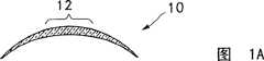
图1A是用于矫正近视的透镜植入物的侧横截面图。Figure 1A is a side cross-sectional view of a lens implant for correcting myopia.
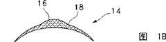
图1B是用于矫正无晶状体的透镜植入物的侧横截面图。Figure IB is a side cross-sectional view of a lens implant for correcting aphakia.
图1C是用于矫正老花眼的透镜植入物的前视图。Figure 1C is a front view of a lens implant for correcting presbyopia.
图1D是用于矫正老花眼的透镜植入物的侧横截面图。Figure ID is a side cross-sectional view of a lens implant for correcting presbyopia.
图1E是包含菲涅耳型透镜部分的透镜植入物的前视图。Figure IE is a front view of a lens implant including a Fresnel-type lens portion.
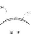
图1F是包含菲涅耳型透镜部分的透镜植入物的侧横截面图。Figure IF is a side cross-sectional view of a lens implant including a Fresnel-type lens portion.
图2A是角膜上皮上吸引器的侧横截面图。Figure 2A is a side cross-sectional view of an epithelial aspirator on the cornea.
图2B是用于接触角膜上皮的吸引器环的底视图。Figure 2B is a bottom view of the aspirator ring used to contact the corneal epithelium.
图2C是包含用于支撑上皮的密孔板的吸引器的变化形式的侧横截面图。Figure 2C is a side cross-sectional view of a variation of an aspirator comprising a microporous plate for supporting epithelium.
图2D是图2C装置的底视图。Figure 2D is a bottom view of the device of Figure 2C.
图2E是包括用于支撑上皮的输送插入体的吸引器的变化形式的侧横截面图。2E is a side cross-sectional view of a variation of a suction device including a delivery insert for supporting epithelium.
图2F是图2E装置的底视图。Figure 2F is a bottom view of the device of Figure 2E.
图2G是图2A的吸引器所形成的吸引水疱的侧横截面图。Figure 2G is a side cross-sectional view of a suction blister formed by the suction device of Figure 2A.
图3A是从角膜间质分离上皮的金属丝的侧横截面图。Figure 3A is a side cross-sectional view of a wire separating epithelium from the corneal stroma.
图3C显示了多种金属丝横截面和构造。Figure 3C shows various wire cross-sections and configurations.
图3D和3E显示了金属丝结构的旋转以分离上皮。Figures 3D and 3E show the rotation of the wire construct to separate the epithelium.
图3F显示了多金属丝的金属丝结构的多种构造。Figure 3F shows various configurations of multi-wire wire structures.
图3G显示了主要用于旋转金属丝分离器组件的具有粗糙表面的金属丝结构。Figure 3G shows a wire structure with a rough surface that is primarily used in a rotating wire separator assembly.
图3H显示了非螺旋性盘绕的分层器金属丝。Figure 3H shows a non-helically coiled delamination wire.
图4A显示了分离器的两种轭状物构造。Figure 4A shows two yoke configurations for the separator.
图4B显示了将分离器施用于角膜的途径。Figure 4B shows the route of application of the separator to the cornea.
图4C显示了分离器的倾斜式轭状物构造。Figure 4C shows the inclined yoke configuration of the separator.
图4D显示了分离器的旋转式轭状物构造。Figure 4D shows the rotating yoke configuration of the separator.
图4E显示了图4D倾斜式轭状物构造的侧视图。Figure 4E shows a side view of the tilted yoke configuration of Figure 4D.
图5A显示了用于限制或控制分离器在角膜上的途径的钩的局部剖视图。Figure 5A shows a partial cross-sectional view of a hook used to limit or control the passage of a separator over the cornea.
图5B显示了图5A钩的特写图。Figure 5B shows a close-up view of the hook of Figure 5A.
图5C显示了置于眼睛上的图5A钩的侧剖视图。Figure 5C shows a side cross-sectional view of the hook of Figure 5A placed on the eye.
图5D和5E显示了图5A钩的两个透视图。Figures 5D and 5E show two perspective views of the hook of Figure 5A.
图5F显示了图5A位于眼睛上的钩和上皮刻痕器。Figure 5F shows the hook and epithelial scorer of Figure 5A positioned on the eye.
图6A是通过钝性剥离悬提部分上皮的弯刮铲的侧横截面图。Figure 6A is a side cross-sectional view of a curved spatula lifting a portion of the epithelium by blunt dissection.
图6B是悬提部分上皮的注入的流体或凝胶的侧横截面图。Figure 6B is a side cross-sectional view of injected fluid or gel suspending a portion of the epithelium.
图6C显示了许多钝头分离器及其在去上皮过程中的潜在移动。Figure 6C shows a number of blunt-tipped separators and their potential movement during de-epithelialization.
图7A显示了在部分角膜上皮上用于悬提上皮的充满化学组合物的孔的侧横截面图。Figure 7A shows a side cross-sectional view of a chemical composition filled well for suspending the epithelium on a portion of the corneal epithelium.
图7B显示了在部分角膜上皮上的用于悬提上皮的含有化学组合物的吸收垫的侧横截面图。Figure 7B shows a side cross-sectional view of an absorbent pad containing a chemical composition for suspending the epithelium on a portion of the corneal epithelium.
图8A显示了悬提和分离了上皮和间质表面的眼球表面。Figure 8A shows the surface of the eyeball with the epithelial and mesenchymal surfaces suspended and separated.
图8B显示了分离的上皮的近视图。Figure 8B shows a close-up view of isolated epithelium.
图8C显示了分离的上皮的更近视图。Figure 8C shows a closer view of isolated epithelium.
图8D显示了眼球的残留间质表面的视图。Figure 8D shows a view of the residual stromal surface of the eyeball.
发明详述 Invention Details
如本作者前面所述,本文描述了用于将上皮下装置(例如,眼内透镜)导入上皮下面和通常在前角膜面上,用于悬提上皮以用于将那些上皮下装置置于角膜上或用于其它外科手术方法,和用于将上皮组织置回在植入的上皮下装置或外科手术位点上的方法。下述的用于从角膜表面悬提上皮的装置是与通过某些上皮下装置与替换的上皮接触而形成的结构相同。As previously stated by the authors, this paper describes methods for introducing subepithelial devices (eg, intraocular lenses) under the epithelium and usually on the anterior corneal surface, for suspending the epithelium for placement of those subepithelial devices in the cornea or for other surgical procedures, and for methods of repositioning epithelial tissue over implanted subepithelial devices or surgical sites. The means described below for suspending the epithelium from the corneal surface are the same structures formed by contacting the replacement epithelium with certain subepithelial means.
方法步骤Method steps
同样,如本作者前面所述,术语“表层角膜镜片术”一般指将供体角膜组织(间质)植于眼球表面上,其基本上位于原来的前角膜表面和多层上皮之间。而合成表层角膜镜片术是指将合成材料(代替供体角膜间质)用于完全相同的方法步骤中。我已经观察到任何表层角膜镜片术方法的成功都要依赖于上皮创伤愈合以及上皮覆盖于非上皮被覆表面(即植入物表面)。Also, as stated by this author earlier, the term "epikeratophakia" generally refers to the implantation of donor corneal tissue (stroma) on the surface of the eye, essentially between the original anterior corneal surface and the multilayered epithelium. In contrast, synthetic epikeratophakia refers to the use of synthetic material (instead of the donor corneal stroma) in exactly the same method steps. I have observed that the success of any epikeratophakia approach is dependent on epithelial wound healing and epithelial coverage of non-epithelialized surfaces (ie, implant surfaces).
非上皮表面上的上皮创伤愈合要依赖上皮细胞的功能。所谓“愈合性”上皮细胞在功能上和表型上与内稳态上皮细胞(通常位于未损伤上皮中)不同。Epithelial wound healing on non-epithelial surfaces is dependent on epithelial cell function. So-called "healing" epithelial cells are functionally and phenotypically distinct from homeostatic epithelial cells (usually located in undamaged epithelium).
内稳态上皮细胞(其在基底细胞层以缓慢的速度增殖并最终分化为子代细胞)。被推向上皮表面在基底细胞层,上皮细胞的一个主要功能是为下面的间质提供粘附作用,而另一主要功能是生成更多的上皮细胞。其为非蛋白水解性、非重塑性的且仅提供于维持状态。Homeostatic epithelial cells (which proliferate at a slow rate in the basal cell layer and eventually differentiate into progeny cells). Pushed to the epithelial surface At the basal cell layer, one major function of epithelial cells is to provide adhesion to the underlying stroma, while another major function is to generate more epithelial cells. It is non-proteolytic, non-remodeling and only provided for maintenance.
另一方面,愈合性上皮细胞在表型和功能上与内稳态上皮细胞不同。愈合性上皮细胞进行移行并对其移行的基质进行改造。愈合性上皮细胞溶解其细胞间连接(桥粒)并生成用于运动能力的肌动蛋白丝。除移行外,愈合性上皮细胞从活性基质中再吸收/溶解无活性基质。如此,这些细胞生成蛋白酶(例如,间质胶原酶,纤溶酶原激活剂和基质金属蛋白酶)。On the other hand, healing epithelial cells are phenotypically and functionally distinct from homeostatic epithelial cells. Healing epithelial cells migrate and remodel the matrix in which they migrate. Healing epithelial cells dissolve their intercellular junctions (desmosomes) and generate actin filaments for motility. In addition to migration, the healing epithelium resorbs/dissolves the inactive matrix from the active matrix. As such, these cells produce proteases (eg, interstitial collagenases, plasminogen activators, and matrix metalloproteinases).
在经典的表层角膜镜片术步骤中,存在能将上皮细胞从静止的内稳态转化为代谢活性重塑的直接刺激。该直接刺激就是表层角膜镜片术装置上的上皮缺失。在经典的表层角膜镜片术步骤中,一个基本的要求是装置表面能够抵抗蛋白水解并被识别为正常组织。如果装置表面不具有这些特性,则移行上皮可或者破坏该表面或者保持转化为再吸收性重塑机械装置。这就是上皮缺乏的直接结果。During the classical epikeratophagectomy step, there is a direct stimulus that converts epithelial cells from quiescent homeostasis to metabolically active remodeling. The immediate stimulus is the loss of epithelium on the epikeratophakia device. In the classical epikeratoscopy procedure, a fundamental requirement is that the device surface be resistant to proteolysis and be recognized as normal tissue. If the device surface does not possess these properties, the transitional epithelium can either destroy the surface or remain transformed into a resorbable remodeling mechanism. This is a direct result of the lack of epithelium.
通过使用本发明的将上皮组织置回植入上皮下装置上或外科手术位点上的方法,可将上皮保持在内稳态。上皮细胞功能的有害改变可得以避免。By using the methods of the present invention for repositioning epithelial tissue onto implanted subepithelial devices or surgical sites, the epithelium can be maintained in homeostasis. Detrimental changes in epithelial cell function can be avoided.
上面所使用的意在将细胞保持在内稳态的外科方法称为层状角膜切除术(lamellar keratotomy),可采用间质显微角膜刀实施。间质显微角膜刀的原型是Barraquer显微角膜刀,其是一种由Dr.JoseBarraquer研制的装置。该装置包括自动化切割系统,该系统在角膜体上以定向的切割平面进行切割,以制备包括角膜和上皮的组织的盘或瓣。The surgical approach used above to maintain cellular homeostasis is called lamellar keratotomy and can be performed using an interstitial microkeratome. The prototype of the interstitial microkeratome is the Barraquer microkeratome, a device developed by Dr. Jose Barraquer. The device includes an automated cutting system that cuts on the corneal body with oriented cutting planes to prepare a disc or flap of tissue including the cornea and epithelium.
自LASIK出现开始,显微角膜刀分离变成普通的方法。由于LASIK能在上皮下切穿角膜,故LASIK能将上皮保持于内稳态。用激光消融术重构得到的手术层。近来,已经制备了水凝胶透镜,这特别意指可采用其它(非消融性)装置改造手术层。实际上,完整的正常上皮降低了LASIK方法中的疼痛感觉是激光屈光被公众广泛接受的主要原因。Since the advent of LASIK, microkeratome dissection has become a common procedure. Because LASIK cuts through the cornea under the epithelium, LASIK maintains the epithelium in homeostasis. The resulting surgical layer was reconstructed with laser ablation. More recently, hydrogel lenses have been produced, which in particular means that other (non-ablative) devices can be used to remodel the surgical layer. In fact, the reduction of pain perception in the LASIK procedure by intact normal epithelium is the main reason why laser refraction is widely accepted by the public.
我所描述的方法的一个方面是,如所需的,即使用于与如LASIK的所述最低限度分离植入/外科方法联合时也可保持上皮细胞的原始状态。然而,我的方法避免了现有一般LASIK方法的一些问题,其中之一为通过悬提上皮并将其保持在内稳态而切入角膜间质的侵入性,攻击性和不可逆的方法。应当认识到,在一般的LASIK方法中,一经在角膜间质中做切口,则该切口将或者保持裂开或将明显地形成不透明纤维疤痕。每一方面都是不可逆的。很明显,“较好的”薄层角膜切开术能产生“较薄的”间质瓣或盘。我的方法制备了被认为是纯的上皮瓣,其“分离”平面位于基底上皮细胞的前细胞层下和在前角膜间质胶原I和胶原III上。本人将制备纯上皮瓣的方法称为上皮分层。One aspect of the method I describe is that, as desired, the epithelial cells can be preserved in their pristine state even when used in conjunction with such minimally dissected implantation/surgical methods as LASIK. However, my method avoids some of the problems of existing general LASIK methods, one of which is the invasive, aggressive and irreversible approach of incising the corneal stroma by suspending the epithelium and keeping it in homeostasis. It should be recognized that in typical LASIK procedures, once an incision is made in the corneal stroma, the incision will either remain open or will visibly form an opaque fibrous scar. Every aspect is irreversible. Apparently, a "better" thin keratotomy produces a "thinner" interstitial flap or disc. My method produces what is believed to be a pure epithelial flap with a "separation" plane below the anterior cell layer of the basal epithelium and on the anterior corneal stroma collagen I and collagen III. I refer to the method of preparing a pure epithelial flap as epithelial delamination.
通过上皮分层可以生成用于植入上皮下装置的外科手术空间。可采用化学、热或机械装置和下述方法实施上皮的分层。模拟上皮分层的一般病理过程是发疱。渗透性发疱(1M NaCl)能获得在基板(透明板)处的分离,其能导致纯上皮瓣。吸引发疱还能促进能导致纯上皮瓣的疱。还可以将拉力施加于基板而在透明板处实施分离。由于透明板是附着的“最弱的连接”,沿着基底膜的力可导致沿透明板的钝性分离。机械探针或流体的导入能用于获得钝性剥离而生成上皮瓣。The stratification of the epithelium creates a surgical space for the implantation of subepithelial devices. Layering of the epithelium can be performed using chemical, thermal or mechanical means and the methods described below. A general pathological process that mimics epithelial stratification is blistering. Osmotic blistering (1M NaCl) can achieve separation at the base (transparent plate), which can result in a pure epithelial flap. Attracting blisters also promotes blisters that can lead to pure epithelial flaps. It is also possible to apply a pulling force to the substrate to effect separation at the transparent plate. Since the transparent plate is the "weakest link" of attachment, forces along the basement membrane can cause blunt detachment along the transparent plate. Introduction of mechanical probes or fluids can be used to obtain blunt dissection to create epithelial flaps.
上皮瓣一经制备,即将“适宜的”上皮下装置置于分层的角膜表面上。可选择适宜的上皮下装置来缓解多种疾病:近视屈光矫正、远视屈光矫正或甚至老花眼矫正。前表面曲率的改变一般通过产生更平的角膜表面而矫正近视。Once the epithelial flap is prepared, a "fit" subepithelial device is placed on the stratified corneal surface. A suitable subepithelial device can be selected for the relief of a variety of conditions: myopic refractive correction, hyperopic refractive correction or even presbyopia correction. Changes in the curvature of the anterior surface generally correct myopia by creating a flatter corneal surface.
可通过上皮下装置提供更陡峭角膜表面实施近视矫正。曲率改变联合例如,提供中心陡峭曲率改变和装置边缘较平曲率的装置可用于治疗老花眼。仅依赖曲率改变而提供视力矫正的装置通常是光学上透明的。依赖衍射光学的透镜可含有不透明或半透明区。Myopia correction can be achieved by subepithelial devices that provide a steeper corneal surface. Changes in curvature combining, for example, devices that provide a steeper change in curvature at the center with flatter curvature at the edges of the device can be used to treat presbyopia. Devices that rely solely on changes in curvature to provide vision correction are typically optically transparent. Lenses that rely on diffractive optics may contain opaque or translucent regions.
基于衍射的、眼内、上皮下装置使用能产生相长干扰的衍射模式。多焦点的衍射光学模式可用于治疗老花眼矫正或矫正存在的单纯屈光不正。衍射光学模式可通过将其印在上皮下装置上而被导入分层角膜的表面。其整体效果与角膜表面的“磨针术(tattooing)”模式相同。Diffraction-based, intraocular, subepithelial devices use diffraction patterns that produce constructive interference. The multifocal diffractive optics mode can be used for presbyopia correction or correction of existing simple refractive errors. A diffractive optical pattern can be introduced into the surface of the stratified cornea by imprinting it on the subepithelial device. The overall effect is the same as a "tattooing" pattern of the corneal surface.
从功能上来说,选用本发明的方法的上皮下装置(结构设计和材料两方面)能使基本上稳定的营养流和代谢物从眼睛前房越过角膜至活性上皮。该流能使装置的长期连续起作用。下面描述了适宜的材料。Functionally, subepithelial devices (both structural design and materials) selected for the methods of the present invention enable a substantially steady flow of nutrients and metabolites from the anterior chamber of the eye across the cornea to the viable epithelium. This flow enables long-term continuous functioning of the device. Suitable materials are described below.
因为此前开发涉及合成表层角膜镜片术装置的方法的努力没有涉及将上皮组织置于装置本身的表面上,装置的底物化学性具有巨大的重要性,因为其提供了蛋白水解稳定的(非可降解的)化学性,其允许粘附和,更重要的,愈合性上皮细胞的移行。Because previous efforts to develop methods involving the synthesis of epikeratophakia devices have not involved placing epithelial tissue on the surface of the device itself, the substrate chemistry of the device is of great importance as it provides a proteolytically stable (non-degradable) degraded) chemistry that allows adhesion and, more importantly, migration of healing epithelial cells.
采用本发明的上皮分层和置换方法,需要材料表面化学性促进理想的细胞粘附环境的要求并不明显。一般而言,本人相信所选的材料仅具有足够的生物学“惰性”,因此并不足以造成外源体反应。LASIK方法已经在临床上证明了“游离”薄层角膜瓣不需要为了非常有效的矫正视力而被粘附于切割的手术层。事实上,LASIK方法成功的改进之处依赖于外科医生从手术层悬提瓣的能力。默认的角膜瓣附着于手术层足以保持LASIK方法的有效性。With the epithelial delamination and replacement methods of the present invention, the requirement for material surface chemistry to promote an ideal cell adhesion environment is not apparent. In general, I believe that the selected materials are only sufficiently biologically "inert" to cause an exosome response. The LASIK method has clinically demonstrated that a "free" thin layer corneal flap does not need to be adhered to the cut surgical layer for very effective vision correction. In fact, successful improvements to the LASIK approach depend on the surgeon's ability to lift the flap from the operative layer. The default flap attachment to the surgical layer is sufficient to maintain the effectiveness of the LASIK approach.
一般而言,所述方法涉及以下步骤:从角膜间质前表面分离上皮,通常希望使用下述装置中的一种,以制备基本上连续的层,并带有瓣(或可以是囊)形状或与眼睛相连的铰状连接,在所述位点施用上皮下透镜或其它植入物装置,以及在植入物外表面再施用至少部分所述上皮。虽然由于放置健康上皮瓣而产生的多数生理学益处在使用不借助瓣或铰连与眼睛相连的置回上皮时将产生效果,但是如果将上皮保持为瓣或囊,则上述优势更佳且方法自身更容易由外科医生(资深和新手)和由非外科医生实施。In general, the method involves the step of separating the epithelium from the anterior surface of the corneal stroma, usually desirably using one of the following devices, to produce a substantially continuous layer with a flap (or possibly a capsule) shape Or a hinged connection to the eye, at which site an epithelial sublens or other implant device is applied, and at least a portion of the epithelium is reapplied to the outer surface of the implant. While most of the physiological benefits resulting from the placement of healthy epithelial flaps will be effective when using repositioned epithelium attached to the eye without a flap or hinge, the advantages described above are even greater if the epithelium is maintained as a flap or capsule and the method In itself is easier to perform by surgeons (veteran and novice) and by non-surgeons.
上皮下装置Subcutaneous device
所述方法适用的上皮下装置包括能矫正或提高视力或单纯在某些方面改变使用者视觉,或通过改变眼睛焦距的天然范围以形成远视或显微效果的装置或植入物。适宜的装置或植入物还可用于其它医疗或美容原因,例如,用于给药或用作绷带或改变眼睛颜色。当然,眼内矫正性植入物在手术原则下可以是屈光性或衍射性的。Subepithelial devices to which the method is applicable include devices or implants that correct or enhance vision or simply alter the user's vision in some way, or by altering the natural range of eye focus to create hypermetropia or microscopic effects. Suitable devices or implants may also be used for other medical or cosmetic reasons, for example, for drug delivery or as a bandage or to change eye color. Of course, intraocular corrective implants may be refractive or diffractive under surgical principles.
因为上皮层自身具有较小的屈光性,适宜的眼内矫正性植入物可具有通常与软性接触透镜大小和构造相似的物理形状。这些植入物(置于角膜之上及置回的上皮瓣之下)辅助角膜曲率,从而矫正异常病症如散光、近视、远视、老花眼和无晶状体。Because the epithelial layer itself has less refractive power, a suitable intraocular corrective implant may have a physical shape that is generally similar in size and configuration to a soft contact lens. These implants (placed above the cornea and below the replaced epithelial flap) assist in corneal curvature, thereby correcting abnormal conditions such as astigmatism, nearsightedness, hyperopia, presbyopia, and aphakia.
这些眼内植入物透镜的外直径在功能上适于实施所需矫正,所述外直径通常小于约25mm,可以是10~15mm,或甚至更小如5~10mm。而且,透镜的厚度在功能上适于实施所需矫正。眼内植入物的厚度通常小于300μm,通常为5~200μm,通常为5~100μm。The outer diameter of these intraocular implant lenses is functionally adapted to implement the desired correction, typically less than about 25mm, may be 10-15mm, or even smaller such as 5-10mm. Also, the thickness of the lens is functionally adapted to implement the desired correction. The thickness of the intraocular implant is usually less than 300 μm, usually 5-200 μm, usually 5-100 μm.
图1A显示了适用于近视患者的透镜植入物(10),其在中心具有其前曲率被压平的通常为圆形的区域(12)。在对无晶状体的矫正中,可使用如图1B所示的适宜透镜(14)。所述透镜具有相对较厚的中心(16)和较薄的周边(18)。而且,一般而言,此处描述的形状与在所谓的“软性”接触透镜中发现的那些相同,可从与选用于矫正特定眼疾病的透镜的总体外形相关联的技术中得到指导。Figure 1A shows a lens implant (10) suitable for myopic patients having a generally circular region (12) in the center of which the anterior curvature is flattened. In the correction of aphakia, a suitable lens (14) as shown in Figure 1B can be used. The lens has a relatively thick center (16) and a thinner periphery (18). Also, in general, the shapes described here are the same as those found in so-called "soft" contact lenses, and guidance can be drawn from techniques associated with the overall shape of the lens chosen to correct a particular ocular disorder.
图1C和1D显示了同样可用于矫正老花眼的透镜。特别地,为治疗老花眼,透镜(20)同样具有与装置中心毗邻的通常不透明的环状区域(22)。开放中心(open center)(24)优选具有plano透镜特性且其有效直径低于约1.5mm,优选约0.5~1.5mm,和最优选0.75mm~1.75mm。开放中心(24)或中心区或“针孔”的直径通常被成形且选择,以小于日光中宿主眼睛的瞳孔直径。这会产生屈光性“针孔”效应,由此增长了眼睛的整体有效焦距并将眼睛适应的需要最小化。双焦点透镜设计也可被并入,例如,同心环、环形区域或环的段或扇区,或步进衍射性。Figures 1C and ID show lenses that can also be used to correct presbyopia. In particular, for the treatment of presbyopia, the lens (20) also has a generally opaque annular region (22) adjacent to the center of the device. The open center (24) preferably has plano lens characteristics and has an effective diameter of less than about 1.5 mm, preferably about 0.5 to 1.5 mm, and most preferably 0.75 to 1.75 mm. The diameter of the open center (24) or central region or "pinhole" is typically shaped and selected to be smaller than the pupil diameter of the host eye in daylight. This creates a refractive "pinhole" effect, thereby increasing the overall effective focal length of the eye and minimizing the need for the eye to adapt. Bifocal lens designs may also incorporate, for example, concentric rings, annular regions or segments or sectors of rings, or step diffractive properties.
图1D显示了如图1C所示的透镜(20)的侧横截面图,与角膜(26)的前表面相邻以说明该变化形式的特点。通常选择不透明环状环(22)的外径(28),以便使其在弱光条件下小于虹膜(34)中瞳孔(32)的直径(30)。这样,眼睛角膜和晶状体和发明的透镜以入射光同时通过不透明环(24)的中心和(而更重要地是)不透明环(22)的周边的方式联合作用,从而能在弱光环境下矫正光线。Figure ID shows a side cross-sectional view of the lens (20) shown in Figure 1C, adjacent to the anterior surface of the cornea (26) to illustrate the features of this variation. The outer diameter (28) of the opaque annular ring (22) is generally selected so that it is smaller than the diameter (30) of the pupil (32) in the iris (34) under low light conditions. In this way, the cornea and lens of the eye and the inventive lens combine to act in such a way that incident light passes through both the center of the opaque ring (24) and (and more importantly) the periphery of the opaque ring (22), thereby enabling correction in low-light conditions. light.
环状环(22)可以多种方式位于透镜植入物上,例如,通过放置适宜的染料,即,通过“墨针术(TATTOOING)”,或通过在后表面上放置基本上不透明的例如,Dacron网等生物相容性部件以滤过光线。挤压,由多种组件组装透镜,着色,染色或任何其它方法放置所需模式基于本说明书均为适宜的。环状环(22)的另一种放置方法包括置于透镜的前表面上。环状环(22)自身优选为相当不透明的,例如,透过低于约80%的入射可见光,但也可以选择较低程度的不透明或通过利用有色光屈光等方法将入射有色光转变为可见范围而矫正其它病症如色盲症。The annular ring (22) can be located on the lens implant in various ways, for example, by placing a suitable dye, i.e., by "TATTOOING", or by placing a substantially opaque, e.g., Biocompatible components such as Dacron mesh to filter light. Extrusion, assembly of lenses from various components, tinting, dyeing or any other method of placing the desired pattern are all suitable based on this specification. Another method of placement of the annular ring (22) involves placement on the front surface of the lens. The annular ring (22) itself is preferably fairly opaque, e.g., transmits less than about 80% of incident visible light, but may choose to be less opaque or convert incident colored light into Correction of other conditions such as color blindness.
其它屈光性植入物设计也是适宜的。图1E和1F分别显示了可用作置回上皮下植入物的Fresnel样透镜植入物(34)的前和侧横截面图。具有三角形横截面的圆状环(36)协同折射入射光以形成最终的影像。虽然所显示的环(36)没有遮盖物,但放置具有适当屈光率的遮盖物以为上皮提供光滑表面也包含在本发明的范围之内。Other refractive implant designs are also suitable. Figures IE and IF show front and side cross-sectional views, respectively, of a Fresnel-like lens implant (34) that can be used as a repositioned subepithelial implant. Circular rings (36) with triangular cross-section cooperate to refract incident light to form the final image. While the annulus (36) is shown without a covering, it is within the scope of the invention to place a covering of appropriate refractive power to provide a smooth surface to the epithelium.
虽然大多屈光性设计通常在形状上为圆形的,本人的方法可以在如美国专利No.6,228,113中所述的通常为矩形角膜镶嵌的其它形状的情况下使用。While most refractive designs are generally circular in shape, my method can be used with other shapes, generally rectangular corneal inlays, as described in US Patent No. 6,228,113.
植入物的衍射性设计也是适用的。例如,包括具有采用已知制备衍射性表面的技术所制备的光影响(light-affecting)表面的透镜元件的衍射性透镜设计是适用的。通常,透镜元件(或聚合物表面中的槽或适当布置的丝状元件的集合)含有具有不同的接近可见光的波长的相邻表面尺寸的光影响表面。Diffractive designs of implants are also suitable. For example, diffractive lens designs comprising lens elements with light-affecting surfaces prepared using known techniques for preparing diffractive surfaces are suitable. Typically, a lens element (or a groove in a polymer surface or a suitably arranged collection of filamentary elements) contains light-affecting surfaces with different adjacent surface dimensions near wavelengths of visible light.
上皮下植入物材料组合物Subepithelial implant material composition
适用于这些上皮下装置的材料种类繁多。装置或植入物可包括,基本上由或由任何特别如下述的和本文其它地方所述的材料构成。A wide variety of materials are suitable for these subepithelial devices. The device or implant may comprise, consist essentially of, or consist of, any material particularly as described below and elsewhere herein.
多种类型的材料都是适用的。例如,亲水性聚合物、疏水性聚合物、能形成水凝胶的聚合物、生物聚合物、多孔聚合物和多孔陶瓷和玻璃。通常,适于用作接触透镜的聚合组合物适用于本发明所述方法。传统的软性透镜通常是主要衍生自多种亲水性单体或聚合物的水凝胶,其已经进行交联或通过某些其它机制,如通过导入结晶性或通过改变相对疏水性/亲水性特性的方法使之在水中不溶解。所述聚合物一般包含高于45%的水,在35℃下其Dk值为8-25(×10-11cm2/sec)(ml O2/ml mmHg)。另一类软性透镜组合物包括高于其玻璃化温度(Tg)的疏水性聚合物系统,例如,硅酮弹性体。Various types of materials are suitable. For example, hydrophilic polymers, hydrophobic polymers, hydrogel-forming polymers, biopolymers, porous polymers, and porous ceramics and glasses. In general, polymeric compositions suitable for use as contact lenses are suitable for use in the methods of the present invention. Traditional soft lenses are usually hydrogels mainly derived from various hydrophilic monomers or polymers, which have been cross-linked or through some other mechanism, such as by introducing crystallinity or by changing the relative hydrophobicity/hydrophilicity. The water-based properties make it insoluble in water. Said polymers generally contain more than 45% water and have a Dk value of 8-25 (×10−11 cm2 /sec) (ml O2 /ml mmHg) at 35° C.. Another class of soft lens compositions includes hydrophobic polymer systems above their glass transition temperature (Tg), eg, silicone elastomers.
本人优选形成水凝胶的聚合物组合物,因为其具有转送或携带大量流体和营养物等越过透镜厚度至上皮层的良好能力。物理聚合物共混物或合金、复合聚合物结构、倾向于增强上皮细胞生长等的被覆或处理的聚合物均是适用的。I prefer hydrogel-forming polymer compositions because of their good ability to transfer or carry large quantities of fluids, nutrients, etc. across the thickness of the lens to the epithelial layer. Physical polymer blends or alloys, composite polymer structures, coated or treated polymers that tend to enhance epithelial cell growth, etc. are suitable.
水凝胶组合物通常包括亲水性聚合物,或者包括一定量的在合成时倾向于吸收水而不是产生溶解的疏水性聚合物。例如,亲水性聚合物可由单体或大分子单体合成,如:羟基取代的C1-C4-烷基丙烯酸酯和甲基丙烯酸酯的单体,包括羟乙基甲基丙烯酸酯(HEMA)、羟乙基丙烯酸酯或羟基丙基丙烯酸酯,丙烯酰胺、甲基丙烯酰胺、N-单-和N,N-二-C1-C4-烷基丙烯酰胺和甲基丙烯酰胺,其烷基部分可被羟基取代,羟基取代的C1-C4-烷基乙烯醚、烯丙醇、醋酸乙烯酯、共含有3~5碳原子的烯键不饱和羧酸,例如丙烯酸或甲基丙烯酸、N-乙烯基吡咯烷酮和N-丙烯酰吗啉、亲水性大分子单体包括乙烯基官能化的(viny1functionalized)聚乙烯醇、聚环氧烷(例如,聚环氧乙烷)或N-乙烯基吡咯烷酮均聚物或共聚合物,其或许含有一个或多个烯键不饱和双键。HEMA和N-乙烯基吡咯烷酮的venerable聚合物或共聚物或这些聚合物的共聚物或其与甲基丙烯酸甲酯或丙烯酸的共聚物(如可通过将这些单体在适宜的催化剂存在下加热进行催化聚合而制备)包含在适宜的形成水凝胶的聚合物类中。参见美国专利4,693,715;5,300,116和5,458,819。Hydrogel compositions generally include a hydrophilic polymer, or include an amount of a hydrophobic polymer that tends to absorb water rather than dissolve when synthesized. For example, hydrophilic polymers can be synthesized from monomers or macromers such as hydroxyl-substituted C1 -C4 -alkyl acrylate and methacrylate monomers, including hydroxyethyl methacrylate ( HEMA), hydroxyethyl acrylate or hydroxypropyl acrylate, acrylamide, methacrylamide, N-mono- and N,N-di-C1 -C4 -alkylacrylamide and methacrylamide, The alkyl moiety may be substituted by hydroxyl, C1 -C4 -alkyl vinyl ether, allyl alcohol, vinyl acetate, ethylenically unsaturated carboxylic acid containing 3 to 5 carbon atoms in total, such as acrylic acid or methacrylic acid Acrylic acid, N-vinylpyrrolidone and N-acryloylmorpholine, hydrophilic macromers including vinylfunctionalized (viny1functionalized) polyvinyl alcohol, polyalkylene oxide (e.g., polyethylene oxide) or Homopolymers or copolymers of N-vinylpyrrolidone, which may contain one or more ethylenically unsaturated double bonds. venerable polymers or copolymers of HEMA and N-vinylpyrrolidone or copolymers of these polymers or copolymers with methyl methacrylate or acrylic acid (such as can be produced by heating these monomers in the presence of a suitable catalyst) prepared by catalyzed polymerization) are included in the class of suitable hydrogel-forming polymers. See US Patents 4,693,715; 5,300,116 and 5,458,819.
美国专利No.5,786,434公开了适宜的水凝胶,吸水性软性接触透镜材料,其由通过聚合15~40重量%的N,N-二烷基甲基丙烯酰胺或N,N-二烷基丙烯酰胺,10~30重量%的N-乙烯基内酰胺和30~70重量%的二(含硅烷基)富马酸的单体混合物而制备的共聚物组成。U.S. Patent No. 5,786,434 discloses a suitable hydrogel, water-absorbing soft contact lens material, which is composed of 15 to 40% by weight of N,N-dialkylmethacrylamide or N,N-dialkyl The copolymer is composed of acrylamide, a monomer mixture of 10-30% by weight of N-vinyllactam and 30-70% by weight of di(silyl-containing) fumaric acid.
如任选取代的聚(烯基)乙二醇(例如,那些在每个烯基单位中含有至多7个碳原子的,特别是聚乙二醇或聚丙二醇)之类多孔剂(Porogen)可在聚合期间加入,以(如有需要)在最终聚合物中提供气孔。Porous agents such as optionally substituted poly(alkenyl)glycols (e.g., those containing up to 7 carbon atoms per alkenyl unit, especially polyethylene glycol or polypropylene glycol) can be used Added during polymerization to provide, if desired, air voids in the final polymer.
多数所述聚合物可通过如下方法进行改性以促进细胞生长,所述方法例如为,使下面的亲水性单体(使用交联剂)与包含磺基基团的单体如具有磺基基团的烯键不饱和的2~18C化合物或其适宜盐的单体进行共聚,所述化合物例如为甲代烯丙基磺酸、苯乙烯磺酸、磺丙基甲基丙烯酸酯、磺丙基丙烯酸酯,2-丙烯酰胺基-2-甲基丙烷磺酸、乙烯基磺酸或它们盐如甲代烯丙基磺酸钠、苯乙烯磺酸钠、磺丙基甲基丙烯酸钾或磺丙基丙烯酸钾。Many of these polymers can be modified to promote cell growth by, for example, combining the following hydrophilic monomers (using crosslinkers) with monomers containing sulfo groups such as The monomers of ethylenically unsaturated 2-18C compounds or their suitable salts are copolymerized, such as methallyl sulfonic acid, styrene sulfonic acid, sulfopropyl methacrylate, sulfopropyl Acrylate, 2-acrylamido-2-methylpropanesulfonic acid, vinylsulfonic acid or their salts such as sodium methallylsulfonate, sodium styrenesulfonate, potassium sulfopropylmethacrylate or Potassium propyl acrylate.
其它支持细胞生长的聚合物系统包括如美国专利4,440,918、4,818,801和5,994,133中所示的聚全氟代聚醚以及如美国专利No.6,225,367中所示的聚全氟代烷基聚醚。如果有特别需要的话,这些聚合物和此处所述的其它聚合物可物理地或化学地制成多孔材料。Other polymer systems that support cell growth include polyperfluoropolyethers as shown in US Patents 4,440,918, 4,818,801 and 5,994,133 and polyperfluoroalkylpolyethers as shown in US Patent No. 6,225,367. These polymers, and others described herein, can be physically or chemically made porous if specifically desired.
包括天然聚合物和疏水性单体的水凝胶也是可接受的。美国专利No.5,632,773中公开了一种这样的聚合物,其是与疏水性聚合物共价结合的胶原的组合物,其中单体具有低于甲基丙烯酸甲酯的部分极性。Hydrogels comprising natural polymers and hydrophobic monomers are also acceptable. One such polymer is disclosed in US Patent No. 5,632,773, which is a composition of collagen covalently bonded to a hydrophobic polymer, wherein the monomer has a lower partial polarity than methyl methacrylate.
可以使用生物聚合物如胶原I、胶原III、胶原IV、明胶、交联肝素、交联透明质酸、硫酸软骨素(chondroitin sulfate)、纤连蛋白、层粘连蛋白等。Biopolymers such as collagen I, collagen III, collagen IV, gelatin, cross-linked heparin, cross-linked hyaluronic acid, chondroitin sulfate, fibronectin, laminin, and the like can be used.
其它非水凝胶聚合物如聚乙烯、聚丙烯、聚氨酯等也是适用的,特别是在用于增强细胞生长或使流体输送时。美国专利No.4,607,617教导了在接触透镜中使用聚砜。例如,美国专利No.6,176,580公开了硅酮弹性体、包括美国专利5,371,147、5,314,960和5,057,578中所述的那些的含硅酮的大分子单体、水凝胶、含有硅酮的水凝胶等和它们的组合的用途。表面包括硅氧烷或硅氧烷官能团如聚二甲基硅氧烷大分子单体,甲基丙烯酰氧丙基聚烷基硅氧烷,及其混合物、硅酮水凝胶或水凝胶,如etafilcon A。Other non-hydrogel polymers such as polyethylene, polypropylene, polyurethane, etc. are also suitable, especially when used to enhance cell growth or to enable fluid transport. US Patent No. 4,607,617 teaches the use of polysulfone in contact lenses. For example, U.S. Patent No. 6,176,580 discloses silicone elastomers, silicone-containing macromers including those described in U.S. Patents 5,371,147, 5,314,960, and 5,057,578, hydrogels, silicone-containing hydrogels, etc. and use of their combinations. Surfaces include siloxane or siloxane functional groups such as dimethicone macromers, methacryloxypropyl polyalkylsiloxanes, and mixtures thereof, silicone hydrogels or hydrogels , such as etafilcon A.
如5,713,957中所示的微孔非水凝胶包括丙烯酸、聚烯烃、含氟聚合物、硅酮、苯乙烯、乙烯、聚酯、聚氨酯、聚碳酸酯、纤维素或蛋白如基于胶原的材料的聚合物和共聚合物是适用的。Microporous non-hydrogels such as those shown in 5,713,957 include acrylics, polyolefins, fluoropolymers, silicones, styrenes, vinyls, polyesters, polyurethanes, polycarbonates, cellulose, or proteins such as collagen-based materials. Polymers and copolymers are suitable.
上皮悬提装置Epithelial Suspension Device
对于任何被覆表面如皮肤、呼吸道上皮、消化道上皮和角膜,都有上皮细胞层附着于下面的基底膜。当将上皮从其基底膜和下面的胶原组织分离时,会形成上皮下疱。一般而言,直径小于1mm的大体分离被称为圆盘小泡化(vesiculation),而直径大于1mm的分离会形成真正的疱。As with any covering surface such as skin, respiratory epithelium, gastrointestinal epithelium, and cornea, there is an epithelial cell layer attached to the underlying basement membrane. Subepithelial blebs form when the epithelium is separated from its basement membrane and underlying collagenous tissue. In general, gross separations less than 1 mm in diameter are referred to as disc vesiculation, while separations greater than 1 mm in diameter form true vesicles.
角膜上皮的连续层可通过对前表面,或对基底细胞层,或对基底细胞层和鲍曼层(“透明板”)间的连接施加多种机械力而从前眼球表面分离或悬提。如本发明书所使用的,术语“连续的”是指“不间断的”。如本发明书所使用的术语“机械力”是指由人,器械或装置产生的任何物理力。机械力的实例包括吸力、剪切力和钝力。The continuous layer of corneal epithelium can be detached or suspended from the anterior ocular surface by applying various mechanical forces to the anterior surface, or to the basal cell layer, or to the junction between the basal cell layer and Bowman's layer ("transparent plate"). As used in this specification, the term "continuous" means "uninterrupted". The term "mechanical force" as used in this specification refers to any physical force produced by a person, machine or device. Examples of mechanical forces include suction, shear and blunt forces.
通过上皮分层器将机械力施加于上皮如角膜上皮。如本发明书所使用的,术语“上皮分层器”是指通过施加机械力而从基底膜上分离上皮的任何器械或装置。还可以通过将能诱导上皮与下面的间质发生分离的化学组合物与前眼球表面接触而从前眼球表面上分离或悬提上皮。Mechanical force is applied to epithelium, such as corneal epithelium, by an epithelial delamination device. As used herein, the term "epithelial delamination device" refers to any instrument or device that separates the epithelium from the basement membrane by the application of mechanical force. Epithelium may also be isolated or suspended from the anterior eyeball surface by contacting the anterior eyeball surface with a chemical composition that induces separation of the epithelium from the underlying stroma.
机械上皮分层器
在机械上皮分层器的第一个变化形式中,分层器是如图2A中所示的吸引器。吸引器(100)包括含有上皮接触面(112)和真空源(114)的吸引室(110)。吸引室(110)和真空源(114)处于真空相通状态并可通过如连接器或软管之类连接装置连接。In a first variation of the mechanical epithelial stratifier, the stratifier is an aspirator as shown in Figure 2A. The suction device (100) includes a suction chamber (110) including an epithelial contact surface (112) and a vacuum source (114). The suction chamber (110) and the vacuum source (114) are in vacuum communication and can be connected by connecting means such as connectors or hoses.
吸引室(110)可以是任何形状的,但一般而言是与眼球前表面相一致的半球杯或杯状。吸引室的壁(116)可由可变形的材料制成且为透明的,以便在将吸引器置于眼球前表面时能够看到上皮(118)。吸引器还可具有优选为环形的上皮接触面(120),而其也可使用任何所需形状,不过无论如何,其形状要在功能上提供与上皮表面适宜的真空密封。图2B显示了图2A的上皮接触面(120)的下视图。该环(120)可用如金属、聚合物、和弹性体,或其混合物的材料制备。The suction chamber (110) can be of any shape, but is generally hemispherical or cup-shaped that conforms to the anterior surface of the eyeball. The walls (116) of the suction chamber may be made of a deformable material and transparent so that the epithelium (118) can be seen when the suction device is placed on the anterior surface of the eyeball. The suction device may also have an epithelial contacting surface (120) which is preferably annular, although any desired shape may be used, but in any event be shaped to provide a functionally suitable vacuum seal against the epithelial surface. Figure 2B shows a lower view of the epithelial contact surface (120) of Figure 2A. The ring (120) can be made from materials such as metals, polymers, and elastomers, or mixtures thereof.
吸引器(100)的真空源(114)通常是手动泵或机动化泵,但也可以是注射器。由泵产生的负压量可用压力表进行监测。当使用注射器时,通过可伸缩活塞的移动调整抽吸。The vacuum source (114) of the aspirator (100) is typically a manual or motorized pump, but could also be a syringe. The amount of negative pressure generated by the pump can be monitored with a pressure gauge. When using a syringe, the suction is adjusted by the movement of the retractable piston.
在使用时,将吸引器(100)置于前眼球表面上以接触上皮(118)。真空由真空源(114)生成,且在由上皮接触面(120)所围成的区域(122)上所施加的负压量足够悬提上皮连续层。如有需要,施用间断的或连续的、平移的、振动的、或扭转的力也可增强上皮分离。In use, the aspirator (100) is placed on the anterior eyeball surface to contact the epithelium (118). The vacuum is generated by a vacuum source (114) and the amount of negative pressure applied on the area (122) bounded by the epithelial contact surface (120) is sufficient to suspend the continuous layer of epithelium. Epithelial separation can also be enhanced by application of intermittent or continuous, translational, vibratory, or torsional force, if desired.
图2C-2E显示了吸引装置的其它变化形式,特别使用了装置真空空间中的支持。在图2C和2D中,吸引装置(140)包括含有小内嵌通道(144)的多孔或穿孔板(142)。通过进行吸引而拉住上皮瓣靠在板(142)的。Figures 2C-2E show other variations of the suction device, specifically using supports in the vacuum space of the device. In Figures 2C and 2D, the suction device (140) comprises a porous or perforated plate (142) containing small inset channels (144). Pull the upper flap against the plate (142) by applying suction.
同样的,吸引装置的变化形式(150)包括输送性插入物(152),其具有制备用来按需要接受上皮层而基本上不变形的下表面(154)。可通过真空的释放而松弛上皮瓣。适宜的输送性插入物在组成上是多种多样的。插入物可以是,例如,多孔熔结金属或聚合物。同样的,插入物可以是网状泡沫材料或用于使真空从真空口(156)传至支持表面的其它聚合插入物。Likewise, a variation (150) of the suction device includes a delivery insert (152) having a lower surface (154) prepared to accept the epithelial layer as desired without substantial deformation. The epithelial flap can be relaxed by the release of the vacuum. Suitable delivery inserts vary in composition. The insert can be, for example, a porous sintered metal or a polymer. Likewise, the insert may be a reticulated foam or other polymeric insert for transferring the vacuum from the vacuum port (156) to the support surface.
图2G显示了由凸起的上皮形成的上皮下疱(124)。然后,可操作分离的上皮(126)从而制备上皮瓣。Figure 2G shows a subepithelial bleb (124) formed by raised epithelium. The separated epithelium can then be manipulated (126) to prepare an epithelial flap.
病理学而言,当半桥粒不与基底膜附着时,上皮(118)即被悬提。然而,与LASEK不同的是(其中上皮细胞死亡),吸引发疱法(suctionbstering)得到的上皮完整,其为基本上连续的层且有活力,即,能够马上恢复正常的上皮功能。Pathologically, the epithelium (118) is suspended when hemidesmosomes are not attached to the basement membrane. However, unlike LASEK (in which the epithelial cells die), suctionbstering results in an intact epithelium that is a substantially continuous layer and is viable, ie, capable of restoring normal epithelial function immediately.
在本发明之前,已知吸引发疱法仅作为皮肤中的上皮分层方法。皮肤中的吸引发疱(suction blister)最初用作确定自身免疫皮肤发疱病的病理生理学的实验室工具。Prior to the present invention, suction blistering was known only as a method of epithelial layering in the skin. Suction blisters in the skin were originally used as a laboratory tool to determine the pathophysiology of autoimmune cutaneous blistering diseases.
机械上皮分层器还可以是钝头分离器。钝头分离器具有适于置于上皮和胶原间质组织间的非切割表面。如本说明书所使用的,术语“非切割”是指当以正常力使用时,钝头分离器不具有切入角膜间质的能力。本人相信本人的钝头分离器可从角膜间质层上在基底膜带中于天然最弱附着点即透明板处,分离上皮。该由此分离的上皮不含大量的角膜间质组织,或为了本发明的目的,当该方法用于“正常”眼睛(其不具有由于损伤或疾病而产生的伪像)时,其仅含非大量的间质组织。该由此分离的上皮不包含I或III型胶原,而在间质组织可发现所述胶原。The mechanical delamination device can also be a blunt-tipped separator. The blunt-tipped separator has a non-cutting surface suitable for placement between epithelial and collagenous stroma. As used herein, the term "non-cutting" means that the blunt-tipped separator does not have the ability to cut into the corneal stroma when used with normal force. I believe my blunt separator can separate the epithelium from the corneal stroma at the natural weakest attachment point, the pellucid plate, in the basilar membrane zone. The thus isolated epithelium does not contain significant amounts of corneal stromal tissue, or, for the purposes of the present invention, only Non-massive interstitial tissue. The thus isolated epithelium does not contain type I or III collagen, which can be found in the mesenchymal tissue.
在一个变化形式中,钝头分离器是细金属丝,该金属丝插入上皮下,然后平行移向角膜表面从而从间质组织分离上皮。图3A显示了金属丝(200)的侧横截面图,该金属丝插入上皮(202)和间质(204)之间从而形成分离区(206)。该金属丝可以是连续地或不间断地旋转,或通过“滚动”金属丝或通过以与眼球前面垂直的轴转动金属丝,例如,如同以约与地板平行的轴旋转的叶片,或在剥离上皮(202)期间振动。然后可将分离的上皮(208)返折或剥离以暴露下面的间质组织(204)。In one variation, the blunt-tipped separator is a thin wire that is inserted under the epithelium and then moved parallel to the corneal surface to separate the epithelium from the stroma. Figure 3A shows a side cross-sectional view of a wire (200) inserted between epithelium (202) and stroma (204) to form a separation zone (206). The wire may be rotated continuously or uninterrupted, either by "rolling" the wire or by turning the wire on an axis perpendicular to the front of the eyeball, for example, like a blade rotating on an axis approximately parallel to the floor, or by peeling The epithelium (202) vibrates during. The isolated epithelium (208) can then be folded back or peeled away to expose the underlying mesenchymal tissue (204).
本人发现金属丝的有效直径与基底细胞层的厚度大小相近,例如,约为1/2mil~3.5mil(0.0005~0.0035”),而通常约为1.0mil~3.0mil(0.001~0.003”),根据经验,直径约为2.0mil的圆形金属丝为佳。另外,通过正确选择金属丝的刚度(通过调节已安装金属丝中的张力和/或选择金属丝中材料的固有刚度),金属丝将分离上皮和间质层间的联接而不切割上皮或鲍曼氏层。金属丝张力的适宜范围为15kpsi~35kpsi,或者为25kpsi~32.5kpsi,通常为25kpsi~30kpsi。从功能上考虑,我将这种从下面的间质分离上皮而不切割下面间质的原纤维的能力称为“非切割性”。I have found that the effective diameter of the wire is similar to the thickness of the basal cell layer, for example, about 1/2 mil to 3.5 mil (0.0005 to 0.0035"), and usually about 1.0 mil to 3.0 mil (0.001 to 0.003"), according to As a rule of thumb, a round wire with a diameter of about 2.0 mil is best. Additionally, with the correct choice of wire stiffness (by adjusting the tension in the installed wire and/or choosing the inherent stiffness of the material in the wire), the wire will separate the junction between the epithelial and mesenchymal layers without cutting the epithelium or abalone. Mann layer. A suitable range of wire tension is 15kpsi-35kpsi, or 25kpsi-32.5kpsi, usually 25kpsi-30kpsi. From functional considerations, I refer to this ability to separate the epithelium from the underlying stroma without cutting the underlying stroma's fibrils as "non-cutting."
参照图3A,我描述了轻微剥离间质表面(204)的金属丝分离器(200)。虽然本人相信这是对我的方法和装置起作用的方式的精确描述,但我并不希望受该理论的限制。然而,在使用此处所描述的金属丝分离器时可观察到在上皮下间质的某些剥离。分离器金属丝,当以通常与金属自身垂直的方向在上皮下跨过眼睛时,能够在金属丝分离器的前缘从上皮上拉低或拉离间质。Referring to Figure 3A, I describe a wire separator (200) that lightly dissects the interstitial surface (204). While I believe this is an accurate description of the way my method and apparatus work, I do not wish to be bound by this theory. However, some dissection of the subepithelial stroma was observed when using the wire separator described here. The separator wire, when crossing the eye under the epithelium in a direction generally perpendicular to the metal itself, is capable of pulling down from the epithelium or away from the stroma at the leading edge of the wire separator.
基于这一认识,金属丝的横截面形状无需是圆形的以获得所述的分离功能,而可以是任何适宜的能钝性分离角膜组织以从间质表面分离上皮而无需移去间质组织的横截面。对于术语“金属丝”和术语“金属丝结构”,我是指多样地包括:a)具有例如,下述横截面形状的单股细长的材料和b)包括未织在一起、织在一起和编在一起的两股或多股的结构。图3C中显示了单股横截面形状的实例:圆形(400)、卵形(402)、平截卵形(404)、正方形(406)、椭圆形(408)、矩形(410)、具有牛鼻前缘(412)的矩形,及其它。而且,形状与构建材料和张力的组合能获得其它地方所述的“非切割”分离功能。相对光滑的表面是这一操作所需的,且能使在间质和上皮间的移动变得容易。Based on this recognition, the cross-sectional shape of the wire need not be circular to achieve the described separation function, but may be any suitable shape capable of bluntly dissecting the corneal tissue to separate the epithelium from the stromal surface without removing the stromal tissue. Cross-section. By the term "wire" and the term "wire structure" I mean variously including: a) a single strand of elongated material having, for example, the following cross-sectional shape and b) including unwoven, woven A structure of two or more strands braided together. Examples of single strand cross-sectional shapes are shown in Figure 3C: circular (400), oval (402), truncated oval (404), square (406), oval (408), rectangular (410), with The rectangle of the bullnose leading edge (412), and others. Furthermore, the combination of shape and construction material and tension can achieve the "non-cutting" separation function described elsewhere. A relatively smooth surface is desirable for this manipulation and facilitates movement between the stroma and epithelium.
而且,金属丝自身不需要具有恒定横截面也不需要其沿长度为均一的,只要其能实施“非切割”分离功能即可。例如,本发明的范围包括扭转的或螺旋状的金属丝(414),如图3C中所示的变化形式,其具有正方形横截面。扭转的金属丝可与其它横截面变化形式如卵圆形、椭圆形、矩形和其它非圆形的形式一起使用。卵圆形和椭圆形,当扭转时,对眼睛是非常柔和的,并可以以使上皮上的悬提力在上皮中心高而在中心附近位置较低的方式进行定位。Also, the wire itself need not be of constant cross-section nor uniform along its length, so long as it can perform a "no cutting" separation function. For example, the scope of the invention includes twisted or helical wire (414), a variation as shown in Figure 3C, which has a square cross-section. Twisted wires can be used with other cross-sectional variations such as oval, oval, rectangular and other non-circular forms. Oval and oval, when twisted, are very gentle to the eye and can be positioned in such a way that the suspending force on the epithelium is high in the center of the epithelium and low near the center.
图3D,3E,和3F中显示了基于金属丝的分离器的其它变化形式,我确信该形式对于眼睛非常柔和。这些金属丝结构由多根金属丝制成,所述丝例如为那些具有图3C中所示横截面结构的丝。Other variations of wire-based separators are shown in Figures 3D, 3E, and 3F, which I believe are very gentle on the eyes. These wire structures are made of a plurality of wires, such as those having the cross-sectional structure shown in Figure 3C.
如图3D中所示,金属丝结构(201)被动的在上皮(405)下滚过角膜表面(403),从而在角膜表面(403)上产生“牵力”,而不会导致在表面上滑动摩擦。如其中显示,在分层器以自左向右的方向(409)移动时,金属丝结构(201)以顺时针方向(407)滚过角膜表面(403)。另一方面,如图3E中所示,可提抓上皮(405)并保持处于紧张状态下,然后将金属丝结构(401)从角膜表面(403)上悬提起来,或者甚至离开角膜表面(403),从而使得金属丝结构(401)能够从角膜上分离上皮。该变化形式具有能够增强从角膜表面上分离上皮容易度的优势。金属丝结构的卷动被设想为能产生一系列的力,每种力几乎垂直于所述上皮的下侧,且每种力在新金属丝卷至与上皮接触时发生。注意,图3E嵌入物中显示了力的箭头(399)。As shown in Figure 3D, the wire structure (201) passively rolls over the corneal surface (403) under the epithelium (405), thereby creating a "pull" on the corneal surface (403) without causing a "pull" on the surface. sliding friction. As shown therein, the wire structure (201 ) rolls across the corneal surface (403) in a clockwise direction (407) as the layerer moves in a left-to-right direction (409). Alternatively, as shown in Figure 3E, the epithelium (405) can be lifted and held under tension, and the wire structure (401) suspended from the corneal surface (403), or even off the corneal surface ( 403), thereby enabling the wire structure (401) to separate the epithelium from the cornea. This variation has the advantage of enhancing the ease of separating the epithelium from the corneal surface. The rolling of the wire structure is conceived to generate a series of forces, each nearly perpendicular to the underside of the epithelium, and each force occurring as a new wire is rolled into contact with the epithelium. Note that force arrows (399) are shown in the Figure 3E inset.
第二个变化形式为金属丝结构或金属丝的滚动动作可以是受动力驱动的。第三个变化形式是多金属丝结构,如与图3C中所示扭转的金属丝相关的其它地方所描述的,其不一定需要卷动。A second variation is that the wire structure or the rolling action of the wire can be powered. A third variation is a multi-wire structure, as described elsewhere in relation to the twisted wire shown in Figure 3C, which does not necessarily require rolling.
图3F显示了金属丝结构的多种构型,例如含有两根金属丝(413),三根金属丝(415),和多根金属丝(417)。结构中能够满足分离功能的任何合理数量的金属丝(例如,3、4、5、6、7、8等)都是适用的。可将金属丝并列放置(419)。其可被简单扭绕(421)或其可被编织(423)而形成确定的结构。所希望的直径(425)可以是上述的“有效的”直径。而且,希望当其移过眼睛时它们能发生卷动以分离上皮。Figure 3F shows various configurations of wire structures, such as two wires (413), three wires (415), and multiple wires (417). Any reasonable number of wires (eg, 3, 4, 5, 6, 7, 8, etc.) in the structure to fulfill the separation function is suitable. The wires can be placed side by side (419). It can be simply twisted (421) or it can be braided (423) to form a defined structure. The desired diameter (425) may be the "effective" diameter described above. Also, it is desirable that they roll to separate the epithelium as it moves across the eye.
最后,图3G中显示了粗糙化的金属丝(427)。该表面可通过蚀刻、喷沙等方法获得。这种去除光滑性的作法使金属丝(427)卷过眼睛时获得了较好的装置控制性和较好的上皮分离。也可对多金属丝结构应用粗糙化的表面。Finally, the roughened wire (427) is shown in Figure 3G. This surface can be obtained by etching, sandblasting, etc. This desmoothness results in better device control and better epithelial separation as the wire (427) is rolled over the eye. Roughened surfaces may also be applied to multi-wire structures.
在图3H中,显示了扭绕的金属丝的另一变化形式,其含有不均匀的横截面和非螺旋状的扭绕方式。在图3H中,显示了含有船首状物(prow)(418)类型的金属丝(416A),或含有前缘(416B)的金属丝的顶视图,其中当牵拉或推挤透明板时,其起到具有相邻肩(420)的钝头分离器的作用,其在相邻组织区域以斜形移动。在该变化形式中,狭窄的轴杆部分(422)以单方向(参见416C)扭绕,从而使自透明板至金属丝(418C)的上表面(424)的距离增加。当沿透明板移动时,该变化形式向上皮层提供了轻微的卷动运动,其外形上类似于儿童卷动舌头。In Fig. 3H, another variation of a twisted wire is shown, which contains a non-uniform cross-section and a non-helical twist. In FIG. 3H, a top view of a wire (416A) of the type containing a prow (prow) (418), or a wire containing a leading edge (416B) is shown, wherein when the transparent plate is pulled or pushed, It functions as a blunt-tipped separator with an adjacent shoulder (420), which moves in a diagonal shape over adjacent tissue regions. In this variation, the narrow shaft portion (422) is twisted in a single direction (see 416C), thereby increasing the distance from the transparent plate to the upper surface (424) of the wire (418C). This variation provided a slight rolling motion to the upper cortex when moved along the transparent plate, resembling in appearance a child rolling his tongue.
制备金属丝的材料对分离器结构来说不是严格的,只要材料能实施所述功能均包括在范围内。该结构的材料可以是金属的,例如钢、不锈钢、包括镍钛记忆合金(nitinol(Ni/Ti))的高弹性合金或聚合物,例如尼龙,聚芳酰胺,聚对苯二甲酸乙二酯和其它制备强纤维聚合物或它们的混合物,例如,涂以PTFE涂层的金属丝。The material from which the wire is made is not critical to the structure of the separator, as long as the material can perform the stated function, it is included in the scope. The material of the structure can be metallic, such as steel, stainless steel, highly elastic alloys including nitinol (Ni/Ti), or polymers, such as nylon, polyaramid, polyethylene terephthalate And other polymers or their mixtures for making strong fibers, for example, PTFE-coated wire.
上述多数变化形式可按照金属丝在两个位点间悬吊,或纵向预拉张,或在将金属沿透明板牵拉时形成纵向张力的方式安装。图4A中显示了金属丝悬带或“轭状物”的适宜的实例。轭状物(440)的第一个实例为在两个轭状物臂(444)之间悬吊分离金属丝(442),所述两个轭状物臂(444)具有相当平的构型从而使得操作装置的眼睛外科手术者能易于看见在上皮下发生的分离并易于定位用于开始分层步骤的工具。同样的,轭状物(446)具有能够使手术者察看轴杆(450)下的步骤和空间的臂(448),通常还有柄,作为抓握空间等。Most of the above variants can be installed with the wire suspended between two points, or pre-tensioned longitudinally, or in longitudinal tension as the metal is drawn along the transparent plate. A suitable example of a wire sling or "yoke" is shown in Figure 4A. A first example of a yoke (440) is to suspend a split wire (442) between two yoke arms (444) which have a rather flat configuration This allows the eye surgeon handling the device to easily see the separation occurring under the epithelium and to easily position the tools for initiating the delamination step. Likewise, the yoke (446) has an arm (448) that enables the operator to view steps and space under the shaft (450), and often a handle, as a gripping space, etc.
通常通过将金属丝在柄轴线上运动而进行移动来使用图4A中所示的变化形式(参见图4B)—注意方向箭头(452)。已经证实了其可以非常有效的容易地移去上皮而不造成损伤。然而,如图4C中所示,可使用为这一目的而构建的轭状物(454)而将金属丝斜穿过透明板。注意,图4C中显示了运动(456)的方向。The variation shown in Fig. 4A (see Fig. 4B) is typically used by moving the wire by moving it on the shaft axis - note the directional arrow (452). It has been shown to be very effective in easily removing the epithelium without causing damage. However, as shown in Figure 4C, the wire can be threaded diagonally through the transparent plate using a yoke (454) constructed for this purpose. Note that the direction of motion (456) is shown in Figure 4C.
而且,金属丝(456)可按图4D和4E所示旋转。在该变化形式中,轭状物(458)的两个臂彼此之间不固定。一个臂(460)相对于旋转臂(462)固定。金属丝(456)与旋转臂一起以部分弧旋转从而,至少部分地,从间质上剥离上皮。Also, the wire (456) can be rotated as shown in Figures 4D and 4E. In this variation, the two arms of the yoke (458) are not fixed relative to each other. One arm (460) is fixed relative to the swivel arm (462). The wire (456) rotates with the rotating arm in a partial arc to at least partially peel the epithelium from the stroma.
如下面将要叙述的,使用金属丝进行的分离可使用构型forms或“钩”来限制上皮膜与瓣的分离。金属丝分离器,如此处所述,还可进行振动或振荡以增强分离步骤。As will be described below, separation using wires may use formations or "hooks" to limit separation of the epithelial membrane from the flap. Wire separators, as described herein, can also be vibrated or oscillated to enhance the separation step.
首先,金属丝分离器的重复运动可有助于分离步骤的速率和容易程度。金属丝可以多种方式振动或振荡,例如,金属丝可仅在平面内振动,通常使运动和结构处于在分离步骤期间金属丝的预期运动方向上。在如图4A,4B,和4C中所示的变化形式中,往复运动将沿着柄的纵轴进行。First, the repetitive motion of the wire separator can aid in the speed and ease of the separation step. The wire can vibrate or oscillate in a variety of ways, for example, the wire can only vibrate in-plane, generally putting the motion and structure in the direction of the wire's intended motion during the separation step. In the variant shown in Figures 4A, 4B, and 4C, the reciprocating motion would be along the longitudinal axis of the handle.
金属丝分离组件可采用适当联接于金属丝上的振动器并沿着金属丝的轴向振动。然而采用这样的振动要特别注意,所述振动中所述运动具有切割角膜的较高可能性。本人发现频率为100~350Hz,200~325Hz,或225-275Hz,或245-255Hz的金属丝可以很好地悬提上皮。1.5-4.5mm或2-3mm的金属丝振动的振幅是非常适用的。The wire separating assembly may employ a vibrator suitably coupled to the wire and vibrate along the wire's axial direction. However, special care must be taken with vibrations in which the movement has a high probability of cutting the cornea. I have found that wires with a frequency of 100-350 Hz, 200-325 Hz, or 225-275 Hz, or 245-255 Hz can suspend the epithelium well. An amplitude of wire vibration of 1.5-4.5mm or 2-3mm is very suitable.
振动中的金属丝的张力范围很广,例如为0kpsi~35kpsi或25kpsi~32.5kpsi、通常为25kpsi~30kpsi。我发现当使用约0.002”不锈钢金属丝时,这些为特别适宜的值,但当在设计这些装置以适应此处所述的去上皮功能的限制条件时,某些试验水平也是适用的。The tension range of the vibrating metal wire is very wide, such as 0kpsi-35kpsi or 25kpsi-32.5kpsi, usually 25kpsi-30kpsi. I have found these to be particularly suitable values when using about 0.002" stainless steel wire, but some level of experimentation is applicable when designing these devices to accommodate the limitations of epithelial function described herein.
如其他部分所述,金属丝或金属丝结构在分离步骤期间可主动转动或被动转动。As described elsewhere, the wire or wire structure can be actively rotated or passively rotated during the separation step.
虽然此处的方法通常用于分离基本上完整的上皮层,即移动至分离器金属丝前侧的上皮部分为连续的,但装置可以较不精巧的方式使用。例如,分离器可用于移去所述膜的选定部分。事实上,当将该装置与LASEK方法结合使用时,上皮可以以软瓣的形式移去从而使得在任何角膜激光改造一经完成时能易于置回或复位。分离器的某些变化形式可用于制备上皮囊。可以考虑将如美国专利No.5,777,719中所述的眼局部解剖学系统,用于测量和矫正高度像差(higher-orderaberrations)的波阵面传感系统与本方法的某些变化形式结合使用。While the methods herein are generally used to separate substantially intact epithelial layers, ie, the portion of epithelium that travels to the front of the separator wire is continuous, the device can be used in a less delicate manner. For example, a separator can be used to remove selected portions of the membrane. In fact, when the device is used in conjunction with the LASEK approach, the epithelium can be removed in the form of a soft flap allowing easy replacement or repositioning once any corneal laser reconstruction has been completed. Certain variations of separators can be used to prepare epithelial vesicles. It is contemplated that an ocular topography system, a wavefront sensing system for measuring and correcting higher-order aberrations, as described in US Patent No. 5,777,719, may be used in conjunction with certain variations of the present method.
图5A-5E显示了适于根据情况将分离器金属丝精确定位在眼睛上并限制分离器金属丝移动以便形成上皮瓣的钩或形状(470)。Figures 5A-5E show a hook or shape (470) suitable for precise positioning of the separator wire on the eye and restricting movement of the separator wire to form an upper flap, as appropriate.
图5A和5B显示了钩(470)的实例的透视图,除去部分剖面以描述与普通眼睛(472)相关的下侧细节。装置的该实例使用具有真空腔(476)的柄(474)。将真空施加于具有一对与眼睛(474)相接触的密封脊(480,482)的密封腔(478)并使得钩(470)抓握眼睛(474)且使钩(470)和眼睛(474)彼此固定。Figures 5A and 5B show perspective views of an example of hook (470), with portions cut away to depict underside detail in relation to a common eye (472). This example of the device uses a handle (474) with a vacuum chamber (476). A vacuum is applied to the seal chamber (478) having a pair of seal ridges (480, 482) in contact with the eye (474) and causes the hook (470) to grasp the eye (474) and the hook (470) and eye (474) to ) are fixed to each other.
图5C显示了在密封脊(486,482)间形成密封腔(478)的钩(470)的侧视横断面图。还可以看到真空腔(476)和密封腔(478)间的真空通道(484)。图3C中清晰显示了导向槽(490),其为仅部分包绕钩(470)的通过钩(470)的槽。导向槽(490)还可如图5A和5B中所示。Figure 5C shows a side cross-sectional view of hook (470) forming seal cavity (478) between seal ridges (486, 482). Also visible is vacuum channel (484) between vacuum chamber (476) and sealed chamber (478). Guide slot (490) is clearly shown in Figure 3C, which is a slot through hook (470) that only partially surrounds hook (470). The guide groove (490) may also be as shown in Figures 5A and 5B.
图5D和5E显示了其终端(492)靠近柄(474)的导向槽(490)的放置范围。当将金属丝分离器导入导向槽(490)时,其在上皮中靠近金属丝入口位点的位置分离上皮,但由于金属丝的行进不超过导向槽的末端(492),故其仅分离上皮瓣。Figures 5D and 5E show the extent of placement of the guide slot (490) with its terminal end (492) close to the handle (474). When the wire separator is introduced into the guide channel (490), it separates the epithelium near the site of wire entry in the epithelium, but since the wire does not travel beyond the end of the guide channel (492), it only separates the upper epithelium. flap.
为了更精确地控制剥离的上皮瓣的形状,可使用如图5F中所示的划线工具(scoring tool)(500),或与如图5A和5B中所示的真空钩结合使用。划线工具(500)可用于计算适于钩(474)的上孔(502)的尺寸。可将适宜的档栓(504)置于划线工具(500)上或钩(474)上以限制切割深度或上皮中的“线痕”。线痕深度应(明显地)不大于上皮层厚度。通常为该深度的10%~90%,更通常为30%~70%是可接受的。For more precise control over the shape of the dissected epithelial flap, a scoring tool (500) as shown in Figure 5F may be used, or in combination with a vacuum hook as shown in Figures 5A and 5B. Scribing tool (500) may be used to calculate the size of upper hole (502) for hook (474). A suitable stop (504) may be placed on the scribing tool (500) or on the hook (474) to limit the depth of the cut or "line marks" in the epithelium. The depth of the scar should be (obviously) no greater than the thickness of the epithelial layer. Usually 10% to 90% of this depth, more usually 30% to 70% is acceptable.
最后,最好稍加热金属丝分离器,例如,可高于周边或眼内温度1℃~5℃,以增加上皮释放的容易性。Finally, it is best to warm the wire separator slightly, eg, 1°C to 5°C above the peripheral or intraocular temperature, to increase the ease of epithelial release.
在另一变化形式中,图6A显示了包括在角膜上皮(210)和间质(212)间注入流体、凝胶和气体的钝头分离器的上皮分层器。可采用针头(214)或插管注入流体或凝胶。可用于注入的流体包括盐溶液,例如,盐溶液(如1M高渗盐水)、基于硅酮的化合物和溶液、去垢剂溶液、CO2、N2和空气。可使用油,通常为疏水性的,例如,矿物油和其它可注入的碳基油或硅油。经注射,流体或凝胶从间质(212)钝性剥离或分离出上皮(210)。如有需要,在流体中附加脉冲,例如振动运动或超声运动以便增强上皮分层。In another variation, Fig. 6A shows an epithelial delamination device comprising a blunt-tipped separator for injecting fluid, gel and gas between the corneal epithelium (210) and stroma (212). The fluid or gel may be injected using a needle (214) or cannula. Fluids that can be used for infusion include saline solutions, eg, saline solutions (eg, 1M hypertonic saline), silicone-based compounds and solutions, detergent solutions, CO2,N2, and air. Oils can be used, typically hydrophobic, eg, mineral oil and other injectable carbon-based oils or silicone oils. Upon injection, the fluid or gel bluntly dissects or separates the epithelium (210) from the stroma (212). Additional pulses, such as vibratory or ultrasonic movements, in the fluid are applied to enhance epithelial delamination if necessary.
在钝头分离器的另一变化形式中,固体器械,例如,未支持在上皮中的分离区的每一边上的刮铲或其它机械分离器,可用作采用分离力来从角膜间质上分离上皮的钝头分离器。图6B显示了从角膜间质悬提连续层上皮(216)的弯刮铲(214)。分离器的刮铲变化形式的其它实例具有如图6C中所示的动作,例如部分旋转(510)、轴向运动(512)和振动(514)。In another variation of the blunt dissector, a solid instrument, such as a spatula or other mechanical dissector unsupported on each side of the dissected zone in the epithelium, can be used to dissect the corneal stroma from the A blunt-tipped separator to separate the epithelium. Figure 6B shows a curved spatula (214) suspending a continuous layer of epithelium (216) from the corneal stroma. Other examples of spatula variations of separators have motions as shown in Figure 6C, such as partial rotation (510), axial movement (512), and vibration (514).
化学上皮分层器Chemical Epithelial Stratifier
通常使用平衡盐溶液中的20%乙醇从而移去上面的上皮来实施LASEK方法。将20%乙醇置于眼睛上的孔中并在2~5分钟期间允许破坏上皮的粘附功能。然而,使用20%乙醇溶液可以杀死部分上皮细胞,妨碍了以连续层悬提的上皮的能力。在本发明的一个实施方案中,如图7A中所示,上皮分层器包括用乙醇溶液(302)注满的孔(300),所述乙醇溶液具有较低的乙醇百分比(0.5%~15%),从而使细胞丢失最小化。通过将乙醇溶液(302)用于眼球前表面,从间质表面(306)以连续层悬提上皮(304)。The LASEK method is typically performed using 20% ethanol in balanced salt solution to remove the overlying epithelium. 20% ethanol was placed in the hole on the eye and allowed to disrupt the adhesive function of the epithelium during 2-5 minutes. However, the use of a 20% ethanol solution can kill some of the epithelial cells, hampering the ability to suspend the epithelium in a continuous layer. In one embodiment of the invention, as shown in FIG. 7A, the epithelial delamination device includes holes (300) filled with an ethanol solution (302) having a lower ethanol percentage (0.5%-15%). %), thereby minimizing cell loss. The epithelium (304) is suspended in a continuous layer from the interstitial surface (306) by applying an ethanol solution (302) to the anterior surface of the eyeball.
其他化学组合物也可包括在化学上皮分层器中以便施用于眼睛。所述组合物可以是水溶液、凝胶例如水凝胶、或固体。所述组合物可直接施用于眼睛或以足够以连续层悬提上皮的量置于吸收垫上来进行施用。可以使用任何类型的吸收垫,不过通常使用以所述化学组合物浸湿的纱布垫。图7B显示了与上皮(310)接触的含有用于上皮分层的化学组合物的吸收垫(308)。Other chemical compositions may also be included in the chemical epithelializer for application to the eye. The composition may be an aqueous solution, a gel such as a hydrogel, or a solid. The composition can be applied directly to the eye or placed on an absorbent pad in an amount sufficient to suspend the epithelium in a continuous layer. Any type of absorbent pad can be used, but typically a gauze pad soaked with the chemical composition is used. Figure 7B shows an absorbent pad (308) in contact with an epithelium (310) containing a chemical composition for epithelial delamination.
用于上皮分层的优选的化学组合物包括发泡剂如1M高渗盐水、乙醇、斑蝥素和CEES。还可以在施用于眼睛前将稀释剂加至该组合物中。用于斑蝥素的适宜稀释剂为丙酮。用于CEES的适宜的稀释剂是水或潮湿空气。典型地,与斑蝥素和CEES一样,化合物通过破坏基底上皮细胞自身,而不损伤位于基底上皮层上的上皮细胞的方式起作用。如果使用1M高渗盐水,基底膜复合物沿着透明板解离。通常不会破坏基底上皮细胞。在1M高渗盐水中温育任何上皮都可实现从下面结缔组织上纯分离上皮。Preferred chemical compositions for epithelial delamination include foaming agents such as 1M hypertonic saline, ethanol, cantharidin, and CEES. Diluents may also be added to the composition prior to application to the eye. A suitable diluent for cantharidin is acetone. Suitable diluents for CEES are water or moist air. Typically, like cantharidin and CEES, the compounds act by destroying the basal epithelial cells themselves, without damaging the epithelial cells lying on the basal epithelial layer. If 1M hypertonic saline is used, the basement membrane complex dissociates along the transparent plate. Basal epithelial cells are usually not destroyed. Incubation of any epithelium in 1M hypertonic saline will achieve pure isolation of the epithelium from the underlying connective tissue.
某些情况下,可能需要加热眼球前表面以加速机械或化学上皮分层。再参照图2A,吸引器(100)可包括口或沟槽(128),其作为温暖眼球表面的加热流体的入口。口或沟槽(128)还可用于作为辅助分层方法的眼科器械的入口。In some cases, it may be necessary to heat the anterior surface of the eye to accelerate mechanical or chemical epithelial delamination. Referring again to FIG. 2A, the aspirator (100) may include a port or groove (128) that acts as an inlet for a heating fluid that warms the surface of the eyeball. The mouth or groove (128) may also be used as an entrance for the ophthalmic device to aid in the layering method.
本说明书所公开的上皮分层方法可与角膜重塑步骤或包括将眼内透镜装置置于眼球表面上的方法联合使用。特别的,所公开的方法可用于制备通常具有连接铰链的上皮瓣。然后可将适宜的眼内透镜置于间质表面上并将上皮瓣置回于透镜上。申请PCT/US01/22633中公开了可与本发明一同使用的一种所述适宜的眼内透镜装置,所述申请全文引入此处作为参考。The epithelial delamination methods disclosed in this specification may be used in conjunction with corneal remodeling procedures or methods involving placement of intraocular lens devices on the surface of the eyeball. In particular, the disclosed methods can be used to prepare epithelial flaps that typically have connecting hinges. A suitable intraocular lens can then be placed on the interstitial surface and the epithelial flap placed back on the lens. One such suitable intraocular lens device that may be used with the present invention is disclosed in application PCT/US01/22633, which is incorporated herein by reference in its entirety.
同样地,可实施角膜重塑过程并置回角膜瓣。Likewise, a corneal remodeling procedure can be performed and the corneal flap replaced.
而且,将振动或振荡力与上述任何上皮分层器结合使用可增强上皮分层。上述任一方法中所用的振动力将通过激发基底膜中的分子的共振频率(这本身将能互解上皮细胞的联结)而加速分层。Also, the use of vibratory or oscillating forces in conjunction with any of the epithelial delamination devices described above can enhance epithelial delamination. The vibratory force used in either of the above methods will accelerate delamination by exciting the resonant frequencies of the molecules in the basement membrane (which in themselves will dissociate the epithelial cells from each other).
实施例Example
采用与图4A,4B,4C,4D和4E中所示相似的装置实施机械上皮分层,其中其含有具有下垂且包括具有2mil(0.002英尺)直径的分离金属丝的轭状物臂的轭状物组合装置。轭状物沿着手柄的轴以约250Hz振动。将如图5A-5E中所示的钩置于新获得的(死后6小时)猪眼睛的前表面上并施用真空。Mechanical epithelial delamination was performed using a device similar to that shown in Figures 4A, 4B, 4C, 4D, and 4E, wherein it contained a yoke with sags and yoke arms comprising a separating wire with a diameter of 2 mil (0.002 feet). Combination device. The yoke vibrates at about 250 Hz along the axis of the handle. Hooks as shown in Figures 5A-5E were placed on the anterior surface of freshly harvested (6 hours post mortem) porcine eyes and vacuum was applied.
将金属丝在上皮下穿过钩并提起上皮瓣。在残留角膜表面、提起的上皮和瓣与角膜连接的“铰链”处提取组织学样品。图8A显示了分离的和提起的上皮(600),角膜(602),和剥离终止处的联接(604)。图8B显示了(高倍放大率下)联接(604)且还显示了钝性机械分离的精确度:在上皮(600)的基底细胞层(606)上缺少可视的角膜,间质组织,同样地,角膜表面(602)上无基底细胞。Thread the wire under the epithelium through the hook and lift the epithelial flap. Histological samples were taken from the residual corneal surface, lifted epithelium, and the "hinge" where the flap attaches to the cornea. Figure 8A shows the isolated and lifted epithelium (600), cornea (602), and junction (604) where the dissection terminates. Figure 8B shows (at high magnification) the junction (604) and also shows the precision of blunt mechanical dissection: lack of visible cornea on the basal cell layer (606) of the epithelium (600), mesenchymal tissue, as well Naturally, there are no basal cells on the corneal surface (602).
图8C显示了(仍为高倍放大)分离的上皮(600)和基底细胞层(606)。而且,即使在此放大率下,也未发现间质组织。Figure 8C shows (still at high magnification) the isolated epithelium (600) and basal cell layer (606). Also, even at this magnification, no interstitial tissue was found.
图8D显示了(高倍放大)角膜表面(602)和鲍曼氏层(610)。未发现上皮细胞。Figure 8D shows (higher magnification) the corneal surface (602) and Bowman's layer (610). No epithelial cells were found.
已经公开了本发明的结构和生理学特性,以及该上皮分层装置的特定变化形式的的具体优势。然而,本发明描述方式不应理解为对本发明范围有任何形式的限制。The structural and physiological properties of the present invention have been disclosed, as well as specific advantages of certain variations of this epithelial layering device. However, the manner in which the invention has been described should not be construed as limiting the scope of the invention in any way.
Claims (71)
Applications Claiming Priority (7)
| Application Number | Priority Date | Filing Date | Title |
|---|---|---|---|
| US35000302P | 2002-01-17 | 2002-01-17 | |
| US60/350,003 | 2002-01-17 | ||
| US39330502P | 2002-07-01 | 2002-07-01 | |
| US60/393,305 | 2002-07-01 | ||
| US40822602P | 2002-09-03 | 2002-09-03 | |
| US60/408,226 | 2002-09-03 | ||
| PCT/US2003/001549WO2003061518A2 (en) | 2002-01-17 | 2003-01-17 | Methods for producing epithelial flaps on the cornea and for placement of ocular devices and lenses beneath an epithelial flap or membrane, epithelial delaminating devices, and structures of epithelium and ocular devices and lenses |
Publications (2)
| Publication Number | Publication Date |
|---|---|
| CN1642499A CN1642499A (en) | 2005-07-20 |
| CN1310629Ctrue CN1310629C (en) | 2007-04-18 |
Family
ID=27617584
Family Applications (1)
| Application Number | Title | Priority Date | Filing Date |
|---|---|---|---|
| CNB038063484AExpired - Fee RelatedCN1310629C (en) | 2002-01-17 | 2003-01-17 | Epithelial layering device for preparation of epithelial flaps on the cornea and placement of intraocular devices and lenses under the epithelial flaps |
Country Status (8)
| Country | Link |
|---|---|
| US (1) | US7497866B2 (en) |
| EP (1) | EP1474084A4 (en) |
| JP (1) | JP2005515019A (en) |
| CN (1) | CN1310629C (en) |
| AU (1) | AU2003207603B2 (en) |
| CA (1) | CA2473703A1 (en) |
| MX (1) | MXPA04006954A (en) |
| WO (1) | WO2003061518A2 (en) |
Families Citing this family (61)
| Publication number | Priority date | Publication date | Assignee | Title |
|---|---|---|---|---|
| EP1173790A2 (en) | 1999-03-01 | 2002-01-23 | Boston Innovative Optics, Inc. | System and method for increasing the depth of focus of the human eye |
| US8668735B2 (en) | 2000-09-12 | 2014-03-11 | Revision Optics, Inc. | Corneal implant storage and delivery devices |
| US7156859B2 (en) | 2001-07-23 | 2007-01-02 | Fos Holding S.A. | Device for separating the epithelium layer from the surface of the cornea of an eye |
| JP4189320B2 (en)* | 2001-11-30 | 2008-12-03 | 国際交易株式会社 | Corneal surgery medical instrument |
| EP1549255A4 (en)* | 2002-09-13 | 2007-12-19 | Coopervision Inc | Devices and methods for improving vision |
| US7628810B2 (en) | 2003-05-28 | 2009-12-08 | Acufocus, Inc. | Mask configured to maintain nutrient transport without producing visible diffraction patterns |
| US20050046794A1 (en) | 2003-06-17 | 2005-03-03 | Silvestrini Thomas A. | Method and apparatus for aligning a mask with the visual axis of an eye |
| CA2539507A1 (en)* | 2003-09-22 | 2005-04-07 | Edward Perez | Corneal retention device or corneal stabilizing tool |
| WO2005049071A2 (en)* | 2003-11-14 | 2005-06-02 | Tissue Engineering Refraction, Inc. | Epithelium treatment methods and devices for treating the epithelium |
| US20050131434A1 (en)* | 2003-12-12 | 2005-06-16 | Peyman Gholam A. | Reciprocating instrument for separating a cornea to form a corneal flap |
| US8057541B2 (en) | 2006-02-24 | 2011-11-15 | Revision Optics, Inc. | Method of using small diameter intracorneal inlays to treat visual impairment |
| US7776086B2 (en) | 2004-04-30 | 2010-08-17 | Revision Optics, Inc. | Aspherical corneal implant |
| US10835371B2 (en) | 2004-04-30 | 2020-11-17 | Rvo 2.0, Inc. | Small diameter corneal inlay methods |
| US20080262610A1 (en)* | 2007-04-20 | 2008-10-23 | Alan Lang | Biomechanical design of intracorneal inlays |
| JP2007537829A (en)* | 2004-05-20 | 2007-12-27 | クーパーヴィジョン インコーポレイテッド | Corneal onlay and wavefront aberration correction for visual enhancement |
| EP1765236A1 (en)* | 2004-06-16 | 2007-03-28 | Tissue Engineering Refraction, Inc. | Epithelial delaminating device |
| US7976577B2 (en) | 2005-04-14 | 2011-07-12 | Acufocus, Inc. | Corneal optic formed of degradation resistant polymer |
| AU2006239235A1 (en)* | 2005-04-28 | 2006-11-02 | Tissue Engineering Refraction Inc. | Epithelial pocket expanding tool and combination epithelial delaminating device and corneal reformer |
| JP5312951B2 (en) | 2006-01-26 | 2013-10-09 | ウェイク・フォレスト・ユニヴァーシティ・ヘルス・サイエンシズ | Medical tools and related methods to facilitate deep endothelial corneal transplantation |
| US10555805B2 (en) | 2006-02-24 | 2020-02-11 | Rvo 2.0, Inc. | Anterior corneal shapes and methods of providing the shapes |
| US7883520B2 (en) | 2006-04-10 | 2011-02-08 | Forsight Labs, Llc | Corneal epithelial pocket formation systems, components and methods |
| US20090099557A1 (en)* | 2006-06-15 | 2009-04-16 | Sedarevic Olivia N | Methods and compositions for optimizing the outcomes of refractive laser surgery of the cornea |
| US9271828B2 (en) | 2007-03-28 | 2016-03-01 | Revision Optics, Inc. | Corneal implant retaining devices and methods of use |
| US9549848B2 (en) | 2007-03-28 | 2017-01-24 | Revision Optics, Inc. | Corneal implant inserters and methods of use |
| US8162953B2 (en) | 2007-03-28 | 2012-04-24 | Revision Optics, Inc. | Insertion system for corneal implants |
| US20080319463A1 (en)* | 2007-06-19 | 2008-12-25 | Dyson William Hickingbotham | Apparatus, system and method for illuminated membrane manipulator |
| US10779818B2 (en) | 2007-10-05 | 2020-09-22 | Covidien Lp | Powered surgical stapling device |
| US10498269B2 (en) | 2007-10-05 | 2019-12-03 | Covidien Lp | Powered surgical stapling device |
| US9539143B2 (en) | 2008-04-04 | 2017-01-10 | Revision Optics, Inc. | Methods of correcting vision |
| JP2011516180A (en) | 2008-04-04 | 2011-05-26 | レヴィジオン・オプティックス・インコーポレーテッド | Corneal inlay design and method for correcting vision |
| US20090306773A1 (en)* | 2008-06-04 | 2009-12-10 | Acufocus, Inc. | Opaque corneal insert for refractive correction |
| MX2011002070A (en)* | 2008-08-25 | 2011-06-21 | Wavelight Gmbh | Coupling of an eye to a laser device. |
| CA2770735C (en) | 2009-08-13 | 2017-07-18 | Acufocus, Inc. | Masked intraocular implants and lenses |
| WO2011020074A1 (en) | 2009-08-13 | 2011-02-17 | Acufocus, Inc. | Corneal inlay with nutrient transport structures |
| US10004593B2 (en) | 2009-08-13 | 2018-06-26 | Acufocus, Inc. | Intraocular lens with elastic mask |
| USD656526S1 (en)* | 2009-11-10 | 2012-03-27 | Acufocus, Inc. | Ocular mask |
| US8469948B2 (en) | 2010-08-23 | 2013-06-25 | Revision Optics, Inc. | Methods and devices for forming corneal channels |
| US9622911B2 (en) | 2010-09-30 | 2017-04-18 | Cxl Ophthalmics, Llc | Ophthalmic treatment device, system, and method of use |
| US9186243B2 (en)* | 2011-05-31 | 2015-11-17 | Novartis Ag | Accommodative intraocular lens and method of implantation |
| WO2013059813A1 (en) | 2011-10-21 | 2013-04-25 | Revision Optics, Inc. | Corneal implant storage and delivery devices |
| IN2014KN00780A (en)* | 2011-11-10 | 2015-10-02 | Wavelight Gmbh | |
| JP6046160B2 (en) | 2011-12-02 | 2016-12-14 | アキュフォーカス・インコーポレーテッド | Ophthalmic mask with selective spectral transmission |
| EP2830554A1 (en) | 2012-03-29 | 2015-02-04 | CXL Ophthalmics, LLC | Ocular cross-linking system and method for sealing corneal wounds |
| EP4420725A3 (en) | 2012-03-29 | 2025-04-16 | Epion Therapeutics, Inc. | Ocular treatment solutions, delivery devices and delivery augmentation methods |
| EP2830637B1 (en) | 2012-03-29 | 2024-08-21 | Epion Therapeutics, Inc. | Compositions and methods for treating or preventing diseases associated with oxidative stress |
| US9204962B2 (en) | 2013-03-13 | 2015-12-08 | Acufocus, Inc. | In situ adjustable optical mask |
| US9427922B2 (en) | 2013-03-14 | 2016-08-30 | Acufocus, Inc. | Process for manufacturing an intraocular lens with an embedded mask |
| US10092393B2 (en) | 2013-03-14 | 2018-10-09 | Allotex, Inc. | Corneal implant systems and methods |
| EP3073316B1 (en) | 2013-11-20 | 2018-12-26 | Iwane Mitsui | Contact lens for cornea-correction crosslinking, cornea-correction crosslinking method, and ring-shaped contact lens |
| US9603616B2 (en) | 2014-04-17 | 2017-03-28 | Covidien Lp | Vibrating surgical instruments for blunt dissection and methods for use thereof |
| US9597105B2 (en) | 2014-04-17 | 2017-03-21 | Covidien Lp | Vibrating surgical instruments for blunt dissection |
| US9943403B2 (en) | 2014-11-19 | 2018-04-17 | Acufocus, Inc. | Fracturable mask for treating presbyopia |
| US10064642B2 (en) | 2015-03-04 | 2018-09-04 | Covidien Lp | Surgical instrument for dissecting tissue |
| AU2015385773A1 (en) | 2015-03-12 | 2017-10-05 | Revision Optics, Inc. | Methods of correcting vision |
| US10449090B2 (en) | 2015-07-31 | 2019-10-22 | Allotex, Inc. | Corneal implant systems and methods |
| ES2972581T3 (en) | 2015-10-05 | 2024-06-13 | Acufocus Inc | Intraocular lens molding methods |
| KR102407311B1 (en) | 2015-11-24 | 2022-06-10 | 아큐포커스, 인크. | Toroidal eyelet intraocular lens with extended depth of focus |
| CN107028700B (en)* | 2017-04-05 | 2020-02-18 | 青岛大学附属医院 | Corneal Epithelial Flap Shaper |
| US10219939B1 (en)* | 2017-10-14 | 2019-03-05 | Samer Jaber Bashir | Surgical ophthalmic intra ocular access tool |
| DE102018203695A1 (en)* | 2018-03-12 | 2019-09-12 | Geuder Ag | Ophthalmic surgical kit and a contact lens |
| EP3790508A4 (en) | 2018-05-09 | 2022-02-09 | AcuFocus, Inc. | INTRAOCULAR IMPLANT WITH REMOVABLE OPTICS |
Citations (5)
| Publication number | Priority date | Publication date | Assignee | Title |
|---|---|---|---|---|
| CN1089127A (en)* | 1992-11-20 | 1994-07-13 | 冈本新生郎 | Method and device for eyeball cornea surgery |
| US5395385A (en)* | 1989-12-14 | 1995-03-07 | Corneal Contouring, Inc. | Apparatus for surgically re-profiling the cornea |
| US5877841A (en)* | 1995-12-05 | 1999-03-02 | Lg Electronics, Inc. | Application and method for self eye examination using a video appliance |
| CN2343998Y (en)* | 1998-11-13 | 1999-10-20 | 河南省眼科研究所 | Electric corneal epithelium curettage device |
| US6099541A (en)* | 1997-05-30 | 2000-08-08 | Summit Technology, Inc. | Surgical microtomes |
Family Cites Families (46)
| Publication number | Priority date | Publication date | Assignee | Title |
|---|---|---|---|---|
| EP0046338B1 (en)* | 1980-08-05 | 1985-04-10 | David Peter Choyce | Intraocular lens |
| US4346482A (en)* | 1981-01-22 | 1982-08-31 | Tennant Jerald L | Living contact lens |
| US4818801A (en)* | 1982-01-18 | 1989-04-04 | Minnesota Mining And Manufacturing Company | Ophthalmic device comprising a polymer of a telechelic perfluoropolyether |
| US4440918A (en)* | 1982-01-18 | 1984-04-03 | Minnesota Mining And Manufacturing Company | Contact lens containing a fluorinated telechelic polyether |
| US4646720A (en)* | 1985-03-12 | 1987-03-03 | Peyman Gholam A | Optical assembly permanently attached to the cornea |
| US4676790A (en)* | 1985-09-25 | 1987-06-30 | Kern Seymour P | Method of manufacture and implantation of corneal inlays |
| US4662881A (en)* | 1986-01-21 | 1987-05-05 | Nordan Lee T | Epikeratophakia process |
| US4693715A (en)* | 1986-06-19 | 1987-09-15 | Abel Robert Jr | Artificial cornea |
| US4715858A (en)* | 1986-07-25 | 1987-12-29 | Lindstrom Richard L | Epicorneal lens |
| US5114627A (en)* | 1986-10-16 | 1992-05-19 | Cbs Lens | Method for producing a collagen hydrogel |
| US4793344A (en)* | 1987-11-02 | 1988-12-27 | Recore, Inc. | Method for preparing corneal donor tissue for refractive eye surgery |
| US5171318A (en)* | 1987-11-09 | 1992-12-15 | Chiron Ophthalmics, Inc. | Treated corneal prosthetic device |
| US5192316A (en)* | 1988-02-16 | 1993-03-09 | Allergan, Inc. | Ocular device |
| US5314960A (en)* | 1990-04-10 | 1994-05-24 | Permeable Technologies, Inc. | Silicone-containing polymers, oxygen permeable hydrophilic contact lenses and methods for making these lenses and treating patients with visual impairment |
| US5057578A (en)* | 1990-04-10 | 1991-10-15 | E. I. Du Pont De Nemours And Company | Silicone-containing block copolymers and macromonomers |
| US5196027A (en)* | 1990-05-02 | 1993-03-23 | Thompson Keith P | Apparatus and process for application and adjustable reprofiling of synthetic lenticules for vision correction |
| US5371147A (en)* | 1990-10-11 | 1994-12-06 | Permeable Technologies, Inc. | Silicone-containing acrylic star polymers, block copolymers and macromonomers |
| US5312330A (en)* | 1992-05-20 | 1994-05-17 | Summit Technology, Inc. | Medical treatment of the eye involving removal of the epithelium |
| AU650156B2 (en)* | 1992-08-05 | 1994-06-09 | Lions Eye Institute Limited | Keratoprosthesis and method of producing the same |
| US5374515A (en)* | 1992-11-13 | 1994-12-20 | Organogenesis, Inc. | In vitro cornea equivalent model |
| US5827641A (en) | 1992-11-13 | 1998-10-27 | Parenteau; Nancy L. | In vitro cornea equivalent model |
| US5634921A (en)* | 1993-08-23 | 1997-06-03 | Hood; Larry | Method and apparatus for modifications of visual acuity by thermal means |
| TW257671B (en)* | 1993-11-19 | 1995-09-21 | Ciba Geigy | |
| US5632773A (en)* | 1994-05-03 | 1997-05-27 | Allergan, Inc. | Biostable corneal implants |
| US5496339A (en)* | 1994-05-17 | 1996-03-05 | Koepnick; Russell G. | Universal automated keratectomy apparatus and method |
| US5599341A (en)* | 1994-06-15 | 1997-02-04 | Keravision, Inc. | Laser surgical procedure and device for treatment of the cornea |
| DE69605778T2 (en) | 1995-04-04 | 2000-07-20 | Novartis Ag, Basel | USE OF A POLYMER AS A SUBSTRATE FOR CELL GROWTH |
| JP3453224B2 (en) | 1995-09-12 | 2003-10-06 | 株式会社メニコン | Hydrous soft contact lens material |
| US5919185A (en) | 1997-04-25 | 1999-07-06 | Peyman; Gholam A. | Universal implant blank for modifying corneal curvature and methods of modifying corneal curvature therewith |
| US5817115A (en)* | 1995-12-04 | 1998-10-06 | Chiron Vision Corporation | Apparatus for resecting corneal tissue |
| EP0891753A4 (en) | 1996-11-13 | 1999-09-22 | Menicon Co Ltd | Artificial cornea |
| US6187053B1 (en) | 1996-11-16 | 2001-02-13 | Will Minuth | Process for producing a natural implant |
| US5777719A (en) | 1996-12-23 | 1998-07-07 | University Of Rochester | Method and apparatus for improving vision and the resolution of retinal images |
| US6036683A (en) | 1997-01-02 | 2000-03-14 | G. Rodenstock Instruments Gmbh | Process and apparatus for changing the curvature of the cornea |
| US6126668A (en)* | 1997-04-25 | 2000-10-03 | Innovative Optics, Inc. | Microkeratome |
| US6231583B1 (en)* | 1997-07-09 | 2001-05-15 | Joseph Y. Lee | Corneal circular channel dissecting device |
| US6161544A (en)* | 1998-01-28 | 2000-12-19 | Keratoform, Inc. | Methods for accelerated orthokeratology |
| ES2277430T3 (en) | 1998-03-04 | 2007-07-01 | Visx Incorporated | LASER PRESBORAGE TREATMENT SYSTEM. |
| EP0956841B1 (en)* | 1998-03-31 | 2006-03-15 | Nidek Co., Ltd. | Corneal surgical apparatus |
| TWI230712B (en) | 1998-09-15 | 2005-04-11 | Novartis Ag | Polymers |
| US6176580B1 (en) | 1999-04-02 | 2001-01-23 | Johnson & Johnson Vision Care, Inc. | Method of designing and fitting contact lenses taking into account material properties of the lenses |
| US6280469B1 (en) | 1999-05-11 | 2001-08-28 | Mark A. Terry | Implantable iris device for the eye, and method of installing same |
| US6228113B1 (en) | 2000-01-10 | 2001-05-08 | Board Of Supervisors Of Louisiana State University And Agricultural And Mechanical College | Intracorneal astigmatic onlay |
| US6544286B1 (en) | 2000-07-18 | 2003-04-08 | Tissue Engineering Refraction, Inc. | Pre-fabricated corneal tissue lens method of corneal overlay to correct vision |
| US7156859B2 (en)* | 2001-07-23 | 2007-01-02 | Fos Holding S.A. | Device for separating the epithelium layer from the surface of the cornea of an eye |
| JP4187648B2 (en) | 2001-07-23 | 2008-11-26 | フォス ホールディング ソシエテ アノニム | Device for separating the epithelial layer from the corneal surface of the eye |
- 2003
- 2003-01-17JPJP2003561464Apatent/JP2005515019A/enactivePending
- 2003-01-17AUAU2003207603Apatent/AU2003207603B2/ennot_activeCeased
- 2003-01-17EPEP03705818Apatent/EP1474084A4/ennot_activeWithdrawn
- 2003-01-17CNCNB038063484Apatent/CN1310629C/ennot_activeExpired - Fee Related
- 2003-01-17MXMXPA04006954Apatent/MXPA04006954A/ennot_activeApplication Discontinuation
- 2003-01-17CACA002473703Apatent/CA2473703A1/ennot_activeAbandoned
- 2003-01-17USUS10/346,664patent/US7497866B2/ennot_activeExpired - Fee Related
- 2003-01-17WOPCT/US2003/001549patent/WO2003061518A2/enactiveApplication Filing
Patent Citations (5)
| Publication number | Priority date | Publication date | Assignee | Title |
|---|---|---|---|---|
| US5395385A (en)* | 1989-12-14 | 1995-03-07 | Corneal Contouring, Inc. | Apparatus for surgically re-profiling the cornea |
| CN1089127A (en)* | 1992-11-20 | 1994-07-13 | 冈本新生郎 | Method and device for eyeball cornea surgery |
| US5877841A (en)* | 1995-12-05 | 1999-03-02 | Lg Electronics, Inc. | Application and method for self eye examination using a video appliance |
| US6099541A (en)* | 1997-05-30 | 2000-08-08 | Summit Technology, Inc. | Surgical microtomes |
| CN2343998Y (en)* | 1998-11-13 | 1999-10-20 | 河南省眼科研究所 | Electric corneal epithelium curettage device |
Also Published As
| Publication number | Publication date |
|---|---|
| CN1642499A (en) | 2005-07-20 |
| WO2003061518A2 (en) | 2003-07-31 |
| WO2003061518A3 (en) | 2004-07-29 |
| AU2003207603B2 (en) | 2008-04-03 |
| EP1474084A2 (en) | 2004-11-10 |
| EP1474084A4 (en) | 2007-04-18 |
| US7497866B2 (en) | 2009-03-03 |
| CA2473703A1 (en) | 2003-07-31 |
| MXPA04006954A (en) | 2005-03-23 |
| US20030220653A1 (en) | 2003-11-27 |
| JP2005515019A (en) | 2005-05-26 |
Similar Documents
| Publication | Publication Date | Title |
|---|---|---|
| CN1310629C (en) | Epithelial layering device for preparation of epithelial flaps on the cornea and placement of intraocular devices and lenses under the epithelial flaps | |
| AU2003207603A1 (en) | Methods for producing epithelial flaps on the cornea and for placement of ocular devices and lenses beneath an epithelial flap or membrane, epithelial delaminating devices, and structures of epithelium and ocular devices and lenses | |
| CN1458849A (en) | Prefabricated Corneal Tissue Lenses and Cornea Overlay Method for Vision Correction (II) | |
| KR20070118580A (en) | Eyeball Applicator | |
| JP2008512208A (en) | Complex epithelial detachment / insertion tool | |
| JP2006508709A (en) | Apparatus and method for improving visual acuity | |
| US20070265649A1 (en) | Epithelial delaminating device | |
| CN87104281A (en) | Method for the phakic treatment of myopia and artificial intraocular lens device | |
| JP2008539033A (en) | Epithelial stripping device (V) and blade useful in the device | |
| WO2005030102A1 (en) | Corneal retention device or corneal stabilizing tool | |
| US10751166B2 (en) | Method for performing ophthalmosurgical operations using an autograft | |
| CN1694658A (en) | Devices and methods for improving vision | |
| JP4189320B2 (en) | Corneal surgery medical instrument | |
| CN101203196A (en) | Epithelium laying device (V) and blade for the device | |
| HK1085113A (en) | Devices and methods for improving vision | |
| HK1099680A (en) | Corneal retention device or corneal stabilizing tool |
Legal Events
| Date | Code | Title | Description |
|---|---|---|---|
| C06 | Publication | ||
| PB01 | Publication | ||
| C10 | Entry into substantive examination | ||
| SE01 | Entry into force of request for substantive examination | ||
| C14 | Grant of patent or utility model | ||
| GR01 | Patent grant | ||
| C19 | Lapse of patent right due to non-payment of the annual fee | ||
| CF01 | Termination of patent right due to non-payment of annual fee |