CN1300741C - Pre-processing method for skin micro image - Google Patents
Pre-processing method for skin micro imageDownload PDFInfo
- Publication number
- CN1300741C CN1300741CCNB2004100535402ACN200410053540ACN1300741CCN 1300741 CCN1300741 CCN 1300741CCN B2004100535402 ACNB2004100535402 ACN B2004100535402ACN 200410053540 ACN200410053540 ACN 200410053540ACN 1300741 CCN1300741 CCN 1300741C
- Authority
- CN
- China
- Prior art keywords
- image
- value
- sigma
- skin
- pixel
- Prior art date
- Legal status (The legal status is an assumption and is not a legal conclusion. Google has not performed a legal analysis and makes no representation as to the accuracy of the status listed.)
- Expired - Fee Related
Links
Images


Landscapes
- Image Processing (AREA)
Abstract
Translated fromChineseDescription
Translated fromChinese技术领域technical field
本发明涉及一种图像处理方法,特别是一种皮肤显微图像的预处理方法。The invention relates to an image processing method, in particular to a preprocessing method for skin microscopic images.
背景技术Background technique
图像的预处理包含两个方面:(1)图像的平滑处理,即图像的去噪处理,(2)图像中的目标区域从背景中分离出来,提取目标区域图像。Image preprocessing includes two aspects: (1) image smoothing, that is, image denoising processing, (2) target area in the image is separated from the background, and the image of the target area is extracted.
图像的去噪处理主要是为了去除实际成像过程中,因成像设备和环境所造成的图像失真,提取有用信息。目前,常用低通空间滤波器和中值滤波器来消除图像中的噪声。这种方法在去除图像中的噪声、干扰的同时,也滤掉了图像的边缘信息、细节和有用的高频信息。Image denoising processing is mainly to remove image distortion caused by imaging equipment and environment in the actual imaging process and extract useful information. At present, low-pass spatial filter and median filter are commonly used to remove noise in the image. While removing the noise and interference in the image, this method also filters out the edge information, details and useful high-frequency information of the image.
提取目标区域图像的最简单的图像分割方法是:采用对比度、边缘、灰度检测的方法,这些方法利用了前景与背景的灰度变化来进行分割,其前提是假设目标与背景相比有明显的灰度对比或颜色变化,但在实际处理中,由于受到光照不均匀等因素的影响,其对比特征并不明显,因此上述方法存在一定局限性。The simplest image segmentation method to extract the image of the target area is to use contrast, edge, and grayscale detection methods. These methods use the grayscale changes of the foreground and background for segmentation. However, in actual processing, due to the influence of uneven illumination and other factors, the contrast characteristics are not obvious, so the above method has certain limitations.
发明目的purpose of invention
本发明的目的在于提供一种皮肤显微图像的预处理方法,能克服已有技术中存在的问题,能适应光线的变化、图像大小的变化和不同皮肤不同症状,从背景皮肤图像中分割提取出清晰的皮肤症状区域的二值图像。The purpose of the present invention is to provide a preprocessing method for skin microscopic images, which can overcome the problems in the prior art, can adapt to changes in light, image size, and different symptoms of different skins, and can be segmented and extracted from background skin images. A clear binary image of the skin symptom area.
为达到上述目的,本发明的构思如下:For achieving the above object, design of the present invention is as follows:
本发明运用二进小波对图像进行变换,计算二阶小波分解后图像的模值,采用动态阈值的方法消除其中的局部极大值,有效地在保护图像有用信息的条件下去除了皮肤图像中的毛发等干扰,使重构的图像去除了噪声而保留了边缘信息。The present invention uses binary wavelet to transform the image, calculates the modulus of the image decomposed by the second-order wavelet, and adopts the method of dynamic threshold to eliminate the local maximum value, effectively removing the skin image under the condition of protecting the useful information of the image. Interference such as hair removes noise and preserves edge information in the reconstructed image.
针对皮肤显微图像的研究,发现以饱和度S(Saturation-S)作为图像的颜色信息能明显提高分割效果。所谓饱和度,它是反映某种色光被白光冲淡程度的物理量。Aiming at the study of skin microscopic images, it is found that using saturation S (Saturation-S) as the color information of the image can significantly improve the segmentation effect. The so-called saturation is a physical quantity that reflects the degree to which a certain color light is diluted by white light.
本发明提出一种针对皮肤症状的自适应图像分割提取方法,将RGB图像转换成S图像,以皮肤症状图像的S域标准差σ为判据,当σ较大(大于20)时,采用最大类间方差动态阈值法,当σ较小时(小于20),采用改进的S域动态阈值分割方法,从而解决了大动态范围图像标准差的图像分割,使图像分割的效果得到了明显的提高。The present invention proposes an adaptive image segmentation and extraction method for skin symptoms, which converts RGB images into S images, and takes the standard deviation σ of the S domain of the skin symptom image as a criterion. When σ is large (greater than 20), the maximum The inter-class variance dynamic threshold method, when σ is small (less than 20), adopts the improved S-domain dynamic threshold segmentation method, thereby solving the image segmentation of the standard deviation of large dynamic range images, and the effect of image segmentation has been significantly improved.
根据上述构思,本发明采用下述技术方案:According to above-mentioned design, the present invention adopts following technical scheme:
一种皮肤显微图像的预处理方法,其特征在于由皮肤显微镜对有症状的皮肤表面拍摄获得的视频图像信号,经图像采集卡取样得到真彩色图像,将真彩色图像输入微机进行二进小波去噪声处理和自适应图像分割提取症状区域,然后在微机显示器上显示出症状与背景皮肤分离的二值图像;具体步骤为:A preprocessing method for microscopic skin images, which is characterized in that the video image signal obtained by shooting the symptomatic skin surface with a skin microscope is sampled by an image acquisition card to obtain a true-color image, and the true-color image is input into a computer for binary wavelet processing. Noise removal processing and adaptive image segmentation extract the symptom area, and then display the binary image of the symptoms separated from the background skin on the computer monitor; the specific steps are:
a.皮肤显微图像的采集:用显微镜拍摄有症状的皮肤表面,视频图像信号经图像采集卡取样,得到红(R)、绿(G)、蓝(B)真彩色图像,输入微机;a. Acquisition of skin microscopic images: use a microscope to photograph the symptomatic skin surface, and the video image signal is sampled by an image acquisition card to obtain red (R), green (G) and blue (B) true color images, which are input into the computer;
b.对皮肤显微图像进行预处理,步骤如下:b. Preprocessing the skin microscopic image, the steps are as follows:
(a)采用二进小波去噪:将24位RGB真彩图像的数据区域分解,得出各像素点R、G、B值;并分别对R、G、B分量进行二维小波分解,得到处理过的R’、G’、B’,将R’、G’、B’组合,得到去噪后的图像;(a) Use binary wavelet denoising: decompose the data area of the 24-bit RGB true-color image to obtain the R, G, and B values of each pixel; and perform two-dimensional wavelet decomposition on the R, G, and B components respectively to obtain The processed R', G', B' are combined to obtain a denoised image;
(b)图像的S域转换:用公式S=max(R,G,B)-min(R,G,B)将RGB图像转换成S域图像;(b) S domain conversion of image: RGB image is converted into S domain image with formula S=max(R, G, B)-min(R, G, B);
(c)症状区域的提取:以皮肤症状图像的S域图像标准方差σ为判据,当σ<20时,采用改进的S域动态阈值分割提取法,当σ>20时,采用(c) Extraction of symptom area: take the standard deviation σ of the S-domain image of the skin symptom image as the criterion, when σ<20, use the improved S-domain dynamic threshold segmentation extraction method, and when σ>20, use
最大类间方差动态阈值分割提取法,上述的S域图像标准方差σ为:The maximum inter-class variance dynamic threshold segmentation extraction method, the standard deviation σ of the above S-domain image is:
式中:In the formula:
(d)形态修正:分割提取的症状图像用形态学方法进行修正,得到清晰的症状区域形态图像;(d) Morphological correction: the symptom image extracted by segmentation is corrected by morphological methods to obtain a clear morphological image of the symptom area;
c.由微机的显示器显示出症状与背景皮肤分离的二维图像。上述的对RGB真彩图像的R、G、B分量进行二维小波分解的具体步骤为:c. The monitor of the microcomputer displays a two-dimensional image of the symptoms separated from the background skin. The above-mentioned concrete steps of carrying out two-dimensional wavelet decomposition to the R, G, and B components of the RGB true-color image are:
a.对于R,保留二阶分解后的低频部分系数ca2;记录一阶水平、垂直、斜对角的高频部分系数chd1、cvd1、cdd1,以及二阶水平、垂直、斜对角的高频部分系数chd2、cvd2、cdd2,以上系数都是以矩阵的形式保存的。分别求一阶、二阶的任意点(i,j)模值:a. For R, retain the low-frequency part coefficient ca2 after the second-order decomposition; record the high-frequency part coefficients chd1, cvd1, and cdd1 of the first-order horizontal, vertical, and diagonal angles, and the high-frequency coefficients of the second-order horizontal, vertical, and diagonal angles Part of the coefficients chd2, cvd2, cdd2, the above coefficients are all stored in the form of matrix. Find the modulus of any point (i, j) of the first order and second order respectively:
b.求
c.找出M2中大于 M2的点的位置,这些点就是信号突变点,往往在皮肤显微图像中就是毛发与表面纹理的位置。如果任意点M2(i,j)> M2,则令chd2(i,j)= chd2,cvd2(i,j)= cvd2,cdd2(i,j)= cdd2;c. Find out that the M2 is greater than The position of the M2 points, these points are the signal mutation points, which are often the positions of the hair and surface texture in the skin microscopic image. If any point M2(i, j) > M2, then let chd2(i, j)= chd2, cvd2 (i, j) = cvd2, cdd2(i, j) = cdd2;
d.将二阶低频部分系数ca2与处理过的二阶高频部分系数重构,得到重构后的一阶低频系数rca1;d. Reconstruct the second-order low-frequency coefficient ca2 and the processed second-order high-frequency coefficient to obtain the reconstructed first-order low-frequency coefficient rca1;
e.找出M1中大于 M1的点的位置,如果任意点M1(i,j)> M1,则令chd1(i,j)= chd1,cvd1(i,j)= cvd1,cdd1(i,j)= cdd1;e. Find out if M1 is greater than The position of the point of M1, if any point M1(i, j) > M1, then let chd1(i, j)= chd1, cvd1 (i, j) = cvd1, cdd1 (i, j) = cdd1;
f.将重构后的一阶低频系数rca1与处理过的一阶高频部分系数重构,得到处理后的R分量R’;f. Reconstructing the reconstructed first-order low-frequency coefficient rca1 and the processed first-order high-frequency part coefficient to obtain the processed R component R';
g.对于G、B重复a)-f)的过程得到处理过的G’、B’。上述的RGB图像转换成S域图像的具体步骤为:g. Repeat the process of a)-f) for G, B to obtain processed G', B'. The specific steps for converting the above-mentioned RGB image into an S-domain image are:
设图像的区域为n行m列,则Suppose the area of the image is n rows and m columns, then
a.开设一个单元,记为T1(i,j),存放图像中第i行、第j列的像素的R、G、B的最大值;a. set up a unit, denoted as T1 (i, j), store the maximum value of R, G, B of the pixel of row i, j column in the image;
b.开设一个单元,记为T2(i,j),存放图像中第i行、第j列的像素的R、G、B的最小值;B. set up a cell, denoted as T2 (i, j), store the minimum value of R, G, B of the pixel of row i, j column in the image;
c.计算图像的第i行、第j列的像素的T1和T2,再计算出它相应的饱和度值c. Calculate the T1 and T2 of the pixel in the i-th row and j-th column of the image, and then calculate its corresponding saturation value
Sij=T1(i,j)-T2(i,j)Sij =T1(i,j)-T2(i,j)
d.固定第i行,依次变化列,j从1到m,根据上式,计算出第i行的所有像素的饱和度值(S)值;d. Fix the i-th row, change the columns in turn, j from 1 to m, according to the above formula, calculate the saturation value (S) value of all pixels in the i-th row;
e.依次变化行,i从1到n,重复步骤c和d,计算出整个图像区各像素的饱和度值,完成将图像从RGB空间转换到S空间的工作。e. Change rows in turn, i from 1 to n, repeat steps c and d, calculate the saturation value of each pixel in the entire image area, and complete the work of converting the image from RGB space to S space.
上述的采用改进的S域动态域值分割提取方法的具体步骤为:The specific steps of the above-mentioned method for segmenting and extracting the dynamic domain value of the improved S domain are as follows:
a.按照下式计算S图像的平均值A:a. Calculate the average value A of the S image according to the following formula:
b.按照下式计算S图像的标准方差σ:b. Calculate the standard deviation σ of the S image according to the following formula:
c.计算动态阈值T=A+ησ,其中的η为经验系数,取η=3;c. Calculate the dynamic threshold T=A+ησ, wherein η is an empirical coefficient, and η=3;
d.对S图像的各象素按照下式进行动态阈值处理,得到二值化的值S′:d. Perform dynamic threshold processing on each pixel of the S image according to the following formula to obtain the binarized value S':
采用最大类间方差动态阈值分割提取方法的具体步骤为:The specific steps of using the maximum inter-class variance dynamic threshold segmentation extraction method are as follows:
a.设S图像的S值为0~L-1,用阈值t划分成小于t和大于t,得到两类C0和C1:C0={0,1,...,t},C1={t+1,t+2,...,L-1}a. Assuming that the S value of the S image is 0~L-1, divide it into smaller than t and larger than t by the threshold t, and obtain two types of C0 and C1 : C0 ={0,1,...,t}, C1 ={t+1,t+2,...,L-1}
b.以下式计算C0类出现的概率:b. The following formula calculates the probability of occurrence of C0 class:
c.以下式计算C1类出现的概率:
d.以下式计算C0类出现的均值U0:d. The following formula calculates the mean value U0 of the appearance of C0 class:
e.以下式计算C1类出现的均值U1:e. The following formula calculates the mean value U1 of the C1 category:
f.定义类间方差:DB=W0(U0-UT)2+W1(U1-UT)2=W0·W1(U1-U0)2由式b-f的公式得知,类间方差DB是t的函数,也可表达成DB(t);f. Define the variance between classes: DB =W0 (U0 -UT )2 +W1 (U1 -UT )2 =W0 ·W1 (U1 -U0 )2 by the formula of formula bf It is known that the inter-class varianceDB is a function of t, which can also be expressed asDB (t);
g.t的区间在[0,L-1],按f中的公式计算类间方差DB(t),从0到L-1以步长1依次改变t值,得到L个类间方差DB(t);The interval of gt is in [0, L-1], calculate the inter-class varianceDB (t) according to the formula in f, change the t value sequentially from 0 to L-1 with a step size of 1, and obtain L inter-class varianceDB (t);
h.比较这L个类间方差DB(t),求得其中最大的为max DB,此时对应的t记作为t*,t*即为最大类间方差法求得的阈值;h. Compare the L inter-class varianceDB (t), and obtain the largest among them as max DB . At this time, the corresponding t is recorded as t* , and t* is the threshold value obtained by the maximum inter-class variance method;
i.当S图像的各象素S值小于等于t*时,S值用0代替;其余的用1代替,从而实现图像症状与背景的分离。i. When the S value of each pixel of the S image is less than or equal to t* , the S value is replaced by 0; the rest are replaced by 1, so as to realize the separation of image symptoms and background.
上述的分割提取的症状区域图像用形态学方法进行形态修正包含如下修正:The morphological correction of the image of the symptom area extracted by the above segmentation using the morphological method includes the following corrections:
a.对于图像中的黑色小区域,若在像素为1的闭环区域内包含值为0像素的像素点,则人为将这些点置1,从而消除了图像中分散的黑色小区域;a. For the small black area in the image, if there are pixels with a value of 0 pixels in the closed-loop area with a pixel of 1, these points are artificially set to 1, thereby eliminating the scattered small black areas in the image;
b.对图像中的孤立白点,根据其所处位置的不同,采用不同的邻域法消除:b. For the isolated white point in the image, according to its location, different neighborhood methods are used to eliminate it:
(a)对于对处于图像四个顶点,即左上角、右上角、左下角、右下角的孤立白点,进行3邻域统计,即若当前像素值为1,如果有2个像素以上的值为0,则人为将其置0,反之维持不变;(a) For the isolated white points in the four vertices of the image, that is, the upper left corner, the upper right corner, the lower left corner, and the lower right corner, perform 3-neighborhood statistics, that is, if the current pixel value is 1, if there are more than 2 pixel values If it is 0, it is artificially set to 0, otherwise it remains unchanged;
(b)对于处于图像四个边界(不包括四个顶点)的孤立白点,进行5邻域统计,即若当前像素值为1,如果有3个像素以上的值为0,则人为将其置0,反之维持不变;(b) For the isolated white points on the four borders of the image (excluding the four vertices), carry out 5-neighborhood statistics, that is, if the current pixel value is 1, if there are more than 3 pixels with a value of 0, artificially set it Set to 0, otherwise remain unchanged;
(c)对图像中不在四周的孤立白点,进行8邻域统计,即若当前像素值为1,如果其周围有5个像素以上的值为0,则人为将其置0,反之维持不变。(c) Perform 8-neighborhood statistics on isolated white points that are not around in the image, that is, if the current pixel value is 1, if there are more than 5 pixels around it with a value of 0, it will be artificially set to 0, otherwise it will remain unchanged Change.
本发明与现有技术相比较,具有如下显而易见的突出实质性特点和显著优点:本发明中采用去噪声处理是运用二进小波图像进行变换,计算二阶小波分解后图像的模值,采用动态阈值的方法消除其中的局部极大值,有效地在保护图像有用信息的条件下去除皮肤图像中毛发等干扰,使重构的图象去除了噪声而保留了边缘信息;本发明中提出一种针对皮肤症状的自适应图像分割提取方法,将RGB图像转换成S图像,以皮肤症状图像的S域标准方差σ为判据,当σ较大(大于20)时,采用最大类间方差动态阈值法,当σ较小时(小于20),采用改进的S域动态阈值分割方法,从而解决了大动态范围图像标准差的图像分割,使图像分割的效果得到了明显的提高;本发明中还采用形态学修正法对图像形态修正,从而最终得到清晰的症状区域二值图像。本发明提供的皮肤显微图像预处理方法是采用微机对皮肤症状进行自动识别的必要的前序环节,为皮肤图像的高层处理提供基础,适用于皮肤疾病诊断和美容等领域。Compared with the prior art, the present invention has the following obvious outstanding substantive features and significant advantages: the denoising process is adopted in the present invention to transform the binary wavelet image, calculate the modulus of the image after the second-order wavelet decomposition, and adopt dynamic The threshold method eliminates the local maximum value, effectively removes the hair and other disturbances in the skin image under the condition of protecting the useful information of the image, and makes the reconstructed image remove the noise and retain the edge information; the present invention proposes a Adaptive image segmentation and extraction method for skin symptoms, converting RGB images into S images, taking the standard variance σ of the S domain of the skin symptom image as the criterion, when σ is large (greater than 20), the maximum between-class variance dynamic threshold is used method, when σ was small (less than 20), the improved S-domain dynamic threshold segmentation method was adopted, thereby solving the image segmentation of the standard deviation of large dynamic range images, and the effect of image segmentation was significantly improved; The morphological correction method corrects the shape of the image, so as to finally obtain a clear binary image of the symptom area. The skin microscopic image preprocessing method provided by the present invention is a necessary pre-order link for automatic recognition of skin symptoms by using a microcomputer, provides a basis for high-level processing of skin images, and is suitable for the fields of skin disease diagnosis and cosmetology.
附图说明Description of drawings
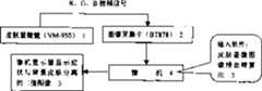
图1是本发明一个实施例采用的系统框图Fig. 1 is a system block diagram that an embodiment of the present invention adopts
图2、图3、图4组成本发明实施例的图像预处理流程框图。FIG. 2 , FIG. 3 , and FIG. 4 constitute a flow chart of image preprocessing in an embodiment of the present invention.
图5是雀斑皮肤的原始显微图像图。Figure 5 is an original photomicrograph of freckled skin.
图6是经小波去噪后的雀斑皮肤显微图像图。Fig. 6 is a microscopic image of freckle skin after wavelet denoising.
图7是经S域转换后的雀斑皮肤的S域图像图。Fig. 7 is an S-domain image diagram of freckle skin after S-domain conversion.
图8是经最大类间方差法分割后的症状区域图像图。Fig. 8 is an image diagram of the symptom area segmented by the method of maximum variance between classes.
图9是经形态学修正后的症状区域图像图。Figure 9 is an image map of the symptom area after morphological correction.
具体实施方式Detailed ways
本发明的一个实施例是以雀斑皮肤为例,按前述的步骤进行其显微图像的预处理。One embodiment of the present invention takes freckle skin as an example, and performs the preprocessing of its microscopic image according to the aforementioned steps.
本实施例采用图1所示系统进行图像预处理:由VM-950型皮肤显微镜1对雀斑皮肤拍摄,其R,G,B视频信号由BT878图像采集卡2输入微机4,将皮肤显微图像预处理软件植入微机而进行预处理运算,然后由微机显示器5显示出症状与背景皮肤分离的二值图像。Present embodiment adopts system shown in Fig. 1 to carry out image pretreatment: by VM-950 type skin microscope 1 pair of freckle skin photographs, its R, G, B video signal is input microcomputer 4 by BT878 image acquisition card 2, skin microscopic image The preprocessing software is implanted into the microcomputer to perform preprocessing calculations, and then the microcomputer display 5 displays a binary image in which the symptoms are separated from the background skin.
微机4的预处理运算程序参见图2、图3、图4组合的图像预处理流程图。For the preprocessing operation program of the microcomputer 4, refer to the combined image preprocessing flowchart of Fig. 2, Fig. 3 and Fig. 4 .
根据前文的操作步骤对雀斑皮肤显微图像进行预处理:Preprocess the microscopic image of freckle skin according to the previous steps:
1.由皮肤显微镜VM-950拍摄的视频信号,经BT878采样得到图5所示的雀斑皮肤的显微图像,可看到其表面有许多毛发。1. The video signal taken by the skin microscope VM-950 was sampled by BT878 to obtain the microscopic image of freckle skin shown in Figure 5, and many hairs can be seen on the surface.
2.采用二进小波去噪:将RGB图像对其R、G、B各分量分别进行。对于R:2. Use binary wavelet denoising: the RGB image is processed separately for its R, G, and B components. For R:
(1)保留二阶分解后的低频部分系数ca2,记录一阶水平、垂直、斜对角的高频部分系数chd1、cvd1、cdd1,以及二阶水平、垂直、斜对角的高频部分系数chd2、cvd2、cdd2,以上系数都是以矩阵的形式保存的。分别求一阶、二阶的任意点M1(i,j)和M2(i,j)模值。(1) Keep the low-frequency part coefficient ca2 after the second-order decomposition, record the first-order horizontal, vertical and diagonal high-frequency part coefficients chd1, cvd1, cdd1, and the second-order horizontal, vertical and diagonal high-frequency part coefficients chd2, cvd2, cdd2, the above coefficients are all stored in the form of matrix. Find the modulus values of the first-order and second-order arbitrary points M1(i, j) and M2(i, j) respectively.
(2)求出二阶小波模、一阶小波模与各阶高频部分系数的平均值 M2, M1,chd2, cvd2, cdd2, chd1, cvd1, cdd1。这里求出的值分别为:26.1582,14.3608,2.8333,0.1209,-0.0245,0.1432,0.0275,-0.0280(2) Calculate the average value of the second-order wavelet mode, first-order wavelet mode and high-frequency coefficients of each order M2, M1, chd2, cvd2, cdd2, chd1, cvd1, cdd1. The values obtained here are: 26.1582, 14.3608, 2.8333, 0.1209, -0.0245, 0.1432, 0.0275, -0.0280
(3)找出M2中大于 M2的点的位置,这些点就是信号突变点,往往在皮肤显微图像中就是毛发与表面纹理的位置。如果任意点M2(i,j)> M2,则令chd2(i,j)= chd2,cvd2(i,j)= cvd2,cdd2(i,j)= cdd2。(3) Find out the value greater than in M2 The position of the M2 points, these points are the signal mutation points, which are often the positions of the hair and surface texture in the skin microscopic image. If any point M2(i, j) > M2, then let chd2(i, j)= chd2, cvd2 (i, j) = cvd2, cdd2(i, j) = cdd2.
(4)将二阶低频部分系数ca2与处理过的二阶高频部分系数重构,得到重构后的一阶低频系数rca1。(4) Reconstruct the second-order low-frequency coefficient ca2 and the processed second-order high-frequency coefficient to obtain the reconstructed first-order low-frequency coefficient rca1.
(5)找出M1中大于 M1的点的位置,如果任意点M1(i,j)> M1,则令chd1(i,j)= chd1,cvd1(i,j)= cvd1,cdd1(i,j)= cdd1。(5) Find out if M1 is greater than The position of the point of M1, if any point M1(i, j) > M1, then let chd1(i, j)= chd1, cvd1 (i, j) = cvd1, cdd1 (i, j) = cdd1.
(6)将重构后的一阶低频系数rca1与处理过的一阶高频部分系数重构,得到处理后的R分量R’。(6) Reconstruct the reconstructed first-order low-frequency coefficient rca1 and the processed first-order high-frequency coefficient to obtain the processed R component R'.
同样的方法得到G’、B’,最后将R’、G’、B’组合,便得到了图6示的去噪后的图像。Get G', B' in the same way, and finally combine R', G', B' to get the denoised image shown in Figure 6.
3.将图像从RGB空间转换到S空间,就得到图7所示的雀斑皮肤的S区域图像。3. Convert the image from the RGB space to the S space to obtain the S area image of the freckle skin shown in Figure 7.
4.症状区域的提取:统计S图中的平均值A=91与标准差σ=36,计算结果显示σ>20,选择最大类间方差法分割方式,求出阈值T=144,得出图8所示的症状区域图像。4. Extraction of symptom area: the average value A=91 and standard deviation σ=36 in the statistical S diagram, the calculation result shows that σ>20, the maximum inter-class variance method is selected for segmentation, and the threshold value T=144 is obtained to obtain the graph 8 shows the image of the symptom area.
5.最后用形态学修正,首先根据图像的闭环区域,消除症状内的黑色区域。然后消除图像中的孤立白点,顶点采用3邻域法、四周采用5邻域法,中间采用8邻域法。得到图9所示的症状区域,是完整地、正确地分离出来的形态修正后的症状区域图像。5. Finally, morphological correction is used, firstly, according to the closed-loop area of the image, the black area in the symptom is eliminated. Then eliminate the isolated white point in the image, use the 3-neighborhood method for the vertex, 5-neighborhood method for the surrounding, and 8-neighborhood method for the middle. The symptom area shown in Figure 9 is obtained, which is a completely and correctly separated symptom area image after morphological correction.
Claims (5)
Priority Applications (1)
| Application Number | Priority Date | Filing Date | Title |
|---|---|---|---|
| CNB2004100535402ACN1300741C (en) | 2004-08-06 | 2004-08-06 | Pre-processing method for skin micro image |
Applications Claiming Priority (1)
| Application Number | Priority Date | Filing Date | Title |
|---|---|---|---|
| CNB2004100535402ACN1300741C (en) | 2004-08-06 | 2004-08-06 | Pre-processing method for skin micro image |
Publications (2)
| Publication Number | Publication Date |
|---|---|
| CN1588428A CN1588428A (en) | 2005-03-02 |
| CN1300741Ctrue CN1300741C (en) | 2007-02-14 |
Family
ID=34602908
Family Applications (1)
| Application Number | Title | Priority Date | Filing Date |
|---|---|---|---|
| CNB2004100535402AExpired - Fee RelatedCN1300741C (en) | 2004-08-06 | 2004-08-06 | Pre-processing method for skin micro image |
Country Status (1)
| Country | Link |
|---|---|
| CN (1) | CN1300741C (en) |
Families Citing this family (4)
| Publication number | Priority date | Publication date | Assignee | Title |
|---|---|---|---|---|
| RU2470576C2 (en)* | 2008-05-23 | 2012-12-27 | Пола Кемикал Индастриз Инк. | Method of automatic assessment of skin and/or wrinkle texture |
| CN103776839B (en)* | 2014-02-10 | 2016-05-04 | 湖州师范学院 | A kind of Surface Crack Inspection Algorithm |
| US11379958B2 (en)* | 2016-09-02 | 2022-07-05 | Casio Computer Co., Ltd. | Diagnosis assisting device, and image processing method in diagnosis assisting device |
| CN110826569B (en)* | 2019-11-05 | 2022-07-19 | 泰康保险集团股份有限公司 | Bill image preprocessing method, device, medium and electronic equipment |
Citations (2)
| Publication number | Priority date | Publication date | Assignee | Title |
|---|---|---|---|---|
| CN1378166A (en)* | 2002-04-29 | 2002-11-06 | 华南理工大学 | Computer aided characteristic registration identifying method for medical homolateral fundus image |
| US20030194119A1 (en)* | 2002-04-15 | 2003-10-16 | General Electric Company | Semi-automatic segmentation algorithm for pet oncology images |
- 2004
- 2004-08-06CNCNB2004100535402Apatent/CN1300741C/ennot_activeExpired - Fee Related
Patent Citations (2)
| Publication number | Priority date | Publication date | Assignee | Title |
|---|---|---|---|---|
| US20030194119A1 (en)* | 2002-04-15 | 2003-10-16 | General Electric Company | Semi-automatic segmentation algorithm for pet oncology images |
| CN1378166A (en)* | 2002-04-29 | 2002-11-06 | 华南理工大学 | Computer aided characteristic registration identifying method for medical homolateral fundus image |
Also Published As
| Publication number | Publication date |
|---|---|
| CN1588428A (en) | 2005-03-02 |
Similar Documents
| Publication | Publication Date | Title |
|---|---|---|
| CN105654436B (en) | A kind of backlight image enhancing denoising method based on prospect background separation | |
| CN106556940B (en) | A kind of background suppression method in TFT-LCD screen automatic optics inspection | |
| JP5941674B2 (en) | Cell contour forming apparatus and method, and cell contour forming program | |
| CN116823686B (en) | Night infrared and visible light image fusion method based on image enhancement | |
| Mahmoud et al. | The automatic identification of melanoma by wavelet and curvelet analysis: study based on neural network classification | |
| CN112801899A (en) | Internal and external circulation driving image blind deblurring method and device based on complementary structure perception | |
| CN112991199A (en) | Image high-low frequency decomposition noise removing method based on residual error dense network | |
| CN111223110A (en) | Microscopic image enhancement method and device and computer equipment | |
| CN113763264B (en) | Image processing method and storage medium based on positive and negative polarity detail layer separation | |
| Ali et al. | Medical images enhanced by using fuzzy logic depending on contrast stretch membership function | |
| CN1921562A (en) | Method for image noise reduction based on transforming domain mathematics morphology | |
| CN1300741C (en) | Pre-processing method for skin micro image | |
| CN116188291A (en) | Rapid airborne dim light image enhancement method | |
| CN114782263B (en) | Direction-aware attention image deraining method, system and device based on wavelet | |
| JP2009098742A (en) | Image processing apparatus, image processing method, and program thereof | |
| CN1254770C (en) | Image merging method based on maximum expectation value and discrete wavelet frame | |
| CN115908155A (en) | NSST domain combined GAN and scale correlation coefficient low-illumination image enhancement and denoising method | |
| Naidu et al. | Enhancement of X-ray images using various Image Processing Approaches | |
| CN1741068A (en) | Histogram equalizing method based on boundary | |
| Guo et al. | Thyroid nodule ultrasonic imaging segmentation based on a deep learning model and data augmentation | |
| Lee et al. | A 4K-capable hardware accelerator of haze removal algorithm using haze-relevant features | |
| JP2023016752A (en) | Processing of image containing overlapping particles | |
| CN111932469B (en) | Method, device, equipment and medium for fusing saliency weight fast exposure images | |
| Wu et al. | Extraction and digitization method of blood vessel in sclera-conjunctiva image | |
| CN114037623B (en) | Water ripple pre-elimination method based on fusion of ripple and environmental characteristics and iterative restoration |
Legal Events
| Date | Code | Title | Description |
|---|---|---|---|
| C06 | Publication | ||
| PB01 | Publication | ||
| C10 | Entry into substantive examination | ||
| SE01 | Entry into force of request for substantive examination | ||
| C14 | Grant of patent or utility model | ||
| GR01 | Patent grant | ||
| C17 | Cessation of patent right | ||
| CF01 | Termination of patent right due to non-payment of annual fee | Granted publication date:20070214 |