CN1242076A - Gel, method and apparatus for biomolecule identification and characterization - Google Patents
Gel, method and apparatus for biomolecule identification and characterizationDownload PDFInfo
- Publication number
- CN1242076A CN1242076ACN97181104ACN97181104ACN1242076ACN 1242076 ACN1242076 ACN 1242076ACN 97181104 ACN97181104 ACN 97181104ACN 97181104 ACN97181104 ACN 97181104ACN 1242076 ACN1242076 ACN 1242076A
- Authority
- CN
- China
- Prior art keywords
- gel
- dimensional array
- biomolecule
- species
- computer
- Prior art date
- Legal status (The legal status is an assumption and is not a legal conclusion. Google has not performed a legal analysis and makes no representation as to the accuracy of the status listed.)
- Granted
Links
Images

Classifications
- G—PHYSICS
- G01—MEASURING; TESTING
- G01N—INVESTIGATING OR ANALYSING MATERIALS BY DETERMINING THEIR CHEMICAL OR PHYSICAL PROPERTIES
- G01N27/00—Investigating or analysing materials by the use of electric, electrochemical, or magnetic means
- G01N27/26—Investigating or analysing materials by the use of electric, electrochemical, or magnetic means by investigating electrochemical variables; by using electrolysis or electrophoresis
- G01N27/416—Systems
- G01N27/447—Systems using electrophoresis
- G01N27/44704—Details; Accessories
- G01N27/44717—Arrangements for investigating the separated zones, e.g. localising zones
- G01N27/44739—Collecting the separated zones, e.g. blotting to a membrane or punching of gel spots
- G—PHYSICS
- G01—MEASURING; TESTING
- G01N—INVESTIGATING OR ANALYSING MATERIALS BY DETERMINING THEIR CHEMICAL OR PHYSICAL PROPERTIES
- G01N27/00—Investigating or analysing materials by the use of electric, electrochemical, or magnetic means
- G01N27/26—Investigating or analysing materials by the use of electric, electrochemical, or magnetic means by investigating electrochemical variables; by using electrolysis or electrophoresis
- G01N27/416—Systems
- G01N27/447—Systems using electrophoresis
- G01N27/44704—Details; Accessories
- G01N27/44717—Arrangements for investigating the separated zones, e.g. localising zones
- B—PERFORMING OPERATIONS; TRANSPORTING
- B01—PHYSICAL OR CHEMICAL PROCESSES OR APPARATUS IN GENERAL
- B01J—CHEMICAL OR PHYSICAL PROCESSES, e.g. CATALYSIS OR COLLOID CHEMISTRY; THEIR RELEVANT APPARATUS
- B01J2219/00—Chemical, physical or physico-chemical processes in general; Their relevant apparatus
- B01J2219/00274—Sequential or parallel reactions; Apparatus and devices for combinatorial chemistry or for making arrays; Chemical library technology
- B01J2219/00277—Apparatus
- B01J2219/00497—Features relating to the solid phase supports
- B01J2219/00527—Sheets
- B—PERFORMING OPERATIONS; TRANSPORTING
- B01—PHYSICAL OR CHEMICAL PROCESSES OR APPARATUS IN GENERAL
- B01J—CHEMICAL OR PHYSICAL PROCESSES, e.g. CATALYSIS OR COLLOID CHEMISTRY; THEIR RELEVANT APPARATUS
- B01J2219/00—Chemical, physical or physico-chemical processes in general; Their relevant apparatus
- B01J2219/00274—Sequential or parallel reactions; Apparatus and devices for combinatorial chemistry or for making arrays; Chemical library technology
- B01J2219/00583—Features relative to the processes being carried out
- B01J2219/00603—Making arrays on substantially continuous surfaces
- B01J2219/00605—Making arrays on substantially continuous surfaces the compounds being directly bound or immobilised to solid supports
- B01J2219/00612—Making arrays on substantially continuous surfaces the compounds being directly bound or immobilised to solid supports the surface being inorganic
- B—PERFORMING OPERATIONS; TRANSPORTING
- B01—PHYSICAL OR CHEMICAL PROCESSES OR APPARATUS IN GENERAL
- B01J—CHEMICAL OR PHYSICAL PROCESSES, e.g. CATALYSIS OR COLLOID CHEMISTRY; THEIR RELEVANT APPARATUS
- B01J2219/00—Chemical, physical or physico-chemical processes in general; Their relevant apparatus
- B01J2219/00274—Sequential or parallel reactions; Apparatus and devices for combinatorial chemistry or for making arrays; Chemical library technology
- B01J2219/00583—Features relative to the processes being carried out
- B01J2219/00603—Making arrays on substantially continuous surfaces
- B01J2219/00605—Making arrays on substantially continuous surfaces the compounds being directly bound or immobilised to solid supports
- B01J2219/00614—Delimitation of the attachment areas
- B01J2219/00621—Delimitation of the attachment areas by physical means, e.g. trenches, raised areas
- B—PERFORMING OPERATIONS; TRANSPORTING
- B01—PHYSICAL OR CHEMICAL PROCESSES OR APPARATUS IN GENERAL
- B01J—CHEMICAL OR PHYSICAL PROCESSES, e.g. CATALYSIS OR COLLOID CHEMISTRY; THEIR RELEVANT APPARATUS
- B01J2219/00—Chemical, physical or physico-chemical processes in general; Their relevant apparatus
- B01J2219/00274—Sequential or parallel reactions; Apparatus and devices for combinatorial chemistry or for making arrays; Chemical library technology
- B01J2219/00583—Features relative to the processes being carried out
- B01J2219/00603—Making arrays on substantially continuous surfaces
- B01J2219/00605—Making arrays on substantially continuous surfaces the compounds being directly bound or immobilised to solid supports
- B01J2219/00623—Immobilisation or binding
- B01J2219/00626—Covalent
- B—PERFORMING OPERATIONS; TRANSPORTING
- B01—PHYSICAL OR CHEMICAL PROCESSES OR APPARATUS IN GENERAL
- B01J—CHEMICAL OR PHYSICAL PROCESSES, e.g. CATALYSIS OR COLLOID CHEMISTRY; THEIR RELEVANT APPARATUS
- B01J2219/00—Chemical, physical or physico-chemical processes in general; Their relevant apparatus
- B01J2219/00274—Sequential or parallel reactions; Apparatus and devices for combinatorial chemistry or for making arrays; Chemical library technology
- B01J2219/00583—Features relative to the processes being carried out
- B01J2219/00603—Making arrays on substantially continuous surfaces
- B01J2219/00605—Making arrays on substantially continuous surfaces the compounds being directly bound or immobilised to solid supports
- B01J2219/00623—Immobilisation or binding
- B01J2219/0063—Other, e.g. van der Waals forces, hydrogen bonding
- B—PERFORMING OPERATIONS; TRANSPORTING
- B01—PHYSICAL OR CHEMICAL PROCESSES OR APPARATUS IN GENERAL
- B01J—CHEMICAL OR PHYSICAL PROCESSES, e.g. CATALYSIS OR COLLOID CHEMISTRY; THEIR RELEVANT APPARATUS
- B01J2219/00—Chemical, physical or physico-chemical processes in general; Their relevant apparatus
- B01J2219/00274—Sequential or parallel reactions; Apparatus and devices for combinatorial chemistry or for making arrays; Chemical library technology
- B01J2219/00583—Features relative to the processes being carried out
- B01J2219/00603—Making arrays on substantially continuous surfaces
- B01J2219/00605—Making arrays on substantially continuous surfaces the compounds being directly bound or immobilised to solid supports
- B01J2219/00632—Introduction of reactive groups to the surface
- B01J2219/00637—Introduction of reactive groups to the surface by coating it with another layer
- B—PERFORMING OPERATIONS; TRANSPORTING
- B01—PHYSICAL OR CHEMICAL PROCESSES OR APPARATUS IN GENERAL
- B01J—CHEMICAL OR PHYSICAL PROCESSES, e.g. CATALYSIS OR COLLOID CHEMISTRY; THEIR RELEVANT APPARATUS
- B01J2219/00—Chemical, physical or physico-chemical processes in general; Their relevant apparatus
- B01J2219/00274—Sequential or parallel reactions; Apparatus and devices for combinatorial chemistry or for making arrays; Chemical library technology
- B01J2219/00583—Features relative to the processes being carried out
- B01J2219/00603—Making arrays on substantially continuous surfaces
- B01J2219/00639—Making arrays on substantially continuous surfaces the compounds being trapped in or bound to a porous medium
- B01J2219/00641—Making arrays on substantially continuous surfaces the compounds being trapped in or bound to a porous medium the porous medium being continuous, e.g. porous oxide substrates
- B—PERFORMING OPERATIONS; TRANSPORTING
- B01—PHYSICAL OR CHEMICAL PROCESSES OR APPARATUS IN GENERAL
- B01J—CHEMICAL OR PHYSICAL PROCESSES, e.g. CATALYSIS OR COLLOID CHEMISTRY; THEIR RELEVANT APPARATUS
- B01J2219/00—Chemical, physical or physico-chemical processes in general; Their relevant apparatus
- B01J2219/00274—Sequential or parallel reactions; Apparatus and devices for combinatorial chemistry or for making arrays; Chemical library technology
- B01J2219/00583—Features relative to the processes being carried out
- B01J2219/00603—Making arrays on substantially continuous surfaces
- B01J2219/00639—Making arrays on substantially continuous surfaces the compounds being trapped in or bound to a porous medium
- B01J2219/00644—Making arrays on substantially continuous surfaces the compounds being trapped in or bound to a porous medium the porous medium being present in discrete locations, e.g. gel pads
- B—PERFORMING OPERATIONS; TRANSPORTING
- B01—PHYSICAL OR CHEMICAL PROCESSES OR APPARATUS IN GENERAL
- B01J—CHEMICAL OR PHYSICAL PROCESSES, e.g. CATALYSIS OR COLLOID CHEMISTRY; THEIR RELEVANT APPARATUS
- B01J2219/00—Chemical, physical or physico-chemical processes in general; Their relevant apparatus
- B01J2219/00274—Sequential or parallel reactions; Apparatus and devices for combinatorial chemistry or for making arrays; Chemical library technology
- B01J2219/00583—Features relative to the processes being carried out
- B01J2219/00603—Making arrays on substantially continuous surfaces
- B01J2219/00659—Two-dimensional arrays
- B—PERFORMING OPERATIONS; TRANSPORTING
- B01—PHYSICAL OR CHEMICAL PROCESSES OR APPARATUS IN GENERAL
- B01J—CHEMICAL OR PHYSICAL PROCESSES, e.g. CATALYSIS OR COLLOID CHEMISTRY; THEIR RELEVANT APPARATUS
- B01J2219/00—Chemical, physical or physico-chemical processes in general; Their relevant apparatus
- B01J2219/00274—Sequential or parallel reactions; Apparatus and devices for combinatorial chemistry or for making arrays; Chemical library technology
- B01J2219/0068—Means for controlling the apparatus of the process
- B01J2219/00686—Automatic
- B01J2219/00689—Automatic using computers
- B—PERFORMING OPERATIONS; TRANSPORTING
- B01—PHYSICAL OR CHEMICAL PROCESSES OR APPARATUS IN GENERAL
- B01J—CHEMICAL OR PHYSICAL PROCESSES, e.g. CATALYSIS OR COLLOID CHEMISTRY; THEIR RELEVANT APPARATUS
- B01J2219/00—Chemical, physical or physico-chemical processes in general; Their relevant apparatus
- B01J2219/00274—Sequential or parallel reactions; Apparatus and devices for combinatorial chemistry or for making arrays; Chemical library technology
- B01J2219/0068—Means for controlling the apparatus of the process
- B01J2219/00686—Automatic
- B01J2219/00691—Automatic using robots
- B—PERFORMING OPERATIONS; TRANSPORTING
- B01—PHYSICAL OR CHEMICAL PROCESSES OR APPARATUS IN GENERAL
- B01J—CHEMICAL OR PHYSICAL PROCESSES, e.g. CATALYSIS OR COLLOID CHEMISTRY; THEIR RELEVANT APPARATUS
- B01J2219/00—Chemical, physical or physico-chemical processes in general; Their relevant apparatus
- B01J2219/00274—Sequential or parallel reactions; Apparatus and devices for combinatorial chemistry or for making arrays; Chemical library technology
- B01J2219/0068—Means for controlling the apparatus of the process
- B01J2219/00702—Processes involving means for analysing and characterising the products
- B01J2219/00704—Processes involving means for analysing and characterising the products integrated with the reactor apparatus
- B—PERFORMING OPERATIONS; TRANSPORTING
- B01—PHYSICAL OR CHEMICAL PROCESSES OR APPARATUS IN GENERAL
- B01J—CHEMICAL OR PHYSICAL PROCESSES, e.g. CATALYSIS OR COLLOID CHEMISTRY; THEIR RELEVANT APPARATUS
- B01J2219/00—Chemical, physical or physico-chemical processes in general; Their relevant apparatus
- B01J2219/00274—Sequential or parallel reactions; Apparatus and devices for combinatorial chemistry or for making arrays; Chemical library technology
- B01J2219/00718—Type of compounds synthesised
- B01J2219/0072—Organic compounds
- G—PHYSICS
- G01—MEASURING; TESTING
- G01N—INVESTIGATING OR ANALYSING MATERIALS BY DETERMINING THEIR CHEMICAL OR PHYSICAL PROPERTIES
- G01N1/00—Sampling; Preparing specimens for investigation
- G01N1/28—Preparing specimens for investigation including physical details of (bio-)chemical methods covered elsewhere, e.g. G01N33/50, C12Q
- G01N1/286—Preparing specimens for investigation including physical details of (bio-)chemical methods covered elsewhere, e.g. G01N33/50, C12Q involving mechanical work, e.g. chopping, disintegrating, compacting, homogenising
- G01N2001/2873—Cutting or cleaving
- G01N2001/288—Filter punches
- G—PHYSICS
- G01—MEASURING; TESTING
- G01N—INVESTIGATING OR ANALYSING MATERIALS BY DETERMINING THEIR CHEMICAL OR PHYSICAL PROPERTIES
- G01N27/00—Investigating or analysing materials by the use of electric, electrochemical, or magnetic means
- G01N27/26—Investigating or analysing materials by the use of electric, electrochemical, or magnetic means by investigating electrochemical variables; by using electrolysis or electrophoresis
- G01N27/416—Systems
- G01N27/447—Systems using electrophoresis
- G01N27/44756—Apparatus specially adapted therefor
- G01N27/44773—Multi-stage electrophoresis, e.g. two-dimensional electrophoresis
- G—PHYSICS
- G01—MEASURING; TESTING
- G01N—INVESTIGATING OR ANALYSING MATERIALS BY DETERMINING THEIR CHEMICAL OR PHYSICAL PROPERTIES
- G01N27/00—Investigating or analysing materials by the use of electric, electrochemical, or magnetic means
- G01N27/26—Investigating or analysing materials by the use of electric, electrochemical, or magnetic means by investigating electrochemical variables; by using electrolysis or electrophoresis
- G01N27/416—Systems
- G01N27/447—Systems using electrophoresis
- G01N27/44756—Apparatus specially adapted therefor
- G01N27/44782—Apparatus specially adapted therefor of a plurality of samples
Landscapes
- Health & Medical Sciences (AREA)
- Life Sciences & Earth Sciences (AREA)
- Molecular Biology (AREA)
- Chemical & Material Sciences (AREA)
- Chemical Kinetics & Catalysis (AREA)
- Electrochemistry (AREA)
- Physics & Mathematics (AREA)
- Analytical Chemistry (AREA)
- Biochemistry (AREA)
- General Health & Medical Sciences (AREA)
- General Physics & Mathematics (AREA)
- Immunology (AREA)
- Pathology (AREA)
- Investigating, Analyzing Materials By Fluorescence Or Luminescence (AREA)
- Investigating Or Analysing Biological Materials (AREA)
- Measuring Or Testing Involving Enzymes Or Micro-Organisms (AREA)
- Apparatus Associated With Microorganisms And Enzymes (AREA)
- Pharmaceuticals Containing Other Organic And Inorganic Compounds (AREA)
- Medicines That Contain Protein Lipid Enzymes And Other Medicines (AREA)
- Colloid Chemistry (AREA)
Abstract
Translated fromChineseDescription
Translated fromChinese1.简介 1 Introduction
本发明涉及计算机辅助的方法和仪器,其用于高效及系统地研究存在于生物学样品中的分子并测定它们在健康和疾病中的作用。尤其是,本发明涉及蛋白质图(protemics)的成像领域,其包括存在于生物学样品中的蛋白质的系统鉴定及表征,其中蛋白质包括糖基化的或展示其它翻译后修饰的蛋白质。蛋白质图方法对于鉴定对诊断、预后或监测应答治疗的有用之蛋白质以及在鉴定用于疾病预防和治疗的蛋白质目标中提供了极大的优点。The present invention relates to computer-aided methods and instruments for efficiently and systematically studying molecules present in biological samples and determining their role in health and disease. In particular, the invention relates to the field of imaging of protemics, which includes the systematic identification and characterization of proteins present in biological samples, including proteins that are glycosylated or display other post-translational modifications. Protein mapping methods offer great advantages for identifying proteins useful for diagnosis, prognosis, or monitoring response to therapy, as well as in identifying protein targets for disease prevention and treatment.
2.发明背景2. Background of the invention
在分子遗传学上的最新进展揭示了用于在特定细胞或组织中核酸表达研究的高通量的测序技术和系统策略的益处。这些进展已经突出了对不依赖操作者的计算机介导的方法的需求,这些方法用于鉴定和选择来自蛋白质、寡糖及其它生物分子的复杂混合物的子集或个体分子,并分离这种选择的生物分子用于进一步分析。Recent advances in molecular genetics have revealed the benefits of high-throughput sequencing technologies and systematic strategies for the study of nucleic acid expression in specific cells or tissues. These advances have highlighted the need for operator-independent, computer-mediated methods for identifying and selecting subsets or individual molecules from complex mixtures of proteins, oligosaccharides, and other biomolecules, and for isolating such selected The biomolecules were used for further analysis.
用于靶物驱动的药物发现以及合理的药物设计的策略需要鉴定在因果关系上涉及疾病过程的关键细胞组分,例如蛋白质,以及使用这种组分作为治疗干预的靶物。然而,现在的分析生物分子例如蛋白质的方法耗费时间并且昂贵,而且在检测、成像、纯化及分析方面要受效率低下的困扰。Strategies for target-driven drug discovery and rational drug design require the identification of key cellular components, such as proteins, that are causally involved in disease processes, and the use of such components as targets for therapeutic intervention. However, current methods for analyzing biomolecules such as proteins are time consuming and expensive, and suffer from inefficiencies in detection, imaging, purification and analysis.
虽然基因组方法已经推进了我们对生物学过程之遗传基础的理解,但有明显的局限性。首先,鉴定的基因,尤其是部分cDNA序列编码的产物的功能经常是未知的。其次,有关蛋白质翻译后修饰的信息很少能从其基因序列的知识推导出,并且现在很明显大部分蛋白质经历能够深刻地影响其生物化学特性的翻译后修饰(例如糖基化及磷酸化)。第三,蛋白质表达通常受翻译后控制,所以细胞的mRNA水平不是必定与其基因产物的表达水平相关联。第四,自动化的核酸随机测序策略包括在确定其显示临床或科学意义(如果有的话)之前大量核酸分子的分析。While genomic approaches have advanced our understanding of the genetic basis of biological processes, there are clear limitations. First, the function of an identified gene, especially a product encoded by a partial cDNA sequence, is often unknown. Second, little information about post-translational modifications of proteins can be deduced from knowledge of their genetic sequences, and it is now clear that most proteins undergo post-translational modifications (such as glycosylation and phosphorylation) that can profoundly affect their biochemical properties . Third, protein expression is often under post-translational control, so a cell's mRNA level does not necessarily correlate with the expression level of its gene product. Fourth, automated random nucleic acid sequencing strategies involve the analysis of large numbers of nucleic acid molecules prior to determining that they show clinical or scientific significance, if any.
因为这些原因,需要补充基因组资料,方法是通过研究蛋白质和碳水化合物的表达模式以及通过对蛋白质、寡糖和其它生物分子的直接分析研究其在生物学或疾病过程中翻译后修饰的模式。然而,技术上的局限到现在为止已经阻碍了在生物学样品中蛋白质或其它生物分子的快速、经济、可重复的系统分析。For these reasons, genomic data need to be supplemented by studying the expression patterns of proteins and carbohydrates and by direct analysis of proteins, oligosaccharides and other biomolecules to study their patterns of post-translational modification during biological or disease processes. However, technical limitations have so far prevented rapid, economical, and reproducible systematic analysis of proteins or other biomolecules in biological samples.
3.发明概述3. Summary of Invention
本发明涉及用于生物学样品中生物分子的鉴定、选择和表征的高效计算机辅助的方法及仪器。根据本发明,通过分离存在于复杂混合物中的生物分子产生一个二维阵列。本发明提供了一种计算机产生的数字化分布图,其表示在二维阵列中检测的多数生物分子的特征性及相对丰度,籍此允许计算机介导的来自多重生物学样品分布图的比较。这种筛选生物学样品并比较其分布图的自动化技术,实现了对个体生物分子快速高效的鉴定,这些分子的存在、缺乏或改变的表达与疾病或目标病况相联系。这种生物分子可用作治疗剂,用作用于治疗干预的靶物以及用作诊断、预后和评价对治疗反应的标记。这种技术也实现了快速并高效的鉴定成套的生物分子,其表达模式与疾病或目标病况相联系;这些成套的生物分子提供了用于诊断、预后及评价对治疗反应标记的图样。The present invention relates to efficient computer-aided methods and apparatus for the identification, selection and characterization of biomolecules in biological samples. According to the present invention, a two-dimensional array is generated by separating biomolecules present in a complex mixture. The present invention provides a computer-generated digitized profile representing the identity and relative abundance of a plurality of biomolecules detected in a two-dimensional array, thereby allowing computer-mediated comparison of profiles from multiple biological samples. This automated technique of screening biological samples and comparing their profiles enables the rapid and efficient identification of individual biomolecules whose presence, absence or altered expression is associated with a disease or condition of interest. Such biomolecules are useful as therapeutic agents, as targets for therapeutic intervention and as markers for diagnosis, prognosis and assessment of response to therapy. This technique also enables rapid and efficient identification of sets of biomolecules whose expression patterns correlate with diseases or conditions of interest; these sets of biomolecules provide patterns of markers for diagnosis, prognosis, and assessment of response to treatment.
本发明的高通量、自动化的方法和仪器进一步实现了根据预先规定标准对个体分离的生物分子(或分离的生物分子的子集)不依赖操作者的选择,而不需要任何序列信息或生物分子的其它结构特征的知识。而这又提供了在生物学样品中检测的多数所选生物分子的自动化、不依赖操作者的分离以及并行表征。因此,本发明在测序或结构表征之前,有利地实现了生物分子的自动化选择。在一个特定的实施方案中,本发明提供了一种适于生物分子(如蛋白质)电泳的凝胶并且其结合于固体支持物上以使凝胶具有二维空间稳定性,并且支持物基本上不干扰关于与凝胶中一个或多个生物分子相结合的标记的检测(例如结合于一个或多个蛋白质的荧光标记)。在另一个特定的实施方案中,本发明提供了一个综合的计算机程序,该计算机程序通过比较数字化分布图来选择在二维阵列中检测的一个或多个生物分子,并产生指令指导一种机器人装置从二维阵列中分离这种选择的生物分子。然而在进一步的实施方案中,这个程序也实现了实验室信息管理系统(LIMS),其跟踪实验室样品以及相关的数据例如临床数据,在样品上进行的操作和通过分析样品产生的数据。The high-throughput, automated methods and apparatus of the present invention further enable operator-independent selection of individual isolated biomolecules (or subsets of isolated biomolecules) according to pre-specified criteria, without any sequence information or biological Knowledge of other structural features of the molecule. This in turn provides automated, operator-independent separation and parallel characterization of most selected biomolecules detected in biological samples. Thus, the present invention advantageously enables automated selection of biomolecules prior to sequencing or structural characterization. In a specific embodiment, the present invention provides a gel suitable for electrophoresis of biomolecules such as proteins and which is bound to a solid support such that the gel is two-dimensionally stable and the support is substantially Does not interfere with detection of labels associated with one or more biomolecules in the gel (eg fluorescent labels bound to one or more proteins). In another specific embodiment, the present invention provides an integrated computer program for selecting one or more biomolecules detected in a two-dimensional array by comparing digitized profiles and generating instructions to direct a robotic The device separates such selected biomolecules from a two-dimensional array. In a further embodiment, however, this program also implements a laboratory information management system (LIMS) that tracks laboratory samples and related data such as clinical data, operations performed on the samples and data generated by analyzing the samples.
4.附图简述4. Brief description of the attached drawings
图1是根据本发明特定的实施方案,在不同蛋白质的混合物上进行的操作流程图。Figure 1 is a flow diagram of operations performed on a mixture of different proteins according to a particular embodiment of the invention.
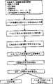
图2是阐明在本发明特定的实施方案中依靠计算机执行的功能的流程图。Figure 2 is a flowchart illustrating computer-implemented functions in a particular embodiment of the invention.
图3是用来从本发明的被支持的凝胶中分离生物分子的机器人装置的图解。Figure 3 is a schematic illustration of a robotic device used to separate biomolecules from a supported gel of the present invention.
5.发明详述5. Detailed description of the invention
本发明提供了用来快速和有效地鉴定及表征生物学样品中生物分子如蛋白质的方法和仪器。在本发明的一个应用中,生物学样品进行两个连续的分离步骤。在第一个分离步骤中,生物分子根据一种物理或化学特性被分离,以产生包含生物分子的一维阵列;例如,蛋白质通过等电聚焦沿着第一轴被分离。在第二个分离步骤中,在该一维阵列中的生物分子根据第二种物理或化学特性被分离,以产生一个分离的生物分子的二维阵列;例如,通过等电聚焦分离的蛋白质沿着与第一个轴垂直的第二个轴进行SDS-PAGE。分离的生物分子稳定地保持在二维阵列中以随后成像。基于对来自成像数据的自动化计算机分析,稳定的二维阵列可保存或存档一段较长的时期(如数月或数年),并且选择的生物分子能在任何要求的时间从阵列中检索。The present invention provides methods and apparatus for the rapid and efficient identification and characterization of biomolecules, such as proteins, in biological samples. In one application of the invention, a biological sample is subjected to two successive separation steps. In the first separation step, biomolecules are separated according to one physical or chemical property to produce a one-dimensional array containing biomolecules; for example, proteins are separated along a first axis by isoelectric focusing. In a second separation step, the biomolecules in the one-dimensional array are separated according to a second physical or chemical property to produce a two-dimensional array of separated biomolecules; for example, proteins separated by isoelectric focusing along SDS-PAGE was performed with the second axis perpendicular to the first axis. Separated biomolecules are held stably in a two-dimensional array for subsequent imaging. Based on automated computer analysis of data from imaging, stable two-dimensional arrays can be stored or archived for extended periods of time (eg, months or years), and selected biomolecules can be retrieved from the array at any desired time.
对每个检测的生物分子,通过检测器对二维阵列成像而产生包含一组x,y坐标及一个信号值的计算机可读的输出信号。如需要,在计算机介导的分析之前或之后计算机可读的输出信号能够在显示器或任何适当的媒介上以计算机产生的图像显示给人类操作者。对计算机可读的输出信号之计算机介导的分析导致计算机可读的分布图,对大多数检测的生物分子而言它表示每个这种生物分子的相对丰度以及从其在二维阵列中x,y坐标推断的属性。例如,来自包含通过等电聚焦后由SDS-PAGE分离蛋白质的凝胶成像的分布图,显示了大多数被检测蛋白质的等电点(pI)、表观分子量(MW)以及相对丰度。For each detected biomolecule, the two-dimensional array is imaged by the detector to generate a computer readable output signal comprising a set of x, y coordinates and a signal value. If desired, the computer-readable output signal can be displayed to a human operator as a computer-generated image on a display or any suitable medium, either before or after computer-mediated analysis. Computer-mediated analysis of the computer-readable output signal results in a computer-readable profile that, for most detected biomolecules, represents the relative abundance of each such biomolecule and its distribution in a two-dimensional array. Attributes for x,y coordinate inference. For example, a profile from a gel image containing proteins separated by SDS-PAGE after isoelectric focusing shows the isoelectric point (pi), apparent molecular weight (MW) and relative abundance of most detected proteins.
本发明的计算机可读的分布图适于计算机介导的分析来鉴定满足特定标准的一个或多个生物分子。在一个实施方案中,第一组分布图与第二组分布图比较以鉴定在所有第一组分布图中存在(或以第一组分布图的第一个百分比)而在第二组分布图中缺失(或第二组分布图的第二个百分比缺失,第一和第二百分比可以独立地指定)的生物分子。在其它实施方案中,比较多组分布图以鉴定与另一样品组分布图的指定百分比相比,在一个样品组分布图中存在以指定的较高水平表达的生物分子,或鉴定一个样品组与另一个样品组在翻译后加工中不同的生物分子。The computer-readable profiles of the invention are suitable for computer-mediated analysis to identify one or more biomolecules meeting certain criteria. In one embodiment, the first set of profiles is compared to the second set of profiles to identify the presence in all of the first set of profiles (or in a first percentage of the first set of profiles) that is present in the second set of profiles Biomolecules that are missing in (or missing in a second percentage of the second set of profiles, the first and second percentages can be specified independently). In other embodiments, multiple sets of profiles are compared to identify the presence of a biomolecule expressed at a specified higher level in one sample set profile compared to a specified percentage of another sample set profile, or to identify a sample set Biomolecules that differ in post-translational processing from another sample group.
选择如此鉴定的一个或多个生物分子来进行分离。在一个实施方案中,这种选择是根据预先规定的编程的标准通过计算机自动进行的,没有更多的人类干预。在另一个实施方案中,人类操作者综合计算机介导的分析结果,然后输入计算机一个选择。为分离每个选择的生物分子,计算机产生指导机器人装置的机器可读性指令(a)取出包含选择的生物分子二维阵列的一个或多个部分并且(b)将取出的部分输运至一个或多个适当的容器用于进一步表征。例如,可以分析选择的蛋白质以确定其全部或部分的氨基酸序列,以检测并表征任何结合的寡糖部分,以及研究翻译后加工的其它方面,例如磷酸化、十四烷基化等等。本发明有利地实现了从二维阵列取出生物分子的自动化并行步骤,从而促进许多选择的生物分子快速及有效的表征。图一表示根据本发明的一个特定实施方案,一个样品的图解处理的流程图。One or more biomolecules so identified are selected for isolation. In one embodiment, this selection is performed automatically by a computer according to pre-specified programmed criteria, without further human intervention. In another embodiment, a human operator synthesizes the results of the computer-mediated analysis and then enters a selection into the computer. To isolate each selected biomolecule, computer-generated machine-readable instructions directing the robotic device to (a) retrieve one or more portions containing the two-dimensional array of selected biomolecules and (b) transport the removed portions to a or multiple appropriate containers for further characterization. For example, selected proteins can be analyzed to determine all or part of their amino acid sequence, to detect and characterize any bound oligosaccharide moieties, and to study other aspects of post-translational processing, such as phosphorylation, myristylation, and the like. The present invention advantageously enables automated parallel steps of retrieval of biomolecules from two-dimensional arrays, thereby facilitating rapid and efficient characterization of many selected biomolecules. Figure 1 shows a flow diagram of the graphical processing of a sample according to a particular embodiment of the invention.
本发明可用于鉴定及分析蛋白质,但是一般地更适于鉴定及分析任何生物分子。如在此使用的,术语“生物分子”指的是存在于生物学样品中的任何有机分子,并包括肽、多肽、蛋白质、寡糖、脂类、类固醇、前列腺素、前列环素以及核酸(包括DNA和RNA)。如在此使用的,术语“蛋白质”包括糖基化的及非糖基化的蛋白质。5.1.生物学样品The present invention can be used to identify and analyze proteins, but is generally more suitable for identifying and analyzing any biomolecule. As used herein, the term "biomolecule" refers to any organic molecule present in a biological sample and includes peptides, polypeptides, proteins, oligosaccharides, lipids, steroids, prostaglandins, prostacyclins, and nucleic acids ( including DNA and RNA). As used herein, the term "protein" includes glycosylated and non-glycosylated proteins. 5.1. Biological samples
如在此使用的,术语“生物学样品”指的是从任何活生物体排泄或分泌物中获得的任何固体或液体样品,包括单细胞微生物(例如细菌和酵母)以及多细胞生物(例如植物和动物,如脊椎动物或哺乳动物,尤其是健康或表面上健康的人或由将被诊断或调查的病情或疾病侵袭的人类患者)。生物学样品可以是从任何部位(例如血液、血浆、血清、尿液、胆汁、脑脊液、含水的或玻璃样体液,或任何身体分泌物)获得的生物学液体,渗出液,分泌液(例如从脓肿或任何感染或发炎的其它部位获得的液体),或从关节(例如正常的关节或被疾病例如风湿性关节炎、骨关节炎、痛风或脓毒性关节炎侵袭的关节)获得的液体。另外,生物学样品可从任何器官或组织(包括活组织检查或尸体解剖样品)获得,或从包括细胞(无论是原代的或培养的细胞)或被任何细胞、组织或器官决定的介质中获得。如需要,生物学样品可以经过初步加工,包括初步分离技术。例如,可以提取细胞或组织并进行亚细胞分级分离,用来在特定的亚细胞部分分离分析生物分子,例如在细胞的不同部分发现的蛋白质或药物。见Deutscher(编),1990,酶学方法(Methods InEnzymology)182卷,147-238(在此引入其全部以供参考)。类似地,可以进行免疫沉淀以鉴定抗原相关的生物分子,例如蛋白质。见Firestone和Winguth在Duetscher中,出处同前,688-699(在此引入其全部以供参考)。As used herein, the term "biological sample" refers to any solid or liquid sample obtained from the excretion or secretion of any living organism, including unicellular microorganisms such as bacteria and yeast, as well as multicellular organisms such as plants and animals, such as vertebrates or mammals, especially healthy or apparently healthy humans or human patients afflicted by the condition or disease to be diagnosed or investigated). A biological sample may be a biological fluid obtained from any site (e.g., blood, plasma, serum, urine, bile, cerebrospinal fluid, aqueous or vitreous body fluid, or any bodily secretion), an exudate, an exudate (e.g. fluid obtained from an abscess or any other site of infection or inflammation), or fluid obtained from a joint such as a normal joint or a joint affected by a disease such as rheumatoid arthritis, osteoarthritis, gout or septic arthritis. Additionally, a biological sample may be obtained from any organ or tissue (including biopsy or autopsy samples), or from a medium comprising cells (whether primary or cultured) or determined by any cell, tissue, or organ get. Biological samples may undergo primary processing, including primary separation techniques, if desired. For example, cells or tissues can be extracted and subjected to subcellular fractionation to isolate and analyze biomolecules, such as proteins or drugs found in different parts of the cell, in specific subcellular fractions. See Deutscher (Ed.), 1990, Methods In Enzymology Vol. 182, 147-238 (herein incorporated by reference in its entirety). Similarly, immunoprecipitation can be performed to identify antigen-associated biomolecules such as proteins. See Firestone and Winguth in Duetscher, supra, 688-699 (herein incorporated by reference in its entirety).
优选地,对分析有用的相关临床信息依相应的样品按目录分类并编为索引;基于计算机的实验室信息管理系统(LIMS)优选地用于此目的。这种信息优选地包括患者资料例如家族史、临床诊断、性别、年龄、民族、居住地、工作地以及病史。在LIMS中,涉及样品本身的信息也优选地编入索引;这种信息可包括样品类型、取样的精确位置、取样品的日时、采集与保存之间的时间、保存的方法及获得样品使用的方法。Preferably, relevant clinical information useful for the analysis is cataloged and indexed against the respective sample; a computer-based laboratory information management system (LIMS) is preferably used for this purpose. Such information preferably includes patient data such as family history, clinical diagnosis, gender, age, ethnicity, place of residence, place of work, and medical history. In the LIMS, information pertaining to the sample itself is preferably also indexed; this information may include the type of sample, the precise location of sampling, the day of sampling, the time between collection and storage, the method of storage, and the method used to obtain the sample. Methods.
将信息记录索引到适合的样品的方法可以包括对记录和样品分配匹配的编号。优选地通过使用条形码和条形码扫描器使这个过程自动化。处理每个样品时,扫描器用于将样品鉴定号码记录进LIMS,通过其不同操作追踪样品,因而在记录和样品之间保持链接。条形码的使用同样实现了自动归档并检索保存的样品及凝胶。5.2.蛋白质分析The method of indexing information records to appropriate samples may include assigning matching numbers to records and samples. This process is preferably automated through the use of barcodes and barcode scanners. As each sample is processed, the scanner is used to record the sample identification number into the LIMS, tracking the sample through its different operations, thus maintaining a link between the record and the sample. The use of barcodes also enables automatic archiving and retrieval of stored samples and gels. 5.2. Protein analysis
在一个实施方案中,使用本发明的方法及仪器鉴定并表征在生物学样品中的一个或多个蛋白质。5.2.1.第一步分离In one embodiment, one or more proteins in a biological sample are identified and characterized using the methods and apparatus of the invention. 5.2.1. The first step of separation
对那些本领域的技术人员来说,很多种分离蛋白质的技术是众所周知的,见例如,Deutscher(编),1990,酶学方法182卷,9-18页和285-554页(在此引入其全部以供参考),并可根据本发明进行应用。以举例方式但并不限于此,蛋白质可以基于等电点(例如通过层析聚焦或等电聚焦)、基于电泳迁移率(例如通过非变性电泳或通过在变性剂如尿素或十二烷基硫酸钠(SDS)存在情况下,在有或没有提前经还原剂例如2-巯基乙醇或二硫苏糖醇处理的电泳),通过在任何合适的基质上的包括FPIC和HPIC的层析(例如凝胶过滤层析,离子交换层析,反相层析或亲和层析,例如带有固定化抗体或凝集素),或通过离心(例如等密度离心或速度离心)分离。A wide variety of techniques for isolating proteins are well known to those skilled in the art, see, e.g., Deutscher (ed.), 1990, Methods in Enzymology, Vol. 182, pp. 9-18 and pp. 285-554 (herein incorporated All are provided for reference), and can be applied in accordance with the present invention. By way of example and not limitation, proteins can be based on isoelectric point (e.g. by tomofocusing or isoelectric focusing), on electrophoretic mobility (e.g. by native In the presence of sodium (SDS), electrophoresis with or without prior treatment with a reducing agent such as 2-mercaptoethanol or dithiothreitol), by chromatography on any suitable matrix including FPIC and HPIC (e.g. gel Gel filtration chromatography, ion exchange chromatography, reverse phase or affinity chromatography, eg with immobilized antibodies or lectins), or separation by centrifugation (eg isopycnic or velocity centrifugation).
任何分离技术,包括上面列举的任何技术,可以用于第一步分离。在一个实施方案中,第一步分离导致一个不连续的一维阵列(例如在亲和层析中收集的级分)。更优选地,第一步分离导致连续的一维阵列;特别优选的是在配有合适电解质的聚丙烯酰胺条形凝胶中的等电聚焦。5.2.2.第二步分离Any separation technique, including any of those listed above, can be used for the first separation step. In one embodiment, the first step of separation results in a discrete one-dimensional array (eg, fractions collected in affinity chromatography). More preferably, the first separation results in a continuous one-dimensional array; particularly preferred is isoelectric focusing in polyacrylamide strip gels equipped with suitable electrolytes. 5.2.2. The second step of separation
第二步分离应用一种不同于在第一步分离中使用的分离技术,产生一个分离蛋白质的二维阵列。在任何介质中可使用任何分离技术(包括上文列举的那些)只要(a)所得的生物分子(例如蛋白质)的二维阵列可以成像以检测在分离介质中许多生物分子的物理位置,并且(b)一个或多个选择的生物分子可以从进行第二步分离的介质中分离。在一个优选的实施方案中,第二步分离在凝胶如聚丙烯酰胺平板凝胶中应用电泳;特别优选的是在十二烷基硫酸钠存在下的聚丙烯酰胺凝胶电泳(SDS-PAGE)。如果第一步分离导致不连续的一维阵列,一维阵列的组分(或其等分样品)经受第二种分离技术。例如来自亲和层析的等分样品上样于进行SDS-PAGE的聚丙烯酰胺凝胶孔中。如果第一步分离导致连续的一维阵列,那么该阵列(或其部分)再经过第二种分离技术。例如通过等电聚焦将包含沿着第一轴分离的蛋白质的带状凝胶加样于聚丙烯酰胺平板凝胶,用于沿着与第一轴垂直的第二轴进行SDS-PAGE。5.2.3.被支持的聚丙烯酰胺凝胶The second separation uses a different separation technique than that used in the first separation, resulting in a two-dimensional array of separated proteins. Any separation technique (including those listed above) may be used in any medium as long as (a) the resulting two-dimensional array of biomolecules (e.g., proteins) can be imaged to detect the physical location of many biomolecules in the separation medium, and ( b) One or more selected biomolecules can be isolated from the medium subjected to the second separation step. In a preferred embodiment, the second separation is performed using electrophoresis in a gel such as a polyacrylamide slab gel; particularly preferred is polyacrylamide gel electrophoresis in the presence of sodium dodecyl sulfate (SDS-PAGE ). If the first step of separation results in a discrete one-dimensional array, components of the one-dimensional array (or aliquots thereof) are subjected to a second separation technique. For example, an aliquot from affinity chromatography is loaded into a polyacrylamide gel well for SDS-PAGE. If the first step of separation results in a contiguous one-dimensional array, then the array (or a portion thereof) is then subjected to a second separation technique. Ribbon gels containing proteins separated along a first axis are loaded onto polyacrylamide slab gels for SDS-PAGE along a second axis perpendicular to the first axis, for example by isoelectric focusing. 5.2.3. Supported polyacrylamide gels
本发明的一个方面是适用于电泳中的被支持的凝胶,其中凝胶稳固地结合于固体支持物上以便凝胶具有二维空间稳定性,此支持物是坚硬的并基本上不干扰结合或以其它方式联结凝胶中的一个或多个生物分子的标记的有关检测。优选地,支持物基本上不干扰荧光标记的有关检测;玻璃支持物适用于此目的,因为玻璃不象塑料,缺乏损害或阻碍荧光成像的光学活性。One aspect of the invention is a supported gel suitable for use in electrophoresis, wherein the gel is firmly bound to a solid support so that the gel has two-dimensional steric stability, the support is rigid and does not substantially interfere with binding or otherwise associated detection of one or more biomolecular labels in the gel. Preferably, the support does not substantially interfere with the associated detection of the fluorescent label; glass supports are suitable for this purpose because, unlike plastic, glass lacks optical activity that impairs or hinders fluorescent imaging.
由于凝胶与固体支持物间稳固的结合,现在凝胶的一个或多个部分可以移开(例如通过切除)而不伴有凝胶剩余部分的位置移动,或只有最小限度的变形,因此在操作及保存期间保持了分离蛋白质二维阵列的完整性;优选地,凝胶共价结合于固体支持物上。在成像确定分离蛋白质的x,y坐标之后,一个或多个部分的包含所选蛋白质的被支持的凝胶可以被移开用于进一步分析,而剩余的蛋白质稳定地保持于先前成像位置,如需要可随后移开。本发明被支持的凝胶代表了在促进分离生物分子精确的、可重复的切除及分离的生物分子的分离方面的主要进展。此外,被支持的凝胶可条形码化以提供在原始样品的特征性与分离的生物分子二维阵列之间不可分离的链接;这种链接可以使用本发明的LIMS来保持。Due to the firm bond between the gel and the solid support, one or more parts of the gel can now be removed (eg, by resection) without moving the rest of the gel, or with minimal deformation, so in The integrity of the two-dimensional array of separated proteins is maintained during handling and storage; preferably, the gel is covalently bound to a solid support. After imaging to determine the x,y coordinates of the separated proteins, one or more portions of the supported gel containing the selected proteins can be removed for further analysis, while the remaining proteins remain stable at the previously imaged positions, as Can be removed later if needed. The presently supported gels represent a major advance in facilitating precise, reproducible excision of isolated biomolecules and separation of isolated biomolecules. Furthermore, supported gels can be barcoded to provide an inseparable link between characteristics of the original sample and the two-dimensional array of isolated biomolecules; this link can be maintained using the LIMS of the present invention.
为制备被支持的凝胶,固体支持物可以被功能化,例如用双功能链接物如γ-异丁烯酰基氧基丙基三甲氧基硅烷;然后将凝胶浇注在功能化的支持物上。在一个优选的实施方案中,支持物通常是平板(如通常的平板玻璃);特别优选的是平直薄板玻璃。如需要,可以在例如低温(如在4℃、-20℃或-70℃)保存被支持的凝胶。保存的适当方法在1997年7月9日提交的国际专利申请号PCT/GB97/01846中描述,在此引入其全部以供参考。对于有些操作(例如凝胶一个或多个部分的切除),外露的被支持的凝胶是优选的;对其它的操作(例如电泳),凝胶可任选地夹在凝胶稳定结合的第一个固体支持物(例如用双功能链接物处理的玻璃板)与凝胶不稳定结合的第二个固体支持物(例如未处理的玻璃板或经硅烷化剂处理的玻璃板)之间。5.2.4.分离蛋白质的检测To prepare supported gels, the solid support can be functionalized, for example with a bifunctional linker such as gamma-methacryloxypropyltrimethoxysilane; the gel is then cast on the functionalized support. In a preferred embodiment, the support is generally a flat plate (eg generally flat glass); particularly preferred is a flat thin plate glass. Supported gels can be stored, eg, at low temperatures (eg, at 4°C, -20°C, or -70°C), if desired. Suitable methods of storage are described in International Patent Application No. PCT/GB97/01846, filed 9 July 1997, which is hereby incorporated by reference in its entirety. For some manipulations (such as excision of one or more portions of the gel), an exposed, supported gel is preferred; for other manipulations (such as electrophoresis), the gel can optionally be sandwiched between gel-stabilized second Between one solid support (such as a glass plate treated with a bifunctional linker) and a second solid support (such as an untreated glass plate or a glass plate treated with a silylating agent) to which the gel is not bound. 5.2.4. Detection of isolated proteins
二维阵列中的蛋白质可用任何期望的技术检测。在一个实施方案中,如本领域内众所周知的,聚丙烯酰胺凝胶中的蛋白质用适当的染料(例如考马斯兰或荧光染料)或通过适当染色技术(例如银染)标记。在一个优选的实施方案中,聚丙烯酰胺凝胶中的蛋白质通过将凝胶用染料浸渍来标记,其中当染料结合或接触到蛋白质时,其变为荧光活性或改变其荧光特性,从而避免了在成像之前清除未结合染料的需要;Sypro Red(Molecular Bioprobes,Inc.,Eugene,俄勒冈州)适用于此目的。在另一个实施方案中,蛋白质可以通过将凝胶用抗体、凝集素或其它适当的配体浸渍来标记,这些配体与报告部分例如放射性核素、酶,或结合生物素的品种联结;清除未结合的抗体、凝集素或其它配体后,报告物可以用任何适当的技术检测。在进一步的实施方案中,蛋白质在分离之前被放射标记,例如用任何适当的放射性核素通过新陈代谢标记(例如氚,一种硫的放射性核素或碳的放射性核素)或用任何适当的放射性核素(例如放射性碘)通过化学或酶法标记。在又一实施方案中,蛋白质被电转移到适当的膜上形成一个二维阵列的复制品,将它用与报告部分联结的抗体、凝集素或其它适当配体探查;这种Western印迹技术在本领域内是众所周知的。Proteins in a two-dimensional array can be detected by any desired technique. In one embodiment, the proteins in the polyacrylamide gel are labeled with an appropriate dye (eg, Coomassie blue or a fluorescent dye) or by an appropriate staining technique (eg, silver staining), as is well known in the art. In a preferred embodiment, proteins in a polyacrylamide gel are labeled by impregnating the gel with a dye, wherein when the dye binds or contacts the protein, it becomes fluorescently active or changes its fluorescent properties, thereby avoiding the Unbound dye needs to be cleared prior to imaging; Sypro Red (Molecular Bioprobes, Inc., Eugene, OR) is suitable for this purpose. In another embodiment, proteins can be labeled by impregnating the gel with antibodies, lectins, or other suitable ligands that are linked to reporter moieties such as radionuclides, enzymes, or biotin-binding species; clearing Following unbound antibody, lectin or other ligand, the reporter can be detected by any suitable technique. In a further embodiment, the protein is radiolabeled prior to isolation, e.g., by metabolic labeling with any suitable radionuclide (e.g., tritium, a sulfur radionuclide or carbon radionuclide) or with any suitable radionuclide. Nuclides, such as radioactive iodine, are chemically or enzymatically labeled. In yet another embodiment, proteins are electrotransferred to an appropriate membrane to form a replica of a two-dimensional array, which is probed with an antibody, lectin, or other appropriate ligand conjugated to a reporter moiety; this Western blotting technique is described in are well known in the art.
标记的蛋白质用任何能够检测所用的报告物(例如通过光密度测定法或光谱法、或通过检测荧光或放射活性)并产生计算机可读输出信号的检测器成像。在一个实施方案中,检测器是激光荧光扫描仪,其中旋转镜面沿着第一轴沿着凝胶扫描激光束,而凝胶沿着与第一轴垂直的第二轴前进。这种扫描仪中凝胶沿直线传送,穿过连续的扫描激光,使凝胶能够从箱中(例如染色箱)被自动地加载于传送机件,扫描并在扫描完成后自动密封,从而提高凝胶的通过量并减少手工处理。优选地,由凝胶发出的荧光进入波导并传送至光电检测器例如光电倍增管。在一个实施方案中,激光束从旋转镜面沿着与凝胶平行的平面传播直至达到第二个弓形镜面,其以直角反射激光到凝胶。由于弓形镜面,激光束的路径长度保持恒定而凝胶被从一边到另一边扫描。恒定的路径长度促进了相位敏感的检测,其中激光束的振幅是循环调制的;由蛋白质染料复合物(信号)发出的荧光信号显示了用于区分信号与背景荧光(干扰)的相位移,在背景荧光中没有观察到相位移(或较小的相位移)。这种激光荧光扫描仪在Basiji,1997,应用内部反射光学及相敏感检测的高通量荧光扫描仪的进展(博士论文,University of Washington,Seattle,WA)中描述,在此引入其全部以供参考。The labeled protein is imaged with any detector capable of detecting the reporter used (eg, by densitometry or spectroscopy, or by detecting fluorescence or radioactivity) and producing a computer-readable output signal. In one embodiment, the detector is a laser fluorescence scanner in which a rotating mirror scans the laser beam along a first axis along the gel while the gel advances along a second axis perpendicular to the first axis. In this scanner, the gel is conveyed in a straight line through a continuous scanning laser, so that the gel can be automatically loaded from a box (such as a staining box) to the conveyor, scanned and automatically sealed after scanning, thereby improving Gel throughput and reduced manual handling. Preferably, fluorescent light emitted by the gel enters the waveguide and is transmitted to a photodetector such as a photomultiplier tube. In one embodiment, the laser beam travels from the rotating mirror along a plane parallel to the gel until it reaches a second arcuate mirror, which reflects the laser light at right angles to the gel. Due to the bowed mirror, the path length of the laser beam remains constant while the gel is scanned from side to side. The constant path length facilitates phase-sensitive detection, where the amplitude of the laser beam is cyclically modulated; the fluorescent signal emitted by the protein-dye complex (signal) shows a phase shift used to distinguish signal from background fluorescence (interference), in No phase shift (or a small phase shift) was observed in the background fluorescence. Such laser fluorescence scanners are described in Basiji, 1997, Advances in High-Throughput Fluorescence Scanners Using Internal Reflection Optics and Phase-Sensitive Detection (Ph.D. Dissertation, University of Washington, Seattle, WA), which is incorporated herein in its entirety for refer to.
需要提供一个或多个由成像装置可检测的参照点,用于确定在分离的蛋白质的二维阵列中检测的任何特征的x,y坐标。参照点可以在凝胶共价结合于其上的支持物(如通常功能化的平板玻璃面)上提供。另外,参照点可以在成像时凝胶固定于其上的框架上提供;匹配的框架可在机器人分离装置中提供。5.3.寡糖分析It is desirable to provide one or more reference points detectable by the imaging device for determining the x,y coordinates of any features detected in the two-dimensional array of separated proteins. The reference point may be provided on a support to which the gel is covalently bound, such as a usually functionalized flat glass surface. Alternatively, a reference point can be provided on the frame on which the gel is immobilized during imaging; a matching frame can be provided in the robotic separation device. 5.3. Oligosaccharide Analysis
生物学样品中的寡糖(聚糖)可以用本发明的方法和仪器,利用已建立的切割、标记和分离寡糖技术鉴定并表征。见例如,Townsend和Hotchkiss(编),1997,糖生物学技术(Techniques in Glycobiology)(Marcel Dekker,Inc.,New York);Takahashi,1996,层析杂志(J.Chromatography)720:217-225,在此引入各自的全部以供参考。Oligosaccharides (glycans) in biological samples can be identified and characterized using the methods and apparatus of the present invention using established techniques for cutting, labeling and isolating oligosaccharides. See, e.g., Townsend and Hotchkiss (eds.), 1997, Techniques in Glycobiology (Marcel Dekker, Inc., New York); Takahashi, 1996, J. Chromatography 720:217-225, Each is incorporated herein by reference in its entirety.
在一个优选的实施方案中,寡糖由荧光标记(如用邻位取代的苯胺衍生物如2-氨基苯甲脒或2-邻氨基苯甲酸)并通过二维聚丙烯酰胺凝胶电泳分离。见Starr等人,1996,层析杂志720:295-321;Bigge等人,1995,分析生物化学(Analyt.Biochem.)230:229-238(在此引入各自全部以供参考)。例如,为了达到主要基于寡糖内在荷质比的分离,荧光标记的寡糖在第一维中可以经历聚丙烯酰胺凝胶电泳(PAGE)(例如使用15%丙烯酰胺凝胶以及3(羟甲基)氨基甲烷(“Tris”)/N-3(羟甲基)甲基-3-氨基丙磺酸(“TAPS”)缓冲液,pH7.4)。然后,为了达到主要基于来自伴随寡糖的硼酸阴离子非共价络合物的诱导电荷的分离,寡糖的一维阵列在第二维中进行PAGE,使用20%丙烯酰胺凝胶及20mM Tris/硼酸缓冲液,pH8.5。所得的二维阵列中的寡糖用荧光扫描仪成像以产生计算机可读的信号输出信号。5.4.检测器输出信号的计算机分析In a preferred embodiment, oligosaccharides are fluorescently labeled (eg with ortho-substituted aniline derivatives such as 2-aminobenzamidine or 2-anthranilic acid) and separated by two-dimensional polyacrylamide gel electrophoresis. See Starr et al., 1996, Journal of Chromatography 720:295-321; Bigge et al., 1995, Analyt. Biochem. 230:229-238 (each incorporated herein by reference in its entirety). For example, fluorescently labeled oligosaccharides can be subjected to polyacrylamide gel electrophoresis (PAGE) in the first dimension (e.g., using 15% acrylamide gels with 3(hydroxymethyl) gels in order to achieve a separation based primarily on the intrinsic charge-to-mass ratio of the oligosaccharides. base)aminomethane ("Tris")/N-3(hydroxymethyl)methyl-3-aminopropanesulfonic acid ("TAPS") buffer, pH 7.4). The one-dimensional array of oligosaccharides was then subjected to PAGE in the second dimension using a 20% acrylamide gel with 20 mM Tris/ Borate buffer, pH 8.5. The oligosaccharides in the resulting two-dimensional array are imaged with a fluorescent scanner to generate a computer-readable signal output signal. 5.4. Computer analysis of the detector output signal
本发明方便地提供了检测器输出信号的计算机介导的分析。以举例方式但不限于此,本发明的此方面在首先通过等电聚焦然后通过SDS-PAGE分离蛋白质的情况中讨论;然而,在此描述的方法和仪器同样地适用于分析来自分离的生物分子之任何二维阵列成像的输出信号,这对本领域的技术人员是显而易见的。The present invention advantageously provides computer-mediated analysis of detector output signals. By way of example and not limitation, this aspect of the invention is discussed in the context of separating proteins first by isoelectric focusing and then by SDS-PAGE; however, the methods and apparatus described herein are equally applicable to the analysis of biomolecules from separations The output signal of any two-dimensional array imaging will be obvious to those skilled in the art.
为了传递输出信号用于分析,检测器有效地连结于计算机上。如在此使用的,术语“有效地连接”包括直接连接(例如通过传导电缆、红外线通信设备等等永久的或间断的连接)或间接连接,籍此数据通过中间存储设备传输(例如服务器或软盘)。检测器的输出信号应该是能被计算机接受的格式,这是容易理解的。位图格式(例如GIF格式)优选地用于此目的。The detector is operatively coupled to a computer for transmitting an output signal for analysis. As used herein, the term "operably connected" includes direct connections (permanent or intermittent connections such as through conductive cables, infrared communication devices, etc.) or indirect connections whereby data is transmitted through intermediate storage devices (such as server or floppy disk ). The output signal of the detector should be in a format that can be accepted by the computer, which is easy to understand. A bitmap format (eg GIF format) is preferably used for this purpose.
一旦传输到合适的编程的计算机,输出信号就可以被处理以检测参照点;过滤并去除人为因素;检测并定量特征;并产生图像文件。特征可以通过计算机介导的潜在的蛋白质斑点与背景的比较来检测。例如,计算机程序可以选择对应于显示超过设定阈值的染色或荧光的凝胶区域的信号。Once transmitted to a suitably programmed computer, the output signal can be processed to detect reference points; filter and remove artifacts; detect and quantify features; and generate image files. Features can be detected by computer-mediated comparison of underlying protein spots to the background. For example, a computer program can select signals corresponding to regions of the gel showing staining or fluorescence above a set threshold.
此外,计算机可以用于编辑检测的特征并对给定样品(或任何数目的复制品)进行匹配的复制品分析。输出信号可以被评价和比较并去掉不合格的图像文件,它们总体上异常或上样低或全部图像亮度太低,或分辨率太差,或复制品太不相似。如果复制品的一个图像文件不合格,那么属于该复制品的图像文件无论其图像质量如何也是不合格的。一经给人类操作者显示一个图像文件,所有这些功能可以根据操作者确定的标准,交互式地或自动地执行。In addition, computers can be used to compile detected signatures and perform matched replicate analysis for a given sample (or any number of replicates). The output signals can be evaluated and compared to remove substandard image files which are abnormal overall or have low loading or too low overall image brightness, or are too poor in resolution, or are too dissimilar in reproduction. If one image file of a reproduction is disqualified, the image files belonging to that reproduction are also disqualified regardless of their image quality. Once an image file is displayed to a human operator, all of these functions can be performed interactively or automatically according to criteria determined by the operator.
参照物鉴定可用于校正凝胶泳动中的任何可变性。这个过程包括在给定生物学样品中已知的或期望发现的一种或多种参照物蛋白质的鉴定,它们具有恒定的等电点以及电泳迁移率。这些参照物蛋白质可以作为内在标准以校正任何可能的凝胶变化或变形。或者,另外一个或多个蛋白质可加到样品中以作为外在的标准。被认为是人为因素的特征可以从分析中滤出;这种人为因素可能主要发生在凝胶边缘,并尤其在或接近样品作用点及染料前沿。Reference identification can be used to correct for any variability in gel run. This process involves the identification of one or more reference proteins known or expected to be found in a given biological sample, which have a constant isoelectric point and electrophoretic mobility. These reference proteins can be used as internal standards to correct for any possible gel changes or distortions. Alternatively, one or more additional proteins can be added to the sample as an extrinsic standard. Features considered to be artifacts can be filtered out of the analysis; such artifacts can occur primarily at the gel edges, and especially at or near the sample application point and the dye front.
如需要,两个或多个试验中的输出信号可以经校准并结合形成全景图像文件;例如,含有蛋白质的样品可以通过双向电泳分离,在第一个试验中使用pH4.0到5.0梯度的等电聚焦,而在第二个试验中使用pH 5.0到6.0梯度的等电聚焦。为了观察或进一步分析,现在计算机可以用于表示从这些试验中得到的为单一全景图像的输出信号。If desired, the output signals from two or more experiments can be aligned and combined to form a panoramic image file; for example, samples containing proteins can be separated by 2-D electrophoresis, using a pH 4.0 to 5.0 gradient in the first experiment, etc. Electrofocusing, while isoelectric focusing with a pH 5.0 to 6.0 gradient was used in the second experiment. Computers are now available to represent the output signal from these experiments as a single panoramic image for observation or further analysis.
复制品凝胶可通过参照物加以排列且进行匹配。匹配过程可包括复制品凝胶上对应特征的配对。因为配对的特征显示了分离的可重复性,这为随后的准确测量等电点及表观分子量提供了进一步的保证。处理的图像文件可以显示于屏幕上为肉眼观察,可作为图形表示打印出来并用于随后的分析。Replica gels can be aligned and matched against reference objects. The matching process may include the pairing of corresponding features on the replica gel. Because the paired features show the reproducibility of the separation, this provides further assurance for the subsequent accurate measurement of the isoelectric point and apparent molecular weight. Processed image files can be displayed on screen for visual inspection, printed as a graphical representation and used for subsequent analysis.
在一个实施方案中,计算机用于测量所有检测蛋白质(或根据操作者确定的标准交互式或自动选择的子集)的x,y坐标。参考用于分离步骤中的实验参数、参照物蛋白质或外在标准,将这些坐标与特定的等电点及表观分子量关联。表示蛋白质特征的信号的强度也被测量和存贮。In one embodiment, a computer is used to measure the x,y coordinates of all detected proteins (or a subset selected interactively or automatically according to operator-determined criteria). These coordinates are related to specific isoelectric points and apparent molecular weights with reference to experimental parameters, reference proteins or extrinsic standards used in the separation steps. The intensity of the signal characteristic of the protein is also measured and stored.
图像处理的适当程序在本领域中是众所周知的。由BioRad实验室,Hercules,加利福尼亚发布的商业化程序(版本2.2,1997),商品名为MELANIE适用于此目的。在优选的实施方案中,MELANIE用来对检测器输出信号执行下面的操作:(a)为了把列与行坐标转化为等电点(pI)及分子量(MW)值,通过参考参照物的定义校正凝胶;(b)凝胶图像中特征的检测;(c)复制品凝胶间的特征配对;(d)为每个检测的特征,计算其绝对特征强度(Vol.)、相对特征强度(%Vol.)、pI及MW;和(e)来自不同样品的凝胶间的特征配对。因此从MELANIE输出的信号可以包括特征报告、参照物报告和配对报告。5.5.分布图的计算机产生及分析Suitable procedures for image processing are well known in the art. A commercial program published by BioRad Laboratories, Hercules, California (version 2.2, 1997) under the tradename MELANIE(R) is suitable for this purpose. In a preferred embodiment,MELANIE® is used to perform the following operations on the detector output signal: (a) in order to convert the column and row coordinates into isoelectric point (pI) and molecular weight (MW) values, by referring to the Define calibration gel; (b) detection of features in gel image; (c) feature pairing between replica gels; (d) for each detected feature, calculate its absolute feature intensity (Vol.), relative feature Intensity (% Vol.), pi and MW; and (e) feature pairing between gels from different samples. So the output signal from MELANIE can include feature report, reference object report and pairing report. 5.5. Computer generation and analysis of distribution maps
图像处理程序(如MELANIE)的输出信号可用计算机进一步处理,产生适于比较分析的数字化分布图。5.5.1分布图的构建The output signal of an image processing program (eg MELANIE(R )) can be further processed by a computer to produce a digitized profile suitable for comparative analysis. 5.5.1 Construction of distribution map
现在可以对MELANIE处理的每个图像文件构建数字化分布图。在优选的实施方案中,每个样品于两个或多个复制品(称为“同胞”) 中分析,其中之一被随意地指定为同胞集合的“代表性的”凝胶。对每个鉴定的特征,数字化分布图优选地包括1)唯一的随意鉴定代码,2)x,y坐标,3)等电点,4)分子量以及5)荧光强度。A digital distribution map can now be constructed for each image file processed by MELANIE(R) . In a preferred embodiment, each sample is analyzed in two or more replicates (termed "siblings"), one of which is arbitrarily designated as the "representative" gel for the set of siblings. For each identified characteristic, the digitized profile preferably includes 1) a unique arbitrary identification code, 2) x,y coordinates, 3) isoelectric point, 4) molecular weight, and 5) fluorescence intensity.
对每个同胞凝胶集合来说,来自同胞图像的特征报告通过匹配特征之间的平均%Vol、pI及MW汇集成综合的合成画面。然后综合的合成画面由分配给取自同胞集合代表性凝胶的特征ID的平均特征参数组成。另外,%Vol的平均数的标准偏差通过下面的公式(用于复制凝胶)计算:For each sibling gel set, feature reports from sibling images were pooled into a comprehensive composite picture by matching the mean %Vol, pi, and MW between features. The comprehensive composite picture then consists of the average feature parameters assigned to feature IDs taken from representative gels of the sibling set. Additionally, the standard deviation of the mean of % Vol was calculated by the following formula (for replicate gels):
D=100*SQRT(sqr(<V>-V1)+sqr(<V>-V2))/<V>D=100* SQRT(sqr(<V>-V1 )+sqr(<V>-V2 ))/<V>
其中<V>=(V1+V2)/2,而V1,V2是成对特征的%Vol值。Where <V>=(V1 +V2 )/2, and V1 , V2 are the %Vol values of the paired features.
另外的信息,例如用于凝胶的蛋白质的总量及凝胶的条形码同样可与综合的合成画面相结合。在优选的实施方案中,综合的合成画面包括一个MELANIE格式的特征报告,参考代表性凝胶并为代表性凝胶的条形码提供线索。Additional information such as the total amount of protein used in the gel and the barcode of the gel can also be combined with the comprehensive synthesis screen. In the preferred embodiment, the integrated synthesis screen includes a feature report in MELANIE(R) format, referencing and providing clues to the barcode of the representative gel.
可通过对用于产生图像的实际储存凝胶追踪分布图,由此构建综合合成画面图像,以致通过计算机分析分布图可检索到鉴定的蛋白质。分布图也可追溯到原始样品或患者。通过使用标准及参照物继之以与主凝胶(master gel)相关斑点的匹配,带有校正的凝胶系统的可重复性使在相同或不同时间用相同或不同样品进行电泳的多个凝胶的比较成为可能。而且,为了进行对基于如年龄或临床结果这样的信息选择的样品横断面的计算机分析,在收集原始生物学样品收集中汇合的数据(如在5.1节中描述)可以与凝胶数据再结合。An integrated synthetic frame image can be constructed by tracing the profile against the actual stored gel used to generate the image such that identified proteins can be retrieved by computer analysis of the profile. Profiles can also be traced back to the original sample or patient. The reproducibility of the gel system with calibration enables multiple gels run with the same or different samples at the same or different times through the use of standards and references followed by matching of associated spots on the master gel. Glue comparisons become possible. Furthermore, data pooled in collecting raw biological sample collections (as described in Section 5.1) can be recombined with gel data for in silico analysis of sample cross-sections selected based on information such as age or clinical outcome.
图2表示根据本发明的一个特定的实施方案阐明的计算机介导的分析的流程图。5.5.2.样品间的交叉匹配Figure 2 represents a flow diagram of a computer-mediated analysis illustrated in accordance with a particular embodiment of the present invention. 5.5.2. Cross-matching between samples
现在将来自不同样品分析中产生的资料进行计算机分析。在优选的实施方案中,每一有意义特征被分配一个索引(“分子簇索引”,“MCI”),其识别特征的分子内容并在所有凝胶的匹配特征中具有相同的值。对样品的每种类型来说,建立唯一地定义了每一凝胶连续映射到其上的坐标系统的“分子簇表”。这种方法消除了试图匹配凝胶集合中每一凝胶与其它凝胶的N×N问题。The data generated from the analysis of the different samples are now subjected to computer analysis. In a preferred embodiment, each meaningful feature is assigned an index ("Molecular Cluster Index", "MCI"), which identifies the molecular content of the feature and has the same value in all matched features of the gel. For each type of sample, a "molecular cluster table" is built that uniquely defines the coordinate system onto which each gel is successively mapped. This approach eliminates the NxN problem of trying to match each gel to every other gel in a collection of gels.
为了产生分子簇表,随意选择代表性的凝胶作为主凝胶,优选地被认为对其样品类型是最佳的凝胶。对同胞集合的综合的合成画面的每个特征(分子簇)产生了分子簇表中的新条目,其中此代表性凝胶是该同胞集合的一员。当另外的分子簇在其它的凝胶中被观察到但在主凝胶中不具有代表性时,可加到表上。这样的其它凝胶被认为是“二级主凝胶”。To generate the molecular cluster table, a representative gel is randomly selected as the master gel, preferably the gel considered optimal for its sample type. Each feature (molecular cluster) of the integrated composite picture for the set of siblings of which this representative gel is a member results in a new entry in the table of molecular clusters. Additional clusters can be added to the table when they are observed in other gels but not representative in the main gel. Such other gels are considered "secondary primary gels".
在一个实施方案中,通过pI/MW空间分层的象限树(quadtree)分解,从特征的pI和MW计算MCI。首先,计算包含整个pI/MW空间的2D坐标方格。作为例子但不限于此,pI可以取0到14的任意值,而MW可以取1000到1000000之间的任意值。因为蛋白质的行位置(代表其凝胶上的位移)与其分子量的对数是大约成比例的,就分子量的自然对数即ln(MW)计算坐标方格的位置。2D pI/ln(MW)空间可被接连的水平及垂直对分分为连续的较小象限,每个象限包括比其母体较少的特征,直到分辨率达到在2D空间的每个单元中特征的数目不太可能大于1。In one embodiment, the MCI is calculated from the pi and MW of the features by a quadtree decomposition of the pi/MW space hierarchy. First, a 2D coordinate grid containing the entire pI/MW space is calculated. By way of example and not limitation, pi can take any value from 0 to 14, and MW can take any value from 1000 to 1000000. Since the row position of a protein (representing its displacement on the gel) is approximately proportional to the logarithm of its molecular weight, the position of the coordinate grid is calculated in terms of the natural logarithm of the molecular weight, ie ln(MW). The 2D pI/ln(MW) space can be divided into consecutive smaller quadrants by successive horizontal and vertical halvings, each quadrant containing fewer features than its parent, until the resolution reaches a feature in each cell of the 2D space is unlikely to be greater than 1.
在详细的实施方案中,生成9个连续的部分,所以全部的pI/ln(MW)空间被垂直和水平地分为512部分(RES=512)。现在从主坐标系统中的pI和MW,通过下面的公式计算主凝胶中特征的MCI:In the detailed embodiment, 9 consecutive fractions are generated, so the entire pi/ln(MW) space is divided vertically and horizontally into 512 fractions (RES=512). Now from the pI and MW in the master coordinate system, the MCI of the features in the master gel is calculated by the following formula:
MCI=((int)((ln(MWmax)-ln(MW))/dM)*8192+(int)(pI/dI))*8192/RESMCI=((int)((ln(MWmax )-ln(MW))/dM)* 8192+(int)(pI/dI))* 8192/RES
其中in
ln(MWmax)=14;ln(MWmin)=7;dM=(ln(MWmax)-ln(MWmin))/RES=0.013672;ln(MWmax )=14; ln(MWmin )=7; dM=(ln(MWmax )-ln(MWmin ))/RES=0.013672;
pImax=14;pImin=0;dI=(pImax-pImin)/RES=0.027344pImax =14; pImin =0; dI=(pImax -pImin )/RES=0.027344
给定类型的所有其它样品的代表性凝胶现在可以与那种样品类型的主凝胶及次级主凝胶匹配(使用MELANIE)。然后对每一匹配的特征,通过加上主凝胶或次级主凝胶分布图中特征的MCI注释数字化分布图。5.5.3.分布图的差分分析Representative gels for all other samples of a given type can now be matched (usingMELANIE® ) with the primary and secondary primary gels for that sample type. Then for each matched feature, the profile is digitized by adding the MCI annotation of the feature in the primary gel or secondary primary gel profile. 5.5.3. Differential Analysis of Distribution Map
一旦分布图用MCI注释,就可进行计算机分析来选择一个或多个代表目标蛋白质或其它生物分子的特征。优选地,通过比较来自单个样品的同胞凝胶复制品分析的综合合成画面分布图进行分析。Once the profile is annotated with MCI, computer analysis can be performed to select one or more features representative of the protein or other biomolecule of interest. Preferably, the analysis is performed by comparing integrated synthetic frame profiles from analysis of sibling gel replicates of a single sample.
一个图像集合是从数据库中用户可选择的样品列表中产生的。图像集合的每个成员包含同胞集合的综合合成画面分布图和用于匹配特征的主分子簇表。An image set is generated from a user-selectable list of samples in the database. Each member of the image set contains a comprehensive synthetic frame distribution map of the sibling set and a table of primary molecular clusters for matching features.
然后定义特征集合,表示已经被发现的在图像集合中的特征集合。一个任意的阈值水平X被指定给一个特征集合;如果它出现在(即其中具有相同的MCI)图像集合至少X%的成员,那么给定特征被定义为该集合的一部分。对特征集合的每一成员来说,定义了下列的特性:(1)MCI,(2)%Vol均值(特征出现的图像集合中所有成员的平均%Vol),(3)%Vol中值,以及(4)特征被鉴定的集合中的图像数目。A feature set is then defined, representing the set of features that have been found in the image set. An arbitrary threshold level X is assigned to a feature set; a given feature is defined as part of the set if it occurs in (ie has the same MCI in) at least X% of the members of the set of images. For each member of the feature set, the following properties are defined: (1) MCI, (2) %Vol mean (average %Vol of all members in the image set where the feature appears), (3) %Vol median, and (4) the number of images in the set for which the feature was identified.
现在可以进行二元集合的运算以比较两图像集合(指的是“背景”和“前景”集合)之间的特征集合。基本二元运算是在两特征集合中匹配特征间折叠变化(fold-change)的计算。折叠变化(G)通过下面的算法确定:Binary set operations can now be performed to compare feature sets between two sets of images (referred to as the "background" and "foreground" sets). The basic binary operation is the computation of a fold-change between matching features in two feature sets. The fold change (G) is determined by the following algorithm:
使V1和V2分别为背景特征集合F1和前景特征集合F2中特征的%Vol均值,(其中缺失的特征用V=0表示),然后:Let V1 and V2 be the %Vol mean values of the features in the background feature set F1 and the foreground feature set F2 respectively, (wherein the missing feature is represented by V=0), then:
G=V2/V1(当V2>V1)G=V2/V1 (when V2>V1)
G=V1/V2(当V1>V2)G=V1/V2 (when V1>V2)
G=+MAX_G(当V1=0)G=+MAX_G(when V1=0)
G=-MAX_G(当V2=0),G=-MAX_G (when V2=0),
其中MAX_G是一些合适的大数目。where MAX_G is some suitably large number.
在折叠变化计算中,这个算法可任选地使用%Vol中值代替%Vol均值进行。结果可以以棒图或其它任何方便的格式报告。This algorithm can optionally be performed using %Vol median instead of %Vol mean in fold change calculations. Results can be reported as bar graphs or in any other convenient format.
作为一些样品记录变量的一个函数,可以进行一系列的集合运算以确定特征集合中每一特征表达中的变动。例如这种比较可以用于(a)在一样品供体集合中表达变化的时间序列研究;(b)对患有不同疾病的个体集合的表达变化比较;或(c)对进行不同治疗个体集合的表达变化比较。一系列的集合运算产生一个结果的矩阵,其中的行计数了特征集合的个体成员,列计数了矩阵中不同的图像集合并且矩阵中的单元包含从上文描述的每一图像集合中每一特征的%Vol均值中计算的数目。A series of aggregate operations can be performed to determine the variation in the expression of each feature in the feature set as a function of some sample record variable. For example, such comparisons can be used for (a) a time-series study of expression changes in a pool of sample donors; (b) comparison of expression changes in a pool of individuals with different diseases; or (c) in a pool of individuals undergoing different treatments. Comparison of expression changes. A series of set operations produces a resulting matrix, where the rows count the individual members of the feature set, the columns count the distinct image sets in the matrix, and the cells in the matrix contain each feature from each image set described above. The number of %Vol mean calculated.
例如,为了比较一个关于三个图像集合(称为S1、S2和S3)的特征(称为MCI(F)),对每个样品V1、V2和V3的MCI(F)的%Vol是作为相应图像集合中MCI(F)的%Vol均值计算的。其次,均值、中值和最大值V1、V2和V3是跨行计算的。而一个样品集合不包括MCI(F),相应的%Vol取为零。使用者选择一个参照列,Vref,它可以是任何一个图像集合或均值或中值。然后每个单元如下计算:For example, to compare a feature (called MCI(F)) with respect to three sets of images (called S1, S2, and S3), the %Vol of the MCI(F) for each sample V1, V2, and V3 is taken as the corresponding Calculated from the mean %Vol of MCI(F) in the image collection. Second, the mean, median and maximum values V1, V2 and V3 are calculated across rows. Whereas a sample set does not include MCI(F), the corresponding %Vol is taken as zero. The user selects a reference column, Vref, which can be any set of images or the mean or median. Each cell is then calculated as follows:
P1=(V1-Vref)/VmaxP1=(V1-Vref)/Vmax
P2=(V2-Vref)/VmaxP2=(V2-Vref)/Vmax
P3=(V3-Vref)/VmaxP3=(V3-Vref)/Vmax
这些数值全部在-1.0到+1.0范围内。这个范围可以分为期望的的亚区间数目(例如七个相等的亚区间),结果以图形显示并且用每个亚区间的符号代表的单元中的数值。These values are all in the range -1.0 to +1.0. This range can be divided into the desired number of subintervals (eg seven equal subintervals) and the results are displayed graphically with values in cells represented by the symbol for each subinterval.
计算机介导的根据操作者指定标准的比较促进了大量分布图快速有效的分析,并允许被比较的分布图间较小差异的鉴定,即使每个分布图可代表成百上千个检测的生物分子。大量样品集合的快速有效的处理能力提高了检测统计学上显著性差异的可能性。Computer-mediated comparison against operator-specified criteria facilitates the rapid and efficient analysis of large numbers of profiles and allows the identification of minor differences between compared profiles, even though each profile may represent hundreds or thousands of organisms tested molecular. Rapid and efficient processing of large sample sets increases the likelihood of detecting statistically significant differences.
同样应该理解,本发明的操作者可以将新产生的每一分布图加到连续增长的数据库中,允许交叉试验比较,或者以原始研究者未想象到的组合进行比较。分布图数据库可以允许进行虚拟试验,其中来自任何研究的分布图可以与来自任何其它研究的分布图比较,而不必重新产生实际的临床资料。例如,提供蛋白质分子量和等电点的任何数据库可以与本发明的来自蛋白质分析的分布图比较。It should also be understood that the operator of the present invention can add each profile newly generated to a continuously growing database, allowing cross-trial comparisons, or comparisons in combinations not imagined by the original investigator. A profile database can allow virtual trials where a profile from any study can be compared to a profile from any other study without having to reproduce the actual clinical data. For example, any database providing molecular weights and isoelectric points of proteins can be compared to profiles from protein analysis of the present invention.
本领域技术人员能够理解,这种分析可采取多种形式,并且随后的实施例通过举例而非限制方式提供。Those skilled in the art will appreciate that such assays can take a variety of forms, and the examples that follow are provided by way of illustration and not limitation.
可比较患病以及正常个体的分布图以鉴定两人群间一贯不同的斑点模式。这些斑点可以包括有治疗或诊断意义的蛋白质。The profiles of diseased and normal individuals can be compared to identify spotting patterns that are consistently different between the two populations. These spots can include proteins of therapeutic or diagnostic interest.
可比较来自单个个体的患病和正常组织的分布图以鉴定在患病与正常组织间一贯不同的斑点模式。这些斑点可以包括用于治疗或诊断目的的感兴趣的生物分子。因为比较是在取自单个个体的样品之间进行的,这对不同个体间变化的控制具有特殊优势。Profiles of diseased and normal tissue from a single individual can be compared to identify spot patterns that are consistently different between diseased and normal tissue. These spots can include biomolecules of interest for therapeutic or diagnostic purposes. Because comparisons are made between samples taken from a single individual, this has particular advantages in controlling for variation among different individuals.
可比较治疗以及未治疗个体的分布图以鉴定与某些治疗或药物处理相关的斑点模式。这在研究抗药性或药物开发中对阐明药物的机理是有帮助的。Profiles of treated and untreated individuals can be compared to identify spot patterns associated with certain treatments or drug treatments. This is helpful for elucidating the mechanism of drugs in the study of drug resistance or drug development.
健康、患病和已治疗的患病个体的三方比较可以鉴定哪种药物能够将患病的分布图恢复到更接近类似正常的分布图。无论在临床试验还是在常规治疗中这种方法均可用于筛选药物、监测治疗效果及检测或预测副作用的发生,并可用于鉴定哪些斑点对疾病的显示及治疗更重要。A three-way comparison of healthy, diseased, and treated diseased individuals can identify which drug restores the diseased profile to a more normal-like profile. Whether in clinical trials or conventional treatment, this method can be used to screen drugs, monitor treatment effects and detect or predict the occurrence of side effects, and can be used to identify which spots are more important for the display and treatment of diseases.
未治疗、用药物A治疗或用药物B治疗的患病个体的三方比较也可以鉴定感兴趣的生物分子。例如,如果已知A是有效的而B是无效的,那么在治疗组A(一方面)与未治疗组及与治疗组B(另一方面)之间不同的生物分子对预后有用,并且是用于治疗目标研究的任选物。Three-way comparisons of diseased individuals untreated, treated with drug A, or treated with drug B can also identify biomolecules of interest. For example, if it is known that A is effective and B is not, then the biomolecules that differ between treated group A (on the one hand) and untreated group and from treated group B (on the other hand) are useful for prognosis, and are Optional for therapeutic target studies.
5.6.被支持凝胶的选择部分的移出5.6. Removal of Selected Portions of Supported Gels
一旦被分离,生物学分子被约束于稳定的阵列中,这是本发明的被支持的凝胶的一个特征。现在可以移出一部分包含单一被检测特征的凝胶而不影响阵列中剩余部分的空间完整性。以举例方式,通过切除部分凝胶、通过局部应用液化凝胶的试剂以使期望的物质可以通过吸出或溢出被转移、或通过局部应用电洗脱装置可以完成转移。基于分布图,包含需要分析物质的部分凝胶的移出能够准确及重复地实施;这些部分可以从已经成像的凝胶中或从复制品或其它成像凝胶的复制物中移出。很容易理解,这很适于计算机控制的分析及选择。Once separated, biological molecules are constrained in a stable array, which is a feature of the supported gels of the present invention. A portion of the gel containing a single detected feature can now be removed without affecting the spatial integrity of the remainder of the array. By way of example, transfer can be accomplished by excising a portion of the gel, by topically applying an agent that liquefies the gel so that the desired substance can be transferred by aspiration or spillage, or by topically applying an electroelution device. Based on the profile, removal of portions of the gel containing the species of interest can be performed accurately and reproducibly; these portions can be removed from already imaged gels or from replicas or replicas of other imaged gels. It is easy to understand that this is very suitable for computer-controlled analysis and selection.
优选地,使用本发明的仪器切除选择的凝胶部分。因此,提供了软件控制的机器人装置,它使用在生物分子分离之后获得的生物学分子分布图作参照。这种装置可以有效地与计算机连接并通过机器可读的指令驱动,根据指令以不依赖操作者的方式在凝胶上进行切除及操作,这些指令是通过计算机介导的对来自多种生物分子混合物分析的多种分布图的分析产生的。计算机设计x,y运动并指引机器人装置的切割头采取单一切割或一系列重叠切割以分离并移走鉴定的特征。Preferably, selected portions of the gel are excised using the apparatus of the invention. Accordingly, a software-controlled robotic device is provided that uses as a reference the profile of the biomolecule obtained after separation of the biomolecule. This device can be effectively connected to a computer and driven by machine-readable instructions to perform excision and manipulation on the gel in an operator-independent manner. The analysis of a variety of distribution plots for mixture analysis is generated. The computer designs the x,y motion and directs the robotic device's cutting head to take a single cut or a series of overlapping cuts to separate and remove identified features.
这种装置可以包括(1)与在成像中凝胶放于其中的框架相同或匹配的规定的一个框架。(2)一个用于控制带有凝胶框架的位置的底板。(3)一个带有附着到可变操作的驱动的可移动的x,y坐标定位机械装置,和用于定位操作者规定的感兴趣的定位凝胶区域的由软件指导的切除部件,并且能够执行要求的操作,输运凝胶或凝胶衍生物到接受盒中规定位置。这种装置的一个实施方案在图3中阐明。The device may include (1) a defined frame identical to or matching the frame in which the gel is placed during imaging. (2) A bottom plate for controlling the position of the frame with the gel. (3) A movable x, y coordinate positioning mechanism with a drive attached to a variable operation, and a software-guided resection component for locating an operator-specified region of interest in the positioning gel, and capable of Perform the required operations to deliver the gel or gel derivative to the designated location in the receiving cassette. One embodiment of such a device is illustrated in FIG. 3 .
这种设备能够从背面为玻璃的凝胶中转移凝胶片段并将转移的凝胶片段转移到适当的容器中。单独的反应管(例如Eppendorf管)可用于每一凝胶片段;更优选地,凝胶片段转移到试管架或盒子中的多容量容器例如96孔收集微量培养板上。将要操作的背面为玻璃的凝胶安装在设备底板上的导轨上,因此用切割装置对正玻璃和凝胶。根据机器可读的指令,这个设备移出一个或多个选择的凝胶部分。在一个实施方案中,对每个特征和用于打断凝胶与玻璃的连接并移出凝胶片段的核心切割、剪切和抽吸的过程,均选择新的尖头。优选地,剪切过程包括尖头沿着第一轴从一边到另一边运动,接着尖头以与第一轴成一定角度(最优选地直角)沿着第二轴从一边到另一边运动。每一凝胶片段被转移到收集板上并排出,优选地投到液体中以帮助凝胶片段从尖头移出。为了预防凝胶片段在切割过程中被抽吸进真空泵并在排出过程将凝胶片段推出尖头的双重目的,优选地在尖头中包含一个可移动的梭形阀。为了在不同特征之间将遗留减到最小,切割工具可以在整体的自动清洁站中清洁。更优选地,切割工具是可更换的,所以对每一特征使用一种新的切割工具,因此防止任何遗留。This device is capable of transferring gel fragments from glass-backed gels and transferring the transferred gel fragments into appropriate containers. A separate reaction tube (such as an Eppendorf tube) can be used for each gel fragment; more preferably, the gel fragments are transferred to a multi-volume container such as a 96-well collection microplate in a test tube rack or box. Mount the glass-backed gel to be manipulated on the rails on the bottom plate of the apparatus, so the glass and gel are aligned with the cutting device. According to machine-readable instructions, the device displaces one or more selected gel portions. In one embodiment, a new tip is selected for each feature and process of core cutting, shearing, and aspiration used to break the gel-glass connection and remove gel fragments. Preferably, the shearing process comprises side-to-side movement of the tip along a first axis followed by side-to-side movement of the tip along a second axis at an angle (most preferably at right angles) to the first axis. Each gel segment is transferred to a collection plate and drained, preferably into a liquid to assist removal of the gel segment from the tip. For the dual purpose of preventing gel fragments from being sucked into the vacuum pump during cutting and pushing gel fragments out of the tip during expulsion, a movable shuttle valve is preferably included in the tip. To minimize carryover between different features, cutting tools can be cleaned in an integrated automated cleaning station. More preferably, the cutting tool is replaceable, so a new cutting tool is used for each feature, thus preventing any carryover.
大的特征通过制作多种相邻或重叠切割被切除,相同特征的所有的部分在收集平板的同一孔中沉淀,或不同的特征部分在不同的孔中沉淀。另外,沿着大特征的周边界切割分布图;然后当在凝胶下面进行切割时,凝胶被提起以从支持物上分离,所有的特征被转移到收集容器中。已经设置这个系统以允许多个凝胶被切割进一个收集板,并且同样允许一个凝胶被切割进任何数目的采集板中,因此允许所有特征被进一步处理。一旦收集板充满或完成,即可进一步处理或保存。在选择的凝胶片段已经切除后,凝胶的剩余部分可以保存,例如在如上文描述的低温条件下。5.7.处理凝胶转移的部分Large features are excised by making multiple adjacent or overlapping cuts, with all sections of the same feature deposited in the same well of the collection plate, or sections of different features deposited in different wells. Alternatively, the profiles are cut along the perimeter boundaries of large features; then when the cut is made under the gel, the gel is lifted to separate from the support and all features are transferred into a collection container. This system has been set up to allow multiple gels to be cut into one collection plate, and likewise to allow one gel to be cut into any number of collection plates, thus allowing all features to be further processed. Once the collection plate is full or complete, it can be further processed or stored. After selected gel fragments have been excised, the remainder of the gel may be stored, for example under cryogenic conditions as described above. 5.7. Handling the gel-transferred sections
凝胶的一个或多个转移部分被传送至工作站,例如一个普通的模块化化学单元,其完全可编程并设置用于蛋白质水解。这个工作站允许任何规程的操作,包括试剂增加、用滴管吸取并转移、孵育、混合、冷却、真空干燥和固相提取技术、或模块能够开发的任何其它技术。板被安装到托架上,通过一个集成的托架操作器托架重新安装在设备的底板上。这些板可以为任意形式,优选地为一种孔的正交阵列,更优选地采取9mm间距微量培养板形式。特别优选地为在每孔的底部带有允许液体流过的管口的微量培养板,如由英国,Shepperton的Porvair有限公司制造的板。在这样的微量培养板中,液体和凝胶片段通过聚四氟乙烯板保留,依靠移液管组件以从上面施加的空气压力通过板提取液体。在提取过程中液体可以作为废物扔掉或在肽洗脱过程中收集于位于管口下面的第二块板中。One or more transferred sections of the gel are delivered to a workstation, such as a common modular chemical unit, which is fully programmable and set up for proteolysis. This workstation allows manipulation of any protocol, including reagent addition, pipetting and transfer, incubation, mixing, cooling, vacuum drying and solid phase extraction techniques, or any other technique the module is capable of developing. The board is mounted to the bracket, which is remounted on the unit's backplane via an integrated bracket operator bracket. The plates may be in any form, preferably an orthogonal array of wells, more preferably in the form of 9 mm pitch microplates. Particularly preferred are microplates with orifices at the bottom of each well to allow liquid to flow through, such as those manufactured by Porvair Ltd, Shepperton, UK. In such microplates, liquid and gel fragments are retained through a Teflon plate, and liquid is drawn through the plate by means of a pipette assembly with air pressure applied from above. Liquid can be discarded as waste during extraction or collected in a second plate located below the orifice during peptide elution.
一种根据本发明分离的蛋白质可以如下面所述进行分析,或用于产生针对分离蛋白质的多克隆或单克隆抗体向实验动物如小鼠、大鼠或兔施用。这种抗体在诊断及预后试验中有用,并用于大量蛋白质的纯化,例如通过抗体亲和层析。An isolated protein according to the invention can be analyzed as described below, or used to raise polyclonal or monoclonal antibodies against the isolated protein and administered to experimental animals such as mice, rats or rabbits. Such antibodies are useful in diagnostic and prognostic assays and for the purification of large quantities of proteins, for example by antibody affinity chromatography.
5.8.蛋白质分析5.8. Protein analysis
工作站可以被设计用于化学法蛋白质水解,或用于单独或结合使用一种或多种适当的酶(例如胰蛋白酶或糜蛋白酶)的酶法蛋白质水解。见如Shevchenko等人,1996,分析化学68:850-858和Houthaeve等人,1995,FEBS Letters 376:91-94(在此引用每篇的全部以供参考)。如需要,电洗脱可任选地用于从凝胶薄片中提取蛋白质。在一个优选的实施方案中,工作站被编程,以使移出的凝胶片段中的蛋白质被切割,产生适于进一步表征的肽库。因此,工作站接收在适当托架中切割的凝胶块的样品,并且将这些凝胶块经历洗涤、还原、烷化、洗涤以及胰蛋白酶水解步骤。所得的肽被提取到第二块板中用于进一步清洗或在分析之前额外制备。见例如Shevchenko,出处同前。孵育可以在高达100℃及以上的任意温度进行;工作站包括一个密封结构,以便在高温或延长期间进行孵育时防止或最小化液体损失。设备被设计成可无人操作,理想地为整夜,并有全面的检测、监视及自检机制,以确保编程的规程正确地执行、报告任何错误、并在预先规定的偶然事故发生时中断操作。多个平板可以并行处理,并且如需要可以根据不同的规程进行处理。工作站也能进行肽的提纯以及其他处理例如肼解和标记。The workstation may be designed for chemical proteolysis, or for enzymatic proteolysis using one or more suitable enzymes, such as trypsin or chymotrypsin, alone or in combination. See, eg, Shevchenko et al., 1996, Analytical Chemistry 68:850-858 and Houthaeve et al., 1995, FEBS Letters 376:91-94 (each incorporated herein by reference in its entirety). Electroelution may optionally be used to extract proteins from gel slices, if desired. In a preferred embodiment, the workstation is programmed so that the proteins in the removed gel fragments are cleaved to generate a library of peptides suitable for further characterization. Thus, the workstation receives samples of gel blocks cut in appropriate racks, and subjects these gel blocks to washing, reduction, alkylation, washing, and trypsinization steps. The resulting peptides were extracted into a second plate for further washing or additional preparation prior to analysis. See eg Shevchenko, op. cit. Incubations can be performed at any temperature up to 100°C and above; the workstation includes a sealed structure to prevent or minimize fluid loss when incubations are performed at elevated temperatures or for extended periods. The equipment is designed to be operated unattended, ideally overnight, with comprehensive testing, monitoring and self-testing mechanisms to ensure that programmed procedures are being executed correctly, reporting any errors, and interrupting in the event of pre-specified contingencies operate. Multiple plates can be processed in parallel and according to different protocols if desired. The workstation can also perform peptide purification and other treatments such as hydrazinolysis and labeling.
5.8.1.氨基酸序列的确定5.8.1. Determination of amino acid sequence
现在可以确定来自转移的蛋白质的一个或多个肽之氨基酸序列,例如通过适当的质谱技术,例如矩阵辅助的与飞行时间质量分析(MALDI-TOF MS)或电喷离子化质谱(ESI MS)结合的激光解吸附作用/离子化。见Jensen等人,1997,通过质谱的蛋白质分析,Creighton编,蛋白质结构,实用方法(牛津大学出版社),牛津,29-57页;Patterson和Aebersold,1995,电泳16:1791-1841;Figeys等人,1996,分析化学(Analyt.Chem.)68:1822-1828(在此引入每篇的全部以供参考)。优选地,一种分离技术例如HPIC或毛细管电泳被直接或间接地与质谱仪结合。见Ducret等人,1996,电泳17:866-876;Gevaert等人,1996,电泳,17:918-924;Clauser等人,1995,美国国家科学院院报(Proc.Natl.Acad.Sci.USA)92:5072-5076(在此引入每篇的全部以供参考)。特别优选地是在美国专利申请08/877,605中描述的从头测序技术(在此引入其全部以供参考)。在从头测序中,肽的分子量通过适当的技术,优选地用质谱仪被精确测定。考虑到形成肽键时的水丢失、质子化、改变测量的氨基酸质量的其它因素以及制约氨基酸的容许组合的实验因素,用计算机确定加入到测量的肽的分子量的氨基酸的所有可能的组合。然后计算机构建在许可组合中氨基酸所有线性组合的容许文库。然后对组合的容许文库中每一成员计算理论上的片段质谱,并与未知肽的通过质谱测定获得的实验片段质谱比较,以确定未知肽的氨基酸序列。最优选地,使用串联的质谱仪以测定未知肽的氨基酸序列。It is now possible to determine the amino acid sequence of one or more peptides from the transferred protein, e.g. by appropriate mass spectrometry techniques such as matrix assisted time-of-flight mass spectrometry (MALDI-TOF MS) or electrospray ionization mass spectrometry (ESI MS) Laser desorption/ionization. See Jensen et al., 1997, Protein Analysis by Mass Spectrometry, eds. Creighton, Protein Structure, A Practical Method (Oxford University Press), Oxford, pp. 29-57; Patterson and Aebersold, 1995, Electrophoresis 16: 1791-1841; Figeys et al. Man, 1996, Analyt. Chem. 68: 1822-1828 (each incorporated herein by reference in its entirety). Preferably, a separation technique such as HPIC or capillary electrophoresis is coupled directly or indirectly to the mass spectrometer. See Ducret et al., 1996, Electrophoresis 17:866-876; Gevaert et al., 1996, Electrophoresis, 17:918-924; Clauser et al., 1995, Proc. Natl. Acad. Sci. USA 92:5072-5076 (each incorporated herein by reference in its entirety). Particularly preferred is the de novo sequencing technique described in US Patent Application 08/877,605 (herein incorporated by reference in its entirety). In de novo sequencing, the molecular weight of the peptide is accurately determined by an appropriate technique, preferably mass spectrometry. All possible combinations of amino acids to add to the measured molecular weight of the peptide were determined in silico, taking into account water loss upon peptide bond formation, protonation, other factors that alter the measured amino acid mass, and experimental factors restricting the allowable combinations of amino acids. A permissive library of all linear combinations of amino acids in the permissive combinations is then constructed in silico. A theoretical fragment mass spectrum is then calculated for each member of the combined permissive library and compared to the experimental fragment mass spectrum obtained by mass spectrometry for the unknown peptide to determine the amino acid sequence of the unknown peptide. Most preferably, a tandem mass spectrometer is used to determine the amino acid sequence of the unknown peptide.
一旦分离的蛋白质之全部或部分氨基酸序列已经用实验方法测定,可用计算机在可得到的数据库中搜寻匹配的氨基酸序列或核苷酸序列,包括表达的序列标记(EST),其预测的氨基酸序列与实验方法测定的氨基酸序列匹配。如果没有发现匹配的核苷酸序列,通过本领域内熟知的技术反向构建编码实验测定的氨基酸序列的一组简并性核苷酸序列;这样的一组简并性核苷酸序列可用于克隆编码经分离的蛋白质的基因及用于表达测序的蛋白质或肽片段的。另外,仅使用蛋白质从其所获得的物种中偏好的密码子(例如在人类中偏好的密码子,其中蛋白质为人类蛋白质),可以反向构建简并性的核苷酸序列组的亚组;如需要,这个亚组可被限定为相关物种中最高度优选的一个核苷酸序列。Once all or part of the amino acid sequence of an isolated protein has been experimentally determined, available databases can be searched for matching amino acid or nucleotide sequences, including expressed sequence tags (ESTs), whose predicted amino acid sequence is consistent with Amino acid sequence matches determined experimentally. If no matching nucleotide sequence is found, a set of degenerate nucleotide sequences encoding the experimentally determined amino acid sequence is reverse constructed by techniques well known in the art; such a set of degenerate nucleotide sequences can be used for Cloning of genes encoding isolated proteins and protein or peptide fragments for expression sequencing. In addition, subgroups of degenerate sets of nucleotide sequences can be reverse constructed using only codons that are preferred in the species from which the protein was derived (eg, codons that are preferred in humans, where the protein is a human protein); If desired, this subgroup can be defined as the most highly preferred one nucleotide sequence in the related species.
一旦编码分离蛋白质的基因在公共或私人数据库中鉴定,这个基因就可以克隆并转入细菌、酵母或哺乳动物宿主细胞中。如果这种基因在数据库中没有被鉴定,那么可以使用简并性组探针克隆这个基因,这个探针编码通过本发明的方法和仪器确定的蛋白质的氨基酸序列。如果数据库包含一个或多个编码实验上确定的蛋白质的氨基酸序列之部分核苷酸序列,那么这样的部分核苷酸序列(或其互补序列)可用作克隆基因的探针,从而避免了使用简并性组的需要。Once a gene encoding an isolated protein has been identified in a public or private database, the gene can be cloned and transformed into bacterial, yeast, or mammalian host cells. If the gene is not identified in the database, the gene can be cloned using a degeneracy probe encoding the amino acid sequence of the protein determined by the methods and apparatus of the present invention. If the database contains one or more partial nucleotide sequences encoding the amino acid sequence of an experimentally determined protein, such partial nucleotide sequences (or their complements) can be used as probes for cloning genes, thus avoiding the use of The need for degeneracy groups.
表达这种重组蛋白质的经基因工程构建的细胞可以在筛选程序中使用,从而鉴定与重组蛋白质有特定相互作用的其它的蛋白质或药物,或生产大量的重组蛋白质,用于如治疗施用。拥有克隆的基因实现了以替代或补充与疾病相关的蛋白质的表达缺失或减少的基因治疗。拥有克隆的基因同样实现了以抑制其存在或其增强表达与疾病相关的蛋白质表达反义或三螺旋治疗。5.8.2.翻译后加工的分析Cells engineered to express such recombinant proteins can be used in screening programs to identify additional proteins or drugs that specifically interact with the recombinant protein, or to produce large quantities of the recombinant protein for, eg, therapeutic administration. Possessing cloned genes enables gene therapy to replace or supplement the absence or reduction of expression of disease-associated proteins. Possessing cloned genes also enables expression of antisense or triple helix treatments to suppress the presence or enhance expression of disease-associated proteins. 5.8.2. Analysis of post-translational processing
许多蛋白质用化学基团而不是用氨基酸进行翻译后修饰,例如磷酸基团及寡糖。这些基团在蛋白质中的存在、定位及化学特性可以通过使用本发明的仪器及方法获得的蛋白质特异性多肽之片段来分析。在一个实施方案中,从凝胶片段中分离的蛋白质中获得的肽库被分开。如上面5.8.1.节中所描述,一部分用于蛋白质的鉴定。其余一部分或多部分通过本领域已知的标准方法用于鉴定个体的翻译后修饰。例如,磷酸化分析在Carr等人,1996,分析生物化学(Analyt.Biochem.)239:180-192以及Townsend等人,1996,蛋白质科学(Protein Science)5:1865-1873中有描述(在此引入每篇的全部以供参考)。聚糖分析在Dwek等人,1993,分析生物化学,62:65-100中(在此引入其全部以供参考)以及上面5.3.节中的参考文献中描述。Many proteins are post-translationally modified with chemical groups other than amino acids, such as phosphate groups and oligosaccharides. The presence, location and chemical properties of these groups in proteins can be analyzed by fragments of protein-specific polypeptides obtained using the apparatus and methods of the present invention. In one embodiment, peptide libraries obtained from proteins separated from gel fragments are separated. One part was used for protein identification as described in Section 5.8.1. above. The remaining part or parts are used to identify individual post-translational modifications by standard methods known in the art. For example, phosphorylation assays are described in Carr et al., 1996, Analyt. Biochem. 239: 180-192 and Townsend et al., 1996, Protein Science 5: 1865-1873 (herein Each is incorporated by reference in its entirety). Glycan analysis is described in Dwek et al., 1993, Analytical Biochemistry, 62:65-100 (which is hereby incorporated by reference in its entirety) and references in Section 5.3. above.
6.实施例:来自风湿性关节炎患者血清和滑液的蛋白质6. Example: Proteins from serum and synovial fluid of patients with rheumatoid arthritis
风湿性关节炎(RA)患者血清和滑液中的蛋白质通过等电聚焦随后SDS-PAGE被分离并比较。6.1.等电聚焦Proteins in serum and synovial fluid of patients with rheumatoid arthritis (RA) were separated and compared by isoelectric focusing followed by SDS-PAGE. 6.1. Isoelectric focusing
对于等电聚焦(IEF),将每一样品用于ImmobilineDryStrip试剂盒(Pharmacia Biotech),随后的步骤在制造商说明书中描述,见ImmobilineDryStrip试剂盒说明书,Pharmacia,#18-1038-63,AB版(在此引入其全部以供参考),带有可选择的修饰,被Sanchez等人,1997,电泳18:324-327描述(在此引入其全部以供参考)。For isoelectric focusing (IEF), each sample was used in theImmobiline® DryStrip Kit (Pharmacia Biotech) and subsequent steps were described in the manufacturer's instructions, seeImmobiline® DryStrip Kit Instructions, Pharmacia, #18-1038-63 , version AB (hereby incorporated by reference in its entirety), with optional modifications, as described by Sanchez et al., 1997, Electrophoresis 18:324-327 (hereby incorporated by reference in its entirety).
在某些事例中,为了增加在特定pH范围内的分辨率或加载大量目标蛋白质到凝胶上,根据Westermeier,1993,电泳实践(Electrophorsis inPractice),(VCH,Weinheim,德国),197-209页(在此引入其全部作为参考)中所描述,使用了pH范围为2pH单位或更小的窄范围的“放大凝胶”(zoom gel)。6.2.凝胶平衡和SDS-PAGEIn some cases, in order to increase the resolution in a specific pH range or to load a large number of target proteins onto the gel, according to Westermeier, 1993, Electrophoresis in Practice, (VCH, Weinheim, Germany), pp. 197-209 As described in (hereby incorporated by reference in its entirety), a narrow range "zoom gel" with a pH range of 2 pH units or less was used. 6.2. Gel Equilibration and SDS-PAGE
如Sanchez等人,同上所描述,通过在SDS缓冲系统中平衡,制备用于SDS-PAGE的IEF凝胶,平衡根据一个两步程序进行,包括最初二硫键的还原,随后烷化游离巯基团。其后,根据Hochstrasser等人,1988,分析生物化学173:412-423(在此引入其全部以供参考)的描述,如下面详细说明修改,进行SDS-PAGE。6.3.被支持的凝胶的制备IEF gels for SDS-PAGE were prepared by equilibrating in an SDS buffer system as described by Sanchez et al., supra. Equilibration was performed according to a two-step procedure involving initial reduction of disulfide bonds followed by alkylation of free sulfhydryl groups. . Thereafter, SDS-PAGE was performed as described in Hochstrasser et al., 1988, Analytical Biochemistry 173: 412-423 (which is hereby incorporated by reference in its entirety), with modifications as detailed below. 6.3. Preparation of supported gels
通过在凝胶附着的玻璃板上(“底板”)使用0.4%Y-异丁烯酰基-氧基丙基三甲氧基硅烷的乙醇溶液,SDS-PAGE凝胶共价粘附到玻璃支持物上。通过用水冲洗将过剩的试剂清除,并使底板干燥。在此阶段,既作为凝胶的标识,又作为鉴定平板涂层面的标记,粘性的条形码被粘到底板不会接触凝胶基质的位置。SDS-PAGE gels were covalently attached to the glass support by using a 0.4% solution of Y-methacryl-oxypropyltrimethoxysilane in ethanol on the glass plate to which the gel was attached ("bottom plate"). Excess reagent was removed by rinsing with water and the bottom plate was allowed to dry. At this stage, an adhesive barcode is affixed to the base plate where it will not contact the gel matrix, both to identify the gel and as a marker to identify the coated side of the plate.
用RepelSilane(pharmacia Biotech)处理相对的玻璃板(“顶板”)以最小化凝胶的粘附。应用了试剂之后,通过将热空气流(如从热空气枪中)作用于板的表面加热顶板。过量的试剂再次用水冲洗清除并使顶板干燥。The opposing glass plate ("top plate") was treated with RepelSilane (pharmacia Biotech) to minimize gel sticking. After the reagents have been applied, the top plate is heated by applying a stream of hot air (such as from a heat gun) to the surface of the plate. Excess reagent was rinsed off again with water and the top plate was allowed to dry.
干燥的平板被装配进带有13个凝胶夹层的浇注箱中。一些浇注箱可以平行地装配以在相同的条件下浇注更多的凝胶。每个夹层的顶板和底板用1mm厚的隔板隔开。夹层用醋酸层间隔以促进在凝胶聚合后夹层的分离。然后根据Hochstrasser等人,出处同前进行浇注。6.4.SDS-PAGEDried panels were assembled into casting boxes with 13 gel sandwiches. Some casting boxes can be assembled in parallel to deposit more gels under the same conditions. The top and bottom plates of each interlayer were separated by a 1 mm thick partition. The interlayers were spaced with layers of acetic acid to facilitate separation of the interlayers after gel polymerization. Pouring was then performed according to Hochstrasser et al., supra. 6.4. SDS-PAGE
来自IEF步骤的凝胶条加于浇注的SDS-PAGE凝胶上部并开始电泳。为了确保电泳进行中凝胶平均冷却,基本上如Amess等人,1995,电泳,16:1255-1267(再次引入其全部以供参考)所描述,设计了一个系统。甚至还需要更有效的冷却,以便在凝胶电泳过程中最小化温度的波动并因此保持蛋白质迁移的一致性。进行电泳直到示踪染料达到凝胶的底边。然后将凝胶立即转移进行染色。6.5.染色The gel strip from the IEF step was loaded on top of the poured SDS-PAGE gel and electrophoresis started. To ensure uniform cooling of the gel during electrophoresis, a system was devised essentially as described by Amess et al., 1995, Electrophoresis, 16: 1255-1267 (which is again incorporated by reference in its entirety). Even more efficient cooling is required in order to minimize temperature fluctuations during gel electrophoresis and thus maintain consistent protein migration. Run the electrophoresis until the tracking dye reaches the bottom edge of the gel. The gel was then transferred immediately for staining. 6.5. Dyeing
小心移去凝胶盒子的顶板,剩下结合到底板的凝胶。然后将带有结合凝胶的底板放进能容纳12片凝胶的染色设备中。凝胶完全浸入固定液中过夜,固定液包括40%(v/v)乙醇,10%(v/v)乙酸,50%(v/v)水。然后将固定液从箱中排出,并且将凝胶浸入7.5%(v/v)乙酸,0.05%(w/v)SDS中引发30分钟。然后将引发液排出,将凝胶完全浸入染色液染色4小时。荧光染色储存液根据制造商的说明书,通过稀释SyproRed(Molecular Bioprobes,Inc.,Eugene,俄勒冈州(Oregon))制备。稀释的溶液通过0.4μm滤器真空过滤。Carefully remove the top plate of the gel cassette, leaving the gel bound to the bottom plate. The bottom plate with the bound gels is then placed into a staining unit that holds 12 gels. The gel was completely immersed overnight in a fixative solution consisting of 40% (v/v) ethanol, 10% (v/v) acetic acid, and 50% (v/v) water. The fixative was then drained from the box and the gel was induced by immersion in 7.5% (v/v) acetic acid, 0.05% (w/v) SDS for 30 minutes. Then the triggering solution was drained, and the gel was completely immersed in the staining solution for 4 hours. Fluorescent staining stocks were prepared by diluting SyproRed (Molecular Bioprobes, Inc., Eugene, Oregon) according to the manufacturer's instructions. The diluted solution was vacuum filtered through a 0.4 μm filter.
为了达到不同溶液连续地、甚至循环地经过所有12片凝胶,溶液通过分配杆被引入箱中,沿着箱的底部扩散跨过其全部宽度并提供使溶液均匀地流过每一凝胶的洞。6.6.凝胶成像In order to achieve a continuous, even cyclic flow of the different solutions through all 12 gels, the solution is introduced into the box through a distribution rod, spreading along the bottom of the box across its full width and providing a means for the solution to flow evenly through each gel. Hole. 6.6. Gel imaging
根据制造商的说明书,(见Storm使用指南,1995,版本4.0,号码149-355,在此引入其全部以供参考),如下文所述修改,通过使用Storm扫描仪(Molecular Dynamics,Sunnyvale,加利福尼亚)对荧光染色的凝胶成像生成计算机可读的输出信号。因为凝胶坚固地结合到玻璃板上,在成像过程中凝胶保持与扫描仪的接触。为了避免由于凝胶与扫描仪工作台不一致的接触产生的干涉图案,在凝胶下面导入水膜,小心避免气泡。而且,将凝胶放于带有两个与凝胶共同成像的荧光按钮的框架中,提供用于确定凝胶中检测的其它特征的x,y坐标的参照点(命名为M1和M2)。为了保持准确的凝胶定位,在机器人凝胶切除器上提供了匹配的框架。在成像之后,凝胶密封于包含少量染色溶液的聚乙烯袋中,然后保存于4℃。According to the manufacturer's instructions, (see Storm Instructions for Use, 1995, Version 4.0, No. 149-355, which is hereby incorporated by reference in its entirety), modified as described below, by using a Storm scanner (Molecular Dynamics, Sunnyvale, Calif. ) generates a computer-readable output signal for imaging fluorescently stained gels. Because the gel is firmly bound to the glass plate, the gel remains in contact with the scanner during imaging. To avoid interference patterns due to inconsistent contact of the gel with the scanner stage, introduce a film of water beneath the gel, being careful to avoid air bubbles. Furthermore, the gel was placed in a frame with two fluorescent buttons co-imaged with the gel, providing reference points (designated M1 and M2) for determining the x,y coordinates of other features detected in the gel. To maintain accurate gel positioning, a matching frame is provided on the robotic gel cutter. After imaging, gels were sealed in polyethylene bags containing a small amount of staining solution and then stored at 4°C.
来自扫描仪的输出信号首先使用MELANIE处理以自动检测记录的点,M1和M2;自动修剪图像(即除去源自凝胶边界,如参考框架之外区域的扫描的图像的信号);滤出由于灰尘的人为因素;检测并定量特征;以及产生GIF格式的图像文件。通过计算机介导的与背景比较潜在的蛋白质斑点以选择与超过代表背景染色给定阈值的信号相关的凝胶区域进行特征检测。The output signal from the scanner is first processed usingMELANIE® to automatically detect the recorded points, M1 and M2; automatically crop the image (i.e. remove signals originating from the scanned image of the gel boundary, such as areas outside the frame of reference); filter out Artifacts due to dust; detect and quantify features; and generate image files in GIF format. Feature detection is performed by computer-mediated comparison of potential protein spots to background to select gel regions associated with signals above a given threshold representing background staining.
使用第二个程序对检测的特征进行交互式编辑并对每一样品匹配复制品凝胶。首先,要估计图像以丢弃总体上不正常的或上样量太低或全部图像亮度太低,或分辨率太差,或复制品太不相似的图像。如果一个复制品的图像被丢弃,那么属于这个复制品的其它图像无论图像的质量如何也被丢弃。安排时间对丢弃的样品进行重复分析。Use a second program to interactively edit detected features and match replica gels for each sample. First, images are evaluated to discard images that are grossly abnormal, or that have too little loading, or that have too low overall image brightness, or that have too poor resolution, or that have too dissimilar replicas. If an image of a duplicate is discarded, other images belonging to this duplicate are also discarded regardless of image quality. Schedule a time to repeat the analysis on discarded samples.
参照物的鉴定用于校正凝胶电泳中的任何变动。这个方法包括鉴定期望在任何给定的生物学样品中发现的某些蛋白质。因为这些普通蛋白质在不同的样品中表现一致的等电点和分子量,它们可以用作校正任何可能的凝胶变异或变形的标准。在参照凝胶中参照物的pI和分子量值通过样品与大肠杆菌蛋白质共同电泳来确定,大肠杆菌蛋白质先前已经用人类血清中的已知蛋白质标定。认为是人为因素的特征(主要地在凝胶图像的边缘,特别是由于样品上样点及染料前沿的那些)被清除掉。然后复制的凝胶通过参照物排列,进行匹配使得在复制的凝胶上有成对的一致斑点。因为成对的斑点显示了分离的可重复性,这为后来测定等电点及分子量是准确的提供了更多保证。校正的凝胶,除了用于随后的分析,也被打印出来进行肉眼检察。The identification of the reference material was used to correct for any variation in the gel electrophoresis. This method involves identifying certain proteins that are expected to be found in any given biological sample. Because these common proteins exhibit consistent isoelectric points and molecular weights from sample to sample, they can be used as standards to correct for any possible gel variation or distortion. The pi and molecular weight values of the reference substances in the reference gel were determined by co-electrophoresis of the samples with E. coli proteins that had been previously calibrated with known proteins in human serum. Features considered to be artifacts (mainly at the edges of the gel image, especially those due to sample loading points and dye fronts) were removed. The replicated gels were then aligned by reference, and matched so that there were pairs of identical spots on the replicated gels. Because paired spots show reproducible separation, this provides more assurance that subsequent determinations of isoelectric points and molecular weights are accurate. Corrected gels, in addition to being used for subsequent analysis, were also printed for visual inspection.
图像的产生继之以计算机测量每一蛋白质的x,y坐标,它通过参考已知的参照物蛋白质或标准与特定等电点和分子量相关联。测量每一蛋白质斑点强度并保存。每一蛋白质斑点被分配一个识别代码并与主凝胶(即在每型样品中可见的包含的多数或全部蛋白质斑点并且用作与其它样品的蛋白质斑点匹配的模板的参考凝胶)上的一个斑点匹配。这一步骤允许横跨许多不同凝胶的假设的相关斑点的鉴定。在原始生物学样品的收集中收集的资料,如在5.1.节中所描述,与凝胶数据再结合,籍此实现了基于如年龄或临床结果等信息的样品之计算机选择的横断面分析。Generation of the images is followed by computer measurements of the x,y coordinates of each protein, which are correlated to specific isoelectric points and molecular weights by reference to known reference proteins or standards. The intensity of each protein spot is measured and saved. Each protein spot is assigned an identification code and is linked to a reference gel on the master gel (i.e., the reference gel that contains most or all of the protein spots seen in each type of sample and is used as a template for matching protein spots from other samples). Spot matching. This step allows the identification of putatively relevant spots across many different gels. Data collected in the collection of primary biological samples, as described in Section 5.1., were recombined with gel data, thereby enabling computer-selected cross-sectional analysis of samples based on information such as age or clinical outcome.
此方面分析的最终结果是数字化分布图的生成,对每一鉴定的斑点,它包含:1)唯一的随机识别代码,2)x,y坐标,3)等电点,4)分子量,5)信号值,6)每一前述方法的标准偏差,以及7)主凝胶上斑点的与其匹配的指向MCI的指针。依靠LIMS,对分布图其所产生于的实际保存的凝胶是可追踪的,所以,通过计算机分析凝胶分布图数据库鉴定的蛋白质可以被检索到。LIMS也可使分布图被追溯到原始样品或患者。6.7.凝胶的数字分析The end result of this aspect of the analysis is the generation of a digital profile containing, for each identified spot: 1) a unique random identification code, 2) x,y coordinates, 3) isoelectric point, 4) molecular weight, 5) Signal values, 6) standard deviations for each of the preceding methods, and 7) pointers to MCIs for spots on the master gel that match them. By means of LIMS, the actual stored gel from which the profile was generated is traceable, so proteins identified by computer analysis of the gel profile database can be retrieved. LIMS also allows profiles to be traced back to the original sample or patient. 6.7. Digital analysis of gels
一旦生成分布图,就针对于有兴趣蛋白质的选择进行分析。Once the profile is generated, it is analyzed against a selection of proteins of interest.
来自成对血清和关节滑液样品的个体图像之蛋白质特征被电子化比较。与在2-D分离中位置的特征性一样,整个图像集合的任一特征的分子特征性在此分析中被阐述。在个体图像中个体特征丰度的定量测定是基于测定该图像中特征的标准化荧光强度。在多个血清及关节滑液样品集合之间丰度不同的那些蛋白质通过在所有相关图像中所有检测特征的电子比较被揭示。6.8.选择的蛋白质的回收和分析Protein profiles of individual images from paired serum and synovial fluid samples were compared electronically. As with positional characterization in 2-D separations, molecular characterization of any feature across the entire image collection is accounted for in this analysis. Quantitative determination of the abundance of individual features in an individual image is based on determining the normalized fluorescence intensity of the features in that image. Those proteins that differed in abundance between multiple collections of serum and synovial fluid samples were revealed by electronic comparison of all detected features in all relevant images. 6.8. Recovery and analysis of selected proteins
差异地表达的蛋白质被机器人切除并处理,产生胰蛋白酶肽;通过质谱分析,使用从头测序,确定这些肽的部分氨基酸序列。6.9.结果Differentially expressed proteins were robotically excised and processed to generate tryptic peptides; the partial amino acid sequences of these peptides were determined by mass spectrometry using de novo sequencing. 6.9. Results
这些最初实验鉴定的12种蛋白质在人类RA关节滑液中水平高于匹配的血清样品,而在人类RA关节滑液中9种蛋白质比匹配的血清样品中存在的水平要低。对每一种差异地表达的蛋白质,确定了部分氨基酸序列。公共数据库的计算机分析揭示了这些部分测序蛋白质中的16种在本领域内是已知的,而5种在检查的所有公共数据库中没有描述。Twelve proteins identified by these initial experiments were present at higher levels in human RA synovial fluid than in matched serum samples, while nine proteins were present at lower levels in human RA synovial fluid than in matched serum samples. For each differentially expressed protein, a partial amino acid sequence was determined. Computer analysis of public databases revealed that 16 of these partially sequenced proteins were known in the art, while 5 were not described in all public databases examined.
Claims (33)
Applications Claiming Priority (2)
| Application Number | Priority Date | Filing Date | Title |
|---|---|---|---|
| GBGB9624927.1AGB9624927D0 (en) | 1996-11-29 | 1996-11-29 | Gels and their use |
| GB9624927.1 | 1996-11-29 |
Publications (2)
| Publication Number | Publication Date |
|---|---|
| CN1242076Atrue CN1242076A (en) | 2000-01-19 |
| CN1125981C CN1125981C (en) | 2003-10-29 |
Family
ID=10803727
Family Applications (1)
| Application Number | Title | Priority Date | Filing Date |
|---|---|---|---|
| CN97181104AExpired - Fee RelatedCN1125981C (en) | 1996-11-29 | 1997-12-01 | Method and apparatus for biomolecule identification and characterization |
Country Status (13)
| Country | Link |
|---|---|
| US (4) | US6064754A (en) |
| EP (2) | EP0941477B1 (en) |
| JP (1) | JP3462220B2 (en) |
| CN (1) | CN1125981C (en) |
| AT (1) | ATE236392T1 (en) |
| AU (1) | AU713259B2 (en) |
| CA (1) | CA2272092C (en) |
| DE (1) | DE69720505T2 (en) |
| DK (1) | DK0941477T3 (en) |
| GB (1) | GB9624927D0 (en) |
| NO (1) | NO992583L (en) |
| WO (1) | WO1998023950A1 (en) |
| ZA (1) | ZA9710792B (en) |
Cited By (5)
| Publication number | Priority date | Publication date | Assignee | Title |
|---|---|---|---|---|
| CN102399888A (en)* | 2011-11-28 | 2012-04-04 | 江苏省农业科学院 | Molecular identification method for cochliobolus miyabeanus pathogens |
| CN105074447A (en)* | 2013-03-29 | 2015-11-18 | 夏普株式会社 | Analytical method |
| CN108693002A (en)* | 2017-04-07 | 2018-10-23 | 中国医学科学院药物研究所 | A kind of imitated biological tissue's thin slice, preparation method and its application and device |
| CN109709199A (en)* | 2019-03-01 | 2019-05-03 | 黄河科技学院 | Serum lipoprotein agarose gel electrophoresis slotter and holder |
| CN114397348A (en)* | 2021-12-28 | 2022-04-26 | 苏州中析生物信息有限公司 | Full-automatic all-in-one of liquid-transfering electrophoresis |
Families Citing this family (186)
| Publication number | Priority date | Publication date | Assignee | Title |
|---|---|---|---|---|
| US6027711A (en)* | 1995-06-07 | 2000-02-22 | Rhomed Incorporated | Structurally determined metallo-constructs and applications |
| GB9624927D0 (en)* | 1996-11-29 | 1997-01-15 | Oxford Glycosciences Uk Ltd | Gels and their use |
| AUPO625497A0 (en) | 1997-04-16 | 1997-05-15 | Macquarie Research Limited | Analysis of molecules |
| US5993627A (en)* | 1997-06-24 | 1999-11-30 | Large Scale Biology Corporation | Automated system for two-dimensional electrophoresis |
| US6554991B1 (en)* | 1997-06-24 | 2003-04-29 | Large Scale Proteomics Corporation | Automated system for two-dimensional electrophoresis |
| EP1017982A4 (en)* | 1997-09-25 | 2004-10-06 | Macquarie Res Ltd | Apparatus for removing a sample from an array of samples and a cutting tool for use with that apparatus |
| JP2001517788A (en)* | 1997-09-25 | 2001-10-09 | マクアリー リサーチ リミテッド | Method and apparatus for removing a specimen from an array of specimens |
| DE69938623T2 (en) | 1998-02-04 | 2009-05-28 | Invitrogen Corp., Carlsbad | MICROARRAYS AND ITS USES |
| US6844315B2 (en) | 1998-05-20 | 2005-01-18 | Erasmus Universiteit Rotterdam | Immunoregulator |
| US6921751B1 (en)* | 1998-05-20 | 2005-07-26 | Erasmus Universiteit Rotterdam | Immunoregulator |
| US20050227925A1 (en)* | 2004-04-08 | 2005-10-13 | Robbert Benner | Compositions capable of reducing elevated blood urea concentration |
| US20040202645A1 (en)* | 2003-04-08 | 2004-10-14 | Khan Nisar Ahmed | Administration of gene-regulatory peptides |
| US20030220258A1 (en) | 2001-12-21 | 2003-11-27 | Robbert Benner | Treatment of ischemic events |
| US8680059B2 (en)* | 1998-05-20 | 2014-03-25 | Biotempt B.V. | Oligopeptide acetate and formulations thereof |
| GB9811656D0 (en)* | 1998-05-29 | 1998-07-29 | Oxford Glycosciences Uk Ltd | Gels, methods and apparatus for identification and characterization of biomolecules |
| US20030138973A1 (en)* | 1998-07-14 | 2003-07-24 | Peter Wagner | Microdevices for screening biomolecules |
| US20020119579A1 (en)* | 1998-07-14 | 2002-08-29 | Peter Wagner | Arrays devices and methods of use thereof |
| US6576478B1 (en)* | 1998-07-14 | 2003-06-10 | Zyomyx, Inc. | Microdevices for high-throughput screening of biomolecules |
| US6780582B1 (en)* | 1998-07-14 | 2004-08-24 | Zyomyx, Inc. | Arrays of protein-capture agents and methods of use thereof |
| US6406921B1 (en)* | 1998-07-14 | 2002-06-18 | Zyomyx, Incorporated | Protein arrays for high-throughput screening |
| US20040224338A1 (en)* | 1998-08-12 | 2004-11-11 | Zycos Inc., A Delaware Corporation | Profiling and cataloging expressed protein tags |
| US6335446B1 (en) | 1998-10-05 | 2002-01-01 | Oxford Glycosciences (Uk) Ltd. | Quinolinium- and pyridinium-based fluorescent dye compounds |
| EP1006362A1 (en)* | 1998-12-02 | 2000-06-07 | Michael Dr. Cahill | Method and apparatus for the separation of components from a biological material |
| EP1166097A1 (en) | 1999-02-17 | 2002-01-02 | Genomic Solutions, Inc. | Method and apparatus for automated excision of samples from two-dimensional electrophoresis gels |
| US6816259B1 (en) | 1999-02-17 | 2004-11-09 | Kevin Auton | Method and apparatus for automated excision of samples from two-dimensional electrophoresis gels |
| AUPP930099A0 (en)* | 1999-03-19 | 1999-04-15 | Campbell Corporation Pty Ltd | Improvements in apparatus and method for removing samples |
| US6937330B2 (en) | 1999-04-23 | 2005-08-30 | Ppd Biomarker Discovery Sciences, Llc | Disposable optical cuvette cartridge with low fluorescence material |
| US6379970B1 (en) | 1999-04-30 | 2002-04-30 | The Arizona Board Of Regents On Behalf Of The University Of Arizona | Analysis of differential protein expression |
| EP1059529A1 (en)* | 1999-06-09 | 2000-12-13 | Alfred Prof. Dr. Nordheim | Reversibly fixed gels for gel electrophoresis |
| US6687395B1 (en)* | 1999-07-21 | 2004-02-03 | Surromed, Inc. | System for microvolume laser scanning cytometry |
| US6969615B2 (en)* | 1999-07-26 | 2005-11-29 | 20/20 Genesystems, Inc. | Methods, devices, arrays and kits for detecting and analyzing biomolecules |
| US7838222B2 (en)* | 1999-07-26 | 2010-11-23 | United States of America/ NIH | Methods, devices and kits for multiplex blotting of biological samples from multi-well plates |
| WO2002048674A2 (en)* | 2000-11-20 | 2002-06-20 | 20/20 Genesystems, Inc. | Methods, devices, arrays and kits for detecting and analyzing biomolecules |
| US7214477B1 (en) | 1999-07-26 | 2007-05-08 | The United States Of America As Represented By The Secretary Of The Department Of Health And Human Services | Layered device with capture regions for cellular analysis |
| JP4137444B2 (en)* | 1999-07-26 | 2008-08-20 | ザ ガバメント オブ ザ ユナイテッドステイツ オブ アメリカ アズ リプレゼンテッド バイ ザ セクレタリー デパートメント オブ ヘルス アンド ヒューマン サービシーズ ザ ナショナル インステ | Layered device with capture area for cell analysis |
| US20030077603A1 (en)* | 1999-08-18 | 2003-04-24 | Parekh Rajesh Bhikhu | Methods and compositions for diagnosis of hepatoma |
| US6301377B1 (en)* | 1999-10-05 | 2001-10-09 | Large Scale Proteomics Corporation | Gel electrophoresis image warping |
| US7329388B2 (en) | 1999-11-08 | 2008-02-12 | Princeton Biochemicals, Inc. | Electrophoresis apparatus having staggered passage configuration |
| US6406604B1 (en) | 1999-11-08 | 2002-06-18 | Norberto A. Guzman | Multi-dimensional electrophoresis apparatus |
| US6342143B1 (en) | 2000-01-06 | 2002-01-29 | Carnegie Mellon University | Cutting tool for multiple sample retrieval from gelatinous material |
| CA2298181C (en) | 2000-02-02 | 2006-09-19 | Dayan Burke Goodnough | Non-targeted complex sample analysis |
| MXPA02007771A (en)* | 2000-02-08 | 2003-03-10 | Univ Michigan | Protein mapping. |
| US20080153711A1 (en)* | 2000-02-08 | 2008-06-26 | Regents Of The University Of Michigan | Protein microarray system |
| US20080280771A1 (en)* | 2000-02-08 | 2008-11-13 | Regents Of The University Of Michigan | Protein MicroarraySystem |
| US6652724B2 (en) | 2001-04-04 | 2003-11-25 | Large Scale Proteomics Corporation | Automated apparatus for separating a biological sample from a two dimensional electrophoresis gel |
| USRE43279E1 (en) | 2000-03-29 | 2012-03-27 | Biotemp B.V. | Compositions capable of reducing elevated blood urea concentration |
| EP1300418A1 (en)* | 2001-10-04 | 2003-04-09 | Erasmus Universiteit Rotterdam | Gene regulation by oligopeptides |
| US7358330B2 (en)* | 2001-03-29 | 2008-04-15 | Biotempt B.V. | Immunoregulatory compositions |
| US6521111B1 (en)* | 2000-04-10 | 2003-02-18 | Invitrogen Corporation | Methods and articles for labeling polymer gels |
| CA2408291C (en) | 2000-05-04 | 2014-07-15 | Yale University | High density protein arrays for screening of protein activity |
| SE0001780D0 (en)* | 2000-05-12 | 2000-05-12 | Amersham Pharm Biotech Ab | Spot picker device and method |
| SE0002041D0 (en) | 2000-05-12 | 2000-06-13 | Amersham Pharm Biotech Ab | Gel spot picker |
| AU2001274445A1 (en)* | 2000-06-07 | 2001-12-17 | Yissum Research Development Company Of The Hebrew University Of Jerusalem | Method and apparatus for direct coupling of planar chromatography with mass spectrometry |
| US20030064372A1 (en)* | 2000-06-22 | 2003-04-03 | Bodnar Jackie S. | Gene and sequence variation associated with lipid disorder |
| GB0016010D0 (en) | 2000-06-30 | 2000-08-23 | Amersham Pharm Biotech Ab | Spot picker device and method for picking gel plugs |
| EP1301632A2 (en)* | 2000-07-19 | 2003-04-16 | Pointilliste, Inc. | Nested sorting and high throughput screening |
| US20060275851A1 (en)* | 2000-07-26 | 2006-12-07 | Emmert-Buck Michael R | Layered peptide/antigen arrays - for high-throughput antibody screening of clinical samples |
| US6404905B1 (en)* | 2000-08-31 | 2002-06-11 | Large Scale Proteomics Corp. | Method and apparatus for impressing a master pattern to a gel image |
| JP3998905B2 (en)* | 2000-09-14 | 2007-10-31 | 日立ソフトウエアエンジニアリング株式会社 | Electrophoresis band selection apparatus and band selection method |
| GB0022978D0 (en) | 2000-09-19 | 2000-11-01 | Oxford Glycosciences Uk Ltd | Detection of peptides |
| US6787761B2 (en)* | 2000-11-27 | 2004-09-07 | Surromed, Inc. | Median filter for liquid chromatography-mass spectrometry data |
| US20020095260A1 (en)* | 2000-11-28 | 2002-07-18 | Surromed, Inc. | Methods for efficiently mining broad data sets for biological markers |
| US20030064411A1 (en)* | 2000-12-08 | 2003-04-03 | Herath Herath Mudiyanselage Athula Chandrasiri | Nucleic acid molecules, polypeptides and uses therefor, including diagnosis and treatment of Alzheimer's disease |
| GB0030051D0 (en)* | 2000-12-08 | 2001-01-24 | Oxford Glycosciences Uk Ltd | Proteins genes and their use for diagnosis and treatment of chronic asthma |
| EP1379879A2 (en)* | 2000-12-08 | 2004-01-14 | Oxford GlycoSciences (UK) Limited | Diagnosis and treatment of alzheimer's disease |
| US7351690B2 (en)* | 2000-12-19 | 2008-04-01 | Palatin Technologies, Inc. | Knockout identification of target-specific sites in peptides |
| US20050014193A1 (en)* | 2003-06-17 | 2005-01-20 | Sharma Shubh D. | Identification of target-specific folding sites in peptides and proteins |
| WO2002063271A2 (en)* | 2001-02-05 | 2002-08-15 | Activx Biosciences, Inc. | Activity based probe analysis |
| US6931325B2 (en) | 2001-02-07 | 2005-08-16 | Regents Of The University Of Michigan | Three dimensional protein mapping |
| US20040092001A1 (en)* | 2001-03-01 | 2004-05-13 | 3M Innovative Properties Company | Automated imaging and harvesting of colonies on thin film culture devices |
| US20030060695A1 (en)* | 2001-03-07 | 2003-03-27 | Connelly Patrick R. | Implantable artificial organ devices |
| AU2002332390A1 (en)* | 2001-03-12 | 2003-03-03 | Affinium Pharmaceuticals, Inc. | Proteins, druggable regions of proteins and target analysis for chemistry of therapeutics |
| US20050019942A1 (en)* | 2001-03-16 | 2005-01-27 | Proteome Systems Ltd. | Array-based biomolecule analysis |
| AUPR378001A0 (en)* | 2001-03-16 | 2001-04-12 | Proteome Systems Ltd | Protein chip |
| US20020186875A1 (en)* | 2001-04-09 | 2002-12-12 | Burmer Glenna C. | Computer methods for image pattern recognition in organic material |
| WO2002090991A2 (en)* | 2001-05-03 | 2002-11-14 | Oxford Glycosciences (Uk) Ltd | Proteins and genes for diagnosis and treatment of erbb2-related cancer |
| US7158888B2 (en)* | 2001-05-04 | 2007-01-02 | Takeda San Diego, Inc. | Determining structures by performing comparisons between molecular replacement results for multiple different biomolecules |
| WO2002090966A1 (en)* | 2001-05-10 | 2002-11-14 | Large Scale Proteomics Corporation | Automated apparatus for separating a biological sample from a two dimensional electrophoresis gel |
| EP1392342B1 (en)* | 2001-05-11 | 2015-09-23 | Yale University | Global analysis of protein activities using proteome chips |
| JP2002365177A (en)* | 2001-05-25 | 2002-12-18 | Proteome Systems Ltd | Sample pretreatment equipment for mass spectrometry |
| AUPR527301A0 (en)* | 2001-05-25 | 2001-06-21 | Proteome Systems Ltd | Liquid handling means for excision apparatus |
| CA2349265A1 (en)* | 2001-05-30 | 2002-11-30 | Andrew Emili | Protein expression profile database |
| CA2453764A1 (en)* | 2001-07-13 | 2003-01-23 | Syngenta Participations Ag | System and method for storing mass spectrometry data |
| EP1428019A4 (en)* | 2001-07-13 | 2006-04-26 | Syngenta Participations Ag | System and method of determining proteomic differences |
| DE60216115T2 (en)* | 2001-08-01 | 2007-05-31 | Merck & Co., Inc. | BENZIMIDAZO 4,5-föISOCHINOLINONE DERIVATIVES |
| WO2003058584A2 (en)* | 2001-08-13 | 2003-07-17 | Late Night Labs Ltd. | System and method for simulating laboratory experiment |
| US20030174147A1 (en)* | 2001-08-13 | 2003-09-18 | David Jaffe | Device, system and method for simulating a physical system |
| US7133864B2 (en) | 2001-08-23 | 2006-11-07 | Syngenta Participations Ag | System and method for accessing biological data |
| US6873915B2 (en)* | 2001-08-24 | 2005-03-29 | Surromed, Inc. | Peak selection in multidimensional data |
| US6855554B2 (en) | 2001-09-21 | 2005-02-15 | Board Of Regents, The University Of Texas Systems | Methods and compositions for detection of breast cancer |
| US6835927B2 (en)* | 2001-10-15 | 2004-12-28 | Surromed, Inc. | Mass spectrometric quantification of chemical mixture components |
| WO2003044485A2 (en)* | 2001-11-20 | 2003-05-30 | Proteex, Inc. | Proteonomic methods for diagnosis and monitoring of breast cancer |
| US20040023295A1 (en)* | 2001-11-21 | 2004-02-05 | Carol Hamilton | Methods and systems for analyzing complex biological systems |
| FR2833082B1 (en)* | 2001-12-03 | 2004-08-27 | Centre Nat Rech Scient | POSITIONING DETECTION AND DETERMINATION OF SEPARATE BIOLOGICAL MOLECULES ON A SURFACE, USING THE MEASUREMENT OF AN ELECTRICAL PROPERTY OR EFFECT |
| US20080242837A1 (en)* | 2001-12-21 | 2008-10-02 | Khan Nisar A | Peptide compositions |
| US7560433B2 (en) | 2001-12-21 | 2009-07-14 | Biotempt B.V. | Treatment of multiple sclerosis (MS) |
| US20080318871A1 (en)* | 2001-12-21 | 2008-12-25 | Khan Nisar A | Treatment of neurological disorders |
| US20030220257A1 (en)* | 2001-12-21 | 2003-11-27 | Robbert Benner | Treatment of trauma |
| US7786084B2 (en)* | 2001-12-21 | 2010-08-31 | Biotempt B.V. | Treatment of burns |
| WO2003058230A1 (en)* | 2002-01-11 | 2003-07-17 | Ludesi Ab | Method and computer program for digital image processing for two-dimensional electrophoresis |
| WO2003067904A2 (en)* | 2002-02-06 | 2003-08-14 | University Of North Carolina At Chapel Hill | High-throughput cell identification and isolation method and apparatus |
| GB0203924D0 (en)* | 2002-02-19 | 2002-04-03 | Univ Bristol | Screening method |
| BR0200690A (en)* | 2002-03-04 | 2004-03-23 | Embrapa Pesquisa Agropecuaria | Method for detection of source proteins in complex mixtures |
| US20040107057A1 (en)* | 2002-03-22 | 2004-06-03 | Capaldi Roderick A | Enhanced protein separation and analysis |
| WO2003095978A2 (en)* | 2002-05-09 | 2003-11-20 | Surromed, Inc. | Methods for time-alignment of liquid chromatography-mass spectrometry data |
| WO2004015061A2 (en)* | 2002-05-17 | 2004-02-19 | Automated Cell, Inc. | Determination of protein function |
| EP1384993A1 (en)* | 2002-07-23 | 2004-01-28 | F. Hoffmann-La Roche Ag | Method and device for localizing and extracting spots in a gel |
| EP1384994A1 (en)* | 2002-07-23 | 2004-01-28 | F.Hoffmann-La Roche Ag | Method and device for localizing and extracting spots in a gel |
| ATE418729T1 (en)* | 2002-08-22 | 2009-01-15 | Applera Corp | METHOD FOR CHARACTERIZING BIOMOLECULES USING RESULT-DRIVEN STRATEGY |
| US6878949B2 (en) | 2002-08-22 | 2005-04-12 | Genextix Limited | Gel imaging and excision |
| US9740817B1 (en) | 2002-10-18 | 2017-08-22 | Dennis Sunga Fernandez | Apparatus for biological sensing and alerting of pharmaco-genomic mutation |
| KR20030005086A (en)* | 2002-11-08 | 2003-01-15 | 주식회사 이롬라이프 | Diet composition containing freeze-dried uncooked food and dietary fiber |
| US7118708B2 (en)* | 2002-11-12 | 2006-10-10 | Automated Biotechnology, Inc. | System of sample medium carriers with built-in memory elements and information input/output station for the carriers |
| WO2004048928A2 (en)* | 2002-11-25 | 2004-06-10 | The United States Of America As Represented By The Secretary Of Health And Human Services, Nih | Method and apparatus for performing multiple simultaneous manipulations of biomolecules in a two-dimensional array |
| US7351574B2 (en)* | 2002-11-27 | 2008-04-01 | 3M Innovative Properties Company | Loading and ejection systems for biological growth plate scanner |
| US7298885B2 (en)* | 2002-11-27 | 2007-11-20 | 3M Innovative Properties Company | Biological growth plate scanner with automated image processing profile selection |
| US20040101954A1 (en) | 2002-11-27 | 2004-05-27 | Graessle Josef A. | Back side plate illumination for biological growth plate scanner |
| US20040102903A1 (en)* | 2002-11-27 | 2004-05-27 | Graessle Josef A. | Biological growth plate scanner |
| US7319031B2 (en)* | 2002-11-27 | 2008-01-15 | 3M Innovative Properties Company | Mounting platform for biological growth plate scanner |
| US7022481B2 (en)* | 2002-12-19 | 2006-04-04 | Rosetta Inpharmatics Llc | Methods of using glucan synthase pathway reporter genes to screen for antifungal compounds |
| US7422865B2 (en)* | 2003-01-13 | 2008-09-09 | Agilent Technologies, Inc. | Method of identifying peptides in a proteomic sample |
| AU2004204502B2 (en)* | 2003-01-13 | 2008-05-29 | The Regents Of The University Of Michigan | Method to form a protein microarray system |
| EP1447412A1 (en)* | 2003-02-17 | 2004-08-18 | Universiteit Utrecht Holding B.V. | Method for the characterization of proteins, isolated proteins and uses therefore |
| US7227984B2 (en)* | 2003-03-03 | 2007-06-05 | Kla-Tencor Technologies Corporation | Method and apparatus for identifying defects in a substrate surface by using dithering to reconstruct under-sampled images |
| US6956208B2 (en)* | 2003-03-17 | 2005-10-18 | Indiana University Research And Technology Corporation | Method and apparatus for controlling position of a laser of a MALDI mass spectrometer |
| US20040183009A1 (en)* | 2003-03-17 | 2004-09-23 | Reilly James P. | MALDI mass spectrometer having a laser steering assembly and method of operating the same |
| US6861647B2 (en)* | 2003-03-17 | 2005-03-01 | Indiana University Research And Technology Corporation | Method and apparatus for mass spectrometric analysis of samples |
| US20050233390A1 (en)* | 2003-04-09 | 2005-10-20 | Allen John W | Device including a proteinaceous factor, a recombinant proteinaceous factor, and a nucleotide sequence encoding the proteinaceous factor |
| US20060177870A1 (en)* | 2003-04-28 | 2006-08-10 | Ciphergen Biosystems, Inc | Immunoassays |
| US20040248323A1 (en)* | 2003-06-09 | 2004-12-09 | Protometrix, Inc. | Methods for conducting assays for enzyme activity on protein microarrays |
| US7860727B2 (en) | 2003-07-17 | 2010-12-28 | Ventana Medical Systems, Inc. | Laboratory instrumentation information management and control network |
| US8719053B2 (en)* | 2003-07-17 | 2014-05-06 | Ventana Medical Systems, Inc. | Laboratory instrumentation information management and control network |
| US20080235055A1 (en)* | 2003-07-17 | 2008-09-25 | Scott Mattingly | Laboratory instrumentation information management and control network |
| WO2005012911A1 (en)* | 2003-07-29 | 2005-02-10 | Solvay Pharmaceuticals Gmbh | Analytical method for pancreatin and comparable compositions |
| US20080050720A1 (en)* | 2003-07-30 | 2008-02-28 | Phillips John W | Methods for Using a Sterol Biosynthesis Pathway Reporter Gene to Screen for Antifungal or Lipid Lowering Compounds |
| US7776584B2 (en) | 2003-08-01 | 2010-08-17 | Genetix Limited | Animal cell colony picking apparatus and method |
| IL157206A0 (en)* | 2003-08-03 | 2004-02-19 | Correlating spatial position of chemical species on a substrate with molecular weight, structure and chemical reactivity | |
| US8346482B2 (en) | 2003-08-22 | 2013-01-01 | Fernandez Dennis S | Integrated biosensor and simulation system for diagnosis and therapy |
| US7496225B2 (en)* | 2003-09-04 | 2009-02-24 | 3M Innovative Properties Company | Biological growth plate scanner with automated intake |
| US7298886B2 (en) | 2003-09-05 | 2007-11-20 | 3M Innovative Properties Company | Counting biological agents on biological growth plates |
| JP2007510935A (en)* | 2003-11-07 | 2007-04-26 | プリンストン・バイオケミカルズ・インコーポレーテッド | Multi-dimensional electrophoresis device |
| US8007724B2 (en) | 2003-11-07 | 2011-08-30 | Princeton Biochemicals, Inc. | Electrophoresis apparatus having at least one auxiliary buffer passage |
| US20090227505A1 (en)* | 2004-01-07 | 2009-09-10 | Biotempt B.V. | Methods and uses for protein breakdown products |
| US7187286B2 (en) | 2004-03-19 | 2007-03-06 | Applera Corporation | Methods and systems for using RFID in biological field |
| BRPI0509148B8 (en)* | 2004-03-22 | 2021-05-25 | Abbott Products Gmbh | oral pharmaceutical compositions of products containing lipase, in particular pancreatin containing surfactants, their uses and respective manufacturing processes |
| US7248360B2 (en)* | 2004-04-02 | 2007-07-24 | Ppd Biomarker Discovery Sciences, Llc | Polychronic laser scanning system and method of use |
| US7247275B2 (en)* | 2004-06-21 | 2007-07-24 | Jeremy Scot Caldwell | Gel extraction device |
| US8054857B2 (en)* | 2004-10-07 | 2011-11-08 | Lsi Corporation | Task queuing methods and systems for transmitting frame information over an I/O interface |
| CA2614084A1 (en)* | 2005-07-05 | 2007-01-11 | Biotempt B.V. | Treatment of tumors |
| GB0514555D0 (en)* | 2005-07-15 | 2005-08-24 | Nonlinear Dynamics Ltd | A method of analysing separation patterns |
| GB0514553D0 (en)* | 2005-07-15 | 2005-08-24 | Nonlinear Dynamics Ltd | A method of analysing a representation of a separation pattern |
| HUE034739T2 (en) | 2005-07-29 | 2018-02-28 | Abbott Laboratories Gmbh | Pancreatin with reduced viral content |
| US11266607B2 (en)* | 2005-08-15 | 2022-03-08 | AbbVie Pharmaceuticals GmbH | Process for the manufacture and use of pancreatin micropellet cores |
| US9198871B2 (en)* | 2005-08-15 | 2015-12-01 | Abbott Products Gmbh | Delayed release pancreatin compositions |
| JP5038311B2 (en) | 2005-09-12 | 2012-10-03 | フェノメノーム ディスカバリーズ インク | Method for diagnosing colorectal cancer and ovarian cancer by measuring vitamin E-related metabolites |
| US8163153B2 (en) | 2005-12-20 | 2012-04-24 | Caldwell Jeremy S | Tool for extracting electrophoretic sample |
| EP1864692A1 (en)* | 2006-06-07 | 2007-12-12 | Biotempt B.V. | Use of peptides for the control of radiation injury |
| US10072256B2 (en)* | 2006-05-22 | 2018-09-11 | Abbott Products Gmbh | Process for separating and determining the viral load in a pancreatin sample |
| GB0611116D0 (en) | 2006-06-06 | 2006-07-19 | Oxford Genome Sciences Uk Ltd | Proteins |
| US8009889B2 (en) | 2006-06-27 | 2011-08-30 | Affymetrix, Inc. | Feature intensity reconstruction of biological probe array |
| US20080220442A1 (en)* | 2006-12-06 | 2008-09-11 | Proteinics | Difference detection methods using isoelectric focusing chips |
| WO2008100140A1 (en)* | 2007-02-12 | 2008-08-21 | Biotempt B.V. | Treatment of trauma hemorrhage with short oligopeptides |
| DK2447719T3 (en) | 2007-02-26 | 2016-10-10 | Oxford Biotherapeutics Ltd | proteins |
| WO2008104803A2 (en) | 2007-02-26 | 2008-09-04 | Oxford Genome Sciences (Uk) Limited | Proteins |
| US20080217002A1 (en)* | 2007-03-07 | 2008-09-11 | Floyd Randolph Simonds | Sand control screen having a micro-perforated filtration layer |
| US20090130063A1 (en)* | 2007-11-15 | 2009-05-21 | Solvay Pharmaceuticals Gmbh | Process for separating and determining the viral load in a pancreatin sample |
| WO2009111301A1 (en) | 2008-03-04 | 2009-09-11 | 3M Innovative Properties Company | Information management in automated processing of biological growth media |
| US9933446B2 (en) | 2008-03-04 | 2018-04-03 | 3M Innovative Properties Company | Processing of biological growth media based on measured manufacturing characteristics |
| US20120282177A1 (en) | 2009-11-02 | 2012-11-08 | Christian Rohlff | ROR1 as Therapeutic and Diagnostic Target |
| WO2012135849A1 (en)* | 2011-04-01 | 2012-10-04 | Life Technologies Corporation | Computer - controlled gel electrophoresis system |
| CA2837768C (en)* | 2011-06-16 | 2019-03-05 | British Columbia Cancer Agency Branch | Automated size selection of nucleic acids |
| EP2753697A1 (en) | 2011-09-05 | 2014-07-16 | ETH Zürich | Biosynthetic gene cluster for the production of peptide/protein analogues |
| EP2574660B1 (en) | 2011-09-29 | 2020-04-08 | BD Kiestra B.V. | Method for picking up cell material and assembly for performing said method |
| US9230185B1 (en)* | 2012-03-30 | 2016-01-05 | Pierce Biotechnology, Inc. | Analysis of electrophoretic bands in a substrate |
| US9005419B2 (en)* | 2012-05-29 | 2015-04-14 | Health Diagnostic Laboratory, Inc. | Composition and method for gel electrophoresis with in-situ calibration |
| US9091628B2 (en) | 2012-12-21 | 2015-07-28 | L-3 Communications Security And Detection Systems, Inc. | 3D mapping with two orthogonal imaging views |
| JP5594501B1 (en) | 2013-04-11 | 2014-09-24 | 株式会社昇竜建設 | Gel plate fragmentation / dispensing device and fragmentation / dispensing method |
| CN104611306B (en)* | 2015-02-13 | 2019-10-18 | 北京大北农科技集团股份有限公司 | Herbicide resistance protein, its encoding gene and use |
| CN105746255B (en)* | 2016-03-22 | 2019-01-11 | 北京大北农科技集团股份有限公司 | The purposes of herbicide tolerant protein |
| CN105766992B (en)* | 2016-03-22 | 2018-06-22 | 北京大北农科技集团股份有限公司 | The purposes of herbicide tolerant protein |
| CN105802933B (en)* | 2016-03-22 | 2020-05-05 | 北京大北农科技集团股份有限公司 | Herbicide tolerance protein, coding gene and application thereof |
| JP7240381B2 (en)* | 2017-08-24 | 2023-03-15 | メルク パテント ゲゼルシャフト ミット ベシュレンクテル ハフツング | Methods and apparatus for providing samples |
| US12387822B2 (en) | 2017-09-05 | 2025-08-12 | Sri International | Mass spectrometry distinguishable synthetic compounds, libraries, and methods thereof |
| KR20200115579A (en) | 2018-01-30 | 2020-10-07 | 라이프 테크놀로지스 코포레이션 | Instruments, devices and consumables for use in the workflow of smart molecular analysis systems |
| EP4038546B1 (en)* | 2019-10-01 | 2024-08-21 | 10X Genomics, Inc. | Systems and methods for identifying morphological patterns in tissue samples |
| US12421558B2 (en) | 2020-02-13 | 2025-09-23 | 10X Genomics, Inc. | Systems and methods for joint interactive visualization of gene expression and DNA chromatin accessibility |
Family Cites Families (36)
| Publication number | Priority date | Publication date | Assignee | Title |
|---|---|---|---|---|
| US3499360A (en)* | 1968-02-12 | 1970-03-10 | Charles L Davis | Pattern cutting device and indexing system |
| DE2823360C3 (en) | 1978-05-29 | 1981-06-25 | Texas Instruments Deutschland Gmbh, 8050 Freising | Device for transferring objects |
| US4341735A (en) | 1980-03-28 | 1982-07-27 | American Cyanamid Company | Sample carrier material handling apparatus |
| DE3032069C2 (en)* | 1980-08-26 | 1982-06-16 | Nikolaus Dr. 6900 Heidelberg Grubhofer | Lacquer-like adhesion promoters for aqueous polyacrylamide gels on smooth polyester films, process for their production and their use for electrophoresis |
| US4613573A (en) | 1982-05-20 | 1986-09-23 | Hitachi, Ltd. | Automatic bacterial colony transfer apparatus |
| JPS59164950A (en)* | 1983-03-11 | 1984-09-18 | Fuji Photo Film Co Ltd | Medium material for electrophoresis |
| JPS59166850A (en)* | 1983-03-11 | 1984-09-20 | Fuji Photo Film Co Ltd | Medium material for electrophoresis |
| JPS59212752A (en)* | 1983-05-19 | 1984-12-01 | Fuji Photo Film Co Ltd | Medium material for electrophoresis |
| US4592089A (en)* | 1983-08-15 | 1986-05-27 | Bio Image Corporation | Electrophoretogram analytical image processing system |
| FR2563343B1 (en) | 1984-04-19 | 1986-06-13 | Rhone Poulenc Sante | DEVICE FOR PERFORMING SAMPLES IN SEMI-SOLID MEDIA |
| JPS60243551A (en)* | 1984-05-18 | 1985-12-03 | Fuji Photo Film Co Ltd | Medium material for electrophoresis and manufacture thereof |
| EP0162693A3 (en)* | 1984-05-21 | 1988-05-11 | Fuji Photo Film Co., Ltd. | Medium for electrophoresis |
| US4897306A (en)* | 1986-04-19 | 1990-01-30 | Fuji Photo Film Co., Ltd. | Medium for electrophoresis |
| US4865707A (en)* | 1986-10-21 | 1989-09-12 | Northeastern University | Capillary gel electrophoresis columns |
| US5275710A (en) | 1990-05-14 | 1994-01-04 | Labintelligence, Inc. | Gel electrophoresis system including optical stage, sample applicator and sample retriever |
| US5217591A (en)* | 1990-05-14 | 1993-06-08 | Labintelligence, Inc. | Gel electrophoresis sample applicator/retriever |
| US5073963A (en)* | 1990-05-25 | 1991-12-17 | Arizona Technology Development Corp. | Computerized method of matching two-dimensional (2-d) patterns |
| US5098539A (en)* | 1991-04-19 | 1992-03-24 | Beckman Instruments, Inc. | Gel-containing microcapillary column |
| US5301671A (en)* | 1991-09-17 | 1994-04-12 | The United States Of America As Represented By The Department Of Health And Human Services | Two- and three-dimensional autoradiographic imaging utilizing charge coupled devices |
| JP3067347B2 (en) | 1991-10-30 | 2000-07-17 | 株式会社島津製作所 | Gel-like bead sorting equipment |
| WO1993015394A1 (en)* | 1992-01-31 | 1993-08-05 | Beckman Instruments, Inc. | Capillary column containing removable separation gel composition and method of use |
| US5340461A (en)* | 1992-02-03 | 1994-08-23 | Nakano Vinegar Co., Ltd. | Electrophoretic medium for electrophoretic separation, gel holder for holding the same medium, slab type electrophoretic apparatus using the same medium and gel holder, and electrophoretic gel cutter |
| AU1183195A (en)* | 1993-11-17 | 1995-06-06 | Applied Hydrogel Technology Corporation | Methods for the separation of biological materials |
| US5631734A (en)* | 1994-02-10 | 1997-05-20 | Affymetrix, Inc. | Method and apparatus for detection of fluorescently labeled materials |
| JPH07260742A (en)* | 1994-03-23 | 1995-10-13 | Sanyo Electric Co Ltd | Electrophoresis device |
| SE9404274D0 (en)* | 1994-12-08 | 1994-12-08 | Pharmacia Biotech Ab | Device at a gel electrophoresis apparatus |
| US5710628A (en)* | 1994-12-12 | 1998-01-20 | Visible Genetics Inc. | Automated electrophoresis and fluorescence detection apparatus and method |
| US6127134A (en) | 1995-04-20 | 2000-10-03 | Carnegie Mellon University | Difference gel electrophoresis using matched multiple dyes |
| AU5775696A (en)* | 1995-06-06 | 1996-12-24 | Beltronics Inc | Automatic protein and/or dna analysis system and method |
| US6660233B1 (en)* | 1996-01-16 | 2003-12-09 | Beckman Coulter, Inc. | Analytical biochemistry system with robotically carried bioarray |
| US5587062A (en)* | 1996-01-24 | 1996-12-24 | Shimadzu Corporation | Sample collecting apparatus by gel electrophoresis |
| US5717602A (en)* | 1996-02-05 | 1998-02-10 | Kenning; Gregory G. | Automated electrophoresis and analysis system |
| US6068753A (en)* | 1996-05-06 | 2000-05-30 | Helena Laboratories Corporation | Automatic electrophoresis apparatus with fluorescent and visible scanning |
| US5949899A (en)* | 1996-10-01 | 1999-09-07 | Nebular Vision Research & Development Inc. | Apparatus for measuring and analyzing electrophoresis images |
| GB9624927D0 (en)* | 1996-11-29 | 1997-01-15 | Oxford Glycosciences Uk Ltd | Gels and their use |
| US5993627A (en) | 1997-06-24 | 1999-11-30 | Large Scale Biology Corporation | Automated system for two-dimensional electrophoresis |
- 1996
- 1996-11-29GBGBGB9624927.1Apatent/GB9624927D0/enactivePending
- 1997
- 1997-12-01CACA002272092Apatent/CA2272092C/ennot_activeExpired - Fee Related
- 1997-12-01USUS08/980,574patent/US6064754A/ennot_activeExpired - Fee Related
- 1997-12-01ATAT97947150Tpatent/ATE236392T1/ennot_activeIP Right Cessation
- 1997-12-01EPEP97947150Apatent/EP0941477B1/ennot_activeRevoked
- 1997-12-01WOPCT/GB1997/003307patent/WO1998023950A1/enactiveIP Right Grant
- 1997-12-01DEDE69720505Tpatent/DE69720505T2/ennot_activeExpired - Fee Related
- 1997-12-01DKDK97947150Tpatent/DK0941477T3/enactive
- 1997-12-01AUAU52300/98Apatent/AU713259B2/ennot_activeCeased
- 1997-12-01EPEP03000559Apatent/EP1302768A3/ennot_activeWithdrawn
- 1997-12-01ZAZA9710792Apatent/ZA9710792B/enunknown
- 1997-12-01CNCN97181104Apatent/CN1125981C/ennot_activeExpired - Fee Related
- 1997-12-01JPJP52445098Apatent/JP3462220B2/ennot_activeExpired - Fee Related
- 1999
- 1999-05-28NONO992583Apatent/NO992583L/ennot_activeApplication Discontinuation
- 2000
- 2000-02-08USUS09/500,775patent/US6278794B1/ennot_activeExpired - Fee Related
- 2000-11-28USUS09/724,399patent/US6480618B1/ennot_activeExpired - Fee Related
- 2002
- 2002-08-22USUS10/226,951patent/US20020191825A1/ennot_activeAbandoned
Cited By (6)
| Publication number | Priority date | Publication date | Assignee | Title |
|---|---|---|---|---|
| CN102399888A (en)* | 2011-11-28 | 2012-04-04 | 江苏省农业科学院 | Molecular identification method for cochliobolus miyabeanus pathogens |
| CN105074447A (en)* | 2013-03-29 | 2015-11-18 | 夏普株式会社 | Analytical method |
| CN108693002A (en)* | 2017-04-07 | 2018-10-23 | 中国医学科学院药物研究所 | A kind of imitated biological tissue's thin slice, preparation method and its application and device |
| CN109709199A (en)* | 2019-03-01 | 2019-05-03 | 黄河科技学院 | Serum lipoprotein agarose gel electrophoresis slotter and holder |
| CN109709199B (en)* | 2019-03-01 | 2024-04-12 | 黄河科技学院 | Serum lipoprotein agarose gel electrophoresis groover and support |
| CN114397348A (en)* | 2021-12-28 | 2022-04-26 | 苏州中析生物信息有限公司 | Full-automatic all-in-one of liquid-transfering electrophoresis |
Also Published As
| Publication number | Publication date |
|---|---|
| US6278794B1 (en) | 2001-08-21 |
| AU5230098A (en) | 1998-06-22 |
| EP0941477B1 (en) | 2003-04-02 |
| NO992583D0 (en) | 1999-05-28 |
| EP1302768A3 (en) | 2004-04-14 |
| DK0941477T3 (en) | 2003-07-28 |
| DE69720505T2 (en) | 2004-03-04 |
| GB9624927D0 (en) | 1997-01-15 |
| US20020191825A1 (en) | 2002-12-19 |
| JP3462220B2 (en) | 2003-11-05 |
| ATE236392T1 (en) | 2003-04-15 |
| DE69720505D1 (en) | 2003-05-08 |
| US6064754A (en) | 2000-05-16 |
| CA2272092A1 (en) | 1998-06-04 |
| US6480618B1 (en) | 2002-11-12 |
| EP1302768A2 (en) | 2003-04-16 |
| CN1125981C (en) | 2003-10-29 |
| ZA9710792B (en) | 1998-12-01 |
| WO1998023950A1 (en) | 1998-06-04 |
| JP2001504938A (en) | 2001-04-10 |
| EP0941477A1 (en) | 1999-09-15 |
| CA2272092C (en) | 2004-03-30 |
| AU713259B2 (en) | 1999-11-25 |
| NO992583L (en) | 1999-07-28 |
Similar Documents
| Publication | Publication Date | Title |
|---|---|---|
| CN1125981C (en) | Method and apparatus for biomolecule identification and characterization | |
| US6539102B1 (en) | Reference database | |
| Kellner | Proteomics. Concepts and perspectives | |
| Somiari et al. | Proteomics of breast carcinoma | |
| CN1549924A (en) | Arrays of buffers for analysing biomolecules by their isoelectric point | |
| US20070059770A1 (en) | Reference Database | |
| WO2003042774A2 (en) | Mass intensity profiling system and uses thereof | |
| WO1999063351A2 (en) | Method for identification and characterisation of biomolecules | |
| Mustafa et al. | Combining laser capture microdissection and proteomics techniques | |
| US20040172200A1 (en) | Constellation mapping and uses thereof | |
| Krieg et al. | Clinical proteomics for cancer biomarker discovery and therapeutic targeting | |
| JP2025504332A (en) | Automated Variant Analysis Model Version Switching for Genomic Analysis Applications | |
| WO2002021428A1 (en) | Reference database | |
| Baudin | Two-dimensional gel electrophoresis (2-DE) | |
| Lopez | Message in a bottleneck: a complete approach to automating | |
| Ventzki et al. | 3D‐Gel Electrophoresis–A New Development in Protein Analysis | |
| Mei et al. | Figure 6 Data collected through the use of microarray technology in which mRNA from patience with the premature aging disorder progeria are “normals” of differing age groups. | |
| Martin | Charting the human genome: a design role for the life-science laboratory instrument engineer | |
| Page et al. | Proteomics | |
| Matheson | Martin J. Page¹, Chris Moyses², Mary J. Cunningham³, Gordon Holt² and | |
| Pavelić et al. | 4. Global Approach to Biomedicine: Functional Genomics and Proteomics | |
| Rowell | The Human Genome and its Impact on Drug Discovery and Therapy | |
| Celis et al. | Protein profiling and proteomic databases | |
| AU2002363690A1 (en) | Mass intensity profiling system and uses thereof |
Legal Events
| Date | Code | Title | Description |
|---|---|---|---|
| C06 | Publication | ||
| PB01 | Publication | ||
| C10 | Entry into substantive examination | ||
| SE01 | Entry into force of request for substantive examination | ||
| C10 | Entry into substantive examination | ||
| SE01 | Entry into force of request for substantive examination | ||
| C14 | Grant of patent or utility model | ||
| GR01 | Patent grant | ||
| C19 | Lapse of patent right due to non-payment of the annual fee | ||
| CF01 | Termination of patent right due to non-payment of annual fee |