CN116376694A - Microfluidic chip for simulating tumor microenvironment and application method thereof - Google Patents
Microfluidic chip for simulating tumor microenvironment and application method thereofDownload PDFInfo
- Publication number
- CN116376694A CN116376694ACN202211674855.3ACN202211674855ACN116376694ACN 116376694 ACN116376694 ACN 116376694ACN 202211674855 ACN202211674855 ACN 202211674855ACN 116376694 ACN116376694 ACN 116376694A
- Authority
- CN
- China
- Prior art keywords
- cell culture
- microfluidic chip
- cells
- cell
- chip
- Prior art date
- Legal status (The legal status is an assumption and is not a legal conclusion. Google has not performed a legal analysis and makes no representation as to the accuracy of the status listed.)
- Pending
Links
Images


Classifications
- C—CHEMISTRY; METALLURGY
- C12—BIOCHEMISTRY; BEER; SPIRITS; WINE; VINEGAR; MICROBIOLOGY; ENZYMOLOGY; MUTATION OR GENETIC ENGINEERING
- C12M—APPARATUS FOR ENZYMOLOGY OR MICROBIOLOGY; APPARATUS FOR CULTURING MICROORGANISMS FOR PRODUCING BIOMASS, FOR GROWING CELLS OR FOR OBTAINING FERMENTATION OR METABOLIC PRODUCTS, i.e. BIOREACTORS OR FERMENTERS
- C12M23/00—Constructional details, e.g. recesses, hinges
- C12M23/02—Form or structure of the vessel
- C12M23/16—Microfluidic devices; Capillary tubes
- B—PERFORMING OPERATIONS; TRANSPORTING
- B01—PHYSICAL OR CHEMICAL PROCESSES OR APPARATUS IN GENERAL
- B01L—CHEMICAL OR PHYSICAL LABORATORY APPARATUS FOR GENERAL USE
- B01L3/00—Containers or dishes for laboratory use, e.g. laboratory glassware; Droppers
- B01L3/50—Containers for the purpose of retaining a material to be analysed, e.g. test tubes
- B01L3/502—Containers for the purpose of retaining a material to be analysed, e.g. test tubes with fluid transport, e.g. in multi-compartment structures
- B01L3/5027—Containers for the purpose of retaining a material to be analysed, e.g. test tubes with fluid transport, e.g. in multi-compartment structures by integrated microfluidic structures, i.e. dimensions of channels and chambers are such that surface tension forces are important, e.g. lab-on-a-chip
- C—CHEMISTRY; METALLURGY
- C12—BIOCHEMISTRY; BEER; SPIRITS; WINE; VINEGAR; MICROBIOLOGY; ENZYMOLOGY; MUTATION OR GENETIC ENGINEERING
- C12M—APPARATUS FOR ENZYMOLOGY OR MICROBIOLOGY; APPARATUS FOR CULTURING MICROORGANISMS FOR PRODUCING BIOMASS, FOR GROWING CELLS OR FOR OBTAINING FERMENTATION OR METABOLIC PRODUCTS, i.e. BIOREACTORS OR FERMENTERS
- C12M23/00—Constructional details, e.g. recesses, hinges
- C12M23/58—Reaction vessels connected in series or in parallel
- C—CHEMISTRY; METALLURGY
- C12—BIOCHEMISTRY; BEER; SPIRITS; WINE; VINEGAR; MICROBIOLOGY; ENZYMOLOGY; MUTATION OR GENETIC ENGINEERING
- C12N—MICROORGANISMS OR ENZYMES; COMPOSITIONS THEREOF; PROPAGATING, PRESERVING, OR MAINTAINING MICROORGANISMS; MUTATION OR GENETIC ENGINEERING; CULTURE MEDIA
- C12N13/00—Treatment of microorganisms or enzymes with electrical or wave energy, e.g. magnetism, sonic waves
- C—CHEMISTRY; METALLURGY
- C12—BIOCHEMISTRY; BEER; SPIRITS; WINE; VINEGAR; MICROBIOLOGY; ENZYMOLOGY; MUTATION OR GENETIC ENGINEERING
- C12N—MICROORGANISMS OR ENZYMES; COMPOSITIONS THEREOF; PROPAGATING, PRESERVING, OR MAINTAINING MICROORGANISMS; MUTATION OR GENETIC ENGINEERING; CULTURE MEDIA
- C12N5/00—Undifferentiated human, animal or plant cells, e.g. cell lines; Tissues; Cultivation or maintenance thereof; Culture media therefor
- C12N5/06—Animal cells or tissues; Human cells or tissues
- C12N5/0602—Vertebrate cells
- C12N5/0625—Epidermal cells, skin cells; Cells of the oral mucosa
- C—CHEMISTRY; METALLURGY
- C12—BIOCHEMISTRY; BEER; SPIRITS; WINE; VINEGAR; MICROBIOLOGY; ENZYMOLOGY; MUTATION OR GENETIC ENGINEERING
- C12N—MICROORGANISMS OR ENZYMES; COMPOSITIONS THEREOF; PROPAGATING, PRESERVING, OR MAINTAINING MICROORGANISMS; MUTATION OR GENETIC ENGINEERING; CULTURE MEDIA
- C12N5/00—Undifferentiated human, animal or plant cells, e.g. cell lines; Tissues; Cultivation or maintenance thereof; Culture media therefor
- C12N5/06—Animal cells or tissues; Human cells or tissues
- C12N5/0602—Vertebrate cells
- C12N5/0634—Cells from the blood or the immune system
- C12N5/0645—Macrophages, e.g. Kuepfer cells in the liver; Monocytes
- C—CHEMISTRY; METALLURGY
- C12—BIOCHEMISTRY; BEER; SPIRITS; WINE; VINEGAR; MICROBIOLOGY; ENZYMOLOGY; MUTATION OR GENETIC ENGINEERING
- C12N—MICROORGANISMS OR ENZYMES; COMPOSITIONS THEREOF; PROPAGATING, PRESERVING, OR MAINTAINING MICROORGANISMS; MUTATION OR GENETIC ENGINEERING; CULTURE MEDIA
- C12N5/00—Undifferentiated human, animal or plant cells, e.g. cell lines; Tissues; Cultivation or maintenance thereof; Culture media therefor
- C12N5/06—Animal cells or tissues; Human cells or tissues
- C12N5/0602—Vertebrate cells
- C12N5/069—Vascular Endothelial cells
- C—CHEMISTRY; METALLURGY
- C12—BIOCHEMISTRY; BEER; SPIRITS; WINE; VINEGAR; MICROBIOLOGY; ENZYMOLOGY; MUTATION OR GENETIC ENGINEERING
- C12N—MICROORGANISMS OR ENZYMES; COMPOSITIONS THEREOF; PROPAGATING, PRESERVING, OR MAINTAINING MICROORGANISMS; MUTATION OR GENETIC ENGINEERING; CULTURE MEDIA
- C12N5/00—Undifferentiated human, animal or plant cells, e.g. cell lines; Tissues; Cultivation or maintenance thereof; Culture media therefor
- C12N5/06—Animal cells or tissues; Human cells or tissues
- C12N5/0602—Vertebrate cells
- C12N5/0693—Tumour cells; Cancer cells
- G—PHYSICS
- G01—MEASURING; TESTING
- G01N—INVESTIGATING OR ANALYSING MATERIALS BY DETERMINING THEIR CHEMICAL OR PHYSICAL PROPERTIES
- G01N33/00—Investigating or analysing materials by specific methods not covered by groups G01N1/00 - G01N31/00
- G01N33/48—Biological material, e.g. blood, urine; Haemocytometers
- G01N33/50—Chemical analysis of biological material, e.g. blood, urine; Testing involving biospecific ligand binding methods; Immunological testing
- G01N33/5005—Chemical analysis of biological material, e.g. blood, urine; Testing involving biospecific ligand binding methods; Immunological testing involving human or animal cells
- G01N33/5008—Chemical analysis of biological material, e.g. blood, urine; Testing involving biospecific ligand binding methods; Immunological testing involving human or animal cells for testing or evaluating the effect of chemical or biological compounds, e.g. drugs, cosmetics
- G01N33/5044—Chemical analysis of biological material, e.g. blood, urine; Testing involving biospecific ligand binding methods; Immunological testing involving human or animal cells for testing or evaluating the effect of chemical or biological compounds, e.g. drugs, cosmetics involving specific cell types
- G01N33/5047—Cells of the immune system
- G01N33/5055—Cells of the immune system involving macrophages
- B—PERFORMING OPERATIONS; TRANSPORTING
- B01—PHYSICAL OR CHEMICAL PROCESSES OR APPARATUS IN GENERAL
- B01L—CHEMICAL OR PHYSICAL LABORATORY APPARATUS FOR GENERAL USE
- B01L2300/00—Additional constructional details
- B01L2300/08—Geometry, shape and general structure
- B01L2300/0861—Configuration of multiple channels and/or chambers in a single devices
- C—CHEMISTRY; METALLURGY
- C12—BIOCHEMISTRY; BEER; SPIRITS; WINE; VINEGAR; MICROBIOLOGY; ENZYMOLOGY; MUTATION OR GENETIC ENGINEERING
- C12N—MICROORGANISMS OR ENZYMES; COMPOSITIONS THEREOF; PROPAGATING, PRESERVING, OR MAINTAINING MICROORGANISMS; MUTATION OR GENETIC ENGINEERING; CULTURE MEDIA
- C12N2503/00—Use of cells in diagnostics
- C12N2503/02—Drug screening
- C—CHEMISTRY; METALLURGY
- C12—BIOCHEMISTRY; BEER; SPIRITS; WINE; VINEGAR; MICROBIOLOGY; ENZYMOLOGY; MUTATION OR GENETIC ENGINEERING
- C12N—MICROORGANISMS OR ENZYMES; COMPOSITIONS THEREOF; PROPAGATING, PRESERVING, OR MAINTAINING MICROORGANISMS; MUTATION OR GENETIC ENGINEERING; CULTURE MEDIA
- C12N2513/00—3D culture
- C—CHEMISTRY; METALLURGY
- C12—BIOCHEMISTRY; BEER; SPIRITS; WINE; VINEGAR; MICROBIOLOGY; ENZYMOLOGY; MUTATION OR GENETIC ENGINEERING
- C12N—MICROORGANISMS OR ENZYMES; COMPOSITIONS THEREOF; PROPAGATING, PRESERVING, OR MAINTAINING MICROORGANISMS; MUTATION OR GENETIC ENGINEERING; CULTURE MEDIA
- C12N2529/00—Culture process characterised by the use of electromagnetic stimulation
- C12N2529/10—Stimulation by light
- C—CHEMISTRY; METALLURGY
- C12—BIOCHEMISTRY; BEER; SPIRITS; WINE; VINEGAR; MICROBIOLOGY; ENZYMOLOGY; MUTATION OR GENETIC ENGINEERING
- C12N—MICROORGANISMS OR ENZYMES; COMPOSITIONS THEREOF; PROPAGATING, PRESERVING, OR MAINTAINING MICROORGANISMS; MUTATION OR GENETIC ENGINEERING; CULTURE MEDIA
- C12N2535/00—Supports or coatings for cell culture characterised by topography
- G—PHYSICS
- G01—MEASURING; TESTING
- G01N—INVESTIGATING OR ANALYSING MATERIALS BY DETERMINING THEIR CHEMICAL OR PHYSICAL PROPERTIES
- G01N2500/00—Screening for compounds of potential therapeutic value
- G01N2500/10—Screening for compounds of potential therapeutic value involving cells
- Y—GENERAL TAGGING OF NEW TECHNOLOGICAL DEVELOPMENTS; GENERAL TAGGING OF CROSS-SECTIONAL TECHNOLOGIES SPANNING OVER SEVERAL SECTIONS OF THE IPC; TECHNICAL SUBJECTS COVERED BY FORMER USPC CROSS-REFERENCE ART COLLECTIONS [XRACs] AND DIGESTS
- Y02—TECHNOLOGIES OR APPLICATIONS FOR MITIGATION OR ADAPTATION AGAINST CLIMATE CHANGE
- Y02A—TECHNOLOGIES FOR ADAPTATION TO CLIMATE CHANGE
- Y02A50/00—TECHNOLOGIES FOR ADAPTATION TO CLIMATE CHANGE in human health protection, e.g. against extreme weather
- Y02A50/30—Against vector-borne diseases, e.g. mosquito-borne, fly-borne, tick-borne or waterborne diseases whose impact is exacerbated by climate change
Landscapes
- Health & Medical Sciences (AREA)
- Engineering & Computer Science (AREA)
- Life Sciences & Earth Sciences (AREA)
- Chemical & Material Sciences (AREA)
- Biomedical Technology (AREA)
- Bioinformatics & Cheminformatics (AREA)
- Zoology (AREA)
- Wood Science & Technology (AREA)
- Organic Chemistry (AREA)
- Biotechnology (AREA)
- Genetics & Genomics (AREA)
- Immunology (AREA)
- General Health & Medical Sciences (AREA)
- Microbiology (AREA)
- Biochemistry (AREA)
- Cell Biology (AREA)
- General Engineering & Computer Science (AREA)
- Hematology (AREA)
- Molecular Biology (AREA)
- Urology & Nephrology (AREA)
- Clinical Laboratory Science (AREA)
- Analytical Chemistry (AREA)
- Dispersion Chemistry (AREA)
- Food Science & Technology (AREA)
- Pathology (AREA)
- Toxicology (AREA)
- Tropical Medicine & Parasitology (AREA)
- General Physics & Mathematics (AREA)
- Physics & Mathematics (AREA)
- Sustainable Development (AREA)
- Medicinal Chemistry (AREA)
- Oncology (AREA)
- Vascular Medicine (AREA)
- Dermatology (AREA)
- Gastroenterology & Hepatology (AREA)
- Chemical Kinetics & Catalysis (AREA)
- Apparatus Associated With Microorganisms And Enzymes (AREA)
- Measuring Or Testing Involving Enzymes Or Micro-Organisms (AREA)
Abstract
Description
Translated fromChinese技术领域technical field
本申请涉及微流控芯片技术领域,特别涉及一种模拟肿瘤微环境的微流控芯片,以及其在调控TAMs极化药物筛选中的应用方法。The present application relates to the field of microfluidic chip technology, in particular to a microfluidic chip simulating a tumor microenvironment, and its application method in regulating TAMs polarization drug screening.
背景技术Background technique
肿瘤微环境是癌细胞与周围健康组织之间的部位,包含了肿瘤细胞、非肿瘤细胞、细胞因子以及细胞外基质等多种成分,这些成分不断地进行相互作用,促进肿瘤的发生、发展和转移,引导着形态学,信号通路等各方面的进程,因此在过去的数十年,对肿瘤微环境的研究已成为癌症领域的热点。The tumor microenvironment is the site between cancer cells and surrounding healthy tissues, including tumor cells, non-tumor cells, cytokines, and extracellular matrix. These components continuously interact to promote the occurrence, development and development of tumors. Metastasis guides the process of morphology, signaling pathways and other aspects. Therefore, in the past few decades, the study of tumor microenvironment has become a hot spot in the field of cancer.
肿瘤微环境中,最主要的免疫细胞是肿瘤相关巨噬细胞(TAMs),它不仅能够促进肿瘤细胞增殖、血管生成来帮助肿瘤转移,而且能够帮助肿瘤细胞免疫逃逸,此外TAMs的浸润与实体瘤的预后不良也有关,因此,肿瘤相关巨噬细胞已经成为癌症治疗的重要靶点。In the tumor microenvironment, the most important immune cells are tumor-associated macrophages (TAMs), which can not only promote tumor cell proliferation and angiogenesis to help tumor metastasis, but also help tumor cells immune escape. It is also associated with poor prognosis, therefore, tumor-associated macrophages have become an important target for cancer therapy.
肿瘤相关巨噬细胞具有M1型和M2型。M1型发挥促进炎症、抗肿瘤的作用,而M2型发挥抑制炎症、促肿瘤的作用。肿瘤相关巨噬细胞具有强可塑性,在特定因子的刺激下,M1型细胞和M2型细胞可以发生相互转化。Tumor-associated macrophages have M1 and M2 types. The M1 type plays the role of promoting inflammation and anti-tumor, while the M2 type plays the role of inhibiting inflammation and promoting tumor. Tumor-associated macrophages have strong plasticity, and under the stimulation of specific factors, M1 cells and M2 cells can undergo mutual transformation.
体外研究TAMs的方法以传统二维细胞实验为主导的,二维细胞模型中的细胞形态、信号传递与体内细胞生长状态有较大差异,很难反应肿瘤微环境中细胞与细胞的相互作用。The method of studying TAMs in vitro is dominated by traditional two-dimensional cell experiments. The cell morphology and signal transmission in two-dimensional cell models are quite different from the growth state of cells in vivo, and it is difficult to reflect the cell-cell interaction in the tumor microenvironment.
微流控芯片又被称为芯片实验室,是一种以在微纳米尺度空间中对流体进行操控为主要特征的科学技术,在生物、化工、材料领域都有应用。在药物的高通量筛选、疾病模型的构建,以及组织修复的模拟等研究中表现出独特的优势。对于肿瘤细胞的培养及其微环境的构建,微流控芯片能提供可控条件以对应某些特定理化因素,单独地研究其作用下肿瘤细胞的生理特性及功能响应。利用微流控技术在体外构建具有整合意义的多因素肿瘤微环境模型,可以解决由于研究方法不足导致药物研发效率低下的问题。Microfluidic chip, also known as lab-on-a-chip, is a science and technology characterized by the manipulation of fluids in the micro-nano scale space, and has applications in the fields of biology, chemical engineering, and materials. It has unique advantages in the research of high-throughput screening of drugs, construction of disease models, and simulation of tissue repair. For the cultivation of tumor cells and the construction of their microenvironment, the microfluidic chip can provide controllable conditions to correspond to some specific physical and chemical factors, and study the physiological characteristics and functional responses of tumor cells separately under its influence. Using microfluidic technology to construct a multi-factor tumor microenvironment model with integrated significance in vitro can solve the problem of low efficiency in drug development due to insufficient research methods.
发明内容Contents of the invention
本申请的目的在于提供一种适于建立体外肿瘤微环境的微流控芯片,为调控TAMs极化药物筛选提供新的解决方案。The purpose of this application is to provide a microfluidic chip suitable for establishing an in vitro tumor microenvironment, and to provide a new solution for regulating TAMs polarization drug screening.
本申请的一方面,公开了一种模拟肿瘤微环境的微流控芯片,包括基底(1)和芯片主体(6),芯片主体(6)上包括多个细胞培养单元,其中,In one aspect of the present application, a microfluidic chip that simulates a tumor microenvironment is disclosed, including a substrate (1) and a chip body (6), and the chip body (6) includes a plurality of cell culture units, wherein,
每个细胞培养单元包括:互相连通的进样孔(2)、细胞培养通道(4)、出样孔(3),细胞培养通道(4)的两侧内壁设置有多边形柱阵列,多边形柱阵列包括沿着细胞培养通道(4)延伸方向排列的多个多边形微柱,每个多边形微柱之间形成空隙;Each cell culture unit includes: interconnected sample inlets (2), cell culture channels (4), sample outlet holes (3), the inner walls of both sides of the cell culture channels (4) are provided with polygonal column arrays, polygonal column arrays It includes a plurality of polygonal microcolumns arranged along the extending direction of the cell culture channel (4), and spaces are formed between each polygonal microcolumn;
多个细胞培养单元的每一个的细胞培养通道(4)互相紧密贴合,细胞培养单元的进样孔(2)、出样孔(3)分别呈发散的辐射状。The cell culture channels (4) of each of the plurality of cell culture units are closely attached to each other, and the sampling holes (2) and the sampling holes (3) of the cell culture units are respectively in a divergent radial shape.
在一个优选例中,所述多边形柱阵列是等边梯形微柱阵列(5),包括多个等边梯形微柱,每个等边梯形微柱的长边侧附接于细胞培养通道(4)的内壁,短边朝向通道限定的空间。In a preferred example, the polygonal column array is an equilateral trapezoidal microcolumn array (5), comprising a plurality of equilateral trapezoidal microcolumns, and the long side of each equilateral trapezoidal microcolumn is attached to the cell culture channel (4 ), the short side faces the space defined by the channel.
在一个优选例中,所述细胞培养通道(4)的长×宽×高为a×b×hchannel;其中,a,b,hchannel的取值范围分别为:10mm≤a≤20mm,0.7mm≤b≤1mm,0.25mm≤hchannel≤0.3mm。In a preferred example, the length×width×height of the cell culture channel (4) is a×b×hchannel; wherein, the value ranges of a, b, and hchannel are respectively: 10mm≤a≤20mm, 0.7mm≤ b≤1mm, 0.25mm≤hchannel≤0.3mm.
在一个优选例中,所述进样孔(2)和所述出样孔(3)数量一致;且所述进样孔(2)的直径为dinlet,高为hinlet,dinlet的取值范围为0.8mm≤d1inlet≤1mm,hinlet的取值范围为4.7mm≤hinlet≤5mm;In a preferred example, the number of the sampling hole (2) and the sampling hole (3) is consistent; and the diameter of the sampling hole (2) is dinlet, and the height is hinlet, and the value range of dinlet is 0.8mm≤d1inlet≤1mm, the value range of hintlet is 4.7mm≤hinlet≤5mm;
所述出样孔(3)的直径为doutlet,高为houtlet,doutlet的取值范围为0.8mm≤doutlet≤1mm,houtlet的取值范围为4.7mm≤houtlet≤5mm。The diameter of the sampling hole (3) is dolet, the height is houtlet, the value range of dolet is 0.8mm≤doutlet≤1mm, and the value range of houtlet is 4.7mm≤houtlet≤5mm.
在一个优选例中,所述细胞培养单元的数量为7个。In a preferred example, the number of cell culture units is 7.
在一个优选例中,所述等边梯形微柱阵列(5)中,每个等边梯形微柱的上宽为50μm,下宽为100μm,高度为25μm,立体高度为300μm;每个等边梯形微柱之间的空隙为50μm~100μm。In a preferred example, in the equilateral trapezoidal microcolumn array (5), the upper width of each equilateral trapezoidal microcolumn is 50 μm, the lower width is 100 μm, the height is 25 μm, and the three-dimensional height is 300 μm; The gap between the trapezoidal micropillars is 50 μm-100 μm.
在一个优选的实施例中,所述芯片主体(6)采用PDMS聚合物制成。In a preferred embodiment, the chip body (6) is made of PDMS polymer.
本申请的另一方面,还公开了一种细胞的三维培养方法,包括如下步骤:Another aspect of the present application also discloses a three-dimensional cell culture method, comprising the following steps:
(S1)调整给定细胞密度的MDA-MB-231细胞悬液;(S1) adjust the MDA-MB-231 cell suspension of given cell density;
(S2)吸取所述MDA-MB-231细胞悬液加入EP管中,离心弃上清;(S2) Draw the MDA-MB-231 cell suspension into the EP tube, centrifuge and discard the supernatant;
(S3)将凝胶材料、光引发剂溶液按照比例混合均匀,并取混合溶液,加入到(S2)所得的细胞沉淀中混合;(S3) Mix the gel material and the photoinitiator solution uniformly according to the ratio, and take the mixed solution, add it to the cell pellet obtained in (S2) and mix;
(S4)以注射泵将凝胶-细胞悬液注入根据前文描述的微流控芯片的一个细胞培养单元的第七进样孔(207)中;(S4) Inject the gel-cell suspension into the seventh injection hole (207) of a cell culture unit of the microfluidic chip described above with a syringe pump;
(S5)用波长为405nm的紫外光源照射(S4)所得的微流控芯片15秒;将所述微流控芯片放入培养箱中培养30min;(S5) irradiating the microfluidic chip obtained in (S4) with an ultraviolet light source with a wavelength of 405nm for 15 seconds; placing the microfluidic chip in an incubator for 30 minutes;
(S6)以注射泵将培养基注入(S5)所得的微流控芯片的相邻细胞培养单元的第三进样孔(203)中;将芯片再度放进培养箱中培养;(S6) Inject the medium into the third injection hole (203) of the adjacent cell culture unit of the microfluidic chip obtained in (S5) with a syringe pump; put the chip into the incubator again for cultivation;
(S7)在固定的时间点用光学显微镜观察进行细胞的形态观察并拍照记录,并对三维培养下细胞进行死细胞和活细胞染色、计数。(S7) Observing the morphology of the cells with an optical microscope at a fixed time point, taking pictures and recording, and staining and counting the dead cells and live cells of the cells under the three-dimensional culture.
在一个优选例中,并继续如下步骤:In a preferred example, continue with the following steps:
(S7)在固定的时间点用光学显微镜观察进行细胞的形态观察并拍照记录,并对细胞所在的细胞培养通道的等边梯形微柱两侧的细胞进行计数。(S7) Observe the morphology of the cells with an optical microscope at a fixed time point, take pictures and record them, and count the cells on both sides of the equilateral trapezoidal microcolumn of the cell culture channel where the cells are located.
本申请的又一方面,还公开了一种体外形成血管的方法,包括如下步骤:Another aspect of the present application also discloses a method for forming blood vessels in vitro, comprising the following steps:
(S1)调整给定细胞密度的HUVEC细胞悬液;(S1) adjusting the HUVEC cell suspension at a given cell density;
(S2)吸取所述HUVEC细胞悬液加入EP管中;(S2) drawing the HUVEC cell suspension into the EP tube;
(S3)将凝胶材料、光引发剂溶液按照比例混合均匀,并取混合溶液,加入到(S2)所得的细胞悬液中混合;(S3) Mix the gel material and the photoinitiator solution uniformly according to the ratio, and take the mixed solution, add it to the cell suspension obtained in (S2) and mix;
(S4)以注射泵将凝胶-细胞悬液注入根据前文描述的微流控芯片的一个细胞培养单元的第一进样孔(201)中;(S4) Inject the gel-cell suspension into the first injection hole (201) of a cell culture unit of the microfluidic chip described above with a syringe pump;
(S5)用波长为405nm的紫外光源照射(S4)所得的微流控芯片15秒;将所述微流控芯片放入培养箱中培养30min;(S5) irradiating the microfluidic chip obtained in (S4) with an ultraviolet light source with a wavelength of 405nm for 15 seconds; placing the microfluidic chip in an incubator for 30 minutes;
(S6)以注射泵将培养基注入(S5)所得的微流控芯片的又一个细胞培养单元的进样孔(202)中;将芯片再度放进培养箱中培养;(S6) Inject the medium into the injection hole (202) of another cell culture unit of the microfluidic chip obtained in (S5) with a syringe pump; put the chip into the incubator again for cultivation;
(S7)在固定的时间点用光学显微镜观察进行细胞的形态观察并拍照记录。(S7) Observing the morphology of the cells with an optical microscope at a fixed time point and taking pictures for recording.
本申请的再一方面,还公开了一种调控TAMs极化的药物筛选的方法,包括如下步骤:Another aspect of the present application also discloses a method of drug screening for regulating the polarization of TAMs, comprising the following steps:
(S1)分别调整给定细胞密度的MDA-MB-231细胞、HUVEC细胞、TAMs细胞的悬液;(S1) respectively adjusting the suspension of MDA-MB-231 cells, HUVEC cells, and TAMs cells at a given cell density;
(S2)将凝胶材料、光引发剂溶液按照比例混合均匀,并取混合溶液,分别加入到(S1)所得的细胞悬液中混合;(S2) Mix the gel material and the photoinitiator solution uniformly according to the ratio, and take the mixed solution, and add them to the cell suspension obtained in (S1) respectively for mixing;
(S3)分别将(S1)所得的三种凝胶-细胞悬液吸取于各自的注射器中;(S3) drawing the three gel-cell suspensions obtained in (S1) into respective syringes;
(S41)以注射泵将MDA-MB-231凝胶-细胞悬液注入根据前文描述的微流控芯片的一个细胞培养单元的第七进样孔(207)中;(S41) Inject the MDA-MB-231 gel-cell suspension into the seventh injection hole (207) of a cell culture unit of the microfluidic chip described above with a syringe pump;
(S42)以注射泵将凝胶注入根据(S41)所述的微流控芯片的第五进样孔(205)中;(S42) using a syringe pump to inject the gel into the fifth injection hole (205) of the microfluidic chip according to (S41);
(S43)以注射泵将TAMs凝胶-细胞悬液注入根据(S42)所述的微流控芯片的第一进样孔(201)中;(S43) Inject the TAMs gel-cell suspension into the first injection hole (201) of the microfluidic chip according to (S42) with a syringe pump;
(S44)以注射泵将凝胶注入根据(S43)所述的微流控芯片的第一进样孔(201)中;(S44) using a syringe pump to inject the gel into the first injection hole (201) of the microfluidic chip according to (S43);
(S45)以注射泵将HUVEC凝胶-细胞悬液注入根据(S44)所述的微流控芯片的第二进样孔(202)中;(S45) injecting the HUVEC gel-cell suspension into the second injection hole (202) of the microfluidic chip according to (S44) with a syringe pump;
(S46)以注射泵将凝胶注入根据(S43)所述的微流控芯片的第四进样孔(204)中;(S46) using a syringe pump to inject the gel into the fourth injection hole (204) of the microfluidic chip according to (S43);
(S5)重复(S41)至(S45),直至制得至少4个具有相同的肿瘤微环境的微流控芯片;(S5) Repeat (S41) to (S45) until at least 4 microfluidic chips with the same tumor microenvironment are prepared;
(S6)用波长为405nm的紫外光源照射所得的所有微流控芯片15秒;将所述微流控芯片放入培养箱中培养30min;(S6) irradiating all the obtained microfluidic chips with an ultraviolet light source with a wavelength of 405nm for 15 seconds; placing the microfluidic chips in an incubator for 30 minutes;
(S7)将培养基配制的化合物通过浓度梯度的微流控芯片,稀释成不同的浓度;(S7) Dilute the compound prepared in the culture medium into different concentrations through a microfluidic chip with a concentration gradient;
(S8)将不同浓度的化合物分别通入(S6)所得的微流控芯片的第六进样孔(206);将芯片再度放进培养箱中培养;(S8) Pass compounds of different concentrations into the sixth injection hole (206) of the microfluidic chip obtained in (S6); put the chip into the incubator again for cultivation;
(S9)向芯片中注入裂解液;(S9) injecting lysate into the chip;
(S10)通过流式检测法,获取不同浓度的化合物下,M1型细胞、M2细胞的标志物的极化变化趋势。(S10) Obtain the polarization change trend of the markers of M1 cells and M2 cells under different concentrations of the compound by flow cytometry.
本申请实施方式至少具有以下有益效果:The embodiments of the present application have at least the following beneficial effects:
1.二维细胞培养的细胞模型不能准确描述和模拟在体内观察到的丰富的环境和复杂的过程,如细胞信号转导、化学梯度或空间结构变化等;本发明的构建的三维肿瘤微环境相较于能够更好的模拟出体内的环境,缩小体外细胞模型与体内组织之间的差距。微流控芯片的多条通道结构更加便于进行多种细胞的共培养,弥补了传统培养方法静态且单一的缺陷。1. The cell model of two-dimensional cell culture cannot accurately describe and simulate the rich environment and complex processes observed in vivo, such as cell signal transduction, chemical gradient or spatial structure changes; the three-dimensional tumor microenvironment constructed by the present invention Compared with being able to better simulate the in vivo environment, narrow the gap between in vitro cell models and in vivo tissues. The multi-channel structure of the microfluidic chip is more convenient for the co-cultivation of various cells, which makes up for the static and single defect of the traditional culture method.
2.微流控芯片的特殊结构设计使得实验者可以随时且快速的检测监控细胞行为的变化,微流控通道降低了细胞和试剂耗量。2. The special structural design of the microfluidic chip enables the experimenter to detect and monitor changes in cell behavior at any time and quickly, and the microfluidic channel reduces the consumption of cells and reagents.
3.本发明的肿瘤微环境微流控芯片在体外构建了乳腺癌微环境,并与浓度梯度芯片联用,探究不同浓度化合物对肿瘤微环境中TAMs的影响,建立了一个用于调控TAMs极化药物的筛选模型,具有低成本、操作简单的优点。3. The tumor microenvironment microfluidic chip of the present invention constructs the breast cancer microenvironment in vitro, and uses it in conjunction with the concentration gradient chip to explore the influence of different concentrations of compounds on TAMs in the tumor microenvironment, and establishes a pole for regulating TAMs. The screening model of chemical drugs has the advantages of low cost and simple operation.
应理解,在本发明范围内中,本发明的上述各技术特征和在下文(如实施例)中具体描述的各技术特征之间都可以互相组合,从而构成新的或优选的技术方案。限于篇幅,在此不再一一累述。It should be understood that within the scope of the present invention, the above-mentioned technical features of the present invention and the technical features specifically described in the following (such as embodiments) can be combined with each other to form new or preferred technical solutions. Due to space limitations, we will not repeat them here.
附图说明Description of drawings
为了更清楚地说明本发明实施例或现有技术中的技术方案,下面将对实施例或现有技术描述中所需要使用的附图作简单地介绍。应理解,下面描述中的附图仅仅是本发明的一些实施实例,本领域的普通技术人员,在不付出创造性劳动的前提下,还可以根据这些附图获得其他的实施实例。In order to more clearly illustrate the embodiments of the present invention or the technical solutions in the prior art, the following briefly introduces the drawings that are required in the description of the embodiments or the prior art. It should be understood that the drawings in the following description are only some implementation examples of the present invention, and those skilled in the art can also obtain other implementation examples according to these drawings without any creative effort.
图1为本发明实例中模拟肿瘤微环境的微流控芯片的结构图。Fig. 1 is a structural diagram of a microfluidic chip simulating a tumor microenvironment in an example of the present invention.
图2为图1中所述微流控芯片的实物图。Fig. 2 is a physical diagram of the microfluidic chip described in Fig. 1 .
图3为根据本申请一个实施例的微流控芯片中的细胞二维培养。Fig. 3 is a two-dimensional culture of cells in a microfluidic chip according to an embodiment of the present application.
图4为根据本申请一个实施例的微流控芯片中细胞三维培养及死活染色图。Fig. 4 is a diagram of three-dimensional cell culture and life-and-death staining in a microfluidic chip according to an embodiment of the present application.
图5为根据本申请一个实施例的微流控芯片中模拟肿瘤细胞侵袭行为图。Fig. 5 is a diagram of simulated tumor cell invasion behavior in a microfluidic chip according to an embodiment of the present application.
图6为根据本申请一个实施例的微流控芯片中模拟血管生成情况图。Fig. 6 is a diagram showing simulated angiogenesis in a microfluidic chip according to an embodiment of the present application.
图7为图1中所述微流控芯片中使用不同浓度药物作用在乳腺癌微环境后,药物对芯片内TAMs细胞极化变化的影响的流式检测结果图。Fig. 7 is a flow cytometric detection result diagram of the effect of drugs on TAMs cell polarization changes in the chip after different concentrations of drugs are used in the microfluidic chip described in Fig. 1 to act on the microenvironment of breast cancer.
图8为根据本申请一个实施例的微流控芯片与浓度梯度芯片联用的照片,其中示出将不同浓度科罗索酸通入到微流控芯片内的过程。Fig. 8 is a photo of a combination of a microfluidic chip and a concentration gradient chip according to an embodiment of the present application, which shows the process of passing different concentrations of corosolic acid into the microfluidic chip.
附图标记reference sign
1-基底;2-进样孔;201-第一进样孔;202-第二进样孔;203-第三进样孔;204-第四进样孔;205-第五进样孔;3-出样孔;4-细胞培养通道;401-第一细胞培养通道;402-第二细胞培养通道;403-第三细胞培养通道;5-等边梯形微柱阵列;6-芯片主体。1-base; 2-injection hole; 201-the first injection hole; 202-the second injection hole; 203-the third injection hole; 204-the fourth injection hole; 205-the fifth injection hole; 3-sample hole; 4-cell culture channel; 401-the first cell culture channel; 402-the second cell culture channel; 403-the third cell culture channel; 5-equilateral trapezoidal microcolumn array; 6-chip main body.
具体实施方式Detailed ways
本发明人通过广泛而深入的研究,首次公开了一种基于构建肿瘤细胞的培养及其微环境的微流控芯片及其应用方法,根据本申请的微流控芯片能提供可控条件以对应某些特定理化因素,利用微流控技术在体外构建具有整合意义的多因素肿瘤微环境模型,以此来研究调控肿瘤相关巨噬细胞极化的药物具有重要意义,可以解决由于研究方法不足导致药物研发效率低下的问题。Through extensive and in-depth research, the inventor disclosed for the first time a microfluidic chip based on the construction of tumor cell culture and its microenvironment and its application method. The microfluidic chip according to the application can provide controllable conditions to correspond to For some specific physical and chemical factors, it is of great significance to use microfluidic technology to construct an integrated multi-factor tumor microenvironment model in vitro, so as to study drugs that regulate the polarization of tumor-associated macrophages, which can solve the problem caused by insufficient research methods. The problem of low efficiency in drug development.
术语the term
如本文所使用的,术语“芯片”、“微流控芯片”、“肿瘤微环境微流控芯片”可互换使用;As used herein, the terms "chip", "microfluidic chip", and "tumor microenvironment microfluidic chip" are used interchangeably;
需要说明的是,在本专利的申请文件中,诸如第一和第二等之类的关系术语仅仅用来将一个实体或者操作与另一个实体或操作区分开来,而不一定要求或者暗示这些实体或操作之间存在任何这种实际的关系或者顺序。而且,术语“包括”、“包含”或者其任何其他变体意在涵盖非排他性的包含,从而使得包括一系列要素的过程、方法、物品或者设备不仅包括那些要素,而且还包括没有明确列出的其他要素,或者是还包括为这种过程、方法、物品或者设备所固有的要素。在没有更多限制的情况下,由语句“包括一个”限定的要素,并不排除在包括所述要素的过程、方法、物品或者设备中还存在另外的相同要素。本专利的申请文件中,如果提到根据某要素执行某行为,则是指至少根据该要素执行该行为的意思,其中包括了两种情况:仅根据该要素执行该行为、和根据该要素和其它要素执行该行为。多个、多次、多种等表达包括2个、2次、2种以及2个以上、2次以上、2种以上。It should be noted that in the application documents of this patent, relative terms such as first and second are only used to distinguish one entity or operation from another entity or operation, and do not necessarily require or imply these No such actual relationship or order exists between entities or operations. Furthermore, the term "comprises", "comprises" or any other variation thereof is intended to cover a non-exclusive inclusion such that a process, method, article, or apparatus comprising a set of elements includes not only those elements, but also includes elements not expressly listed. other elements of or also include elements inherent in such a process, method, article, or device. Without further limitations, an element defined by the statement "comprising a" does not exclude the presence of additional identical elements in the process, method, article or apparatus comprising said element. In the application documents of this patent, if it is mentioned that an action is performed according to a certain element, it means that the action is performed based on at least the element, which includes two situations: the action is only performed based on the element, and the action is performed based on the element and Other elements perform this behavior. Expressions such as multiple, multiple, and multiple include 2, 2 times, 2 types, and 2 or more, 2 or more times, or 2 or more types.
在本发明中,所有方向性指示(诸如上、下、左、右、前、后等)仅用于解释在某一特定姿态(如附图所示)下各部件之间的相对位置关系、运动情况等,如果该特定姿态发生改变时,则该方向性指示也相应地随之改变。In the present invention, all directional indications (such as up, down, left, right, front, back, etc.) are only used to explain the relative positional relationship between the various components in a certain posture (as shown in the drawings). Sports conditions, etc., if the specific posture changes, the directional indication will also change accordingly.
在以下的叙述中,为了使读者更好地理解本申请而提出了许多技术细节。但是,本领域的普通技术人员可以理解,即使没有这些技术细节和基于以下各实施方式的种种变化和修改,也可以实现本申请所要求保护的技术方案。In the following description, many technical details are proposed in order to enable readers to better understand the application. However, those skilled in the art can understand that the technical solutions claimed in this application can be realized even without these technical details and various changes and modifications based on the following implementation modes.
本申请的模拟肿瘤微环境的微流控芯片如图1至图2所示,包括基底1和芯片主体6。芯片主体6具有一定厚度,附着在作为底板得基底1上。在可选的实施方式中,芯片主体6可以通过光刻蚀、氢氟酸湿法刻蚀、干法刻蚀、热压花法、注塑成型、3D打印制备。其中优选的实施方式是光刻蚀。芯片主体6采用PDMS聚合物制成,具有一定的生物相容性,为细胞培养提供更好的环境。芯片主体6高为7000μm≤hbody≤10000μm。基底1的尺寸尽可能地大于芯片主体6。基底1的材质可以采用选自下组:玻璃、石英、塑料、陶瓷、或其组合。优选地,基底1的材质采用透明玻璃,便于观察细胞培养通道4内细胞的情况。The microfluidic chip for simulating the tumor microenvironment of the present application is shown in FIGS. 1 to 2 , including a
芯片主体6包括多个细胞培养单元。细胞培养单元是通过一定的注塑成型工艺形成在芯片主体6上表面的、向内凹陷但不贯穿芯片主体的通道,每个细胞培养单元都是包括进样孔2、细胞培养通道4、出样孔3,形成相互连通的通道。这些多个细胞培养单元的细胞培养通道4互相紧密贴合,而其各自的进样孔2、出样孔3分别呈发散的辐射状。在优选的实施方式中,芯片主体6共包括7个细胞培养单元,即进样孔和出样孔分别有7个,使得不同种介质可以流入或流出,其用于研究多种不同介质的协同。其中,进样孔的高度hinlet优选为5mm,进样孔的直径dinlet优选为800μm。出样孔的高度houtlet优选为5mm,出样孔的直径doutlet优选为800μm。The chip
细胞培养通道4的长×宽×高为a×b×hchannel。此处的高hchannel即为细胞培养通道4的深度。其中,a优选的取值范围10mm≤a≤20mm。在一优选的实施方式中,a=15mm。b优选的取值范围0.7mm≤b≤1mm。在一优选的实施方式中,b=1mm。hchannel优选的取值范围0.25mm≤hchannel≤0.3mm。在一个优选的实施方式中,高为0.3mm。The length×width×height of the
在细胞培养通道4的内部,其两侧内壁设置有等边梯形微柱阵列5。等边梯形微柱阵列5是具有毛细效应的微结构,利于间隔三维培养介质,进行细胞间接共培养。其包括沿着细胞培养通道4延伸方向排列的多个等边梯形微柱。每个等边梯形微柱短边(即上宽)50μm,长边(即下宽)100μm,长边侧附接于细胞培养通道4的内壁,短边朝向通道限定的空间。等边梯形微柱的高度为25μm,立体高度为300μm(此处,高度指的是平面梯形的高度,立体高度指的是梯形柱的高度,与通道培养高度一致)每个等边梯形微柱之间形成空隙,每个微柱之间间距为50μm。Inside the
对于肿瘤细胞的培养及其微环境的构建,微流控芯片能提供可控条件以对应某些特定理化因素,单独地研究其作用下肿瘤细胞的生理特性及功能响应。因此模拟肿瘤微环境已成为微流控模型的重要发展方向。目前针对肿瘤微环境的微流控芯片主要分为三类,分别为模拟肿瘤迁移、模拟血管生成、以及构建肿瘤微环境,以及与浓度梯度芯片联用进行针对某种因素的药物筛选。利用微流控技术在体外构建具有整合意义的多因素肿瘤微环境模型,以此来研究调控肿瘤相关巨噬细胞极化的药物具有重要意义,可以解决由于研究方法不足导致药物研发效率低下的问题。For the cultivation of tumor cells and the construction of their microenvironment, the microfluidic chip can provide controllable conditions to correspond to some specific physical and chemical factors, and study the physiological characteristics and functional responses of tumor cells separately under its influence. Therefore, simulating the tumor microenvironment has become an important development direction of microfluidic models. At present, microfluidic chips for tumor microenvironment are mainly divided into three categories, namely simulating tumor migration, simulating angiogenesis, constructing tumor microenvironment, and combining with concentration gradient chip for drug screening for certain factors. Using microfluidic technology to construct an integrated multi-factor tumor microenvironment model in vitro is of great significance to study drugs that regulate the polarization of tumor-associated macrophages, which can solve the problem of low efficiency in drug development due to insufficient research methods .
为使本发明的目的、技术方案和优点更加清楚,下面将结合附图对本发明的实施例作进一步地详细描述。应该理解,这些仅仅为读者提供本发明可能采取的一些实例,但不旨在限制本发明的范围。In order to make the object, technical solution and advantages of the present invention clearer, the embodiments of the present invention will be further described in detail below in conjunction with the accompanying drawings. It should be understood that these merely provide the reader with some examples of possible implementations of the invention and are not intended to limit the scope of the invention.
微流控芯片的制作步骤Manufacturing steps of microfluidic chip
本申请的微流控芯片制作步骤如下:The manufacturing steps of the microfluidic chip of the present application are as follows:
(S1)将设计完成的芯片结构图(如图1所示)制成硅板模具,采用光刻技术加工制作硅片模具,主要包括以下步骤:(S1) Make the designed chip structure diagram (as shown in Figure 1) into a silicon plate mold, and use photolithography to process and manufacture the silicon wafer mold, which mainly includes the following steps:
(S101)硅片预处理;例如,去离子清洗、脱水、和硅片表面成底膜处理,目的在于增强硅片和光刻胶之间的粘附性。(S101) Pretreatment of the silicon wafer; for example, deionization cleaning, dehydration, and bottom film treatment on the surface of the silicon wafer, the purpose of which is to enhance the adhesion between the silicon wafer and the photoresist.
(S102)光刻胶涂布;方式为旋转涂胶,硅片真空吸附在载台上,一定毫升的光刻胶滴在硅片上,通过低速和高速的旋转,将光刻胶均匀得涂在硅片表面。(S102) Photoresist coating; the method is spin coating, the silicon wafer is vacuum adsorbed on the stage, a certain milliliter of photoresist is dropped on the silicon wafer, and the photoresist is evenly coated by low-speed and high-speed rotation on the silicon wafer surface.
(S103)前烘;目的是去除光刻胶里面的溶剂,同时提升粘附性,以及光刻胶在硅片上的均一性,使刻蚀能更好的控制线宽。(S103) Pre-baking; the purpose is to remove the solvent in the photoresist, and at the same time improve the adhesion and the uniformity of the photoresist on the silicon wafer, so that the etching can better control the line width.
(S104)第二层光刻胶涂布;(S104) second layer of photoresist coating;
(S105)第二层烘胶;(S105) second layer of glue baking;
(S106)对准和曝光;目的是将设计的芯片图形转移到硅片上。该步骤可选为掩膜版转移或无掩膜版转移。优选的实施方式是无掩膜版转移。(S106) Alignment and exposure; the purpose is to transfer the designed chip pattern to the silicon wafer. This step can be selected as mask transfer or maskless transfer. A preferred embodiment is maskless transfer.
(S107)中烘;目的是改善曝光中产生的驻波效应。(S107) Intermediate baking; the purpose is to improve the standing wave effect generated during exposure.
(S108)显影;在曝光之后,光刻胶对于显影液的溶解度发生了改变,可溶解区域将被化学显影液溶解,将需要刻蚀的地方裸露起来。(S108) Developing; after the exposure, the solubility of the photoresist to the developing solution changes, and the soluble area will be dissolved by the chemical developing solution, exposing the place to be etched.
(S109)坚膜;目的是改善光刻胶的抗刻蚀能力、注入能力。(S109) Hardening the film; the purpose is to improve the etching resistance and injection ability of the photoresist.
(S110)修饰,即检查的目的是确认显影后的线宽控制以及图形形貌是否符合设计需求。(S110) Modification, that is, the purpose of inspection is to confirm whether the line width control and graphic appearance after development meet the design requirements.
(S2)取PDMS原料和固化剂并按照10:1的比例混合,充分搅拌混匀后抽取真空约20min除气泡;(S2) Take the PDMS raw material and the curing agent and mix them according to the ratio of 10:1, stir and mix well, then draw a vacuum for about 20 minutes to remove air bubbles;
(S3)将PDMS混合液倒入用锡纸包好的硅板模具中再次放入冷冻干燥机中抽真空;放在50℃加热台上烘烤固化过夜,将固化的PDMS从模具上小心剥离;(S3) Pour the PDMS mixture into a silicon plate mold wrapped with tinfoil and put it into a freeze dryer again to evacuate; put it on a heating platform at 50°C and bake and solidify overnight, and carefully peel off the cured PDMS from the mold;
(S4)根据相应进出孔用打孔器进行打孔;(S4) Punching holes with a puncher according to the corresponding inlet and outlet holes;
(S5)将PDMS芯片和玻璃板放入等离子体发生器中,处理4min结束后迅速拿出将内侧面对齐;将芯片放在50℃加热台上烘烤过夜,得到芯片(图2)。(S5) Put the PDMS chip and the glass plate into the plasma generator, and quickly take them out after 4 minutes of treatment to align the inner sides; place the chip on a heating platform at 50°C and bake overnight to obtain the chip (Figure 2).
实施例1、细胞的三维培养
本实施例中所使用的实验材料和试剂,若无特别说明,均为常规可从商业途径所获得的耗材和试剂。The experimental materials and reagents used in this example, unless otherwise specified, are conventional consumables and reagents that can be obtained from commercial channels.
本实例中所制备得到的肿瘤微环境芯片,不影响细胞的正常生长及增殖,可用于多种细胞的三维培养。The tumor microenvironment chip prepared in this example does not affect the normal growth and proliferation of cells, and can be used for three-dimensional culture of various cells.
首先设置一组以细胞二维培养的对照组。调整人乳腺癌细胞MDA-MB-231细胞悬液,细胞密度为1×10⁵cells/mL。进行二维培养时,利用注射泵以10μL/min的流速将细胞悬液直接通入到普通培养器皿(例如,零液流细胞静态培养芯片)进行培养,并在0h、48h用光学显微镜观察细胞(如图3所示)。单层平面培养的细胞在形态、结构和功能方面无基质支持,细胞仅能贴壁生长,从而失去其原有的形态特征及生长分化能力。First, a group of control groups cultured in two-dimensional cells was set up. Adjust the human breast cancer cell MDA-MB-231 cell suspension to a cell density of 1×10⁵ cells/mL. When carrying out two-dimensional culture, use a syringe pump to directly pass the cell suspension into a common culture vessel (for example, a static cell culture chip with zero liquid flow) at a flow rate of 10 μL/min for culture, and observe the cells with an optical microscope at 0h and 48h (As shown in Figure 3). Cells cultured in a monolayer have no matrix support in terms of shape, structure and function, and the cells can only grow on the wall, thus losing their original morphological characteristics and growth and differentiation capabilities.
使用本申请的微流控芯片进行细胞三维培养。The microfluidic chip of the present application is used for three-dimensional cell culture.
吸取上述密度的细胞悬液1mL加入EP管中,离心弃上清。然后,将5%(重量/体积,w/v)的GelMA水凝胶与1%的光引发剂LAP溶液按照9:1比例混合均匀,取100μL混合溶液加入到上述细胞沉淀中混合,形成水凝胶-细胞悬液。水凝胶具有良好的力学性能,并且具有类似组织的硬度,有利于液滴固化为水凝胶微球,在一定程度上可以完全模拟人体细胞外基质,生成3D形态,模拟肿瘤实质,是最常用的支架材料之一。这种多孔材料作为一个细胞外基质,可以存储营养物质和可溶性因子如细胞因子和生长因子,并且控制其通过凝胶。这些可溶性因子是由细胞分泌的,这些细胞除了直接接触之外,还能进行细胞之间的通讯交流。
将注射泵的入口端连接悬液,并将注射泵与微流控芯片的细胞培养单元的第七进样孔207接。用注射泵以10μL/min的速度将水凝胶-细胞悬液灌注至芯片中。Connect the inlet end of the syringe pump to the suspension, and connect the syringe pump to the
当细胞悬液流入第七细胞培养通道407时,迅速用波长为405nm的紫外光源固化15s。随后,将芯片放入培养箱中培养30min,取出芯片。When the cell suspension flows into the seventh cell culture channel 407, it is rapidly cured by an ultraviolet light source with a wavelength of 405 nm for 15 seconds. Subsequently, the chip was placed in an incubator and incubated for 30 min, and the chip was taken out.
将注射泵的入口端与培养基连接,并将注射泵与芯片的又一个细胞培养单元的第三进样孔203接。接有培养基的注射泵以10μL/min的流速向芯片中通培养基。泵送适量培养基后,将芯片再度放进培养箱中培养。培养基流入自身的三细胞培养通道403后,通过等边梯形微柱阵列5向第七细胞培养通道407扩散和渗透。Connect the inlet end of the syringe pump to the medium, and connect the syringe pump to the
在第0h、48h用光学显微镜观察进行细胞的形态观察并拍照记录,并对三维培养下细胞进行死细胞和活细胞染色、计数(如图4所示)。其中:At 0h and 48h, observe the morphology of the cells with an optical microscope, take pictures and record them, and stain and count the dead cells and live cells of the cells under the three-dimensional culture (as shown in Figure 4). in:
细胞存活率=活细胞数/细胞总数×100%Cell viability = number of viable cells/total number of cells × 100%
计数结果显示,对照组中普通培养器皿的贴壁细胞比例仅占2.8%,而使用根据本申请的微流控芯片培养的贴壁细胞比例为97.1%。以1mm/s的流速灌流1h后PDMS通道表面几乎没有细胞存留。上述结果说明,MDA-MB-231细胞与水凝胶、培养基的基质成分相互作用促进了细胞的贴壁和粘附,这些基质成分与细胞表面相应受体结合,从而在芯片的3D培养中建立了细胞-间质的相互作用。Counting results showed that the proportion of adherent cells in common culture vessels in the control group was only 2.8%, while the proportion of adherent cells cultured using the microfluidic chip according to the present application was 97.1%. After perfusion at a flow rate of 1 mm/s for 1 h, almost no cells remained on the surface of the PDMS channel. The above results indicate that the interaction between MDA-MB-231 cells and the matrix components of the hydrogel and medium promotes the attachment and adhesion of the cells. A cell-matrix interaction is established.
细胞存活率测试结果显示0h,16h,32h和48h时细胞生存率均大于90%;连续监测发现;细胞密度分别为3.9×103,1.2×104,2.9×104和9×10⁴μL-1。微球中细胞总数持续增加提示细胞在水凝胶间隙中不断增殖,活性良好。芯片3D培养条件下的MDA-MB-231细胞在种植后48h内保持增殖状态,其倍增时间约为16h。相比较2D培养(倍增时间约4h),3D环境中由于生长空间和养分供应限制造成细胞增长率相对较低,比较接近体内肿瘤细胞增殖的模式。The cell viability test results showed that the cell viability was greater than 90% at 0h, 16h, 32h and 48h; continuous monitoring revealed that the cell densities were 3.9×10³, 1.2×10⁴ , 2.9×10⁴ and 9×10⁴ μL- 1 . The continuous increase of the total number of cells in the microspheres indicates that the cells are constantly proliferating in the hydrogel gap and have good activity. The MDA-MB-231 cells under the chip 3D culture conditions maintained a proliferating state within 48 hours after planting, and their doubling time was about 16 hours. Compared with 2D culture (the doubling time is about 4 hours), the growth rate of cells in 3D environment is relatively low due to the limitation of growth space and nutrient supply, which is closer to the mode of tumor cell proliferation in vivo.
实施例2、肿瘤细胞侵袭研究Example 2, Tumor Cell Invasion Research
本实例中所制备得到的肿瘤微环境芯片,可用于肿瘤细胞侵袭研究。The tumor microenvironment chip prepared in this example can be used for tumor cell invasion research.
首先调整人乳腺癌细胞MDA-MB-231细胞悬液,细胞密度为1×10⁵cells/mL。吸取上述密度的细胞悬液1mL加入EP管中,离心弃上清。然后,将5%(w/v)的GelMA水凝胶与1%的光引发剂LAP溶液按照9:1比例混合均匀,取100μL混合溶液加入到上述细胞沉淀中混合,形成水凝胶-细胞悬液。First adjust the human breast cancer cell MDA-MB-231 cell suspension, and the cell density is 1×10⁵ cells/mL.
将注射泵的入口端连接悬液,并将注射泵与微流控芯片的一个细胞培养单元的第七进样孔207连接。用注射泵以10μL/min的速度将水凝胶-细胞悬液灌注至芯片中。Connect the inlet end of the syringe pump to the suspension, and connect the syringe pump to the
当细胞悬液流入七细胞培养通道407时,迅速用波长为405nm的紫外光源固化15s。随后,将芯片放入培养箱中培养30min,取出芯片。When the cell suspension flows into the seven-cell culture channel 407, it is quickly cured by an ultraviolet light source with a wavelength of 405nm for 15s. Subsequently, the chip was placed in an incubator and incubated for 30 min, and the chip was taken out.
将注射泵的入口端与培养基连接,并将注射泵与芯片的又一个细胞培养单元的三进样孔203连接。接有培养基的注射泵以10μL/min的流速向芯片中通培养基。泵送适量培养基后,将芯片再度放进培养箱中培养。培养基流入自身的细胞培养通道403后,通过等边梯形微柱阵列5向第七细胞培养通道407扩散和渗透。Connect the inlet end of the syringe pump to the culture medium, and connect the syringe pump to the three
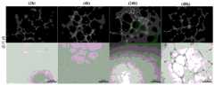
在第0h、16h、24h用光学显微镜观察进行细胞的形态观察并拍照记录(如图5所示)。在微流控芯片的俯视视角下,等边梯形微柱阵列5的两侧分别属于两个细胞培养单元的两个不同的细胞培养通道4:在等边梯形微柱的短边侧是第七细胞培养通道407,在等边梯形微柱的长边侧是与它紧邻的细胞培养单元的第三细胞培养通道403。每个等边梯形微柱之间形成空隙,每个微柱之间间距为50μm。细胞通过上述空隙从第七细胞培养通道407扩散到第三细胞培养通道403。对等边梯形微柱两侧的细胞进行计数。At 0h, 16h, and 24h, the morphology of the cells was observed with an optical microscope and photographed and recorded (as shown in FIG. 5 ). From the top view of the microfluidic chip, the two sides of the equilateral
计数结果显示,在16h时,第三细胞培养通道403内的细胞占细胞总数比例为28.6%;第三细胞培养通道403内的细胞占细胞总数比例为57.3%。上述结果表明,MDA-MB-231细胞可以在本申请的微流控芯片的梯形微柱之间的空隙正常通过、扩散。由于细胞在芯片中以类似体内条件的微环境生长,实验以MDA-MB-231细胞为模型体系考察了芯片系统的基本性能。结果显示这种芯片适宜于MDA-MB-231细胞生存、侵袭,并可以形成类似组织的3D结构,其生长和代谢接近体内肿瘤情况。这种芯片3D细胞培养方法操作简便可靠,仿真度高,适合于肿瘤细胞研究。The counting results showed that at 16 hours, the proportion of the cells in the third
实施例3、体外形成血管实验
本实例中所制备得到的肿瘤微环境芯片,可用于体外形成血管实验。The tumor microenvironment chip prepared in this example can be used for in vitro blood vessel formation experiments.
首先调整HUVEC细胞悬液,细胞密度为1×10⁵cells/mL。吸取上述密度的细胞悬液1mL加入EP管中。然后,将5%(w/v)的GelMA水凝胶与1%的光引发剂LAP溶液按照9:1比例混合均匀,取100μL混合溶液加入到上述细胞沉淀中混合,形成水凝胶-细胞悬液。First adjust the HUVEC cell suspension to a cell density of 1×10⁵ cells/mL.
将注射泵的入口端连接悬液,并将注射泵与微流控芯片的一个细胞培养单元的第一进样孔201连接。用注射泵以10μL/min的速度将水凝胶-细胞悬液灌注至芯片中。Connect the inlet end of the syringe pump to the suspension, and connect the syringe pump to the
当细胞悬液流入第一细胞培养通道401时,迅速用波长为405nm的紫外光源固化20s。随后,将芯片放入培养箱中培养30min。取出芯片。When the cell suspension flows into the first
将注射泵的入口端与培养基连接,并将注射泵与芯片的又一个细胞培养单元的第二进样孔202连接。接有培养基的注射泵以10μL/min的流速向芯片中通培养基。泵送适量培养基后,将芯片再度放进培养箱中培养。培养基流入自身的第二细胞培养通道402后,通过等边梯形微柱阵列5向第一细胞培养通道401扩散和渗透。Connect the inlet end of the syringe pump to the culture medium, and connect the syringe pump to the
分别在第2h、4h、24h、48h用光学显微镜观察进行细胞的形态观察并拍照记录,如图6所示。At 2h, 4h, 24h, and 48h, observe the morphology of the cells with an optical microscope, take pictures and record them, as shown in FIG. 6 .
可以看到,芯片模拟了MDA-MB-231细胞在体内侵略性生长并形成类似于具有明显腺体分化的人浸润性导管癌的肿瘤的过程;在人体中,所述肿瘤在组织和骨骼肌周围局部扩散。As can be seen, the chip mimics the process by which MDA-MB-231 cells grow aggressively in vivo and form tumors resembling human invasive ductal carcinoma with marked glandular differentiation; Local spread around.
实施例4、调控TAMs极化的药物筛选
肿瘤相关巨噬细胞(TAMs)是肿瘤微环境炎症产生的基础细胞,有两种主要来源,并受到不同细胞因子和代谢产物的调控,在肿瘤中存在分布差异和功能差异。和普通巨噬细胞相似的是,TAMs也具有类似M1和M2极化的活化过程,同样具有杀瘤和促瘤的“两面性”。TAMs的高度异质性是许多免疫细胞疗法在不同适应症之间存在较大疗效差异的原因。本实例中所制备得到的肿瘤微环境芯片,可用于体外构建肿瘤微环境,并针对特定肿瘤的TAMs亚群研究突破适应症筛选的思路,进行调控TAMs极化的药物筛选。Tumor-associated macrophages (TAMs) are the basic cells of inflammation in the tumor microenvironment. There are two main sources, and they are regulated by different cytokines and metabolites. There are differences in distribution and function in tumors. Similar to ordinary macrophages, TAMs also have an activation process similar to M1 and M2 polarization, and also have the "two-sidedness" of killing and promoting tumors. The high degree of heterogeneity of TAMs is the reason for the large variation in the efficacy of many immune cell therapies among different indications. The tumor microenvironment chip prepared in this example can be used to construct a tumor microenvironment in vitro, and to break through the idea of screening indications for TAMs subgroups of specific tumors, and to screen drugs that regulate the polarization of TAMs.
首先将MDA-MB-231细胞、HUVEC细胞、TAMs细胞分别调整为1×10⁵cells/mL的细胞悬液。然后,将5%(w/v)的GelMA水凝胶与1%的光引发剂LAP溶液按照9:1比例混合均匀,取100μL混合溶液加入到上述细胞沉淀中混合,形成水凝胶-细胞悬液。Firstly, the MDA-MB-231 cells, HUVEC cells, and TAMs cells were respectively adjusted to a cell suspension of 1×10⁵ cells/mL. Then, 5% (w/v) GelMA hydrogel and 1% photoinitiator LAP solution were mixed evenly at a ratio of 9:1, and 100 μL of the mixed solution was added to the above cell pellet and mixed to form a hydrogel-cell Suspension.
用1mL无菌注射器分别吸取这三种细胞悬液于注射器中。按照MDA-MB-231细胞悬液、水凝胶、TAMs细胞悬液、水凝胶、HUVEC细胞悬液、水凝胶的顺序,用注射泵以10μL/min的速度将注射器中的细胞悬液注入到芯片的细胞培养通道内,构建乳腺癌微环境,即:Use a 1mL sterile syringe to draw the three cell suspensions into the syringe respectively. According to the order of MDA-MB-231 cell suspension, hydrogel, TAMs cell suspension, hydrogel, HUVEC cell suspension, hydrogel, the cell suspension in the syringe was pumped at a speed of 10 μL/min with a syringe pump. Inject into the cell culture channel of the chip to build a breast cancer microenvironment, namely:
将一个注射泵的入口端连接MDA-MB-231细胞悬液注射器,并将注射泵与微流控芯片的一个细胞培养单元的第七进样孔207连接。用注射泵以10μL/min的速度将MDA-MB-231细胞悬液灌注至芯片中;Connect the inlet end of a syringe pump to the MDA-MB-231 cell suspension syringe, and connect the syringe pump to the
将另一个注射泵的入口端连接水凝胶注射器,并将注射泵与微流控芯片的一个细胞培养单元的第五进样孔205连接。用注射泵以10μL/min的速度将水凝胶灌注至芯片中;Connect the inlet end of another syringe pump to the hydrogel syringe, and connect the syringe pump to the
将再一个注射泵的入口端连接TAMs细胞悬液注射器,并将注射泵与微流控芯片的一个细胞培养单元的第一进样孔201连接。用注射泵以10μL/min的速度将水凝胶灌注至芯片中;Connect the inlet end of another syringe pump to the TAMs cell suspension syringe, and connect the syringe pump to the
将又一个注射泵的入口端连接水凝胶注射器,并将注射泵与微流控芯片的一个细胞培养单元的第一进样孔201连接。用注射泵以10μL/min的速度将水凝胶灌注至芯片中;Connect the inlet end of another syringe pump to the hydrogel syringe, and connect the syringe pump to the
将又一个注射泵的入口端连接HUVEC细胞悬液注射器,并将注射泵与微流控芯片的一个细胞培养单元的第二进样孔202连接。用注射泵以10μL/min的速度将水凝胶灌注至芯片中;Connect the inlet end of another syringe pump to the HUVEC cell suspension syringe, and connect the syringe pump to the
将又一个注射泵的入口端连接水凝胶注射器,并将注射泵与微流控芯片的一个细胞培养单元的第四进样孔204连接。用注射泵以10μL/min的速度将水凝胶灌注至芯片中。Connect the inlet end of another syringe pump to the hydrogel syringe, and connect the syringe pump to the
以上述方法注入多个(至少4个)芯片,构件相同的肿瘤微环境。Inject multiple (at least 4) chips with the above method to construct the same tumor microenvironment.
注入完成后,立即用405nm手持固化光源照射15s左右使水凝胶进行交联,然后断开注射泵与芯片的连接,将芯片放入培养箱中培养30min固化。Immediately after the injection is completed, irradiate the hydrogel with a 405nm hand-held curing light source for about 15 seconds to cross-link the hydrogel, then disconnect the syringe pump from the chip, and place the chip in an incubator for 30 minutes to cure.
利用具有浓度梯度的微流控芯片将科罗索酸进行稀释。科罗索酸在芯片的微孔阵列中的扩散定律符合Fick扩散定律,形成0μmoL/mL、2.2μmoL/mL、6.7μmoL/mL、20μmoL/mL的浓度梯度。将含有不同浓度科罗索酸的培养基分别与注射泵连接,并各自以10μL/min的流速通入上述各个芯片中,如图8所示。通入适量科罗索酸后,将芯片放进培养箱中培养。Corosolic acid was diluted using a microfluidic chip with a concentration gradient. The diffusion law of corosolic acid in the microwell array of the chip conforms to Fick's diffusion law, forming a concentration gradient of 0 μmoL/mL, 2.2 μmoL/mL, 6.7 μmoL/mL, and 20 μmoL/mL. The media containing different concentrations of corosolic acid were respectively connected to syringe pumps, and flowed into each of the above-mentioned chips at a flow rate of 10 μL/min, as shown in FIG. 8 . After injecting an appropriate amount of corosolic acid, put the chip into the incubator for cultivation.
分别在第0h、48h用光学显微镜观察进行细胞的形态观察并拍照记录。At 0h and 48h respectively, the morphology of the cells was observed with an optical microscope and photographed and recorded.
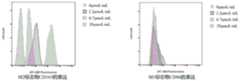
向芯片中注入GelMA裂解液,将TAMs细胞-水凝胶中的TAMs细胞裂解。然后,用流式检测所获得细胞极化变化,如图7所示,其中,横坐标表示荧光信号相对强弱纵坐标表示细胞数。M1型细胞以CD86蛋白表达;M2型细胞以CD163蛋白表达。Inject the GelMA lysate into the chip to lyse the TAMs cells in the TAMs cell-hydrogel. Then, the obtained cell polarization changes are detected by flow cytometry, as shown in FIG. 7 , wherein the abscissa indicates the relative strength of the fluorescent signal and the ordinate indicates the number of cells. M1 cells express CD86 protein; M2 cells express CD163 protein.
试验表明,随着科罗索酸的浓度的增高,M2型细胞逐渐减少,证明本模型用作调控TAMs极化药物筛选的可行性。The test showed that with the increase of the concentration of corosolic acid, the M2 type cells gradually decreased, which proved the feasibility of this model as a drug screen for regulating the polarization of TAMs.
在本申请提及的所有文献都被认为是整体性地包括在本申请的公开内容中,以便在必要时可以作为修改的依据。此外应理解,在阅读了本申请的上述公开内容之后,本领域技术人员可以对本申请作各种改动或修改,这些等价形式同样落于本申请所要求保护的范围。All documents mentioned in this application are considered to be included in the disclosure content of this application in their entirety so that they can be used as a basis for amendments when necessary. In addition, it should be understood that after reading the above disclosure of the present application, those skilled in the art may make various changes or modifications to the present application, and these equivalent forms also fall within the scope of protection claimed in the present application.
Claims (10)
Translated fromChinesePriority Applications (1)
| Application Number | Priority Date | Filing Date | Title |
|---|---|---|---|
| CN202211674855.3ACN116376694A (en) | 2022-12-26 | 2022-12-26 | Microfluidic chip for simulating tumor microenvironment and application method thereof |
Applications Claiming Priority (1)
| Application Number | Priority Date | Filing Date | Title |
|---|---|---|---|
| CN202211674855.3ACN116376694A (en) | 2022-12-26 | 2022-12-26 | Microfluidic chip for simulating tumor microenvironment and application method thereof |
Publications (1)
| Publication Number | Publication Date |
|---|---|
| CN116376694Atrue CN116376694A (en) | 2023-07-04 |
Family
ID=86977484
Family Applications (1)
| Application Number | Title | Priority Date | Filing Date |
|---|---|---|---|
| CN202211674855.3APendingCN116376694A (en) | 2022-12-26 | 2022-12-26 | Microfluidic chip for simulating tumor microenvironment and application method thereof |
Country Status (1)
| Country | Link |
|---|---|
| CN (1) | CN116376694A (en) |
Cited By (1)
| Publication number | Priority date | Publication date | Assignee | Title |
|---|---|---|---|---|
| CN119530004A (en)* | 2024-10-30 | 2025-02-28 | 中国科学院杭州医学研究所 | An intelligent microfluidic living cell culture observation and analysis equipment |
- 2022
- 2022-12-26CNCN202211674855.3Apatent/CN116376694A/enactivePending
Cited By (1)
| Publication number | Priority date | Publication date | Assignee | Title |
|---|---|---|---|---|
| CN119530004A (en)* | 2024-10-30 | 2025-02-28 | 中国科学院杭州医学研究所 | An intelligent microfluidic living cell culture observation and analysis equipment |
Similar Documents
| Publication | Publication Date | Title |
|---|---|---|
| CN110997900B (en) | Microfluidic platform for rapid generation of organoids/spheroids for compound screening | |
| CN103981096B (en) | Two-layer cell culture system organ chip and preparation method thereof | |
| Kim et al. | Microphysiological systems as enabling tools for modeling complexity in the tumor microenvironment and accelerating cancer drug development | |
| CN101165161B (en) | Micro-fluid concentration gradient cell culture chip and its preparation method and application | |
| Dereli-Korkut et al. | Three dimensional microfluidic cell arrays for ex vivo drug screening with mimicked vascular flow | |
| Wu et al. | Rapid microfluidic formation of uniform patient-derived breast tumor spheroids | |
| CN111218404A (en) | A bionic multi-organ chip and its preparation method and application | |
| Zuchowska et al. | Studies of anticancer drug cytotoxicity based on long‐term HepG2 spheroid culture in a microfluidic system | |
| Gil et al. | Cancer models on chip: paving the way to large‐scale trial applications | |
| CN111971384B (en) | A bionic intestinal-liver organ chip and its preparation method and application | |
| CN104974935A (en) | device with annular micro-channel chip for cell culture | |
| CN103087912A (en) | Micro-fluidic chip capable of producing stable concentration gradient and cell co-culture method | |
| CN113814010B (en) | Multi-cell and multi-tissue co-culture bionic micro-fluidic chip and preparation method thereof | |
| CN102899249A (en) | Device and method of generating in vitro blood vessels | |
| CN112608841A (en) | Microfluidic system for tumor organoid culture and drug experiments and use method thereof | |
| CN110257249A (en) | A kind of micro-fluidic chip and administration cultural method for tumour cell dimensional culture | |
| CN110331096A (en) | Simulate the micro-fluidic chip of tumor microenvironment and the construction method of tumor microenvironment | |
| CN212316139U (en) | Bionic multi-organ chip | |
| Johnson et al. | The applications and challenges of the development of in vitro tumor microenvironment chips | |
| CN115074246A (en) | Microfluidic organ chip with soluble temporary barrier and preparation method thereof | |
| Lin et al. | 3D microfluidic tumor models for biomimetic engineering of glioma niche and detection of cell morphology, migration and phenotype change | |
| CN116376694A (en) | Microfluidic chip for simulating tumor microenvironment and application method thereof | |
| CN116333881A (en) | Three-channel organ chip and application and use method thereof | |
| CN211445767U (en) | Microfluidic chip for three-dimensional culture of cell clusters | |
| CN108504571B (en) | Device and method for constructing artificial liver lobule functional unit |
Legal Events
| Date | Code | Title | Description |
|---|---|---|---|
| PB01 | Publication | ||
| PB01 | Publication |