CN116269454A - Bone microstructure analysis method, device, electronic equipment and medium around bone prosthesis - Google Patents
Bone microstructure analysis method, device, electronic equipment and medium around bone prosthesisDownload PDFInfo
- Publication number
- CN116269454A CN116269454ACN202310101829.XACN202310101829ACN116269454ACN 116269454 ACN116269454 ACN 116269454ACN 202310101829 ACN202310101829 ACN 202310101829ACN 116269454 ACN116269454 ACN 116269454A
- Authority
- CN
- China
- Prior art keywords
- bone
- image
- prosthesis
- around
- preset
- Prior art date
- Legal status (The legal status is an assumption and is not a legal conclusion. Google has not performed a legal analysis and makes no representation as to the accuracy of the status listed.)
- Pending
Links
Images


Classifications
- A—HUMAN NECESSITIES
- A61—MEDICAL OR VETERINARY SCIENCE; HYGIENE
- A61B—DIAGNOSIS; SURGERY; IDENTIFICATION
- A61B6/00—Apparatus or devices for radiation diagnosis; Apparatus or devices for radiation diagnosis combined with radiation therapy equipment
- A61B6/50—Apparatus or devices for radiation diagnosis; Apparatus or devices for radiation diagnosis combined with radiation therapy equipment specially adapted for specific body parts; specially adapted for specific clinical applications
- A61B6/505—Apparatus or devices for radiation diagnosis; Apparatus or devices for radiation diagnosis combined with radiation therapy equipment specially adapted for specific body parts; specially adapted for specific clinical applications for diagnosis of bone
- A—HUMAN NECESSITIES
- A61—MEDICAL OR VETERINARY SCIENCE; HYGIENE
- A61B—DIAGNOSIS; SURGERY; IDENTIFICATION
- A61B6/00—Apparatus or devices for radiation diagnosis; Apparatus or devices for radiation diagnosis combined with radiation therapy equipment
- A61B6/52—Devices using data or image processing specially adapted for radiation diagnosis
- A61B6/5211—Devices using data or image processing specially adapted for radiation diagnosis involving processing of medical diagnostic data
- G—PHYSICS
- G06—COMPUTING OR CALCULATING; COUNTING
- G06T—IMAGE DATA PROCESSING OR GENERATION, IN GENERAL
- G06T7/00—Image analysis
- G06T7/0002—Inspection of images, e.g. flaw detection
- G06T7/0012—Biomedical image inspection
- G—PHYSICS
- G06—COMPUTING OR CALCULATING; COUNTING
- G06T—IMAGE DATA PROCESSING OR GENERATION, IN GENERAL
- G06T7/00—Image analysis
- G06T7/10—Segmentation; Edge detection
- G06T7/11—Region-based segmentation
- G—PHYSICS
- G06—COMPUTING OR CALCULATING; COUNTING
- G06T—IMAGE DATA PROCESSING OR GENERATION, IN GENERAL
- G06T2207/00—Indexing scheme for image analysis or image enhancement
- G06T2207/30—Subject of image; Context of image processing
- G06T2207/30004—Biomedical image processing
- G06T2207/30008—Bone
Landscapes
- Engineering & Computer Science (AREA)
- Health & Medical Sciences (AREA)
- Life Sciences & Earth Sciences (AREA)
- Medical Informatics (AREA)
- Physics & Mathematics (AREA)
- Computer Vision & Pattern Recognition (AREA)
- General Health & Medical Sciences (AREA)
- Radiology & Medical Imaging (AREA)
- Nuclear Medicine, Radiotherapy & Molecular Imaging (AREA)
- Pathology (AREA)
- Animal Behavior & Ethology (AREA)
- Optics & Photonics (AREA)
- Biophysics (AREA)
- Theoretical Computer Science (AREA)
- Biomedical Technology (AREA)
- Heart & Thoracic Surgery (AREA)
- Molecular Biology (AREA)
- Surgery (AREA)
- High Energy & Nuclear Physics (AREA)
- General Physics & Mathematics (AREA)
- Public Health (AREA)
- Veterinary Medicine (AREA)
- Orthopedic Medicine & Surgery (AREA)
- Quality & Reliability (AREA)
- Dentistry (AREA)
- Oral & Maxillofacial Surgery (AREA)
- Prostheses (AREA)
Abstract
Description
Translated fromChinese技术领域technical field
本公开涉及植入到人体内的骨假体技术领域,具体涉及一种骨假体周围的骨微结构分析方法、装置、电子设备及介质。The present disclosure relates to the technical field of bone prosthesis implanted in the human body, in particular to a bone microstructure analysis method, device, electronic equipment and medium around the bone prosthesis.
背景技术Background technique
骨假体在医学上也被称作修复体,是一种用来置换人体某一肢体、器官或者组织的医疗器械。骨假体置换术在骨科疾病的治疗中具有广泛的应用,例如,关节置换术(TJA)是治疗晚期关节疾病的最有效方法之一。骨假体的无菌性松动是骨假体置换术后最主要的远期并发症,是限制骨假体使用寿命的瓶颈。最近的研究表明,术后早期对有松动风险的骨假体进行干预(如应用双膦酸盐),可以使术后10年松动风险率从5.3%降为2.8%,即降低约47%。因此,早期预测骨假体稳定性对延长骨假体使用寿命来说至关重要。A bone prosthesis, also called a prosthesis in medicine, is a medical device used to replace a certain limb, organ or tissue of the human body. Bone prosthetic replacement has a wide range of applications in the treatment of orthopedic diseases, for example, joint arthroplasty (TJA) is one of the most effective methods for the treatment of advanced joint diseases. Aseptic loosening of bone prosthesis is the most important long-term complication after bone prosthesis replacement, and it is the bottleneck that limits the service life of bone prosthesis. Recent studies have shown that early postoperative intervention on bone prostheses with a risk of loosening (such as the application of bisphosphonates) can reduce the risk of loosening from 5.3% to 2.8% at 10 years after surgery, which is about 47%. Therefore, early prediction of bone prosthesis stability is crucial to prolong the service life of bone prosthesis.
放射立体测绘分析(RadiostereometricAnalysis,RSA)测量的早期骨假体位移(术后1-2年内)与远期无菌性松动率高度相关,被认为是目前早期评估骨假体稳定性最有效的指标,但因其特殊的摄片设备要求及有创的钽珠植入,至今无法被广泛应用于所有的关节置换患者中。Early bone prosthesis displacement (within 1-2 years after surgery) measured by Radiostereometric Analysis (RSA) is highly correlated with long-term aseptic loosening rate, and is considered to be the most effective index for early assessment of bone prosthesis stability , but because of special imaging equipment requirements and invasive tantalum bead implantation, it has not been widely used in all joint replacement patients so far.
骨假体周围骨质量是影响骨假体稳定性的另一个关键因素,既往大量临床数据证实了骨假体周围骨质量与骨假体位移、远期松动率都显著相关。骨密度(BMD,即骨矿物成分的含量)与骨微结构是决定骨质量的两个主要因素,骨密度下降和骨微结构破坏,都会表现为骨的脆性增加,所能承受的压力变差。The bone quality around the bone prosthesis is another key factor affecting the stability of the bone prosthesis. A large number of previous clinical data have confirmed that the bone quality around the bone prosthesis is significantly related to the displacement and long-term loosening rate of the bone prosthesis. Bone mineral density (BMD, that is, the content of bone mineral components) and bone microstructure are the two main factors that determine bone quality. The decline in bone density and the destruction of bone microstructure will both manifest as increased bone fragility and poor pressure resistance. .
目前发现骨假体周围/系统性骨密度(BMD)与远期无菌性松动相关;骨假体周围/系统性BMD与骨假体早期位移相关。因此,骨假体周围骨BMD已成为评估骨假体稳定性的另一个重要指标。双能量X射线吸收仪(DXA)是评估骨假体周围骨BMD的金标准,骨假体周围骨质通常根据Gruen分区被分为7个区域对BMD进行评估,但该方法尚存一些局限性。首先,骨质量主要由骨矿物质的含量与骨微结构共同决定,但BMD只代表骨矿物质的含量,无法衡量骨微结构。其次,根据最新的研究,与骨假体表面相邻的厚度小于1毫米的骨质才是影响骨假体稳定性的关键因,但骨假体周围骨质的Gruen分区各区块面积大、总区块数量少、且同时包含骨皮质与骨松质——总体“分辨率”较差,不足以捕捉小区块内骨质的变化。即便对局部关键区块进行重新勾画,由于骨假体周围骨的形态的个体性差异,也会导致关键区块很难在不同患者之间进行匹配和比较,从而难以根据骨假体周围骨质量对骨假体稳定性进行预测。It has been found that periprosthetic/systemic bone mineral density (BMD) is associated with long-term aseptic loosening; periprosthetic/systemic BMD is associated with early displacement of the prosthesis. Therefore, BMD around the bone prosthesis has become another important index to evaluate the stability of the bone prosthesis. Dual-energy X-ray absorptiometry (DXA) is the gold standard for evaluating bone BMD around a bone prosthesis. The bone around a bone prosthesis is usually divided into 7 regions according to the Gruen division to evaluate BMD, but this method still has some limitations. . First of all, bone quality is mainly determined by bone mineral content and bone microstructure, but BMD only represents bone mineral content and cannot measure bone microstructure. Secondly, according to the latest research, the bone thickness less than 1 mm adjacent to the surface of the bone prosthesis is the key factor affecting the stability of the bone prosthesis, but the Gruen partition of bone around the bone prosthesis has a large area and a total The number of blocks is small, and it contains both cortical bone and cancellous bone -- the overall "resolution" is poor, and it is not enough to capture the changes in bone quality in small blocks. Even if the local key areas are redrawn, due to the individual differences in the shape of the bone around the bone prosthesis, it will be difficult to match and compare the key areas between different patients, making it difficult to determine the quality of the bone around the bone prosthesis. Prediction of bone prosthesis stability.
发明内容Contents of the invention
为了解决相关技术中的问题,本公开实施例提供一种骨假体周围的骨微结构分析方法、装置、电子设备及介质。In order to solve problems in related technologies, embodiments of the present disclosure provide a bone microstructure analysis method, device, electronic equipment, and medium around a bone prosthesis.
第一方面,本公开实施例中提供了一种骨假体周围的骨微结构分析方法,包括:In a first aspect, an embodiment of the present disclosure provides a method for analyzing bone microstructure around a bone prosthesis, including:
获取骨假体所在的人体区域的透视图像;obtaining a perspective image of the region of the body where the bone prosthesis is located;
从所述透视图像中分割出骨假体周围骨图像;segmenting bone images around the bone prosthesis from the fluoroscopic images;
将所述骨假体周围骨图像转换为预设矩形的骨质图像;Converting the bone image around the bone prosthesis into a preset rectangular bone image;
将所述预设矩形的骨质图像划分为多个矩形区块图像,所述矩形区块图像用于对所述骨假体周围的骨微结构进行定量分析。The preset rectangular bone image is divided into a plurality of rectangular block images, and the rectangular block images are used for quantitative analysis of the bone microstructure around the bone prosthesis.
根据本公开的实施例,所述将所述骨假体周围骨图像转换为预设矩形的骨质图像,包括:According to an embodiment of the present disclosure, the converting the bone image around the bone prosthesis into a preset rectangular bone image includes:
使用以下任意一种算法将所述骨假体周围骨图像转换为预设矩形的骨质图像:分段仿射算法、基于点阵网格的图像变形转换算法、基于线性的图像变形转换算法、基于非线性的图像变形转换算法。Use any of the following algorithms to convert the bone image around the bone prosthesis into a preset rectangular bone image: a segmented affine algorithm, an image deformation conversion algorithm based on a lattice grid, a linear image deformation conversion algorithm, Based on non-linear image deformation transformation algorithm.
根据本公开的实施例,所述透视图像为包含所述骨假体的透视图像,所述从所述透视图像中分割出骨假体周围骨图像,包括:According to an embodiment of the present disclosure, the fluoroscopic image is a fluoroscopic image containing the bone prosthesis, and the segmentation of bone images around the bone prosthesis from the fluoroscopic image includes:
通过图形分割算法从所述透视图像中分割出骨假体与周围骨质部分;Segmenting the bone prosthesis and surrounding bone parts from the perspective image by a graphic segmentation algorithm;
通过图形分割算法从所述骨假体与周围骨质部分中去除骨假体部分,分割出骨假体周围骨图像。The bone prosthesis part is removed from the bone prosthesis and the surrounding bone part by means of a graphic segmentation algorithm, and a bone image around the bone prosthesis is segmented.
根据本公开的实施例,所述将所述骨假体周围骨图像转换为预设矩形的骨质图像,包括:According to an embodiment of the present disclosure, the converting the bone image around the bone prosthesis into a preset rectangular bone image includes:
将所述骨假体周围骨图像作为一个整体转换为预设矩形的骨质图像;或者converting the bone image around the bone prosthesis as a whole into a preset rectangular bone image; or
将所述骨假体周围骨图像划分为多个区域,将所述多个区域分别转换为预设矩形的骨质图像的相应区域,从而得到所述预设矩形的骨质图像。The bone image around the bone prosthesis is divided into multiple regions, and the multiple regions are converted into corresponding regions of the preset rectangular bone image, thereby obtaining the preset rectangular bone image.
根据本公开的实施例,所述将所述骨假体周围骨图像作为一个整体转换为预设矩形的骨质图像,包括使用以下任意一种算法将所述骨假体周围骨图像作为一个整体转换为预设矩形的骨质图像:分段仿射算法、基于点阵网格的图像变形转换算法、基于线性的图像变形转换算法、基于非线性的图像变形转换算法;According to an embodiment of the present disclosure, the converting the bone image around the bone prosthesis as a whole into a preset rectangular bone image includes using any of the following algorithms to convert the bone image around the bone prosthesis as a whole Bone images converted into preset rectangles: segmented affine algorithm, image deformation conversion algorithm based on lattice grid, linear-based image deformation conversion algorithm, nonlinear image deformation conversion algorithm;
所述将所述多个区域分别转换为预设矩形的骨质图像的相应区域,包括使用以下任意一种算法将所述多个区域分别转换为所述预设矩形的骨质图像的相应区域:分段仿射算法、基于点阵网格的图像变形转换算法、基于线性的图像变形转换算法、基于非线性的图像变形转换算法。The converting the plurality of regions into corresponding regions of the preset rectangular bone image includes converting the plurality of regions into corresponding regions of the preset rectangular bone image using any of the following algorithms : Segmented affine algorithm, image deformation conversion algorithm based on lattice grid, linear image deformation conversion algorithm, nonlinear image deformation conversion algorithm.
根据本公开的实施例,所述使用分段仿射算法将所述骨假体周围骨图像作为一个整体转换为预设矩形的骨质图像,包括:According to an embodiment of the present disclosure, the use of a segmented affine algorithm to convert the bone image around the bone prosthesis as a whole into a preset rectangular bone image includes:
将第一网点阵均匀覆盖至所述预设矩形;Evenly covering the first dot matrix to the preset rectangle;
将与所述第一网点阵相同分布的第二网点阵根据所述骨假体周围骨的形状缩放并覆盖到所述骨假体周围骨图像上;Scaling and overlaying the second grid lattice with the same distribution as the first lattice lattice on the bone image around the bone prosthesis according to the shape of the bone around the bone prosthesis;
基于所述第一网点阵和所述第二网点阵,建立分段仿射转换模型;Establishing a segmented affine transformation model based on the first lattice and the second lattice;
通过所述分段仿射转换模型将所述骨假体周围骨图像转换为所述预设矩形的骨质图像。The bone image around the bone prosthesis is converted into the preset rectangular bone image by using the segmented affine transformation model.
根据本公开的实施例,所述使用分段仿射算法将所述多个区域分别转换为所述预设矩形的骨质图像的相应区域,包括:According to an embodiment of the present disclosure, converting the multiple regions into corresponding regions of the preset rectangular bone image using a segmented affine algorithm includes:
将多个第一网点阵分别均匀覆盖至所述相应区域;Uniformly covering a plurality of first mesh lattices to the corresponding areas respectively;
将分别与所述多个第一网点阵相同分布的多个第二网点阵根据所述骨假体周围骨的形状分别缩放并覆盖到所述多个区域上;Scaling and covering the plurality of second mesh lattices distributed in the same distribution as the plurality of first mesh lattices respectively on the plurality of regions according to the shape of the bone around the bone prosthesis;
基于所述第一网点阵和相应的第二网点阵,建立分段仿射转换模型;Establishing a segmented affine transformation model based on the first lattice and the corresponding second lattice;
通过所述分段仿射转换模型将所述多个区域转换为所述预设矩形的骨质图像的相应区域。The plurality of regions are transformed into corresponding regions of the preset rectangular bone image through the segmented affine transformation model.
根据本公开的实施例,所述将所述预设矩形的骨质图像划分为多个矩形区块图像,包括:According to an embodiment of the present disclosure, the dividing the preset rectangular bone image into a plurality of rectangular block images includes:
将所述预设矩形的骨质图像均匀划分为多个矩形区块图像。The preset rectangular bone image is evenly divided into a plurality of rectangular block images.
根据本公开的实施例,所述方法还包括:使用所述矩形区块图像对所述骨假体周围的骨微结构进行定量分析。According to an embodiment of the present disclosure, the method further includes: using the rectangular block image to quantitatively analyze the bone microstructure around the bone prosthesis.
根据本公开的实施例,所述使用所述矩形区块图像对所述骨假体周围的骨微结构进行定量分析,包括:According to an embodiment of the present disclosure, the quantitative analysis of the bone microstructure around the bone prosthesis using the rectangular block image includes:
将所述预设矩形的骨质图像作为一个整体进行骨微结构的定量分析;performing quantitative analysis of the bone microstructure on the bone image of the preset rectangle as a whole;
统计所述矩形区块图像中的骨结构参数,所述骨结构参数是所述骨微结构的定量分析结果。The bone structure parameters in the rectangular block image are counted, and the bone structure parameters are quantitative analysis results of the bone microstructure.
根据本公开的实施例,所述将所述预设矩形的骨质图像作为一个整体进行骨微结构的定量分析,包括:According to an embodiment of the present disclosure, the quantitative analysis of the bone microstructure of the preset rectangular bone image as a whole includes:
使用以下任意一种方式将所述预设矩形的骨质图像作为一个整体进行骨微结构的定量分析:支柱分析、基于灰阶共生矩阵的Haralick特征分析、本地二值化图案分析、阈值邻接统计算法。Use any of the following methods to perform quantitative analysis of the bone microstructure on the preset rectangular bone image as a whole: pillar analysis, Haralick feature analysis based on gray-scale co-occurrence matrix, local binary pattern analysis, threshold adjacency statistics algorithm.
根据本公开的实施例,所述使用支柱分析将所述预设矩形的骨质图像作为一个整体进行骨微结构的定量分析,包括:According to an embodiment of the present disclosure, the quantitative analysis of the bone microstructure of the preset rectangular bone image as a whole using pillar analysis includes:
对所述预设矩形的骨质图像进行上采样,得到上采样图像;Upsampling the bone image of the preset rectangle to obtain an upsampled image;
对所述上采样图像进行密度矫正,移除由骨密度造成的图像阴暗;Carrying out density correction to the upsampled image to remove image darkness caused by bone density;
将密度矫正后的图像转换为二值化图像;Convert the density-corrected image into a binarized image;
将所述二值化图像转换为仅包含骨微结构的拓扑结构支柱的拓扑结构支柱图像;converting the binarized image to a topological strut image comprising only topological struts of bone microstructure;
对所述二值化图像和所述拓扑结构支柱图像中各支柱的参数进行量化分析,得到定量分析结果。Quantitative analysis is performed on the binarized image and the parameters of each pillar in the topological structure pillar image to obtain a quantitative analysis result.
根据本公开的实施例,所述使用所述矩形区块图像对所述骨假体周围的骨微结构进行定量分析,包括:According to an embodiment of the present disclosure, the quantitative analysis of the bone microstructure around the bone prosthesis using the rectangular block image includes:
将所述骨假体周围骨图像作为一个整体进行骨微结构的定量分析;Taking the bone image around the bone prosthesis as a whole to perform quantitative analysis of bone microstructure;
确定所述骨假体周围骨图像中与每个矩形区块图像对应的相应部分;determining a corresponding part corresponding to each rectangular block image in the bone image around the bone prosthesis;
统计所述矩形区块图像中的骨结构参数,所述骨结构参数是所述骨假体周围骨图像中与所述矩形区块对应的相应部分的骨微结构定量分析结果。The bone structure parameters in the rectangular block image are counted, and the bone structure parameters are the quantitative analysis results of the bone microstructure corresponding to the corresponding part of the bone image around the bone prosthesis corresponding to the rectangular block.
根据本公开的实施例,所述将所述骨假体周围骨图像作为一个整体进行骨微结构的定量分析,包括:According to an embodiment of the present disclosure, the quantitative analysis of the bone microstructure of the bone image around the bone prosthesis as a whole includes:
使用以下任意一种方式将所述骨假体周围骨图像作为一个整体进行骨微结构的定量分析:支柱分析、基于灰阶共生矩阵的Haralick特征分析、本地二值化图案分析、阈值邻接统计算法。Use any of the following methods to perform quantitative analysis of the bone microstructure of the bone image around the bone prosthesis as a whole: pillar analysis, Haralick feature analysis based on gray-scale co-occurrence matrix, local binary pattern analysis, threshold adjacency statistical algorithm .
根据本公开的实施例,所述使用支柱分析将所述骨假体周围骨图像作为一个整体进行骨微结构的定量分析,包括:According to an embodiment of the present disclosure, the quantitative analysis of the bone microstructure of the bone image around the bone prosthesis as a whole using pillar analysis includes:
对所述骨假体周围骨图像进行上采样,得到上采样图像;Upsampling the bone image around the bone prosthesis to obtain an upsampling image;
对所述上采样图像进行密度矫正,移除由骨密度造成的图像阴暗;Carrying out density correction to the upsampled image to remove image darkness caused by bone density;
将密度矫正后的图像转换为二值化图像;Convert the density-corrected image into a binarized image;
将所述二值化图像转换为仅包含骨微结构的拓扑结构支柱的拓扑结构支柱图像;converting the binarized image to a topological strut image comprising only topological struts of bone microstructure;
对所述二值化图像和所述拓扑结构支柱图像中各支柱的参数进行量化分析,得到定量分析结果。Quantitative analysis is performed on the binarized image and the parameters of each pillar in the topological structure pillar image to obtain a quantitative analysis result.
根据本公开的实施例,所述方法还包括:According to an embodiment of the present disclosure, the method further includes:
基于与所述骨假体的位移的相关度满足预设条件的矩形区块图像中的骨结构参数对所述骨假体的稳定性进行预测。The stability of the bone prosthesis is predicted based on the bone structure parameters in the rectangular block image whose correlation with the displacement of the bone prosthesis satisfies a preset condition.
根据本公开的实施例,所述方法还包括:According to an embodiment of the present disclosure, the method further includes:
基于位于骨干下部表面的矩形区块图像中的骨结构参数对所述骨假体的稳定性进行预测。The stability of the bone prosthesis is predicted based on the bone structure parameters in the image of the rectangular block located on the lower surface of the diaphysis.
第二方面,本公开实施例中提供了一种骨假体周围的骨微结构分析装置,包括:In the second aspect, an embodiment of the present disclosure provides a bone microstructure analysis device around a bone prosthesis, including:
获取模块,被配置为获取骨假体所在的人体区域的透视图像;an acquisition module configured to acquire a perspective image of the human body area where the bone prosthesis is located;
分割模块,被配置为从所述透视图像中分割出骨假体周围骨图像;a segmentation module configured to segment bone images around the bone prosthesis from the fluoroscopic images;
转换模块,被配置为将所述骨假体周围骨图像转换为预设矩形的骨质图像;A conversion module configured to convert the bone image around the bone prosthesis into a preset rectangular bone image;
划分模块,被配置为将所述预设矩形的骨质图像划分为多个矩形区块图像,所述矩形区块图像用于对所述骨假体周围的骨微结构进行定量分析。The dividing module is configured to divide the preset rectangular bone image into a plurality of rectangular block images, and the rectangular block images are used for quantitative analysis of the bone microstructure around the bone prosthesis.
根据本公开的实施例,所述将所述骨假体周围骨图像转换为预设矩形的骨质图像,包括:According to an embodiment of the present disclosure, the converting the bone image around the bone prosthesis into a preset rectangular bone image includes:
使用以下任意一种算法将所述骨假体周围骨图像转换为预设矩形的骨质图像:分段仿射算法、基于点阵网格的图像变形转换算法、基于线性的图像变形转换算法、基于非线性的图像变形转换算法。Use any of the following algorithms to convert the bone image around the bone prosthesis into a preset rectangular bone image: a segmented affine algorithm, an image deformation conversion algorithm based on a lattice grid, a linear image deformation conversion algorithm, Based on non-linear image deformation transformation algorithm.
根据本公开的实施例,所述透视图像为包含所述骨假体的透视图像,所述从所述透视图像中分割出骨假体周围骨图像,包括:According to an embodiment of the present disclosure, the fluoroscopic image is a fluoroscopic image containing the bone prosthesis, and the segmentation of bone images around the bone prosthesis from the fluoroscopic image includes:
通过图形分割算法从所述透视图像中分割出骨假体与周围骨质部分;Segmenting the bone prosthesis and surrounding bone parts from the perspective image by a graphic segmentation algorithm;
通过图形分割算法从所述骨假体与周围骨质部分中去除骨假体部分,分割出骨假体周围骨图像。The bone prosthesis part is removed from the bone prosthesis and the surrounding bone part by means of a graphic segmentation algorithm, and a bone image around the bone prosthesis is segmented.
根据本公开的实施例,所述将所述骨假体周围骨图像转换为预设矩形的骨质图像,包括:According to an embodiment of the present disclosure, the converting the bone image around the bone prosthesis into a preset rectangular bone image includes:
将所述骨假体周围骨图像作为一个整体转换为预设矩形的骨质图像;或者converting the bone image around the bone prosthesis as a whole into a preset rectangular bone image; or
将所述骨假体周围骨图像划分为多个区域,将所述多个区域分别转换为预设矩形的骨质图像的相应区域,从而得到所述预设矩形的骨质图像。The bone image around the bone prosthesis is divided into multiple regions, and the multiple regions are converted into corresponding regions of the preset rectangular bone image, thereby obtaining the preset rectangular bone image.
根据本公开的实施例,所述将所述骨假体周围骨图像作为一个整体转换为预设矩形的骨质图像,包括使用以下任意一种算法将所述骨假体周围骨图像作为一个整体转换为预设矩形的骨质图像:分段仿射算法、基于点阵网格的图像变形转换算法、基于线性的图像变形转换算法、基于非线性的图像变形转换算法;According to an embodiment of the present disclosure, the converting the bone image around the bone prosthesis as a whole into a preset rectangular bone image includes using any of the following algorithms to convert the bone image around the bone prosthesis as a whole Bone images converted into preset rectangles: segmented affine algorithm, image deformation conversion algorithm based on lattice grid, linear-based image deformation conversion algorithm, nonlinear image deformation conversion algorithm;
所述将所述多个区域分别转换为预设矩形的骨质图像的相应区域,包括使用以下任意一种算法将所述多个区域分别转换为所述预设矩形的骨质图像的相应区域:分段仿射算法、基于点阵网格的图像变形转换算法、基于线性的图像变形转换算法、基于非线性的图像变形转换算法。The converting the plurality of regions into corresponding regions of the preset rectangular bone image includes converting the plurality of regions into corresponding regions of the preset rectangular bone image using any of the following algorithms : Segmented affine algorithm, image deformation conversion algorithm based on lattice grid, linear image deformation conversion algorithm, nonlinear image deformation conversion algorithm.
根据本公开的实施例,所述使用分段仿射算法将所述骨假体周围骨图像作为一个整体转换为预设矩形的骨质图像,包括:According to an embodiment of the present disclosure, the use of a segmented affine algorithm to convert the bone image around the bone prosthesis as a whole into a preset rectangular bone image includes:
将第一网点阵均匀覆盖至所述预设矩形;Evenly covering the first dot matrix to the preset rectangle;
将与所述第一网点阵相同分布的第二网点阵根据所述骨假体周围骨的形状缩放并覆盖到所述骨假体周围骨图像上;Scaling and overlaying the second grid lattice with the same distribution as the first lattice lattice on the bone image around the bone prosthesis according to the shape of the bone around the bone prosthesis;
基于所述第一网点阵和所述第二网点阵,建立分段仿射转换模型;Establishing a segmented affine transformation model based on the first lattice and the second lattice;
通过所述分段仿射转换模型将所述骨假体周围骨图像转换为所述预设矩形的骨质图像。The bone image around the bone prosthesis is converted into the preset rectangular bone image by using the segmented affine transformation model.
根据本公开的实施例,所述使用分段仿射算法将所述多个区域分别转换为所述预设矩形的骨质图像的相应区域,包括:According to an embodiment of the present disclosure, converting the multiple regions into corresponding regions of the preset rectangular bone image using a segmented affine algorithm includes:
将多个第一网点阵分别均匀覆盖至所述相应区域;Uniformly covering a plurality of first mesh lattices to the corresponding areas respectively;
将分别与所述多个第一网点阵相同分布的多个第二网点阵根据所述骨假体周围骨的形状分别缩放并覆盖到所述多个区域上;Scaling and covering the plurality of second mesh lattices distributed in the same distribution as the plurality of first mesh lattices respectively on the plurality of regions according to the shape of the bone around the bone prosthesis;
基于所述第一网点阵和相应的第二网点阵,建立分段仿射转换模型;Establishing a segmented affine transformation model based on the first lattice and the corresponding second lattice;
通过所述分段仿射转换模型将所述多个区域转换为所述预设矩形的骨质图像的相应区域。The plurality of regions are transformed into corresponding regions of the preset rectangular bone image through the segmented affine transformation model.
根据本公开的实施例,所述将所述预设矩形的骨质图像划分为多个矩形区块图像,包括:According to an embodiment of the present disclosure, the dividing the preset rectangular bone image into a plurality of rectangular block images includes:
将所述预设矩形的骨质图像均匀划分为多个矩形区块图像。The preset rectangular bone image is evenly divided into a plurality of rectangular block images.
根据本公开的实施例,骨假体周围的骨微结构分析装置还包括:分析模块,被配置为使用所述矩形区块图像对所述骨假体周围的骨微结构进行定量分析。According to an embodiment of the present disclosure, the bone microstructure analysis device around the bone prosthesis further includes: an analysis module configured to quantitatively analyze the bone microstructure around the bone prosthesis by using the rectangular block image.
根据本公开的实施例,所述使用所述矩形区块图像对所述骨假体周围的骨微结构进行定量分析,包括:According to an embodiment of the present disclosure, the quantitative analysis of the bone microstructure around the bone prosthesis using the rectangular block image includes:
将所述预设矩形的骨质图像作为一个整体进行骨微结构的定量分析;performing quantitative analysis of the bone microstructure on the bone image of the preset rectangle as a whole;
统计所述矩形区块图像中的骨结构参数,所述骨结构参数是所述骨微结构的定量分析结果。The bone structure parameters in the rectangular block image are counted, and the bone structure parameters are quantitative analysis results of the bone microstructure.
根据本公开的实施例,所述将所述预设矩形的骨质图像作为一个整体进行骨微结构的定量分析,包括:According to an embodiment of the present disclosure, the quantitative analysis of the bone microstructure of the preset rectangular bone image as a whole includes:
使用以下任意一种方式将所述预设矩形的骨质图像作为一个整体进行骨微结构的定量分析:支柱分析、基于灰阶共生矩阵的Haralick特征分析、本地二值化图案分析、阈值邻接统计算法。Use any of the following methods to perform quantitative analysis of the bone microstructure on the preset rectangular bone image as a whole: pillar analysis, Haralick feature analysis based on gray-scale co-occurrence matrix, local binary pattern analysis, threshold adjacency statistics algorithm.
根据本公开的实施例,所述使用支柱分析将所述预设矩形的骨质图像作为一个整体进行骨微结构的定量分析,包括:According to an embodiment of the present disclosure, the quantitative analysis of the bone microstructure of the preset rectangular bone image as a whole using pillar analysis includes:
对所述预设矩形的骨质图像进行上采样,得到上采样图像;Upsampling the bone image of the preset rectangle to obtain an upsampled image;
对所述上采样图像进行密度矫正,移除由骨密度造成的图像阴暗;Carrying out density correction to the upsampled image to remove image darkness caused by bone density;
将密度矫正后的图像转换为二值化图像;Convert the density-corrected image into a binarized image;
将所述二值化图像转换为仅包含骨微结构的拓扑结构支柱的拓扑结构支柱图像;converting the binarized image to a topological strut image comprising only topological struts of bone microstructure;
对所述二值化图像和所述拓扑结构支柱图像中各支柱的参数进行量化分析,得到定量分析结果。Quantitative analysis is performed on the binarized image and the parameters of each pillar in the topological structure pillar image to obtain a quantitative analysis result.
根据本公开的实施例,所述使用所述矩形区块图像对所述骨假体周围的骨微结构进行定量分析,包括:According to an embodiment of the present disclosure, the quantitative analysis of the bone microstructure around the bone prosthesis using the rectangular block image includes:
将所述骨假体周围骨图像作为一个整体进行骨微结构的定量分析;Taking the bone image around the bone prosthesis as a whole to perform quantitative analysis of bone microstructure;
确定所述骨假体周围骨图像中与每个矩形区块图像对应的相应部分;determining a corresponding part corresponding to each rectangular block image in the bone image around the bone prosthesis;
统计所述矩形区块图像中的骨结构参数,所述骨结构参数是所述骨假体周围骨图像中与所述矩形区块对应的相应部分的骨微结构定量分析结果。The bone structure parameters in the rectangular block image are counted, and the bone structure parameters are the quantitative analysis results of the bone microstructure corresponding to the corresponding part of the bone image around the bone prosthesis corresponding to the rectangular block.
根据本公开的实施例,所述将所述骨假体周围骨图像作为一个整体进行骨微结构的定量分析,包括:According to an embodiment of the present disclosure, the quantitative analysis of the bone microstructure of the bone image around the bone prosthesis as a whole includes:
使用以下任意一种方式将所述骨假体周围骨图像作为一个整体进行骨微结构的定量分析:支柱分析、基于灰阶共生矩阵的Haralick特征分析、本地二值化图案分析、阈值邻接统计算法。Use any of the following methods to perform quantitative analysis of the bone microstructure of the bone image around the bone prosthesis as a whole: pillar analysis, Haralick feature analysis based on gray-scale co-occurrence matrix, local binary pattern analysis, threshold adjacency statistical algorithm .
根据本公开的实施例,所述使用支柱分析将所述骨假体周围骨图像作为一个整体进行骨微结构的定量分析,包括:According to an embodiment of the present disclosure, the quantitative analysis of the bone microstructure of the bone image around the bone prosthesis as a whole using pillar analysis includes:
对所述骨假体周围骨图像进行上采样,得到上采样图像;Upsampling the bone image around the bone prosthesis to obtain an upsampling image;
对所述上采样图像进行密度矫正,移除由骨密度造成的图像阴暗;Carrying out density correction to the upsampled image to remove image darkness caused by bone density;
将密度矫正后的图像转换为二值化图像;Convert the density-corrected image into a binarized image;
将所述二值化图像转换为仅包含骨微结构的拓扑结构支柱的拓扑结构支柱图像;converting the binarized image to a topological strut image comprising only topological struts of bone microstructure;
对所述二值化图像和所述拓扑结构支柱图像中各支柱的参数进行量化分析,得到定量分析结果。Quantitative analysis is performed on the binarized image and the parameters of each pillar in the topological structure pillar image to obtain a quantitative analysis result.
根据本公开的实施例,骨假体周围的骨微结构分析装置还包括:According to an embodiment of the present disclosure, the bone microstructure analysis device around the bone prosthesis further includes:
第一预测模块,被配置为基于与所述骨假体的位移的相关度满足预设条件的矩形区块图像中的骨结构参数对所述骨假体的稳定性进行预测。The first prediction module is configured to predict the stability of the bone prosthesis based on the bone structure parameters in the rectangular block image whose correlation with the displacement of the bone prosthesis satisfies a preset condition.
根据本公开的实施例,骨假体周围的骨微结构分析装置还包括:According to an embodiment of the present disclosure, the bone microstructure analysis device around the bone prosthesis further includes:
第二预测模块,被配置为基于位于骨干下部表面的矩形区块图像中的骨结构参数对所述骨假体的稳定性进行预测。The second prediction module is configured to predict the stability of the bone prosthesis based on the bone structure parameters in the rectangular block image located on the lower surface of the diaphysis.
第三方面,本公开实施例提供了一种电子设备,包括存储器和处理器,其中,所述存储器用于存储一条或多条计算机指令,其中,所述一条或多条计算机指令被所述处理器执行以实现如第一方面任一项所述的方法。In a third aspect, an embodiment of the present disclosure provides an electronic device, including a memory and a processor, wherein the memory is used to store one or more computer instructions, wherein the one or more computer instructions are processed by the The device is executed to implement the method as described in any one of the first aspect.
第四方面,本公开实施例中提供了一种计算机可读存储介质,其上存储有计算机指令,该计算机指令被处理器执行时实现如第一方面所述的方法。In a fourth aspect, embodiments of the present disclosure provide a computer-readable storage medium, on which computer instructions are stored, and when the computer instructions are executed by a processor, the method described in the first aspect is implemented.
第五方面,本公开实施例中提供了一种计算机程序产品,包括计算机指令,该计算机指令被处理器执行时实现如第一方面所述的方法。In a fifth aspect, an embodiment of the present disclosure provides a computer program product, including computer instructions, and when the computer instructions are executed by a processor, the method described in the first aspect is implemented.
根据本公开实施例提供的技术方案,首先将透视图像上的骨假体周围骨质转换为预设矩形的骨假体周围骨图像,再分割成多个小的矩形区块图像,每个矩形区块图像都可以在不同患者间进行匹配。在此基础上,可以通过骨微结构的定量分析技术来量化每个矩形区块图像的骨微结构。本公开为骨假体无菌性松动的早期预测提供一种新思路,根据预测结果,后续可对具有松动风险的骨假体实施早期干预以降低远期无菌性松动发生的可能性。According to the technical solution provided by the embodiments of the present disclosure, firstly, the bone quality around the bone prosthesis on the fluoroscopic image is converted into a preset rectangular bone image around the bone prosthesis, and then divided into multiple small rectangular block images, each rectangle Block images can be matched between different patients. On this basis, the bone microstructure of each rectangular block image can be quantified by quantitative analysis techniques of bone microstructure. The present disclosure provides a new idea for early prediction of aseptic loosening of bone prosthesis. According to the prediction result, early intervention can be implemented on bone prosthesis with loosening risk to reduce the possibility of long-term aseptic loosening.
需要说明的是,基于本公开的技术方案得到的骨微结构分析结果无法直接用于诊断骨假体是否松动。骨微结构分析结果与骨假体松动的可能性具有相关性,但是不能作为骨假体松动的诊断指标。骨假体松动的诊断指标是骨假体与骨的病理组织检查结果,因此,本公开的技术方案并非用于诊断或治疗目的。It should be noted that the bone microstructural analysis results obtained based on the technical solution of the present disclosure cannot be directly used for diagnosing whether the bone prosthesis is loose. Bone microarchitectural analysis results are correlated with the possibility of bone prosthesis loosening, but cannot be used as a diagnostic indicator of bone prosthesis loosening. The diagnostic index of bone prosthesis loosening is the result of histopathological examination of bone prosthesis and bone. Therefore, the technical solutions disclosed in the present disclosure are not for diagnosis or treatment purposes.
应当理解的是,以上的一般描述和后文的细节描述仅是示例性和解释性的,并不能限制本公开。It is to be understood that both the foregoing general description and the following detailed description are exemplary and explanatory only and are not restrictive of the present disclosure.
附图说明Description of drawings
结合附图,通过以下非限制性实施方式的详细描述,本公开的其它特征、目的和优点将变得更加明显。在附图中:Other features, objects and advantages of the present disclosure will become more apparent through the following detailed description of non-limiting embodiments in conjunction with the accompanying drawings. In the attached picture:
图1示出根据本公开的实施例的骨假体周围的骨微结构分析方法的流程图。FIG. 1 shows a flowchart of a bone microstructure analysis method around a bone prosthesis according to an embodiment of the present disclosure.
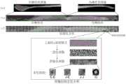
图2示出了根据本公开实施例从包含所述髋关节骨假体的透视图像中分割出骨假体周围骨图像的示意图。Fig. 2 shows a schematic diagram of segmenting a bone image around a bone prosthesis from a perspective image containing the hip joint bone prosthesis according to an embodiment of the present disclosure.
图3示出了将骨假体周围骨图像转换为预设矩形的骨质图像并划分为矩形区块图像的示意图。Fig. 3 shows a schematic diagram of converting a bone image around a bone prosthesis into a preset rectangular bone image and dividing it into rectangular block images.
图4示出了根据本公开实施例的一种矩形区块图像中的骨结构参数统计方法。Fig. 4 shows a statistical method for bone structure parameters in a rectangular block image according to an embodiment of the present disclosure.
图5示出了根据本公开另一实施例的一种矩形区块图像中的骨结构参数统计方法。Fig. 5 shows a statistical method for bone structure parameters in a rectangular block image according to another embodiment of the present disclosure.
图6A和6B示出了由支柱参数估计的CT密度与BMD的相关性。Figures 6A and 6B show the correlation of CT density estimated from pillar parameters with BMD.
图7A示出了骨假体周围骨图像转换得到的预设矩形的骨质图像中骨假体周围骨结构支柱参数随时间变化的主要区域。FIG. 7A shows the main area where the bone structure pillar parameters around the bone prosthesis change over time in the preset rectangular bone image obtained by converting the bone image around the bone prosthesis.
图7B示出了骨假体周围骨图像转换得到的预设矩形的骨质图像中各支柱参数随时间的变化趋势。FIG. 7B shows the time-varying trend of each pillar parameter in the preset rectangular bone image converted from the bone image around the bone prosthesis.
图7C示出了根据本公开实施例的预设矩形的骨质图像的区域相关性热图。FIG. 7C shows a region correlation heat map of a preset rectangular bone image according to an embodiment of the present disclosure.
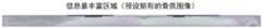
图7D示出了根据本公开实施例的预设矩形的骨质图像中的信息最丰富区域。Fig. 7D shows the most informative region in a preset rectangular bone image according to an embodiment of the present disclosure.
图7E示出了根据本公开实施例基于信息最丰富区域的骨结构参数对术后1年骨假体位移的预测效果远和基于Gruen分区的预测效果。FIG. 7E shows the prediction effect of the bone structure parameters based on the most informative region on the displacement of the bone prosthesis at 1 year after operation and the prediction effect based on the Gruen partition according to an embodiment of the present disclosure.
图8示出根据本公开的实施例的骨假体周围的骨微结构分析装置的结构框图。Fig. 8 shows a structural block diagram of a bone microstructure analysis device around a bone prosthesis according to an embodiment of the present disclosure.
图9示出根据本公开的实施例的电子设备的结构框图。FIG. 9 shows a structural block diagram of an electronic device according to an embodiment of the present disclosure.
图10示出适于用来实现根据本公开实施例的方法的计算机系统的结构示意图。FIG. 10 shows a schematic structural diagram of a computer system suitable for implementing the method according to an embodiment of the present disclosure.
具体实施方式Detailed ways
下文中,将参考附图详细描述本公开的示例性实施例,以使本领域技术人员可容易地实现它们。此外,为了清楚起见,在附图中省略了与描述示例性实施例无关的部分。Hereinafter, exemplary embodiments of the present disclosure will be described in detail with reference to the accompanying drawings so that those skilled in the art can easily realize them. Also, for clarity, parts not related to describing the exemplary embodiments are omitted in the drawings.
在本公开中,应理解,诸如“包括”或“具有”等的术语旨在指示本说明书中所公开的特征、数字、步骤、行为、部件、部分或其组合的存在,并且不欲排除一个或多个其他特征、数字、步骤、行为、部件、部分或其组合存在或被添加的可能性。In the present disclosure, it should be understood that terms such as "comprising" or "having" are intended to indicate the presence of features, numbers, steps, acts, components, parts or combinations thereof disclosed in the specification, and are not intended to exclude one or a plurality of other features, numbers, steps, acts, parts, parts or combinations thereof exist or are added.
另外还需要说明的是,在不冲突的情况下,本公开中的实施例及实施例中的特征可以相互组合。下面将参考附图并结合实施例来详细说明本公开。In addition, it should be noted that, in the case of no conflict, the embodiments in the present disclosure and the features in the embodiments can be combined with each other. The present disclosure will be described in detail below with reference to the accompanying drawings and embodiments.
在本公开中,如涉及对用户信息或用户数据的获取操作或向他人展示用户信息或用户数据的操作,则所述操作均为经用户授权、确认,或由用户主动选择的操作。In this disclosure, if it involves the operation of acquiring user information or user data or displaying user information or user data to others, the operation is authorized, confirmed by the user, or actively selected by the user.
如前所述,早期预测骨假体稳定性对延长骨假体使用寿命来说至关重要,骨假体周围骨质量是影响骨假体稳定性的关键因素之一,BMD与骨微结构是决定骨质量的两个主要因素,BMD下降和骨微结构破坏,都会表现为骨的脆性增加,所能承受的压力变差。骨微结构是由大量骨小梁相互交织形成的网状结构,其中骨小梁的网状分布方向与骨所承受的压力和张力的方向一致,其结构分布的优劣性决定了局部骨质能在多大程度上承受如支持、拉伸、冲击等外部力量。但是,目前的评估手段局限于BMD的检测,而未考虑骨微结构的检测和分析,因此无法全面地根据骨假体周围骨质量对骨假体稳定性进行预测。As mentioned above, early prediction of bone prosthesis stability is crucial to prolonging the service life of bone prosthesis. Bone quality around bone prosthesis is one of the key factors affecting the stability of bone prosthesis. BMD and bone microstructure are important The two main factors that determine bone quality, BMD decline and bone microarchitecture destruction, will be manifested as increased bone fragility and poor pressure resistance. Bone microstructure is a network structure formed by the interweaving of a large number of bone trabeculae, in which the network distribution direction of the bone trabecula is consistent with the direction of the pressure and tension on the bone, and the quality of the structure distribution determines the local bone quality. The extent to which it can withstand external forces such as support, stretching, impact, etc. However, the current evaluation methods are limited to the detection of BMD, without considering the detection and analysis of bone microstructure, so it is impossible to comprehensively predict the stability of bone prosthesis based on the bone quality around the bone prosthesis.
根据最新的研究,与骨假体表面相邻的厚度小于1毫米的骨质是影响骨假体稳定性的关键因,但骨假体周围骨质的Gruen分区各区块面积大、总区块数量少、且同时包含骨皮质与骨松质——总体“分辨率”较差,不足以捕捉小区块内骨质的变化。即便对局部关键区块进行重新勾画,由于骨假体周围骨的形态的个体性差异,也会导致关键区块很难在不同患者之间进行匹配和比较。According to the latest research, the bone thickness less than 1 mm adjacent to the surface of the bone prosthesis is the key factor affecting the stability of the bone prosthesis, but the Gruen partition of the bone around the bone prosthesis has a large area and the total number of blocks Fewer and containing both cortical bone and cancellous bone - the overall "resolution" is poor, not enough to capture changes in bone quality in a small area. Even if the local critical areas are redrawn, due to the individual differences in the morphology of the bone around the prosthesis, it will be difficult to match and compare the key areas between different patients.
为进一步对骨假体周围骨的微结构进行分析,并且能更好的捕捉关键小区块内骨质的变化,以及实现各小区块在不同患者间的匹配,本公开提出了一种基于分区和骨微结构的定量分析方法。根据本公开的实施例,首先将透视图像上的骨假体周围骨质转换为预设矩形,再分割成多个小的矩形区块,每个矩形区块都可以在不同患者间进行匹配。通过骨微结构的定量分析技术来量化每个矩形区块的骨微结构。本公开为骨假体无菌性松动的早期预测提供一种新思路,根据预测结果,后续可对具有松动风险的骨假体实施早期干预以降低远期无菌性松动发生的可能性。In order to further analyze the microstructure of the bone around the bone prosthesis, better capture the changes in bone quality in the key small blocks, and realize the matching of each small block between different patients, the present disclosure proposes a method based on partitioning and Quantitative analysis methods of bone microstructure. According to the embodiment of the present disclosure, the bone around the bone prosthesis on the fluoroscopic image is first converted into a preset rectangle, and then divided into a plurality of small rectangular blocks, and each rectangular block can be matched among different patients. The bone microstructure of each rectangular block was quantified by a quantitative analysis technique of bone microstructure. The present disclosure provides a new idea for early prediction of aseptic loosening of bone prosthesis. According to the prediction result, early intervention can be implemented on bone prosthesis with loosening risk to reduce the possibility of long-term aseptic loosening.
图1示出根据本公开的实施例的骨假体周围的骨微结构分析方法的流程图。如图1所示,所述骨假体周围的骨微结构分析方法包括以下步骤S101-S104:FIG. 1 shows a flowchart of a bone microstructure analysis method around a bone prosthesis according to an embodiment of the present disclosure. As shown in Figure 1, the bone microstructure analysis method around the bone prosthesis includes the following steps S101-S104:
在步骤S101中,获取骨假体所在的人体区域的透视图像;In step S101, a perspective image of the human body area where the bone prosthesis is located is acquired;
在步骤S102中,从所述透视图像中分割出骨假体周围骨图像;In step S102, a bone image around the bone prosthesis is segmented from the perspective image;
在步骤S103中,将所述骨假体周围骨图像转换为预设矩形的骨质图像;In step S103, converting the bone image around the bone prosthesis into a preset rectangular bone image;
在步骤S104中,将所述预设矩形的骨质图像划分为多个矩形区块图像,所述矩形区块图像用于对所述骨假体周围的骨微结构进行定量分析。In step S104, the preset rectangular bone image is divided into a plurality of rectangular block images, and the rectangular block images are used for quantitative analysis of the bone microstructure around the bone prosthesis.
根据本公开的实施例,透视图像可以是X光片。According to an embodiment of the present disclosure, the fluoroscopic image may be an X-ray film.
根据本公开的实施例,所述透视图像为包含所述骨假体的透视图像,所述从所述透视图像中分割出骨假体周围骨图像,包括:通过图形分割算法从所述透视图像中分割出骨假体与周围骨质部分;通过图形分割算法从所述骨假体与周围骨质部分中去除骨假体部分,分割出骨假体周围骨图像。According to an embodiment of the present disclosure, the fluoroscopic image is a fluoroscopic image containing the bone prosthesis, and the segmenting the bone image around the bone prosthesis from the fluoroscopic image includes: using a graph segmentation algorithm to obtain the fluoroscopic image The bone prosthesis and the surrounding bone parts are segmented; the bone prosthesis part is removed from the bone prosthesis and the surrounding bone parts by a graph segmentation algorithm, and the bone image around the bone prosthesis is segmented.
例如,骨假体可以是髋关节骨假体(髋关节骨假体例如可以是全髋关节骨假体或半髋关节骨假体),所述透视图像可以是包含所述髋关节骨假体的透视图像,例如股骨中上端正位片、骨盆正位片等等。所述从所述透视图像中分割出骨假体周围骨图像,包括:通过图形分割算法从所述透视图像中分割出髋关节骨假体与周围骨质部分(即,股骨部分);通过图形分割算法从所述骨假体与周围骨质部分中去除髋关节骨假体部分,分割出骨假体周围骨图像。For example, the bone prosthesis can be a hip bone prosthesis (the hip bone prosthesis can be, for example, a total hip bone prosthesis or a hemihip bone prosthesis), and the fluoroscopic image can be an image containing the hip bone prosthesis. X-ray images, such as the mid-upper end of the femur, the pelvis, and so on. The said segmenting the bone image around the bone prosthesis from the perspective image includes: segmenting the bone prosthesis of the hip joint and the surrounding bone part (that is, the femoral part) from the perspective image by a graphic segmentation algorithm; The segmentation algorithm removes the bone prosthesis part of the hip joint from the bone prosthesis and the surrounding bone part, and segments the bone image around the bone prosthesis.
图2示出了根据本公开实施例从包含所述骨假体的透视图像中分割出骨假体周围骨图像的示意图。Fig. 2 shows a schematic diagram of segmenting a bone image around a bone prosthesis from a perspective image containing the bone prosthesis according to an embodiment of the present disclosure.
根据本公开的实施例,可以使用BoneFinder算法,从包含所述骨假体的透视图像中勾勒出骨假体与周围骨质部分的轮廓,分割出骨假体与周围骨质部分。或者,也可以使用其他基于人工智能的图像分割算法、基于图像的灰度值或图像轮廓的图像分割算法等,从包含所述骨假体的透视图像中分割出骨假体与周围骨质部分。According to an embodiment of the present disclosure, the BoneFinder algorithm can be used to outline the bone prosthesis and the surrounding bone parts from the perspective image containing the bone prosthesis, and segment the bone prosthesis and the surrounding bone parts. Alternatively, other image segmentation algorithms based on artificial intelligence, image segmentation algorithms based on image gray values or image contours, etc. can be used to segment the bone prosthesis and surrounding bone parts from the perspective image containing the bone prosthesis. .
图2所示示例为包含髋关节骨假体的透视图像,其中(a)部分为包含髋关节骨假体的透视图像,其中白色框线围住的部分即为骨假体与周围骨质部分。The example shown in Figure 2 is a perspective image containing a hip bone prosthesis, where part (a) is a perspective image containing a hip bone prosthesis, and the part surrounded by the white frame line is the bone prosthesis and the surrounding bone parts .
然后,可以使用基于灰度值的算法从所述骨假体与周围骨质部分中去除骨假体部分,分割出骨假体周围骨图像。或者,也可以使用其他基于人工智能的图像分割算法、基于图像轮廓的图像分割算法等,从所述骨假体与周围骨质部分中去除骨假体部分,分割出骨假体周围骨图像。图2的(b)部分即为去除髋关节骨假体部分的图像,图2的(c)部分即为骨假体周围骨图像。Then, the bone prosthesis part can be removed from the bone prosthesis and the surrounding bone part by using an algorithm based on the gray value, and a bone image around the bone prosthesis can be segmented. Alternatively, other artificial intelligence-based image segmentation algorithms, image contour-based image segmentation algorithms, etc. may be used to remove the bone prosthesis part from the bone prosthesis and surrounding bone parts, and segment the bone image around the bone prosthesis. Part (b) of Fig. 2 is the image of the hip joint bone prosthesis removed, and part (c) of Fig. 2 is the bone image around the bone prosthesis.
由于骨假体周围骨的形态的个体性差异,会导致关键区块很难在不同患者之间进行匹配和比较,因此将图2的(c)部分所示骨假体周围骨图像转换为预设矩形的骨质图像,将所述预设矩形的骨质图像划分为多个矩形区块图像。根据本公开的实施例,所述预设矩形具有预设的尺寸,所述矩形区块图像也具有预设的尺寸且其尺寸小于预设矩形。将不同患者的骨假体周围骨图像均转换为相同尺寸的预设矩形的骨质图像并将其划分为多个更小的矩形区块图像,便于骨图像在不同患者之间进行匹配和比较。Due to the individual differences in the shape of the bone around the prosthesis, it will be difficult to match and compare the key blocks between different patients. Therefore, the bone image around the prosthesis shown in part (c) of Figure 2 A rectangular bone image is assumed, and the preset rectangular bone image is divided into a plurality of rectangular block images. According to an embodiment of the present disclosure, the preset rectangle has a preset size, and the rectangular block image also has a preset size and its size is smaller than the preset rectangle. Convert the bone images around the bone prosthesis of different patients into a preset rectangular bone image of the same size and divide it into multiple smaller rectangular block images, which facilitates the matching and comparison of bone images between different patients .
根据本公开的实施例,所述将所述骨假体周围骨图像转换为预设矩形的骨质图像,包括:使用以下任意一种算法将所述骨假体周围骨图像转换为预设矩形的骨质图像:分段仿射算法、基于点阵网格的图像变形转换算法、基于线性的图像变形转换算法、基于非线性的图像变形转换算法。According to an embodiment of the present disclosure, converting the bone image around the bone prosthesis into a preset rectangular bone image includes: using any of the following algorithms to convert the bone image around the bone prosthesis into a preset rectangle Bone image: segmented affine algorithm, image deformation transformation algorithm based on lattice grid, linear image deformation transformation algorithm, nonlinear image deformation transformation algorithm.
根据本公开的实施例,所述透视图像为包含所述骨假体的透视图像,所述从所述透视图像中分割出骨假体周围骨图像,包括:通过图形分割算法从所述透视图像中分割出骨假体与周围骨质部分;通过图形分割算法从所述骨假体与周围骨质部分中去除骨假体部分,分割出骨假体周围骨图像。According to an embodiment of the present disclosure, the fluoroscopic image is a fluoroscopic image containing the bone prosthesis, and the segmenting the bone image around the bone prosthesis from the fluoroscopic image includes: using a graph segmentation algorithm to obtain the fluoroscopic image The bone prosthesis and the surrounding bone parts are segmented; the bone prosthesis part is removed from the bone prosthesis and the surrounding bone parts by a graph segmentation algorithm, and the bone image around the bone prosthesis is segmented.
根据本公开的实施例,所述将所述骨假体周围骨图像转换为预设矩形的骨质图像,包括:将所述骨假体周围骨图像作为一个整体转换为预设矩形的骨质图像;或者将所述骨假体周围骨图像划分为多个区域,将所述多个区域分别转换为预设矩形的骨质图像的相应区域,从而得到所述预设矩形的骨质图像。According to an embodiment of the present disclosure, the converting the bone image around the bone prosthesis into a preset rectangular bone image includes: converting the bone image around the bone prosthesis as a whole into a preset rectangular bone quality image image; or divide the bone image around the bone prosthesis into multiple regions, and convert the multiple regions into corresponding regions of the preset rectangular bone image, so as to obtain the preset rectangular bone image.
根据本公开的实施例,所述将所述骨假体周围骨图像作为一个整体转换为预设矩形的骨质图像,包括使用以下任意一种算法将所述骨假体周围骨图像作为一个整体转换为预设矩形的骨质图像:分段仿射算法、基于点阵网格的图像变形转换算法、基于线性的图像变形转换算法、基于非线性的图像变形转换算法;所述将所述多个区域分别转换为预设矩形的骨质图像的相应区域,包括使用以下任意一种算法将所述多个区域分别转换为所述预设矩形的骨质图像的相应区域:分段仿射算法、基于点阵网格的图像变形转换算法、基于线性的图像变形转换算法、基于非线性的图像变形转换算法。According to an embodiment of the present disclosure, the converting the bone image around the bone prosthesis as a whole into a preset rectangular bone image includes using any of the following algorithms to convert the bone image around the bone prosthesis as a whole Bone images converted into preset rectangles: segmented affine algorithm, image deformation conversion algorithm based on lattice grid, linear-based image deformation conversion algorithm, nonlinear image deformation conversion algorithm; The regions are respectively converted into corresponding regions of the preset rectangular bone image, including using any of the following algorithms to respectively convert the multiple regions into corresponding regions of the preset rectangular bone image: segmented affine algorithm , Image deformation conversion algorithm based on lattice grid, linear image deformation conversion algorithm, nonlinear image deformation conversion algorithm.
根据本公开的实施例,所述使用分段仿射算法将所述骨假体周围骨图像作为一个整体转换为预设矩形的骨质图像,包括:将第一网点阵均匀覆盖至所述预设矩形;将与所述第一网点阵相同分布的第二网点阵根据所述骨假体周围骨的形状缩放并覆盖到所述骨假体周围骨图像上;基于所述第一网点阵和所述第二网点阵,建立分段仿射转换模型;通过所述分段仿射转换模型将所述骨假体周围骨图像转换为所述预设矩形的骨质图像。According to an embodiment of the present disclosure, the converting the bone image around the bone prosthesis as a whole into a preset rectangular bone image using a segmented affine algorithm includes: uniformly covering the first grid lattice to the preset Assume a rectangle; scale and cover the second lattice with the same distribution as the first lattice on the bone image around the bone prosthesis according to the shape of the bone around the bone prosthesis; based on the first lattice and The second network lattice establishes a segmented affine transformation model; the bone image around the bone prosthesis is converted into the preset rectangular bone image through the segmented affine transformation model.
根据本公开的实施例,所述使用分段仿射算法将所述多个区域分别转换为所述预设矩形的骨质图像的相应区域,包括:将多个第一网点阵分别均匀覆盖至所述相应区域;将分别与所述多个第一网点阵相同分布的多个第二网点阵根据所述骨假体周围骨的形状分别缩放并覆盖到所述多个区域上;基于所述第一网点阵和相应的第二网点阵,建立分段仿射转换模型;通过所述分段仿射转换模型将所述多个区域转换为所述预设矩形的骨质图像的相应区域。According to an embodiment of the present disclosure, the converting the plurality of regions into corresponding regions of the preset rectangular bone image using a segmented affine algorithm includes: uniformly covering a plurality of first grid lattices into The corresponding area; the multiple second grid lattices distributed in the same manner as the multiple first grid lattices are respectively scaled and covered on the multiple areas according to the shape of the bone around the bone prosthesis; based on the The first dot matrix and the corresponding second dot matrix establish a segmented affine transformation model; the plurality of regions are transformed into corresponding regions of the preset rectangular bone image through the segmental affine transformation model.
根据本公开的实施例,与所述第一网点阵相同分布的第二网点阵,是指第二网点阵的网点的行、列数及各网点之间的间距与第一网点阵相同。According to an embodiment of the present disclosure, the second dot matrix having the same distribution as the first dot matrix means that the number of rows and columns of dots in the second dot matrix and the spacing between dots are the same as those in the first dot matrix.
图3示出了将骨假体周围骨图像转换为预设矩形的骨质图像并划分为矩形区块图像的示意图。Fig. 3 shows a schematic diagram of converting a bone image around a bone prosthesis into a preset rectangular bone image and dividing it into rectangular block images.
以髋关节骨假体为例,根据本公开的实施例,所述将所述骨假体周围骨图像转换为预设矩形的骨质图像,包括:将所述骨假体周围骨图像划分为两个区域(即,内侧骨质图像和外侧骨质图像);将所述内侧骨质图像和所述外侧骨质图像分别转换为预设矩形的骨质图像的相应区域,从而得到预设矩形的骨质图像。Taking a hip joint bone prosthesis as an example, according to an embodiment of the present disclosure, converting the bone image around the bone prosthesis into a preset rectangular bone image includes: dividing the bone image around the bone prosthesis into Two regions (that is, the inner bone image and the outer bone image); the inner bone image and the outer bone image are respectively converted into corresponding areas of the bone image of a preset rectangle, thereby obtaining a preset rectangle bone image.
具体地,如图3的(a)部分所示,将图2的(c)部分的骨假体周围骨图像划分为内侧骨质图像和外侧骨质图像,然后分别转换为预设矩形的骨质图像的相应区域。Specifically, as shown in part (a) of Figure 3, the bone image around the bone prosthesis in part (c) of Figure 2 is divided into an inner bone image and an outer bone image, and then converted into preset rectangular bone images respectively. the corresponding area of the quality image.
根据本公开的实施例,所述预设矩形包括分别对应于所述内侧骨质图像和所述外侧骨质图像的两个区域,这两个区域分别称为内侧部分和外侧部分。例如,预设矩形可以设置为外侧部分具有100*1200像素,内侧部分具有100*1000像素。所述将所述内侧骨质图像和所述外侧骨质图像分别转换为预设矩形的骨质图像的相应区域,包括:使用以下任意一种算法将所述内侧骨质图像和所述外侧骨质图像分别转换为所述预设矩形的骨质图像的内侧部分和外侧部分:分段仿射算法、基于点阵网格的图像变形转换算法、基于线性的图像变形转换算法、基于非线性的图像变形转换算法。According to an embodiment of the present disclosure, the preset rectangle includes two areas respectively corresponding to the inner bone image and the outer bone image, and these two areas are respectively referred to as an inner portion and an outer portion. For example, the preset rectangle may be set such that the outer part has 100*1200 pixels, and the inner part has 100*1000 pixels. The converting the inner bone image and the outer bone image into corresponding regions of the preset rectangular bone image includes: using any of the following algorithms to convert the inner bone image and the outer bone image The bone quality image is respectively converted into the inner part and the outer part of the bone image of the preset rectangle: segmented affine algorithm, image deformation conversion algorithm based on lattice grid, image deformation conversion algorithm based on linear, nonlinear based Image warping transformation algorithm.
作为示例性的,本公开实施例以分段仿射算法作为详细说明,但是本领域技术人员可以理解的是,其他算法如基于点阵网格的图像变形转换算法、基于线性的图像变形转换算法、基于非线性的图像变形转换算法也可以达到相同的技术效果。在该实施例中,使用分段仿射算法将所述内侧骨质图像和所述外侧骨质图像分别转换为所述预设矩形的骨质图像的内侧部分和外侧部分,包括:将两个第一网点阵分别均匀覆盖至所述内侧部分和所述外侧部分;将与所述两个第一网点阵相同分布的两个第二网点阵根据所述骨假体周围骨的形状分别缩放并覆盖到所述内侧骨质图像和所述外侧骨质图像上;基于所述第一网点阵和所述第二网点阵,建立分段仿射转换模型;通过所述分段仿射转换模型将所述内侧骨质图像和所述外侧骨质图像转换为所述预设矩形的骨质图像。As an example, the embodiment of the present disclosure takes the segmented affine algorithm as a detailed description, but those skilled in the art can understand that other algorithms such as image deformation conversion algorithm based on lattice grid, image deformation conversion algorithm based on linear , The same technical effect can also be achieved based on the non-linear image deformation conversion algorithm. In this embodiment, the segmented affine algorithm is used to convert the inner bone image and the outer bone image into the inner part and the outer part of the preset rectangular bone image, including: converting the two The first mesh lattice covers the inner portion and the outer portion uniformly; the two second mesh lattices distributed in the same manner as the two first mesh lattices are respectively scaled and scaled according to the shape of the bone around the bone prosthesis. overlaid on the inner bone image and the outer bone image; based on the first lattice matrix and the second lattice matrix, a segmented affine transformation model is established; through the segmentation affine transformation model, the The inner bone image and the outer bone image are converted into the preset rectangular bone image.
将两个密集的网点阵分别均匀覆盖至内侧部分和外侧部分,这两个网点阵称为第一网点阵。将与两个第一网点阵同样分布的两个第二网点阵根据骨假体周围骨的形状分别缩放并覆盖到内侧骨质图像和外侧骨质图像上,即与覆盖内侧部分的第一网点阵相同分布的第二网点阵缩放后覆盖到内侧骨质图像上,与覆盖外侧部分的第一网点阵相同分布的第二网点阵缩放后覆盖到外侧骨质图像上。Evenly cover the inner part and the outer part with two dense dot matrixes respectively, and these two dot matrixes are called the first dot matrix. Scale and overlay the two second dot matrixes distributed in the same manner as the two first dot matrix respectively on the inner bone image and the outer bone image according to the shape of the bone around the bone prosthesis, that is, the same as the first dots covering the inner part The second dot matrix with the same distribution as the dot matrix is scaled and overlaid on the inner bone image, and the second dot matrix with the same distribution as the first dot matrix covering the outer part is scaled and overlaid on the outer bone image.
基于第一网点阵和第二网点阵建立分段仿射转换模型,通过该分段仿射转换模型将骨假体周围骨图像转换为预设矩形的骨质图像,如图3的(b)部分所示。Establish a segmented affine transformation model based on the first lattice and the second lattice, and convert the bone image around the bone prosthesis into a preset rectangular bone image through the segmental affine transformation model, as shown in (b) of Figure 3 part shown.
根据本公开的实施例,所述将所述预设矩形的骨质图像划分为多个矩形区块图像,包括:将所述预设矩形的骨质图像均匀划分为多个矩形区块图像。According to an embodiment of the present disclosure, the dividing the preset rectangular bone image into a plurality of rectangular block images includes: evenly dividing the preset rectangular bone image into a plurality of rectangular block images.
例如,如图3的(c)部分所示,将图3的(b)部分的预设矩形的骨质图像均匀地划分为多个矩形区块图像,其中外侧划分为10*24个矩形区块图像,内侧划分为10*20个矩形区块图像,每个矩形区块图像可以具有相同的尺寸。For example, as shown in part (c) of Figure 3, the preset rectangular bone image of part (b) of Figure 3 is evenly divided into a plurality of rectangular block images, wherein the outer side is divided into 10*24 rectangular areas The block image is divided into 10*20 rectangular block images inside, and each rectangular block image may have the same size.
通过这样的方式,可以将不同患者的骨假体周围骨图像转换为相同尺寸的矩形区块图像,从而便于关键区块图像在不同患者之间进行匹配和比较。In this way, the bone images around the bone prosthesis of different patients can be converted into rectangular block images of the same size, so as to facilitate the matching and comparison of key block images among different patients.
根据本公开的实施例,在获得矩形区块图像之后,统计矩形区块图像中的骨结构参数,从而实现骨假体周围的骨微结构的定量分析。According to an embodiment of the present disclosure, after the rectangular block image is obtained, the bone structure parameters in the rectangular block image are counted, so as to realize the quantitative analysis of the bone microstructure around the bone prosthesis.
本公开实施例提供了两种矩形区块图像中的骨结构参数统计方法。The embodiments of the present disclosure provide two statistical methods for bone structure parameters in rectangular block images.
图4示出了根据本公开实施例的一种矩形区块图像中的骨结构参数统计方法。Fig. 4 shows a statistical method for bone structure parameters in a rectangular block image according to an embodiment of the present disclosure.
具体地,在步骤S401中,将预设矩形的骨质图像作为一个整体进行骨微结构的定量分析,在步骤S402中,统计所述矩形区块图像中的骨结构参数,所述骨结构参数是所述骨微结构的定量分析结果。Specifically, in step S401, quantitative analysis of bone microstructure is performed on the preset rectangular bone image as a whole, and in step S402, bone structure parameters in the rectangular block image are counted, and the bone structure parameters is the quantitative analysis result of the bone microstructure.
根据本公开的实施例,所述将所述预设矩形的骨质图像作为一个整体进行骨微结构的定量分析,包括:对所述预设矩形的骨质图像进行上采样,得到上采样图像;对所述上采样图像进行密度矫正,移除由骨密度造成的图像阴暗;将密度矫正后的图像转换为二值化图像;将所述二值化图像转换为仅包含骨微结构的拓扑结构支柱的拓扑结构支柱图像;对所述二值化图像和所述拓扑结构支柱图像中各支柱的参数进行量化分析,得到定量分析结果。According to an embodiment of the present disclosure, the quantitative analysis of the bone microstructure of the preset rectangular bone image as a whole includes: performing up-sampling on the preset rectangular bone image to obtain an up-sampled image ; Density correction is performed on the upsampled image to remove the dark image caused by bone density; the density-corrected image is converted into a binary image; the binary image is converted to a topology containing only bone microstructure A topological structure pillar image of a structural pillar; performing quantitative analysis on the binarized image and parameters of each pillar in the topological structure pillar image to obtain a quantitative analysis result.
具体地,对所述预设矩形的骨质图像进行上采样的上采样比例可以是400%,也可以采用其他的上采样比例。作为示例,上采样可以使用二次插值法实现,但是本领域技术人员可以理解,也可以采用其他上采样方法,例如基于线性插值的上采样方法、基于深度学习的上采样方法、Unpooling的上采样方法,等等。Specifically, the upsampling ratio for upsampling the preset rectangular bone image may be 400%, or other upsampling ratios may be used. As an example, upsampling can be implemented using the quadratic interpolation method, but those skilled in the art can understand that other upsampling methods can also be used, such as upsampling methods based on linear interpolation, upsampling methods based on deep learning, and upsampling methods based on Unpooling method, and so on.
根据本公开的实施例,通过对上采样图像进行高斯模糊处理(sd=5)实现图像数据的平滑化处理,以得到平滑图像,从上采样图像图像中减去平滑图像来完成密度矫正。在该实施例中,作为示例,通过高斯模糊处理实现图像数据的平滑化处理,但是本领域技术人员可以理解,也可以使用其他平滑算法,例如基于线性或非线性的图像数据平滑算法,或者基于深度学习的图像数据平滑算法,来实现图像数据的平滑化处理。According to the embodiment of the present disclosure, the image data is smoothed by performing Gaussian blur processing (sd=5) on the up-sampled image to obtain a smooth image, and subtracting the smooth image from the up-sampled image to complete density correction. In this embodiment, as an example, the smoothing of image data is realized by Gaussian blur processing, but those skilled in the art can understand that other smoothing algorithms can also be used, such as linear or nonlinear image data smoothing algorithms, or based on The image data smoothing algorithm of deep learning is used to realize the smoothing processing of image data.
根据本公开的实施例,可以将密度矫正后的图像转换为二值化图像,例如根据中位数转换为二值化图像。According to an embodiment of the present disclosure, the density-corrected image may be converted into a binarized image, for example, converted into a binarized image according to a median.
根据本公开的实施例,将所述二值化图像转换为仅包含骨微结构的拓扑结构支柱的拓扑结构支柱图像,也称为骨架化转换。According to an embodiment of the present disclosure, the binarized image is converted into a topological strut image including only topological struts of the bone microstructure, which is also referred to as skeletonization conversion.
根据本公开的实施例,对所述二值化图像和所述拓扑结构支柱图像中各支柱的参数进行量化分析,包括根据所述二值化图像和所述拓扑结构支柱图像确定以下至少一项参数作为所述定量分析结果:According to an embodiment of the present disclosure, the quantitative analysis of the parameters of each pillar in the binarized image and the topological pillar image includes determining at least one of the following according to the binary image and the topological pillar image Parameters as a result of the quantitative analysis:
·HDA:二值化图像中高密度区域的比例HDA: The proportion of high-density areas in the binarized image
●TSL:单位面积上所有支柱长度的总和●TSL: the sum of the lengths of all struts per unit area
●nS:单位面积上支柱的总数● nS: the total number of pillars per unit area
●支柱E-E:端-端支柱的比例Props E-E: ratio of end-to-end props
·支柱J-E:节-端支柱的比例Prop J-E: ratio of node-to-end struts
●支柱J-J:节-节支柱的比例●Pillar J-J: the ratio of section-section support
●支柱IC:独立环形支柱的比例Pillar IC: Ratio of independent ring-shaped pillars
图5示出了根据本公开另一实施例的一种矩形区块图像中的骨结构参数统计方法。Fig. 5 shows a statistical method for bone structure parameters in a rectangular block image according to another embodiment of the present disclosure.
具体地,在步骤S501,将所述骨假体周围骨图像作为一个整体进行骨微结构的定量分析。在步骤S502,确定所述骨假体周围骨图像中与每个矩形区块对应的相应部分。在步骤S503,统计所述矩形区块图像中的骨结构参数,所述骨结构参数是所述骨假体周围骨图像中与所述矩形区块对应的相应部分的骨微结构定量分析结果。Specifically, in step S501, the bone image around the bone prosthesis is taken as a whole for quantitative analysis of bone microstructure. In step S502, a corresponding part corresponding to each rectangular block in the bone image around the bone prosthesis is determined. In step S503, the bone structure parameters in the rectangular block image are counted, and the bone structure parameters are the quantitative analysis results of the bone microstructure of the corresponding part of the bone image around the bone prosthesis corresponding to the rectangular block.
根据本公开的实施例,所述将所述骨假体周围骨图像作为一个整体进行骨微结构的定量分析,包括:对所述骨假体周围骨图像进行上采样,得到上采样图像;对所述上采样图像进行密度矫正,移除由骨密度造成的图像阴暗;将密度矫正后的图像转换为二值化图像;将所述二值化图像转换为仅包含骨微结构的拓扑结构支柱的拓扑结构支柱图像;对所述二值化图像和所述拓扑结构支柱图像中各支柱的参数进行量化分析,得到定量分析结果。According to an embodiment of the present disclosure, the quantitative analysis of the bone microstructure of the bone image around the bone prosthesis as a whole includes: performing up-sampling on the bone image around the bone prosthesis to obtain an up-sampled image; Perform density correction on the upsampled image to remove image darkness caused by bone density; convert the density-corrected image into a binarized image; convert the binarized image into a topological structure pillar containing only bone microstructure The topological structure pillar image; performing quantitative analysis on the binarized image and the parameters of each pillar in the topological structure pillar image to obtain a quantitative analysis result.
具体地,对所述骨假体周围骨图像进行上采样的上采样比例可以是400%,也可以采用其他的上采样比例。作为示例,上采样可以使用二次插值法实现,但是本领域技术人员可以理解,也可以采用其他上采样方法,例如基于线性插值的上采样方法、基于深度学习的上采样方法、Unpooling的上采样方法,等等。Specifically, the upsampling ratio for upsampling the bone image around the bone prosthesis may be 400%, or other upsampling ratios may be used. As an example, upsampling can be implemented using the quadratic interpolation method, but those skilled in the art can understand that other upsampling methods can also be used, such as upsampling methods based on linear interpolation, upsampling methods based on deep learning, and upsampling methods based on Unpooling method, and so on.
根据本公开的实施例,通过对上采样图像进行高斯模糊处理(sd=5)实现图像数据的平滑化处理,以得到平滑图像,从上采样图像图像中减去平滑图像来完成密度矫正。在该实施例中,作为示例,通过高斯模糊处理实现图像数据的平滑化处理,但是本领域技术人员可以理解,也可以使用其他平滑算法,例如基于线性或非线性的图像数据平滑算法,或者基于深度学习的图像数据平滑算法,来实现图像数据的平滑化处理。According to the embodiment of the present disclosure, the image data is smoothed by performing Gaussian blur processing (sd=5) on the up-sampled image to obtain a smooth image, and subtracting the smooth image from the up-sampled image to complete density correction. In this embodiment, as an example, the smoothing of image data is realized by Gaussian blur processing, but those skilled in the art can understand that other smoothing algorithms can also be used, such as linear or nonlinear image data smoothing algorithms, or based on The image data smoothing algorithm of deep learning is used to realize the smoothing processing of image data.
根据本公开的实施例,可以将密度矫正后的图像转换为二值化图像,例如根据中位数转换为二值化图像。According to an embodiment of the present disclosure, the density-corrected image may be converted into a binarized image, for example, converted into a binarized image according to a median.
根据本公开的实施例,将所述二值化图像转换为仅包含骨微结构的拓扑结构支柱的拓扑结构支柱图像,也称为骨架化转换。According to an embodiment of the present disclosure, the binarized image is converted into a topological strut image including only topological struts of the bone microstructure, which is also referred to as skeletonization conversion.
根据本公开的实施例,对所述二值化图像和所述拓扑结构支柱图像中各支柱的参数进行量化分析,包括根据所述二值化图像和所述拓扑结构支柱图像确定以下至少一项参数作为所述定量分析结果:According to an embodiment of the present disclosure, the quantitative analysis of the parameters of each pillar in the binarized image and the topological pillar image includes determining at least one of the following according to the binary image and the topological pillar image Parameters as a result of the quantitative analysis:
·HDA:二值化图像中高密度区域的比例HDA: The proportion of high-density areas in the binarized image
●TSL:单位面积上所有支柱长度的总和●TSL: the sum of the lengths of all struts per unit area
●nS:单位面积上支柱的总数● nS: the total number of pillars per unit area
●支柱E-E:端-端支柱的比例Props E-E: ratio of end-to-end props
·支柱J-E:节-端支柱的比例Prop J-E: ratio of node-to-end struts
●支柱J-J:节-节支柱的比例●Pillar J-J: the ratio of section-section support
●支柱IC:独立环形支柱的比例Pillar IC: Ratio of independent ring-shaped pillars
由于上述定量分析结果是针对骨假体周围骨图像得到的,为了统计每个矩形区块图像的骨结构参数,需要确定所述骨假体周围骨图像中与每个矩形区块图像对应的相应部分。即,针对每个矩形区块图像,确定该矩形区块图像对应于哪个骨假体周围图像部分,对该骨假体周围图像部分中的骨结构参数进行统计。Since the above quantitative analysis results are obtained for the bone image around the bone prosthesis, in order to count the bone structure parameters of each rectangular block image, it is necessary to determine the corresponding corresponding to each rectangular block image in the bone image around the bone prosthesis. part. That is, for each rectangular block image, it is determined which part of the image around the bone prosthesis corresponds to the rectangular block image, and the bone structure parameters in the image part around the bone prosthesis are counted.
以上结合图4和图5描述了基于支柱分析算法的骨微结构定量分析,但是,本领域技术人员可以理解,也可以采用纹理分析算法或特征提取算法来实现骨微结构定量分析,其中,特征提取算法可以包括基于灰阶共生矩阵(gray-level co-occurrence matrix)的Haralick特征、本地二值化图案(Local Binary Patterns)、阈值邻接统计算法(ThresholdAdjacency Statistics)等。The quantitative analysis of bone microstructure based on the pillar analysis algorithm is described above in conjunction with Fig. 4 and Fig. 5, but those skilled in the art can understand that the quantitative analysis of bone microstructure can also be realized by using a texture analysis algorithm or a feature extraction algorithm, wherein the feature Extraction algorithms may include Haralick features based on gray-level co-occurrence matrix, local binary patterns (Local Binary Patterns), threshold adjacency statistical algorithm (Threshold Adjacency Statistics), etc.
根据本公开的实施例实现了骨假体周围骨质的微结构定量检测。DXA测量的骨密度BMD代表了局部骨矿物的含量,而骨结构则代表了骨矿物的分布情况。μCT(显微计算机断层扫描成像)能够准确重建骨小梁的微观结构,但由于金属伪影影响,基于CT(计算机断层扫描成像)的骨假体周围骨结构评估无法实现。通过骨微结构的定量检测技术可以清晰直观地对X光片中骨小梁结构进行参数化。本发明的实施结果表明,由本方法测量的骨结构参数与CT测量的密度值显著相关,由骨结构参数预估的CT值与DXA测量的骨密度值显著相关。因此本发明实施例所测量的骨结构参数可作为DXA测量骨密度的额外补充,共同用于骨假体周围骨质量的评估。According to the embodiments of the present disclosure, the quantitative detection of the microstructure of the bone around the bone prosthesis is realized. The bone density BMD measured by DXA represents the content of local bone minerals, while the bone structure represents the distribution of bone minerals. μCT (micro-computed tomography) can accurately reconstruct the microstructure of trabecular bone, but due to metal artifacts, CT-based assessment of bone structure around bone prostheses cannot be achieved. The quantitative detection technology of bone microstructure can clearly and intuitively parameterize the structure of trabecular bone in X-ray films. The implementation results of the present invention show that the bone structure parameters measured by the method are significantly correlated with the density values measured by CT, and the CT values estimated from the bone structure parameters are significantly correlated with the bone density values measured by DXA. Therefore, the bone structure parameters measured in the embodiment of the present invention can be used as an additional supplement to the bone density measured by DXA, and can be used together for the assessment of bone quality around the bone prosthesis.
图6A和图6B示出了由支柱参数估计的CT密度与BMD的相关性。Figures 6A and 6B show the correlation of CT density estimated from pillar parameters with BMD.
CT密度值已被证明与BMD呈线性相关。为测试本公开实施例的性能,首先利用在同一时间段内(一周内)完成的健侧股骨近端X光片和CT检查的10名患者建立基于骨结构参数的CT密度估计模型。随后对另外28名同时进行了髋关节X光片和骨假体周围DXA随访的患者进行分析。骨假体周围骨结构参数被用来通过上述CT密度估计模型估计CT密度,并与DXA测量的BMD进行相关性分析。CT density values have been shown to be linearly correlated with BMD. To test the performance of the embodiments of the present disclosure, a CT density estimation model based on bone structure parameters was first established using X-ray films and CT examinations of the uninjured proximal femur of 10 patients completed within the same period of time (within one week). An additional 28 patients who underwent both hip radiographs and periprosthetic DXA follow-up were subsequently analyzed. Bone structure parameters around the bone prosthesis were used to estimate CT density by the above CT density estimation model and correlated with BMD measured by DXA.
分析结果显示,髋关节X光片中各分区的骨结构参数与CT中相应区域的平均HU值显著相关(复相关系数=0.69,P<0.01)。通过线性回归建立基于骨结构参数的CT密度估计模型,R2=0.34,mse=230.61。随后根据各Gruen分区的骨结构参数估计CT密度值(见图6A)。其中3、5、6、7区所估计的CT密度与金标准DXA测量的BMD有明显的相关性(Pearson's r=0.36-0.56,P≤0.05),在1、2区虽然没有统计显著性,但发现了相关性的趋势。根据DXA测量的BMD结果,进一步将各Gruen分区的骨样本分为高、中、低骨量3类样本。在Gruen 2、3、5、6和7区,基于骨结构参数估计的CT密度对低骨量样本显示出良好的分辨性能(准确率=0.64%-0.86%;见图6B)。The analysis results showed that the bone structure parameters of each subregion in the hip joint X-ray film were significantly correlated with the average HU value of the corresponding region in the CT (multiple correlation coefficient=0.69, P<0.01). A CT density estimation model based on bone structure parameters was established by linear regression, R2=0.34, mse=230.61. CT density values were subsequently estimated from the bone structure parameters of each Gruen division (see Figure 6A). Among them, the estimated CT density in
根据本公开的实施例,将骨假体周围骨图像转换为预设矩形的骨质图像,将所述预设矩形的骨质图像划分为多个矩形区块图像显著提高了检测的“分辨率”,并可实现在不同患者间的匹配与比较。According to the embodiment of the present disclosure, the bone image around the bone prosthesis is converted into a preset rectangular bone image, and the preset rectangular bone image is divided into a plurality of rectangular block images to significantly improve the "resolution" of detection. ", and can achieve matching and comparison among different patients.
在骨假体植入后,由于骨质的内部应力发生了明显变化,各部分骨质都会发生普遍性的骨重塑改变。但目前对骨假体-骨界面的病理学研究表明,骨假体的稳定性主要取决于与骨假体相邻的厚度小于1mm的骨质。Gruen区的面积较大,更容易捕捉到普遍的骨重塑,而与松动相关的小区域变化可能会被遗漏。本公开实施例首先通过分段仿射算法将不规则的骨假体周围骨转化为规则的预设矩形,随后将其统一划分为10*44个小的矩形区块图像,相应的骨假体周围骨尺寸约为1-2mm(厚度)*8mm(长度),这使得矩形区块图像有可能捕捉到与松动有关的小区域变化。本公开的实施结果表明,预设矩形统一了骨假体周围骨的形状,其中每个矩形区块都可以在不同患者间进行匹配,能够更好的分析各矩形区块中骨结构的变化趋势。After the bone prosthesis is implanted, due to the obvious changes in the internal stress of the bone, all parts of the bone will undergo general bone remodeling changes. However, the current pathological research on the bone prosthesis-bone interface shows that the stability of the bone prosthesis mainly depends on the bone thickness less than 1mm adjacent to the bone prosthesis. The larger area of Gruen's area makes it easier to capture general bone remodeling, while small regional changes associated with loosening may be missed. In the embodiment of the present disclosure, the irregular bone around the bone prosthesis is first converted into a regular preset rectangle through a segmented affine algorithm, and then it is uniformly divided into 10*44 small rectangular block images, and the corresponding bone prosthesis The surrounding bone size is approximately 1-2 mm (thickness) * 8 mm (length), which makes it possible for rectangular block images to capture small regional changes associated with loosening. The implementation results of the present disclosure show that the preset rectangle unifies the shape of the bone around the bone prosthesis, and each rectangular block can be matched among different patients, which can better analyze the change trend of the bone structure in each rectangular block .
为验证本公开实施例的效果,对共计91位病例完成了1年的髋关节X线随访,对术后随访的髋关节X线中骨假体周围骨质依次按照上述方法进行划分矩形区块图像、骨微结构的定量分析、获得各个矩形区块图像的骨结构参数。将所有患者的矩形区块图像进行匹配,对于其中每一个矩形区块图像,将该矩形区块图像的骨结构参数与随访时间做相关性分析以明确各矩形区块图像内骨结构参数随时间变化的趋势。In order to verify the effect of the disclosed embodiment, a total of 91 cases were followed up for 1 year by hip joint X-ray, and the bone mass around the bone prosthesis in the postoperative follow-up hip joint X-ray was divided into rectangular blocks according to the above method. Quantitative analysis of images, bone microstructure, and obtaining bone structure parameters of each rectangular block image. Match the rectangular block images of all patients, and for each rectangular block image, do a correlation analysis between the bone structure parameters of the rectangular block image and the follow-up time to clarify the bone structure parameters in each rectangular block image over time. changing trends.
图7A示出了骨假体周围骨图像转换得到的预设矩形的骨质图像中骨假体周围骨结构支柱参数随时间变化的主要区域,其中,颜色越深,表示支柱参数随时间的变化程度越大。Fig. 7A shows the main area of the pillar parameters of the bone structure around the bone prosthesis in the preset rectangular bone image converted from the bone image around the bone prosthesis, where the darker the color, the change of the pillar parameters over time The greater the degree.
图7B示出了骨假体周围骨图像转换得到的预设矩形的骨质图像中各支柱参数随时间的变化趋势,其中,颜色越深,表示支柱参数随时间的变化程度越大。在图7B中,“HDA”表示二值化后图片中高密度区域所占比例,“TSL”表示单位面积中所有支柱长度总和,“nS”表示单位面积中所有支柱总数,支柱“E-E”表示所有支柱中端-端支柱所占比例,“支柱J-E”表示所有支柱中节-端支柱所占比例,“支柱J-J”表示所有支柱中节-节支柱所占比例,“支柱IC”表示所有支柱中独立环型支柱所占比例。FIG. 7B shows the time-varying trend of each pillar parameter in the preset rectangular bone image converted from the bone image around the bone prosthesis, where the darker the color, the greater the change degree of the pillar parameter over time. In Figure 7B, "HDA" represents the proportion of high-density areas in the image after binarization, "TSL" represents the sum of the lengths of all pillars per unit area, "nS" represents the total number of all pillars per unit area, and pillars "E-E" represent all Proportion of end-to-end pillars in pillars, "pillar J-E" indicates the proportion of node-end pillars in all pillars, "pillar J-J" indicates the proportion of node-end pillars in all pillars, "pillar IC" indicates the proportion of node-end pillars in all pillars The proportion of independent ring pillars.
分析结果显示,各患者的骨假体周围骨质在根据矩形区块图像进行匹配后,骨假体周围骨的各矩形区块图像中以J-J支柱(节-节支柱)和J-E支柱(节-端支柱)为主,分别占各支柱总数的57.93%和36.87%。大转子区(1区)E-E支柱(端-端支柱)的百分比为7.32±1.6%,明显高于其他区域(平均4.32%),这可能与大转子区的骨结构较稀疏有关。在1年的随访中,骨假体周围的骨微结构的变化主要集中在股骨距、股骨内侧皮质和股骨外侧中段区域。这些区域的总体HDA和nS随着随访时间的推移显著下降(HDA:-0.25±0.25%/月,nS:-0.94±1.10个/区·月)。所有支柱中J-J支柱的占比随着时间的推移显著下降(-0.61±0.98%/月),而所有支柱中E-E支柱和J-E支柱的占比则随时间增加(E-E支柱:0.19±0.48%/月;J-E支柱:0.46±0.95%/月),揭示了随访期间骨假体周围骨结构的骨质疏松性改变。The analysis results showed that after the bone quality around the bone prosthesis of each patient was matched according to the rectangular block images, the J-J pillar (node-node pillar) and J-E pillar (node-node pillar) in each rectangular block image of the bone around the bone prosthesis were matched end pillars), accounting for 57.93% and 36.87% of the total number of pillars respectively. The percentage of E-E pillars (end-to-end pillars) in the greater trochanter region (region 1) was 7.32±1.6%, which was significantly higher than that in other regions (average 4.32%), which may be related to the sparser bone structure in the greater trochanter region. During the 1-year follow-up, changes in bone microarchitecture around the bone prosthesis were mainly concentrated in the femoral calcar, medial femoral cortex, and lateral midsection of the femur. The overall HDA and nS of these regions decreased significantly with the time of follow-up (HDA: -0.25±0.25%/month, nS: -0.94±1.10 units/district·month). The share of J-J pillars in all pillars decreased significantly over time (-0.61±0.98%/month), while the shares of E-E pillars and J-E pillars in all pillars increased over time (E-E pillar: 0.19±0.48%/month months; J-E pillar: 0.46±0.95%/month), revealing osteoporotic changes in the bone structure around the bone prosthesis during follow-up.
根据本公开的实施例,将骨假体周围骨图像转换为预设矩形的骨质图像,将所述预设矩形的骨质图像划分为多个矩形区块图像,能够进一步识别信息最丰富的区域,更好地预测术后1年骨假体微位移。According to an embodiment of the present disclosure, the bone image around the bone prosthesis is converted into a preset rectangular bone image, and the preset rectangular bone image is divided into a plurality of rectangular block images, which can further identify the most informative region, better predicts bone prosthesis microdisplacement at 1 year postoperatively.
根据本公开的实施例,基于与所述骨假体的位移的相关度满足预设条件的矩形区块图像中的骨结构参数对所述骨假体的稳定性进行预测。例如,在获得矩形区块图像中的骨结构参数后,可进一步与术后1年骨假体位移做相关性分析,其中相关的前5%区域(22个)即被定义为信息最丰富的区域,或者,也可以将相关度高于预设相关度阈值的矩形区块图像定位为信息最丰富的区域。基于该信息最丰富的区域的骨结构参数预测骨假体位移。According to an embodiment of the present disclosure, the stability of the bone prosthesis is predicted based on the bone structure parameters in the rectangular block image whose correlation with the displacement of the bone prosthesis satisfies a preset condition. For example, after obtaining the bone structure parameters in the rectangular block image, further correlation analysis can be done with the displacement of the bone prosthesis at 1 year after operation, and the relevant top 5% areas (22) are defined as the most informative Alternatively, a rectangular block image whose correlation degree is higher than a preset correlation degree threshold can also be positioned as the most informative region. Bone prosthesis displacement is predicted based on bone structure parameters in this most informative region.
图7C示出了根据本公开实施例的预设矩形的骨质图像的区域相关性热图,其中,矩形区块图像的颜色越深,其与术后1年骨假体位移的相关性越高。Fig. 7C shows the region correlation heat map of the preset rectangular bone image according to an embodiment of the present disclosure, wherein the darker the color of the rectangular block image, the more correlated it is with the displacement of the bone prosthesis at 1 year after operation high.
图7D示出了根据本公开实施例的预设矩形的骨质图像中的信息最丰富区域。如图7B所示,与骨假体移位有关的信息最丰富的区域位于骨干下部的表面,这与骨假体-骨界面的病理研究结果一致。因此,可以基于位于骨干下部表面的矩形区块图像中的骨结构参数对所述骨假体的稳定性进行预测。Fig. 7D shows the most informative region in a preset rectangular bone image according to an embodiment of the present disclosure. As shown in Figure 7B, the most informative region related to bone prosthesis displacement was located on the surface of the lower part of the diaphysis, which is consistent with pathological studies of the bone prosthesis-bone interface. Therefore, the stability of the bone prosthesis can be predicted based on the bone structure parameters in the image of the rectangular block located on the lower surface of the diaphysis.
图7E示出了根据本公开实施例基于信息最丰富区域的骨结构参数对术后1年骨假体位移的预测效果远和基于Gruen分区的预测效果。FIG. 7E shows the prediction effect of the bone structure parameters based on the most informative region on the displacement of the bone prosthesis at 1 year after operation and the prediction effect based on the Gruen partition according to an embodiment of the present disclosure.
如图7E所示,基于该信息最丰富区域的骨结构参数对术后1年骨假体位移的预测效果远优于基于Gruen分区的预测效果。该结果证实了可通过本公开的方法预测术后1年的微位移,而术后1年的骨假体微位移被认为是无菌性松动的早期表现,因此本公开可为骨假体无菌性松动的早期评估提供新思路,后续可直接对具有松动风险的骨假体实施早期干预以降低远期无菌性松动发生的可能性。As shown in Figure 7E, the bone structure parameters based on this most informative region were much better at predicting bone prosthesis displacement at 1 year after surgery than those based on Gruen's partition. This result confirmed that the micro-displacement of the bone prosthesis at 1 year after the operation can be predicted by the method of the present disclosure, and the micro-displacement of the bone prosthesis at 1 year after the operation is considered to be an early manifestation of aseptic loosening, so the present disclosure can be used for bone prosthesis. The early assessment of bacterial loosening provides a new idea, and the follow-up can directly implement early intervention on bone prostheses with a risk of loosening to reduce the possibility of long-term aseptic loosening.
图8示出根据本公开的实施例的骨假体周围的骨微结构分析装置的结构框图。其中,该装置可以通过软件、硬件或者两者的结合实现成为电子设备的部分或者全部。Fig. 8 shows a structural block diagram of a bone microstructure analysis device around a bone prosthesis according to an embodiment of the present disclosure. Wherein, the device can be implemented as part or all of the electronic equipment through software, hardware or a combination of the two.
如图8所示,一种骨假体周围的骨微结构分析装置800包括:获取模块801、分割模块802、转换模块803、划分模块804。As shown in FIG. 8 , a bone
获取模块801被配置为获取骨假体所在的人体区域的透视图像。The obtaining
分割模块802被配置为从所述透视图像中分割出骨假体周围骨图像。The
转换模块803被配置为将所述骨假体周围骨图像转换为预设矩形的骨质图像。The
划分模块804被配置为将所述预设矩形的骨质图像划分为多个矩形区块图像,所述矩形区块图像用于对所述骨假体周围的骨微结构进行定量分析。The
根据本公开的实施例,所述将所述骨假体周围骨图像转换为预设矩形的骨质图像,包括:According to an embodiment of the present disclosure, the converting the bone image around the bone prosthesis into a preset rectangular bone image includes:
使用以下任意一种算法将所述骨假体周围骨图像转换为预设矩形的骨质图像:分段仿射算法、基于点阵网格的图像变形转换算法、基于线性的图像变形转换算法、基于非线性的图像变形转换算法。Use any of the following algorithms to convert the bone image around the bone prosthesis into a preset rectangular bone image: a segmented affine algorithm, an image deformation conversion algorithm based on a lattice grid, a linear image deformation conversion algorithm, Non-linear based image warping transformation algorithm.
根据本公开的实施例,所述透视图像为包含所述骨假体的透视图像,所述从所述透视图像中分割出骨假体周围骨图像,包括:According to an embodiment of the present disclosure, the fluoroscopic image is a fluoroscopic image containing the bone prosthesis, and the segmentation of bone images around the bone prosthesis from the fluoroscopic image includes:
通过图形分割算法从所述透视图像中分割出骨假体与周围骨质部分;Segmenting the bone prosthesis and surrounding bone parts from the perspective image by a graphic segmentation algorithm;
通过图形分割算法从所述骨假体与周围骨质部分中去除骨假体部分,分割出骨假体周围骨图像。The bone prosthesis part is removed from the bone prosthesis and the surrounding bone part by means of a graphic segmentation algorithm, and a bone image around the bone prosthesis is segmented.
根据本公开的实施例,所述将所述骨假体周围骨图像转换为预设矩形的骨质图像,包括:According to an embodiment of the present disclosure, the converting the bone image around the bone prosthesis into a preset rectangular bone image includes:
将所述骨假体周围骨图像作为一个整体转换为预设矩形的骨质图像;或者converting the bone image around the bone prosthesis as a whole into a preset rectangular bone image; or
将所述骨假体周围骨图像划分为多个区域,将所述多个区域分别转换为预设矩形的骨质图像的相应区域,从而得到所述预设矩形的骨质图像。The bone image around the bone prosthesis is divided into multiple regions, and the multiple regions are converted into corresponding regions of the preset rectangular bone image, thereby obtaining the preset rectangular bone image.
根据本公开的实施例,所述将所述骨假体周围骨图像作为一个整体转换为预设矩形的骨质图像,包括使用以下任意一种算法将所述骨假体周围骨图像作为一个整体转换为预设矩形的骨质图像:分段仿射算法、基于点阵网格的图像变形转换算法、基于线性的图像变形转换算法、基于非线性的图像变形转换算法;According to an embodiment of the present disclosure, the converting the bone image around the bone prosthesis as a whole into a preset rectangular bone image includes using any of the following algorithms to convert the bone image around the bone prosthesis as a whole Bone images converted into preset rectangles: segmented affine algorithm, image deformation conversion algorithm based on lattice grid, linear-based image deformation conversion algorithm, nonlinear image deformation conversion algorithm;
所述将所述多个区域分别转换为预设矩形的骨质图像的相应区域,包括使用以下任意一种算法将所述多个区域分别转换为所述预设矩形的骨质图像的相应区域:分段仿射算法、基于点阵网格的图像变形转换算法、基于线性的图像变形转换算法、基于非线性的图像变形转换算法。The converting the plurality of regions into corresponding regions of the preset rectangular bone image includes converting the plurality of regions into corresponding regions of the preset rectangular bone image using any of the following algorithms : Segmented affine algorithm, image deformation conversion algorithm based on lattice grid, linear image deformation conversion algorithm, nonlinear image deformation conversion algorithm.
根据本公开的实施例,所述使用分段仿射算法将所述骨假体周围骨图像作为一个整体转换为预设矩形的骨质图像,包括:According to an embodiment of the present disclosure, the use of a segmented affine algorithm to convert the bone image around the bone prosthesis as a whole into a preset rectangular bone image includes:
将第一网点阵均匀覆盖至所述预设矩形;Evenly covering the first dot matrix to the preset rectangle;
将与所述第一网点阵相同分布的第二网点阵根据所述骨假体周围骨的形状缩放并覆盖到所述骨假体周围骨图像上;Scaling and overlaying the second grid lattice with the same distribution as the first lattice lattice on the bone image around the bone prosthesis according to the shape of the bone around the bone prosthesis;
基于所述第一网点阵和所述第二网点阵,建立分段仿射转换模型;Establishing a segmented affine transformation model based on the first lattice and the second lattice;
通过所述分段仿射转换模型将所述骨假体周围骨图像转换为所述预设矩形的骨质图像。The bone image around the bone prosthesis is converted into the preset rectangular bone image by using the segmented affine transformation model.
根据本公开的实施例,所述使用分段仿射算法将所述多个区域分别转换为所述预设矩形的骨质图像的相应区域,包括:According to an embodiment of the present disclosure, converting the multiple regions into corresponding regions of the preset rectangular bone image using a segmented affine algorithm includes:
将多个第一网点阵分别均匀覆盖至所述相应区域;Uniformly covering a plurality of first mesh lattices to the corresponding areas respectively;
将分别与所述多个第一网点阵相同分布的多个第二网点阵根据所述骨假体周围骨的形状分别缩放并覆盖到所述多个区域上;Scaling and covering the plurality of second mesh lattices distributed in the same distribution as the plurality of first mesh lattices respectively on the plurality of regions according to the shape of the bone around the bone prosthesis;
基于所述第一网点阵和相应的第二网点阵,建立分段仿射转换模型;Establishing a segmented affine transformation model based on the first lattice and the corresponding second lattice;
通过所述分段仿射转换模型将所述多个区域转换为所述预设矩形的骨质图像的相应区域。The plurality of regions are transformed into corresponding regions of the preset rectangular bone image through the segmented affine transformation model.
根据本公开的实施例,所述将所述预设矩形的骨质图像划分为多个矩形区块图像,包括:According to an embodiment of the present disclosure, the dividing the preset rectangular bone image into a plurality of rectangular block images includes:
将所述预设矩形的骨质图像均匀划分为多个矩形区块图像。The preset rectangular bone image is evenly divided into a plurality of rectangular block images.
根据本公开的实施例,骨假体周围的骨微结构分析装置还包括:分析模块,被配置为使用所述矩形区块图像对所述骨假体周围的骨微结构进行定量分析。According to an embodiment of the present disclosure, the bone microstructure analysis device around the bone prosthesis further includes: an analysis module configured to quantitatively analyze the bone microstructure around the bone prosthesis by using the rectangular block image.
根据本公开的实施例,所述使用所述矩形区块图像对所述骨假体周围的骨微结构进行定量分析,包括:According to an embodiment of the present disclosure, the quantitative analysis of the bone microstructure around the bone prosthesis using the rectangular block image includes:
将所述预设矩形的骨质图像作为一个整体进行骨微结构的定量分析;performing quantitative analysis of the bone microstructure on the bone image of the preset rectangle as a whole;
统计所述矩形区块图像中的骨结构参数,所述骨结构参数是所述骨微结构的定量分析结果。The bone structure parameters in the rectangular block image are counted, and the bone structure parameters are quantitative analysis results of the bone microstructure.
根据本公开的实施例,所述将所述预设矩形的骨质图像作为一个整体进行骨微结构的定量分析,包括:According to an embodiment of the present disclosure, the quantitative analysis of the bone microstructure of the preset rectangular bone image as a whole includes:
对所述预设矩形的骨质图像进行上采样,得到上采样图像;Upsampling the bone image of the preset rectangle to obtain an upsampled image;
对所述上采样图像进行密度矫正,移除由骨密度造成的图像阴暗;Carrying out density correction to the upsampled image to remove image darkness caused by bone density;
将密度矫正后的图像转换为二值化图像;Convert the density-corrected image into a binarized image;
将所述二值化图像转换为仅包含骨微结构的拓扑结构支柱的拓扑结构支柱图像;converting the binarized image to a topological strut image comprising only topological struts of bone microstructure;
对所述二值化图像和所述拓扑结构支柱图像中各支柱的参数进行量化分析,得到定量分析结果。Quantitative analysis is performed on the binarized image and the parameters of each pillar in the topological structure pillar image to obtain a quantitative analysis result.
根据本公开的实施例,所述使用所述矩形区块图像对所述骨假体周围的骨微结构进行定量分析,包括:According to an embodiment of the present disclosure, the quantitative analysis of the bone microstructure around the bone prosthesis using the rectangular block image includes:
将所述骨假体周围骨图像作为一个整体进行骨微结构的定量分析;Taking the bone image around the bone prosthesis as a whole to perform quantitative analysis of bone microstructure;
确定所述骨假体周围骨图像中与每个矩形区块图像对应的相应部分;determining a corresponding part corresponding to each rectangular block image in the bone image around the bone prosthesis;
统计所述矩形区块图像中的骨结构参数,所述骨结构参数是所述骨假体周围骨图像中与所述矩形区块对应的相应部分的骨微结构定量分析结果。The bone structure parameters in the rectangular block image are counted, and the bone structure parameters are the quantitative analysis results of the bone microstructure corresponding to the corresponding part of the bone image around the bone prosthesis corresponding to the rectangular block.
根据本公开的实施例,所述将所述骨假体周围骨图像作为一个整体进行骨微结构的定量分析,包括:According to an embodiment of the present disclosure, the quantitative analysis of the bone microstructure of the bone image around the bone prosthesis as a whole includes:
对所述骨假体周围骨图像进行上采样,得到上采样图像;Upsampling the bone image around the bone prosthesis to obtain an upsampling image;
对所述上采样图像进行密度矫正,移除由骨密度造成的图像阴暗;Carrying out density correction to the upsampled image to remove image darkness caused by bone density;
将密度矫正后的图像转换为二值化图像;Convert the density-corrected image into a binarized image;
将所述二值化图像转换为仅包含骨微结构的拓扑结构支柱的拓扑结构支柱图像;converting the binarized image to a topological strut image comprising only topological struts of bone microstructure;
对所述二值化图像和所述拓扑结构支柱图像中各支柱的参数进行量化分析,得到定量分析结果。Quantitative analysis is performed on the binarized image and the parameters of each pillar in the topological structure pillar image to obtain a quantitative analysis result.
根据本公开的实施例,骨假体周围的骨微结构分析装置还包括:According to an embodiment of the present disclosure, the bone microstructure analysis device around the bone prosthesis further includes:
第一预测模块,被配置为基于与所述骨假体的位移的相关度满足预设条件的矩形区块图像中的骨结构参数对所述骨假体的稳定性进行预测。The first prediction module is configured to predict the stability of the bone prosthesis based on the bone structure parameters in the rectangular block image whose correlation with the displacement of the bone prosthesis satisfies a preset condition.
根据本公开的实施例,骨假体周围的骨微结构分析装置还包括:According to an embodiment of the present disclosure, the bone microstructure analysis device around the bone prosthesis further includes:
第二预测模块,被配置为基于位于骨干下部表面的矩形区块图像中的骨结构参数对所述骨假体的稳定性进行预测。The second prediction module is configured to predict the stability of the bone prosthesis based on the bone structure parameters in the rectangular block image located on the lower surface of the diaphysis.
本公开还公开了一种电子设备,图9示出根据本公开的实施例的电子设备的结构框图。The present disclosure also discloses an electronic device, and FIG. 9 shows a structural block diagram of the electronic device according to an embodiment of the present disclosure.
如图9所示,所述电子设备包括存储器和处理器,其中,存储器用于存储一条或多条计算机指令,其中,所述一条或多条计算机指令被所述处理器执行以实现根据本公开的实施例的方法。As shown in FIG. 9, the electronic device includes a memory and a processor, wherein the memory is used to store one or more computer instructions, wherein the one or more computer instructions are executed by the processor to implement the The method of the embodiment.
本公开实施例提供了一种骨假体周围的骨微结构分析方法,包括:An embodiment of the present disclosure provides a method for analyzing bone microstructure around a bone prosthesis, including:
获取骨假体所在的人体区域的透视图像;obtaining a perspective image of the region of the body where the bone prosthesis is located;
从所述透视图像中分割出骨假体周围骨图像;segmenting bone images around the bone prosthesis from the fluoroscopic images;
将所述骨假体周围骨图像转换为预设矩形的骨质图像;Converting the bone image around the bone prosthesis into a preset rectangular bone image;
将所述预设矩形的骨质图像划分为多个矩形区块图像,所述矩形区块图像用于对所述骨假体周围的骨微结构进行定量分析。The preset rectangular bone image is divided into a plurality of rectangular block images, and the rectangular block images are used for quantitative analysis of the bone microstructure around the bone prosthesis.
根据本公开的实施例,所述将所述骨假体周围骨图像转换为预设矩形的骨质图像,包括:According to an embodiment of the present disclosure, the converting the bone image around the bone prosthesis into a preset rectangular bone image includes:
使用以下任意一种算法将所述骨假体周围骨图像转换为预设矩形的骨质图像:分段仿射算法、基于点阵网格的图像变形转换算法、基于线性的图像变形转换算法、基于非线性的图像变形转换算法。Use any of the following algorithms to convert the bone image around the bone prosthesis into a preset rectangular bone image: a segmented affine algorithm, an image deformation conversion algorithm based on a lattice grid, a linear image deformation conversion algorithm, Based on non-linear image deformation transformation algorithm.
根据本公开的实施例,所述透视图像为包含所述骨假体的透视图像,所述从所述透视图像中分割出骨假体周围骨图像,包括:According to an embodiment of the present disclosure, the fluoroscopic image is a fluoroscopic image containing the bone prosthesis, and the segmentation of bone images around the bone prosthesis from the fluoroscopic image includes:
通过图形分割算法从所述透视图像中分割出骨假体与周围骨质部分;Segmenting the bone prosthesis and surrounding bone parts from the perspective image by a graphic segmentation algorithm;
通过图形分割算法从所述骨假体与周围骨质部分中去除骨假体部分,分割出骨假体周围骨图像。The bone prosthesis part is removed from the bone prosthesis and the surrounding bone part by means of a graphic segmentation algorithm, and a bone image around the bone prosthesis is segmented.
根据本公开的实施例,所述将所述骨假体周围骨图像转换为预设矩形的骨质图像,包括:According to an embodiment of the present disclosure, the converting the bone image around the bone prosthesis into a preset rectangular bone image includes:
将所述骨假体周围骨图像作为一个整体转换为预设矩形的骨质图像;或者converting the bone image around the bone prosthesis as a whole into a preset rectangular bone image; or
将所述骨假体周围骨图像划分为多个区域,将所述多个区域分别转换为预设矩形的骨质图像的相应区域,从而得到所述预设矩形的骨质图像。The bone image around the bone prosthesis is divided into multiple regions, and the multiple regions are converted into corresponding regions of the preset rectangular bone image, thereby obtaining the preset rectangular bone image.
根据本公开的实施例,所述将所述骨假体周围骨图像作为一个整体转换为预设矩形的骨质图像,包括使用以下任意一种算法将所述骨假体周围骨图像作为一个整体转换为预设矩形的骨质图像:分段仿射算法、基于点阵网格的图像变形转换算法、基于线性的图像变形转换算法、基于非线性的图像变形转换算法;According to an embodiment of the present disclosure, the converting the bone image around the bone prosthesis as a whole into a preset rectangular bone image includes using any of the following algorithms to convert the bone image around the bone prosthesis as a whole Bone images converted into preset rectangles: segmented affine algorithm, image deformation conversion algorithm based on lattice grid, linear-based image deformation conversion algorithm, nonlinear image deformation conversion algorithm;
所述将所述多个区域分别转换为预设矩形的骨质图像的相应区域,包括使用以下任意一种算法将所述多个区域分别转换为所述预设矩形的骨质图像的相应区域:分段仿射算法、基于点阵网格的图像变形转换算法、基于线性的图像变形转换算法、基于非线性的图像变形转换算法。The converting the plurality of regions into corresponding regions of the preset rectangular bone image includes converting the plurality of regions into corresponding regions of the preset rectangular bone image using any of the following algorithms : Segmented affine algorithm, image deformation conversion algorithm based on lattice grid, linear image deformation conversion algorithm, nonlinear image deformation conversion algorithm.
根据本公开的实施例,所述使用分段仿射算法将所述骨假体周围骨图像作为一个整体转换为预设矩形的骨质图像,包括:According to an embodiment of the present disclosure, the use of a segmented affine algorithm to convert the bone image around the bone prosthesis as a whole into a preset rectangular bone image includes:
将第一网点阵均匀覆盖至所述预设矩形;Evenly covering the first dot matrix to the preset rectangle;
将与所述第一网点阵相同分布的第二网点阵根据所述骨假体周围骨的形状缩放并覆盖到所述骨假体周围骨图像上;Scaling and overlaying the second grid lattice with the same distribution as the first lattice lattice on the bone image around the bone prosthesis according to the shape of the bone around the bone prosthesis;
基于所述第一网点阵和所述第二网点阵,建立分段仿射转换模型;Establishing a segmented affine transformation model based on the first lattice and the second lattice;
通过所述分段仿射转换模型将所述骨假体周围骨图像转换为所述预设矩形的骨质图像。The bone image around the bone prosthesis is converted into the preset rectangular bone image by using the segmented affine transformation model.
根据本公开的实施例,所述使用分段仿射算法将所述多个区域分别转换为所述预设矩形的骨质图像的相应区域,包括:According to an embodiment of the present disclosure, converting the multiple regions into corresponding regions of the preset rectangular bone image using a segmented affine algorithm includes:
将多个第一网点阵分别均匀覆盖至所述相应区域;Uniformly covering a plurality of first mesh lattices to the corresponding areas respectively;
将分别与所述多个第一网点阵相同分布的多个第二网点阵根据所述骨假体周围骨的形状分别缩放并覆盖到所述多个区域上;Scaling and covering the plurality of second mesh lattices distributed in the same distribution as the plurality of first mesh lattices respectively on the plurality of regions according to the shape of the bone around the bone prosthesis;
基于所述第一网点阵和相应的第二网点阵,建立分段仿射转换模型;Establishing a segmented affine transformation model based on the first lattice and the corresponding second lattice;
通过所述分段仿射转换模型将所述多个区域转换为所述预设矩形的骨质图像的相应区域。The plurality of regions are transformed into corresponding regions of the preset rectangular bone image through the segmented affine transformation model.
根据本公开的实施例,所述将所述预设矩形的骨质图像划分为多个矩形区块图像,包括:According to an embodiment of the present disclosure, the dividing the preset rectangular bone image into a plurality of rectangular block images includes:
将所述预设矩形的骨质图像均匀划分为多个矩形区块图像。The preset rectangular bone image is evenly divided into a plurality of rectangular block images.
根据本公开的实施例,所述方法还包括:使用所述矩形区块图像对所述骨假体周围的骨微结构进行定量分析。According to an embodiment of the present disclosure, the method further includes: using the rectangular block image to quantitatively analyze the bone microstructure around the bone prosthesis.
根据本公开的实施例,所述使用所述矩形区块图像对所述骨假体周围的骨微结构进行定量分析,包括:According to an embodiment of the present disclosure, the quantitative analysis of the bone microstructure around the bone prosthesis using the rectangular block image includes:
将所述预设矩形的骨质图像作为一个整体进行骨微结构的定量分析;performing quantitative analysis of the bone microstructure on the bone image of the preset rectangle as a whole;
统计所述矩形区块图像中的骨结构参数,所述骨结构参数是所述骨微结构的定量分析结果。The bone structure parameters in the rectangular block image are counted, and the bone structure parameters are quantitative analysis results of the bone microstructure.
根据本公开的实施例,所述将所述预设矩形的骨质图像作为一个整体进行骨微结构的定量分析,包括:According to an embodiment of the present disclosure, the quantitative analysis of the bone microstructure of the preset rectangular bone image as a whole includes:
使用以下任意一种方式将所述预设矩形的骨质图像作为一个整体进行骨微结构的定量分析:支柱分析、基于灰阶共生矩阵的Haralick特征分析、本地二值化图案分析、阈值邻接统计算法。Use any of the following methods to perform quantitative analysis of the bone microstructure on the preset rectangular bone image as a whole: pillar analysis, Haralick feature analysis based on gray-scale co-occurrence matrix, local binary pattern analysis, threshold adjacency statistics algorithm.
根据本公开的实施例,所述使用支柱分析将所述预设矩形的骨质图像作为一个整体进行骨微结构的定量分析,包括:According to an embodiment of the present disclosure, the quantitative analysis of the bone microstructure of the preset rectangular bone image as a whole using pillar analysis includes:
对所述预设矩形的骨质图像进行上采样,得到上采样图像;Upsampling the bone image of the preset rectangle to obtain an upsampled image;
对所述上采样图像进行密度矫正,移除由骨密度造成的图像阴暗;Carrying out density correction to the upsampled image to remove image darkness caused by bone density;
将密度矫正后的图像转换为二值化图像;Convert the density-corrected image into a binarized image;
将所述二值化图像转换为仅包含骨微结构的拓扑结构支柱的拓扑结构支柱图像;converting the binarized image to a topological strut image comprising only topological struts of bone microstructure;
对所述二值化图像和所述拓扑结构支柱图像中各支柱的参数进行量化分析,得到定量分析结果。Quantitative analysis is performed on the binarized image and the parameters of each pillar in the topological structure pillar image to obtain a quantitative analysis result.
根据本公开的实施例,所述使用所述矩形区块图像对所述骨假体周围的骨微结构进行定量分析,包括:According to an embodiment of the present disclosure, the quantitative analysis of the bone microstructure around the bone prosthesis using the rectangular block image includes:
将所述骨假体周围骨图像作为一个整体进行骨微结构的定量分析;Taking the bone image around the bone prosthesis as a whole to perform quantitative analysis of bone microstructure;
确定所述骨假体周围骨图像中与每个矩形区块图像对应的相应部分;determining a corresponding part corresponding to each rectangular block image in the bone image around the bone prosthesis;
统计所述矩形区块图像中的骨结构参数,所述骨结构参数是所述骨假体周围骨图像中与所述矩形区块对应的相应部分的骨微结构定量分析结果。The bone structure parameters in the rectangular block image are counted, and the bone structure parameters are the quantitative analysis results of the bone microstructure corresponding to the corresponding part of the bone image around the bone prosthesis corresponding to the rectangular block.
根据本公开的实施例,所述将所述骨假体周围骨图像作为一个整体进行骨微结构的定量分析,包括:According to an embodiment of the present disclosure, the quantitative analysis of the bone microstructure of the bone image around the bone prosthesis as a whole includes:
使用以下任意一种方式将所述骨假体周围骨图像作为一个整体进行骨微结构的定量分析:支柱分析、基于灰阶共生矩阵的Haralick特征分析、本地二值化图案分析、阈值邻接统计算法。Use any of the following methods to perform quantitative analysis of the bone microstructure of the bone image around the bone prosthesis as a whole: pillar analysis, Haralick feature analysis based on gray-scale co-occurrence matrix, local binary pattern analysis, threshold adjacency statistical algorithm .
根据本公开的实施例,所述使用支柱分析将所述骨假体周围骨图像作为一个整体进行骨微结构的定量分析,包括:According to an embodiment of the present disclosure, the quantitative analysis of the bone microstructure of the bone image around the bone prosthesis as a whole using pillar analysis includes:
对所述骨假体周围骨图像进行上采样,得到上采样图像;Upsampling the bone image around the bone prosthesis to obtain an upsampling image;
对所述上采样图像进行密度矫正,移除由骨密度造成的图像阴暗;Carrying out density correction to the upsampled image to remove image darkness caused by bone density;
将密度矫正后的图像转换为二值化图像;Convert the density-corrected image into a binarized image;
将所述二值化图像转换为仅包含骨微结构的拓扑结构支柱的拓扑结构支柱图像;converting the binarized image to a topological strut image comprising only topological struts of bone microstructure;
对所述二值化图像和所述拓扑结构支柱图像中各支柱的参数进行量化分析,得到定量分析结果。Quantitative analysis is performed on the binarized image and the parameters of each pillar in the topological structure pillar image to obtain a quantitative analysis result.
根据本公开的实施例,所述方法还包括:According to an embodiment of the present disclosure, the method further includes:
基于与所述骨假体的位移的相关度满足预设条件的矩形区块图像中的骨结构参数对所述骨假体的稳定性进行预测。The stability of the bone prosthesis is predicted based on the bone structure parameters in the rectangular block image whose correlation with the displacement of the bone prosthesis satisfies a preset condition.
根据本公开的实施例,所述方法还包括:According to an embodiment of the present disclosure, the method further includes:
基于位于骨干下部表面的矩形区块图像中的骨结构参数对所述骨假体的稳定性进行预测。The stability of the bone prosthesis is predicted based on the bone structure parameters in the image of the rectangular block located on the lower surface of the diaphysis.
图10示出适于用来实现根据本公开实施例的方法的计算机系统的结构示意图。FIG. 10 shows a schematic structural diagram of a computer system suitable for implementing the method according to an embodiment of the present disclosure.
如图10所示,计算机系统包括处理单元,其可以根据存储在只读存储器(ROM)中的程序或者从存储部分加载到随机访问存储器(RAM)中的程序而执行上述实施例中的各种方法。在RAM中,还存储有计算机系统操作所需的各种程序和数据。处理单元、ROM以及RAM通过总线彼此相连。输入/输出(I/O)接口也连接至总线。As shown in FIG. 10, the computer system includes a processing unit that can execute various procedures in the above-described embodiments according to a program stored in a read-only memory (ROM) or a program loaded from a storage section into a random access memory (RAM). method. In RAM, various programs and data necessary for the operation of the computer system are also stored. The processing unit, ROM, and RAM are connected to each other through a bus. Input/output (I/O) interfaces are also connected to the bus.
以下部件连接至I/O接口:包括键盘、鼠标等的输入部分;包括诸如阴极射线管(CRT)、液晶显示器(LCD)等以及扬声器等的输出部分;包括硬盘等的存储部分;以及包括诸如LAN卡、调制解调器等的网络接口卡的通信部分。通信部分经由诸如因特网的网络执行通信过程。驱动器也根据需要连接至I/O接口。可拆卸介质,诸如磁盘、光盘、磁光盘、半导体存储器等等,根据需要安装在驱动器上,以便于从其上读出的计算机程序根据需要被安装入存储部分。其中,所述处理单元可实现为CPU、GPU、TPU、FPGA、NPU等处理单元。The following parts are connected to the I/O interface: an input section including a keyboard, a mouse, etc.; an output section including a cathode ray tube (CRT), a liquid crystal display (LCD), etc., and a speaker; a storage section including a hard disk, etc.; The communication part of the network interface card of LAN card, modem, etc. The communication section performs a communication process via a network such as the Internet. Drives are also connected to the I/O interface as required. A removable medium, such as a magnetic disk, an optical disk, a magneto-optical disk, a semiconductor memory, etc., is mounted on the drive as necessary so that a computer program read therefrom is installed into the storage section as necessary. Wherein, the processing unit may be realized as a processing unit such as a CPU, GPU, TPU, FPGA, or NPU.
特别地,根据本公开的实施例,上文描述的方法可以被实现为计算机软件程序。例如,本公开的实施例包括一种计算机程序产品,其包括有形地包含在机器可读介质上的计算机程序,所述计算机程序包含用于执行上述方法的程序代码。在这样的实施例中,该计算机程序可以通过通信部分从网络上被下载和安装,和/或从可拆卸介质被安装。In particular, according to an embodiment of the present disclosure, the methods described above can be implemented as a computer software program. For example, embodiments of the present disclosure include a computer program product comprising a computer program tangibly embodied on a machine-readable medium, the computer program comprising program code for performing the method described above. In such an embodiment, the computer program can be downloaded and installed from a network via the communication part, and/or installed from a removable medium.
附图中的流程图和框图,图示了按照本公开各种实施例的系统、方法和计算机程序产品的可能实现的体系架构、功能和操作。在这点上,流程图或框图中的每个方框可以代表一个模块、程序段或代码的一部分,所述模块、程序段或代码的一部分包含一个或多个用于实现规定的逻辑功能的可执行指令。也应当注意,在有些作为替换的实现中,方框中所标注的功能也可以以不同于附图中所标注的顺序发生。例如,两个接连地表示的方框实际上可以基本并行地执行,它们有时也可以按相反的顺序执行,这依所涉及的功能而定。也要注意的是,框图和/或流程图中的每个方框、以及框图和/或流程图中的方框的组合,可以用执行规定的功能或操作的专用的基于硬件的系统来实现,或者可以用专用硬件与计算机指令的组合来实现。The flowchart and block diagrams in the Figures illustrate the architecture, functionality, and operation of possible implementations of systems, methods and computer program products according to various embodiments of the present disclosure. In this regard, each block in a flowchart or block diagram may represent a module, program segment, or part of code that includes one or more Executable instructions. It should also be noted that, in some alternative implementations, the functions noted in the block may occur out of the order noted in the figures. For example, two blocks shown in succession may, in fact, be executed substantially concurrently, or they may sometimes be executed in the reverse order, depending upon the functionality involved. It should also be noted that each block of the block diagrams and/or flowchart illustrations, and combinations of blocks in the block diagrams and/or flowchart illustrations, can be implemented by a dedicated hardware-based system that performs the specified functions or operations , or may be implemented by a combination of dedicated hardware and computer instructions.
描述于本公开实施例中所涉及到的单元或模块可以通过软件的方式实现,也可以通过可编程硬件的方式来实现。所描述的单元或模块也可以设置在处理器中,这些单元或模块的名称在某种情况下并不构成对该单元或模块本身的限定。The units or modules involved in the embodiments described in the present disclosure may be implemented by means of software, or may be implemented by means of programmable hardware. The described units or modules may also be set in the processor, and the names of these units or modules do not constitute limitations on the units or modules themselves in some cases.
作为另一方面,本公开还提供了一种计算机可读存储介质,该计算机可读存储介质可以是上述实施例中电子设备或计算机系统中所包含的计算机可读存储介质;也可以是单独存在,未装配入设备中的计算机可读存储介质。计算机可读存储介质存储有一个或者一个以上程序,所述程序被一个或者一个以上的处理器用来执行描述于本公开的方法。As another aspect, the present disclosure also provides a computer-readable storage medium. The computer-readable storage medium may be the computer-readable storage medium contained in the electronic device or the computer system in the above-mentioned embodiments; it may also be a separate , a computer-readable storage medium not incorporated into a device. The computer-readable storage medium stores one or more programs, and the programs are used by one or more processors to execute the methods described in the present disclosure.
以上描述仅为本公开的较佳实施例以及对所运用技术原理的说明。本领域技术人员应当理解,本公开中所涉及的发明范围,并不限于上述技术特征的特定组合而成的技术方案,同时也应涵盖在不脱离所述发明构思的情况下,由上述技术特征或其等同特征进行任意组合而形成的其它技术方案。例如上述特征与本公开中公开的(但不限于)具有类似功能的技术特征进行互相替换而形成的技术方案。The above description is only a preferred embodiment of the present disclosure and an illustration of the applied technical principles. Those skilled in the art should understand that the scope of the invention involved in this disclosure is not limited to the technical solution formed by the specific combination of the above-mentioned technical features, but should also cover the technical solutions made by the above-mentioned technical features without departing from the inventive concept. Other technical solutions formed by any combination of or equivalent features thereof. For example, a technical solution formed by replacing the above-mentioned features with (but not limited to) technical features with similar functions disclosed in this disclosure.
Claims (21)
Priority Applications (1)
| Application Number | Priority Date | Filing Date | Title |
|---|---|---|---|
| CN202310101829.XACN116269454A (en) | 2023-02-08 | 2023-02-08 | Bone microstructure analysis method, device, electronic equipment and medium around bone prosthesis |
Applications Claiming Priority (1)
| Application Number | Priority Date | Filing Date | Title |
|---|---|---|---|
| CN202310101829.XACN116269454A (en) | 2023-02-08 | 2023-02-08 | Bone microstructure analysis method, device, electronic equipment and medium around bone prosthesis |
Publications (1)
| Publication Number | Publication Date |
|---|---|
| CN116269454Atrue CN116269454A (en) | 2023-06-23 |
Family
ID=86815923
Family Applications (1)
| Application Number | Title | Priority Date | Filing Date |
|---|---|---|---|
| CN202310101829.XAPendingCN116269454A (en) | 2023-02-08 | 2023-02-08 | Bone microstructure analysis method, device, electronic equipment and medium around bone prosthesis |
Country Status (1)
| Country | Link |
|---|---|
| CN (1) | CN116269454A (en) |
Citations (5)
| Publication number | Priority date | Publication date | Assignee | Title |
|---|---|---|---|---|
| KR20010002090A (en)* | 1999-06-11 | 2001-01-05 | 정선종 | Method for constructing a trabecular index using trabecular pattern and method for estimating bone mineral density |
| US20120127297A1 (en)* | 2010-11-24 | 2012-05-24 | Baxi Vipul A | Digital microscopy with focus grading in zones distinguished for comparable image structures |
| CN103458830A (en)* | 2011-03-07 | 2013-12-18 | 莱普医疗公司 | Device for monitoring a medical prosthesis and the human body |
| US10070962B1 (en)* | 2015-02-13 | 2018-09-11 | Nextstep Arthropedix, LLC | Medical implants having desired surface features and methods of manufacturing |
| CN111292363A (en)* | 2020-02-13 | 2020-06-16 | 张逸凌 | A joint image processing method, device and computing device |
- 2023
- 2023-02-08CNCN202310101829.XApatent/CN116269454A/enactivePending
Patent Citations (5)
| Publication number | Priority date | Publication date | Assignee | Title |
|---|---|---|---|---|
| KR20010002090A (en)* | 1999-06-11 | 2001-01-05 | 정선종 | Method for constructing a trabecular index using trabecular pattern and method for estimating bone mineral density |
| US20120127297A1 (en)* | 2010-11-24 | 2012-05-24 | Baxi Vipul A | Digital microscopy with focus grading in zones distinguished for comparable image structures |
| CN103458830A (en)* | 2011-03-07 | 2013-12-18 | 莱普医疗公司 | Device for monitoring a medical prosthesis and the human body |
| US10070962B1 (en)* | 2015-02-13 | 2018-09-11 | Nextstep Arthropedix, LLC | Medical implants having desired surface features and methods of manufacturing |
| CN111292363A (en)* | 2020-02-13 | 2020-06-16 | 张逸凌 | A joint image processing method, device and computing device |
Non-Patent Citations (2)
| Title |
|---|
| 孙皖苏 等: "锥形束CT在种植体周围骨小梁结构临床研究中的应用", 安徽医药, vol. 22, no. 9, 30 September 2018 (2018-09-30), pages 1722 - 1725* |
| 黄桂武 等: "臼杯假体高度对臼杯- 骨界面应力应变 影响的有限元分析", 中华关节外科杂志, vol. 14, no. 5, 31 October 2020 (2020-10-31), pages 578 - 583* |
Similar Documents
| Publication | Publication Date | Title |
|---|---|---|
| Hwang et al. | Strut analysis for osteoporosis detection model using dental panoramic radiography | |
| AU2018200612B2 (en) | Analysis method and system | |
| Zhang et al. | Fast segmentation of bone in CT images using 3D adaptive thresholding | |
| White et al. | Alterations of the trabecular pattern of the jaws in patients with osteoporosis | |
| JP3656695B2 (en) | Bone measuring method and apparatus | |
| CN102324090B (en) | Method and device for removing scanning table from CTA (Computed Tomography Angiography) image | |
| EP3011538B1 (en) | Lung measurements | |
| Guha et al. | A comparative study of trabecular bone micro-structural measurements using different CT modalities | |
| Xiang et al. | Evaluating tooth segmentation accuracy and time efficiency in CBCT images using artificial intelligence: a systematic review and Meta-analysis | |
| KR102477991B1 (en) | Medical image processing method and apparatus | |
| Väänänen et al. | Automated segmentation of cortical and trabecular bone to generate finite element models for femoral bone mechanics | |
| Chen et al. | Detection of various dental conditions on dental panoramic radiography using Faster R-CNN | |
| KR100419573B1 (en) | Method for evaluating trabecular bone using X-ray image | |
| US11257214B2 (en) | Two-stage variational image segmentation of medical images using fracture mechanics | |
| Nazia Fathima et al. | A Survey on Osteoporosis Detection Methods with a Focus on X-ray and DEXA Images | |
| WO2019220825A1 (en) | Chest x-ray image tone scale conversion method, image tone scale conversion program, image tone scale conversion device, server device, and conversion method | |
| Widyaningrum et al. | Automatic segmentation of periapical radiograph using color histogram and machine learning for osteoporosis detection | |
| Marar et al. | Mandible bone osteoporosis detection using cone-beam computed tomography | |
| Leydon et al. | Bone segmentation in contrast enhanced whole-body computed tomography | |
| Sela et al. | Segmentation on the dental periapical X-ray images for osteoporosis screening | |
| TWI399194B (en) | Semi-automatic knee cartilage mri image segmentation based on cellular automata | |
| CN116269454A (en) | Bone microstructure analysis method, device, electronic equipment and medium around bone prosthesis | |
| WO2024157202A1 (en) | System and method for analysis of medical images | |
| US20230105966A1 (en) | Bone fracture risk prediction using low-resolution clinical computed tomography (ct) scans | |
| Anandan et al. | Strut and radio-morphometric analysis of mandibular trabecular structure in pre-and post-menopausal women to aid in the diagnosis of osteoporosis |
Legal Events
| Date | Code | Title | Description |
|---|---|---|---|
| PB01 | Publication | ||
| PB01 | Publication | ||
| SE01 | Entry into force of request for substantive examination | ||
| SE01 | Entry into force of request for substantive examination |