CN116196082A - Esophageal heat loss protector - Google Patents
Esophageal heat loss protectorDownload PDFInfo
- Publication number
- CN116196082A CN116196082ACN202310242056.7ACN202310242056ACN116196082ACN 116196082 ACN116196082 ACN 116196082ACN 202310242056 ACN202310242056 ACN 202310242056ACN 116196082 ACN116196082 ACN 116196082A
- Authority
- CN
- China
- Prior art keywords
- esophagus
- negative pressure
- esophageal
- space
- air bag
- Prior art date
- Legal status (The legal status is an assumption and is not a legal conclusion. Google has not performed a legal analysis and makes no representation as to the accuracy of the status listed.)
- Granted
Links
- 230000001012protectorEffects0.000title1
- 210000003238esophagusAnatomy0.000claimsabstractdescription91
- 238000002679ablationMethods0.000claimsabstractdescription50
- 239000002872contrast mediaSubstances0.000claimsdescription18
- 238000000034methodMethods0.000claimsdescription12
- 238000005192partitionMethods0.000claimsdescription6
- 230000002093peripheral effectEffects0.000claimsdescription3
- 230000002265preventionEffects0.000claims7
- 210000004712air sacAnatomy0.000claims2
- 230000000903blocking effectEffects0.000claims2
- 206010003658Atrial FibrillationDiseases0.000abstractdescription22
- 230000006378damageEffects0.000abstractdescription18
- 208000014674injuryDiseases0.000abstractdescription10
- 208000027418Wounds and injuryDiseases0.000abstractdescription9
- 230000001746atrial effectEffects0.000abstractdescription9
- 238000013153catheter ablationMethods0.000abstractdescription6
- 208000006687Esophageal FistulaDiseases0.000abstractdescription5
- 206010065835Oesophageal fistulaDiseases0.000abstractdescription5
- 230000015572biosynthetic processEffects0.000abstractdescription3
- 210000005246left atriumAnatomy0.000description21
- CURLTUGMZLYLDI-UHFFFAOYSA-NCarbon dioxideChemical compoundO=C=OCURLTUGMZLYLDI-UHFFFAOYSA-N0.000description16
- 238000006073displacement reactionMethods0.000description15
- 210000003492pulmonary veinAnatomy0.000description10
- 238000010586diagramMethods0.000description9
- 229910002092carbon dioxideInorganic materials0.000description8
- 239000001569carbon dioxideSubstances0.000description8
- 230000000694effectsEffects0.000description6
- 230000002496gastric effectEffects0.000description6
- 230000003685thermal hair damageEffects0.000description6
- 210000001519tissueAnatomy0.000description6
- 238000002604ultrasonographyMethods0.000description5
- 239000008280bloodSubstances0.000description4
- 210000004369bloodAnatomy0.000description4
- 230000007423decreaseEffects0.000description4
- 210000002837heart atriumAnatomy0.000description4
- 238000007674radiofrequency ablationMethods0.000description4
- 208000032843HemorrhageDiseases0.000description3
- 208000034158bleedingDiseases0.000description3
- 230000000740bleeding effectEffects0.000description3
- 230000005672electromagnetic fieldEffects0.000description3
- 210000002216heartAnatomy0.000description3
- 238000012544monitoring processMethods0.000description3
- 208000006017Cardiac TamponadeDiseases0.000description2
- 206010008469Chest discomfortDiseases0.000description2
- 208000005189EmbolismDiseases0.000description2
- 208000001435ThromboembolismDiseases0.000description2
- 230000017531blood circulationEffects0.000description2
- 230000008602contractionEffects0.000description2
- 230000006870functionEffects0.000description2
- 238000003384imaging methodMethods0.000description2
- 208000015181infectious diseaseDiseases0.000description2
- 210000001370mediastinumAnatomy0.000description2
- 238000012986modificationMethods0.000description2
- 230000004048modificationEffects0.000description2
- 230000033764rhythmic processEffects0.000description2
- XLYOFNOQVPJJNP-UHFFFAOYSA-NwaterSubstancesOXLYOFNOQVPJJNP-UHFFFAOYSA-N0.000description2
- 206010001526Air embolismDiseases0.000description1
- 241000894006BacteriaSpecies0.000description1
- 206010011224CoughDiseases0.000description1
- 206010012289DementiaDiseases0.000description1
- 206010014561EmphysemaDiseases0.000description1
- 206010019280Heart failuresDiseases0.000description1
- HTTJABKRGRZYRN-UHFFFAOYSA-NHeparinChemical compoundOC1C(NC(=O)C)C(O)OC(COS(O)(=O)=O)C1OC1C(OS(O)(=O)=O)C(O)C(OC2C(C(OS(O)(=O)=O)C(OC3C(C(O)C(O)C(O3)C(O)=O)OS(O)(=O)=O)C(CO)O2)NS(O)(=O)=O)C(C(O)=O)O1HTTJABKRGRZYRN-UHFFFAOYSA-N0.000description1
- 206010049941Mediastinal haematomaDiseases0.000description1
- 206010056343Mediastinal haemorrhageDiseases0.000description1
- 206010028851NecrosisDiseases0.000description1
- 206010033557PalpitationsDiseases0.000description1
- 229910000566Platinum-iridium alloyInorganic materials0.000description1
- 208000001431Psychomotor AgitationDiseases0.000description1
- 206010038743RestlessnessDiseases0.000description1
- 208000006011StrokeDiseases0.000description1
- 230000001154acute effectEffects0.000description1
- 210000000577adipose tissueAnatomy0.000description1
- 230000002411adverseEffects0.000description1
- 230000010100anticoagulationEffects0.000description1
- 206010003119arrhythmiaDiseases0.000description1
- 230000006793arrhythmiaEffects0.000description1
- 230000000747cardiac effectEffects0.000description1
- 230000006999cognitive declineEffects0.000description1
- 208000010877cognitive diseaseDiseases0.000description1
- 210000004351coronary vesselAnatomy0.000description1
- 230000003247decreasing effectEffects0.000description1
- 230000007812deficiencyEffects0.000description1
- 230000007850degenerationEffects0.000description1
- 238000013461designMethods0.000description1
- 230000003205diastolic effectEffects0.000description1
- 238000004090dissolutionMethods0.000description1
- 230000002169extracardiacEffects0.000description1
- 230000005182global healthEffects0.000description1
- 229960002897heparinDrugs0.000description1
- 229920000669heparinPolymers0.000description1
- 238000002955isolationMethods0.000description1
- 230000003907kidney functionEffects0.000description1
- 210000005240left ventricleAnatomy0.000description1
- 238000013507mappingMethods0.000description1
- 239000000463materialSubstances0.000description1
- 229910052751metalInorganic materials0.000description1
- 239000002184metalSubstances0.000description1
- 208000010125myocardial infarctionDiseases0.000description1
- 210000004165myocardiumAnatomy0.000description1
- 230000017074necrotic cell deathEffects0.000description1
- HLXZNVUGXRDIFK-UHFFFAOYSA-Nnickel titaniumChemical group[Ti].[Ti].[Ti].[Ti].[Ti].[Ti].[Ti].[Ti].[Ti].[Ti].[Ti].[Ni].[Ni].[Ni].[Ni].[Ni].[Ni].[Ni].[Ni].[Ni].[Ni].[Ni].[Ni].[Ni].[Ni]HLXZNVUGXRDIFK-UHFFFAOYSA-N0.000description1
- 229940127216oral anticoagulant drugDrugs0.000description1
- 210000000056organAnatomy0.000description1
- 230000008855peristalsisEffects0.000description1
- HWLDNSXPUQTBOD-UHFFFAOYSA-Nplatinum-iridium alloyChemical class[Ir].[Pt]HWLDNSXPUQTBOD-UHFFFAOYSA-N0.000description1
- 230000002685pulmonary effectEffects0.000description1
- 239000000523sampleSubstances0.000description1
- 230000002269spontaneous effectEffects0.000description1
- 238000010561standard procedureMethods0.000description1
- 230000008961swellingEffects0.000description1
- 208000024891symptomDiseases0.000description1
- 238000012360testing methodMethods0.000description1
- 238000002560therapeutic procedureMethods0.000description1
- 230000000451tissue damageEffects0.000description1
- 231100000827tissue damageToxicity0.000description1
- 230000008733traumaEffects0.000description1
- 230000003313weakening effectEffects0.000description1
Images


Classifications
- A—HUMAN NECESSITIES
- A61—MEDICAL OR VETERINARY SCIENCE; HYGIENE
- A61B—DIAGNOSIS; SURGERY; IDENTIFICATION
- A61B18/00—Surgical instruments, devices or methods for transferring non-mechanical forms of energy to or from the body
- A—HUMAN NECESSITIES
- A61—MEDICAL OR VETERINARY SCIENCE; HYGIENE
- A61B—DIAGNOSIS; SURGERY; IDENTIFICATION
- A61B18/00—Surgical instruments, devices or methods for transferring non-mechanical forms of energy to or from the body
- A61B2018/00315—Surgical instruments, devices or methods for transferring non-mechanical forms of energy to or from the body for treatment of particular body parts
- A61B2018/00345—Vascular system
- A61B2018/00351—Heart
- A61B2018/00375—Ostium, e.g. ostium of pulmonary vein or artery
- A—HUMAN NECESSITIES
- A61—MEDICAL OR VETERINARY SCIENCE; HYGIENE
- A61B—DIAGNOSIS; SURGERY; IDENTIFICATION
- A61B18/00—Surgical instruments, devices or methods for transferring non-mechanical forms of energy to or from the body
- A61B2018/00315—Surgical instruments, devices or methods for transferring non-mechanical forms of energy to or from the body for treatment of particular body parts
- A61B2018/00482—Digestive system
- A61B2018/00488—Esophagus
- A—HUMAN NECESSITIES
- A61—MEDICAL OR VETERINARY SCIENCE; HYGIENE
- A61B—DIAGNOSIS; SURGERY; IDENTIFICATION
- A61B18/00—Surgical instruments, devices or methods for transferring non-mechanical forms of energy to or from the body
- A61B2018/00571—Surgical instruments, devices or methods for transferring non-mechanical forms of energy to or from the body for achieving a particular surgical effect
- A61B2018/00577—Ablation
Landscapes
- Health & Medical Sciences (AREA)
- Surgery (AREA)
- Life Sciences & Earth Sciences (AREA)
- Biomedical Technology (AREA)
- Otolaryngology (AREA)
- Engineering & Computer Science (AREA)
- Nuclear Medicine, Radiotherapy & Molecular Imaging (AREA)
- Heart & Thoracic Surgery (AREA)
- Medical Informatics (AREA)
- Molecular Biology (AREA)
- Animal Behavior & Ethology (AREA)
- General Health & Medical Sciences (AREA)
- Public Health (AREA)
- Veterinary Medicine (AREA)
- Surgical Instruments (AREA)
Abstract
Translated fromChinese
Description
Translated fromChinese技术领域technical field
本申请涉及一种用于心房颤动导管消融术中食管热损伤防护器械,尤其涉及一种食管热损失防护装置。The present application relates to an esophageal thermal injury protection device used in catheter ablation of atrial fibrillation, in particular to an esophageal thermal loss protection device.
背景技术Background technique
心房颤动是最常见的心律失常之一,其患病率和发病率随年龄增长逐步增加,全球心房颤动患者约6000余万。心房颤动导致全因死亡率男性增加1.5倍,女性增加2倍。心房颤动危害巨大,可导致脑卒中和血栓栓塞、心力衰竭、心肌梗死、认知功能下降和痴呆、肾功能损伤和生活质量的下降,是全球健康的公害。Atrial fibrillation is one of the most common arrhythmias, and its prevalence and incidence gradually increase with age. There are more than 60 million patients with atrial fibrillation worldwide. Atrial fibrillation was associated with a 1.5-fold increase in all-cause mortality in men and a 2-fold increase in women. Atrial fibrillation is a serious hazard, which can lead to stroke and thromboembolism, heart failure, myocardial infarction, cognitive decline and dementia, renal function damage and decline in quality of life. It is a public hazard to global health.
正常人的心脏由窦性心律控制,表现为稳定和快慢有度的频率。但当心房颤动发生时,有规律的心脏收缩和舒张功能丧失,而出现快速、极端紊乱的心律,心房有效收缩功能丧失和内部血流缓慢,心脏输出量下降,从而出现心悸、胸闷等临床症状、血栓栓塞等一系列严重结果。The heart of normal people is controlled by sinus rhythm, showing a stable and moderate frequency. However, when atrial fibrillation occurs, the regular systolic and diastolic functions of the heart are lost, resulting in a rapid and extremely disordered heart rhythm, loss of effective atrial contraction function and slow internal blood flow, and decreased cardiac output, resulting in clinical symptoms such as palpitations and chest tightness. , thromboembolism and a series of serious consequences.
目前认为心房颤动与肺静脉和其他与心房连接的静脉系统有关,在这些静脉系统中,存在能自发产生电活动的心肌,为心房颤动的触发灶。有效地消除这些触发灶,或使这些触发灶的电活动不能传递至心房组织,就能控制心房颤动的发生。而最常见的触发灶为与左心房相连接的肺静脉,既往的文献表明,90%的心房颤动与肺静脉触发灶有关。It is currently believed that atrial fibrillation is related to the pulmonary veins and other venous systems connected to the atrium. In these venous systems, there is a myocardium that can spontaneously generate electrical activity, which is the trigger focus of atrial fibrillation. The occurrence of atrial fibrillation can be controlled by effectively eliminating these trigger foci, or preventing the electrical activity of these trigger foci from being transmitted to the atrial tissue. The most common trigger is the pulmonary vein connected to the left atrium. Previous literature has shown that 90% of atrial fibrillation is related to the pulmonary vein trigger.
导管消融已成为用于治疗心房颤动的主要疗法,其中的经导管射频消融是目前最主流的治疗方法。此方法主要利用强生公司(Biosense Webster)的三维电解剖标测系统实施。射频消融通过在心房内膜面发放射频能量,在组织内转化为热能,并通过热损伤导致心房内相关组织的变性和坏死,形成心房透壁损伤,使产生心房颤动的肺静脉与心房之间出现电学的隔离,并通过消除肺静脉电活动的触发灶,从而控制心房颤动的发生。但是如果不能形成永久性的透壁损伤,那么肺静脉和左心房之间的电传导依然存在,这是心房颤动导管消融术失败的重要原因之一。Catheter ablation has become the main therapy for the treatment of atrial fibrillation, among which transcatheter radiofrequency ablation is currently the most mainstream treatment method. This method is mainly implemented using the three-dimensional electroanatomical mapping system of Johnson & Johnson (Biosense Webster). Radiofrequency ablation emits radiofrequency energy on the intima surface of the atrium, converts it into heat energy in the tissue, and causes degeneration and necrosis of related tissues in the atrium through thermal damage, forming atrial transmural damage, and causing atrial fibrillation between the pulmonary vein and the atrium. Electrical isolation and control of atrial fibrillation by eliminating trigger foci of pulmonary venous electrical activity. However, if permanent transmural damage cannot be formed, the electrical conduction between the pulmonary veins and the left atrium still exists, which is one of the important reasons for the failure of catheter ablation of atrial fibrillation.
要形成肺静脉和左心房连接处的永久性透壁损伤,尽管可通过提高消融时间、功率等方法来实现,但这一方法同时可能导致患者手术风险的显著升高,因为在一些特殊的部位,延长消融时间和提高消融功率可能会损伤与消融部位邻近的心脏外重要脏器。To form a permanent transmural injury at the connection between the pulmonary vein and the left atrium, although it can be achieved by increasing the ablation time, power, etc., this method may also lead to a significant increase in the surgical risk of the patient, because in some special parts, Prolonging the ablation time and increasing the ablation power may damage important extracardiac organs adjacent to the ablation site.
食管位于后纵隔,与左房后壁仅以心包斜窦相隔。Cummings研究显示28.4%(23/81)的患者其食管在左房后壁沿右侧肺静脉开口走行,38.3%(31/81)沿左侧肺静脉开口走行,33.3%(37/81)在双侧肺静脉中间走行。左房后壁和食管前壁均很薄,左房后壁厚度为2.2±0.9(0.9~7.4)mm,与左房相邻的食管前壁厚3.6±1.7(1.3~6.2)mm,左房与食管间存在厚0.9±0.2mm的不连续的脂肪垫。左房与食管在长轴方向有>5cm的直接接触面,在短轴方向有10~15mm的直接接触面。从上述解剖特点可以看出,在进行房颤射频消融时,某些消融“靶区”会落在左房与食管直接接触的部分内,过强的消融会导致左心房与食管同时损伤,损伤达到一定程度就会发生灾难性后果——左心房食道瘘的出现。这一并发症的一旦发生,文献报道的死亡率可达到50-90%。所以,在射频消融中减轻、甚至是完全避免食道的热损伤具有重要意义。The esophagus is located in the posterior mediastinum, separated from the posterior wall of the left atrium only by the oblique pericardial sinus. Cummings study showed that in 28.4% (23/81) patients, the esophagus ran along the right pulmonary vein opening on the left atrium posterior wall, 38.3% (31/81) along the left pulmonary vein opening, and 33.3% (37/81) on both sides The pulmonary veins run in the middle. The posterior wall of the left atrium and the anterior wall of the esophagus are both very thin. There is a discontinuous fat pad with a thickness of 0.9±0.2mm between the esophagus and the esophagus. There is a direct contact surface between the left atrium and the esophagus > 5 cm in the direction of the long axis, and 10-15 mm in the direction of the short axis. From the above anatomical characteristics, it can be seen that during radiofrequency ablation of atrial fibrillation, some ablation "target areas" will fall in the part where the left atrium and esophagus are in direct contact. Excessive ablation will cause simultaneous damage to the left atrium and esophagus. When it reaches a certain level, catastrophic consequences will occur-the emergence of left atrial esophageal fistula. Once this complication occurs, the reported mortality rate can reach 50-90%. Therefore, it is of great significance to reduce or even completely avoid thermal damage to the esophagus during radiofrequency ablation.
为避免左心房食道瘘的发生,一些研究者提出了各自的解决方案,包括:①控制消融时功率、设计远离食管的消融线和食管腔内温度的监测,但消融功率的下降可能导致不能形成透壁损伤,而由于解剖上左心房和肺静脉的紧密相邻,以及食管有自发收缩发生位置改变等原因,远离食管的消融线在实际操作中很难准确划定。而食管腔内温度监测,避免因食管内温度过高而出现热损伤,但这种方法不能保证达到足够的消融强度,往往造成手术成功率的下降。②在消融术中使用机械性力量使食管移位,来避免热损伤。这些技术的共同点是在消融时使用机械方法(在食管内置入成形球囊、超声探头、杆、镍钛诺结构或可塑形的金属管芯)从内部推动食管,使之远离消融热源,而减少热损伤。这些方法虽然在一些患者中有一定的作用,但存在显著不足。首先,机械性的拉动发生于食管腔内部,机械力只能作用于希望远离消融热源的食管壁的对侧(人体平卧时的左或右侧),由于食管是柔软、有弹性的管状结构,在一侧食管壁的推动首先会导致食管形态的变形,导致另一侧食管壁(接近消融热源)的同向运动幅度显著减弱,而如果要使这一侧食管壁产生足够的位置偏移,就必要在机械推动的另一侧给予更大的力,但过大的受力有可能会损伤食管。所以食管内部机械性的拉动,是一个效率和安全性均低的方案;③有的研究者通过专门设计的经食道导管和穿刺针将二氧化碳气体注射于左心房和食道之间的结缔脂肪组织内,形成局部类似于“肺气肿”的结构,从而增加左心房与食道的距离,以减少或消除热损伤。这一方法可能对有些患者有效,但弊端也是显见的。风险之一:穿刺造成食管创伤,进而有出血、感染的可能。心房颤动患者往往同时服用口服抗凝药物,血液凝固性下降,且在房颤消融术中还要静肿脉给予肝素进行抗凝,这样穿刺食管导致出血的风险不容忽视。且正常食管内为带菌状态,穿刺食管有可能导致食管外纵隔的感染。风险之二:更为重要的是有可能损伤左心房,导致危及生命的心包填塞和纵隔血钏(肿)。Cummings等利用心腔内超声(ICE)测得:左心房后壁仅为2.2±0.9(0.9~7.4)mm,食管内膜至左房内膜的距离为4.2±2.1mm。也就是说,从食管内向外穿刺最小可能仅有2mm左右的距离即能进入左心房,一旦食管自发蠕动、患者躯体位置突发变动(咳嗽等)发生,极有可能发生左房后壁损伤出血,甚至出现致命的心包填塞和纵隔血肿,其后果是极为严重的。风险之三:尽管此方法的研究者认为二氧化碳可溶解于血液中,一定量的二氧化碳不会产生不良后果。但首先,二氧化碳仅为能溶于水,并非易溶于水,其溶解速度并不快,且人体血液中已融解有一定数量二氧化碳,所以当二氧化碳被不慎注入左心房时,是有可能形成气泡的。其二,由于人体血液的流动性,这些二氧化碳气泡可能很快通过左心室,并进入患者的右冠动脉,从而可能出现程度不同的气栓而阻塞冠脉血流。此时患者将出现严重的胸闷、躁动,进一步增加了已置入食管的穿刺系统的潜在风险。风险之四:文献报道,左心房后壁与食管前壁之间的脂肪组织薄厚不一(约在0.9±0.2mm左右),且不连续存在。所以在一些患者中,能否形成的足够的二氧化碳“气垫”的可靠性尚有待于验证。In order to avoid the occurrence of left atrial esophageal fistula, some researchers have proposed their own solutions, including: ① control the power during ablation, design the ablation line away from the esophagus, and monitor the temperature in the esophagus lumen, but the decrease of ablation power may lead to failure to form Due to the close proximity of the left atrium and pulmonary vein anatomically, and the spontaneous contraction of the esophagus, the position of the ablation line far away from the esophagus is difficult to accurately delineate in practice. The temperature monitoring in the esophageal lumen can avoid thermal damage due to excessive temperature in the esophagus, but this method cannot guarantee sufficient ablation intensity, which often leads to a decrease in the success rate of the operation. ② Use mechanical force to displace the esophagus during ablation to avoid thermal injury. What these techniques have in common is the use of mechanical means (intraesophageal placement of shaped balloons, ultrasound probes, rods, nitinol structures, or shapeable metal stylets) during ablation to propel the esophagus from the inside away from the ablation heat source, whereas Reduce heat damage. Although these methods have a certain effect in some patients, there are significant deficiencies. First of all, the mechanical pulling occurs inside the esophageal lumen, and the mechanical force can only act on the opposite side of the esophageal wall (the left or right side when the human body is supine), which is expected to be far away from the ablation heat source, because the esophagus is soft and elastic For the tubular structure, the push of one side of the esophageal wall will first lead to the deformation of the esophageal shape, resulting in a significant weakening of the same direction of motion of the other side of the esophagus (close to the ablation heat source). Sufficient positional deviation requires greater force on the other side of the mechanical push, but excessive force may damage the esophagus. Therefore, mechanical pulling inside the esophagus is a solution with low efficiency and safety; ③ some researchers inject carbon dioxide gas into the connective fat tissue between the left atrium and esophagus through a specially designed transesophageal catheter and puncture needle , forming a local structure similar to "emphysema", thereby increasing the distance between the left atrium and the esophagus to reduce or eliminate thermal damage. This method may be effective for some patients, but the disadvantages are also obvious. One of the risks: the puncture causes esophageal trauma, which may lead to bleeding and infection. Patients with atrial fibrillation often take oral anticoagulant drugs at the same time, and the blood coagulability decreases. In addition, heparin is given intravenously for anticoagulation during atrial fibrillation ablation, so the risk of bleeding caused by puncture of the esophagus cannot be ignored. And the normal esophagus is in a state of bacteria, and puncture of the esophagus may lead to infection of the mediastinum outside the esophagus. The second risk: more importantly, it is possible to damage the left atrium, resulting in life-threatening pericardial tamponade and mediastinal hemorrhage (swelling). Cummings et al. used intracardiac ultrasound (ICE) to measure: the posterior wall of the left atrium was only 2.2±0.9 (0.9-7.4) mm, and the distance from the esophageal intima to the left atrial intima was 4.2±2.1 mm. That is to say, the puncture from the inside of the esophagus to the outside can only enter the left atrium with a minimum distance of about 2mm. Once the esophagus peristalsis spontaneously or the patient's body position suddenly changes (cough, etc.), the posterior wall of the left atrium is very likely to be damaged and bleeding , and even fatal cardiac tamponade and mediastinal hematoma, the consequences are extremely serious. Risk 3: Although the researchers of this method believe that carbon dioxide can be dissolved in the blood, a certain amount of carbon dioxide will not have adverse consequences. But first of all, carbon dioxide is only soluble in water, not easy to dissolve in water, its dissolution rate is not fast, and a certain amount of carbon dioxide has been dissolved in human blood, so when carbon dioxide is accidentally injected into the left atrium, it is possible to form bubbles of. Second, due to the fluidity of human blood, these carbon dioxide bubbles may quickly pass through the left ventricle and enter the patient's right coronary artery, which may cause air embolism of varying degrees to block coronary blood flow. At this time, the patient will experience severe chest tightness and restlessness, further increasing the potential risk of the puncture system that has been placed in the esophagus. Risk 4: It has been reported in the literature that the fat tissue between the posterior wall of the left atrium and the anterior wall of the esophagus varies in thickness (about 0.9±0.2mm) and exists discontinuously. Therefore, in some patients, the reliability of sufficient carbon dioxide "air cushion" remains to be verified.
发明内容Contents of the invention
鉴于上述问题,本申请旨在提出一种食管热损失防护装置,其用于心房颤动导管消融术中,预防食管热损伤和左心房食管瘘形成。In view of the above problems, the present application aims to propose an esophageal heat loss protection device, which is used in catheter ablation of atrial fibrillation to prevent esophageal thermal injury and left atrial esophageal fistula formation.
本申请提出食管热损失防护装置,其包括主管体、负压管路;This application proposes an esophageal heat loss protection device, which includes a main body and a negative pressure pipeline;
主管体用于放置于食管中;main body for placement in the esophagus;
主管体的上部通过上端片封闭,下端通过下端片封闭,由此形成主管体内部空间;The upper part of the main pipe body is closed by the upper end piece, and the lower end is closed by the lower end piece, thus forming the inner space of the main pipe body;
围绕主管体的上端设置有环状的上气囊,围绕主管体的下端设置有环状的下气囊;上气囊和下气囊用于在上部和下部支撑于食管内;食管内由上气囊、下气囊以及主管体的外周面所限定的空间为食管坍缩空间;An annular upper air bag is arranged around the upper end of the main body, and an annular lower air bag is arranged around the lower end of the main body; the upper air bag and the lower air bag are used to support in the esophagus at the upper and lower parts; the upper air bag and the lower air bag are arranged in the esophagus And the space defined by the outer peripheral surface of the main body is the esophageal collapse space;
主管体位于上气囊和下气囊的偏心位置,使得上气囊和下气囊在主管体的朝向消融术区方向上的向外延伸程度大于在主管体的其他方向上的延伸程度,由此,在消融术区方向的空间占据食管坍缩空间的大部分空间;The main body is located at the eccentric position of the upper air bag and the lower air bag, so that the outward extension of the upper air bag and the lower air bag in the direction of the main body toward the ablation area is greater than the extension in other directions of the main body, thus, in ablation The space in the direction of the operation area occupies most of the space in the collapsed space of the esophagus;
上气囊连接有上管路,上管路自主管体的内部穿过上端片向上延伸到外部,通过上管路对上气囊充气或放气;下气囊连接有下管路,下管路自主管体的内部穿过上端片向上延伸到外部,通过下管路对下气囊充气或放气;The upper airbag is connected with an upper pipeline, and the upper pipeline extends from the inside of the main body through the upper end piece to the outside, through which the upper airbag is inflated or deflated; the lower airbag is connected with a lower pipeline, and the lower pipeline is from the main pipe. The inside of the body extends upwards to the outside through the upper end piece, and the lower airbag is inflated or deflated through the lower pipeline;
主管体上形成有多个通孔;该多个通孔沿着主管体的长度方向设置,与主管体内部空间连通;A plurality of through holes are formed on the main body; the plurality of through holes are arranged along the length direction of the main body and communicate with the inner space of the main body;
负压管路的上端延伸到外部,负压管路的下端穿过上端片与主管体内部空间连通;负压管路用于对主管体内部空间以及食管坍缩空间进行抽吸,使得食管在食管坍缩空间形成坍缩而远离消融术区。The upper end of the negative pressure pipeline extends to the outside, and the lower end of the negative pressure pipeline passes through the upper end piece to communicate with the internal space of the main body; the negative pressure pipeline is used to suck the internal space of the main body and the collapsed space of the esophagus, so that the esophagus is in the esophagus The collapsed space forms a collapse away from the ablation zone.
优选地,还包括导丝管;Preferably, a wire guide tube is also included;
导丝管形成在主管体内;导丝管的上端穿过上端片,下端穿过下端片;The guide wire tube is formed in the main body; the upper end of the guide wire tube passes through the upper end piece, and the lower end passes through the lower end piece;
导丝管用于容纳导丝,导丝用于引导主管体到达食管的预定位置。The guide wire tube is used to accommodate a guide wire, and the guide wire is used to guide the main body to a predetermined position of the esophagus.
优选地,主管体的上部还包括手柄,以通过手柄调整主管体在食管中的角度和位置。Preferably, the upper part of the main body further includes a handle, through which the angle and position of the main body in the esophagus can be adjusted.
优选地,手柄形成在导丝管的上端或主管体的上端。Preferably, a handle is formed on the upper end of the wire guide tube or the upper end of the main body.
优选地,主管体内形成有隔板,隔板沿着主管体的长度方向形成,将主管体内部空间分隔为独立的第一内部空间和第二内部空间;Preferably, a partition is formed in the main body, and the partition is formed along the length direction of the main body to separate the internal space of the main body into an independent first internal space and a second internal space;
负压管路包括第一负压管路和第二负压管路;第一负压管路与第一内部空间连通;第二负压管路与第二内部空间连通;The negative pressure pipeline includes a first negative pressure pipeline and a second negative pressure pipeline; the first negative pressure pipeline communicates with the first internal space; the second negative pressure pipeline communicates with the second internal space;
所述通孔中至少一部分与第一内部空间连通;所述通孔中至少一部分与第二内部空间连通。At least a part of the through holes communicates with the first internal space; at least a part of the through holes communicates with the second internal space.
优选地,所述第一内部空间对应于消融术区方向。Preferably, the first internal space corresponds to the direction of the ablation zone.
优选地,所述负压管路的上端进一步形成有造影剂输入端口,造影剂用于利用主管体内部空间和食管坍缩空间的负压而喷射到食管的内壁上。Preferably, the upper end of the negative pressure pipeline is further formed with a contrast agent input port, and the contrast agent is used to inject the contrast agent onto the inner wall of the esophagus by utilizing the negative pressure in the inner space of the main body and the collapsed space of the esophagus.
优选地,所述第一负压管路的上端进一步形成有第一造影剂输入端口,所述第二负压管路的上端进一步形成有第二造影剂输入端口。Preferably, the upper end of the first negative pressure pipeline is further formed with a first contrast medium input port, and the upper end of the second negative pressure pipeline is further formed with a second contrast medium input port.
优选地,所述导丝的下端形成有挡头。Preferably, a stopper is formed at the lower end of the guide wire.
优选地,所述挡头为弧线状或圆球状的。Preferably, the stopper is arc-shaped or spherical.
本申请的食管热损失防护装置,通过在食管内制造坍缩,使得心房颤动导管消融术中食管壁可以远离消融术区,预防食管热损伤和左心房食管瘘形成。The esophageal heat loss protection device of the present application creates a collapse in the esophagus, so that the esophageal wall can be kept away from the ablation area during atrial fibrillation catheter ablation, preventing esophageal thermal injury and formation of left atrial esophageal fistula.
附图说明Description of drawings
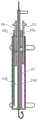
图1为本申请的食管热损失防护装置的第一实施例的主视结构示意图;Fig. 1 is a front structural schematic view of the first embodiment of the esophageal heat loss protection device of the present application;
图2为图1的食管热损失防护装置的剖面结构示意图;Fig. 2 is a schematic cross-sectional structure diagram of the esophagus heat loss protection device in Fig. 1;
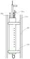
图3为本申请的食管热损失防护装置的第二实施例的主视结构示意图;Fig. 3 is a front structural schematic view of the second embodiment of the esophageal heat loss protection device of the present application;
图4为图3的食管热损失防护装置的剖面结构示意图;Fig. 4 is a schematic cross-sectional structure diagram of the esophageal heat loss protection device in Fig. 3;
图5为利用胃管将导丝放置到食管中的示意图;Fig. 5 is a schematic diagram of placing a guide wire into the esophagus using a gastric tube;
图6为导丝放置到食管中后撤出胃管的示意图;Figure 6 is a schematic diagram of withdrawing the gastric tube after the guide wire is placed in the esophagus;
图7、8为利用导丝将本申请的食管热损失防护装置放置到食管中的示意图;7 and 8 are schematic diagrams of placing the esophageal heat loss protection device of the present application into the esophagus using a guide wire;
图9为对本申请的食管热损失防护装置进行充气使其固定在食管中的示意图;Fig. 9 is a schematic diagram of inflating the esophageal heat loss protection device of the present application to fix it in the esophagus;
图10为通过负压抽吸本申请的食管热损失防护装置而使得食管的对应段坍缩以远离消融区域的示意图;Fig. 10 is a schematic diagram of the corresponding segment of the esophagus being collapsed away from the ablation area by negative pressure suction of the esophageal heat loss protection device of the present application;
图11为向食管的坍缩段喷射造影剂的示意图;Fig. 11 is a schematic diagram of injecting a contrast agent into the collapsed section of the esophagus;
图12为本申请的食管热损失防护装置导致试管内壁的偏移量示意图。Fig. 12 is a schematic diagram of the deviation of the inner wall of the test tube caused by the esophageal heat loss protection device of the present application.
具体实施方式Detailed ways
下面,结合附图对本申请的食管热损失防护装置进行详细说明。Next, the esophageal heat loss protection device of the present application will be described in detail with reference to the accompanying drawings.
图1-2为本申请的第一实施例。1-2 are the first embodiment of the present application.
图1-2的食管热损失防护装置,其包括主管体10、负压管路15。The esophageal heat loss protection device shown in FIGS. 1-2 includes a
主管体10用于放置于食管40中。The
主管体40的上部通过上端片12a封闭,下端通过下端片12b封闭,由此形成主管体内部空间14。The upper part of the
围绕主管体10的上端设置有环状的上气囊11a,围绕主管体10的下端设置有环状的下气囊11b;上气囊11a和下气囊11b用于在上部和下部支撑于食管40内;食管40内由上气囊11a、下气囊11b以及主管体10的外周面所限定的空间为食管坍缩空间。The upper end of the
主管体10位于上气囊11a和下气囊11b的偏心位置,使得上气囊11a和下气囊11b在主管体10的朝向消融术区方向上的向外延伸程度大于在主管体10的其他方向上的延伸程度,由此,在消融术区方向的空间占据食管坍缩空间的大部分空间,也就是说食管在消融术区方向所对应的部分能够有更大的坍缩可能性。The
上气囊11a连接有上管路16a,上管路16a自主管体10的内部穿过上端片12a向上延伸到外部,通过上管路16a对上气囊11a充气或放气;下气囊11b连接有下管路16b,下管路16b自主管体10的内部穿过上端片12a向上延伸到外部,通过下管路16b对下气囊11b充气或放气。上管路16a和下管路16b分别用于连接气源。The
主管体10上形成有多个通孔18;该多个通孔18沿着主管体10的长度方向设置,与主管体内部空间14连通。A plurality of through
负压管路15的上端延伸到外部,负压管路15的下端穿过上端片12a与主管体内部空间14连通;负压管路15用于对主管体内部空间14以及食管坍缩空间进行抽吸,使得食管40在食管坍缩空间形成坍缩而远离消融术区。The upper end of the
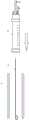
为了方便将主管体10放置到食管的预定位置,本申请的食管热损失防护装置还可以包括导丝管13。In order to facilitate placing the
导丝管13形成在主管体10内;导丝管13的上端穿过上端片12a,下端穿过下端片12b。A
导丝管13用于容纳导丝20,导丝20用于引导主管体10到达食管40的预定位置。The
主管体10的上部还包括手柄17,以通过手柄17调整主管体10在食管40中的角度和位置。The upper part of the
手柄17形成在导丝管20的上端或主管体10的上端。A
负压管路15的上端进一步形成有造影剂输入端口15b;这样负压管路15上端不但包括负压源连接端15a,还包括造影剂输入端15b。造影剂用于利用主管体内部空间14和食管坍缩空间的负压而喷射到食管40的内壁上。当注入造影剂时,其扩散至靠近消融术区的食管壁上,方便在X线下显示该侧食管壁的内部轮廓。The upper end of the
导丝20的下端形成有挡头21。挡头21为弧线状或圆球状的。A
本申请的第二实施例的结构与第一实施例的结构大部分相同,不同之处在于,在本申请的第二实施例中,如图3-4所示,主管体10内形成有隔板19,隔板19沿着主管体10的长度方向形成,将主管体内部空间14分隔为独立的第一内部空间14a和第二内部空间14b。The structure of the second embodiment of the present application is mostly the same as that of the first embodiment, the difference is that in the second embodiment of the present application, as shown in Figures 3-4, a partition is formed in the
在本申请的第二实施例中,负压管路14分为第一负压管路15和第二负压管路18;第一负压管路15与第一内部空间14a连通;第二负压管路18与第二内部空间14b连通。In the second embodiment of the present application, the
通孔18中的一部分与第一内部空间14a连通;通孔18中的一部分与第二内部空间14b连通。A part of the through
第一内部空间14a对应于消融术区方向。The first
第一负压管路15的上端进一步形成有第一造影剂输入端口,第二负压管路18的上端进一步形成有第二造影剂输入端口。这里的第一、第二造影剂输入端口与第一实施例中的造影剂输入端15b相同,只不过由于主管体内部空间14被分为第一内部空间14a和第二内部空间14b,对应于第一内部空间14a和第二内部空间14b的每个空间,分别形成负压管路。The upper end of the first
此外,不管是第一实施例还是第二实施,上气囊和下气囊上都可以设置标记物(markers),以方便利用房颤消融中所应用的影像工具(超声、X线、电磁场等)显示主管体的位置,从而能实现把食管偏移的区间与左心房消融靶区最佳的配合,最大限度的减少甚至避免消融产生的食管热损伤。例如,标记物可以是在超声、电磁场和X线下显示的铂铱合金小块。另外,还可通过压力监测手段来监测上气囊和下气囊中的压力,不管是在上气囊和下气囊中设置微型的压力传感器还是自上气囊和下气囊所连接的对应的管路上设置压力感测器件,均可以实现对上气囊和下气囊中的压力的监测。In addition, regardless of the first embodiment or the second implementation, markers (markers) can be set on the upper balloon and the lower balloon to facilitate the use of imaging tools (ultrasound, X-ray, electromagnetic field, etc.) applied in atrial fibrillation ablation to display The position of the main body can achieve the best match between the esophageal offset area and the left atrium ablation target area, and minimize or even avoid the esophageal thermal damage caused by ablation. For example, markers can be small pieces of platinum-iridium alloy that show up under ultrasound, electromagnetic fields, and x-rays. In addition, the pressure in the upper airbag and the lower airbag can also be monitored by means of pressure monitoring, no matter whether a miniature pressure sensor is set in the upper airbag and the lower airbag or a pressure sensor is installed on the corresponding pipeline connected to the upper airbag and the lower airbag. The measuring device can both realize the monitoring of the pressure in the upper air bag and the lower air bag.
下面,以图1-2中所示的第一实施例为例,说明本申请的食管热损失防护装置使用过程。Next, taking the first embodiment shown in FIGS. 1-2 as an example, the use process of the esophageal heat loss protection device of the present application will be described.
1.术前放置已置入导丝的胃管,置入深度达到食管中下段,或胃腔内,并通过常规标准方法验证胃管的位置,如图5。1. Preoperatively place the gastric tube with the guide wire inserted to the depth of the middle and lower esophagus, or into the gastric cavity, and verify the position of the gastric tube by conventional standard methods, as shown in Figure 5.
2.在拟进行左心房后壁消融前,先撤出胃管而导丝仍在食管内,如图6;。沿导丝送入主管体,如图7、8。2. Before the ablation of the posterior wall of the left atrium is planned, the gastric tube is withdrawn while the guide wire is still in the esophagus, as shown in Figure 6;. Send it into the main body along the guide wire, as shown in Figures 7 and 8.
3.通过操控手柄将主管体置于食管适当的位置,对上、下气囊充气,上、下气囊紧贴食管内壁,由此,通过上、下气囊以及主管体,在食管内形成密闭的空间,即食管坍缩空间,如图9;3. Place the main body in the proper position of the esophagus through the control handle, inflate the upper and lower air bags, and the upper and lower air bags are close to the inner wall of the esophagus, thus forming a closed space in the esophagus through the upper, lower air bags and the main body , that is, the collapsed space of the esophagus, as shown in Figure 9;
4.停止气囊充气后,通过负压源与负压管路连接二对主管体内部空间和食管坍缩空间进行抽吸,食管壁向食管坍缩空间进行不同程度的凹陷,如图10所示;由于上、下气囊相对于主管体的偏心设置,对应于消融术区的一侧食管壁凹陷更为明显,使之显著远离形成负压前的位置。4. After the inflation of the balloon is stopped, the inner space of the main body and the collapsed space of the esophagus are sucked through the negative pressure source and the negative pressure pipeline, and the esophageal wall is sunken to different degrees in the collapsed space of the esophagus, as shown in Figure 10; Due to the eccentric setting of the upper and lower balloons relative to the main body, the esophageal wall depression on the side corresponding to the ablation area is more obvious, making it far away from the position before negative pressure is formed.
5.通过房颤消融中所应用的影像工具(超声、X线、电磁场等),结合上、下气囊上的标记物,可将主管体定位在合适的食道节段,并且在消融术中根据所要消融的左心房后壁的具体位置进行动态调整,以保证在消融部位,使食管产生最大食管壁位移,从而最大程度减轻、甚至避免食管热损伤。5. Through the imaging tools (ultrasound, X-ray, electromagnetic field, etc.) used in atrial fibrillation ablation, combined with the markers on the upper and lower balloons, the main body can be positioned at the appropriate esophageal segment, and the ablation can be performed according to the The specific position of the posterior wall of the left atrium to be ablated is dynamically adjusted to ensure maximum esophageal wall displacement at the ablation site, thereby minimizing or even avoiding thermal damage to the esophagus.
本申请的食管热损失防护装置导致的食管壁位移程度足够用于避免热损伤。The degree of displacement of the esophageal wall caused by the esophageal heat loss protection device of the present application is sufficient to avoid thermal injury.
在食管壁显著位移的一侧,位移的食管壁、主管体及上、下气囊构成的空间近似于彼此相对的两个直角三角形,如图12,其中,直角边a相当于食管壁的显著位移,直角边b的两倍为上、下气囊间的距离。对于产生同样的位移a,如果直角边b更长,则意味着利用了更长一段的食管壁在负压下产生相同的位移,这样,需要的负压和食管壁的局部张力将更小(可以用斜角边c与直角边b形成的锐角θ大小来衡量,此角度越小,位移时食管壁的局部张力越小),从而给患者带来的不适也会更小。既往文献表明,食管壁产生2cm的位移,就足以避开热损伤,根据公式两直角边的平方和等于斜边的平方,即a2+b2=c2,当θ=30°,a=2cm时,b=3.464cm,上、下端气囊间的距离2b即为6.928cm。而上、下端气囊距离更远时,产生2cm位移时,θ<30°,这意味着产生更小的食管局部张力。On the side where the esophageal wall is significantly displaced, the space formed by the displaced esophageal wall, the main body, and the upper and lower balloons is similar to two right-angled triangles facing each other, as shown in Figure 12, where the right-angled side a is equivalent to the esophageal wall The significant displacement of the right-angled side b is twice the distance between the upper and lower airbags. For the same displacement a, if the right-angle side b is longer, it means that a longer section of the esophageal wall is used to produce the same displacement under negative pressure, so that the required negative pressure and local tension of the esophageal wall will be smaller (It can be measured by the acute angle θ formed by the oblique side c and the right-angled side b, the smaller the angle, the smaller the local tension of the esophageal wall during displacement), so that the patient will suffer less discomfort. Previous literature has shown that a 2cm displacement of the esophageal wall is enough to avoid thermal injury. According to the formula, the sum of the squares of the two right-angled sides is equal to the square of the hypotenuse, that is, a2 +b2 =c2 , when θ=30°, a When =2cm, b=3.464cm, the distance 2b between the upper and lower airbags is 6.928cm. However, when the distance between the upper and lower balloons is farther, θ < 30° when a displacement of 2 cm occurs, which means that a smaller local tension of the esophagus is generated.
既往文献报道,食管可移动的距离为4cm左右,其他产品通过局部机械性的推动产生食管位移,食管受力局部的组织可能产生损伤。According to previous literature reports, the movable distance of the esophagus is about 4cm, and other products produce esophageal displacement through local mechanical push, and the local tissue of the esophagus may be damaged due to force.
更为重要的是,用机械推动时,必然是推动远离消融侧的食管壁,而使邻近消融部位的食管壁(是发生热损伤的主要部位)被动性牵引,由于食管的可延展性,这种对侧机械牵引的效果受限,要使邻近消融处的食管壁产生足够的位移,是必在对侧施加更大的作用力,这无疑会增加食管组织损伤的可能性。More importantly, when mechanically propelled, the esophageal wall away from the ablation side must be pushed, and the esophageal wall adjacent to the ablation site (which is the main site of thermal injury) is passively pulled. Due to the extensibility of the esophagus , the contralateral mechanical traction effect is limited, so that sufficient displacement of the esophageal wall adjacent to the ablation site must be exerted on the opposite side, which will undoubtedly increase the possibility of esophageal tissue damage.
而本申请中,1.首先通过操控手柄,将装置置于消融部位的对侧,通过负压使消融侧食管壁自然产生向远离消融侧的位移,由于负压的特点,整个位移食管段的受力相对均匀,损伤小,2.且能使邻近消融的食管壁产生最大位移,而对侧食管壁几乎不产生位移。3.因负压导致的食管壁位移不同于机械推动产生的位移。机械推动是在一个水平面上的作用,而负压则是使上、下气囊间的整个食管段向内部收缩,能使这一食管节段整体远离消融热损伤源关,所以效果更好,如图12所示。In this application, 1. First, place the device on the opposite side of the ablation site by manipulating the handle, and the negative pressure causes the esophageal wall on the ablation side to naturally move away from the ablation side. Due to the characteristics of negative pressure, the entire displacement of the esophagus 2. It can cause the largest displacement of the esophageal wall adjacent to the ablation, and almost no displacement of the contralateral esophageal wall. 3. The displacement of the esophageal wall caused by negative pressure is different from the displacement caused by mechanical pushing. The mechanical push works on a horizontal plane, while the negative pressure makes the entire esophageal segment between the upper and lower balloons shrink inward, which can keep this esophageal segment away from the source of ablation heat damage as a whole, so the effect is better, such as Figure 12 shows.
除非另有定义,本申请中使用的所有技术和/或科学术语具有与由本发明所涉及的领域的普通技术人员通常理解的相同含义。本申请中提到的材料、方法和实施例仅为说明性的,而非限制性的。Unless otherwise defined, all technical and/or scientific terms used in this application have the same meaning as commonly understood by one of ordinary skill in the art to which this invention pertains. The materials, methods, and examples mentioned in this application are illustrative only and not limiting.
虽然已结合具体实施方式对本发明进行了描述,在本申请的发明主旨下,本领域的技术人员可以进行适当的替换、修改和变化,这种替换、修改和变化仍属于本申请的保护范围。Although the present invention has been described in conjunction with specific embodiments, those skilled in the art can make appropriate replacements, modifications and changes under the inventive spirit of the application, and such replacements, modifications and changes still belong to the protection scope of the application.
Claims (10)
Priority Applications (1)
| Application Number | Priority Date | Filing Date | Title |
|---|---|---|---|
| CN202310242056.7ACN116196082B (en) | 2023-03-14 | 2023-03-14 | Esophageal heat injury protection device |
Applications Claiming Priority (1)
| Application Number | Priority Date | Filing Date | Title |
|---|---|---|---|
| CN202310242056.7ACN116196082B (en) | 2023-03-14 | 2023-03-14 | Esophageal heat injury protection device |
Publications (2)
| Publication Number | Publication Date |
|---|---|
| CN116196082Atrue CN116196082A (en) | 2023-06-02 |
| CN116196082B CN116196082B (en) | 2025-03-11 |
Family
ID=86511143
Family Applications (1)
| Application Number | Title | Priority Date | Filing Date |
|---|---|---|---|
| CN202310242056.7AActiveCN116196082B (en) | 2023-03-14 | 2023-03-14 | Esophageal heat injury protection device |
Country Status (1)
| Country | Link |
|---|---|
| CN (1) | CN116196082B (en) |
Citations (12)
| Publication number | Priority date | Publication date | Assignee | Title |
|---|---|---|---|---|
| CN86100323A (en)* | 1986-02-16 | 1987-06-24 | 李亚林 | The double balloon catheter of treatment ureteral calculus |
| CN2257197Y (en)* | 1996-06-04 | 1997-07-02 | 河南石油勘探局职工医院 | Multipurpose nasal tube, gastro intestinal canal for upper digestive tract |
| US20130006139A1 (en)* | 2011-07-01 | 2013-01-03 | Joseph Tiano | Medical probe and method of using same |
| US20180042691A1 (en)* | 2015-03-05 | 2018-02-15 | Medfact Engineering Gmbh | Device for the Displacement of a Hollow Organ of a Patient |
| WO2018065047A1 (en)* | 2016-10-05 | 2018-04-12 | Dr. Philippe Debruyne Bvba | Method for protecting a patient's esophagus while ablating tissue of the patient's heart and intra-esophageal system therefor |
| CN108430301A (en)* | 2015-09-17 | 2018-08-21 | 贝勒医学院 | Esophageal Probes and Methods |
| US20180311497A1 (en)* | 2017-04-28 | 2018-11-01 | Iowa Approach, Inc. | Systems, devices, and methods for delivery of pulsed electric field ablative energy to esophageal tissue |
| WO2020068601A1 (en)* | 2018-09-24 | 2020-04-02 | Cottone Robert J | Systems and methods for tissue displacement |
| US20210030458A1 (en)* | 2018-01-23 | 2021-02-04 | E-Safe Medical, Inc. | Esophageal probes and methods |
| CN215653319U (en)* | 2020-11-19 | 2022-01-28 | 傅蓉 | Anti-backflow mis-suction channel plugging device |
| CN217548579U (en)* | 2022-01-20 | 2022-10-11 | 北京大学深圳医院 | Stomach tube guiding device |
| CN117531100A (en)* | 2023-12-06 | 2024-02-09 | 邓科委 | A disposable H-type tracheo-esophageal fistula angiography catheter |
- 2023
- 2023-03-14CNCN202310242056.7Apatent/CN116196082B/enactiveActive
Patent Citations (12)
| Publication number | Priority date | Publication date | Assignee | Title |
|---|---|---|---|---|
| CN86100323A (en)* | 1986-02-16 | 1987-06-24 | 李亚林 | The double balloon catheter of treatment ureteral calculus |
| CN2257197Y (en)* | 1996-06-04 | 1997-07-02 | 河南石油勘探局职工医院 | Multipurpose nasal tube, gastro intestinal canal for upper digestive tract |
| US20130006139A1 (en)* | 2011-07-01 | 2013-01-03 | Joseph Tiano | Medical probe and method of using same |
| US20180042691A1 (en)* | 2015-03-05 | 2018-02-15 | Medfact Engineering Gmbh | Device for the Displacement of a Hollow Organ of a Patient |
| CN108430301A (en)* | 2015-09-17 | 2018-08-21 | 贝勒医学院 | Esophageal Probes and Methods |
| WO2018065047A1 (en)* | 2016-10-05 | 2018-04-12 | Dr. Philippe Debruyne Bvba | Method for protecting a patient's esophagus while ablating tissue of the patient's heart and intra-esophageal system therefor |
| US20180311497A1 (en)* | 2017-04-28 | 2018-11-01 | Iowa Approach, Inc. | Systems, devices, and methods for delivery of pulsed electric field ablative energy to esophageal tissue |
| US20210030458A1 (en)* | 2018-01-23 | 2021-02-04 | E-Safe Medical, Inc. | Esophageal probes and methods |
| WO2020068601A1 (en)* | 2018-09-24 | 2020-04-02 | Cottone Robert J | Systems and methods for tissue displacement |
| CN215653319U (en)* | 2020-11-19 | 2022-01-28 | 傅蓉 | Anti-backflow mis-suction channel plugging device |
| CN217548579U (en)* | 2022-01-20 | 2022-10-11 | 北京大学深圳医院 | Stomach tube guiding device |
| CN117531100A (en)* | 2023-12-06 | 2024-02-09 | 邓科委 | A disposable H-type tracheo-esophageal fistula angiography catheter |
Also Published As
| Publication number | Publication date |
|---|---|
| CN116196082B (en) | 2025-03-11 |
Similar Documents
| Publication | Publication Date | Title |
|---|---|---|
| US20230380896A1 (en) | Methods and devices for endovascular ablation of a splanchnic nerve | |
| US10543033B2 (en) | Systems and methods for cryoablation of a tissue | |
| JP2022511318A (en) | Heated steam ablation system and method for treating heart disease | |
| EP1420702B1 (en) | Percutaneous pringle occlusion device | |
| JP2024057070A (en) | Directional baloon transseptal insertion device for medical treatment | |
| US20140058294A1 (en) | Tissue treatment and monitoring by application of energy | |
| US20080312643A1 (en) | Tissue ablation system including guidewire with sensing element | |
| US8506589B2 (en) | Nasogastric tube for use during an ablation procedure | |
| JP6905595B2 (en) | Vascular isolation ablation device | |
| JP2002535033A (en) | Equipment for treatment of atrial arrhythmias | |
| CN111989059B (en) | Soft balloon device and system | |
| JPS60227774A (en) | Heart muscle treating method and inverse injection catheter of physiological agent | |
| CN211409192U (en) | Dilator and interatrial puncture system | |
| CN109223169A (en) | Pulmonary vein is electrically isolated balloon structure | |
| WO2019072047A1 (en) | Traction device for curved balloon catheter and traction method thereof | |
| WO2021031616A1 (en) | Triple micro-catheter device and method for transthoracic and epicardial intramyocardial injection under ultrasound guidance | |
| CN205458830U (en) | A Surgical Instrument Delivery System for Transthoracic Minimally Invasive Treatment of Structural Heart Disease | |
| CN116196082A (en) | Esophageal heat loss protector | |
| CN112754600A (en) | Split type sawtooth-shaped thrombus breaking balloon catheter | |
| CN219185515U (en) | Cardiac Vein Chemical Ablation Kit | |
| CN214965572U (en) | Ultrasonic-guided special atrial septal defect diameter measurement balloon for surgery | |
| CN114869431A (en) | Interatrial septum puncture system | |
| CN214260328U (en) | Balloon catheter for cardiac interventional therapy and dilation | |
| CN115024815A (en) | Cardiac muscle ablation device | |
| CN215228130U (en) | Split type sawtooth-shaped thrombus breaking balloon catheter |
Legal Events
| Date | Code | Title | Description |
|---|---|---|---|
| PB01 | Publication | ||
| PB01 | Publication | ||
| SE01 | Entry into force of request for substantive examination | ||
| SE01 | Entry into force of request for substantive examination | ||
| GR01 | Patent grant | ||
| GR01 | Patent grant |