Movatterモバイル変換


[0]ホーム

URL:


CN116115256A - Method and system for dynamically adjusting imaging parameters during an ultrasound scan - Google Patents

Method and system for dynamically adjusting imaging parameters during an ultrasound scan
Download PDF

Info

Publication number
CN116115256A
CN116115256ACN202211392612.0ACN202211392612ACN116115256ACN 116115256 ACN116115256 ACN 116115256ACN 202211392612 ACN202211392612 ACN 202211392612ACN 116115256 ACN116115256 ACN 116115256A
Authority
CN
China
Prior art keywords
image quality
imaging parameters
test
frame
ultrasound
Prior art date
Legal status (The legal status is an assumption and is not a legal conclusion. Google has not performed a legal analysis and makes no representation as to the accuracy of the status listed.)
Granted
Application number
CN202211392612.0A
Other languages
Chinese (zh)
Other versions
CN116115256B (en
Inventor
辛西娅·A·欧文
梅纳凯姆·哈尔曼
K·克里斯托弗森
A·索库林
Current Assignee (The listed assignees may be inaccurate. Google has not performed a legal analysis and makes no representation or warranty as to the accuracy of the list.)
GE Precision Healthcare LLC
Original Assignee
GE Precision Healthcare LLC
Priority date (The priority date is an assumption and is not a legal conclusion. Google has not performed a legal analysis and makes no representation as to the accuracy of the date listed.)
Filing date
Publication date
Application filed by GE Precision Healthcare LLCfiledCriticalGE Precision Healthcare LLC
Publication of CN116115256ApublicationCriticalpatent/CN116115256A/en
Application grantedgrantedCritical
Publication of CN116115256BpublicationCriticalpatent/CN116115256B/en
Activelegal-statusCriticalCurrent
Anticipated expirationlegal-statusCritical

Links

Images

Classifications

Landscapes

Abstract

A system and method for dynamically adjusting imaging parameters during ultrasound scanning is provided. The method includes acquiring a conventional ultrasound image frame based on a first set of imaging parameters and displaying the conventional frame at a display system. The method includes intermittently acquiring test ultrasound image frames based on a second set of imaging parameters having at least one imaging parameter different from the first set of imaging parameters. The test frame is not displayed. The method includes determining a normal frame image quality of at least one frame of the normal frames and a test frame image quality of the test frames. The method includes applying a second set of imaging parameters to capture an additional conventional frame and displaying the additional conventional frame at the display system in response to determining that the test frame image quality exceeds the conventional frame image quality.

Description

Translated fromChinese
用于在超声扫描期间动态调整成像参数的方法和系统Method and system for dynamically adjusting imaging parameters during an ultrasound scan

技术领域technical field

某些实施方案涉及超声成像。更具体地,特定实施方案涉及一种用于在超声扫描期间动态调整成像参数的方法和系统,其通过间歇性地采集测试超声帧以及将该测试超声帧的图像质量与所采集的常规超声帧的图像质量进行比较以确定最佳图像参数。Certain embodiments relate to ultrasound imaging. More specifically, certain embodiments relate to a method and system for dynamically adjusting imaging parameters during an ultrasound scan by intermittently acquiring a test ultrasound frame and comparing the image quality of the test ultrasound frame to the acquired conventional ultrasound frame The image quality is compared to determine the best image parameters.

背景技术Background technique

超声成像是用于对人体中的器官和软组织进行成像的医学成像技术。超声成像使用实时的、非侵入性高频声波来产生一系列二维(2D)图像和/或三维(3D)图像。Ultrasound imaging is a medical imaging technique used to image organs and soft tissues in the human body. Ultrasound imaging uses real-time, non-invasive, high-frequency sound waves to produce a series of two-dimensional (2D) images and/or three-dimensional (3D) images.

在超声成像程序期间,由于存在流体类型的病理状态(诸如腹水、血液、脑脊液等),成像组织可能具有出乎意料的特征。与组织相比,流体类型的不同声学特性可导致出现成像伪影以及降低声束衰减。在一些情况下,流体可使腹腔或其它解剖结构膨胀,以及增加扫描距离。例如,在孕期(即,羊水过多)中带有大量腹水或过多羊膜流体的患者的腹腔可能由于流体而变大;然而,与固体组织相比,声衰减要小得多。在此类情况下,无需降低频率,这会降低空间分辨率;然而,可以执行其它成像参数调整以提高成像质量并减少伪影的存在(诸如对扫描序列、脉冲重复频率(PRF)、聚焦位置、散斑抑制、时间增益补偿(TGC)和/或任何合适的成像参数进行成像参数调整)。由流体(诸如声衰变、多路径伪影、回响伪影和后声增强)引起的超声图像伪影可能降低图像质量并且模糊解剖结构的可视化。During an ultrasound imaging procedure, the imaged tissue may have unexpected characteristics due to the presence of fluid-type pathology (such as ascites, blood, cerebrospinal fluid, etc.). Different acoustic properties of fluid types compared to tissue can lead to imaging artifacts and reduced beam attenuation. In some cases, the fluid may expand the abdominal cavity or other anatomical structures, as well as increase the scan distance. For example, in a patient with large amounts of ascites or excess amniotic fluid during pregnancy (ie, polyhydramnios), the peritoneal cavity may become enlarged due to fluid; however, the acoustic attenuation is much less than that of solid tissue. In such cases, there is no need to reduce frequency, which would reduce spatial resolution; however, other imaging parameter adjustments can be performed to improve image quality and reduce the presence of artifacts (such as adjustments to scan sequence, pulse repetition frequency (PRF), focus position , speckle suppression, time gain compensation (TGC) and/or any suitable imaging parameter for imaging parameter adjustment). Ultrasound image artifacts caused by fluids, such as acoustic decay, multipath artifacts, reverberation artifacts, and postacoustic enhancement, can degrade image quality and obscure visualization of anatomy.

通过将此类系统与本申请的其余部分中参考附图阐述的本公开的一些方面进行比较,常规和传统方法的更多限制和缺点对本领域的技术人员将变得显而易见。Further limitations and disadvantages of conventional and traditional approaches will become apparent to those skilled in the art by comparing such systems with some aspects of the disclosure set forth in the remainder of this application with reference to the accompanying figures.

发明内容Contents of the invention

提供了一种用于在超声扫描期间动态调整成像参数的系统和/或方法,该系统和/或方法基本上如结合至少一个附图所示和/或所述,如在权利要求书中更完整地阐述。There is provided a system and/or method for dynamically adjusting imaging parameters during an ultrasound scan substantially as shown and/or described in connection with at least one of the accompanying drawings, as further described in the claims fully elaborated.

从以下描述和附图将更全面地理解本公开的这些和其他优点、方面和新颖特征、以及其例示的实施方案的细节。These and other advantages, aspects and novel features of the present disclosure, together with details of illustrated embodiments thereof, will be more fully understood from the following description and accompanying drawings.

附图说明Description of drawings

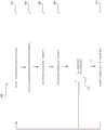
图1是根据各种实施方案的可操作为在超声扫描期间动态调整成像参数的示例性超声系统的框图。1 is a block diagram of an exemplary ultrasound system operable to dynamically adjust imaging parameters during an ultrasound scan, according to various embodiments.

图2是示出根据各种实施方案的可用于在超声扫描期间动态调整成像参数的示例性步骤的流程图。2 is a flowchart illustrating exemplary steps that may be used to dynamically adjust imaging parameters during an ultrasound scan, according to various embodiments.

具体实施方式Detailed ways

特定实施方案可存在于用于在超声扫描期间动态调整成像参数的方法和系统中。本公开的各方面具有基于第一组成像参数采集常规超声图像帧以及基于第二组成像参数间歇性地采集测试超声图像帧的技术效应。各种实施方案具有确定常规超声图像帧的图像质量以及确定间歇性地采集的测试超声图像帧的图像质量的技术效应。特定实施方案具有当测试超声图像帧的图像质量优于常规超声图像帧的图像质量时动态切换到第二组成像参数的技术效应。本公开的各方面具有在显示系统处呈现关于得到改善的图像参数调整的反馈的技术效应。Particular implementations may reside in methods and systems for dynamically adjusting imaging parameters during an ultrasound scan. Aspects of the present disclosure have the technical effect of acquiring regular ultrasound image frames based on a first set of imaging parameters and intermittently acquiring test ultrasound image frames based on a second set of imaging parameters. Various embodiments have the technical effect of determining the image quality of regular ultrasound image frames as well as determining the image quality of intermittently acquired test ultrasound image frames. Certain embodiments have the technical effect of dynamically switching to the second set of imaging parameters when the image quality of the test ultrasound image frame is better than the image quality of the regular ultrasound image frame. Aspects of the present disclosure have the technical effect of presenting feedback on improved image parameter adjustments at the display system.

当结合附图阅读时,将更好地理解前述发明内容以及以下对某些实施方案的详细描述。就附图示出各种实施方案的功能块的图的范围而言,这些功能块不一定表示硬件电路之间的划分。因此,例如,一个或多个功能块(例如,处理器或存储器)可以在单件硬件(例如,通用信号处理器或随机存取存储器块、硬盘等)或多件硬件中来实现。类似地,程序可以是独立程序,可以作为子例程包含在操作系统中,可以是安装的软件包中的功能等。应当理解,各种实施方案不限于附图中所示的布置和工具。还应当理解,可以组合实施方案,或者可以利用其他实施方案,并且可以在不脱离各种实施方案的范围的情况下做出结构的、逻辑的和电气的改变。因此,以下详述不应视为限制性意义,并且本公开的范围由所附权利要求书及其等同物限定。The foregoing summary, as well as the following detailed description of certain embodiments, are better understood when read in conjunction with the accompanying drawings. To the extent that the figures illustrate the functional blocks of various embodiments, these functional blocks are not necessarily indicative of the division between hardware circuitry. Thus, for example, one or more functional blocks (eg, a processor or memory) may be implemented in a single piece of hardware (eg, a general-purpose signal processor or random access memory block, hard disk, etc.) or in multiple pieces of hardware. Similarly, a program can be a stand-alone program, included as a subroutine in an operating system, a function in an installed software package, etc. It should be understood that the various embodiments are not limited to the arrangements and instrumentalities shown in the drawings. It is also to be understood that the embodiments may be combined or other embodiments may be utilized and structural, logical, and electrical changes may be made without departing from the scope of the various embodiments. Accordingly, the following detailed description should not be taken in a limiting sense, and the scope of the present disclosure is defined by the appended claims and their equivalents.

如本文所用,以单数形式列举并且以字词“一”或“一个”开头的元件或步骤应当被理解为不排除多个所述元件或步骤,除非明确说明此类排除。此外,对“示例性实施方案”、“各种实施方案”、“某些实施方案”、“代表性的实施方案”等的引用不旨在被解释为排除存在也结合了叙述的特征的附加实施方案。此外,除非明确地相反说明,否则“包含”、“包括”或“具有”具有特定属性的一个元素或多个元素的实施方案可以包括不具有该属性的附加元素。As used herein, an element or step recited in the singular and proceeded with the word "a" or "an" should be understood as not excluding plural of said elements or steps, unless such exclusion is explicitly stated. Furthermore, references to "exemplary embodiments," "various embodiments," "certain embodiments," "representative embodiments," etc. are not intended to be interpreted as excluding the existence of additional embodiments that also incorporate the recited features. implementation plan. Furthermore, an embodiment that "comprises", "comprises" or "has" an element or elements having a particular attribute may include additional elements not having that attribute unless expressly stated to the contrary.

另外,如本文所用,术语“图像”广义地是指可视图像和表示可视图像的数据两者。然而,许多实施方案生成(或被配置为生成)至少一个可视图像。此外,如本文所用,短语“图像”用于指超声模式,诸如B模式(2D模式)、M模式、三维(3D)模式、CF模式、PW多普勒、CW多普勒、对比增强超声(CEUS),和/或B模式和/或CF的子模式,诸如谐波成像、剪切波弹性成像(SWEI)、应变弹性成像、TVI、PDI、B-flow、MVI、UGAP,并且在某些情况下还包括MM、CM、TVD,其中“图像”和/或“平面”包括单个波束或多个波束。Additionally, as used herein, the term "image" broadly refers to both a viewable image and data representing a viewable image. However, many embodiments generate (or are configured to generate) at least one viewable image. Also, as used herein, the phrase "image" is used to refer to ultrasound modalities, such as B-mode (2D mode), M-mode, three-dimensional (3D) mode, CF mode, PW Doppler, CW Doppler, contrast-enhanced ultrasound ( CEUS), and/or B-mode and/or sub-modalities of CF such as harmonic imaging, shear wave elastography (SWEI), strain elastography, TVI, PDI, B-flow, MVI, UGAP, and in some Cases also include MM, CM, TVD, where an "image" and/or "plane" comprises a single beam or multiple beams.

此外,如本文所用,术语处理器或处理单元是指可执行各种实施方案需要的所需计算的任何类型的处理单元,诸如单核或多核:CPU、加速处理单元(APU)、图形处理单元(GPU)、DSP、FPGA、ASIC或它们的组合。Furthermore, as used herein, the term processor or processing unit refers to any type of processing unit, such as single or multi-core, that can perform the required computations required by various implementations: CPU, Accelerated Processing Unit (APU), Graphics Processing Unit (GPU), DSP, FPGA, ASIC or their combination.

应当指出的是,本文所述的生成或形成图像的各种实施方案可包括用于形成图像的处理,该处理在一些实施方案中包括波束形成,而在其他实施方案中不包括波束形成。例如,可在不进行波束形成的情况下形成图像,诸如通过将解调数据的矩阵乘以系数矩阵,使得乘积是图像,并且其中该过程不形成任何“波束”。另外,可使用可能源自多于一个发射事件的信道组合(例如,合成孔径技术)来执行图像的形成。It should be noted that the various embodiments described herein for generating or forming an image may include a process for forming the image that in some embodiments includes beamforming and in other embodiments does not include beamforming. For example, an image may be formed without beamforming, such as by multiplying a matrix of demodulated data by a matrix of coefficients, such that the product is an image, and wherein the process does not form any "beams". Additionally, image formation may be performed using channel combining (eg, synthetic aperture techniques), possibly resulting from more than one transmission event.

在各种实施方案中,例如,在软件、固件、硬件或它们的组合中执行超声处理以形成图像,包括超声波束形成,诸如接收波束形成。具有根据各种实施方案形成的软件波束形成器架构的超声系统的一个具体实施在图1中示出。In various embodiments, ultrasound processing to form images, including ultrasound beamforming, such as receive beamforming, is performed, for example, in software, firmware, hardware, or a combination thereof. One implementation of an ultrasound system with a software beamformer architecture formed according to various embodiments is shown in FIG. 1 .

图1是根据各种实施方案的可操作为在超声扫描期间动态调整成像参数的示例性超声系统100的框图。参考图1,示出了超声系统100和训练系统200。超声系统100包括发射器102、超声探头104、发射波束形成器110、接收器118、接收波束形成器120、A/D转换器122、RF处理器124、RF/IQ缓冲器126、用户输入设备130、信号处理器132、图像缓冲器136、显示系统134和档案138。1 is a block diagram of anexemplary ultrasound system 100 operable to dynamically adjust imaging parameters during an ultrasound scan, according to various embodiments. Referring to FIG. 1 , anultrasound system 100 and atraining system 200 are shown.Ultrasound system 100 includes transmitter 102,ultrasound probe 104,transmit beamformer 110,receiver 118, receivebeamformer 120, A/D converter 122,RF processor 124, RF/IQ buffer 126,user input device 130 ,signal processor 132 , image buffer 136 ,display system 134 andarchive 138 .

发射器102可包括合适的逻辑、电路、接口和/或代码,该逻辑、电路、接口和/或代码可操作为驱动超声探头104。超声探头104可包括压电元件的二维(2D)阵列。超声探头104可包括通常构成相同元件的一组发射换能器元件106和一组接收换能器元件108。在特定实施方案中,超声探头104可操作以采集覆盖解剖结构,诸如心脏、血管、胎儿或任何合适的解剖结构的至少大部分的超声图像数据。Transmitter 102 may include suitable logic, circuitry, interfaces, and/or code operable to driveultrasound probe 104 . Theultrasound probe 104 may include a two-dimensional (2D) array of piezoelectric elements. Theultrasound probe 104 may include a set oftransmit transducer elements 106 and a set of receivetransducer elements 108 that generally constitute identical elements. In particular embodiments, theultrasound probe 104 is operable to acquire ultrasound image data covering at least a substantial portion of an anatomical structure, such as a heart, blood vessel, fetus, or any suitable anatomical structure.

发射波束形成器110可包括合适的逻辑、电路、接口和/或代码,该逻辑、电路、接口和/或代码可操作为控制发射器102,该发射器通过发射子孔径波束形成器114驱动该组发射换能器元件106以将超声发射信号发到感兴趣区域(例如,人类、动物、地下空腔、物理结构等)中。发射的超声信号可从感兴趣对象中的结构(如血细胞或组织)反向散射,以产生回波。回波由接收换能器元件108接收。Thetransmit beamformer 110 may include suitable logic, circuitry, interfaces, and/or code operable to control the transmitter 102 that drives the beamformer through the transmit sub-aperture beamformer 114. The set oftransmit transducer elements 106 transmits ultrasonic transmit signals into an area of interest (eg, a human, an animal, a subterranean cavity, a physical structure, etc.). The transmitted ultrasound signal can be backscattered from structures in the object of interest, such as blood cells or tissue, to generate echoes. The echoes are received by thereceiving transducer element 108 .

超声探头104中的该组接收换能器元件108可操作为将所接收的回波转换为模拟信号,通过接收子孔径波束形成器116进行子孔径波束形成,并且然后传送到接收器118。接收器118可包括合适的逻辑、电路、接口和/或代码,该逻辑、电路、接口和/或代码可操作为接收来自接收子孔径波束形成器116的信号。可将模拟信号传送至多个A/D转换器122中的一个或多个A/D转换器。The set of receivetransducer elements 108 in theultrasound probe 104 is operable to convert received echoes to analog signals, sub-aperture beamforming by receivesub-aperture beamformer 116 , and then transmitted toreceiver 118 .Receiver 118 may include suitable logic, circuitry, interfaces, and/or code operable to receive signals from receivesub-aperture beamformer 116 . The analog signal may be sent to one or more A/D converters of plurality of A/D converters 122 .

多个A/D转换器122可包括合适的逻辑、电路、接口和/或代码,该逻辑、电路、接口和/或代码可操作为将来自接收器118的模拟信号转换为对应的数字信号。多个A/D转换器122设置在接收器118与RF处理器124之间。尽管如此,本公开在这方面并不受限制。因此,在一些实施方案中,可将多个A/D转换器122集成在接收器118内。A/D converters 122 may include suitable logic, circuitry, interfaces, and/or code operable to convert analog signals fromreceivers 118 to corresponding digital signals. A plurality of A/D converters 122 are disposed betweenreceiver 118 andRF processor 124 . Nonetheless, the present disclosure is not limited in this respect. Thus, in some implementations, multiple A/D converters 122 may be integrated withinreceiver 118 .

RF处理器124可包括合适的逻辑、电路、接口和/或代码,该逻辑、电路、接口和/或代码可操作为解调由多个A/D转换器122输出的数字信号。根据一个实施方案,RF处理器124可包括复解调器(未示出),该复解调器可操作为解调数字信号,以形成代表对应回波信号的I/Q数据对。然后可将RF或I/Q信号数据传送到RF/IQ缓冲器126。RF/IQ缓冲器126可包括合适的逻辑、电路、接口和/或代码,该逻辑、电路、接口和/或代码可操作为提供由RF处理器124生成的RF或I/Q信号数据的临时存储。RF processor 124 may include suitable logic, circuits, interfaces, and/or code operable to demodulate digital signals output by number of A/D converters 122 . According to one embodiment,RF processor 124 may include a complex demodulator (not shown) operable to demodulate digital signals to form I/Q data pairs representative of corresponding echo signals. The RF or I/Q signal data may then be transferred to RF/IQ buffer 126 . RF/IQ buffer 126 may include suitable logic, circuitry, interfaces, and/or code operable to provide temporary storage of RF or I/Q signal data generated byRF processor 124. storage.

接收波束形成器120可包括合适的逻辑、电路、接口和/或代码,该逻辑、电路、接口和/或代码可操作为执行数字波束形成处理,以例如对经由RF/IQ缓冲器126从RF处理器124接收的延迟信道信号求和并输出波束求和信号。所得到的经处理的信息可以是从接收波束形成器120输出并且传送到信号处理器132的波束求和信号。根据一些实施方案,可将接收器118、多个A/D转换器122、RF处理器124和波束形成器120集成到单个波束形成器中,该单个波束形成器可以是数字的。在各种实施方案中,超声系统100包括多个接收波束形成器120。Receivebeamformer 120 may include suitable logic, circuitry, interfaces, and/or code operable to perform a digital beamforming process to, for example, receive data from RF signals via RF/IQ buffer 126.Processor 124 sums the received delayed channel signals and outputs a beam-summed signal. The resulting processed information may be a beam-summed signal output from receivebeamformer 120 and communicated to signalprocessor 132 . According to some embodiments,receiver 118, multiple A/D converters 122,RF processor 124, andbeamformer 120 may be integrated into a single beamformer, which may be digital. In various implementations, theultrasound system 100 includes a plurality of receivebeamformers 120 .

用户输入设备130可用于输入患者数据、常规帧图像参数、测试帧图像参数、设置,选择协议和/或模板,接受所推荐的图像参数变化等。在示例性实施方案中,用户输入设备130可操作为配置、管理和/或控制超声系统100中的一个或多个部件和/或模块的操作。就这一点而言,用户输入设备130可操作为配置、管理和/或控制发射器102、超声探头104、发射波束形成器110、接收器118、接收波束形成器120、RF处理器124、RF/IQ缓冲器126、用户输入设备130、信号处理器132、图像缓冲器136、显示系统134和/或档案138的操作。用户输入设备130可包括一个或多个按钮、一个或多个旋转编码器、触摸屏、运动跟踪、语音识别、鼠标设备、键盘、相机和/或能够接收用户指令的任何其它设备。在特定实施方案中,例如,可将用户输入设备130中的一个或多个用户输入设备集成到其他部件(诸如显示系统134或超声探头104)中。例如,用户输入设备130可包括触摸屏显示器。Theuser input device 130 may be used to enter patient data, routine frame image parameters, test frame image parameters, settings, select protocols and/or templates, accept recommended image parameter changes, and the like. In an exemplary embodiment,user input device 130 is operable to configure, manage and/or control the operation of one or more components and/or modules inultrasound system 100 . In this regard,user input device 130 is operable to configure, manage and/or control transmitter 102,ultrasound probe 104, transmitbeamformer 110,receiver 118, receivebeamformer 120,RF processor 124, RF Operation of IQ buffer 126 ,user input device 130 ,signal processor 132 , image buffer 136 ,display system 134 and/orarchive 138 .User input devices 130 may include one or more buttons, one or more rotary encoders, touch screens, motion tracking, voice recognition, mouse devices, keyboards, cameras, and/or any other device capable of receiving user commands. In certain embodiments, for example, one or more ofuser input devices 130 may be integrated into other components such asdisplay system 134 orultrasound probe 104 . For example,user input device 130 may include a touch screen display.

信号处理器132可包括合适的逻辑部件、电路、接口和/或代码,该合适的逻辑部件、电路、接口和/或代码可操作为处理超声扫描数据(例如,求和IQ信号),该超声扫描数据用于生成根据第一组成像参数所采集的常规超声图像帧,以及根据第二组成像参数所采集的测试超声图像帧。信号处理器132可被配置为生成用于进行图像质量分析以及呈现在显示系统134上的常规超声图像帧。信号处理器132以被配置为生成用于进行图像质量分析的测试超声图像帧。在各种实施方案中,可以不显示测试超声图像帧。信号处理器132可操作为根据所采集的超声扫描数据上的多个可选超声模态来执行一个或多个处理操作。在示例性实施方案中,信号处理器132可用于执行显示处理和/或控制处理等。随着接收到回波信号,可以在扫描会话期间实时处理采集的超声扫描数据。附加地或另选地,超声扫描数据可在扫描会话期间暂时存储在RF/IQ缓冲器126中并且在在线操作或离线操作中以不太实时的方式处理。在各种实施方案中,经处理的图像数据可呈现在显示系统134处和/或可存储在档案138处。档案138可以是本地档案、图片归档和通信系统(PACS)或用于存储图像和相关信息的任何合适的设备。Signal processor 132 may include suitable logic components, circuits, interfaces, and/or code operable to process ultrasound scan data (e.g., summed IQ signals), the ultrasound The scan data is used to generate conventional ultrasound image frames acquired according to the first set of imaging parameters, and test ultrasound image frames acquired according to the second set of imaging parameters.Signal processor 132 may be configured to generate conventional ultrasound image frames for image quality analysis and presentation ondisplay system 134 . Thesignal processor 132 is configured to generate test ultrasound image frames for image quality analysis. In various implementations, the test ultrasound image frames may not be displayed. Thesignal processor 132 is operable to perform one or more processing operations based on a plurality of selectable ultrasound modalities on the acquired ultrasound scan data. In an exemplary embodiment, thesignal processor 132 may be used to perform display processing and/or control processing, among others. Acquired ultrasound scan data may be processed in real-time during a scan session as echo signals are received. Additionally or alternatively, ultrasound scan data may be temporarily stored in the RF/IQ buffer 126 during a scan session and processed in a less real-time manner during online or offline operation. In various implementations, the processed image data may be presented atdisplay system 134 and/or may be stored atarchive 138 . Archive 138 may be a local archive, a picture archiving and communication system (PACS), or any suitable device for storing images and related information.

信号处理器132可以是一个或多个中央处理单元、微处理器、微控制器等。例如,信号处理器132可以是集成部件,或者可分布在各个位置。在示例性实施方案中,信号处理器132可包括常规帧处理器140、测试帧处理器150和图像质量处理器160。信号处理器132可能够从用户输入设备130和/或档案138接收输入信息、生成可由显示系统134显示的输出并且响应于来自用户输入设备130的输入信息来操纵输出等。例如,信号处理器132、常规帧处理器140、测试帧处理器150和图像质量处理器160能够执行在本文根据各种实施方案所讨论的方法和/或指令集中的任一者。Signal processor 132 may be one or more central processing units, microprocessors, microcontrollers, or the like. For example,signal processor 132 may be an integrated component, or may be distributed at various locations. In an exemplary embodiment, thesignal processor 132 may include aconventional frame processor 140 , a test frame processor 150 and animage quality processor 160 .Signal processor 132 may be capable of receiving input information fromuser input device 130 and/orprofile 138 , generating output displayable bydisplay system 134 , manipulating output in response to input information fromuser input device 130 , and the like. For example,signal processor 132,regular frame processor 140, test frame processor 150, andimage quality processor 160 are capable of performing any of the methods and/or instruction sets discussed herein according to various embodiments.

超声系统100可操作为以适于所考虑的成像情况的帧速率连续采集超声扫描数据。连续采集的超声扫描数据可包括常规超声图像帧和间歇性地采集的测试超声图像帧。例如,超声系统100可在常规超声图像帧之间周期性地采集测试超声图像帧(例如,每十帧、二十帧等)。超声系统100可交织采集完整的常规超声图像帧和完整的测试超声图像帧。附加地或另选地,超声系统100可交织进行常规帧发射和测试帧发射。典型的帧速率在20至120的范围内,但可更低或更高。所采集的常规超声图像帧可以与帧速率相同、或更慢或更快的显示速率显示在显示系统134处。在各种实施方案中,不显示所采集的测试超声图像帧。图像缓冲器136被包括以用于存储未被安排立即显示的所采集的常规超声扫描数据的处理的帧。优选地,图像缓冲器136具有足够的容量来存储至少若干分钟的常规超声图像帧。常规超声图像数帧的存储方式便于根据其采集顺序或时间进行检索。图像缓冲器136可体现为任何已知的数据存储介质。Theultrasound system 100 is operable to continuously acquire ultrasound scan data at a frame rate appropriate for the imaging situation under consideration. The continuously acquired ultrasound scan data may include regular ultrasound image frames and intermittently acquired test ultrasound image frames. For example,ultrasound system 100 may periodically acquire test ultrasound image frames (eg, every tenth frame, twenty frames, etc.) between regular ultrasound image frames. Theultrasound system 100 may interleave the acquisition of a complete conventional ultrasound image frame and a complete test ultrasound image frame. Additionally or alternatively, theultrasound system 100 may interleave regular frame transmissions and test frame transmissions. Typical frame rates are in the range of 20 to 120, but can be lower or higher. The acquired conventional ultrasound image frames may be displayed at thedisplay system 134 at the same frame rate, or at a slower or faster display rate. In various implementations, the acquired test ultrasound image frames are not displayed. An image buffer 136 is included for storing processed frames of acquired conventional ultrasound scan data that are not scheduled for immediate display. Preferably, the image buffer 136 has sufficient capacity to store at least several minutes of conventional ultrasound image frames. Conventional ultrasound images are stored in several frames for easy retrieval based on their acquisition sequence or time. Image buffer 136 may embody any known data storage medium.

信号处理器132可包括常规帧处理器140,该常规帧处理器包括合适的逻辑部件、电路、接口和/或代码,该合适的逻辑部件、电路、接口和/或代码可操作为接收和处理用于进行图像质量分析以及呈现在显示系统134上的常规超声图像帧。常规帧处理器140可被配置为施用第一组成像参数以采集常规超声图像帧。该第一组成像参数可包括脉冲重复频率(PRF)(例如,高PRF或低PRF)、谐波开启或关闭、频率(例如,高、中等或低)、孔径、聚焦位置、声学功率、散斑抑制、时间增益补偿(TGC)和/或任何合适的成像参数。如下所述,常规帧处理器140可被配置为处理用于呈现在显示系统134处以及由图像质量处理器160进行分析的常规超声图像帧。常规帧处理器140还可将常规超声图像帧存储在档案138和/或任何合适的数据存储介质处。Signal processor 132 may include aconventional frame processor 140 comprising suitable logic components, circuits, interfaces and/or code operable to receive and process Conventional ultrasound image frames for image quality analysis and presentation on thedisplay system 134 . Theconventional frame processor 140 may be configured to apply the first set of imaging parameters to acquire conventional ultrasound image frames. The first set of imaging parameters may include pulse repetition frequency (PRF) (e.g., high PRF or low PRF), harmonics on or off, frequency (e.g., high, medium, or low), aperture, focus position, acoustic power, dispersion Speckle suppression, time gain compensation (TGC) and/or any suitable imaging parameter. As described below,conventional frame processor 140 may be configured to process conventional ultrasound image frames for presentation atdisplay system 134 and analysis byimage quality processor 160 .Regular frame processor 140 may also store regular ultrasound image frames atarchive 138 and/or any suitable data storage medium.

信号处理器132可包括测试帧处理器150,该测试帧处理器包括合适的逻辑部件、电路、接口和/或代码,该合适的逻辑部件、电路、接口和/或代码可操作为接收和处理用于进行图像质量分析的测试超声图像帧。测试帧处理器150可被配置为施用第二组成像参数以采集测试超声图像帧。可间歇性地采集测试超声图像帧,诸如在已经采集了10个、20个或者任何合适数量的常规超声图像帧之后进行周期性地采集。在代表性实施方案中,不显示测试超声图像帧。被施用以采集测试超声图像帧的第二组成像参数可具有不同于第一组成像参数的至少一个成像参数,该第一组成像参数被施用以采集常规超声图像帧。例如,当第一组成像参数包括低PRF参数时,第二组成像参数可包括高PRF参数。又如,当第一组成像参数包括谐波关闭参数时,第二组成像参数可包括谐波开启参数。又如,当第一组成像参数包括低频率参数时,第二组成像参数可包括中等或高频率参数。不同的至少一个参数值可允许图像质量处理器160确定被施用以采集测试超声图像帧的第二组成像参数是否提供比被施用以获取呈现在显示系统134处的常规超声图像帧的第一组成像参数更高的图像质量,使得图像质量处理器160可提示用户和/或自动更新第一组成像参数,如下所述。测试帧处理器150可被配置为处理测试超声图像帧以供图像质量处理器160进行分析。测试帧处理器150还可将测试超声图像帧存储在档案138和/或任何合适的数据存储介质处。Signal processor 132 may include a test frame processor 150 comprising suitable logic components, circuits, interfaces and/or code operable to receive and process Test ultrasound image frames for image quality analysis. The test frame processor 150 may be configured to apply the second set of imaging parameters to acquire the test ultrasound image frames. Test ultrasound image frames may be acquired intermittently, such as periodically after 10, 20, or any suitable number of regular ultrasound image frames have been acquired. In a representative embodiment, the test ultrasound image frames are not displayed. The second set of imaging parameters applied to acquire the test ultrasound image frames may have at least one imaging parameter different from the first set of imaging parameters applied to acquire the regular ultrasound image frames. For example, when the first set of imaging parameters includes low PRF parameters, the second set of imaging parameters may include high PRF parameters. As another example, when the first set of imaging parameters includes a harmonic off parameter, the second set of imaging parameters may include a harmonic on parameter. As another example, when the first set of imaging parameters includes low frequency parameters, the second set of imaging parameters may include medium or high frequency parameters. A different value for at least one parameter may allow theimage quality processor 160 to determine whether the second set of imaging parameters applied to acquire the test ultrasound image frame provides better imaging parameters than the first set of imaging parameters applied to acquire the conventional ultrasound image frame presented at thedisplay system 134. The higher image quality of the imaging parameters allows theimage quality processor 160 to prompt the user and/or automatically update the first set of imaging parameters, as described below. Test frame processor 150 may be configured to process test ultrasound image frames for analysis byimage quality processor 160 . Test frame processor 150 may also store the test ultrasound image frames atarchive 138 and/or any suitable data storage medium.

在各种实施方案中,测试帧处理器150可被配置为接收和处理同时采集的多个测试帧。例如,测试帧处理器150可被配置为施用第二组成像参数以间歇性地采集第一测试超声图像帧以及施用第三组成像参数以间歇性地采集第二测试超声图像帧。例如,可对第二组成像参数进行优化以改善分辨率,并且可对第三组成像参数进行优化以提高渗透。测试帧处理器150可被配置为处理第一测试超声图像帧和第二测试超声图像帧以供图像质量处理器160进行分析。测试帧处理器150还可将第一测试超声图像帧和第二测试超声图像帧存储在档案138和/或任何合适的数据存储介质处。In various embodiments, test frame processor 150 may be configured to receive and process multiple test frames acquired simultaneously. For example, the test frame processor 150 may be configured to apply a second set of imaging parameters to intermittently acquire a first test ultrasound image frame and a third set of imaging parameters to intermittently acquire a second test ultrasound image frame. For example, a second set of imaging parameters can be optimized for improved resolution and a third set of imaging parameters can be optimized for increased penetration. The test frame processor 150 may be configured to process the first test ultrasound image frame and the second test ultrasound image frame for analysis by theimage quality processor 160 . Test frame processor 150 may also store the first and second test ultrasound image frames atarchive 138 and/or any suitable data storage medium.

信号处理器132可包括图像质量处理器160,该图像质量处理器包括合适的逻辑部件、电路、接口和/或代码,该合适的逻辑部件、电路、接口和/或代码可操作为分析和确定常规超声图像帧以及至少一种类型的测试超声图像帧的图像质量。图像质量处理器160可包括图像分析算法、人工智能算法、一个或多个深度神经网络(例如,卷积神经网络)和/或可利用被配置为确定常规超声图像帧和测试超声图像帧的图像质量的任何合适形式的图像分析技术或机器学习处理功能。在各种实施方案中,图像质量处理器160可被配置为基于伪像(诸如声衰变伪影、多路径伪影、回响伪影、后声增强伪影、次级回波伪影、旁瓣、光栅瓣和/或任何合适的图像伪影)的存在或不存在来确定常规超声图像帧和测试超声图像帧的图像质量。附加地和/或另选地,图像质量处理器160可被配置为基于图像分辨量和渗透量和/或任何合适的图像质量度量来确定常规超声图像帧和测试超声图像帧的图像质量。Signal processor 132 may includeimage quality processor 160 comprising suitable logic components, circuits, interfaces and/or code operable to analyze and determine Image quality of regular ultrasound image frames and at least one type of test ultrasound image frames.Image quality processor 160 may include image analysis algorithms, artificial intelligence algorithms, one or more deep neural networks (e.g., convolutional neural networks), and/or may utilize image data configured to determine conventional ultrasound image frames and test ultrasound image frames. Any suitable form of image analysis techniques or machine learning processing capabilities of quality. In various embodiments, theimage quality processor 160 may be configured to based on artifacts such as acoustic decay artifacts, multipath artifacts, reverberation artifacts, rear acoustic enhancement artifacts, secondary echo artifacts, sidelobe , grating lobes, and/or any suitable image artifacts) to determine the image quality of the conventional ultrasound image frame and the test ultrasound image frame. Additionally and/or alternatively,image quality processor 160 may be configured to determine the image quality of the conventional ultrasound image frames and the test ultrasound image frames based on image resolution and penetration and/or any suitable image quality metric.

在特定实施方案中,可将图像质量处理器160提供作为图像分析工具和/或算法,该图像分析工具和/或算法被配置为分析柱状图变化、常规超声图像帧的变化等。附加地和/或另选地,可将图像质量处理器160提供作为深度神经网络,该深度神经网络可由例如输入层、输出层以及输入层和输出层之间的一个或多个隐藏层构成。每个层可由可称为神经元的多个处理节点构成。例如,图像质量处理器160可包括输入层,该输入层具有用于来自常规超声图像帧或测试超声图像帧的每个像素或一组像素的神经元。输出层可具有对应于图像质量评分的神经元。每个层的每个神经元可执行处理功能,并且将处理的超声图像信息传递到下游层的多个神经元中的一个神经元以用于进一步处理。例如,第一层的神经元可学习识别超声图像帧中的结构的边缘。第二层的神经元可学习以基于来自第一层的检测边缘识别形状。第三层的神经元可学习所识别的形状相对于超声图像帧中的界标的位置。由图像质量处理器160深度神经网络(例如,卷积神经网络)执行的处理可基于具有高度盖然性的图像质量度量(例如,伪影的存在、渗透量/分辨量等)来分配图像质量评分。在各种实施方案中,可为整个帧提供图像质量评分。附加地和/或另选地,帧可包括针对帧的不同部分的多个图像质量评分。例如,可将具有伪影的图像帧的部分评分为低于不具有伪影的图像帧的部分。In certain embodiments,image quality processor 160 may be provided as an image analysis tool and/or algorithm configured to analyze histogram changes, changes in conventional ultrasound image frames, and the like. Additionally and/or alternatively, theimage quality processor 160 may be provided as a deep neural network, which may consist of, for example, an input layer, an output layer and one or more hidden layers between the input layer and the output layer. Each layer can be made up of multiple processing nodes, which can be called neurons. For example,image quality processor 160 may include an input layer with a neuron for each pixel or group of pixels from a regular ultrasound image frame or a test ultrasound image frame. The output layer may have neurons corresponding to image quality scores. Each neuron of each layer may perform a processing function and pass processed ultrasound image information to one of the plurality of neurons of a downstream layer for further processing. For example, neurons in the first layer may learn to recognize the edges of structures in ultrasound image frames. The neurons of the second layer can learn to recognize shapes based on detected edges from the first layer. The neurons of the third layer may learn the positions of the recognized shapes relative to landmarks in the ultrasound image frames. Processing performed byimage quality processor 160 deep neural network (eg, convolutional neural network) may assign an image quality score based on image quality metrics with a high degree of probability (eg, presence of artifacts, amount of penetration/resolution, etc.). In various implementations, an image quality score may be provided for the entire frame. Additionally and/or alternatively, a frame may include multiple image quality scores for different portions of the frame. For example, portions of image frames with artifacts may be scored lower than portions of image frames without artifacts.

图像质量处理器160可被配置为确定与常规超声图像帧相关联的图像质量评分是否高于与测试超声图像帧相关联的图像质量评分。例如,图像质量处理器160可将测试超声图像帧的图像质量评分与紧接在测试超声图像帧之前所采集的常规超声图像帧的图像质量评分进行比较。附加地和/或另选地,图像质量处理器160可将测试超声图像帧的图像质量评分与紧接在测试超声图像帧之后所采集的常规超声图像帧的图像质量评分进行比较。又如,图像质量处理器160可将测试超声图像帧的图像质量评分与在测试超声图像帧之前所采集的常规超声图像帧的平均图像质量评分进行比较(例如,直到先前所采集的测试超声图像帧)。图像质量处理器160可被配置为在常规超声图像帧图像质量优于测试超声图像帧图像质量时保持第一组成像参数和第二组成像参数,并且在采集间歇测试超声图像帧时继续检测图像质量。图像质量处理器160可被配置为在常规超声图像帧图像质量劣于测试超声图像帧图像质量时,切换第一组成像参数和第二组成像参数并且继续基于被切换的第一组成像参数和第二组成像参数检测图像质量。例如,图像质量处理器160可确定并且分析以高PRF所采集的常规超声图像帧以及以低PRF所采集的间歇测试超声图像帧的图像质量。当图像质量处理器160检测到不存在于测试超声图像帧中的常规超声图像帧中的声学伪影时,图像质量处理器160可将常规超声图像帧采集变更为低PRF和将间歇测试超声图像帧采集变更为高PRF,从而导致与测试超声图像帧相比更低的常规超声图像帧的图像质量。当测试超声图像帧中不再存在声学伪影时,图像质量处理器160继续检测图像质量,并且可切换回高PRF的常规超声图像帧采集。Theimage quality processor 160 may be configured to determine whether the image quality score associated with the regular ultrasound image frame is higher than the image quality score associated with the test ultrasound image frame. For example, theimage quality processor 160 may compare the image quality score of the test ultrasound image frame to the image quality score of a conventional ultrasound image frame acquired immediately before the test ultrasound image frame. Additionally and/or alternatively,image quality processor 160 may compare the image quality score of the test ultrasound image frame to the image quality score of a conventional ultrasound image frame acquired immediately after the test ultrasound image frame. As another example,image quality processor 160 may compare the image quality score of the test ultrasound image frame to the average image quality score of conventional ultrasound image frames acquired prior to the test ultrasound image frame (e.g., until the test ultrasound image previously acquired frame). Theimage quality processor 160 may be configured to maintain the first set of imaging parameters and the second set of imaging parameters when the image quality of the conventional ultrasound image frame is better than the image quality of the test ultrasound image frame, and to continue to detect images while acquiring the intermittent test ultrasound image frame quality. Theimage quality processor 160 may be configured to switch between the first set of imaging parameters and the second set of imaging parameters and continue based on the switched first set of imaging parameters and A second set of imaging parameters measures image quality. For example, theimage quality processor 160 may determine and analyze the image quality of regular ultrasound image frames acquired at a high PRF and intermittent test ultrasound image frames acquired at a low PRF. When theimage quality processor 160 detects an acoustic artifact in a regular ultrasound image frame that is not present in the test ultrasound image frame, theimage quality processor 160 may alter the regular ultrasound image frame acquisition to a low PRF and the intermittent test ultrasound image The frame acquisition is changed to a high PRF, resulting in lower image quality of the conventional ultrasound image frames compared to the test ultrasound image frames. When the acoustic artifacts are no longer present in the test ultrasound image frame, theimage quality processor 160 continues to monitor image quality and may switch back to high PRF conventional ultrasound image frame acquisition.

在各种实施方案中,图像质量处理器160可在确定与常规超声图像帧相关联的图像质量评分是否高于与测试超声图像帧相关联的图像质量评分时将帧速率或任何合适的度量因素考虑在内。例如,可基于帧速率区分以其他方式被分配有相同图像质量评分的常规超声图像帧和测试超声图像帧。例如,可确定高帧速率的测试超声图像帧的图像质量超过低帧速率的常规超声图像帧的图像质量以及以其他方式的相同图像质量。In various embodiments,image quality processor 160 may factor frame rate or any suitable metric in determining whether the image quality score associated with a regular ultrasound image frame is higher than the image quality score associated with a test ultrasound image frame. within consideration. For example, regular ultrasound image frames and test ultrasound image frames that would otherwise be assigned the same image quality score may be distinguished based on frame rate. For example, it may be determined that the image quality of the test ultrasound image frames at the high frame rate exceeds the image quality of the conventional ultrasound image frames at the low frame rate and otherwise the same.

图像质量处理器160可被配置为自动切换第一成像参数组和第二成像参数组且不进行反馈,自动切换且进行反馈,或可提示用户切换第一成像参数组和第二成像参数组。例如,图像质量处理器160可响应于图像质量分析而动态切换第一成像参数组和第二成像参数组,而不通知用户该改变。又如,图像质量处理器160可响应于图像质量分析而动态切换第一成像参数组和第二成像参数组,并且可在显示系统134处呈现成像参数变更的指示。该成像参数变更的指示可由图标、文本消息或任何合适的反馈表示。例如,当第一成像参数组变更为低PRF时,图像质量处理器160可呈现说明“已施用更长的衰变时间”的文本消息。在示例性实施方案中,图像质量处理器160可响应于图像质量分析而动态切换第一成像参数组和第二成像参数组,并且可向分流屏幕显示器呈现具有新的第一组成像参数的实时常规超声图像帧以及具有旧的第一组成像参数的当前冻结的最后采集的常规超声图像帧。在各种实施方案中,图像质量处理器160可提供切换回旧的第一组成像参数的选项。在另一示例性实施方案中,图像质量处理器160可呈现推荐以切换第一组成像参数和第二组成像参数,并且提供接受或拒绝该推荐的可选选项。在各种实施方案中,第一组成像参数的自动优化可以默认为“打开”。附加地和/或另选地,自动优化可由与超声系统100交互的用户选择性地“打开”或“关闭”。Theimage quality processor 160 may be configured to automatically switch between the first imaging parameter set and the second imaging parameter set without feedback, automatically switch with feedback, or may prompt the user to switch between the first imaging parameter set and the second imaging parameter set. For example,image quality processor 160 may dynamically switch between the first imaging parameter set and the second imaging parameter set in response to image quality analysis without notifying the user of the change. As another example,image quality processor 160 may dynamically switch between the first imaging parameter set and the second imaging parameter set in response to the image quality analysis, and may present an indication of the imaging parameter change atdisplay system 134 . The indication of this imaging parameter change may be represented by an icon, a text message, or any suitable feedback. For example,image quality processor 160 may present a text message stating "Longer decay time applied" when the first imaging parameter set is changed to low PRF. In an exemplary embodiment,image quality processor 160 may dynamically switch between the first set of imaging parameters and the second set of imaging parameters in response to image quality analysis, and may present a real-time image with the new first set of imaging parameters to the split screen display. The conventional ultrasound image frame and the currently frozen last acquired conventional ultrasound image frame with the old first set of imaging parameters. In various implementations,image quality processor 160 may provide an option to switch back to the old first set of imaging parameters. In another exemplary embodiment,image quality processor 160 may present a recommendation to switch between the first set of imaging parameters and the second set of imaging parameters, and provide a selectable option to accept or reject the recommendation. In various embodiments, automatic optimization of the first set of imaging parameters can be "on" by default. Additionally and/or alternatively, automatic optimization may be selectively turned “on” or “off” by a user interacting withultrasound system 100 .

在特定实施方案中,图像质量处理器160可被配置为仅针对帧的一部分切换第一成像参数组和第二成像参数组。例如,如果测试超声图像帧的第一部分的图像质量评分高于常规超声图像帧的对应部分的图像质量评分,但测试超声图像帧的第二部分的图像质量评分低于常规超声图像帧的对应部分的图像质量评分,则图像质量处理器160可切换仅用于采集对应于测试超声图像帧的第一部分的常规超声图像帧的一部分的第一成像参数组和第二成像参数组。In particular embodiments,image quality processor 160 may be configured to switch between the first set of imaging parameters and the second set of imaging parameters for only a portion of a frame. For example, if a first portion of a test ultrasound image frame has an image quality score higher than a corresponding portion of a regular ultrasound image frame, but a second portion of the test ultrasound image frame has a lower image quality score than a corresponding portion of a regular ultrasound image frame , theimage quality processor 160 may switch between the first imaging parameter set and the second imaging parameter set for acquiring only a portion of the regular ultrasound image frame corresponding to the first portion of the test ultrasound image frame.

显示系统134可以是能够将视觉信息传送给用户的任何设备。例如,显示系统134可包括液晶显示器、发光二极管显示器、和/或任何合适的一种或多种显示器。显示系统134可操作为呈现常规超声图像帧、关于成像参数变更的反馈、将成像参数变更为推荐的成像参数的提示和/或任何合适的信息。Display system 134 may be any device capable of communicating visual information to a user. For example,display system 134 may include a liquid crystal display, a light emitting diode display, and/or any suitable display or displays. Thedisplay system 134 is operable to present regular ultrasound image frames, feedback regarding changes to imaging parameters, prompts to change imaging parameters to recommended imaging parameters, and/or any suitable information.

档案138可以是与超声系统100集成和/或(例如,通过网络)通信地耦接到超声系统100的一个或多个计算机可读存储器,诸如图片归档和通信系统(PACS)、服务器、硬盘、软盘、CD、CD-ROM、DVD、紧凑存储装置、闪存存储器、随机存取存储器、只读存储器、电可擦除和可编程只读存储器和/或任何合适的存储器。档案138可包括例如由信号处理器132访问和/或与信号处理器132结合的数据库、库、信息集或其他存储器。例如,档案138能够暂时或永久地存储数据。档案138可能能够存储医学图像数据、由信号处理器132生成的数据和/或信号处理器132可读取的指令等。例如,在各种实施方案中,档案138存储常规超声图像帧、测试超声图像帧、成像参数组、用于确定超声图像帧的图像质量的指令、用于切换成像参数组的指令和反馈指令。Archive 138 may be one or more computer-readable memories integrated withultrasound system 100 and/or communicatively coupled (e.g., via a network) toultrasound system 100, such as a picture archiving and communication system (PACS), server, hard disk, Floppy disk, CD, CD-ROM, DVD, compact storage device, flash memory, random access memory, read only memory, electrically erasable and programmable read only memory, and/or any suitable memory. Archive 138 may include, for example, a database, library, information set, or other memory that is accessed by and/or integrated withsignal processor 132 . For example, archive 138 can store data temporarily or permanently. Archive 138 may be capable of storing medical image data, data generated bysignal processor 132 and/or instructions readable bysignal processor 132, and the like. For example, in various embodiments, archive 138 stores regular ultrasound image frames, test ultrasound image frames, imaging parameter sets, instructions for determining image quality of ultrasound image frames, instructions for switching imaging parameter sets, and feedback instructions.

超声系统100的部件可在软件、硬件、固件等中实现。超声系统100的各种部件可以通信地连接。超声系统100的部件可单独实现和/或以各种形式集成。例如,显示系统134和用户输入设备130可集成为触摸屏显示器。Components ofultrasound system 100 may be implemented in software, hardware, firmware, or the like. Various components of theultrasound system 100 may be communicatively connected. The components ofultrasound system 100 may be implemented separately and/or integrated in various forms. For example,display system 134 anduser input device 130 may be integrated into a touch screen display.

仍然参见图1,训练系统200可包括训练引擎210和训练数据库220。训练引擎210可包括合适的逻辑部件、电路、接口和/或代码,该逻辑部件、电路、接口和/或代码可操作为训练由图像质量处理器160推断(即,部署)的深度神经网络(例如,人工智能模型)的神经元。例如,可训练由图像质量处理器160推断的人工智能模型以用于自动标识超声图像帧中的伪影、渗透和/或分辨率。例如,训练引擎210可使用分类超声图像帧的数据库220来训练由图像质量处理器160部署的深度神经网络。超声图像帧可包括具体解剖特征的超声图像帧,包括具体伪影类型或任何合适的超声图像帧和特征。Still referring to FIG. 1 , thetraining system 200 may include a training engine 210 and atraining database 220 . The training engine 210 may include suitable logic components, circuits, interfaces, and/or code operable to train the deep neural network ( For example, the neurons of an artificial intelligence model). For example, an artificial intelligence model inferred byimage quality processor 160 may be trained for automatically identifying artifacts, bleeding, and/or resolution in ultrasound image frames. For example, training engine 210 may usedatabase 220 of classified ultrasound image frames to train a deep neural network deployed byimage quality processor 160 . The ultrasound image frames may include ultrasound image frames of specific anatomical features, including specific artifact types, or any suitable ultrasound image frames and features.

在各种实施方案中,训练图像的数据库220可以是图片归档和通信系统(PACS)或任何合适的数据存储介质。在特定实施方案中,训练引擎210和/或训练图像数据库220可以是经由有线或无线连接通信地耦接到超声系统100的远程系统,如图1所示。附加地和/或另选地,训练系统200的部件或全部可以以各种形式与超声系统100集成。In various embodiments, thedatabase 220 of training images may be a picture archiving and communication system (PACS) or any suitable data storage medium. In particular embodiments, training engine 210 and/ortraining image database 220 may be a remote system communicatively coupled toultrasound system 100 via a wired or wireless connection, as shown in FIG. 1 . Additionally and/or alternatively, components or all oftraining system 200 may be integrated withultrasound system 100 in various forms.

图2是示出根据各种实施方案的可用于在超声扫描期间动态调整成像参数的示例性步骤302至312的流程图200。参考图2,示出了包括示例性步骤302至312的流程图300。某些实施方案可省略一个或多个步骤,和/或以与所列顺序不同的顺序执行步骤,和/或组合下文讨论的某些步骤。例如,在特定实施方案中可能不执行一些步骤。又如,某些步骤可能以与下面所列时间顺序不同的时间顺序执行,包括同时执行。2 is aflowchart 200 illustrating exemplary steps 302-312 that may be used to dynamically adjust imaging parameters during an ultrasound scan, according to various embodiments. Referring to FIG. 2 , aflowchart 300 includingexemplary steps 302 through 312 is shown. Certain embodiments may omit one or more steps, and/or perform steps in an order different from that listed, and/or combine certain steps discussed below. For example, some steps may not be performed in certain embodiments. As another example, certain steps may be performed in a different chronological order than listed below, including concurrently.

在步骤302处,超声系统100基于第一组成像参数采集常规超声图像帧。例如,超声系统100可使用定位在感兴趣的解剖结构上方的扫描位置处的超声探头104根据第一组成像参数采集常规超声图像帧。超声系统100的信号处理器132的常规帧处理器140可被配置为施用第一组成像参数以采集常规超声图像帧。该第一组成像参数可包括脉冲重复频率(PRF)(例如,高PRF或低PRF)、谐波开启或关闭、频率(例如,高、中等或低)、孔径、聚焦位置、声学功率、散斑抑制、时间增益补偿(TGC)和/或任何合适的成像参数。常规帧处理器140可被配置为处理用于在显示系统134处呈现并且进行图像质量分析的常规超声图像帧。Atstep 302, theultrasound system 100 acquires conventional ultrasound image frames based on a first set of imaging parameters. For example,ultrasound system 100 may acquire conventional ultrasound image frames according to a first set of imaging parameters usingultrasound probe 104 positioned at a scanning position over an anatomical structure of interest. Theconventional frame processor 140 of thesignal processor 132 of theultrasound system 100 may be configured to apply the first set of imaging parameters to acquire a conventional ultrasound image frame. The first set of imaging parameters may include pulse repetition frequency (PRF) (e.g., high PRF or low PRF), harmonics on or off, frequency (e.g., high, medium, or low), aperture, focus position, acoustic power, dispersion Speckle suppression, time gain compensation (TGC) and/or any suitable imaging parameter.Conventional frame processor 140 may be configured to process conventional ultrasound image frames for presentation atdisplay system 134 and image quality analysis.

在步骤304处,超声系统100基于第二组成像参数间歇性地采集测试超声图像帧。例如,超声系统100可使用定位在感兴趣的解剖结构上方的扫描位置处的超声探头104根据第二组成像参数周期性地采集测试超声图像帧。例如,超声系统100可以每十帧、二十帧或任何合适数量的帧采集测试超声图像帧。被施用以采集测试超声图像帧的第二组成像参数可具有不同于第一组成像参数的至少一个成像参数,该第一组成像参数被施用以采集常规超声图像帧。例如,当第一组成像参数包括低PRF参数时,第二组成像参数可包括高PRF参数。又如,当第一组成像参数包括谐波关闭参数时,第二组成像参数可包括谐波开启参数。又如,当第一组成像参数包括低频率参数时,第二组成像参数可包括中等或高频率参数。超声系统100的信号处理器132的测试帧处理器150可被配置为处理用于进行图像质量分析的测试超声图像帧。在各种实施方案中,不在显示系统134处显示测试超声图像帧。在特定实施方案中,超声系统100可被配置为同时采集多个测试帧。例如,超声系统100可被配置为施用第二组成像参数以间歇性地采集第一测试超声图像帧以及施用第三组成像参数以间歇性地采集第二测试超声图像帧。例如,可对第二组成像参数进行优化以改善分辨率,并且可对第三组成像参数进行优化以提高渗透。Atstep 304, theultrasound system 100 intermittently acquires test ultrasound image frames based on the second set of imaging parameters. For example, theultrasound system 100 may periodically acquire test ultrasound image frames according to the second set of imaging parameters using theultrasound probe 104 positioned at the scanning position over the anatomical structure of interest. For example,ultrasound system 100 may acquire test ultrasound image frames every ten frames, twenty frames, or any suitable number of frames. The second set of imaging parameters applied to acquire the test ultrasound image frames may have at least one imaging parameter different from the first set of imaging parameters applied to acquire the regular ultrasound image frames. For example, when the first set of imaging parameters includes low PRF parameters, the second set of imaging parameters may include high PRF parameters. As another example, when the first set of imaging parameters includes a harmonic off parameter, the second set of imaging parameters may include a harmonic on parameter. As another example, when the first set of imaging parameters includes low frequency parameters, the second set of imaging parameters may include medium or high frequency parameters. Test frame processor 150 ofsignal processor 132 ofultrasound system 100 may be configured to process test ultrasound image frames for image quality analysis. In various implementations, the test ultrasound image frames are not displayed at thedisplay system 134 . In certain embodiments,ultrasound system 100 may be configured to acquire multiple test frames simultaneously. For example,ultrasound system 100 may be configured to apply a second set of imaging parameters to intermittently acquire a first test ultrasound image frame and a third set of imaging parameters to intermittently acquire a second test ultrasound image frame. For example, a second set of imaging parameters can be optimized for improved resolution and a third set of imaging parameters can be optimized for increased penetration.

在步骤306处,超声系统100的信号处理器132可确定常规超声图像帧的第一图像质量。例如,信号处理器132的图像质量处理器160可以分析常规超声图像帧中的至少一个图像帧的图像质量。图像质量处理器160可在测试超声图像帧之前或之后分析常规超声图像帧。附加地和/或另选地,图像质量处理器160可分析测试超声图像帧之间的一子组或所有常规超声图像帧。图像质量处理器160可包括图像分析算法、人工智能算法、一个或多个深度神经网络(例如,卷积神经网络)和/或可利用被配置为确定常规超声图像帧的图像质量的任何合适形式的图像分析技术或机器学习处理功能。在各种实施方案中,图像质量处理器160可被配置为基于伪像(诸如声衰变伪影、多路径伪影、回响伪影、后声增强伪影、次级回波伪影、旁瓣、光栅瓣和/或任何合适的图像伪影)的存在或不存在来确定常规超声图像帧的图像质量。附加地和/或另选地,图像质量处理器160可被配置为基于图像分辨量和渗透量和/或任何合适的图像质量度量来确定常规超声图像帧的图像质量。在特定实施方案中,可将图像质量处理器160提供作为图像分析工具和/或算法,该图像分析工具和/或算法被配置为提供基于柱状图变化、测试超声图像帧的变化等的图像质量评分。附加地和/或另选地,可将图像质量处理器160提供作为深度神经网络。由图像质量处理器160深度神经网络(例如,卷积神经网络)执行的处理可基于图像质量度量(例如,伪影的存在、渗透量/分辨量等)来分配图像质量评分。可为整个帧提供图像质量评分和/或帧可包括针对帧的不同部分的多个图像质量评分。Atstep 306, thesignal processor 132 of theultrasound system 100 may determine a first image quality for the conventional ultrasound image frame. For example, theimage quality processor 160 of thesignal processor 132 may analyze the image quality of at least one of the conventional ultrasound image frames. Theimage quality processor 160 may analyze the regular ultrasound image frames before or after the test ultrasound image frames. Additionally and/or alternatively,image quality processor 160 may analyze a subset or all of the regular ultrasound image frames between the test ultrasound image frames.Image quality processor 160 may include image analysis algorithms, artificial intelligence algorithms, one or more deep neural networks (e.g., convolutional neural networks), and/or may utilize any suitable format configured to determine the image quality of conventional ultrasound image frames advanced image analysis techniques or machine learning processing capabilities. In various embodiments, theimage quality processor 160 may be configured to based on artifacts such as acoustic decay artifacts, multipath artifacts, reverberation artifacts, rear acoustic enhancement artifacts, secondary echo artifacts, sidelobe , grating lobes, and/or any suitable image artifacts) to determine the image quality of conventional ultrasound image frames. Additionally and/or alternatively, theimage quality processor 160 may be configured to determine the image quality of the regular ultrasound image frames based on the image resolution and penetration amounts and/or any suitable image quality metric. In particular embodiments,image quality processor 160 may be provided as an image analysis tool and/or algorithm configured to provide image quality based on histogram changes, changes in test ultrasound image frames, etc. score. Additionally and/or alternatively, theimage quality processor 160 may be provided as a deep neural network. Processing performed byimage quality processor 160 deep neural network (eg, convolutional neural network) may assign an image quality score based on image quality metrics (eg, presence of artifacts, amount of penetration/resolution, etc.). An image quality score may be provided for an entire frame and/or a frame may include multiple image quality scores for different portions of the frame.

在步骤308处,超声系统100的信号处理器132可确定测试超声图像帧的第二图像质量。例如,信号处理器132的图像质量处理器160可被配置为基于伪像(诸如声衰变伪影、多路径伪影、回响伪影、后声增强伪影、次级回波伪影、旁瓣、光栅瓣和/或任何合适的图像伪影)的存在或不存在来确定测试超声图像帧的图像质量。附加地和/或另选地,图像质量处理器160可被配置为基于图像分辨量和渗透量和/或任何合适的图像质量度量来确定测试超声图像帧的图像质量。被施用以评估测试超声图像帧的图像质量度量是相同的被施用以评估常规超声图像帧的图像质量度量。在特定实施方案中,可将图像质量处理器160提供作为图像分析工具和/或算法,该图像分析工具和/或算法被配置为提供基于柱状图变化、常规超声图像帧的变化等的图像质量评分。附加地和/或另选地,可将图像质量处理器160提供作为深度神经网络。由图像质量处理器160深度神经网络(例如,卷积神经网络)执行的处理可基于图像质量度量(例如,伪影的存在、渗透量/分辨量等)来分配图像质量评分。可为整个帧提供图像质量评分和/或帧可包括针对帧的不同部分的多个图像质量评分。在各种实施方案中,在步骤304处采集多个测试超声图像帧类型的实施方案中,图像质量处理器160可被配置为确定第二类型的测试超声图像帧的第三图像质量。Atstep 308, thesignal processor 132 of theultrasound system 100 may determine a second image quality for the test ultrasound image frame. For example, theimage quality processor 160 of thesignal processor 132 may be configured to based on artifacts such as acoustic decay artifacts, multipath artifacts, reverberation artifacts, rear acoustic enhancement artifacts, secondary echo artifacts, sidelobe , grating lobes, and/or any suitable image artifacts) to determine the image quality of the test ultrasound image frame. Additionally and/or alternatively, theimage quality processor 160 may be configured to determine the image quality of the test ultrasound image frame based on the image resolution and penetration and/or any suitable image quality metric. The image quality metric applied to evaluate the test ultrasound image frames is the same image quality metric applied to evaluate the regular ultrasound image frames. In particular embodiments,image quality processor 160 may be provided as an image analysis tool and/or algorithm configured to provide image quality based on histogram changes, changes in conventional ultrasound image frames, etc. score. Additionally and/or alternatively, theimage quality processor 160 may be provided as a deep neural network. Processing performed byimage quality processor 160 deep neural network (eg, convolutional neural network) may assign an image quality score based on image quality metrics (eg, presence of artifacts, amount of penetration/resolution, etc.). An image quality score may be provided for an entire frame and/or a frame may include multiple image quality scores for different portions of the frame. In various embodiments, in embodiments where multiple test ultrasound image frame types are acquired atstep 304, theimage quality processor 160 may be configured to determine a third image quality for the second type of test ultrasound image frame.

在步骤310处,超声系统100的信号处理器132可确定常规超声图像帧的第一图像质量是否优于测试超声图像帧的第二图像质量。例如,信号处理器132的图像质量处理器160可被配置为确定与常规超声图像帧相关联的图像质量评分是否高于与测试超声图像帧相关联的图像质量评分。例如,图像质量处理器160可将测试超声图像帧的图像质量评分与常规超声图像帧的图像质量评分(或多个常规超声图像帧的平均图像质量评分)进行比较。图像质量处理器160可在确定与常规超声图像帧相关联的图像质量评分是否高于与测试超声图像帧相关联的图像质量评分时将帧速率或任何合适的度量因素考虑在内。过程300可返回到步骤302以在常规超声图像帧图像质量优于测试超声图像帧图像质量时保持第一组成像参数和第二组成像参数,并且在采集间歇测试超声图像帧时继续检测图像质量。当常规超声图像帧图像质量劣于测试超声图像帧图像质量时,过程300可前进至步骤312。在各种实施方案中,在步骤304处采集多个测试超声图像帧类型并且在步骤308处确定图像质量的实施方案中,图像质量处理器160可被配置为确定常规超声图像帧的第一图像质量是否优于第二类型的测试超声图像帧的第三图像质量。Atstep 310, thesignal processor 132 of theultrasound system 100 may determine whether the first image quality of the regular ultrasound image frame is better than the second image quality of the test ultrasound image frame. For example,image quality processor 160 ofsignal processor 132 may be configured to determine whether an image quality score associated with a regular ultrasound image frame is higher than an image quality score associated with a test ultrasound image frame. For example, theimage quality processor 160 may compare the image quality score of the test ultrasound image frame to the image quality score of the conventional ultrasound image frame (or the average image quality score of a plurality of conventional ultrasound image frames). Theimage quality processor 160 may factor the frame rate or any suitable metric in determining whether the image quality score associated with the regular ultrasound image frame is higher than the image quality score associated with the test ultrasound image frame. Theprocess 300 may return to step 302 to maintain the first set of imaging parameters and the second set of imaging parameters when the image quality of the regular ultrasound image frame is better than the image quality of the test ultrasound image frame, and continue to check the image quality while acquiring the intermittent test ultrasound image frame .Process 300 may proceed to step 312 when the regular ultrasound image frame image quality is inferior to the test ultrasound image frame image quality. In various embodiments, in embodiments where multiple test ultrasound image frame types are acquired atstep 304 and the image quality is determined atstep 308, theimage quality processor 160 may be configured to determine a first image of a regular ultrasound image frame Whether the quality is better than a third image quality of the second type of test ultrasound image frame.

在步骤312处,超声系统100的信号处理器132可切换第一组成像参数和第二组成像参数。例如,信号处理器132的图像质量处理器160可被配置为在图像质量处理器160确定常规超声图像帧图像质量劣于在步骤310处的测试超声图像帧图像质量时,切换第一组成像参数和第二组成像参数并且继续基于被切换的第一组成像参数和第二组成像参数检测图像质量。例如,图像质量处理器160可在图像质量处理器160检测到与测试超声图像帧相比更低的常规超声图像帧的图像质量(例如,由于存在声学伪影)时,将常规超声图像帧采集从高PRF变更为低PRF,并且将间歇测试超声图像帧采集从低PRF变更为高PRF。又如,图像质量处理器160可在图像质量处理器160检测到与测试超声图像帧相比更低的常规超声图像帧的图像质量(例如,由于测试超声图像帧的图像分辨率和渗透得到更好组合)时,将常规超声图像帧采集从低频率变更为中等频率或高频率,并且将间歇测试超声图像帧采集从高频率或中等频率变更为低频率。从第一组成像参数切换到第二组成像参数可以是自动进行且没有反馈,自动进行且有反馈,或响应于基于所显示的带有用户选择提示的推荐的用户选择等。过程300可返回到步骤302以继续检测常规超声图像帧和测试超声图像帧的图像质量,直到完成超声检查为止。在各种实施方案中,在步骤304处采集多个测试超声图像帧类型并且在步骤308处确定图像质量的实施方案中,图像质量处理器160可以被配置为在图像质量处理器确定常规超声图像帧的第一图像质量劣于第二类型的测试超声图像帧的第三图像质量时,切换第一组成像参数和第三组成像参数并且继续基于被切换的第一组成像参数和第三组成像参数检测图像质量。Atstep 312, thesignal processor 132 of theultrasound system 100 may switch between the first set of imaging parameters and the second set of imaging parameters. For example, theimage quality processor 160 of thesignal processor 132 may be configured to switch the first set of imaging parameters when theimage quality processor 160 determines that the image quality of the regular ultrasound image frame is inferior to the image quality of the test ultrasound image frame atstep 310 and the second set of imaging parameters and continue to detect image quality based on the switched first set of imaging parameters and the second set of imaging parameters. For example,image quality processor 160 may acquire a conventional ultrasound image frame whenimage quality processor 160 detects that the image quality of the conventional ultrasound image frame is lower than that of the test ultrasound image frame (e.g., due to the presence of acoustic artifacts). Change from high PRF to low PRF and change intermittent test ultrasound image frame acquisition from low PRF to high PRF. As another example,image quality processor 160 may detect lower image quality of the regular ultrasound image frame as compared to the test ultrasound image frame (e.g., due to better image resolution and penetration of the test ultrasound image frame) whenimage quality processor 160 detects that the image quality of the test ultrasound image frame is lower. good combination), change the regular ultrasound image frame acquisition from low frequency to medium frequency or high frequency, and change the intermittent test ultrasound image frame acquisition from high frequency or medium frequency to low frequency. Switching from the first set of imaging parameters to the second set of imaging parameters may be automatic with no feedback, automatic with feedback, or in response to user selection based on displayed recommendations with prompts for user selection, and the like.Process 300 may return to step 302 to continue inspecting the image quality of the regular and test ultrasound image frames until the ultrasound examination is complete. In various embodiments, in embodiments where a plurality of test ultrasound image frame types are acquired atstep 304 and image quality is determined atstep 308, theimage quality processor 160 may be configured to determine a conventional ultrasound image When the first image quality of the frame is inferior to the third image quality of the second type of test ultrasound image frame, switch the first set of imaging parameters and the third set of imaging parameters and continue to use the switched first set of imaging parameters and the third set of imaging parameters image parameters to detect image quality.

本公开的各方面提供了用于在超声扫描期间动态调整成像参数的方法300和系统100。根据各种实施方案,方法300可包括由超声系统100基于第一组成像参数采集302常规超声图像帧,并且在超声系统100的显示系统134处显示常规超声图像帧。方法300可包括由超声系统100基于第二组成像参数间歇性地采集304测试超声图像帧。第二组成像参数中的至少一个成像参数不同于第一组成像参数。不在显示系统134处显示测试超声图像。方法300可包括由超声系统100的至少一个处理器132、160确定306、308常规超声图像帧中的至少一个图像帧的常规帧图像质量和测试超声图像帧的测试帧图像质量。方法300可包括由至少一个处理器132、140、160以及响应于确定310测试帧图像质量超过常规帧图像质量而施用312、302第二组成像参数以采集附加的常规超声图像帧并且在显示系统134处显示附加的常规超声图像帧。Aspects of the present disclosure provide amethod 300 andsystem 100 for dynamically adjusting imaging parameters during an ultrasound scan. According to various embodiments, themethod 300 may include acquiring 302, by theultrasound system 100 , conventional ultrasound image frames based on the first set of imaging parameters, and displaying the conventional ultrasound image frames at thedisplay system 134 of theultrasound system 100 . Themethod 300 may include intermittently acquiring 304, by theultrasound system 100 , test ultrasound image frames based on the second set of imaging parameters. At least one imaging parameter of the second set of imaging parameters is different from the first set of imaging parameters. The test ultrasound image is not displayed atdisplay system 134 . Themethod 300 may include determining 306 , 308 , by at least oneprocessor 132 , 160 of theultrasound system 100 , a regular frame image quality of at least one of the regular ultrasound image frames and a test frame image quality of the test ultrasound image frame. Themethod 300 may include, by the at least oneprocessor 132, 140, 160 and in response to determining 310 that the test frame image quality exceeds the conventional frame image quality, applying 312, 302 a second set of imaging parameters to acquire additional conventional ultrasound image frames and displaying them on the display system Additional conventional ultrasound image frames are shown at 134 .

在代表性实施方案中,方法300可包括由至少一个处理器132、150、160以及响应于确定测试帧图像质量超过常规帧图像质量而施用312、304第一组成像参数以间歇性地采集附加的测试超声图像帧。在示例性实施方案中,常规帧图像质量和测试帧图像质量基于至少一个伪影的存在或不存在,该至少一个伪影由常规超声图像帧和测试超声图像帧中的至少一个图像帧中的至少一个处理器132、160检测。至少一个伪影可包括声衰变伪影、多路径伪影、回响伪影、后声增强伪影、次级回波伪影、旁瓣或光栅瓣中的至少一者。在各种实施方案中,常规帧图像质量和测试帧图像质量基于常规超声图像帧和测试超声图像帧中的至少一个图像帧的分辨量和渗透量。在特定实施方案中,不同于第一组成像参数的第二组成像参数中的至少一个成像参数是不同的脉冲重复频率、谐波开启或关闭、频率、孔径尺寸、聚焦位置或声学功率量中的一者。在代表性实施方案中,响应于确定测试帧图像质量超过常规帧图像质量而施用312、302第二组成像参数的执行方式是以下之一:自动执行并且不显示变更成像参数的通知,或者自动执行并且在显示系统134处呈现变更成像参数的通知。在示例性实施方案中,方法300可包括由超声系统100基于第三组成像参数间歇性地采集304第二测试超声图像帧。第三组成像参数中的至少一个成像参数不同于第一组成像参数。不在显示系统134处显示第二测试超声图像。方法300可包括由至少一个处理器确定308第二测试超声图像帧的第二测试帧图像质量。方法300可包括由至少一个处理器132、140、160以及响应于确定第二测试帧图像质量超过常规帧图像质量而施用312、302第三组成像参数以采集附加的常规超声图像帧并且在显示系统134处显示附加的常规超声图像帧。在特定实施方案中,方法300可包括显示312、302基于第一组成像参数所采集的常规超声图像帧中的最后一个图像帧,其中,基于第二组成像参数采集附加的常规超声图像帧,以及显示用于切换回第一组成像参数的可选选项。In a representative embodiment,method 300 may include applying 312, 304, by at least oneprocessor 132, 150, 160, and in response to determining that test frame image quality exceeds conventional frame image quality, a first set of imaging parameters to intermittently acquire additional The test ultrasound image frame. In an exemplary embodiment, the regular frame image quality and the test frame image quality are based on the presence or absence of at least one artifact caused by at least one of the regular ultrasound image frame and the test ultrasound image frame. At least oneprocessor 132, 160 detects. The at least one artifact may include at least one of acoustic decay artifacts, multipath artifacts, reverberation artifacts, rear acoustic enhancement artifacts, secondary echo artifacts, side lobes, or grating lobes. In various embodiments, the regular frame image quality and the test frame image quality are based on the resolution and penetration of at least one of the regular ultrasound image frame and the test ultrasound image frame. In particular embodiments, at least one imaging parameter in the second set of imaging parameters that is different from the first set of imaging parameters is a different pulse repetition frequency, harmonic on or off, frequency, aperture size, focus position, or acoustic power amount. one of. In an exemplary embodiment, administering 312, 302 the second set of imaging parameters in response to determining that the test frame image quality exceeds the conventional frame image quality is performed either automatically and without displaying a notification to change the imaging parameters, or automatically Notifications of altered imaging parameters are executed and presented at thedisplay system 134 . In an exemplary embodiment,method 300 may include intermittently acquiring 304, byultrasound system 100 , a second test ultrasound image frame based on a third set of imaging parameters. At least one imaging parameter of the third set of imaging parameters is different from the first set of imaging parameters. The second test ultrasound image is not displayed atdisplay system 134 .Method 300 may include determining 308, by at least one processor, a second test frame image quality of a second test ultrasound image frame. Themethod 300 may include, by the at least oneprocessor 132, 140, 160 and in response to determining that the second test frame image quality exceeds the conventional frame image quality, applying 312, 302 a third set of imaging parameters to acquire additional conventional ultrasound image frames and displaying Additional conventional ultrasound image frames are displayed atsystem 134 . In a particular embodiment, themethod 300 may include displaying 312, 302 a last image frame of conventional ultrasound image frames acquired based on the first set of imaging parameters, wherein additional conventional ultrasound image frames were acquired based on the second set of imaging parameters, and displays a selectable option for switching back to the first set of imaging parameters.

各种实施方案提供用于在超声扫描期间动态调整成像参数的系统100。该系统可包括超声系统100、至少一个处理器132、140、150、160和显示系统134。超声系统100可被配置为基于第一组成像参数采集常规超声图像帧。超声系统100可被配置为基于第二组成像参数间歇性地采集测试超声图像帧。第二组成像参数中的至少一个成像参数不同于第一组成像参数。不显示测试超声图像。超声系统100可被配置为基于第二组成像参数采集附加的常规超声图像帧。至少一个处理器132、160可被配置为确定至少一个常规超声图像帧的常规帧图像质量和测试超声图像帧的测试帧图像质量。至少一个处理器132、140、160可被配置为响应于确定测试帧图像质量超过常规帧图像质量而施用第二组成像参数以基于第二组成像参数采集附加的常规超声图像帧。显示系统134可被配置为呈现常规超声图像帧和附加的常规超声图像帧。Various embodiments provide asystem 100 for dynamically adjusting imaging parameters during an ultrasound scan. The system may include anultrasound system 100 , at least oneprocessor 132 , 140 , 150 , 160 and adisplay system 134 . Theultrasound system 100 may be configured to acquire conventional ultrasound image frames based on a first set of imaging parameters. Theultrasound system 100 may be configured to intermittently acquire test ultrasound image frames based on the second set of imaging parameters. At least one imaging parameter of the second set of imaging parameters is different from the first set of imaging parameters. The test ultrasound image is not displayed. Theultrasound system 100 may be configured to acquire additional conventional ultrasound image frames based on the second set of imaging parameters. The at least oneprocessor 132, 160 may be configured to determine a regular frame image quality of at least one regular ultrasound image frame and a test frame image quality of a test ultrasound image frame. The at least oneprocessor 132, 140, 160 may be configured to apply a second set of imaging parameters to acquire additional frames of conventional ultrasound images based on the second set of imaging parameters in response to determining that the test frame image quality exceeds the conventional frame image quality. Thedisplay system 134 may be configured to present the conventional ultrasound image frames and the additional conventional ultrasound image frames.

在示例性实施方案中,至少一个处理器132、150、160被配置为响应于确定测试帧图像质量超过常规帧图像质量而施用第一组成像参数以间歇性地采集附加的测试超声图像帧。在各种实施方案中,常规帧图像质量和测试帧图像质量基于至少一个伪影的存在或不存在,该至少一个伪影由常规超声图像帧和测试超声图像帧中的至少一个图像帧中的至少一个处理器132、160检测。至少一个伪影可包括声衰变伪影、多路径伪影、回响伪影、后声增强伪影、次级回波伪影、旁瓣或光栅瓣中的至少一者。在特定实施方案中,常规帧图像质量和测试帧图像质量基于常规超声图像帧和测试超声图像帧中的至少一个图像帧的分辨量和渗透量。在代表性实施方案中,不同于第一组成像参数的第二组成像参数中的至少一个成像参数是不同的脉冲重复频率、谐波开启或关闭、频率、孔径尺寸、聚焦位置或声学功率量中的一者。在示例性实施方案中,至少一个处理器132、140、160可被配置为响应于确定测试帧图像质量超过常规帧图像质量而自动地施用第二组成像参数,并且不会使显示系统134呈现变更成像参数的通知。至少一个处理器132、140、160可被配置为响应于确定测试帧图像质量超过常规帧图像质量而自动地施用第二组成像参数,并且在显示系统134处呈现变更成像参数的通知。在各种实施方案中,显示系统134可被配置为呈现基于第一组成像参数所采集的常规超声图像帧中的最后一个图像帧,其中,基于第二组成像参数采集附加的常规超声图像帧,以及用于切换回第一组成像参数的可选选项。In an exemplary embodiment, at least oneprocessor 132, 150, 160 is configured to apply the first set of imaging parameters to intermittently acquire additional test ultrasound image frames in response to determining that the test frame image quality exceeds conventional frame image quality. In various embodiments, the regular frame image quality and the test frame image quality are based on the presence or absence of at least one artifact caused by at least one of the regular ultrasound image frame and the test ultrasound image frame. At least oneprocessor 132, 160 detects. The at least one artifact may include at least one of acoustic decay artifacts, multipath artifacts, reverberation artifacts, rear acoustic enhancement artifacts, secondary echo artifacts, side lobes, or grating lobes. In certain embodiments, the regular frame image quality and the test frame image quality are based on the resolution and penetration of at least one of the regular ultrasound image frame and the test ultrasound image frame. In representative embodiments, at least one imaging parameter in the second set of imaging parameters that is different from the first set of imaging parameters is a different pulse repetition frequency, harmonic on or off, frequency, aperture size, focus position, or amount of acoustic power one of. In an exemplary embodiment, at least oneprocessor 132, 140, 160 may be configured to automatically apply a second set of imaging parameters in response to determining that the test frame image quality exceeds conventional frame image quality, without causing thedisplay system 134 to render Notification of changes to imaging parameters. The at least oneprocessor 132 , 140 , 160 may be configured to automatically apply the second set of imaging parameters in response to determining that the test frame image quality exceeds the regular frame image quality and present a notification at thedisplay system 134 of the altered imaging parameters. In various embodiments, thedisplay system 134 can be configured to present the last image frame of conventional ultrasound image frames acquired based on a first set of imaging parameters, wherein additional conventional ultrasound image frames were acquired based on a second set of imaging parameters , and an optional option to switch back to the first set of imaging parameters.

特定实施方案提供一种非暂态计算机可读介质,在该非暂态计算机可读介质上存储有计算机程序,该计算机程序具有至少一个代码段。该至少一个代码段可由机器执行以使超声系统执行步骤300。步骤300可包括接收302基于第一组成像参数所采集的常规超声图像帧并且在超声系统100中的显示系统134处显示常规超声图像帧。步骤300可包括接收302基于第二组成像参数间歇性地采集的测试超声图像帧。第二组成像参数中的至少一个成像参数与第一组成像参数不同。不在显示系统134处显示测试超声图像。步骤300可包括确定306、308至少一个常规超声图像帧的常规帧图像质量和测试超声图像帧的测试帧图像质量。步骤300可包括响应于确定310测试帧图像质量超过常规帧图像质量而施用312、302第二组成像参数以采集附加的常规超声图像帧并且在显示系统134处显示附加的常规超声图像帧。Certain embodiments provide a non-transitory computer readable medium on which is stored a computer program, the computer program having at least one code segment. The at least one code segment is executable by a machine to cause the ultrasound system to performstep 300 . Step 300 may include receiving 302 acquired conventional ultrasound image frames based on the first set of imaging parameters and displaying the conventional ultrasound image frames atdisplay system 134 inultrasound system 100 . Step 300 may include receiving 302 intermittently acquired test ultrasound image frames based on a second set of imaging parameters. At least one imaging parameter of the second set of imaging parameters is different from the first set of imaging parameters. The test ultrasound image is not displayed atdisplay system 134 . Step 300 may comprise determining 306, 308 a regular frame image quality of at least one regular ultrasound image frame and a test frame image quality of a test ultrasound image frame. Step 300 may include applying 312 , 302 a second set of imaging parameters to acquire and display additional conventional ultrasound image frames atdisplay system 134 in response to determining 310 that test frame image quality exceeds conventional frame image quality.

在各种实施方案中,步骤300可包括响应于确定310测试帧图像质量超过常规帧图像质量而施用312、304第一组成像参数以间歇性地采集附加的测试超声图像帧。在代表性实施方案中,常规帧图像质量和测试帧图像质量基于至少一个伪影的存在或不存在,该至少一个伪影在常规超声图像帧和测试超声图像帧中的至少一个图像帧中被检测。至少一个伪影可包括声衰变伪影、多路径伪影、回响伪影、后声增强伪影、次级回波伪影、旁瓣或光栅瓣中的至少一者。在示例性实施方案中,常规帧图像质量和测试帧图像质量基于常规超声图像帧和测试超声图像帧中的至少一个图像帧的分辨量和渗透量。在特定实施方案中,响应于确定310测试帧图像质量超过常规帧图像质量而施用312、302第二组成像参数的执行方式是以下之一:自动执行并且不显示变更成像参数的通知,或者自动执行并且在显示系统134处呈现变更成像参数的通知。In various embodiments,step 300 may include applying 312, 304 the first set of imaging parameters to intermittently acquire additional test ultrasound image frames in response to determining 310 that the test frame image quality exceeds the conventional frame image quality. In an exemplary embodiment, the regular frame image quality and the test frame image quality are based on the presence or absence of at least one artifact detected in at least one of the regular ultrasound image frame and the test ultrasound image frame. detection. The at least one artifact may include at least one of acoustic decay artifacts, multipath artifacts, reverberation artifacts, rear acoustic enhancement artifacts, secondary echo artifacts, side lobes, or grating lobes. In an exemplary embodiment, the regular frame image quality and the test frame image quality are based on the resolution and penetration of at least one of the regular ultrasound image frame and the test ultrasound image frame. In particular embodiments, administering 312, 302 the second set of imaging parameters in response to determining 310 that the test frame image quality exceeds the conventional frame image quality is performed either automatically and without displaying a notification to change the imaging parameters, or automatically Notifications of altered imaging parameters are executed and presented at thedisplay system 134 .

如本文所用,术语“电路”是指物理电子部件(例如,硬件)以及可配置硬件、由硬件执行和/或以其他方式与硬件相关联的任何软件和/或固件(“代码”)。例如,如本文所用,当执行一条或多条第一代码时,特定处理器和存储器可包括第一“电路”,并且在执行一条或多条第二代码时,特定处理器和存储器可包括第二“电路”。如本文所用,“和/或”表示列表中的由“和/或”连结的项中的任一个或多个项。例如,“x和/或y”表示三元素集{(x),(y),(x,y)}中的任何元素。又如,“x、y和/或z”表示七元素集{(x),(y),(z),(x,y),(x,z),(y,z),(x,y,z)}中的任何元素。如本文所用,术语“示例性”表示用作非限制性示例、实例或例证。如本文所用,术语“例如(e.g.)”和“例如(for example)”引出一个或多个非限制性示例、实例或例证的列表。如本文所用,电路“可操作为”和/或“被配置为”每当该电路包括执行功能的必需硬件和代码(如果需要的话)时就执行该功能,不管是否通过某些用户可配置的设置禁用或不启用该功能的执行。As used herein, the term "circuitry" refers to physical electronic components (eg, hardware) as well as any software and/or firmware ("code") that configures, executes by, and/or is otherwise associated with the hardware. For example, as used herein, a particular processor and memory may include a first "circuit" when executing one or more first pieces of code, and a particular processor and memory may include a first "circuit" when executing one or more second pieces of code. Two "circuits". As used herein, "and/or" means any one or more of the items in a list joined by "and/or". For example, "x and/or y" means any element in the three-element set {(x),(y),(x,y)}. As another example, "x, y, and/or z" means the seven-element set {(x),(y),(z),(x,y),(x,z),(y,z),(x, y,z)} in any element. As used herein, the term "exemplary" means serving as a non-limiting example, instance or illustration. As used herein, the terms "e.g." and "for example" introduce a list of one or more non-limiting examples, instances, or illustrations. As used herein, a circuit is "operable to" and/or "configured to" perform a function whenever the circuit includes the necessary hardware and code (if necessary) to perform the function, whether or not through some user-configurable Set to disable or not enable execution of this function.

其他实施方案可提供一种计算机可读设备和/或一种非暂态计算机可读介质,和/或一种机器可读设备和/或一种非暂态机器可读介质,其上存储有具有可由机器和/或计算机执行的至少一个代码段的机器代码和/或计算机程序,从而使该机器和/或计算机执行如本文所述的用于在超声扫描期间动态调整成像参数所描述的步骤。Other embodiments may provide a computer-readable device and/or a non-transitory computer-readable medium, and/or a machine-readable device and/or a non-transitory machine-readable medium having stored thereon Machine code and/or computer program having at least one code segment executable by a machine and/or computer, thereby causing the machine and/or computer to perform the steps described herein for dynamically adjusting imaging parameters during an ultrasound scan .

因此,本公开可在硬件、软件或硬件和软件的组合中实现。本公开可能以集中方式在至少一个计算机系统中实现,或以分布式方式实现,其中不同的元件分布在若干互连的计算机系统上。适于执行本文所述的方法的任何种类的计算机系统或其他装置都是合适的。Therefore, the present disclosure can be realized in hardware, software, or a combination of hardware and software. The present disclosure may be implemented in a centralized fashion, in at least one computer system, or in a distributed fashion, where different elements are spread over several interconnected computer systems. Any kind of computer system or other apparatus adapted for carrying out the methods described herein is suited.

各种实施方案也可嵌入计算机程序产品中,该计算机程序产品包括能够实现本文所述的方法的所有特征,并且当加载到计算机系统中时能够执行这些方法。本文中的计算机程序是指以任何语言、代码或符号表示的一组指令的任何表达,这些指令旨在使具有信息处理能力的系统直接执行特定功能或在以下两项或其中一项之后执行特定功能:a)转换为另一种语言、代码或符号;b)以不同的物质形式进行复制。The various embodiments can also be embedded in a computer program product comprising all the features enabling the implementation of the methods described herein and which, when loaded into a computer system, are able to carry out these methods. A computer program in this context means any expression, in any language, code, or symbol, of a set of instructions intended to cause a system with information processing capabilities to perform a specific function either directly or after one or both of the following Function: a) conversion into another language, code or symbol; b) reproduction in a different material form.

虽然已经参考某些实施方案来描述了本公开,但是本领域的技术人员应当理解,在不脱离本公开的范围的情况下,可以进行各种改变并可以替换等同物。另外,在不脱离本公开的范围的情况下,可以进行许多修改以使特定情况或材料适应于本公开的教导。因此,本公开不旨在限于所公开的特定实施方案,而是本公开将包括落入所附权利要求书的范围内的所有实施方案。While the disclosure has been described with reference to certain embodiments, it will be understood by those skilled in the art that various changes may be made and equivalents may be substituted without departing from the scope of the disclosure. In addition, many modifications may be made to adapt a particular situation or material to the teachings of the disclosure without departing from its scope. Therefore, it is intended that the disclosure not be limited to the particular embodiments disclosed, but that the disclosure will include all embodiments falling within the scope of the appended claims.

Claims (20)

Translated fromChinese
1.一种成像方法,所述成像方法包括:1. An imaging method, said imaging method comprising:由超声系统基于第一组成像参数采集常规超声图像帧,并且在所述超声系统的显示系统处显示所述常规超声图像帧;acquiring, by an ultrasound system, frames of conventional ultrasound images based on a first set of imaging parameters, and displaying the frames of conventional ultrasound images at a display system of the ultrasound system;由所述超声系统基于第二组成像参数间歇性地采集测试超声图像帧,其中,所述第二组成像参数中的至少一个成像参数不同于所述第一组成像参数,并且其中,所述测试超声图像不显示在所述显示系统处;intermittently acquiring test ultrasound image frames by the ultrasound system based on a second set of imaging parameters, wherein at least one imaging parameter of the second set of imaging parameters is different than the first set of imaging parameters, and wherein the the test ultrasound image is not displayed at the display system;由所述超声系统的至少一个处理器确定至少一个所述常规超声图像帧的常规帧图像质量以及所述测试超声图像帧的测试帧图像质量;以及determining, by at least one processor of the ultrasound system, a regular frame image quality of at least one of the regular ultrasound image frames and a test frame image quality of the test ultrasound image frame; and由所述至少一个处理器以及响应于确定所述测试帧图像质量超过所述常规帧图像质量而施用所述第二组成像参数以采集附加的常规超声图像帧并且在所述显示系统处显示所述附加的常规超声图像帧。applying, by the at least one processor and in response to determining that the test frame image quality exceeds the regular frame image quality, applying the second set of imaging parameters to acquire additional regular ultrasound image frames and displaying at the display system the Additional conventional ultrasound image frames as described above.2.根据权利要求1所述的方法,所述方法包括由所述至少一个处理器以及响应于所述确定所述测试帧图像质量超过所述常规帧图像质量而施用所述第一组成像参数以间歇性地采集附加的测试超声图像帧。2. The method of claim 1 , comprising applying, by said at least one processor, said first set of imaging parameters in response to said determining that said test frame image quality exceeds said normal frame image quality Additional test ultrasound image frames are acquired intermittently.3.根据权利要求1所述的方法,其中,所述常规帧图像质量和所述测试帧图像质量基于至少一个伪影的存在或不存在,所述至少一个伪影由所述至少一个处理器在所述常规超声图像帧和所述测试超声图像帧中的至少一个图像帧中检测到,并且其中,所述至少一个伪影包括以下项中的至少一个:3. The method of claim 1 , wherein the normal frame image quality and the test frame image quality are based on the presence or absence of at least one artifact, the at least one artifact being controlled by the at least one processor detected in at least one of the regular ultrasound image frame and the test ultrasound image frame, and wherein the at least one artifact comprises at least one of:声衰变伪影,Acoustic decay artifacts,多路径伪影,multipath artifacts,回响伪影,echo artifacts,后声增强伪影,Backsound enhancement artifacts,次级回波伪影,secondary echo artifacts,旁瓣或side lobe or光栅瓣。Grating lobe.4.根据权利要求1所述的方法,其中,所述常规帧图像质量和所述测试帧图像质量基于所述常规超声图像帧和所述测试超声图像帧中的所述至少一个图像帧的分辨量和渗透量。4. The method of claim 1, wherein the normal frame image quality and the test frame image quality are based on the resolution of the at least one of the normal ultrasound image frames and the test ultrasound image frames volume and penetration.5.根据权利要求1所述的方法,其中,不同于所述第一组成像参数的所述第二组成像参数中的所述至少一个成像参数是以下不同项中的一个:5. The method of claim 1, wherein the at least one imaging parameter in the second set of imaging parameters different from the first set of imaging parameters is one of the following different items:脉冲重复频率,pulse repetition frequency,谐波开启或关闭,harmonics on or off,频率,frequency,孔径尺寸,aperture size,聚焦位置,或focus position, or声学功率量。The amount of acoustic power.6.根据权利要求1所述的方法,其中,所述响应于确定所述测试帧图像质量超过所述常规帧图像质量而施用所述第二组成像参数的执行方式是以下之一:6. The method of claim 1, wherein said applying said second set of imaging parameters in response to determining that said test frame image quality exceeds said normal frame image quality is performed by one of:自动执行并且不显示变更成像参数的通知,或Performed automatically and without notification of changing imaging parameters, or自动执行并且在所述显示系统处呈现所述变更成像参数的通知。A notification of the altered imaging parameters is performed automatically and presented at the display system.7.根据权利要求1所述的方法,所述方法包括:7. The method of claim 1, comprising:由所述超声系统基于第三组成像参数间歇性地采集第二测试超声图像帧,其中,所述第三组成像参数中的至少一个成像参数不同于所述第一组成像参数,并且其中,所述第二测试超声图像不显示在所述显示系统处;intermittently acquiring second test ultrasound image frames by the ultrasound system based on a third set of imaging parameters, wherein at least one imaging parameter of the third set of imaging parameters is different than the first set of imaging parameters, and wherein, the second test ultrasound image is not displayed at the display system;由所述至少一个处理器确定所述第二测试超声图像帧的第二测试帧图像质量;以及determining, by the at least one processor, a second test frame image quality of the second test ultrasound image frame; and由所述至少一个处理器以及响应于确定所述第二测试帧图像质量超过所述常规帧图像质量而施用所述第三组成像参数以采集附加的常规超声图像帧并且在所述显示系统处显示所述附加的常规超声图像帧。applying, by the at least one processor and in response to determining that the second test frame image quality exceeds the regular frame image quality, applying the third set of imaging parameters to acquire additional regular ultrasound image frames and displaying at the display system The additional conventional ultrasound image frames are displayed.8.根据权利要求1所述的方法,所述方法包括:8. The method of claim 1, comprising:显示基于所述第一组成像参数所采集的所述常规超声图像帧中的最后一个图像帧以及基于所述第二组成像参数所采集的所述附加的常规超声图像帧,以及displaying the last image frame of the conventional ultrasound image frames acquired based on the first set of imaging parameters and the additional conventional ultrasound image frames acquired based on the second set of imaging parameters, and显示用于切换回所述第一组成像参数的可选选项。A selectable option for switching back to the first set of imaging parameters is displayed.9.一种超声系统,所述超声系统包括至少一个处理器;9. An ultrasound system comprising at least one processor;其中,所述超声系统被配置为:Wherein, the ultrasound system is configured as:基于第一组成像参数采集常规超声图像帧;acquiring conventional ultrasound image frames based on the first set of imaging parameters;基于第二组成像参数间歇性地采集测试超声图像帧,其中,所述第二组成像参数中的至少一个成像参数不同于所述第一组成像参数,并且其中,不显示所述测试超声图像;并且intermittently acquiring test ultrasound image frames based on a second set of imaging parameters, wherein at least one imaging parameter of the second set of imaging parameters is different than the first set of imaging parameters, and wherein the test ultrasound image is not displayed ;and基于所述第二组成像参数采集附加的常规超声图像帧;acquiring additional conventional ultrasound image frames based on the second set of imaging parameters;所述至少一个处理器被配置为:The at least one processor is configured to:确定所述常规超声图像帧中的至少一个图像帧的常规帧图像质量和所述测试超声图像帧的测试帧图像质量;并且determining a regular frame image quality of at least one of the regular ultrasound image frames and a test frame image quality of the test ultrasound image frame; and响应于确定所述测试帧图像质量超过所述常规帧图像质量而施用所述第二组成像参数以基于所述第二组成像参数采集所述附加的常规超声图像帧;和applying the second set of imaging parameters to acquire the additional frames of conventional ultrasound images based on the second set of imaging parameters in response to determining that the test frame image quality exceeds the conventional frame image quality; and显示系统,所述显示系统被配置为呈现所述常规超声图像帧和所述附加的常规超声图像帧。A display system configured to present the conventional ultrasound image frame and the additional conventional ultrasound image frame.10.根据权利要求9所述的系统,其中,所述至少一个处理器被配置为响应于所述确定所述测试帧图像质量超过所述常规帧图像质量而施用所述第一组成像参数以间歇性地采集附加的测试超声图像帧。10. The system of claim 9, wherein the at least one processor is configured to apply the first set of imaging parameters in response to the determining that the test frame image quality exceeds the normal frame image quality Additional test ultrasound image frames are acquired intermittently.11.根据权利要求9所述的系统,其中,所述常规帧图像质量和所述测试帧图像质量基于至少一个伪影的存在或不存在,所述至少一个伪影由所述至少一个处理器在所述常规超声图像帧和所述测试超声图像帧中的所述至少一个图像帧中检测到,并且其中,所述至少一个伪影包括以下项中的至少一个:11. The system of claim 9 , wherein the normal frame image quality and the test frame image quality are based on the presence or absence of at least one artifact, the at least one artifact being controlled by the at least one processor detected in the at least one image frame of the conventional ultrasound image frame and the test ultrasound image frame, and wherein the at least one artifact comprises at least one of:声衰变伪影,Acoustic decay artifacts,多路径伪影,multipath artifacts,回响伪影,echo artifacts,后声增强伪影,Backsound enhancement artifacts,次级回波伪影,secondary echo artifacts,旁瓣或side lobe or光栅瓣。Grating lobe.12.根据权利要求9所述的系统,其中,所述常规帧图像质量和所述测试帧图像质量基于所述常规超声图像帧和所述测试超声图像帧中的所述至少一个图像帧的分辨量和渗透量。12. The system of claim 9, wherein the normal frame image quality and the test frame image quality are based on a resolution of the at least one of the normal ultrasound image frames and the test ultrasound image frames volume and penetration.13.根据权利要求9所述的系统,其中,不同于所述第一组成像参数的所述第二组成像参数中的所述至少一个成像参数是以下不同项中的一个:13. The system of claim 9, wherein the at least one imaging parameter in the second set of imaging parameters different from the first set of imaging parameters is one of the following:脉冲重复频率,pulse repetition frequency,谐波开启或关闭,harmonics on or off,频率,frequency,孔径尺寸,aperture size,聚焦位置,或focus position, or声学功率量。The amount of acoustic power.14.根据权利要求9所述的系统,其中,所述至少一个处理器被配置为响应于确定所述测试帧图像质量超过所述常规帧图像质量而施用所述第二组成像参数,以下之一:14. The system of claim 9, wherein the at least one processor is configured to apply the second set of imaging parameters in response to determining that the test frame image quality exceeds the normal frame image quality, one:自动执行并且不会导致所述显示系统呈现变更成像参数的通知,或is performed automatically and does not cause the display system to present a notification of changed imaging parameters, or自动执行并且在所述显示系统处呈现所述变更成像参数的通知。A notification of the altered imaging parameters is performed automatically and presented at the display system.15.根据权利要求9所述的系统,其中,所述显示系统被配置为呈现:15. The system of claim 9, wherein the display system is configured to present:基于所述第一组成像参数所采集的所述常规超声图像帧中的最后一个图像帧以及基于所述第二组成像参数所采集的所述附加的常规超声图像帧,以及a last image frame of the conventional ultrasound image frames acquired based on the first set of imaging parameters and the additional conventional ultrasound image frames acquired based on the second set of imaging parameters, and用于切换回所述第一组成像参数的可选选项。An optional option to switch back to the first set of imaging parameters.16.一种非暂态计算机可读介质,在所述非暂态计算机可读介质上存储有计算机程序,所述计算机程序具有至少一个代码段,所述至少一个代码段能够由机器执行以使得超声系统执行包括以下各项的步骤:16. A non-transitory computer readable medium on which is stored a computer program, the computer program having at least one code segment executable by a machine such that The ultrasound system performs steps that include the following:接收基于第一组成像参数所采集的常规超声图像帧以及在所述超声系统的显示系统处显示所述常规超声图像帧;receiving conventional ultrasound image frames acquired based on a first set of imaging parameters and displaying the conventional ultrasound image frames at a display system of the ultrasound system;接收基于第二组成像参数间歇性地采集的测试超声图像帧,其中,所述第二组成像参数中的至少一个成像参数不同于所述第一组成像参数,并且其中,所述测试超声图像不显示在所述显示系统处;receiving test ultrasound image frames intermittently acquired based on a second set of imaging parameters, wherein at least one imaging parameter of the second set of imaging parameters is different from the first set of imaging parameters, and wherein the test ultrasound image not displayed at said display system;确定所述常规超声图像帧中的至少一个图像帧的常规帧图像质量和所述测试超声图像帧的测试帧图像质量;并且determining a regular frame image quality of at least one of the regular ultrasound image frames and a test frame image quality of the test ultrasound image frame; and响应于确定所述测试帧图像质量超过所述常规帧图像质量而施用所述第二组成像参数以采集附加的常规超声图像帧并且在所述显示系统处显示所述附加的常规超声图像帧。Applying the second set of imaging parameters to acquire additional frames of conventional ultrasound images and display the additional frames of conventional ultrasound images at the display system in response to determining that the test frame image quality exceeds the conventional frame image quality.17.根据权利要求16所述的非暂态计算机可读介质,包括响应于所述确定所述测试帧图像质量超过所述常规帧图像质量而施用所述第一组成像参数以间歇性地采集附加的测试超声图像帧。17. The non-transitory computer readable medium of claim 16 , comprising applying the first set of imaging parameters to intermittently acquire in response to the determining that the test frame image quality exceeds the regular frame image quality. Additional test ultrasound image frames.18.根据权利要求16所述的非暂态计算机可读介质,其中,所述常规帧图像质量和所述测试帧图像质量基于至少一个伪影的存在或不存在,所述至少一个伪影在所述常规超声图像帧和所述测试超声图像帧中的所述至少一个图像帧中检测到,并且其中,所述至少一个伪影包括以下项中的至少一个:18. The non-transitory computer readable medium of claim 16 , wherein the normal frame image quality and the test frame image quality are based on the presence or absence of at least one artifact, the at least one artifact being detected in the at least one image frame of the regular ultrasound image frame and the test ultrasound image frame, and wherein the at least one artifact comprises at least one of:声衰变伪影,Acoustic decay artifacts,多路径伪影,multipath artifacts,回响伪影,echo artifacts,后声增强伪影,Backsound enhancement artifacts,次级回波伪影,secondary echo artifacts,旁瓣或side lobe or光栅瓣。Grating lobe.19.根据权利要求16所述的非暂态计算机可读介质,其中,所述常规帧图像质量和所述测试帧图像质量基于所述常规超声图像帧和所述测试超声图像帧中的所述至少一个图像帧的分辨量和渗透量。19. The non-transitory computer readable medium of claim 16, wherein the regular frame image quality and the test frame image quality are based on the An amount of resolution and an amount of osmosis for at least one image frame.20.根据权利要求16所述的非暂态计算机可读介质,其中,所述响应于确定所述测试帧图像质量超过所述常规帧图像质量而施用所述第二组成像参数的执行方式是以下之一:20. The non-transitory computer readable medium of claim 16 , wherein said applying said second set of imaging parameters in response to determining that said test frame image quality exceeds said normal frame image quality is performed by one of the following:自动执行并且不显示变更成像参数的通知,或Performed automatically and without notification of changing imaging parameters, or自动执行并且在所述显示系统处呈现所述变更成像参数的通知。A notification of the altered imaging parameters is performed automatically and presented at the display system.
CN202211392612.0A2021-11-152022-11-08 Method and system for dynamically adjusting imaging parameters during ultrasound scanningActiveCN116115256B (en)

Applications Claiming Priority (2)

Application NumberPriority DateFiling DateTitle
US17/526,145US11974884B2 (en)2021-11-152021-11-15Method and system for dynamically adjusting imaging parameters during an ultrasound scan
US17/526,1452021-11-15

Publications (2)

Publication NumberPublication Date
CN116115256Atrue CN116115256A (en)2023-05-16
CN116115256B CN116115256B (en)2025-06-03

Family

ID=86310674

Family Applications (1)

Application NumberTitlePriority DateFiling Date
CN202211392612.0AActiveCN116115256B (en)2021-11-152022-11-08 Method and system for dynamically adjusting imaging parameters during ultrasound scanning

Country Status (2)

CountryLink
US (1)US11974884B2 (en)
CN (1)CN116115256B (en)

Families Citing this family (4)

* Cited by examiner, † Cited by third party
Publication numberPriority datePublication dateAssigneeTitle
EP4539745A1 (en)*2022-06-162025-04-23BFLY Operations, Inc.Method and system for managing ultrasound operations using machine learning and/or non-gui interactions
US12396707B2 (en)*2022-08-102025-08-26EchoNous, Inc.Systems and methods for automated ultrasound image recording based on quality scores
GB2634873A (en)*2023-10-162025-04-30Elekta ltdA method for modifying medical imaging protocol parameters
EP4599771A1 (en)*2024-02-062025-08-13Koninklijke Philips N.V.Defining imaging parameters of a medical imaging system

Citations (10)

* Cited by examiner, † Cited by third party
Publication numberPriority datePublication dateAssigneeTitle
US20090268953A1 (en)*2008-04-242009-10-29Apteryx, Inc.Method for the automatic adjustment of image parameter settings in an imaging system
US20100240992A1 (en)*2009-03-232010-09-23Imsonic Medical, Inc.Method and apparatus for an automatic ultrasound imaging system
CN107003394A (en)*2014-12-102017-08-01通用电气公司 Method and system for enhanced visualization of individual images in real-time scans
US20180214134A1 (en)*2017-02-022018-08-02Samsung Medison Co., Ltd.Ultrasound diagnosis apparatus and method of operating the same
CN112641462A (en)*2019-10-102021-04-13通用电气精准医疗有限责任公司 System and method for reducing abnormalities in ultrasound images
CN112741648A (en)*2019-10-292021-05-04通用电气精准医疗有限责任公司Method and system for multi-mode ultrasound imaging
CN112773393A (en)*2019-11-042021-05-11通用电气精准医疗有限责任公司Method and system for providing ultrasound image enhancement by automatically adjusting beamformer parameters based on ultrasound image analysis
CN112890854A (en)*2019-12-042021-06-04通用电气精准医疗有限责任公司System and method for sequential scan parameter selection
US20210169455A1 (en)*2019-12-042021-06-10GE Precision Healthcare LLCSystem and methods for joint scan parameter selection
CN113397589A (en)*2020-03-162021-09-17通用电气精准医疗有限责任公司System and method for ultrasound image quality determination

Family Cites Families (14)

* Cited by examiner, † Cited by third party
Publication numberPriority datePublication dateAssigneeTitle
US6056691A (en)1998-06-242000-05-02Ecton, Inc.System for collecting ultrasound imaging data at an adjustable collection image frame rate
US6488629B1 (en)*2001-07-312002-12-03Ge Medical Systems Global Technology Company, LlcUltrasound image acquisition with synchronized reference image
JP5171829B2 (en)*2007-08-302013-03-27パナソニック株式会社 Ultrasonic diagnostic apparatus and ultrasonic diagnostic system
JP5508801B2 (en)*2009-09-302014-06-04株式会社東芝 Ultrasonic diagnostic apparatus and ultrasonic diagnostic apparatus control program
US20120065510A1 (en)*2010-09-092012-03-15General Electric CompanyUltrasound system and method for calculating quality-of-fit
BR112015032573B1 (en)*2013-06-282022-04-19Koninklijke Philips N.V. Apparatus configured for guidance in capturing ultrasound images of an individual to obtain a target view, computer-readable media incorporating a program for guidance in capturing ultrasound images of an object to obtain a target view, and method for providing guidance in acquiring ultrasound images of an individual to obtain a target view
CN104739452B (en)*2013-12-302019-02-12深圳迈瑞生物医疗电子股份有限公司A kind of supersonic imaging device and method
US20170090571A1 (en)*2015-09-292017-03-30General Electric CompanySystem and method for displaying and interacting with ultrasound images via a touchscreen
US10813595B2 (en)2016-12-092020-10-27General Electric CompanyFully automated image optimization based on automated organ recognition
US20180206820A1 (en)*2017-01-262018-07-26Carestream Health, Inc.Ultrasound apparatus and method
US10799219B2 (en)*2017-04-282020-10-13General Electric CompanyUltrasound imaging system and method for displaying an acquisition quality level
US11839515B2 (en)*2017-08-212023-12-12Koninklijke Philips N.V.Detection, presentation and reporting of B-lines in lung ultrasound
TW201923776A (en)*2017-10-272019-06-16美商蝴蝶網路公司 Automated measurements on ultrasound images and collected quality indicators for automated measurements
EP3709890A4 (en)2017-11-152021-07-21Butterfly Network, Inc.Methods and apparatus for configuring an ultrasound device with imaging parameter values

Patent Citations (11)

* Cited by examiner, † Cited by third party
Publication numberPriority datePublication dateAssigneeTitle
US20090268953A1 (en)*2008-04-242009-10-29Apteryx, Inc.Method for the automatic adjustment of image parameter settings in an imaging system
US20100240992A1 (en)*2009-03-232010-09-23Imsonic Medical, Inc.Method and apparatus for an automatic ultrasound imaging system
CN107003394A (en)*2014-12-102017-08-01通用电气公司 Method and system for enhanced visualization of individual images in real-time scans
US20180214134A1 (en)*2017-02-022018-08-02Samsung Medison Co., Ltd.Ultrasound diagnosis apparatus and method of operating the same
CN112641462A (en)*2019-10-102021-04-13通用电气精准医疗有限责任公司 System and method for reducing abnormalities in ultrasound images
CN112741648A (en)*2019-10-292021-05-04通用电气精准医疗有限责任公司Method and system for multi-mode ultrasound imaging
CN112773393A (en)*2019-11-042021-05-11通用电气精准医疗有限责任公司Method and system for providing ultrasound image enhancement by automatically adjusting beamformer parameters based on ultrasound image analysis
CN112890854A (en)*2019-12-042021-06-04通用电气精准医疗有限责任公司System and method for sequential scan parameter selection
US20210174496A1 (en)*2019-12-042021-06-10GE Precision Healthcare LLCSystem and methods for sequential scan parameter selection
US20210169455A1 (en)*2019-12-042021-06-10GE Precision Healthcare LLCSystem and methods for joint scan parameter selection
CN113397589A (en)*2020-03-162021-09-17通用电气精准医疗有限责任公司System and method for ultrasound image quality determination

Also Published As

Publication numberPublication date
US11974884B2 (en)2024-05-07
CN116115256B (en)2025-06-03
US20230148998A1 (en)2023-05-18

Similar Documents

PublicationPublication DateTitle
US11593933B2 (en)Systems and methods for ultrasound image quality determination
CN116115256B (en) Method and system for dynamically adjusting imaging parameters during ultrasound scanning
US11798677B2 (en)Method and system for providing a guided workflow through a series of ultrasound image acquisitions with reference images updated based on a determined anatomical position
US20220071595A1 (en)Method and system for adapting user interface elements based on real-time anatomical structure recognition in acquired ultrasound image views
US11903768B2 (en)Method and system for providing ultrasound image enhancement by automatically adjusting beamformer parameters based on ultrasound image analysis
US20210321978A1 (en)Fat layer identification with ultrasound imaging
US11980495B2 (en)Method and system for providing enhanced color flow doppler and pulsed wave doppler ultrasound images by applying clinically specific flow profiles
CN114554966A (en)System and method for image optimization
US20210030402A1 (en)Method and system for providing real-time end of ultrasound examination analysis and reporting
US10537305B2 (en)Detecting amniotic fluid position based on shear wave propagation
JP2022508150A (en) Ultrasonic control unit
US11219430B2 (en)Method and system for automatically providing artifact warnings in pulsed-wave doppler imaging
US11540812B2 (en)Method and system for increasing effective line density of volume compound ultrasound images
US20240041430A1 (en)Method and system for defining a boundary of a region of interest by applying threshold values to outputs of a probabilistic automatic segmentation model based on user-selected segmentation sensitivity levels
US12039697B2 (en)Method and system for reconstructing high resolution versions of low resolution images of a cine loop sequence
US12213830B2 (en)Method and system for automatically detecting an ultrasound image view and focus to provide measurement suitability feedback
US11980501B2 (en)Method and system for providing enhanced ultrasound images simulating acquisition at high acoustic power by processing ultrasound images acquired at low acoustic power
US11109841B2 (en)Method and system for simultaneously presenting doppler signals of a multi-gated doppler signal corresponding with different anatomical structures
US12036071B2 (en)Method and system for automatically setting an elevational tilt angle of a mechanically wobbling ultrasound probe
US20230404533A1 (en)System and method for automatically tracking a minimal hiatal dimension plane of an ultrasound volume in real-time during a pelvic floor examination
US12354257B2 (en)Method and system for automatic segmentation and phase prediction in ultrasound images depicting anatomical structures that change over a patient menstrual cycle
US20250095107A1 (en)System and method for improved panoramic ultrasound images
US20210390685A1 (en)Method and system for providing clutter suppression in vessels depicted in b-mode ultrasound images
US20250318807A1 (en)Preset optimization quick guide for improved image quality
US20230248331A1 (en)Method and system for automatic two-dimensional standard view detection in transesophageal ultrasound images

Legal Events

DateCodeTitleDescription
PB01Publication
PB01Publication
SE01Entry into force of request for substantive examination
GR01Patent grant
GR01Patent grant

[8]ページ先頭

©2009-2025 Movatter.jp