CN115991883A - A kind of hydrogel for digestive tract ESD and its preparation method and application - Google Patents
A kind of hydrogel for digestive tract ESD and its preparation method and applicationDownload PDFInfo
- Publication number
- CN115991883A CN115991883ACN202211676020.1ACN202211676020ACN115991883ACN 115991883 ACN115991883 ACN 115991883ACN 202211676020 ACN202211676020 ACN 202211676020ACN 115991883 ACN115991883 ACN 115991883A
- Authority
- CN
- China
- Prior art keywords
- solution
- carboxymethyl chitosan
- hydrogel
- modified
- alginate
- Prior art date
- Legal status (The legal status is an assumption and is not a legal conclusion. Google has not performed a legal analysis and makes no representation as to the accuracy of the status listed.)
- Granted
Links
- 239000000017hydrogelSubstances0.000titleclaimsabstractdescription155
- 238000002360preparation methodMethods0.000titleclaimsabstractdescription26
- 210000001035gastrointestinal tractAnatomy0.000titleabstractdescription11
- 229920001661ChitosanPolymers0.000claimsabstractdescription74
- 125000002057carboxymethyl groupChemical group[H]OC(=O)C([H])([H])[*]0.000claimsabstractdescription58
- FHVDTGUDJYJELY-UHFFFAOYSA-N6-{[2-carboxy-4,5-dihydroxy-6-(phosphanyloxy)oxan-3-yl]oxy}-4,5-dihydroxy-3-phosphanyloxane-2-carboxylic acidChemical compoundO1C(C(O)=O)C(P)C(O)C(O)C1OC1C(C(O)=O)OC(OP)C(O)C1OFHVDTGUDJYJELY-UHFFFAOYSA-N0.000claimsabstractdescription56
- 229940072056alginateDrugs0.000claimsabstractdescription54
- 235000010443alginic acidNutrition0.000claimsabstractdescription54
- 229920000615alginic acidPolymers0.000claimsabstractdescription54
- 239000000499gelSubstances0.000claimsabstractdescription48
- 239000002904solventSubstances0.000claimsabstractdescription36
- 239000000463materialSubstances0.000claimsabstractdescription34
- 125000003396thiol groupChemical group[H]S*0.000claimsabstractdescription28
- 230000023597hemostasisEffects0.000claimsabstractdescription15
- 238000002156mixingMethods0.000claimsabstractdescription15
- 239000002994raw materialSubstances0.000claimsabstractdescription7
- 239000000243solutionSubstances0.000claimsdescription180
- IXPNQXFRVYWDDI-UHFFFAOYSA-N1-methyl-2,4-dioxo-1,3-diazinane-5-carboximidamideChemical groupCN1CC(C(N)=N)C(=O)NC1=OIXPNQXFRVYWDDI-UHFFFAOYSA-N0.000claimsdescription47
- 235000010413sodium alginateNutrition0.000claimsdescription44
- 239000000661sodium alginateSubstances0.000claimsdescription44
- 229940005550sodium alginateDrugs0.000claimsdescription44
- XLYOFNOQVPJJNP-UHFFFAOYSA-NwaterChemical compoundOXLYOFNOQVPJJNP-UHFFFAOYSA-N0.000claimsdescription32
- 238000006243chemical reactionMethods0.000claimsdescription27
- 238000000034methodMethods0.000claimsdescription25
- 238000000502dialysisMethods0.000claimsdescription23
- 229910021538boraxInorganic materials0.000claimsdescription20
- 239000004328sodium tetraborateSubstances0.000claimsdescription20
- 235000010339sodium tetraborateNutrition0.000claimsdescription20
- 239000000126substanceSubstances0.000claimsdescription20
- LFQSCWFLJHTTHZ-UHFFFAOYSA-NEthanolChemical compoundCCOLFQSCWFLJHTTHZ-UHFFFAOYSA-N0.000claimsdescription18
- 239000008367deionised waterSubstances0.000claimsdescription14
- 229910021641deionized waterInorganic materials0.000claimsdescription14
- 238000003756stirringMethods0.000claimsdescription13
- 230000002439hemostatic effectEffects0.000claimsdescription12
- PEEHTFAAVSWFBL-UHFFFAOYSA-NMaleimideChemical compoundO=C1NC(=O)C=C1PEEHTFAAVSWFBL-UHFFFAOYSA-N0.000claimsdescription11
- 230000008569processEffects0.000claimsdescription11
- OGMADIBCHLQMIP-UHFFFAOYSA-N2-aminoethanethiol;hydron;chlorideChemical compoundCl.NCCSOGMADIBCHLQMIP-UHFFFAOYSA-N0.000claimsdescription10
- 229940097265cysteamine hydrochlorideDrugs0.000claimsdescription10
- 125000005439maleimidyl groupChemical groupC1(C=CC(N1*)=O)=O0.000claimsdescription9
- 238000012323Endoscopic submucosal dissectionMethods0.000claimsdescription7
- 238000005507sprayingMethods0.000claimsdescription7
- 239000000853adhesiveSubstances0.000claimsdescription6
- 230000001070adhesive effectEffects0.000claimsdescription6
- 238000004458analytical methodMethods0.000claimsdescription5
- 238000004108freeze dryingMethods0.000claimsdescription5
- GXBDUKNALWQPMX-UHFFFAOYSA-Nhydron;pyrrole-2,5-dione;chlorideChemical compoundCl.O=C1NC(=O)C=C1GXBDUKNALWQPMX-UHFFFAOYSA-N0.000claimsdescription5
- 238000006467substitution reactionMethods0.000claimsdescription5
- 239000007853buffer solutionSubstances0.000claimsdescription3
- 230000014759maintenance of locationEffects0.000claimsdescription2
- 238000007254oxidation reactionMethods0.000claimsdescription2
- 238000001556precipitationMethods0.000claimsdescription2
- 150000003573thiolsChemical class0.000claims6
- 239000012295chemical reaction liquidSubstances0.000claims1
- 238000001035dryingMethods0.000claims1
- 239000012467final productSubstances0.000claims1
- 238000004519manufacturing processMethods0.000claims1
- 230000002194synthesizing effectEffects0.000claims1
- 238000005303weighingMethods0.000claims1
- 208000027418Wounds and injuryDiseases0.000abstractdescription63
- 206010052428WoundDiseases0.000abstractdescription62
- 230000002980postoperative effectEffects0.000abstractdescription27
- 230000000694effectsEffects0.000abstractdescription22
- 238000007789sealingMethods0.000abstractdescription14
- 239000007788liquidSubstances0.000abstractdescription3
- 230000009977dual effectEffects0.000abstractdescription2
- 239000012567medical materialSubstances0.000abstractdescription2
- 239000007924injectionSubstances0.000description47
- 238000002347injectionMethods0.000description47
- 208000032843HemorrhageDiseases0.000description24
- 238000011065in-situ storageMethods0.000description24
- 108010080379Fibrin Tissue AdhesiveProteins0.000description23
- 210000003238esophagusAnatomy0.000description23
- 238000001514detection methodMethods0.000description20
- 230000008961swellingEffects0.000description20
- 208000034158bleedingDiseases0.000description19
- 230000000740bleeding effectEffects0.000description19
- 210000001519tissueAnatomy0.000description19
- 239000007864aqueous solutionSubstances0.000description18
- 238000001879gelationMethods0.000description18
- 238000012360testing methodMethods0.000description18
- 230000015556catabolic processEffects0.000description14
- 238000006731degradation reactionMethods0.000description14
- 238000002474experimental methodMethods0.000description14
- 239000008280bloodSubstances0.000description13
- 210000004369bloodAnatomy0.000description13
- 239000000872bufferSubstances0.000description12
- 239000000203mixtureSubstances0.000description12
- 241000282472Canis lupus familiarisSpecies0.000description11
- 230000009172burstingEffects0.000description11
- 238000010586diagramMethods0.000description11
- 210000004877mucosaAnatomy0.000description11
- 238000007920subcutaneous administrationMethods0.000description11
- 238000012986modificationMethods0.000description10
- 230000004048modificationEffects0.000description10
- 206010028980NeoplasmDiseases0.000description9
- 230000003902lesionEffects0.000description9
- -1mercaptocarboxymethylChemical group0.000description9
- 238000005033Fourier transform infrared spectroscopyMethods0.000description8
- 208000025865UlcerDiseases0.000description8
- 231100000397ulcerToxicity0.000description8
- 241000700159RattusSpecies0.000description7
- FAPWRFPIFSIZLT-UHFFFAOYSA-MSodium chlorideChemical compound[Na+].[Cl-]FAPWRFPIFSIZLT-UHFFFAOYSA-M0.000description7
- 230000015572biosynthetic processEffects0.000description7
- 230000035876healingEffects0.000description7
- 210000004185liverAnatomy0.000description7
- 210000003491skinAnatomy0.000description7
- 208000031737Tissue AdhesionsDiseases0.000description6
- 230000008859changeEffects0.000description6
- 238000011156evaluationMethods0.000description6
- 230000001788irregularEffects0.000description6
- 229920000642polymerPolymers0.000description6
- 230000029663wound healingEffects0.000description6
- 241000282465CanisSpecies0.000description5
- PEDCQBHIVMGVHV-UHFFFAOYSA-NGlycerineChemical compoundOCC(O)COPEDCQBHIVMGVHV-UHFFFAOYSA-N0.000description5
- 241000283973Oryctolagus cuniculusSpecies0.000description5
- 238000001228spectrumMethods0.000description5
- 230000003068static effectEffects0.000description5
- 238000001356surgical procedureMethods0.000description5
- 206010002091AnaesthesiaDiseases0.000description4
- 241000872931Myoporum sandwicenseSpecies0.000description4
- 239000002253acidSubstances0.000description4
- 125000003172aldehyde groupChemical group0.000description4
- 230000037005anaesthesiaEffects0.000description4
- 230000009286beneficial effectEffects0.000description4
- 230000007547defectEffects0.000description4
- 239000012530fluidSubstances0.000description4
- 238000005469granulationMethods0.000description4
- 230000003179granulationEffects0.000description4
- 229910052739hydrogenInorganic materials0.000description4
- 239000001257hydrogenSubstances0.000description4
- 125000004435hydrogen atomChemical group[H]*0.000description4
- 239000011259mixed solutionSubstances0.000description4
- 210000000214mouthAnatomy0.000description4
- 210000003205muscleAnatomy0.000description4
- IOLCXVTUBQKXJR-UHFFFAOYSA-Mpotassium bromideChemical compound[K+].[Br-]IOLCXVTUBQKXJR-UHFFFAOYSA-M0.000description4
- 239000000047productSubstances0.000description4
- 230000008439repair processEffects0.000description4
- 238000002271resectionMethods0.000description4
- LYCAIKOWRPUZTN-UHFFFAOYSA-NEthylene glycolChemical compoundOCCOLYCAIKOWRPUZTN-UHFFFAOYSA-N0.000description3
- UFHFLCQGNIYNRP-UHFFFAOYSA-NHydrogenChemical compound[H][H]UFHFLCQGNIYNRP-UHFFFAOYSA-N0.000description3
- 206010048038Wound infectionDiseases0.000description3
- 210000000683abdominal cavityAnatomy0.000description3
- 238000010521absorption reactionMethods0.000description3
- 150000001408amidesChemical class0.000description3
- 230000006378damageEffects0.000description3
- 238000009297electrocoagulationMethods0.000description3
- 238000001839endoscopyMethods0.000description3
- 210000005161hepatic lobeAnatomy0.000description3
- 125000000325methylidene groupChemical group[H]C([H])=*0.000description3
- 230000008855peristalsisEffects0.000description3
- 230000001737promoting effectEffects0.000description3
- 238000011160researchMethods0.000description3
- 231100000241scarToxicity0.000description3
- 239000000565sealantSubstances0.000description3
- 239000011734sodiumSubstances0.000description3
- 210000004876tela submucosaAnatomy0.000description3
- 238000012546transferMethods0.000description3
- XBWVGDOUANLQHM-BAOOBMCLSA-N(3s,4r,5r)-1,3,4,5,6-pentahydroxyhexan-2-one;propane-1,2,3-triolChemical compoundOCC(O)CO.OC[C@@H](O)[C@@H](O)[C@H](O)C(=O)COXBWVGDOUANLQHM-BAOOBMCLSA-N0.000description2
- MSWZFWKMSRAUBD-IVMDWMLBSA-N2-amino-2-deoxy-D-glucopyranoseChemical compoundN[C@H]1C(O)O[C@H](CO)[C@@H](O)[C@@H]1OMSWZFWKMSRAUBD-IVMDWMLBSA-N0.000description2
- 206010063560Excessive granulation tissueDiseases0.000description2
- 206010016654FibrosisDiseases0.000description2
- 229930091371FructoseNatural products0.000description2
- 239000005715FructoseSubstances0.000description2
- RFSUNEUAIZKAJO-ARQDHWQXSA-NFructoseChemical compoundOC[C@H]1O[C@](O)(CO)[C@@H](O)[C@@H]1ORFSUNEUAIZKAJO-ARQDHWQXSA-N0.000description2
- 206010061218InflammationDiseases0.000description2
- 208000032984Intraoperative ComplicationsDiseases0.000description2
- LSDPWZHWYPCBBB-UHFFFAOYSA-NMethanethiolChemical groupSCLSDPWZHWYPCBBB-UHFFFAOYSA-N0.000description2
- 206010028116Mucosal inflammationDiseases0.000description2
- 238000005481NMR spectroscopyMethods0.000description2
- QGMRQYFBGABWDR-UHFFFAOYSA-MPentobarbital sodiumChemical compound[Na+].CCCC(C)C1(CC)C(=O)NC(=O)[N-]C1=OQGMRQYFBGABWDR-UHFFFAOYSA-M0.000description2
- 208000037062PolypsDiseases0.000description2
- 208000035965Postoperative ComplicationsDiseases0.000description2
- 206010057765Procedural complicationDiseases0.000description2
- 208000002847Surgical WoundDiseases0.000description2
- 238000000862absorption spectrumMethods0.000description2
- 125000003277amino groupChemical group0.000description2
- 230000000844anti-bacterial effectEffects0.000description2
- MSWZFWKMSRAUBD-UHFFFAOYSA-Nbeta-D-galactosamineNatural productsNC1C(O)OC(CO)C(O)C1OMSWZFWKMSRAUBD-UHFFFAOYSA-N0.000description2
- 238000007906compressionMethods0.000description2
- 230000006835compressionEffects0.000description2
- 239000000385dialysis solutionSubstances0.000description2
- 239000006185dispersionSubstances0.000description2
- 210000002615epidermisAnatomy0.000description2
- 210000002409epiglottisAnatomy0.000description2
- 230000004761fibrosisEffects0.000description2
- 230000002496gastric effectEffects0.000description2
- 210000004704glottisAnatomy0.000description2
- 229960002442glucosamineDrugs0.000description2
- 235000011187glycerolNutrition0.000description2
- 210000001126granulation tissueAnatomy0.000description2
- 230000012010growthEffects0.000description2
- 210000003128headAnatomy0.000description2
- 125000002887hydroxy groupChemical group[H]O*0.000description2
- 238000000338in vitroMethods0.000description2
- 208000015181infectious diseaseDiseases0.000description2
- 230000004054inflammatory processEffects0.000description2
- 208000014674injuryDiseases0.000description2
- 238000002372labellingMethods0.000description2
- 230000002045lasting effectEffects0.000description2
- 238000003760magnetic stirringMethods0.000description2
- 238000012423maintenanceMethods0.000description2
- 239000012528membraneSubstances0.000description2
- 229960003151mercaptamineDrugs0.000description2
- 230000000877morphologic effectEffects0.000description2
- 231100000956nontoxicityToxicity0.000description2
- 229960002275pentobarbital sodiumDrugs0.000description2
- 229920001282polysaccharidePolymers0.000description2
- 239000005017polysaccharideSubstances0.000description2
- 150000004804polysaccharidesChemical class0.000description2
- 210000005163right hepatic lobeAnatomy0.000description2
- 230000036573scar formationEffects0.000description2
- 210000002784stomachAnatomy0.000description2
- 238000003860storageMethods0.000description2
- 238000003786synthesis reactionMethods0.000description2
- 210000003437tracheaAnatomy0.000description2
- 230000000007visual effectEffects0.000description2
- 1500000039232,5-pyrroledionesChemical class0.000description1
- 241000894006BacteriaSpecies0.000description1
- 208000031648Body Weight ChangesDiseases0.000description1
- 208000005623CarcinogenesisDiseases0.000description1
- 208000032544CicatrixDiseases0.000description1
- 238000001157Fourier transform infrared spectrumMethods0.000description1
- 108010010803GelatinProteins0.000description1
- XLYOFNOQVPJJNP-ZSJDYOACSA-NHeavy waterChemical compound[2H]O[2H]XLYOFNOQVPJJNP-ZSJDYOACSA-N0.000description1
- 238000004566IR spectroscopyMethods0.000description1
- DGAQECJNVWCQMB-PUAWFVPOSA-MIlexoside XXIXChemical compoundC[C@@H]1CC[C@@]2(CC[C@@]3(C(=CC[C@H]4[C@]3(CC[C@@H]5[C@@]4(CC[C@@H](C5(C)C)OS(=O)(=O)[O-])C)C)[C@@H]2[C@]1(C)O)C)C(=O)O[C@H]6[C@@H]([C@H]([C@@H]([C@H](O6)CO)O)O)O.[Na+]DGAQECJNVWCQMB-PUAWFVPOSA-M0.000description1
- OVRNDRQMDRJTHS-UHFFFAOYSA-NN-acelyl-D-glucosamineNatural productsCC(=O)NC1C(O)OC(CO)C(O)C1OOVRNDRQMDRJTHS-UHFFFAOYSA-N0.000description1
- OVRNDRQMDRJTHS-FMDGEEDCSA-NN-acetyl-beta-D-glucosamineChemical compoundCC(=O)N[C@H]1[C@H](O)O[C@H](CO)[C@@H](O)[C@@H]1OOVRNDRQMDRJTHS-FMDGEEDCSA-N0.000description1
- MBLBDJOUHNCFQT-LXGUWJNJSA-NN-acetylglucosamineNatural productsCC(=O)N[C@@H](C=O)[C@@H](O)[C@H](O)[C@H](O)COMBLBDJOUHNCFQT-LXGUWJNJSA-N0.000description1
- 206010072208Oesophageal fibrosisDiseases0.000description1
- 240000007817Olea europaeaSpecies0.000description1
- 206010051077Post procedural haemorrhageDiseases0.000description1
- IUHFWCGCSVTMPG-UHFFFAOYSA-N[C].[C]Chemical class[C].[C]IUHFWCGCSVTMPG-UHFFFAOYSA-N0.000description1
- 210000001015abdomenAnatomy0.000description1
- 210000003815abdominal wallAnatomy0.000description1
- 125000002777acetyl groupChemical group[H]C([H])([H])C(*)=O0.000description1
- 230000002378acidificating effectEffects0.000description1
- 230000009471actionEffects0.000description1
- 229920001586anionic polysaccharidePolymers0.000description1
- 150000004836anionic polysaccharidesChemical class0.000description1
- 230000004888barrier functionEffects0.000description1
- 238000005452bendingMethods0.000description1
- 230000001588bifunctional effectEffects0.000description1
- 230000004071biological effectEffects0.000description1
- 238000001574biopsyMethods0.000description1
- 230000004579body weight changeEffects0.000description1
- 238000004364calculation methodMethods0.000description1
- 230000036952cancer formationEffects0.000description1
- 229910052799carbonInorganic materials0.000description1
- 239000011203carbon fibre reinforced carbonSubstances0.000description1
- 125000003178carboxy groupChemical group[H]OC(*)=O0.000description1
- 150000007942carboxylatesChemical class0.000description1
- 231100000504carcinogenesisToxicity0.000description1
- 150000001768cationsChemical class0.000description1
- 238000012512characterization methodMethods0.000description1
- RNFNDJAIBTYOQL-UHFFFAOYSA-Nchloral hydrateChemical compoundOC(O)C(Cl)(Cl)ClRNFNDJAIBTYOQL-UHFFFAOYSA-N0.000description1
- 229960002327chloral hydrateDrugs0.000description1
- 238000004140cleaningMethods0.000description1
- 238000010276constructionMethods0.000description1
- UFULAYFCSOUIOV-UHFFFAOYSA-NcysteamineChemical compoundNCCSUFULAYFCSOUIOV-UHFFFAOYSA-N0.000description1
- 230000003247decreasing effectEffects0.000description1
- 210000004207dermisAnatomy0.000description1
- 238000009792diffusion processMethods0.000description1
- 201000010099diseaseDiseases0.000description1
- 208000037265diseases, disorders, signs and symptomsDiseases0.000description1
- 238000002224dissectionMethods0.000description1
- 238000012377drug deliveryMethods0.000description1
- 238000005516engineering processMethods0.000description1
- 230000037406food intakeEffects0.000description1
- 235000012631food intakeNutrition0.000description1
- 125000000524functional groupChemical group0.000description1
- 239000008273gelatinSubstances0.000description1
- 229920000159gelatinPolymers0.000description1
- 235000019322gelatineNutrition0.000description1
- 235000011852gelatine dessertsNutrition0.000description1
- 210000004907glandAnatomy0.000description1
- 239000004519greaseSubstances0.000description1
- 239000003102growth factorSubstances0.000description1
- 230000002401inhibitory effectEffects0.000description1
- 230000005764inhibitory processEffects0.000description1
- 238000003780insertionMethods0.000description1
- 230000037431insertionEffects0.000description1
- 238000007689inspectionMethods0.000description1
- 230000003434inspiratory effectEffects0.000description1
- 239000007927intramuscular injectionSubstances0.000description1
- 238000010255intramuscular injectionMethods0.000description1
- 239000007928intraperitoneal injectionSubstances0.000description1
- 150000002500ionsChemical class0.000description1
- 230000000670limiting effectEffects0.000description1
- 239000003550markerSubstances0.000description1
- 238000005259measurementMethods0.000description1
- 230000007246mechanismEffects0.000description1
- 125000002496methyl groupChemical group[H]C([H])([H])*0.000description1
- 239000011812mixed powderSubstances0.000description1
- 230000004682mucosal barrier functionEffects0.000description1
- 229950006780n-acetylglucosamineDrugs0.000description1
- 230000017074necrotic cell deathEffects0.000description1
- 230000000474nursing effectEffects0.000description1
- 230000003647oxidationEffects0.000description1
- 230000001590oxidative effectEffects0.000description1
- 230000001575pathological effectEffects0.000description1
- 210000003800pharynxAnatomy0.000description1
- 239000002504physiological saline solutionSubstances0.000description1
- 239000000843powderSubstances0.000description1
- 239000002244precipitateSubstances0.000description1
- 238000004321preservationMethods0.000description1
- 238000011084recoveryMethods0.000description1
- 150000003839saltsChemical class0.000description1
- 230000037387scarsEffects0.000description1
- 230000028327secretionEffects0.000description1
- 238000004904shorteningMethods0.000description1
- 238000004088simulationMethods0.000description1
- 229910052708sodiumInorganic materials0.000description1
- 210000004872soft tissueAnatomy0.000description1
- 230000003595spectral effectEffects0.000description1
- 239000007921spraySubstances0.000description1
- 238000004659sterilization and disinfectionMethods0.000description1
- 210000004003subcutaneous fatAnatomy0.000description1
- PXQLVRUNWNTZOS-UHFFFAOYSA-NsulfanylChemical compound[SH]PXQLVRUNWNTZOS-UHFFFAOYSA-N0.000description1
- 238000010189synthetic methodMethods0.000description1
- 238000004154testing of materialMethods0.000description1
- 238000002560therapeutic procedureMethods0.000description1
- 230000000451tissue damageEffects0.000description1
- 231100000827tissue damageToxicity0.000description1
- 230000017423tissue regenerationEffects0.000description1
- 238000002627tracheal intubationMethods0.000description1
- 230000008733traumaEffects0.000description1
- 238000002604ultrasonographyMethods0.000description1
- 210000003462veinAnatomy0.000description1
- 238000005406washingMethods0.000description1
Images


Landscapes
- Materials For Medical Uses (AREA)
Abstract
Translated fromChinese
Description
Translated fromChinese技术领域technical field
本发明属于医用材料领域,具体涉及一种用于消化道ESD的水凝胶及其制备方法和用途。The invention belongs to the field of medical materials, and in particular relates to a hydrogel for digestive tract ESD, a preparation method and application thereof.
背景技术Background technique
内镜黏膜下剥离术(Endoscopic submucosal dissection,ESD)是消化道早期肿瘤治疗的一种有效方法,具有手术创伤小,病理组织保存完整等特点。但ESD术中及术后常伴有出血和穿孔等风险。因此,ESD术前需在黏膜下注射液体制剂,在黏膜层和黏膜下肌层之间形成一个缓冲区,可避免黏膜剥离过程中对黏膜下肌层造成损伤,引起出血、穿孔等严重并发症。现有的黏膜下注射材料存在易扩散、维持时间短、价格昂贵、易致瘤等缺陷。Endoscopic submucosal dissection (Endoscopic submucosal dissection, ESD) is an effective method for the treatment of early-stage tumors of the digestive tract, with the characteristics of small surgical trauma and complete preservation of pathological tissues. However, ESD is often accompanied by risks such as bleeding and perforation during and after surgery. Therefore, it is necessary to inject liquid preparations under the mucosa before ESD to form a buffer zone between the mucosa and the submucosa, which can avoid damage to the submucosa during mucosal dissection, causing serious complications such as bleeding and perforation. . The existing submucosal injection materials have defects such as easy diffusion, short maintenance time, high price, and easy tumorigenesis.
ESD术后由于黏膜屏障功能的丧失,伤口暴露于管腔开放环境,极易出现感染、溃疡甚至坏死等风险。因此,ESD术后还常需要其他方法或材料辅助创面封闭止血。目前,临床常用的机械止血(止血夹、套扎等)法对术后创面封闭止血效果有限,易造成术后出血穿孔。现有研究材料存在覆盖物降解与溃疡愈合不匹配,促溃疡愈合效果有限的问题。此外,由于消化道具有较强的蠕动性和复杂的生理结构,单纯医用粘合剂在原位保留时间短,且存在递送不方便,手术操作繁琐,手术时间长等问题。因此亟需开发一种能用于黏膜下注射和术后对伤口起封闭作用“双功能”的材料,满足临床ESD的使用要求。Due to the loss of mucosal barrier function after ESD, the wound is exposed to the open environment of the lumen, which is prone to risks such as infection, ulcer and even necrosis. Therefore, other methods or materials are often needed to assist wound sealing and hemostasis after ESD. At present, the commonly used mechanical hemostasis (hemostatic clip, ligation, etc.) in clinical practice has limited effect on postoperative wound sealing and hemostasis, and may easily cause postoperative bleeding and perforation. The existing research materials have the problem that the degradation of the covering does not match the ulcer healing, and the effect of promoting ulcer healing is limited. In addition, due to the strong peristalsis and complex physiological structure of the digestive tract, simple medical adhesives have short retention time in situ, and there are problems such as inconvenient delivery, cumbersome operation, and long operation time. Therefore, there is an urgent need to develop a "dual-function" material that can be used for submucosal injection and seal the wound after surgery, so as to meet the requirements of clinical ESD.
海藻酸钠是一种阴离子多糖,具有无毒性、良好生物相容性和胶凝机制。海藻酸钠的水溶液具有非牛顿特性和剪切稀化特性,是一种廉价的高粘度溶液。海藻酸钠保水能力强,且在胃的酸性环境中可形成低密度粘性凝胶。海藻酸钠溶液也可作为黏膜下注射材料用于内镜治疗。羧甲基壳聚糖是壳聚糖的衍生物,由氨基葡萄糖和N-乙酰氨基葡萄糖组成,氨基葡萄糖存在活性羟基和氨基,可通过接枝改性来获得新的或改进的性能,扩大应用范围。羧化后的壳聚糖不仅溶解性得到改善,还保留了壳聚糖独特的生物学特性,如良好的生物相容性、生物降解性、无毒和优良的抗菌特性。公开号为CN114159586A的专利申请公开了一种内镜用黏膜下注射标记物载体凝胶,由海藻酸钠和羧甲基壳聚糖为原料制备而成。该凝胶可作为黏膜衬垫,使黏膜层与肌肉层分离,减少手术过程中出血与穿孔等手术并发症,并有利于创面愈合。但是从该专利使用形式来看,是由海藻酸钠和羧甲基壳聚糖溶液通过物理混合制得,即两溶液之间不能通过共价键或分子间作用力而作用成胶,严格来说不属于水凝胶,只是一种高粘度混合溶液;该混合溶液也不能与组织中基团反应而起粘附和组织封闭作用,因此只能单纯的满足ESD或EMR中黏膜下的衬垫作用,并不能起到对术后创面的封闭作用和实现预防创面感染和溃疡的目的。且由于混合溶液没有组织粘附能力,即使掺入了促进组织修复的生长因子也会随着组织蠕动迅速流失,使得伤口继续暴露在腔道开放的环境中。Sodium alginate is an anionic polysaccharide with non-toxicity, good biocompatibility and gelation mechanism. The aqueous solution of sodium alginate has non-Newtonian characteristics and shear thinning characteristics, and is an inexpensive high-viscosity solution. Sodium alginate has a strong water retention capacity and can form a low-density viscous gel in the acidic environment of the stomach. Sodium alginate solution can also be used as a submucosal injection material for endoscopic therapy. Carboxymethyl chitosan is a derivative of chitosan, which is composed of glucosamine and N-acetylglucosamine. Glucosamine has active hydroxyl and amino groups, which can be modified by grafting to obtain new or improved properties and expand applications. scope. The carboxylated chitosan not only improves the solubility, but also retains the unique biological properties of chitosan, such as good biocompatibility, biodegradability, non-toxicity and excellent antibacterial properties. The patent application with publication number CN114159586A discloses a submucosal injection marker carrier gel for endoscopy, which is prepared from sodium alginate and carboxymethyl chitosan as raw materials. The gel can be used as a mucosal liner to separate the mucosal layer from the muscle layer, reduce surgical complications such as bleeding and perforation during surgery, and facilitate wound healing. However, judging from the use form of this patent, it is made by physical mixing of sodium alginate and carboxymethyl chitosan solution, that is, the two solutions cannot be gelled by covalent bonds or intermolecular forces. Strictly speaking, It is said that it is not a hydrogel, but a high-viscosity mixed solution; the mixed solution cannot react with the groups in the tissue to play the role of adhesion and tissue sealing, so it can only simply meet the submucosal lining in ESD or EMR. However, it cannot play a role in sealing the postoperative wound and realize the purpose of preventing wound infection and ulcer. And because the mixed solution has no tissue adhesion ability, even if it is mixed with growth factors that promote tissue repair, it will be lost rapidly with tissue peristalsis, making the wound continue to be exposed to the open environment of the cavity.
现有的研究都仅限于寻找理想的黏膜下注射液的替代材料,如温敏水凝胶、光交联水凝胶、剪切稀化水凝胶等,抑或是起术后封闭伤口、抑制术后延迟出血、促进溃疡愈合防止纤维化及瘢痕产生的材料,如明胶膜片、纤维蛋白胶、医用粘合剂等,尚未见有既能作为黏膜下注射液又能在ESD后对伤口起封闭、止血、促进伤口愈合的材料的研究报道。而且,现有研究的用于ESD黏膜下注射材料或是术后起伤口封闭作用的材料都存在相应问题,如材料递送不方便、手术操作繁琐、手术时间长、对组织损伤等的问题。Existing studies are limited to finding ideal alternative materials for submucosal injections, such as temperature-sensitive hydrogels, photo-crosslinked hydrogels, shear-thinning hydrogels, etc., or to seal wounds and inhibit postoperative Materials that delay bleeding, promote ulcer healing and prevent fibrosis and scar formation, such as gelatin membranes, fibrin glue, medical adhesives, etc., have not yet been seen that can be used as submucosal injections and can seal wounds after ESD. Research reports on materials that stop bleeding and promote wound healing. Moreover, the materials used for ESD submucosal injection or the materials used for wound sealing after surgery have corresponding problems, such as inconvenient delivery of materials, cumbersome operation, long operation time, and tissue damage.
针对现有ESD中黏膜注射材料存在的不足及术中/后并发症的问题,开发一种兼具黏膜衬垫和对术后伤口起封闭、止血、促修复和抑制纤维化的“双功能”的材料,同时解决现有ESD中黏膜下注射材料的缺陷及术后伤口封闭的问题很有必要。Aiming at the shortage of mucosal injection materials in the existing ESD and the problems of intraoperative/postoperative complications, a "dual function" that has both mucosal liner and postoperative wound sealing, hemostasis, promoting repair and inhibiting fibrosis has been developed. It is necessary to solve the defects of submucosal injection materials in the existing ESD and the problem of postoperative wound closure at the same time.
发明内容Contents of the invention
本发明的目的在于提供一种用于消化道ESD的水凝胶及其制备方法和用途。该水凝胶是兼具ESD黏膜垫作用和对术后伤口起封闭、止血、促修复作用的“双功能”材料,同时解决现有ESD中液体垫材料衬垫效果差、术中/后并发症等问题。The object of the present invention is to provide a hydrogel for digestive tract ESD, its preparation method and application. The hydrogel is a "dual-functional" material that has both the function of ESD mucosal cushion and the effect of sealing, hemostasis, and promoting repair on postoperative wounds. disease and other issues.
本发明提供了一种水凝胶,它是将如下重量配比的原料分别溶解于溶剂后混合成胶而得:The invention provides a hydrogel, which is obtained by dissolving the following raw materials in a solvent and then mixing them into a gel:
不饱和碳碳双键接枝改性的氧化海藻酸盐1~10份,硫醇基接枝改性的羧甲基壳聚糖1~10份。1-10 parts of oxidized alginate modified by grafting of unsaturated carbon-carbon double bonds, and 1-10 parts of carboxymethyl chitosan modified by grafting of thiol groups.
进一步地,前述的水凝胶是将如下重量配比的原料溶解于溶剂后混合成胶而得:Further, the aforementioned hydrogel is obtained by dissolving the raw materials in the following weight ratio in a solvent and then mixing them into a gel:
不饱和碳碳双键接枝改性的氧化海藻酸盐3~7份,硫醇基接枝改性的羧甲基壳聚糖3~7份;3-7 parts of oxidized alginate modified by unsaturated carbon-carbon double bond grafting, 3-7 parts of carboxymethyl chitosan modified by mercaptan group grafting;
优选地,不饱和碳碳双键接枝改性的氧化海藻酸盐7份,硫醇基接枝改性的羧甲基壳聚糖3份。Preferably, there are 7 parts of unsaturated carbon-carbon double bond grafted modified oxidized alginate, and 3 parts of thiol group modified carboxymethyl chitosan.
进一步地,所述不饱和碳碳双键接枝改性的氧化海藻酸盐中海藻酸盐氧化度为14%~50%;和/或,所述不饱和碳碳双键接枝改性的氧化海藻酸盐中不饱和碳碳双键接枝率为24%~37%;Further, the oxidation degree of alginate in the oxidized alginate modified by unsaturated carbon-carbon double bond grafting is 14% to 50%; and/or, the unsaturated carbon-carbon double bond graft modified The grafting rate of unsaturated carbon-carbon double bonds in oxidized alginate is 24%-37%;
和/或,所述硫醇基接枝改性的羧甲基壳聚糖中硫醇接枝率为24%~52%。And/or, the thiol grafting rate in the carboxymethyl chitosan modified by thiol group grafting is 24%-52%.
进一步地,所述不饱和碳碳双键接枝改性的氧化海藻酸盐为马来酰亚胺接枝改性的氧化海藻酸盐;和/或,所述硫醇基接枝改性的羧甲基壳聚糖为半胱胺盐酸盐接枝改性的羧甲基壳聚糖;Further, the unsaturated carbon-carbon double bond graft modified oxidized alginate is maleimide graft modified oxidized alginate; and/or, the thiol group graft modified Carboxymethyl chitosan is carboxymethyl chitosan grafted with cysteamine hydrochloride;
优选地,所述海藻酸盐为海藻酸钠;和/或,所述羧甲基壳聚糖为取代度小于1的O-羧甲基壳聚糖。Preferably, the alginate is sodium alginate; and/or, the carboxymethyl chitosan is O-carboxymethyl chitosan with a degree of substitution less than 1.
进一步地,所述不饱和碳碳双键接枝改性的氧化海藻酸盐为马来酰亚胺接枝改性的氧化海藻酸钠,所述马来酰亚胺接枝改性的氧化海藻酸钠的制备方法包括如下步骤:Further, the unsaturated carbon-carbon double bond graft modified oxidized alginate is maleimide graft modified oxidized alginate, and the maleimide graft modified oxidized alginate The preparation method of Sodium Acid may further comprise the steps:
(1)将氧化海藻酸钠溶于溶剂中,溶解得到氧化海藻酸钠溶液;(1) dissolving oxidized sodium alginate in a solvent to obtain oxidized sodium alginate solution;
(2)在氧化海藻酸钠溶液中加入EDC和NHS,再加入马来酰亚胺盐酸盐,反应;(2) Add EDC and NHS in the oxidized sodium alginate solution, then add maleimide hydrochloride, and react;
(3)反应后,透析,干燥,即得马来酰亚胺接枝改性的氧化海藻酸钠;(3) After the reaction, dialyze and dry to obtain oxidized sodium alginate modified by maleimide grafting;
优选地,Preferably,
步骤(1)中,所述溶剂为MES溶液、PBS溶液或去离子水;和/或,步骤(1)中,所述氧化海藻酸钠溶液的质量百分含量为1%~3%;In step (1), the solvent is MES solution, PBS solution or deionized water; and/or, in step (1), the mass percentage of the oxidized sodium alginate solution is 1% to 3%;
和/或,步骤(2)中,所述氧化海藻酸钠物质的量与EDC和NHS总物质的量之比为1:1.5~3;和/或,步骤(2)中,所述EDC和NHS的摩尔比或质量比是1:1~1.5;和/或,步骤(2)中,所述氧化海藻酸钠物质的量与马来酰亚胺盐酸盐物质的量之比为1:1~2;和/或,步骤(2)中,所述反应为室温反应12~24h;And/or, in step (2), the ratio of the amount of oxidized sodium alginate to the total amount of EDC and NHS is 1:1.5-3; and/or, in step (2), the EDC and The molar ratio or mass ratio of NHS is 1:1~1.5; And/or, in step (2), the ratio of the amount of described oxidized sodium alginate substance and the amount of maleimide hydrochloride substance is 1: 1-2; and/or, in step (2), the reaction is at room temperature for 12-24 hours;
和/或,步骤(3)中,所述透析时先置于0.01~0.05M HCl去离子水溶液中透析3~5天,再置于去离子水中透析12~24h;And/or, in step (3), during the dialysis, the dialysis is first placed in 0.01-0.05M HCl deionized aqueous solution for 3-5 days, and then placed in deionized water for 12-24 hours;
更优选地,More preferably,
步骤(2)中,所述反应时,每6h调一次反应液的pH,保持pH值在5.0~5.5;In step (2), during the reaction, adjust the pH of the reaction solution every 6 hours to keep the pH value at 5.0-5.5;
和/或,步骤(3)中,所述透析袋截留量为3.5~8kDa;和/或,步骤(3)中,所述透析时每6~12h换一次水。And/or, in step (3), the cutoff of the dialysis bag is 3.5-8 kDa; and/or, in step (3), the water is changed every 6-12 hours during the dialysis.
进一步地,所述硫醇基接枝改性的羧甲基壳聚糖合成方法包括如下步骤:Further, the carboxymethyl chitosan synthetic method of described mercaptan graft modification comprises the steps:
1)将羧甲基壳聚糖溶于溶剂中,溶解得到羧甲基壳聚糖溶液;1) carboxymethyl chitosan is dissolved in a solvent, and dissolved to obtain carboxymethyl chitosan solution;
2)在羧甲基壳聚糖溶液中加入EDC,再加入半胱胺盐酸盐,反应;2) Add EDC to the carboxymethyl chitosan solution, then add cysteamine hydrochloride, and react;
3)反应后,透析,冷冻干燥,醇析,即得巯基羧甲基壳聚糖;3) after the reaction, dialysis, freeze-drying, and alcohol analysis to obtain mercaptocarboxymethyl chitosan;
优选地,Preferably,
步骤1)中,所述溶剂为去离子水或PBS溶液;和/或,步骤1)中,所述羧甲基壳聚糖溶液的质量百分含量为2~5%;In step 1), the solvent is deionized water or PBS solution; and/or, in step 1), the mass percentage of the carboxymethyl chitosan solution is 2-5%;
和/或,步骤2)中,所述羧甲基壳聚糖物质的量与EDC物质的量之比为1:1.5~3;和/或,步骤2)中,所述羧甲基壳聚糖物质的量与半胱胺盐酸盐物质的量之比为1:1~2;和/或,步骤2)中,所述反应为室温反应12~24h;And/or, in step 2), the ratio of the amount of the carboxymethyl chitosan substance to the amount of the EDC substance is 1:1.5~3; and/or, in the step 2), the carboxymethyl chitosan The ratio of the amount of sugar substance to the amount of cysteamine hydrochloride substance is 1:1~2; and/or, in step 2), the reaction is room temperature reaction for 12~24h;
和/或,步骤3)中,所述透析时置于0.005~0.05M的硼砂溶液中透析3~5天,再置于去离子水中透析12~24h;和/或,步骤3)中,所述冷冻干燥前加入DTT搅拌反应;和/或,步骤3)中,所述醇析使用无水乙醇;And/or, in step 3), the dialysis is placed in a 0.005-0.05M borax solution for 3-5 days, and then placed in deionized water for 12-24 hours; and/or, in step 3), the Add DTT and stir the reaction before the freeze-drying; and/or, in step 3), the ethanol is used for the alcohol analysis;
更优选地,More preferably,
步骤3)中,所述透析袋截留量为3.5~8kDa;和/或,步骤(3)中,所述透析时每12h换一次水。In step 3), the cutoff of the dialysis bag is 3.5-8 kDa; and/or, in step (3), the water is changed every 12 hours during the dialysis.
进一步地,所述不饱和碳碳双键接枝改性的氧化海藻酸盐与溶剂的质量体积比为(1~10)g:100mL;和/或:所述硫醇基接枝改性的羧甲基壳聚糖与溶剂的质量体积比为(1~10)g:100mL;Further, the mass-volume ratio of the unsaturated carbon-carbon double bond graft-modified oxidized alginate to the solvent is (1-10) g: 100 mL; and/or: the thiol group graft-modified The mass-volume ratio of carboxymethyl chitosan to solvent is (1-10) g: 100 mL;
优选地,所述不饱和碳碳双键接枝改性的氧化海藻酸盐与溶剂的质量体积比为(3~7)g:100mL;和/或:所述硫醇基接枝改性的羧甲基壳聚糖与溶剂的质量体积比为(3~7)g:100mL;Preferably, the mass-volume ratio of the unsaturated carbon-carbon double bond graft-modified oxidized alginate to the solvent is (3-7) g: 100 mL; and/or: the thiol group graft-modified The mass-volume ratio of carboxymethyl chitosan to solvent is (3-7) g: 100 mL;
更优选地,所述不饱和碳碳双键接枝改性的氧化海藻酸盐与溶剂的质量体积比为7g:100mL;和/或:所述硫醇基接枝改性的羧甲基壳聚糖与溶剂的质量体积比为3g:100mL。More preferably, the mass volume ratio of the unsaturated carbon-carbon double bond grafted oxidized alginate to the solvent is 7g:100mL; and/or: the thiol group grafted modified carboxymethyl shell The mass volume ratio of polysaccharide to solvent is 3g:100mL.
进一步地,所述溶解不饱和碳碳双键接枝改性的氧化海藻酸盐的溶剂为水或PBS缓冲液;和/或,所述溶解硫醇基接枝改性的羧甲基壳聚糖的溶剂为浓度为0.005~0.05M的硼砂水溶液;Further, the solvent for dissolving the oxidized alginate modified by grafting of unsaturated carbon-carbon double bonds is water or PBS buffer; and/or, the solvent for dissolving the modified carboxymethyl chitosan The solvent of sugar is borax aqueous solution whose concentration is 0.005~0.05M;
优选地,所述溶解不饱和碳碳双键接枝改性的氧化海藻酸盐的溶剂为PBS缓冲液;和/或,所述溶解硫醇基接枝改性的羧甲基壳聚糖的溶剂为浓度为0.01M的硼砂水溶液。Preferably, the solvent for dissolving the oxidized alginate modified by unsaturated carbon-carbon double bond grafting is PBS buffer; The solvent is borax aqueous solution with a concentration of 0.01M.
进一步地,所述混合时不饱和碳碳双键接枝改性的氧化海藻酸盐溶液与硫醇基接枝改性的羧甲基壳聚糖溶液的体积比为1:(1~10);Further, when mixing, the volume ratio of the unsaturated carbon-carbon double bond graft-modified oxidized alginate solution to the thiol group graft-modified carboxymethyl chitosan solution is 1: (1-10) ;
优选地,所述混合时不饱和碳碳双键接枝改性的氧化海藻酸盐溶液与硫醇基接枝改性的羧甲基壳聚糖溶液的体积比为1:1。Preferably, the volume ratio of the unsaturated carbon-carbon double bond graft-modified oxidized alginate solution to the thiol group graft-modified carboxymethyl chitosan solution is 1:1 during the mixing.
进一步地,所述成胶时间为3~10s;Further, the gelation time is 3-10s;
和/或,所述混合是将不饱和碳碳双键接枝改性的氧化海藻酸盐溶液和硫醇基接枝改性的羧甲基壳聚糖溶液直接混合,或是在不饱和碳碳双键接枝改性的氧化海藻酸盐溶液上喷洒硫醇基接枝改性的羧甲基壳聚糖溶液;And/or, the mixing is to directly mix the oxidized alginate solution modified by unsaturated carbon-carbon double bond grafting and the carboxymethyl chitosan solution modified by thiol group grafting, or Carboxymethyl chitosan solution modified by thiol group grafting is sprayed on the oxidized alginate solution modified by carbon double bond grafting;
优选地,所述成胶时间为5s。Preferably, the gelation time is 5s.
本发明还提供了前述的水凝胶的制备方法,它包括如下步骤:The present invention also provides the preparation method of aforementioned hydrogel, it comprises the steps:
(1)按照重量配比称取不饱和碳碳双键接枝改性的氧化海藻酸盐和硫醇基接枝改性的羧甲基壳聚糖,并将它们分别溶解于溶剂中;(1) Take unsaturated carbon-carbon double bond graft-modified oxidized alginate and thiol group graft-modified carboxymethyl chitosan according to the weight ratio, and dissolve them in the solvent respectively;
(2)将两种溶液混合成胶,即得。(2) Mix the two solutions to form a gel.
本发明还提供了前述的水凝胶在制备黏膜下衬垫和/或组织止血材料中的用途;The present invention also provides the use of the aforementioned hydrogel in the preparation of submucosal liner and/or tissue hemostatic material;
优选地,所述黏膜下衬垫为用于内镜黏膜下剥离术的黏膜下衬垫;和/或,所述组织止血材料为用于内镜黏膜下剥离术中创面止血的材料。Preferably, the submucosal liner is a submucosal liner for endoscopic submucosal dissection; and/or, the tissue hemostatic material is a material for wound hemostasis in endoscopic submucosal dissection.
本发明的术语解释:Explanation of terms in the present invention:
本发明所述“接枝改性”是指:聚合物主链上通过化学键结合连接特定的支链结构或功能性侧基的反应。本发明“接枝率”是指:接枝率(%)=(结合连接到聚合物主链上的支链结构或功能性侧基的物质的量/聚合物主链的物质的量)*100%。The "grafting modification" in the present invention refers to the reaction in which specific branched chain structures or functional side groups are connected on the polymer main chain through chemical bonds. "Grafting rate" of the present invention refers to: grafting rate (%)=(in conjunction with the amount of the substance of the branched chain structure or the functional side group connected to the polymer backbone/the amount of the substance of the polymer backbone) * 100%.
本发明所述“氧化海藻酸盐”是指:对海藻盐进行氧化改性,使得海藻盐结构中的部分糖醛酸单元C2和C3位置的羟基转变为醛基,示意结构式为:
本发明所述海藻酸盐优选为海藻酸钠,氧化海藻酸盐优选为氧化海藻酸钠,示意结构式为:
本发明所述“含有不饱和碳碳双键接枝改性的氧化海藻酸盐”是指:海藻酸盐分子糖醛酸单元经过NaIO4氧化生成醛基,并接枝了含有不饱和碳碳双键基团的结构,进而形成的聚合物。The "oxidized alginate containing unsaturated carbon-carbon double bond graft modification" in the present invention means that the uronic acid unit of the alginate molecule is oxidized by NaIO to form an aldehyde group, and grafted with unsaturated carbon-carbon The structure of the double bond group, and then the polymer formed.
本发明所述“不饱和碳碳双键接枝改性的氧化海藻酸盐”是指氧化海藻盐分子链上接枝修饰上“含有不饱和碳碳双键”的马来酰亚胺基形成的改性氧化海藻盐(改性氧化海藻酸盐钠)的聚合物;马来酰亚胺基团的结构式为:
本发明所述“含硫醇基接枝改性的羧甲基壳聚糖”是指:羧甲基壳聚糖的分子链上接枝了含有巯基的半胱胺盐酸盐,进而形成的聚合物。巯基基团结构式为:
本发明所述“消化道ESD”,是指:在内镜辅助下对消化道病变组织进行的内镜黏膜下剥离术。The "digestive tract ESD" mentioned in the present invention refers to: endoscopic submucosal dissection performed on digestive tract diseased tissue with the assistance of an endoscope.
本发明所述“一种ESD术后喷洒溶液”是指:在消化道ESD术后在创面喷洒巯基接枝改性的羧甲基壳聚糖溶液。The "spraying solution after ESD" in the present invention refers to spraying the carboxymethyl chitosan solution modified by mercapto grafting on the wound surface after ESD in the digestive tract.
本发明所述“室温”是指:25±5℃。The "room temperature" in the present invention refers to: 25±5°C.
与现有技术相比,本发明的有益效果在于:Compared with prior art, the beneficial effect of the present invention is:
1、本发明提供的改性海藻酸钠溶液具有良好的组织匹配性,可适应任何形状的不规则创面;1. The modified sodium alginate solution provided by the present invention has good tissue matching and can adapt to irregular wounds of any shape;
2、本发明提供的改性海藻酸钠溶液在食管黏膜下能维持持久的衬垫高度;2. The modified sodium alginate solution provided by the present invention can maintain a lasting cushion height under the esophageal mucosa;
3、本发明提供的改性海藻酸钠溶液递送方便,可通过内镜针递送到任何部位和形状的病灶部位处,并通过喷洒改性羧甲基壳聚糖溶液原位成胶,成胶时间可控制在3~10s;更优选为5s;3. The modified sodium alginate solution provided by the present invention is convenient to deliver, and can be delivered to any location and shape of the lesion through an endoscopic needle, and can be gelled in situ by spraying the modified carboxymethyl chitosan solution. The time can be controlled within 3-10s; more preferably 5s;
4、本发明提供的原位水凝胶具有良好的组织粘附性,不易随组织蠕动脱落;所述原位水凝胶顶破强度为9.51±1.20kPa,与纤维蛋白胶的顶破强度为10.16±0.78kPa无显著差异;4. The in-situ hydrogel provided by the present invention has good tissue adhesion and is not easy to wriggle and fall off with the tissue; the burst strength of the in-situ hydrogel is 9.51 ± 1.20kPa, which is 10.16±0.78kPa no significant difference;
5、本发明提供的原位水凝胶具有快速封闭伤口,起快速止血的作用;所述原位水凝胶止血时间为91.33±16.65s,与纤维蛋白胶的止血时间78.00±29.21s无显著差异;5. The in-situ hydrogel provided by the present invention can quickly seal the wound and play a role in rapid hemostasis; the hemostatic time of the in-situ hydrogel is 91.33±16.65s, which is not significantly different from the hemostatic time of fibrin glue of 78.00±29.21s difference;
6、本发明提供的原位水凝胶良好的抗菌性能,可预防创面感染细菌,减小炎症和溃疡的发生率;所述水凝胶在犬食管ESD术后能封闭手术创面,能加快创面愈合,抑制食管纤维化形成。6. The in-situ hydrogel provided by the present invention has good antibacterial properties, which can prevent bacteria from wound infection and reduce the incidence of inflammation and ulcers; the hydrogel can seal the surgical wound after ESD in dogs, and can accelerate the recovery of the wound surface. Healing, inhibition of esophageal fibrosis formation.
不管是公开号为CN114159586A的专利申请公开的黏膜下注射材料,还是纤维蛋白胶,在用于ESD中只能起到单方面的作用;或者是黏膜下衬垫作用(公开号为CN114159586A的专利申请公开的材料),或者是组织封闭作用(纤维蛋白胶)。本发明的“双功能”水凝胶旨在同时解决上述两个问题,水凝胶中组分a——改性海藻酸钠(AM溶液),在ESD术前能起到黏膜下衬垫作用,ESD术后通过在伤口处喷洒组分b——改性羧甲基壳聚糖(CS溶液),CS溶液与ESD术后暴露的AM溶液接触后快速成胶,形成的水凝胶对创面起封闭作用,能有效防止创面与腔道开放环境接触而导致感染或溃疡形成,起到止血和愈合伤口的作用。Whether it is the submucosal injection material disclosed by the patent application CN114159586A with the publication number or the fibrin glue, it can only play a unilateral role in ESD; published materials), or tissue sealant (fibrin glue). The "bifunctional" hydrogel of the present invention aims to solve the above two problems at the same time. Component a in the hydrogel - modified sodium alginate (AM solution) can act as a submucosal liner before ESD surgery After ESD, by spraying component b—modified carboxymethyl chitosan (CS solution) on the wound, the CS solution will quickly form a gel after contacting the AM solution exposed after ESD, and the formed hydrogel has a good effect on the wound surface. It acts as a seal, which can effectively prevent the wound surface from contacting with the open environment of the cavity, which will cause infection or ulcer formation, and play the role of hemostasis and wound healing.
综上,本发明开发了一种兼具ESD黏膜垫作用和对术后伤口起封闭、止血、修复的“双功能”的材料,同时解决现有ESD中黏膜下液体垫(SFC)材料衬垫效果差、术中/后并发症等问题,具有非常好的临床应用前景。In summary, the present invention has developed a "dual-function" material that has the function of an ESD mucosal cushion and can seal, hemostasis, and repair the postoperative wound, and at the same time solve the problem of the submucosal fluid cushion (SFC) material liner in the existing ESD. Poor effect, intraoperative / postoperative complications and other problems, it has a very good clinical application prospect.
显然,根据本发明的上述内容,按照本领域的普通技术知识和惯用手段,在不脱离本发明上述基本技术思想前提下,还可以做出其它多种形式的修改、替换或变更。Apparently, according to the above content of the present invention, according to common technical knowledge and conventional means in this field, without departing from the above basic technical idea of the present invention, other various forms of modification, replacement or change can also be made.
以下通过实施例形式的具体实施方式,对本发明的上述内容再作进一步的详细说明。但不应将此理解为本发明上述主题的范围仅限于以下的实例。凡基于本发明上述内容所实现的技术均属于本发明的范围。The above-mentioned content of the present invention will be further described in detail below through specific implementation in the form of examples. However, this should not be construed as limiting the scope of the above-mentioned subject matter of the present invention to the following examples. All technologies realized based on the above contents of the present invention belong to the scope of the present invention.
附图说明Description of drawings
图1为ADA-Mal和CMCS-SH两种物质的合成示意图。Figure 1 is a schematic diagram of the synthesis of two substances, ADA-Mal and CMCS-SH.
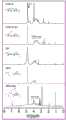
图2为马来酰亚胺基氧化海藻酸钠(ADA-Mal)和巯基羧甲基壳聚糖(CMCS-SH)改性前后FTIR图。Figure 2 is the FTIR images before and after modification of maleimide-based oxidized sodium alginate (ADA-Mal) and mercaptocarboxymethyl chitosan (CMCS-SH).
图3为马来酰亚胺基氧化海藻酸钠(ADA-Mal)和巯基羧甲基壳聚糖(CMCS-SH)改性前后的1HNMR图(400MHz)。Fig. 3 is a1 HNMR chart (400 MHz) before and after modification of maleimide-based oxidized sodium alginate (ADA-Mal) and mercaptocarboxymethyl chitosan (CMCS-SH).
图4为不同质量百分含量的ADA-Mal和CMCS-SH的粘度流变学分析:A为ADA-Mal(AM);B为CMCS-SH(CS)。Figure 4 is the viscorheological analysis of ADA-Mal and CMCS-SH with different mass percentages: A is ADA-Mal (AM); B is CMCS-SH (CS).
图5为不同质量百分含量的ADA-Mal溶液的皮下衬垫效果的超声检测结果:A为超声图像;B为统计结果;*P<0.05,**P<0.01,***P<0.001,****P<0.0001。Figure 5 is the ultrasonic detection results of the subcutaneous liner effect of ADA-Mal solutions with different mass percentages: A is the ultrasonic image; B is the statistical result; *P<0.05, **P<0.01, ***P<0.001 , ****P<0.0001.
图6为不同质量百分含量的ADA-Mal和CMCS-SH水凝胶(AxCy)的力学检测结果:A为应力应变曲线;B为压缩强度;C为杨氏模量;*P<0.05,**P<0.01,***P<0.001,****P<0.0001。Figure 6 shows the mechanical testing results of ADA-Mal and CMCS-SH hydrogels (AxCy) with different mass percentages: A is the stress-strain curve; B is the compressive strength; C is the Young's modulus; *P<0.05, **P<0.01, ***P<0.001, ****P<0.0001.
图7为不同质量百分含量的ADA-Mal和CMCS-SH水凝胶(AxCy)的溶胀与降解性能检测结果:A为溶胀率;B为残留质量百分比。Figure 7 shows the test results of swelling and degradation properties of ADA-Mal and CMCS-SH hydrogels (AxCy) with different mass percentages: A is the swelling rate; B is the residual mass percentage.
图8为不同质量百分含量的ADA-Mal和CMCS-SH混合后的成胶时间;其中,水凝胶组合以AxCy表示,A代表ADA-Mal,C表示CMCS-SH,x和y分别表示ADA-Mal和CMCS-SH的质量百分含量;*P<0.05,**P<0.01,***P<0.001,****P<0.0001。Figure 8 shows the gelation time after mixing ADA-Mal and CMCS-SH with different mass percentages; where, the hydrogel combination is represented by AxCy, A represents ADA-Mal, C represents CMCS-SH, and x and y represent Mass percentage of ADA-Mal and CMCS-SH; *P<0.05, **P<0.01, ***P<0.001, ****P<0.0001.
图9为对水凝胶的组织粘附性进行的评价;A为A7C3水凝胶在猪皮表面粘附行为;B为模拟水凝胶对不规则伤口的粘附行为。Figure 9 is the evaluation of the tissue adhesion of the hydrogel; A is the adhesion behavior of the A7C3 hydrogel on the pigskin surface; B is the adhesion behavior of the simulated hydrogel to the irregular wound.
图10为A7C3组水凝胶(A7C3 Gel)和纤维蛋白胶组(Fibrin Glue,图中为Fib Gel)的顶破强度检测;A为水凝胶顶破强度检测装置;B为顶破强度检测装置工作原理示意图;C为纤维蛋白胶与A7C3水凝胶组顶破强度大小统计。Figure 10 is the bursting strength detection of A7C3 group hydrogel (A7C3 Gel) and fibrin glue group (Fibrin Glue, Fib Gel in the figure); A is the hydrogel bursting strength detection device; B is the bursting strength detection Schematic diagram of the working principle of the device; C is the statistics of the bursting strength of the fibrin glue and A7C3 hydrogel groups.
图11为评价A7C3组水凝胶作为组织封闭剂在兔肝脏出血模型(体外)中的止血效果;A为未经治疗的对照组和使用纤维蛋白胶和A7C3组水凝胶治疗的样本组在0~120s内的止血效果图片;B、C分别为与纤维蛋白胶组和对照组(未经任何治疗)相比,使用A7C3水凝胶干预后兔肝脏的出血量和出血时间(*P<0.05)。Figure 11 is to evaluate the hemostatic effect of the A7C3 group hydrogel as a tissue sealant in the rabbit liver hemorrhage model (in vitro); A is the untreated control group and the sample group treated with fibrin glue and A7C3 group hydrogel in Pictures of the hemostatic effect within 0-120s; B and C are respectively compared with the fibrin glue group and the control group (without any treatment), the bleeding volume and bleeding time of the rabbit liver after the intervention of A7C3 hydrogel (*P< 0.05).
图12为犬食道ESD操作流程。Figure 12 shows the operation flow of canine esophagus ESD.
图13为ESD手术操作过程及AM7溶液在犬食管黏膜下衬垫效果评价;A为便携式内镜系统及ESD手术操作;B为ESD黏膜下注射流程,包括病灶定位、电刀标记、黏膜下注射溶液;C为比较甘油果糖注射液(GFI)和AM7溶液在犬食管黏膜下垫起高度随时间变化(30min内)。Figure 13 shows the ESD operation process and the evaluation of the effect of AM7 solution on the dog esophageal submucosal liner; A is the portable endoscope system and ESD operation; B is the ESD submucosal injection process, including lesion positioning, electrosurgical marking, and submucosal injection Solution; C is to compare the cushioning height of glycerol fructose injection (GFI) and AM7 solution in dog esophageal submucosal over time (within 30min).
图14为水凝胶在犬食管ESD中的应用;A为内镜黏膜下注射AM7溶液垫起病灶;B为对病灶进行切除;C为清理创面暴露病灶(针对黏膜下肿瘤);D为切除表面黏膜后暴露的AM7溶液(或切除黏膜下肿瘤后回填的AM7溶液);E为在AM7溶液上喷洒CS3溶液后原位水凝胶形态;F为原位水凝胶在创面稳定10min后的形态;(“Δ”指示为AM7溶液;“→”指示为创面水凝胶)。Figure 14 shows the application of hydrogel in dog esophagus ESD; A is endoscopic submucosal injection of AM7 solution to cushion the lesion; B is excision of the lesion; C is cleaning the exposed lesion (for submucosal tumor); D is excision AM7 solution exposed after superficial mucosa (or AM7 solution backfilled after resection of submucosal tumor); E is the in situ hydrogel morphology after spraying CS3 solution on AM7 solution; F is the in situ hydrogel after the wound is stable for 10 minutes Morphology; (“Δ” indicates AM7 solution; “→” indicates wound hydrogel).
图15为ESD分组及术后各时间点创面愈合情况观察:A为ESD手术分组:在每只犬的食管的近心端(离心脏近的位点定义为近心端。)和远心端(离口近的手术位点定义为远心端)开展两个手术点,每个食道的两个手术点处理方式如图A中分组所示(n=5);B为观察每个实验组在0、3、9、14、21、28天的伤口愈合情况(黄色“△”指示处为腺体)。Figure 15 is the observation of ESD grouping and postoperative wound healing at each time point: A is ESD operation grouping: at the proximal end of the esophagus of each dog (the position near the heart is defined as the proximal end.) and the distal end (The surgical site close to the mouth is defined as the distal end) Two surgical sites are carried out, and the treatment methods of the two surgical sites for each esophagus are shown in Figure A (n=5); B is the observation of each experimental group Wound healing at 0, 3, 9, 14, 21, and 28 days (glands indicated by yellow "△").
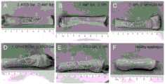
图16为ESD术后28天各实验组取材后食管形态图;A为A7C3 Gel组和AM7溶液组术后食管形态图;B为AM7溶液组和GFI组术后食管形态图;C为GFI组和GFI+CS3组术后食管形态图;D为GFI+CS3溶液组和A7C3 Gel组术后食管形态图;E为A7C3 Gel组和GFI+CS3溶液组术后食管形态图;F为正常食管的形态;(图中①表示远心端第一个实验位点;②表示第二个手术位点)。Figure 16 is the morphology of esophagus in each experimental group 28 days after ESD; A is the morphology of esophagus in A7C3 Gel group and AM7 solution group; B is the morphology of esophagus in AM7 solution group and GFI group; C is the GFI group and GFI+CS3 group postoperative esophagus morphology; D is postoperative esophagus morphology of GFI+CS3 solution group and A7C3 Gel group; E is postoperative esophagus morphology of A7C3 Gel group and GFI+CS3 solution group; F is normal esophagus Morphology; (in the figure ① indicates the first experimental site at the distal end; ② indicates the second surgical site).
具体实施方式Detailed ways
除另有说明外,本发明所用原料与设备均为已知产品,通过购买市售产品所得。Unless otherwise specified, the raw materials and equipment used in the present invention are known products obtained by purchasing commercially available products.
本发明所用氧化海藻酸钠(ADA)为购买市售产品或按照如下方法合成:The oxidized sodium alginate (ADA) used in the present invention is to buy commercially available products or synthesize according to the following method:
1、称取10g海藻酸钠分散于50mL无水乙醇中,磁力搅拌器至分散均匀,制备得到质量百分含量为20%的海藻酸钠的乙醇分散体系。1. Weigh 10 g of sodium alginate and disperse it in 50 mL of absolute ethanol, and disperse evenly with a magnetic stirrer to prepare an ethanol dispersion system of sodium alginate with a mass percentage of 20%.
2、称取2.5g NaIO4,避光条件环境下溶于50mL去离子水中,搅拌至完全溶解,制备得到质量百分含量为5%的NaIO4溶液;2. Weigh 2.5g NaIO4 , dissolve it in 50mL deionized water under dark conditions, stir until completely dissolved, and prepare a 5% NaIO4 solution by mass;
3、避光环境下,将NaIO4溶液逐滴、缓慢滴加到磁力搅拌下的海藻酸钠乙醇分散溶液中;室温避光环境下,持续搅拌反应6h;3. In a dark environment, add the NaIO4 solution dropwise and slowly to the sodium alginate ethanol dispersion solution under magnetic stirring; at room temperature and in a dark environment, continue to stir for 6 hours;
4、避光反应6h后,避光环境下往反应体系中逐滴加入10mL乙二醇,并继续搅拌反应30min,终止反应;4. After reacting in the dark for 6 hours, add 10 mL of ethylene glycol dropwise to the reaction system in a dark environment, and continue stirring for 30 minutes to terminate the reaction;
5、终止反应后,停止搅拌,室温静置、沉积10min,待沉淀析出;5. After terminating the reaction, stop stirring, let it stand at room temperature, and deposit for 10 minutes, and wait for precipitation;
6、小心倒出上层溶液,收集沉淀并装入截留分子量3.5kDa透析袋中,置于5L0.01M HCl去离子水溶液中透析3天,每12h换一次透析液;6. Carefully pour out the upper solution, collect the precipitate and put it into a dialysis bag with a molecular weight cut-off of 3.5kDa, place it in 5L of 0.01M HCl deionized aqueous solution for dialysis for 3 days, and change the dialysis solution every 12 hours;
7、将透析后的氧化海藻酸钠溶液收集后冷冻干燥,即得氧化海藻酸钠(ADA)。7. Collect the oxidized sodium alginate solution after dialysis and then freeze-dry to obtain oxidized sodium alginate (ADA).
本发明所用“含醛基和不饱和碳碳双键结构的改性的海藻酸盐”,即马来酰亚胺基氧化海藻酸钠(ADA-Mal),按照如下方法合成:The "modified alginate containing aldehyde group and unsaturated carbon-carbon double bond structure" used in the present invention, that is, maleimide-based oxidized sodium alginate (ADA-Mal), is synthesized according to the following method:
1、称取2g ADA溶于200mL MES溶液中,搅拌至完全溶解,制备质量百分含量为1%的ADA溶液;1. Weigh 2g ADA and dissolve it in 200mL MES solution, stir until it is completely dissolved, and prepare an ADA solution with a mass percentage of 1%;
2、在ADA溶液中加入EDC/NHS,使得ADA与EDC/NHS物质的量之比为1:1.5(EDC和NHS的摩尔比为1:1);往反应体系中加入马来酰亚胺盐酸盐(Mal-HCl),使得ADA与Mal-HCl的物质的量之比为1:1,室温搅拌反应24h,每6h调一次反应液的pH,保持pH值在5.0~5.5;2. Add EDC/NHS to the ADA solution so that the ratio of ADA to EDC/NHS is 1:1.5 (the molar ratio of EDC to NHS is 1:1); add maleimide salt to the reaction system acid salt (Mal-HCl), so that the ratio of the amount of ADA to Mal-HCl is 1:1, stir and react at room temperature for 24 hours, adjust the pH of the reaction solution every 6 hours, and keep the pH value at 5.0-5.5;
3、反应24h后,将反应液转移至截留分子量3.5kDa的透析袋中,置于5L 0.01M HCl去离子水溶液中透析5天,每12h换一次透析液;待透析第5天时,将透析袋转入5L去离子水中,继续透析12h。3. After 24 hours of reaction, transfer the reaction solution to a dialysis bag with a molecular weight cut-off of 3.5kDa, place it in 5L 0.01M HCl deionized aqueous solution for dialysis for 5 days, and change the dialysis solution every 12 hours; Transfer to 5L deionized water and continue dialysis for 12h.
4、取出透析后的马来酰亚胺基氧化海藻酸钠溶液,并进行冷冻干燥,即得马来酰亚胺基氧化海藻酸钠(ADA-Mal)。4. Take out the maleimide-based oxidized sodium alginate solution after dialysis, and freeze-dry it to obtain maleimide-based oxidized sodium alginate (ADA-Mal).
本发明所用“含有硫醇基的改性羧甲基壳聚糖”,即巯基羧甲基壳聚糖(CMCS-SH),按照如下方法合成:"Modified carboxymethyl chitosan containing thiol group" used in the present invention, i.e. mercapto carboxymethyl chitosan (CMCS-SH), is synthesized according to the following method:
1、称取4g CMCS(羧甲基壳聚糖)溶于200mL去离子水中,搅拌至完全溶解,制备质量百分含量为2%的CMCS溶液;1. Weigh 4g of CMCS (carboxymethyl chitosan) and dissolve it in 200mL of deionized water, stir until completely dissolved, and prepare a CMCS solution with a mass percentage of 2%;
2、CMCS溶液中加入EDC,使得CMCS与EDC物质的量之比为1:1.5;往反应体系中加入半胱胺盐酸盐(CSA-HCl),使得CMCS与CSA-HCl的物质量之比为1:1.5,室温搅拌反应24h;2. Add EDC to the CMCS solution so that the ratio of CMCS to EDC is 1:1.5; add cysteamine hydrochloride (CSA-HCl) to the reaction system to make the ratio of CMCS to CSA-HCl 1:1.5, stirred at room temperature for 24 hours;
3、反应24h后,将反应液转移至截留分子量为3.5kDa中,并置于5L0.01M的硼砂溶液中透析5天,每12h换透析液一次;3. After reacting for 24 hours, transfer the reaction solution to a medium with a molecular weight cut-off of 3.5kDa, and dialyze it in 5L of 0.01M borax solution for 5 days, and change the dialysate every 12 hours;
4、透析后的CMCS-SH溶液倒入烧杯中,并加入2g DTT室温继续搅拌反应1~2h后,取出并进行冷冻干燥;4. Pour the dialyzed CMCS-SH solution into a beaker, add 2g of DTT and continue to stir and react at room temperature for 1-2 hours, then take it out and freeze-dry it;
5、将冷冻干燥后的CMCS-SH加入无水乙醇中,密封置于低温摇床中(4℃)60rpm振荡1h后,用抽滤瓶滤去乙醇,重复此操作5次;5. Add the freeze-dried CMCS-SH to absolute ethanol, seal and place it in a low-temperature shaker (4°C) at 60 rpm for 1 hour, then filter out the ethanol with a suction filter bottle, and repeat this
6、醇析后的CMCS-SH用旋转蒸发仪除去残留乙醇,即得到巯基羧甲基壳聚糖(CMCS-SH)。6. The CMCS-SH after alcohol analysis was removed with a rotary evaporator to remove residual ethanol to obtain mercaptocarboxymethyl chitosan (CMCS-SH).
ADA-Mal和CMCS-SH两种物质的合成示意图如图1所示。The schematic diagram of the synthesis of ADA-Mal and CMCS-SH two substances is shown in Figure 1.
制备完成后通过傅立叶变换红外吸收光谱仪(FTIR),核磁氢谱仪(1HNMR)对海藻酸钠和羧甲基壳聚糖改性前后产物进行表征,证实对海藻酸钠接枝改性后成功制备得到马来酰亚胺基氧化海藻酸钠(ADA-Mal);对羧甲基壳聚糖接枝改性后成功制备得到巯基羧甲基壳聚糖(CMCS-SH)。After the preparation was completed, Fourier transform infrared absorption spectrometer (FTIR) and hydrogen nuclear magnetic spectrometer (1HNMR) were used to characterize the products before and after modification of sodium alginate and carboxymethyl chitosan, which confirmed the successful preparation of sodium alginate after graft modification. Maleimide oxidized sodium alginate (ADA-Mal) was obtained; mercaptocarboxymethyl chitosan (CMCS-SH) was successfully prepared after graft modification of carboxymethyl chitosan.
实施例1、本发明原位水凝胶的制备
1、称取冻干后的ADA-Mal溶于PBS缓冲液中,制备质量百分含量为7%的ADA-Mal溶液;1. Weigh the lyophilized ADA-Mal and dissolve it in PBS buffer to prepare an ADA-Mal solution with a mass percentage of 7%;
2、称取冻干后的CMCS-SH溶于浓度为0.01M的硼砂水溶液中,制备质量百分含量为3%的CMCS-SH溶液;浓度为0.01M的硼砂水溶液的配制方法如下:称取19.05g Na2B4O7·10H2O(常温,硼砂)溶于5L去离子水中,搅拌至完全溶解;2. Weigh the freeze-dried CMCS-SH and dissolve it in a borax aqueous solution with a concentration of 0.01M to prepare a CMCS-SH solution with a mass percent content of 3%; the preparation method of the 0.01M borax aqueous solution is as follows: weigh Dissolve 19.05g Na2 B4 O7 ·10H2 O (normal temperature, borax) in 5L deionized water, stir until completely dissolved;
3、将步骤(1)和(2)中的溶液按体积比为1:1均匀混合,即可制备得到A7C3水凝胶。3. Mix the solutions in steps (1) and (2) uniformly at a volume ratio of 1:1 to prepare the A7C3 hydrogel.
实施例2、本发明原位水凝胶的制备
1、称取冻干后的ADA-Mal溶于PBS缓冲液中,制备质量百分含量为7%的ADA-Mal溶液;1. Weigh the lyophilized ADA-Mal and dissolve it in PBS buffer to prepare an ADA-Mal solution with a mass percentage of 7%;
2、称取冻干后的CMCS-SH溶于浓度为0.01M的硼砂水溶液中,制备质量百分含量为7%的CMCS-SH溶液;浓度为0.01M的硼砂水溶液的配制方法如实施例1;2. Take the freeze-dried CMCS-SH and dissolve it in the borax aqueous solution with a concentration of 0.01M to prepare a 7% CMCS-SH solution by mass percentage; the preparation method of the borax aqueous solution with a concentration of 0.01M is as in Example 1 ;
3、将步骤(1)和(2)中的溶液按体积比为1:1均匀混合,即可制备得到A7C7水凝胶。3. Mix the solutions in steps (1) and (2) uniformly at a volume ratio of 1:1 to prepare the A7C7 hydrogel.
实施例3、本发明原位水凝胶的制备
1、称取冻干后的ADA-Mal溶于PBS缓冲液中,制备质量百分含量为3%的ADA-Mal溶液;1. Weigh the lyophilized ADA-Mal and dissolve it in PBS buffer to prepare an ADA-Mal solution with a mass percentage of 3%;
2、称取冻干后的CMCS-SH溶于浓度为0.01M的硼砂水溶液中,制备质量百分含量为7%的CMCS-SH溶液;浓度为0.01M的硼砂水溶液的配制方法如实施例1;2. Take the freeze-dried CMCS-SH and dissolve it in the borax aqueous solution with a concentration of 0.01M to prepare a 7% CMCS-SH solution by mass percentage; the preparation method of the borax aqueous solution with a concentration of 0.01M is as in Example 1 ;
3、将步骤(1)和(2)中的溶液按体积比为1:1均匀混合,即可制备得到A3C7水凝胶。3. Mix the solutions in steps (1) and (2) uniformly at a volume ratio of 1:1 to prepare the A3C7 hydrogel.
实施例4、本发明原位水凝胶的制备
1、称取冻干后的ADA-Mal溶于PBS缓冲液中,制备质量百分含量为4%的ADA-Mal溶液;1. Weigh the lyophilized ADA-Mal and dissolve it in PBS buffer to prepare an ADA-Mal solution with a mass percentage of 4%;
2、称取冻干后的CMCS-SH溶于浓度为0.01M的硼砂水溶液中,制备质量百分含量为6%的CMCS-SH溶液;浓度为0.01M的硼砂水溶液的配制方法如实施例1;2. Take the freeze-dried CMCS-SH and dissolve it in the borax aqueous solution with a concentration of 0.01M to prepare a CMCS-SH solution with a mass percentage of 6%; the preparation method of the borax aqueous solution with a concentration of 0.01M is as in Example 1 ;
3、将步骤(1)和(2)中的溶液按体积比为1:1均匀混合,即可制备得到A4C6水凝胶。3. Mix the solutions in steps (1) and (2) uniformly at a volume ratio of 1:1 to prepare the A4C6 hydrogel.
实施例5、本发明原位水凝胶的制备
1、称取冻干后的ADA-Mal溶于PBS缓冲液中,制备质量百分含量为5%的ADA-Mal溶液;1. Weigh the lyophilized ADA-Mal and dissolve it in PBS buffer to prepare an ADA-Mal solution with a mass percentage of 5%;
2、称取冻干后的CMCS-SH溶于浓度为0.01M的硼砂水溶液中,制备质量百分含量为5%的CMCS-SH溶液;浓度为0.01M的硼砂水溶液的配制方法如实施例1;2. Take the freeze-dried CMCS-SH and dissolve it in the borax aqueous solution with a concentration of 0.01M to prepare a 5% CMCS-SH solution by mass percentage; the preparation method of the borax aqueous solution with a concentration of 0.01M is as in Example 1 ;
3、将步骤(1)和(2)中的溶液按体积比为1:1均匀混合,即可制备得到A5C5水凝胶。3. Mix the solutions in steps (1) and (2) uniformly at a volume ratio of 1:1 to prepare the A5C5 hydrogel.
实施例6、本发明原位水凝胶的制备
1、称取冻干后的ADA-Mal溶于PBS缓冲液中,制备质量百分含量为6%的ADA-Mal溶液;1. Weigh the lyophilized ADA-Mal and dissolve it in PBS buffer to prepare an ADA-Mal solution with a mass percentage of 6%;
2、称取冻干后的CMCS-SH溶于浓度为0.01M的硼砂水溶液中,制备质量百分含量为4%的CMCS-SH溶液;浓度为0.01M的硼砂水溶液的配制方法如实施例1;2. Take the freeze-dried CMCS-SH and dissolve it in the borax aqueous solution with a concentration of 0.01M to prepare a CMCS-SH solution with a mass percentage of 4%; the preparation method of the borax aqueous solution with a concentration of 0.01M is as in Example 1 ;
3、将步骤(1)和(2)中的溶液按体积比为1:1均匀混合,即可制备得到A6C4水凝胶。3. Mix the solutions in steps (1) and (2) uniformly at a volume ratio of 1:1 to prepare the A6C4 hydrogel.
以下通过具体实验例证明本发明的有益效果。The beneficial effects of the present invention are demonstrated through specific experimental examples below.
实验例1、ADA-Mal和CMCS-SH结构表征Experimental example 1, structure characterization of ADA-Mal and CMCS-SH
1、实验方法1. Experimental method
ADA-Mal/CMCS-SH的FTIR检测:利用溴化钾压片法检测材料红外吸收光谱,具体操作如下:分别称取5~10mg SA、ADA、ADA-Mal、CMCS及CMCS-SH 5种冻干样品,将材料剪成细小碎片状,并加入50~60mg的KBr粉末混合研磨均匀,然后将研磨好的混合粉末经压片机,在10kPa的压力下压成薄片,取出,进行红外光谱测试。测试的具体条件设定为:ATR检测模式下,检测波谱范围是400-4000cm-1,光谱分辨率4cm-1/次,扫描频率是0.0625Hz。FTIR detection of ADA-Mal/CMCS-SH: Use the potassium bromide tablet method to detect the infrared absorption spectrum of the material. Dry the sample, cut the material into fine pieces, add 50-60 mg of KBr powder, mix and grind evenly, then pass the ground mixed powder into a thin tablet under a pressure of 10kPa through a tablet machine, take it out, and perform infrared spectroscopy test . The specific conditions of the test are set as follows: in the ATR detection mode, the detection spectrum range is 400-4000cm-1 , the spectral resolution is 4cm-1 per time, and the scanning frequency is 0.0625Hz.
ADA-Mal/CMCS-SH的1HNMR检测:1HNMR样品制备及检测如下:分别称取5mg SA、ADA、ADA-Mal、CMCS及CMCS-SH 5种冻干样品,装入核磁管中,每管加入0.55mL氘代水(D2O),振荡使材料充分溶解。用核磁共振仪(400MHz,Bruker AV II-400,Switzerland)测定样品化学位移在0-12ppm区间的吸收波谱,设多糖主链中1H峰积分面积为参比峰,根据测得的得到-Mal及-SH波谱图中1H峰面积与参比峰面积之比计算其马来酰亚胺及硫醇基团的接枝度(DS)。1 HNMR detection of ADA-Mal/CMCS-SH:1 HNMR sample preparation and detection are as follows: Weigh 5 mg of SA, ADA, ADA-Mal, CMCS and CMCS-
2、实验结果2. Experimental results
氧化后的海藻酸钠中FTIR图中在1734cm-1伸缩振动吸收峰,表明有醛基生成,生成氧化海藻酸钠(ADA);经马来酰亚胺盐酸盐改性后的ADA的FTIR图谱中在1657cm-1和1542cm-1振动峰为酰胺键I和酰胺II(图2)。1HNMR谱δ=2.88ppm,δ=2.95ppm两个峰分别为马来酰亚胺基(-CH2CH2-)引入的亚甲基质子峰;δ=6.88ppm处为引入-Mal官能团中-C=C-的质子峰(图3)。ADA-Mal中马来酰亚胺基取代度(DS)通过马来酰亚胺基中的碳碳双键(-C=C-)(δ=6.88ppm)中氢原子对应的峰面积和ADA中主链(δ=3.2~4.0ppm)上的氢原子对应的峰面积的比值计算取代度(DS),DS约为27.29%。FTIR和1HNMR结果表明本发明成功制得ADA-Mal。The stretching vibration absorption peak at 1734cm-1 in the FTIR diagram of the oxidized sodium alginate indicates that there is an aldehyde group formed to generate oxidized sodium alginate (ADA); the FTIR of ADA modified by maleimide hydrochloride The vibration peaks at 1657cm-1 and 1542cm-1 in the spectrum are amide bond I and amide II (Figure 2).1 HNMR spectrum δ=2.88ppm, δ=2.95ppm The two peaks are respectively the methylene proton peak introduced by the maleimide group (-CH2 CH2 -); δ=6.88ppm is introduced into the -Mal functional group The proton peak of -C=C- (Figure 3). The degree of substitution (DS) of the maleimide group in ADA-Mal passes the peak area corresponding to the hydrogen atom in the carbon-carbon double bond (-C=C-) (δ=6.88ppm) in the maleimide group and ADA The degree of substitution (DS) is calculated from the ratio of the peak areas corresponding to the hydrogen atoms on the main chain (δ=3.2-4.0ppm), and the DS is about 27.29%. FTIR and1 HNMR results show that the present invention successfully prepared ADA-Mal.
在CMCS及CMCS-SH的FTIR光谱中可以看出,CMCS-SH中在1657和1542cm-1处出现吸收峰,为酰胺I(-C=O)伸缩振动峰和酰胺II(-C-N)弯曲振动峰,表明半胱胺巯基中的氨基(-NH2)与CMCS上的羧基(-COO-)发生了反应,形成了酰胺键(图2)。从CMCS氢谱图中可见,δ=2.02ppm为CMCS中的乙酰基中的甲基质子峰(-CO-CH3)。与CMCS相比,CMCS-SH氢谱中出现的δ=2.86ppm和δ=2.91ppm归属于半胱胺巯基的亚甲基质子峰(-CH2CH2SH),其中δ=2.91ppm处的峰归属于与酰胺键邻近的亚甲基氢(图3)。根据双键δ=2.91ppm和(δ=2.86-2.91ppm位置处氢原子对应的峰面积和亚甲基峰(δ=2.56ppm)处氢原子对应的峰面积,计算巯基的取代度(DS),DS约为48.2%。FTIR和1HNMR结果表明本发明成功制得CMCS-SH。In the FTIR spectra of CMCS and CMCS-SH, it can be seen that the absorption peaks appear at 1657 and 1542 cm-1 in CMCS-SH, which are the stretching vibration peaks of amide I (-C=O) and the bending vibration of amide II (-CN) peak, indicating that the amino group (-NH2 ) in the sulfhydryl group of cysteamine reacted with the carboxyl group (-COO-) on CMCS to form an amide bond (Figure 2). It can be seen from the CMCS hydrogen spectrogram that δ=2.02ppm is the methyl proton peak (-CO-CH3 ) in the acetyl group in CMCS. Compared with CMCS, the δ=2.86ppm and δ=2.91ppm in the CMCS-SH hydrogen spectrum are attributed to the methylene proton peak (-CH2 CH2 SH) of cysteamine thiol, where δ=2.91ppm Peaks were assigned to methylene hydrogens adjacent to the amide bond (Figure 3). According to the peak area corresponding to the hydrogen atom at the position of the double bond δ=2.91ppm and (δ=2.86-2.91ppm) and the peak area corresponding to the hydrogen atom at the methylene peak (δ=2.56ppm), calculate the degree of substitution (DS) of the mercapto group , DS is about 48.2%. The results of FTIR and1 HNMR show that the present invention successfully prepared CMCS-SH.
实验例2、ADA-Mal和CMCS-SH溶液粘度检测Experimental example 2, ADA-Mal and CMCS-SH solution viscosity detection
1、实验方法1. Experimental method
对不同质量百分含量的ADA-Mal和CMCS-SH溶液的粘度进行检测,其中:ADA-Mal简写为AMx,x=3,4,5,6,7,8,分别表示ADA-Mal的质量百分含量为3%、4%、5%、6%、7%、8%;CMCS-SH简写为CSy,y=3,4,5,6,7,分别表示CMCS-SH的质量百分含量为3%、4%、5%、6%、7%。具体操作如下:分别配制不同质量百分比(w/v%)的AMx溶液和CSy溶液,以临床ESD常用的黏膜注射液甘油果糖注射液(GFI)作为对照。使用旋转流变仪(MCR302),50mm的平板转子(PP50)作为测试部件,仪器测试参数设置为:测量的上下平板间隙设为0.25mm,平台温度设置为37℃。初试剪切速率初始值设置为0.1rad/s,终值为100rad/s,在旋转模式下分别检测GFI、AM3、AM4、AM5、AM6、AM7、AM8,及CS3、CS4、CS5、CS6、CS7共12种液体的黏度曲线。The viscosities of ADA-Mal and CMCS-SH solutions with different mass percentages are detected, where: ADA-Mal is abbreviated as AMx, and x=3,4,5,6,7,8, respectively representing the mass of ADA-Mal The percentage content is 3%, 4%, 5%, 6%, 7%, 8%; CMCS-SH is abbreviated as CSy, y=3, 4, 5, 6, 7, which respectively represent the mass percentage of CMCS-SH The content is 3%, 4%, 5%, 6%, 7%. The specific operation is as follows: AMx solutions and CSy solutions with different mass percentages (w/v%) were prepared respectively, and Glycerin-Fructose Injection (GFI), a mucosal injection commonly used in clinical ESD, was used as a control. Using a rotational rheometer (MCR302), a 50mm plate rotor (PP50) is used as the test component, and the instrument test parameters are set as follows: the gap between the upper and lower plates for measurement is set to 0.25mm, and the platform temperature is set to 37°C. The initial value of the initial shear rate is set to 0.1rad/s, and the final value is 100rad/s. In the rotation mode, GFI, AM3, AM4, AM5, AM6, AM7, AM8, and CS3, CS4, CS5, CS6, CS7 are detected respectively Viscosity curves of a total of 12 liquids.
2、实验结果2. Experimental results
对AMx、CSy两种改性溶液粘度曲线如图4A和图4B。图4A为GFI和不同浓度AMx溶液的粘度测试曲线;图4B为不同浓度CSy溶液的粘度测试曲线,图4A和4B反应的是各不同浓度溶液的粘度随剪切速率变化的趋势。图4A和4B与纵标相交的是横坐标为0.1s时的粘度值。而检测得到GFI、AM3、AM4、AM5、AM6、AM7、AM8的各组溶液的静态粘度值分别约为13.409、3783.7、21663、52487、69562、88476、168940mPs;CS3、CS4、CS5、CS6、CS7各组溶液静态粘度值分别约为136、137.07、148.44、148.28、184.96mPs。两种溶液粘度随着流变仪转子剪切速率增大,粘度降低,表现出剪切稀化特性,为典型的非牛顿流体。可见ADA-Mal溶液与CMCS-SH溶液均具有可注射性。The viscosity curves of AMx and CSy two modified solutions are shown in Figure 4A and Figure 4B. Figure 4A is the viscosity test curves of GFI and AMx solutions with different concentrations; Figure 4B is the viscosity test curves of CSy solutions with different concentrations. Figures 4A and 4B intersect the ordinate with the viscosity value at 0.1 s on the abscissa. The static viscosity values of the solutions of GFI, AM3, AM4, AM5, AM6, AM7, and AM8 are about 13.409, 3783.7, 21663, 52487, 69562, 88476, 168940mPs; CS3, CS4, CS5, CS6, CS7 The static viscosity values of the solutions in each group are about 136, 137.07, 148.44, 148.28, 184.96mPs respectively. The viscosities of the two solutions decreased with the increase of the shear rate of the rotor of the rheometer, showing shear thinning characteristics, which were typical non-Newtonian fluids. It can be seen that both ADA-Mal solution and CMCS-SH solution are injectable.
从两种溶液的静态粘度检测结果可以发现,AMx溶液粘度远高于CSy溶液及GFI溶液粘度,溶液粘性对于维持衬垫高度至关重要,因此为实现持久的黏膜下衬垫效果,优选AMx溶液作为黏膜下注射液。从AMx静态粘度曲线可以看出,随着AM溶液质量百分含量增大,溶液静态粘度增大,且当x=8时,粘度突增,可能原因是由于随着AM百分含量增大,溶液粘度增大,高粘度阻止水分子进入材料内部与未溶解的AM中的糖链结合,导致溶质中糖链不能充分展开,以卷曲的形式存在于溶液中,进一步增大了溶液体系的粘度。在行内镜黏膜下注射时,会因粘度过大而堵塞针管,影响手术质量,因此在选择作为黏膜下注射液的AMx浓度时,不考虑x=8组的AM溶液。From the results of the static viscosity test of the two solutions, it can be found that the viscosity of the AMx solution is much higher than that of the CSy solution and the GFI solution. The viscosity of the solution is very important for maintaining the pad height. Therefore, in order to achieve a lasting submucosal pad effect, the AMx solution is preferred As a submucosal injection. From the AMx static viscosity curve, it can be seen that as the mass percentage of AM solution increases, the static viscosity of the solution increases, and when x=8, the viscosity increases suddenly. The possible reason is that as the AM percentage increases, The viscosity of the solution increases, and the high viscosity prevents water molecules from entering the interior of the material and combining with the sugar chains in the undissolved AM, resulting in the inability of the sugar chains in the solute to fully unfold and exist in the solution in the form of curls, which further increases the viscosity of the solution system . When endoscopic submucosal injection is performed, the needle tube will be blocked due to excessive viscosity, which will affect the quality of the operation. Therefore, when selecting the concentration of AMx as the submucosal injection, the AM solution of the x=8 group is not considered.
实验例3、ADA-Mal溶液的皮下衬垫效果的超声检测结果Ultrasonic detection results of the subcutaneous liner effect of experimental example 3, ADA-Mal solution
1、实验方法1. Experimental method
如实验例2所述,制备AM3、AM4、AM5、AM6、AM7五种不同浓度AM溶液。水合氯醛麻醉SD大鼠(300g/mL),背部备皮,暴露背部两侧皮肤,并标记注射点。用1mL注射器吸取100μL上述各浓度AM溶液,将注射器与背部呈小角度将AM溶液从标记点注入到大鼠皮下,观察到隆起后立即进行超声检测,以该检测点检测高度作为初始高度,以该检测点时间作为初始时间(T0=0min)。生理盐水和GFI作为对照组,每组注射液设置三个平行注射点(n=3)。分别在0min、15min、30min三个时间点检测各组溶液在皮下的垫起高度,记录、统计各组AM溶液在皮下垫起高度随时间的变化情况。As described in Experimental Example 2, five AM solutions with different concentrations of AM3, AM4, AM5, AM6, and AM7 were prepared. SD rats were anesthetized with chloral hydrate (300g/mL), the back skin was prepared, the skin on both sides of the back was exposed, and the injection point was marked. Use a 1mL syringe to draw 100 μL of the above-mentioned AM solution of each concentration, inject the AM solution from the marked point into the subcutaneous of the rat with the syringe at a small angle to the back, and perform ultrasonic detection immediately after the bulge is observed, and take the detection height of the detection point as the initial height, and The detection point time is taken as the initial time (T0 =0 min). Physiological saline and GFI were used as the control group, and three parallel injection points (n=3) were set for each injection solution. At three time points of 0 min, 15 min, and 30 min, the subcutaneous cushioning height of the solutions in each group was detected, and the changes in the subcutaneous cushioning height of the AM solution in each group over time were recorded and counted.
2、实验结果2. Experimental results
图5为将生理盐水(NS)、甘油果糖注射液(GFI)和质量百分含量分别为3%、4%、5%、6%、7%的AM溶液注入大鼠皮下后超声检测到的溶液在大鼠皮下垫起高度随时间变化的形态图。从图中可以清楚的看到,在注射相同体积的注射液的前提下,随着AM百分含量增大,检测到的初始(T0=0min)垫起高度增大,且当浓度大于4%时(垫起高度为0.48±0.08cm),AM溶液在皮下垫起高度已显著高于NS和GFI组(分别为0.32±0.05cm,0.34±0.05cm)(图5B)。在注射30min后,AM6和AM7两组依然保持明显的垫起高度,分别为0.36±0.03cm、0.42±0.06cm,均高于NS和GFI初始检测高度。通过超声检测AM溶液的皮下衬垫效果表明,相对于临床常用的NS和GFI,一定浓度的AM溶液在大鼠皮下能维持较好的衬垫高度和衬垫时间,且衬垫效果随浓度可调。超声检测AM溶液在大鼠皮下衬垫效果评价结果表明,AM溶液可作为ESD黏膜下注射潜在替代产品。从AMx溶液的大鼠皮下衬垫效果来看,浓度越高,初始衬垫高度越高,相同时间内皮下维持高度越高,因此,为了维持稳定的黏膜下衬垫效果,拟选用AM7溶液作为食管黏膜下注射材料。Fig. 5 is that normal saline (NS), glycerol-fructose injection (GFI) and mass percentage content are respectively 3%, 4%, 5%, 6%, 7% AM solution injected subcutaneously into rats and detected by ultrasound The morphological diagram of the change of the height of the solution under the skin of the rat with time. It can be clearly seen from the figure that under the premise of injecting the same volume of injection solution, as the AM percentage increases, the detected initial (T0 =0min) cushion height increases, and when the concentration is greater than 4 % (0.48±0.08cm), the subcutaneous cushion height of AM solution was significantly higher than that of NS and GFI groups (0.32±0.05cm, 0.34±0.05cm, respectively) (Fig. 5B). After 30 minutes of injection, the AM6 and AM7 groups still maintained a significant pad height, respectively 0.36±0.03cm and 0.42±0.06cm, which were higher than the initial detection heights of NS and GFI. Ultrasonic detection of the subcutaneous padding effect of AM solution shows that, compared with NS and GFI commonly used in clinical practice, a certain concentration of AM solution can maintain a better pad height and padding time under the skin of rats, and the padding effect varies with the concentration. Tune. Ultrasonic detection of AM solution in subcutaneous liner effect evaluation results in rats showed that AM solution can be used as a potential substitute for ESD submucosal injection. From the perspective of the subcutaneous liner effect of AMx solution in rats, the higher the concentration, the higher the initial liner height, and the higher the subcutaneous maintenance height within the same time period. Therefore, in order to maintain a stable submucosal liner effect, AM7 solution is proposed to be used as the subcutaneous liner. Esophageal submucosal injection of material.
实验例4、AxCy水凝胶力学性能检测Experimental example 4, detection of mechanical properties of AxCy hydrogel
1、实验方法1. Experimental method
用电子万能材料试验机(Instron5967)测试实施例1~6制备的A3C7、A4C6、A5C5、A6C4、A7C3、A7C7,6组水凝的压缩强度和杨氏模量。具体操作如下:将水凝胶制备成直径0.8cm,高1.0cm的两头平整的圆柱状,将水凝胶柱竖立放置在测试机压头的中心位置。试验机压头连接有量程为30kN的传感器,试验机参数设置为,运行速度5mm/min,压缩应变设置为测试物体长度的80%。运行试验机,记录水凝胶应力-应变曲线(n=5),并记录凝胶压缩应力的最大值,及最大值对应的最大压缩应变。压缩模量取测试物体应变发生在15%~25%压缩阶段的斜率(n=5)。The hydraulic compressive strength and Young's modulus of A3C7, A4C6, A5C5, A6C4, A7C3, and A7C7 prepared in Examples 1-6 were tested with an electronic universal material testing machine (Instron5967). The specific operation is as follows: the hydrogel is prepared into a cylindrical shape with a diameter of 0.8 cm and a height of 1.0 cm, and the hydrogel column is placed upright at the center of the pressure head of the testing machine. The indenter of the testing machine is connected with a sensor with a measuring range of 30kN. The parameters of the testing machine are set at a running speed of 5mm/min, and the compressive strain is set at 80% of the length of the test object. Run the testing machine, record the hydrogel stress-strain curve (n=5), and record the maximum value of the compressive stress of the gel, and the maximum compressive strain corresponding to the maximum value. The compressive modulus is taken as the slope (n=5) of the strain of the test object occurring in the compression stage of 15% to 25%.
水凝胶的杨氏模量可通过下面公式计算:The Young's modulus of the hydrogel can be calculated by the following formula:

式中Δδ为水凝胶的杨氏模量;ΔF为应力,其物理意义是单位截面积所受到的力;ΔL应变,其物理意义是单位长度所对应的伸长量;F为水凝胶承受的压力;S为水凝胶的横截面积;L水凝胶的高度;ΔL'水凝胶的压缩高度,r水凝胶圆柱体的半径。In the formula, Δδ is the Young's modulus of the hydrogel; ΔF is the stress, and its physical meaning is the force per unit cross-sectional area; ΔL strain, its physical meaning is the elongation corresponding to the unit length; F is the hydrogel Under pressure; S is the cross-sectional area of the hydrogel; L is the height of the hydrogel; ΔL' is the compressed height of the hydrogel, r is the radius of the hydrogel cylinder.
2、实验结果2. Experimental results
图6为六组水凝胶的压缩应力与应变的关系曲线。从图中可以看出水凝胶在最大压缩应力时坍塌,此时对应的压缩距离为应变,不同水凝胶的压缩应力及对应的应变各不相同,A7C7水凝胶发生坍塌的应变最小,A3C7水凝胶发生坍塌的应变最大。从压缩应力大小来看,A5C5组水凝胶所能承受的最大应力为50.30±5.54kPa,A7C3组能承受的最大应力为76.82±4.93kPa,两组能承受的最大应力之间差异显著(P<0.01);A7C7组水凝胶能承受的最大压缩应力为54.57±4.72kPa,显著小于A7C3组(P<0.05)。六组水凝胶中A7C7组水凝胶压缩模量最大,其次是A7C3组,最小的是A3C7组。其中A4C6、A5C5、A6C4三组压缩模量差异不显著(P>0.05)。从力学结果来看,A7C3水凝胶有较大的压缩强度和杨氏模量,同时A7C3水凝胶成胶时间较短。Fig. 6 is the relationship curve of compressive stress and strain of six groups of hydrogels. It can be seen from the figure that the hydrogel collapses at the maximum compressive stress. At this time, the corresponding compression distance is the strain. The compressive stress and the corresponding strain of different hydrogels are different. A7C7 hydrogel collapses with the smallest strain, and A3C7 The strain at which the hydrogel collapses is the largest. From the perspective of compressive stress, the maximum stress that the A5C5 group hydrogel can withstand is 50.30±5.54kPa, and the maximum stress that the A7C3 group can withstand is 76.82±4.93kPa, and the difference between the two groups is significant (P <0.01); the maximum compressive stress that the hydrogel of A7C7 group could withstand was 54.57±4.72kPa, which was significantly smaller than that of A7C3 group (P<0.05). Among the six groups of hydrogels, the compressive modulus of hydrogels in group A7C7 was the largest, followed by group A7C3, and group A3C7 was the smallest. Among them, there was no significant difference in compressive modulus among A4C6, A5C5 and A6C4 groups (P>0.05). From the mechanical results, the A7C3 hydrogel has higher compressive strength and Young's modulus, and the gelation time of A7C3 hydrogel is shorter.
实验例5、AxCy水凝胶溶胀与降解性能评价Experimental example 5, AxCy hydrogel swelling and degradation performance evaluation
1、实验方法1. Experimental method
(1)水凝胶溶胀:实施例1~6制备的六组柱状水凝胶,每组分别取一定量的水凝胶,滤纸吸干表面的水分,称重(W0);将每组称重后的柱状水凝胶浸入10mL PBS缓冲液中,并置于37℃,60rpm恒温摇床中。一定时间后取出水凝胶,用滤纸吸干表面水分后称重(Wt)。重复上述过程直至各组水凝胶重量保持不变为止。每组水凝胶设置3个平行(n=3)。水凝胶溶胀率计算公式:(1) Hydrogel swelling: for six groups of columnar hydrogels prepared in Examples 1 to 6, a certain amount of hydrogel was taken for each group, and the moisture on the surface was blotted by filter paper, and weighed (W0 ); The weighed columnar hydrogel was immersed in 10 mL of PBS buffer, and placed in a constant temperature shaker at 37° C. at 60 rpm. After a certain period of time, the hydrogel was taken out, and the surface moisture was blotted dry with filter paper, and then weighed (Wt). Repeat the above process until the weight of each group of hydrogels remains unchanged. Three parallels were set up for each group of hydrogels (n=3). Calculation formula of hydrogel swelling rate:
溶胀率(%)=(Wt-Wo)/Wo×100%Swelling rate (%)=(Wt-Wo)/Wo×100%
式中:Wt为t时刻水凝胶块溶胀后的质量,W0为水凝胶块初始的质量。In the formula: Wt is the mass of the hydrogel block after swelling at time t, W0 is the initial mass of the hydrogel block.
(2)水凝胶降解:取实施例1~6制备的水凝胶,冻干,得冻干水凝胶,称重(W0),并分别置于10mL PBS缓冲液中,放置于37℃,60rpm恒温摇床中,一定时间后取出,去离子水冲洗掉表面,经冷冻干燥后称重(Wt),每组水凝胶设置3个平行(n=3)。计算残留质量百分比。水凝胶的降解性能表示方法可以用公式计算:(2) Hydrogel degradation: Take the hydrogels prepared in Examples 1-6 and freeze-dry them to obtain freeze-dried hydrogels, weigh them (W0 ), place them in 10 mL of PBS buffer, and place them at 37 ℃, 60rpm constant temperature shaker, take it out after a certain period of time, rinse the surface with deionized water, weigh (Wt) after freeze-drying, set 3 parallels for each group of hydrogels (n=3). Calculate residual mass percent. The expression method of the degradation performance of the hydrogel can be calculated by the formula:
残留质量百分比(%)=Wt/Wo×100%Residual mass percentage (%)=Wt/Wo×100%
式中:Wt,W0分别为t时刻和初始水凝胶冻干支架的重量。In the formula: Wt, W0 are the weight of time t and the initial hydrogel freeze-dried scaffold, respectively.
2、实验结果2. Experimental results
对六组水凝胶的溶胀及降解性能检的测结果如图7A和7B所示。图7A为水凝胶的溶胀曲线,从溶胀曲线可以看出六组不同水凝胶在PBS缓冲液中在30h左右几乎趋于溶胀平衡。其中A7C3和A3C7组溶胀率分别达到101.9±14.63%和94.34±10.75%;A7C7组最小为55.77±6.39%;A4C6、A5C5、A6C4组水凝胶溶胀率接近,分别为85.38±6.94%、81.28±9.06%、82.87±4.10%。30h后,在水分子和离子作用下,水凝胶开始降解,其中A3C7组降解最明显。The test results of the swelling and degradation properties of the six groups of hydrogels are shown in Figures 7A and 7B. Fig. 7A is the swelling curve of the hydrogel. It can be seen from the swelling curve that the six groups of different hydrogels almost tend to swelling equilibrium in PBS buffer solution at about 30 hours. Among them, the swelling rates of A7C3 and A3C7 groups reached 101.9±14.63% and 94.34±10.75%, respectively; the minimum swelling rate of A7C7 group was 55.77±6.39%; 9.06%, 82.87±4.10%. After 30h, under the action of water molecules and ions, the hydrogel began to degrade, and the degradation of A3C7 group was the most obvious.
六组冻干水凝胶在PBS缓冲液中的降解如图7B所示,从图中可以看出,水凝胶在降解前3天时,降解曲线斜率最大,降解速率最大;随后降解速率减缓,并趋于平衡。其中,A3C7组水凝胶降解最快,其次是A4C6、A5C5、A6C4和A7C7,A7C3组降解最慢。The degradation of the six groups of freeze-dried hydrogels in PBS buffer is shown in Figure 7B. It can be seen from the figure that the slope of the degradation curve is the largest and the degradation rate is the largest 3 days before the degradation of the hydrogel; and tend to balance. Among them, the A3C7 group hydrogel degraded the fastest, followed by A4C6, A5C5, A6C4 and A7C7, and the A7C3 group degraded the slowest.
A7C3组水凝胶相比于其他组水凝胶具更大的溶胀率,溶胀率为100%左右。A7C3水凝胶中含有丰富的亲水性基团(如-CHO、-CONH、-C=C-等)增加了水凝胶的亲水性,导致水凝胶的平衡溶胀率增大(图7A)。此外,A7C3水凝胶内亲水基团能与水通过氢键发生水合作用,形成大的分子网络,可减缓A7C3水凝胶的降解速率(图7B)。因此,A7C3组水凝胶相对于其他组水凝胶有更大的溶胀率和更小的降解速率。Compared with the other groups of hydrogels, the hydrogels in the A7C3 group had a greater swelling rate, and the swelling rate was about 100%. The rich hydrophilic groups (such as -CHO, -CONH, -C=C-, etc.) in A7C3 hydrogel increased the hydrophilicity of the hydrogel, resulting in an increase in the equilibrium swelling rate of the hydrogel (Fig. 7A). In addition, the hydrophilic groups in the A7C3 hydrogel can hydrate with water through hydrogen bonds to form a large molecular network, which can slow down the degradation rate of the A7C3 hydrogel (Figure 7B). Therefore, the hydrogels of A7C3 group had a larger swelling rate and a smaller degradation rate than other hydrogels.
从各组水凝胶力学性能,溶胀及降解性能等各物理性能综合来看,A7C3水凝胶都具有更优的性能;因此,后续实验拟选用A7C3组水凝胶进行进一步研究。From the comprehensive view of the mechanical properties, swelling and degradation properties of each group of hydrogels, the A7C3 hydrogels have better performance; therefore, the follow-up experiments plan to select the A7C3 group of hydrogels for further research.
实验例6、AxCy水凝胶成胶时间Experimental example 6, AxCy hydrogel gelation time
1、实验方法1. Experimental method
成胶时间检测操作如下:吸取500μL AMx(x=3,4,5,6,7)溶液,加入到5mL EP管中,加入磁力搅拌子(C型,0.5cm,橄榄型),置于磁力搅拌器上,120rpm;待搅拌子转速稳定后,吸取500μL CSy(y=3,4,5,6,7)溶液加入到EP管中,同时秒表计时。每3s倒置EP管,同时停止计时,当观察到混合液在EP底部稳定成胶,并且不会从EP管底部滑落时,记录时间;若有凝胶不能在底部稳定停留,倒置时往下滑落,则继续磁力搅拌反应,直至倒置EP管凝胶不会滑落,记录最终成胶时间。每组重复5次(n=5),统计每组凝胶最终成胶时间。水凝胶组合以AxCy表示,A代表ADA-Mal,B表示CMCS-SH,x和y分别表示ADA-Mal和CMCS-SH的质量百分含量。同时,以7wt%氧化海藻酸钠的(ADA)分别与3wt%的CMCS-SHCS(巯基羧甲基壳聚糖)和3wt%的CMCS(羧甲基壳聚糖),混合制备ADA-CS及ADA-CMCS水凝胶,记录两组水凝胶的成胶时间。The detection operation of the gelation time is as follows: draw 500 μL of AMx (x=3,4,5,6,7) solution, add it to a 5mL EP tube, add a magnetic stirrer (C type, 0.5cm, olive type), and place it under a magnetic force. On the stirrer, 120rpm; after the speed of the stirrer is stable, draw 500μL CSy (y=3,4,5,6,7) solution into the EP tube, and stop the timer at the same time. Invert the EP tube every 3s, and stop the timing at the same time. When the mixed solution is observed to form a gel at the bottom of the EP tube and does not slide down from the bottom of the EP tube, record the time; if any gel cannot stay at the bottom stably, slide down when inverting , then continue the magnetic stirring reaction until the inverted EP tube gel does not slip off, and record the final gelation time. Each group was repeated 5 times (n=5), and the final gelation time of each group was counted. The hydrogel combination is represented by AxCy, A represents ADA-Mal, B represents CMCS-SH, and x and y represent the mass percentages of ADA-Mal and CMCS-SH, respectively. At the same time, ADA-CS and ADA-CS were prepared by mixing 7wt% oxidized sodium alginate (ADA) with 3wt% CMCS-SHCS (mercaptocarboxymethyl chitosan) and 3wt% CMCS (carboxymethyl chitosan) respectively. ADA-CMCS hydrogel, record the gelation time of the two groups of hydrogels.
2、实验结果2. Experimental results
图8为A3C7、A4C6、A5C5、A6C4、A7C3、A7C7六组凝胶通过搅拌-倒置法测得的各组凝胶的成胶时间统计结果。从图中可以看出,本研究制备的凝胶成胶时间短,3~10s内即可成胶。六组凝胶中,成胶时间最长的是A3C7组,平均8.3±1.5s成胶;A7C7组成胶时间最快,平均3.7±0.56s成胶。而ADA-CS及ADA-CMCS水凝胶两组水凝胶稳定成胶时间均大于30min,不符合缩短手术操作时间的要求。Figure 8 shows the statistical results of the gelation time of each group of gels measured by the stirring-inversion method for six groups of gels A3C7, A4C6, A5C5, A6C4, A7C3 and A7C7. It can be seen from the figure that the gelation time of the gel prepared in this study is short, and it can be gelled within 3-10s. Among the six groups of gels, the gelation time of the A3C7 group was the longest, with an average of 8.3±1.5s; the gelation time of A7C7 was the fastest, with an average of 3.7±0.56s. However, the stable gelation time of the two groups of ADA-CS and ADA-CMCS hydrogels were all longer than 30 minutes, which did not meet the requirements of shortening the operation time.
实验例7、A7C3水凝胶组织粘附性能Experimental example 7, tissue adhesion performance of A7C3 hydrogel
1、实验方法1. Experimental method
取真皮层厚约0.3cm的新鲜猪皮,剥离掉皮下脂肪层,并清洗掉表皮层的油脂。将猪皮裁剪为5cm×3cm大小的条状。吸取100μL AM7溶液(事先滴加少量红色组织标记液,经实验证明该红色组织标记液不会影响成胶)滴加在猪皮中心位置,再吸取100μL CS3溶液,均匀滴加在AM7溶液上,并用枪头轻轻搅拌、混匀,室温静置30min,让凝胶成胶稳定。待凝胶稳定后,用镊子夹取猪皮两端,依次反复进行拉伸、弯曲、挤压、扭曲,观察凝胶与猪皮粘附情况。Take fresh pigskin with a dermis thickness of about 0.3 cm, peel off the subcutaneous fat layer, and wash off the fat in the epidermis. Cut the pigskin into 5cm×3cm strips. Draw 100 μL of AM7 solution (drop a small amount of red tissue labeling solution in advance, and the experiment proves that the red tissue labeling solution will not affect the gelation) and add it dropwise to the center of the pigskin, then draw 100 μL of CS3 solution, and drop evenly on the AM7 solution, Gently stir and mix with a pipette tip, and let stand at room temperature for 30 minutes to allow the gel to form a stable gel. After the gel was stabilized, the two ends of the pigskin were picked up with tweezers, stretched, bent, squeezed, and twisted repeatedly in sequence to observe the adhesion between the gel and the pigskin.
取去油脂新鲜猪皮,裁剪为10cm×6cm大小片状,并在猪皮表皮层雕刻一个深约0.1~0.15cm深的“☆”,吸取60μL AM7溶液加入到“☆”凹槽中,使AM7溶液填满凹槽。吸取60μL CS3溶液滴加入凹槽中,用枪尖轻轻搅拌使两种溶液充分混匀,并铺满整个凹槽。室温静置30min,让水凝胶成胶稳定。待成胶稳定后用洗瓶往凝胶周边冲水,观察凝胶是否脱落。Take fresh pigskin with grease removed, cut it into 10cm×6cm pieces, and engrave a "☆" about 0.1-0.15cm deep on the epidermis of the pigskin, absorb 60μL of AM7 solution and add it to the "☆" groove, so that AM7 solution fills the grooves. Draw 60 μL of CS3 solution dropwise into the groove, stir gently with the tip of the gun to fully mix the two solutions, and cover the entire groove. Let stand at room temperature for 30 minutes to allow the hydrogel to form a stable gel. After the gel is stable, use a washing bottle to flush around the gel and observe whether the gel falls off.
2、实验结果2. Experimental results
图9A为在猪皮表面原位成胶后对猪皮进行反复拉伸、弯曲、挤压、扭曲,以模拟凝胶用于体表皮肤、体内软组织(如消化道)蠕动情况下凝胶是否依然能牢固粘附。从图9A中可以看出,经反复作用后水凝胶依然能与猪皮牢固粘附。图9B为将在猪皮上不规则伤口表面进行的原位成胶,评价凝胶在不规则伤口上的粘附情况。原位成胶并稳定后,用流水冲洗不规则伤口边缘,可见凝胶依然牢固粘附,未出现脱落情况。上述结果说明本发明制备的水凝胶粘附性能良好。Figure 9A shows that the pigskin is repeatedly stretched, bent, squeezed, and twisted after in-situ gelation on the surface of the pigskin to simulate whether the gel is applied to the skin on the surface of the body or the peristalsis of soft tissues (such as the digestive tract) in the body. Still able to adhere firmly. It can be seen from Figure 9A that the hydrogel can still adhere firmly to the pigskin after repeated application. Fig. 9B shows the in situ gel formation on the surface of the irregular wound on the pig skin, and the adhesion of the gel on the irregular wound is evaluated. After the in-situ gel was formed and stabilized, the irregular wound edge was rinsed with running water. It can be seen that the gel was still firmly adhered and did not fall off. The above results show that the hydrogel prepared by the present invention has good adhesion performance.
实验例8、A7C3水凝胶顶破强度Experimental example 8, A7C3 hydrogel bursting strength
1、实验方法1. Experimental method
装置有一个三通阀连接,三通阀的一段连接有50mL注射器,一段连接有电子压力阀(16kPa,MD-S280),另一端连接一个圆柱形储水小槽(φ3cm),小槽下端连接进水管,上端开口,小槽上均匀分布有四个螺丝孔,用于储水和固定猪皮(图10A、10B)。将去油脂的新鲜猪皮裁剪成直径约为5cm圆片状,并通过螺丝固定在装置的上方,以密封直径为3cm的储水槽。在猪皮的中心处有一个直径为0.2cm×0.1cm的孔。吸取100μL AM7溶液滴加在猪皮中心孔的周围,同时滴加100μL CS3溶液,并用枪尖轻轻将两种溶液混合,室温静置30min,让凝胶稳定成胶。待水凝胶稳定成胶后,将压力表数值调零,待压力表稳定时,缓慢推动注射器,水通过三通阀同时流向压力表和水槽,持续缓慢推动注射器,同时观察压力表头的读数,记录凝胶顶破时压力表读数。相同方法重复测试3次(n=3)。以商品化猪源纤维蛋白胶作为对照(n=3),压力表读数即为凝胶的顶破强度(单位:kPa)。The device is connected by a three-way valve. One end of the three-way valve is connected to a 50mL syringe, the other end is connected to an electronic pressure valve (16kPa, MD-S280), and the other end is connected to a small cylindrical water storage tank (φ3cm). The lower end of the small tank is connected to The water inlet pipe is open at the upper end, and four screw holes are evenly distributed on the small groove, which are used for storing water and fixing the pigskin (Fig. 10A, 10B). Cut the degreased fresh pigskin into a disc shape with a diameter of about 5 cm, and fix it on the top of the device by screws to seal the water storage tank with a diameter of 3 cm. There is a hole with a diameter of 0.2 cm x 0.1 cm at the center of the pigskin. Draw 100 μL of AM7 solution and drop it around the center hole of the pigskin, and add 100 μL of CS3 solution dropwise at the same time, and gently mix the two solutions with a gun tip, and let stand at room temperature for 30 minutes to allow the gel to stabilize into a gel. After the hydrogel is stabilized into a gel, adjust the value of the pressure gauge to zero. When the pressure gauge is stable, slowly push the syringe, and the water will flow through the three-way valve to the pressure gauge and the water tank at the same time. Continue to slowly push the syringe while observing the reading of the pressure gauge head , record the pressure gauge reading when the gel bursts. The same method was repeated 3 times (n=3). The commercial porcine fibrin glue was used as a control (n=3), and the pressure gauge reading was the bursting strength of the gel (unit: kPa).
2、实验结果2. Experimental results
对水凝胶进行了顶破强度检测以模拟伤口中出血情况对封闭剂的破坏,以商品化猪源纤维蛋白胶作为对照。图10A为设计的顶破强度检测装置;图10B为顶破强度检测装置工作原理图;图10C中分别为纤维蛋白胶(Fibrin Glue,图中为Fib Gel)和A7C3组水凝胶的顶破强度,大小分别为10.16±0.78kPa、9.51±1.20kPa,两组凝胶顶破强度对比,差异不显著(P>0.05)。该实验结果说明A7C3水凝胶与商用纤维蛋白胶具有相似的组织粘附能力,证明水凝胶在ESD术后能够对创面起封闭作用,进而起到预防ESD术后创面感染或溃疡形成的作用。The bursting strength of the hydrogel was tested to simulate the destruction of the sealant by the hemorrhage in the wound, and the commercial porcine fibrin glue was used as a control. Figure 10A is the designed bursting strength testing device; Figure 10B is the working principle diagram of the bursting strength testing device; Figure 10C shows the bursting of fibrin glue (Fib Gel in the figure) and A7C3 group hydrogel respectively The strength and size were 10.16±0.78kPa and 9.51±1.20kPa respectively, and the difference in the bursting strength of the two groups of gels was not significant (P>0.05). The experimental results show that A7C3 hydrogel has similar tissue adhesion ability to commercial fibrin glue, which proves that the hydrogel can seal the wound after ESD, and then play a role in preventing wound infection or ulcer formation after ESD .
实验例9、A7C3水凝胶封闭止血性能Experimental example 9, A7C3 hydrogel sealing hemostatic performance
1、实验方法1. Experimental method
构建肝出血模型:取15只新西兰雄兔,随机分为三组,每组五只(n=5):空白对照组(Blank)、纤维蛋白胶组(Fibrin glue)、A7C3水凝胶组。用3%戊巴比妥钠从耳缘静脉注射麻醉(3mL/kg),仰卧固定于手术台上,腹部备皮,消毒后逐层打开腹腔,以纱布吸干腹腔、腹壁上的组织液和血液,充分暴露右肝叶,并将肝叶托出腹腔,用纱布吸干肝叶表面组织液及血液,并在肝叶下方垫两层无菌滤纸(直径10cm)。用手术刀在右肝叶同一部位切开长1cm,深0.5cm的“1”字型切口,建立出血模型;Construction of liver hemorrhage model: 15 New Zealand male rabbits were randomly divided into three groups, five in each group (n=5): blank control group (Blank), fibrin glue group (Fibrin glue), A7C3 hydrogel group. Inject anesthesia with 3% pentobarbital sodium from the ear vein (3mL/kg), fix it on the operating table in supine position, prepare the skin on the abdomen, open the abdominal cavity layer by layer after disinfection, and dry the tissue fluid and blood in the abdominal cavity and abdominal wall with gauze , fully expose the right liver lobe, lift the liver lobe out of the abdominal cavity, blot dry the tissue fluid and blood on the surface of the liver lobe with gauze, and place two layers of sterile filter paper (10 cm in diameter) under the liver lobe. Use a scalpel to cut a "1"-shaped incision with a length of 1 cm and a depth of 0.5 cm at the same part of the right liver lobe to establish a bleeding model;
肝脏止血:空白组不处理,直至自然止血;纤维蛋白胶组在出血点滴加猪纤维蛋白胶封住出血位点;实验组首先在出血口滴加AM7溶液,然后在AM7溶液上滴加CS3溶液,原位成胶。空白组在损伤后记录出血时间;纤维蛋白胶组及实验组在干预后记录出血时间;滤纸吸取流出的血液,直到血液停止流出。称量吸血后滤纸的总质量,计算出血量。Liver hemostasis: no treatment in the blank group until natural hemostasis; in the fibrin glue group, porcine fibrin glue was added to the bleeding site to seal the bleeding site; in the experimental group, AM7 solution was first added to the bleeding site, and then CS3 solution was added to the AM7 solution , in-situ gelling. The blank group recorded the bleeding time after the injury; the fibrin glue group and the experimental group recorded the bleeding time after the intervention; the filter paper sucked out the blood until the blood stopped flowing out. Weigh the total mass of the filter paper after blood aspiration, and calculate the amount of bleeding.
2、实验结果2. Experimental results
图11A为兔肝出血模型,从图中可以看出肝脏造模后出血明显。将A7C3水凝胶在伤口处原位成胶,水凝胶覆盖在出血口处能起到物理屏障的作用,可防止血液继续渗出。以未处理组和市售纤维蛋白胶作为对照。从图11A中可以看出,未处理组的滤纸上出现较大的血迹面积,失血量为0.945±0.161g(图11B)。在纤维蛋白胶组的滤纸上血迹面积较小,出血量为0.603±0.040g。A7C3水凝胶组血迹面积略大于纤维蛋白胶组,出血量为0.658±0.075g。A7C3水凝胶组出血量小于未处理组(P<0.05);略大于纤维蛋白胶组,但无明显统计学差异(P>0.05)。说明本发明水凝胶具有良好的组织粘附力。从止血过程的直观图可以看出,在出血口使用纤维蛋白胶和水凝胶治疗后,能在伤口表面形成一层水凝胶膜,对伤口封闭起作用,可以阻止伤口渗血。兔肝脏体外止血实验表明A7C3原位水凝胶具有良好的伤口封闭和止血性能。Fig. 11A is a rabbit liver hemorrhage model, and it can be seen from the figure that the hemorrhage is obvious after liver modeling. The A7C3 hydrogel is gelled in situ at the wound, and the hydrogel covering the bleeding port can act as a physical barrier to prevent blood from leaking out. The untreated group and commercially available fibrin glue were used as controls. It can be seen from FIG. 11A that a large area of blood stains appeared on the filter paper of the untreated group, and the blood loss was 0.945±0.161 g ( FIG. 11B ). The area of blood stains on the filter paper of the fibrin glue group was small, and the amount of bleeding was 0.603±0.040g. The area of blood stain in the A7C3 hydrogel group was slightly larger than that in the fibrin glue group, and the bleeding volume was 0.658±0.075g. The amount of blood loss in the A7C3 hydrogel group was smaller than that in the untreated group (P<0.05); slightly larger than that in the fibrin glue group, but there was no significant statistical difference (P>0.05). It shows that the hydrogel of the present invention has good tissue adhesion. From the visual diagram of the hemostasis process, it can be seen that after the bleeding port is treated with fibrin glue and hydrogel, a layer of hydrogel film can be formed on the surface of the wound, which can seal the wound and prevent the wound from bleeding. In vitro hemostatic experiments in rabbit liver showed that A7C3 in situ hydrogel had good wound sealing and hemostatic properties.
实验例10、犬食管ESD及术后护理与检查Experimental Example 10: Canine Esophagus ESD and Postoperative Nursing and Inspection
1、实验方法1. Experimental method
(1)术前准备:选取健康成年Beagle犬,术前禁食、禁水6h;(1) Preoperative preparation: select healthy adult Beagle dogs, fasting for 6 hours before the operation;
(2)实验分组:实验分为四组:①以AM7作为黏膜下注射液,术后创面加CS3溶液封闭(A7C3 Gel组);②以AM7作为黏膜下注射液,术后创面不干预(AM7组);③以甘油果糖注射液(GFI)作为黏膜下注射液,术后创面加CS3溶液封闭(GFI+CS3组);④以GFI作为黏膜下注射液,术后创面不干预(GFI组);(2) Experimental grouping: The experiment was divided into four groups: ① AM7 was used as submucosal injection, and the postoperative wound was sealed with CS3 solution (A7C3 Gel group); ② AM7 was used as submucosal injection, and the postoperative wound was not intervened (AM7 Gel group). Glycerin fructose injection (GFI) was used as submucosal injection, and the postoperative wound was sealed with CS3 solution (GFI+CS3 group); ④ GFI was used as submucosal injection, and the postoperative wound was not intervened (GFI group) ;
(2)麻醉、固定:3%戊巴比妥钠腹腔注射麻醉(1mL/kg),待犬完全麻醉后,肌肉注射1mL速眠新。将犬仰卧位固定在手术台上,后颈垫高,调整头部位置使口腔、咽喉及气管处于同一纵轴方向;(2) Anesthesia and immobilization: intraperitoneal injection of 3% pentobarbital sodium for anesthesia (1 mL/kg), after the dog is fully anesthetized, intramuscular injection of 1 mL Sumianxin. Fix the dog on the operating table in the supine position, with the back of the neck raised, and adjust the head position so that the mouth, throat and trachea are in the same longitudinal direction;
(3)气管插管:手持喉镜,沿舌背弯度徐徐插入,至舌根部轻轻挑起会厌软骨,露出声门。待吸气声门开放,将气管导管迅速插入气管内。拔出管芯,放置牙垫,退出喉镜,并将气管插管和牙垫固定;(3) Endotracheal intubation: Hold a laryngoscope and slowly insert it along the curvature of the back of the tongue until the base of the tongue gently lifts the epiglottis cartilage to expose the glottis. After the inspiratory glottis is opened, the endotracheal tube is quickly inserted into the trachea. Pull out the stylet, place the dental pad, withdraw the laryngoscope, and fix the endotracheal tube and the dental pad;
(4)黏膜下注射:使用可视内镜系统(4.0-mm,VLS2-01)经口沿会厌、梨状窝进入食管,在内镜引导下用吸引器,吸掉口腔、食管中残留的多余分泌液。用活检钳夹取外径约为1.5cm的灭菌橡皮圈送入食管,并分别放置在距离食管入口1.5cm、4cm位置处(模拟病灶定位),使用电凝刀在橡皮圈周围组织进行标记,构建一个直径约为1.5cm大小的模拟病灶区,如图12中“标记”过程。使用一次性内镜用注射针(1650mm,23G)在标记黏膜下注射400~500μL AM7溶液,内镜下可见明显隆起。对照组注射GFI;(4) Submucosal injection: Use a visual endoscope system (4.0-mm, VLS2-01) to enter the esophagus through the mouth along the epiglottis and pyriform fossa, and use an aspirator under the guidance of the endoscope to suck out the remaining residue in the oral cavity and esophagus. Excess secretions. Use biopsy forceps to grab a sterilized rubber ring with an outer diameter of about 1.5 cm and send it into the esophagus, and place it at a position 1.5 cm and 4 cm away from the entrance of the esophagus (simulated lesion positioning), and use an electrocoagulation knife to mark the tissue around the rubber ring , construct a simulated lesion area with a diameter of about 1.5cm, as shown in the "marking" process in Figure 12. A disposable endoscopic injection needle (1650mm, 23G) was used to inject 400-500 μL of AM7 solution under the marked mucosa, and an obvious bulge was seen under the endoscope. The control group was injected with GFI;
(5)环切、剥离:使用电凝钩沿着隆起部位基底的标记点进行切割,并剥离掉隆起部位的黏膜上层,构建一个直径约为1.5cm的缺损区。对黏膜下肿瘤的切除,需要使用生理盐水将渗出的血液和残留的黏膜表注射液冲洗掉,以获得清洗的视野;(5) Circumcision and stripping: Use an electrocoagulation hook to cut along the marked points on the base of the raised area, and peel off the upper mucosal layer of the raised area to construct a defect area with a diameter of about 1.5 cm. For the resection of submucosal tumors, it is necessary to use normal saline to wash away the oozing blood and residual mucosal surface injection to obtain a clean field of view;
(6)封闭:将一次性内镜用注射针,更换为一次性使用内窥镜给药管(1200mm,φ1.8),并将CS3溶液喷洒在伤口上,CS3溶液与伤口中残留或黏膜下肿瘤切除后补充的AM7溶液接触后成胶,并对伤口起封闭作用;(6) Sealing: Replace the disposable endoscopic injection needle with a disposable endoscopic drug delivery tube (1200mm, φ1.8), and spray the CS3 solution on the wound. The supplemented AM7 solution after tumor resection forms a gel after contact and seals the wound;
(7)插胃管:术后,在内镜辅助下,经鼻置入胃管。(7) Stomach tube insertion: After the operation, a gastric tube is placed through the nose with the assistance of endoscope.
犬食道ESD操作流程如图12。Figure 12 shows the operation flow chart of canine esophagus ESD.
术后前三天,鼻饲饮食,三天后拔出胃管正常喂食。术后观察指标:a.一般情况观察:观察犬进食、体重变化等情况;b.食管内镜检查:分别于术后第3、9、14、21、28天,麻醉后(舒泰,肌肉注射,0.1mg/kg)行食管内镜检查,观察食管局部缺损区域黏膜炎症、肉芽生长、闭合情况。For the first three days after the operation, the patients were given nasal feeding, and three days later, the gastric tube was removed for normal feeding. Observation indicators after operation: a. General observation: observe the dog’s food intake, body weight changes, etc.; b. Esophageal endoscopy: on the 3rd, 9th, 14th, 21st, and 28th days after the operation, after anesthesia (Shutai, Muscle Injection, 0.1mg/kg) for esophageal endoscopy to observe mucosal inflammation, granulation growth, and closure of esophageal local defects.
2、实验结果2. Experimental results
(1)衬垫效果:以AM溶液作为犬食管ESD的黏膜下衬垫材料,评价其在黏膜下的衬垫效果。图13A为犬食管ESD术的内镜系统以及手术过程的实操图。图13B为犬食道ESD的标记与黏膜下注射过程,分为病灶(模拟)定位、病灶标记,黏膜下注射。图中橡皮圈(φ1.5cm)的位置为模拟病灶区。定位后使用电凝刀对病灶周围进行标定。在标定区域黏膜下层注射AM7溶液,将正常或病变(如息肉)的黏膜层与黏膜下肌层隔离,便于电刀剥离黏膜层(切除息肉或暴露黏膜下肿瘤)。图13C为AM7溶液和GFI在食管黏膜下衬垫效果评价。从图中可以看出,GFI注射后在黏膜下呈宽基峰状隆起,而AM7溶液注射后呈尖峰状隆起。15min后,GFI组峰面趋于平坦,并伴有黏膜褶皱出现;而AM7组隆起形态基本没有变化;30min后,GFI组隆起峰面基本消失,隆起部位黏膜出现明显的褶皱,说明GFI在黏膜下30min内不能很好的维持衬垫作用;而AM7组30min后隆起高度与注射初始高度没有明显差异。食管黏膜下注射效果评价结果说明,AM7溶液作为黏膜下注射液具有维持较高的衬垫高度,衬垫时间长,不易流失的特点。(1) Lining effect: AM solution was used as the submucosal lining material for dog esophagus ESD, and its submucosal lining effect was evaluated. Fig. 13A is a practical diagram of the endoscopic system and operation process of dog esophagus ESD. Figure 13B shows the process of marking and submucosal injection of ESD in the canine esophagus, which is divided into focus (simulation) positioning, focus marking, and submucosal injection. The position of the rubber ring (φ1.5cm) in the figure is the simulated lesion area. After positioning, the electrocoagulation knife was used to mark around the lesion. Inject AM7 solution in the submucosa of the marked area to separate the normal or diseased (such as polyp) mucosa from the submucosal muscle layer, so that the electric knife can be easily stripped of the mucosa (removal of polyps or exposure of submucosal tumors). Figure 13C is the evaluation of the effect of AM7 solution and GFI on the submucosal lining of the esophagus. It can be seen from the figure that after GFI injection, there is a broad-based peak-like bulge in the submucosal membrane, while AM7 solution injection shows a sharp peak-like bulge. After 15 minutes, the peak surface of the GFI group tended to be flat, accompanied by mucosal folds; while the shape of the bulge in the AM7 group basically did not change; after 30 minutes, the peak surface of the GFI group basically disappeared, and obvious folds appeared in the mucosa of the bulge, indicating that GFI was in the mucosa. In the next 30 minutes, the cushioning effect could not be well maintained; while in the AM7 group, there was no significant difference between the height of the bulge after 30 minutes and the initial height of the injection. The evaluation results of esophageal submucosal injection showed that AM7 solution, as a submucosal injection, has the characteristics of maintaining a high pad height, a long pad time, and not easy to lose.
(2)A7C3水凝胶术后封闭效果:图14为本发明开发的用于黏膜下衬垫和术后伤口封闭的“双功能”原位水凝胶的使用过程的操作图。图14A为AM7溶液在内镜辅助下的黏膜下注射过程,图中白色“Δ”指示为内镜针退针后回流的AM7溶液。图14B为黏膜剥离后的大体图,图中白色“Δ”所示为剥离后暴露出的AM7残留液。图14C为针对黏膜下肿瘤的术后处理,用生理盐水冲洗掉术后伤口残留的血液和黏膜下注射液,有利于提供清晰的黏膜下视野,便于肿瘤的定位。图14D为黏膜剥离后或肿瘤切除后伤口处回填AM7溶液,同时在创面喷洒CS3溶液。图14E为喷洒CS3溶液后,AM7和CS3溶液在创面接触后成胶形态(0min)。图14F为原位水凝胶成胶10min后的形态,从图中可以看出,水凝胶与伤口的不规则形态完全匹配,很好的将黏膜下肌层与外界环境隔离。(2) Postoperative sealing effect of A7C3 hydrogel: Figure 14 is an operation diagram of the use process of the "dual-function" in situ hydrogel developed by the present invention for submucosal liner and postoperative wound sealing. Fig. 14A is the submucosal injection process of AM7 solution assisted by endoscope, and the white "Δ" in the figure indicates the AM7 solution refluxed after the endoscope needle is withdrawn. Figure 14B is a general view of the mucosa after peeling, and the white "Δ" in the figure shows the AM7 residue exposed after peeling. Figure 14C shows the postoperative treatment for submucosal tumors. The residual blood and submucosal injection in the postoperative wound were washed away with normal saline, which is beneficial to provide a clear submucosal field of view and facilitate the location of the tumor. Fig. 14D shows that the wound was backfilled with AM7 solution after mucosa stripping or tumor resection, and CS3 solution was sprayed on the wound surface at the same time. Fig. 14E shows that after spraying the CS3 solution, the AM7 and CS3 solutions form a gel after contacting the wound surface (0 min). Figure 14F shows the morphology of the in situ hydrogel after gelation for 10 minutes. It can be seen from the figure that the hydrogel completely matches the irregular shape of the wound and well isolates the submucosal muscle layer from the external environment.
(3)A7C3水凝胶对ESD术后修复效果:(3) The effect of A7C3 hydrogel on repairing after ESD:
分别用GFI和AM7溶液进行食管黏膜下注射,并对术后伤口进行不同方式的处理(如图15A所示),观察术后28天各组创面黏膜炎症、肉芽生长、闭合情况,如图15B所示。从愈合过程图(图15B)中可以看出,GFI组、GFI+CS3组和AM7组术后创面未封闭,术后3天创面有明显的红肿和炎症,肉芽组织未完全填充伤口;在术后9~28天的愈合过程中,创面形成明显的疤痕组织,且在28天时疤痕依然明显。A7C3 Gel组在创面形成原位水凝胶后,对伤口起到封闭作用,将伤口与消化道管腔环境隔离。镜下检查发现,术后3天伤口无明显红肿,可见明显的肉芽组织填充;术后第9天时,肉芽缩小;第14天时,肉芽成熟,创面趋于平坦;第21天时,创面基本愈合;第28天时,创面平坦,形态与正常组织无显著差异。图16为ESD术后28天取材后食管形态图,从图16中可以看出,与正常食管组织相比(图16F),A7C3水凝胶组(图16A-①、16D-②、16E-①)创面平整,无明显疤痕,而其它组都有不同程度的疤痕形成。犬食管ESD术后愈合效果表明,本发明水凝胶术后对伤口封闭有利于伤口愈合。GFI and AM7 solutions were used for submucosal injection of the esophagus, and postoperative wounds were treated in different ways (as shown in Figure 15A), and the wound mucosal inflammation, granulation growth, and closure of each group were observed 28 days after the operation, as shown in Figure 15B shown. From the diagram of the healing process (Fig. 15B), it can be seen that the postoperative wounds in the GFI group, GFI+CS3 group and AM7 group were not closed, and the wounds had obvious redness, swelling and inflammation on the 3rd postoperative day, and the granulation tissue did not completely fill the wounds; During the healing process of the last 9 to 28 days, obvious scar tissue was formed on the wound surface, and the scar was still obvious at 28 days. After the in situ hydrogel was formed on the wound surface, the A7C3 Gel group could seal the wound and isolate the wound from the lumen environment of the digestive tract. Microscopic examination found that the wound had no obvious redness and swelling on the 3rd day after the operation, and obvious granulation tissue was filled; on the 9th day after the operation, the granulation shrunk; on the 14th day, the granulation matured, and the wound surface became flat; On the 28th day, the wound surface was flat, and there was no significant difference in shape from normal tissue. Figure 16 is the morphological diagram of the esophagus after 28 days after ESD. It can be seen from Figure 16 that compared with the normal esophageal tissue (Figure 16F), the A7C3 hydrogel group (Figure 16A-①, 16D-②, 16E- ①) The wound surface was smooth without obvious scars, while the other groups had different degrees of scar formation. The postoperative healing effect of canine esophageal ESD shows that the hydrogel of the present invention is beneficial to wound healing after postoperative wound sealing.
综上所述,本发明开发了一种兼具ESD黏膜垫作用和对术后伤口起封闭、止血、修复的“双功能”的材料,同时解决现有ESD中SFC材料衬垫效果差、术中/后并发症等问题,具有非常好的临床应用前景。In summary, the present invention has developed a "dual-function" material that has the function of ESD mucosal cushion and can seal, hemostasis, and repair postoperative wounds, and at the same time solve the problem of poor cushioning effect of SFC materials in existing ESD and surgical wounds. It has a very good clinical application prospect.
Claims (12)
Priority Applications (1)
| Application Number | Priority Date | Filing Date | Title |
|---|---|---|---|
| CN202211676020.1ACN115991883B (en) | 2022-12-26 | 2022-12-26 | Hydrogel for digestive tract ESD and preparation method and application thereof |
Applications Claiming Priority (1)
| Application Number | Priority Date | Filing Date | Title |
|---|---|---|---|
| CN202211676020.1ACN115991883B (en) | 2022-12-26 | 2022-12-26 | Hydrogel for digestive tract ESD and preparation method and application thereof |
Publications (2)
| Publication Number | Publication Date |
|---|---|
| CN115991883Atrue CN115991883A (en) | 2023-04-21 |
| CN115991883B CN115991883B (en) | 2024-07-16 |
Family
ID=85994768
Family Applications (1)
| Application Number | Title | Priority Date | Filing Date |
|---|---|---|---|
| CN202211676020.1AActiveCN115991883B (en) | 2022-12-26 | 2022-12-26 | Hydrogel for digestive tract ESD and preparation method and application thereof |
Country Status (1)
| Country | Link |
|---|---|
| CN (1) | CN115991883B (en) |
Cited By (1)
| Publication number | Priority date | Publication date | Assignee | Title |
|---|---|---|---|---|
| CN118903186A (en)* | 2024-08-05 | 2024-11-08 | 北京化工大学 | Oral antibacterial material and preparation method and application thereof |
Citations (16)
| Publication number | Priority date | Publication date | Assignee | Title |
|---|---|---|---|---|
| US20020022588A1 (en)* | 1998-06-23 | 2002-02-21 | James Wilkie | Methods and compositions for sealing tissue leaks |
| US20070269488A1 (en)* | 2002-11-13 | 2007-11-22 | Nathan Ravi | Hydrogel Nanocompsites for Ophthalmic Applications |
| WO2008008859A2 (en)* | 2006-07-11 | 2008-01-17 | University Of Utah Research Foundation | Macromolecules modified with electrophilic groups and methods of making and using thereof |
| KR100837860B1 (en)* | 2007-02-28 | 2008-06-13 | 한국과학기술원 | Hydrophilic Polymer Nanocapsules and Manufacturing Method Thereof |
| CN102585303A (en)* | 2012-03-27 | 2012-07-18 | 天津大学 | Chitosan/polylysine in-situ gel and preparation method thereof |
| US20120225101A1 (en)* | 2011-03-02 | 2012-09-06 | Kao Weiyuan J | Multifunctional in situ polymerized network via thiol-ene and thiol-maleimide chemistry |
| CN105920652A (en)* | 2016-04-29 | 2016-09-07 | 华南理工大学 | Antibacterial gel in covalent grafting with antibacterial polypeptide and preparation method of antibacterial gel |
| WO2017139341A1 (en)* | 2016-02-08 | 2017-08-17 | The Johns Hopkins University | Dendrimer-bioadhesive polymer hydrogel nanoglue and use thereof |
| WO2017165389A2 (en)* | 2016-03-24 | 2017-09-28 | Millennium Pharmaceuticals, Inc. | Alginate hydrogel compositions |
| KR20180137914A (en)* | 2017-06-20 | 2018-12-28 | 홍선기 | medical appliance for protecting an anstomosis leakage as double-layered membrane |
| CN110128682A (en)* | 2018-02-02 | 2019-08-16 | 华东理工大学 | Mercapto-aldehyde group cross-linked hydrogel material and its preparation method and application |
| CN111588913A (en)* | 2020-05-15 | 2020-08-28 | 四川大学 | Self-crosslinking hyaluronic acid and hydrogel injection of composite collagen thereof and application of hydrogel injection |
| CN111909401A (en)* | 2020-08-14 | 2020-11-10 | 杭州仪文生物医药有限公司 | Bi-component cross-linked medical composite material, preparation method and application thereof |
| CN112142870A (en)* | 2017-11-15 | 2020-12-29 | 中山光禾医疗科技有限公司 | Preparation, raw materials, products and application of photo-coupling synergetic cross-linked hydrogel material |
| CN112675355A (en)* | 2020-12-16 | 2021-04-20 | 北京大清生物技术股份有限公司 | Degradable medical hydrogel material and preparation method and application thereof |
| US20230285577A1 (en)* | 2020-08-14 | 2023-09-14 | The University Of Tokyo | Polysaccharide derivative, polysaccharide derivative-drug conjugate, and method for producing same |
- 2022
- 2022-12-26CNCN202211676020.1Apatent/CN115991883B/enactiveActive
Patent Citations (16)
| Publication number | Priority date | Publication date | Assignee | Title |
|---|---|---|---|---|
| US20020022588A1 (en)* | 1998-06-23 | 2002-02-21 | James Wilkie | Methods and compositions for sealing tissue leaks |
| US20070269488A1 (en)* | 2002-11-13 | 2007-11-22 | Nathan Ravi | Hydrogel Nanocompsites for Ophthalmic Applications |
| WO2008008859A2 (en)* | 2006-07-11 | 2008-01-17 | University Of Utah Research Foundation | Macromolecules modified with electrophilic groups and methods of making and using thereof |
| KR100837860B1 (en)* | 2007-02-28 | 2008-06-13 | 한국과학기술원 | Hydrophilic Polymer Nanocapsules and Manufacturing Method Thereof |
| US20120225101A1 (en)* | 2011-03-02 | 2012-09-06 | Kao Weiyuan J | Multifunctional in situ polymerized network via thiol-ene and thiol-maleimide chemistry |
| CN102585303A (en)* | 2012-03-27 | 2012-07-18 | 天津大学 | Chitosan/polylysine in-situ gel and preparation method thereof |
| WO2017139341A1 (en)* | 2016-02-08 | 2017-08-17 | The Johns Hopkins University | Dendrimer-bioadhesive polymer hydrogel nanoglue and use thereof |
| WO2017165389A2 (en)* | 2016-03-24 | 2017-09-28 | Millennium Pharmaceuticals, Inc. | Alginate hydrogel compositions |
| CN105920652A (en)* | 2016-04-29 | 2016-09-07 | 华南理工大学 | Antibacterial gel in covalent grafting with antibacterial polypeptide and preparation method of antibacterial gel |
| KR20180137914A (en)* | 2017-06-20 | 2018-12-28 | 홍선기 | medical appliance for protecting an anstomosis leakage as double-layered membrane |
| CN112142870A (en)* | 2017-11-15 | 2020-12-29 | 中山光禾医疗科技有限公司 | Preparation, raw materials, products and application of photo-coupling synergetic cross-linked hydrogel material |
| CN110128682A (en)* | 2018-02-02 | 2019-08-16 | 华东理工大学 | Mercapto-aldehyde group cross-linked hydrogel material and its preparation method and application |
| CN111588913A (en)* | 2020-05-15 | 2020-08-28 | 四川大学 | Self-crosslinking hyaluronic acid and hydrogel injection of composite collagen thereof and application of hydrogel injection |
| CN111909401A (en)* | 2020-08-14 | 2020-11-10 | 杭州仪文生物医药有限公司 | Bi-component cross-linked medical composite material, preparation method and application thereof |
| US20230285577A1 (en)* | 2020-08-14 | 2023-09-14 | The University Of Tokyo | Polysaccharide derivative, polysaccharide derivative-drug conjugate, and method for producing same |
| CN112675355A (en)* | 2020-12-16 | 2021-04-20 | 北京大清生物技术股份有限公司 | Degradable medical hydrogel material and preparation method and application thereof |
Non-Patent Citations (5)
| Title |
|---|
| VISHWAJEET SAMPATRAO GHORPADE: "Preparation of hydrogels based on natural polymers via chemical reaction and cross-Linking", HYDROGELS BASED ON NATURAL POLYMERS, 25 November 2019 (2019-11-25), pages 91 - 118* |
| XIONG-XIN LEI ET AL: "A Self-Assembly Pro-Coagulant Powder Capable of Rapid Gelling Transformation and Wet Adhesion for the Efficient Control of Non-Compressible Hemorrhage", ADVANCED SCIENCE, pages 1 - 17* |
| XIONG-XIN LEI ET AL: "Click-crosslinked in-situ hydrogel improves the therapeutic effect in wound infections through antibacterial, antioxidant and anti-inflammatory activities", CHEMICAL ENGINEERING JOURNAL, pages 142092* |
| XIONG-XIN LEI ET AL: "Multifunctional two-component in-situ hydrogel for esophageal submucosal dissection for mucosa uplift, postoperative wound closure and rapid healing", BIOACTIVE MATERIALS, pages 461* |
| 解慧琪: "软骨细胞-海藻酸钠水凝胶-SIS复合体修复全层关节软骨缺损的实验研究", 中国修复重建外科杂志, 15 August 2009 (2009-08-15), pages 974 - 979* |
Cited By (1)
| Publication number | Priority date | Publication date | Assignee | Title |
|---|---|---|---|---|
| CN118903186A (en)* | 2024-08-05 | 2024-11-08 | 北京化工大学 | Oral antibacterial material and preparation method and application thereof |
Also Published As
| Publication number | Publication date |
|---|---|
| CN115991883B (en) | 2024-07-16 |
Similar Documents
| Publication | Publication Date | Title |
|---|---|---|
| CN105412975B (en) | A kind of biocompatible hemostatic product and preparation method thereof | |
| EP3731791B1 (en) | Tissue adherent chitosan material that resists dissolution | |
| JP5489999B2 (en) | Surgical hydrogel | |
| Lei et al. | Multifunctional two-component in-situ hydrogel for esophageal submucosal dissection for mucosa uplift, postoperative wound closure and rapid healing | |
| CN101053669B (en) | A kind of water-soluble chitosan-based hemostatic guaiac sponge and its preparation method and application | |
| Ni et al. | Lactobionic acid-modified chitosan thermosensitive hydrogels that lift lesions and promote repair in endoscopic submucosal dissection | |
| CN112043874B (en) | A kind of three-phase hydrogel and preparation method thereof | |
| CN101861174B (en) | Derivatized tertiary amines and uses thereof | |
| CN113174063B (en) | Preparation and application of bioadhesive enhanced temperature-sensitive chitosan-based postoperative adhesion prevention hydrogel | |
| CN107929816A (en) | It is a kind of that there is hemostasis, antibacterial, the adherence preventing material and preparation method thereof of promoting healing | |
| JP7325853B2 (en) | Protective adhesive for gastrointestinal mucosa | |
| CN111588916A (en) | A kind of injectable hydrogel, preparation method and use thereof | |
| CN114159586A (en) | A kind of endoscopic submucosal injection marker carrier gel and its application | |
| Liu et al. | Design and validation of performance-oriented injectable chitosan thermosensitive hydrogels for endoscopic submucosal dissection | |
| CN112334169A (en) | Extracellular matrix (ECM) hydrogels as submucosal fluid pads | |
| CN115991883B (en) | Hydrogel for digestive tract ESD and preparation method and application thereof | |
| Ni et al. | Chitosan thermosensitive hydrogels based on lyophilizate powders demonstrate significant potential for clinical use in endoscopic submucosal dissection procedures | |
| CN115869458B (en) | Composition for stopping bleeding and preparation method and application thereof | |
| CN120267903A (en) | Immune regulation hydrogel and preparation method and application thereof | |
| CN111298188A (en) | Self-curing double-component ion and temperature double-sensitive digestive tract mucosa protective adhesive and application thereof | |
| CN114767920B (en) | A kind of polyethylene glycol-based adhesive and its preparation method and application | |
| CN114984252A (en) | Application of poloxamer in-vivo ultrasonic coupling agent and ultrasonic coupling agent | |
| CN110585489A (en) | Digestive tract submucosal injection swelling agent and application thereof | |
| WO2021146291A1 (en) | Self-assembling peptide gel formulation and methods of use | |
| CN119770707B (en) | Preparation method of hydrogel hemostatic patch with underwater adhesiveness |
Legal Events
| Date | Code | Title | Description |
|---|---|---|---|
| PB01 | Publication | ||
| PB01 | Publication | ||
| SE01 | Entry into force of request for substantive examination | ||
| SE01 | Entry into force of request for substantive examination | ||
| GR01 | Patent grant | ||
| GR01 | Patent grant |