CN115975009A - A kind of enhanced fluorescent antibody, its preparation method and application - Google Patents
A kind of enhanced fluorescent antibody, its preparation method and applicationDownload PDFInfo
- Publication number
- CN115975009A CN115975009ACN202211711212.1ACN202211711212ACN115975009ACN 115975009 ACN115975009 ACN 115975009ACN 202211711212 ACN202211711212 ACN 202211711212ACN 115975009 ACN115975009 ACN 115975009A
- Authority
- CN
- China
- Prior art keywords
- igg
- polylysine
- fluorescein
- solution
- fluorescent antibody
- Prior art date
- Legal status (The legal status is an assumption and is not a legal conclusion. Google has not performed a legal analysis and makes no representation as to the accuracy of the status listed.)
- Pending
Links
Images


Classifications
- Y—GENERAL TAGGING OF NEW TECHNOLOGICAL DEVELOPMENTS; GENERAL TAGGING OF CROSS-SECTIONAL TECHNOLOGIES SPANNING OVER SEVERAL SECTIONS OF THE IPC; TECHNICAL SUBJECTS COVERED BY FORMER USPC CROSS-REFERENCE ART COLLECTIONS [XRACs] AND DIGESTS
- Y02—TECHNOLOGIES OR APPLICATIONS FOR MITIGATION OR ADAPTATION AGAINST CLIMATE CHANGE
- Y02A—TECHNOLOGIES FOR ADAPTATION TO CLIMATE CHANGE
- Y02A50/00—TECHNOLOGIES FOR ADAPTATION TO CLIMATE CHANGE in human health protection, e.g. against extreme weather
- Y02A50/30—Against vector-borne diseases, e.g. mosquito-borne, fly-borne, tick-borne or waterborne diseases whose impact is exacerbated by climate change
Landscapes
- Investigating Or Analysing Biological Materials (AREA)
- Investigating Or Analysing Materials By The Use Of Chemical Reactions (AREA)
Abstract
Translated fromChinese
Description
Translated fromChinese技术领域technical field
本发明涉及一种增强型荧光抗体、其制备方法及其应用,属于生物检测技术领域。The invention relates to an enhanced fluorescent antibody, its preparation method and application, and belongs to the technical field of biological detection.
背景技术Background technique
免疫荧光染色技术是一种研究组织形态和原位蛋白表达的常见技术,随着免疫治疗的崛起,越来越多肿瘤微环境中的标志物被发现与疾病治疗、进展密切相关。免疫荧光染色及成像分析是研究组织形态和组织原位抗原表达不可或缺的检测技术,广泛应用于临床病理诊断和医学及生物学研究的各个领域。组织切片样本中蕴含着丰富的信息,但是受制于传统单标记免疫组织化学染色方法限制。传统荧光染色的步骤包括脱蜡,水化,封闭及一抗孵育,二抗荧光染色,封片。需要多轮染色,抗体孵育时间较久(大约2-3天),染色数目较少(3-4色),而且定量结果的判读往往依赖于肉眼观测,缺乏客观标准。随着蛋白组学的发展,对现代组织学分析提出了更高的要求,例如不同的蛋白共表达和共定位分析、低丰度分子的检测、异质性分析、细胞表型统计乃至复杂组织微环境的描绘等,都需要在同一张组织切片样本上同时检测多种靶标分子。Immunofluorescence staining is a common technique for studying tissue morphology and protein expression in situ. With the rise of immunotherapy, more and more markers in the tumor microenvironment have been found to be closely related to disease treatment and progression. Immunofluorescence staining and imaging analysis are indispensable detection techniques for the study of tissue morphology and antigen expression in situ, and are widely used in various fields of clinical pathological diagnosis and medical and biological research. Tissue section samples contain a wealth of information, but are limited by traditional single-label immunohistochemical staining methods. The steps of traditional fluorescent staining include dewaxing, hydration, blocking, incubation with primary antibody, fluorescent staining with secondary antibody, and mounting. Multiple rounds of staining are required, the antibody incubation time is long (about 2-3 days), the number of staining is small (3-4 colors), and the interpretation of quantitative results often relies on visual observation, lacking objective standards. With the development of proteomics, higher requirements are put forward for modern histological analysis, such as different protein co-expression and co-localization analysis, detection of low-abundance molecules, heterogeneity analysis, cell phenotype statistics and even complex tissues Delineation of the microenvironment, etc., all require simultaneous detection of multiple target molecules on the same tissue slice sample.
目前有一些染色方法能够解决上述问题。1.TSA酪胺信号放大分子沉淀技术。在HRP酶的催化下,酪胺会被短暂活化,并与相接蛋白分子的酪氨酸残基形成稳定的共价结合;在下一轮的热修复过程中,抗体被洗脱,只有抗原上的荧光信号被保留。可以达到标记7色左右的蛋白标记,有荧光增强效果,不需要考虑一二抗配对。但是存在操作时间长,需要反复清洗&标记,花费时间较久(2-3天),对技术人员要求高等缺点。2.CODEX技术。CODEX设计原理是在每种抗体上标记特异性寡核苷酸“条码标签”(Barcode),成像所需的荧光染料是通过和Barcode互补的寡核苷酸序列特异性结合,使得CODEX突破可见光谱荧光成像通道数量的限制,轻松实现50种或更多蛋白指标的同时检测及分析。但是存在荧光信号较弱,设备、试剂比较昂贵的问题。3.Ultivue技术。通过在每种抗体上标记特异性寡核苷酸“条码标签”(Barcode),对抗体标记的探针加长成线型,通过对探针进行扩增后添加特异结合的荧光素实现蛋白的检测。优势是可多色,可反复染色。但是也存在如探针需要一次延伸增长,操作相对复杂,需要荧光素标记互补探针染色的问题。There are currently some dyeing methods that can solve the above problems. 1. TSA tyramide signal amplification molecular precipitation technology. Under the catalysis of HRP enzyme, tyramine will be temporarily activated and form a stable covalent bond with the tyrosine residue of the adjacent protein molecule; in the next round of heat repair process, the antibody is eluted, and only the antigen on the The fluorescent signal is preserved. Protein labeling with about 7 colors can be achieved, and the effect of fluorescence enhancement is achieved, and there is no need to consider the pairing of primary and secondary antibodies. However, there are disadvantages such as long operation time, repeated cleaning & marking, time-consuming (2-3 days), and high requirements for technical personnel. 2. CODEX technology. The design principle of CODEX is to label each antibody with a specific oligonucleotide "barcode label" (Barcode), and the fluorescent dye required for imaging is specifically combined with the oligonucleotide sequence complementary to Barcode, so that CODEX breaks through the visible spectrum The limitation of the number of fluorescence imaging channels makes it easy to simultaneously detect and analyze 50 or more protein indicators. However, there are problems such as weak fluorescent signal and expensive equipment and reagents. 3. Ultivue technology. By labeling each antibody with a specific oligonucleotide "barcode label" (Barcode), the antibody-labeled probe is lengthened into a linear form, and the detection of the protein is realized by adding specific binding fluorescein after the probe is amplified . The advantage is that it can be multi-colored and can be dyed repeatedly. However, there are also problems such as that the probe needs to be extended once, the operation is relatively complicated, and the dyeing of the fluorescein-labeled complementary probe is required.
发明内容Contents of the invention
为了克服现有技术的不足,本发明的第一个目的在于提供一种增强型荧光抗体,抗体标记多种目的蛋白,具有信号放大功能,有良好的灵敏度。In order to overcome the deficiencies of the prior art, the first object of the present invention is to provide an enhanced fluorescent antibody, which labels various target proteins, has signal amplification function, and has good sensitivity.
本发明的第二个目的在于提供一种上述增强型荧光抗体的制备方法,使得多聚赖氨酸能够结合更多的荧光素,而且直接与一抗IgG偶联直接孵育,节省了步骤。The second object of the present invention is to provide a method for preparing the above-mentioned enhanced fluorescent antibody, which enables poly-lysine to bind more fluorescein, and directly incubates directly with the primary antibody IgG, saving steps.
本发明的第三个目的在于提供一种上述增强型荧光抗体的应用。The third object of the present invention is to provide an application of the above-mentioned enhanced fluorescent antibody.
实现本发明的第一个目的可以通过采取如下技术方案达到:一种增强型荧光抗体,增强型荧光抗体包括多聚赖氨酸;多聚赖氨酸上连接有荧光素和IgG。The first object of the present invention can be achieved by adopting the following technical solutions: an enhanced fluorescent antibody, the enhanced fluorescent antibody includes polylysine; the polylysine is connected with fluorescein and IgG.
进一步地,多聚赖氨酸的分子式为C103H193N31O15·16HCL。Further, the molecular formula of polylysine is C103 H193 N31 O15 ·16HCL.
进一步地,多聚赖氨酸含有-NH2官能团;荧光素含有-COOH官能团;IgG含有-COOH官能团;多聚赖氨酸与荧光素、IgG以共价键分别进行连接。Further, polylysine contains-NH2 functional group; fluorescein contains -COOH functional group; IgG contains -COOH functional group; polylysine is connected with fluorescein and IgG respectively by covalent bonds.
进一步地,IgG含有Fab段肽链。Further, IgG contains Fab fragment peptide chain.
实现本发明的第二个目的可以通过采取如下技术方案达到:一种增强型荧光抗体的制备方法,多聚赖氨酸上的-NH2官能团与荧光素上的-COOH、IgG上的-COOH分别进行酰胺化反应,使荧光素和IgG以共价键结合在多聚赖氨酸上,得到增强型荧光抗体;Realize the second object of the present invention and can reach by adopting following technical scheme: a kind of preparation method of enhanced fluorescent antibody, -NH on thepolylysine Functional group and -COOH on the fluorescein, -COOH on the IgG Carry out amidation reaction separately, so that fluorescein and IgG are covalently bonded to polylysine to obtain enhanced fluorescent antibody;
增强型荧光抗体包括多聚赖氨酸;多聚赖氨酸上连接有荧光素和IgG。Enhanced fluorescent antibodies include poly-lysine; fluorescein and IgG are linked to poly-lysine.
进一步地,包括:Further, include:
一次孵育步骤:将荧光素溶液和多聚赖氨酸溶液进行混合,于10℃条件下孵育,然后在交联葡聚糖凝胶柱进行过滤,洗脱液为0.1M、pH=7.4的磷酸氢钠溶液,收集深棕色物质并进行离心浓缩得到浓缩液,2-8℃冷藏备用;One incubation step: mix the fluorescein solution and the polylysine solution, incubate at 10°C, and then filter on a cross-linked Sephadex column, the eluent is 0.1M phosphoric acid at pH=7.4 Sodium hydrogen solution, collect the dark brown substance and concentrate it by centrifugation to obtain the concentrated solution, refrigerate at 2-8°C for later use;
二次孵育步骤:在IgG溶液中加入浓缩液,搅拌后孵育,再加入抗原封闭剂,搅拌均匀后得到增强型荧光抗体。The second incubation step: add the concentrated solution to the IgG solution, stir and incubate, then add the antigen blocking agent, and stir evenly to obtain the enhanced fluorescent antibody.
进一步地,荧光素溶液中溶剂为浓度0.1M、pH=7.4的磷酸氢钠溶液,荧光素的浓度为1M。Further, the solvent in the fluorescein solution is a sodium hydrogen phosphate solution with a concentration of 0.1M and pH=7.4, and the concentration of the fluorescein is 1M.
进一步地,多聚赖氨酸溶液中溶剂为浓度0.1M、pH=7.4的磷酸氢钠溶液,多聚赖氨酸的浓度为0.1g/mL。Further, the solvent in the polylysine solution is a sodium hydrogen phosphate solution with a concentration of 0.1M and pH=7.4, and the concentration of polylysine is 0.1 g/mL.
进一步地,IgG溶液为将IgG加入pH=7.4的PBS溶液中,充分搅拌均匀后加入BMPS溶液得到;IgG的浓度为0.05g/mL。Further, the IgG solution is obtained by adding IgG into PBS solution with pH=7.4, stirring well and then adding BMPS solution; the concentration of IgG is 0.05 g/mL.
实现本发明的第三个目的可以通过采取如下技术方案达到:一种增强型荧光抗体的应用,将增强型荧光抗体用于荧光染色技术;增强型荧光抗体包括多聚赖氨酸;多聚赖氨酸上连接有荧光素和IgG。Achieving the third purpose of the present invention can be achieved by taking the following technical scheme: an application of enhanced fluorescent antibody, which is used for fluorescent staining technology; enhanced fluorescent antibody includes polylysine; polylysine Fluorescein and IgG are attached to amino acid.
相比现有技术,本发明的有益效果在于:Compared with the prior art, the beneficial effects of the present invention are:
1、本发明的增强型的荧光抗体是通过在一抗IgG上面偶联多聚赖氨酸作为骨架蛋白,直接通过与特异的荧光素结合成为“荧光素-多聚赖氨酸-IgG”聚合物来标记目的蛋白;1. The enhanced fluorescent antibody of the present invention is made by coupling polylysine as a skeleton protein on the primary antibody IgG, and directly combines with specific fluorescein to form a "fluorescein-polylysine-IgG" polymerization to label the target protein;
2、本发明的增强型的荧光抗体因为多聚赖氨酸树枝状的结构能够结合更多的荧光素基团与IgG,具有信号放大功能,有良好的灵敏度;2. The enhanced fluorescent antibody of the present invention can combine more fluorescein groups and IgG because of the dendritic structure of polylysine, and has a signal amplification function and good sensitivity;
3、相比传统荧光染色,本发明的增强型的荧光抗体可以标记多色,染色时间大幅缩短(大约1-2天),解决了传统聚合物空间位阻大、灵敏度不高、非特异性染色的问题;3. Compared with traditional fluorescent staining, the enhanced fluorescent antibody of the present invention can mark multi-color, and the staining time is greatly shortened (about 1-2 days), which solves the problem of large steric hindrance, low sensitivity and non-specific staining of traditional polymers. The problem;
4、本发明的增强型的荧光抗体对于肿瘤免疫,神经科学,代谢等方向的研究提供了很好的工具;4. The enhanced fluorescent antibody of the present invention provides a good tool for the research of tumor immunity, neuroscience, metabolism, etc.;
5、本发明的增强型的荧光抗体的制备方法使得多聚赖氨酸能够结合更多的荧光素,而且直接与一抗IgG偶联直接孵育、洗涤、贴片及拍照,荧光抗体直接标记组织目的抗原,省去了二抗孵育以及洗涤的过程,节省了孵育荧光抗体的步骤。5. The preparation method of the enhanced fluorescent antibody of the present invention enables polylysine to bind more fluorescein, and is directly coupled with the primary antibody IgG for direct incubation, washing, patching and taking pictures, and the fluorescent antibody directly marks the tissue For the target antigen, the process of secondary antibody incubation and washing is omitted, and the steps of incubating fluorescent antibodies are saved.
附图说明Description of drawings
图1为增强型荧光抗体的结构示意图;Fig. 1 is the schematic diagram of the structure of the enhanced fluorescent antibody;
图2为实施例1的染色情况;Fig. 2 is the dyeing situation of
图3为实施例2的染色情况;Fig. 3 is the dyeing situation of
图4为实施例3的染色情况;Fig. 4 is the dyeing situation of embodiment 3;
图5为传统荧光法的染色情况。Figure 5 shows the staining situation of the traditional fluorescence method.
图中,1、荧光素;2、多聚赖氨酸;3、IgG。In the figure, 1, fluorescein; 2, polylysine; 3, IgG.
具体实施方式Detailed ways
下面,结合附图以及具体实施方式,对本发明做进一步描述:Below, in conjunction with accompanying drawing and specific embodiment, the present invention is described further:
一种增强型荧光抗体,增强型荧光抗体包括多聚赖氨酸;多聚赖氨酸上连接有荧光素和IgG。An enhanced fluorescent antibody, the enhanced fluorescent antibody includes poly-lysine; the poly-lysine is linked with fluorescein and IgG.
其中,多聚赖氨酸(DGL)的分子式为C103H193N31O15·16HCL,分子量为2688,为D3代大分子多聚赖氨酸,其表面拥有高密度的活性氨基高分子团;Among them, the molecular formula of polylysine (DGL) is C103 H193 N31 O15 16HCL, and its molecular weight is 2688. It is a D3 generation macromolecular polylysine, and its surface has high density of active amino polymer groups. ;
DGL的结构式为:The structural formula of DGL is:

多聚赖氨酸含有-NH2官能团;荧光素含有-COOH官能团;IgG含有-COOH官能团;多聚赖氨酸与荧光素、IgG以共价键分别进行连接。Polylysine contains -NH2 functional group; fluorescein contains -COOH functional group; IgG contains -COOH functional group; polylysine is linked with fluorescein and IgG respectively by covalent bonds.
其中,IgG只含有Fab段肽链,分子量小,空间位阻低,更加容易与DGL分子进行酰胺化结合。Among them, IgG only contains the Fab segment peptide chain, which has a small molecular weight and low steric hindrance, and is more likely to be amidated and combined with DGL molecules.
增强型荧光抗体的结构如图1所示。The structure of the enhanced fluorescent antibody is shown in Figure 1.
上述增强型荧光抗体的制备方法,多聚赖氨酸上的-NH2官能团与荧光素上的-COOH、IgG上的-COOH分别进行酰胺化反应,使荧光素和IgG以共价键结合在多聚赖氨酸上,使IgG蛋白分子紧密结合在多聚赖氨酸骨架结构上,增加单位体积蛋白的浓度,荧光染料特异性结合,最终实现对目标蛋白的标记得到增强型荧光抗体。In the preparation method of the above-mentioned enhanced fluorescent antibody, the-NH2 functional group on the polylysine and the -COOH on the fluorescein and the -COOH on the IgG carry out amidation reaction respectively, so that the fluorescein and IgG are covalently bonded to each other. On the polylysine, the IgG protein molecules are tightly bound to the polylysine skeleton structure, the concentration of the protein per unit volume is increased, the fluorescent dye is specifically bound, and finally the target protein is marked to obtain an enhanced fluorescent antibody.
具体包括以下步骤:Specifically include the following steps:
准备溶液步骤:0.1mmol含有-COOH官能团的荧光素基团(Thermo FisherScientific)加入1mL、0.1M、pH=7.4的磷酸氢钠溶液中震荡混匀,得到荧光素溶液,2-8℃冷藏备用;Solution preparation step: add 0.1 mmol of fluorescein group (Thermo Fisher Scientific) containing -COOH functional group to 1 mL, 0.1 M, pH=7.4 sodium hydrogen phosphate solution, shake and mix to obtain fluorescein solution, and refrigerate at 2-8°C for later use;
0.1g多聚赖氨酸溶于1mL、0.1M、pH=7.4的磷酸氢钠溶液中,得到多聚赖氨酸溶液;0.1g polylysine was dissolved in 1mL, 0.1M, pH=7.4 sodium hydrogen phosphate solution to obtain polylysine solution;
0.1g IgG(去除Fc段肽链,保留Fab段肽链)溶解到1mL、pH=7.4的PB S溶液中,充分搅拌均匀后加入1mL BMPS溶液,低速磁力搅拌均匀,得到Ig G溶液;异端双功能蛋白交联剂BMPS(N-β-马来酰亚胺丙基氧化丁二酰亚胺酯)可加剧聚合物的合成;Dissolve 0.1g IgG (remove the Fc peptide chain and keep the Fab peptide chain) into 1mL PBS solution with pH=7.4, stir well, add 1mL BMPS solution, and stir evenly with low-speed magnetic force to obtain IgG solution; Functional protein cross-linker BMPS (N-β-maleimide propyl oxysuccinimide ester) can intensify the synthesis of polymers;
一次孵育步骤:将荧光素溶液和多聚赖氨酸溶液进行混合,于10℃条件下孵育20h,然后在交联葡聚糖凝胶柱(G50)进行过滤,洗脱液为0.1M、pH=7.4的磷酸氢钠溶液,收集深棕色物质并进行离心浓缩得到浓缩液,2-8℃冷藏备用;One incubation step: mix the fluorescein solution and the polylysine solution, incubate at 10°C for 20 hours, then filter on a Sephadex column (G50), the eluent is 0.1M, pH = Sodium hydrogen phosphate solution of 7.4, collect the dark brown substance and carry out centrifugation and concentration to obtain a concentrated solution, which is refrigerated at 2-8°C for later use;
二次孵育步骤:在IgG溶液中加入浓缩液,搅拌5min后孵育16h,再加入抗原封闭剂牛血清白蛋白1g,搅拌均匀后得到增强型荧光抗体,2-8℃保存。The second incubation step: add the concentrated solution to the IgG solution, stir for 5 minutes and incubate for 16 hours, then add 1 g of antigen blocking agent bovine serum albumin, stir evenly to obtain the enhanced fluorescent antibody, and store at 2-8°C.
荧光素溶液的浓度为1M,保证了荧光素充分结合在多聚赖氨酸,浓度过低会导致荧光信号较弱,失去了多聚赖氨酸荧光放大的优势;The concentration of fluorescein solution is 1M, which ensures that fluorescein is fully combined with poly-lysine. If the concentration is too low, the fluorescence signal will be weak, and the advantage of poly-lysine fluorescence amplification will be lost;
多聚赖氨酸溶液在经过多轮试验后浓度0.1g/mL为最佳,浓度过高引起荧光信号过曝;After several rounds of tests, the concentration of polylysine solution is 0.1g/mL, which is the best concentration, and the concentration is too high to cause overexposure of the fluorescent signal;
IgG溶液的浓度0.05g/mL,浓度过低引起标记的荧光抗体浓度过低,荧光信号不高,浓度过高易引起荧光信号过曝。The concentration of the IgG solution is 0.05g/mL. If the concentration is too low, the concentration of the labeled fluorescent antibody will be too low and the fluorescence signal will not be high. If the concentration is too high, the fluorescence signal will be overexposed.
增强型荧光抗体用于荧光染色技术中,染色过程如下:Enhanced fluorescent antibodies are used in fluorescent staining techniques, and the staining process is as follows:
1、将待测组织进行常规的抗原修复液混合,微波处理,0.1M、pH=7.4的磷酸氢钠溶液中进行洗涤,以及加入血清封闭液进行封闭;1. Mix the tissue to be tested with conventional antigen retrieval solution, microwave treatment, wash in 0.1M, pH=7.4 sodium hydrogen phosphate solution, and add serum blocking solution for blocking;
2、将封闭好的组织与增强型荧光抗体充分混合,孵育,洗涤;2. Fully mix the sealed tissue with the enhanced fluorescent antibody, incubate and wash;
3、选择荧光素和IgG制备多种增强型荧光抗体,多种增强型荧光抗体以不同的激发波段逐一重复步骤1-2以与待测组织结合,每一种增强型荧光抗体的得到多重标记复合物;3. Select fluorescein and IgG to prepare a variety of enhanced fluorescent antibodies. Repeat steps 1-2 one by one with different excitation bands for various enhanced fluorescent antibodies to bind to the tissue to be tested. Each enhanced fluorescent antibody is multi-labeled Complex;
4、向步骤3所得的多重标记复合物中添加核染色剂,孵育,洗涤,封片,以显微镜成像、检测。4. Add a nuclear staining agent to the multiple labeling complex obtained in step 3, incubate, wash, cover the slides, image and detect with a microscope.
实施例1:Example 1:
一、IgG的预处理:取800mg山羊抗兔免疫球蛋白IgG、40mg木瓜蛋白酶、20μL、0.1mol/L、pH=7.0的EDTA溶液、20μL 0.5mol/L的半胱氨酸溶液溶于100mL、0.1mol/L市售的磷酸盐缓冲液,于37℃恒温水浴中酶解4h后,加入0.1mL、0.2mol/L的碘乙酰胺溶液冰浴0.5h终止反应;1. Pretreatment of IgG: Dissolve 800mg goat anti-rabbit immunoglobulin IgG, 40mg papain, 20μL, 0.1mol/L, EDTA solution with pH=7.0, and 20μL 0.5mol/L cysteine solution in 100mL, 0.1mol/L commercially available phosphate buffer solution, after enzymatic hydrolysis in a constant temperature water bath at 37°C for 4h, add 0.1mL, 0.2mol/L iodoacetamide solution in ice bath for 0.5h to terminate the reaction;
酶解产物经过亲和色谱分离,平衡液为含0.1mol/L NaCl的0.1mol/L的K2HPO4-HCl缓冲液,酶解液过滤后进行色谱分离,先用0.1mol/L、pH=5.5的HA c-NaAc缓冲液洗脱未结合蛋白,再用含35mmol/L NaCl的0.1mol/L K2HPO4-HCl溶液洗脱Fc段肽链,最后用含1mol/LNaCl的50mmol/L NaOH溶液进行洗脱,收集洗脱液,洗脱液进行超滤膜浓缩至40mL,并用超纯水透析循环多次,此液体为含Fab段肽链的酶解原液,于2℃-8℃保存备用;The enzymatic hydrolyzate is separated by affinity chromatography. The equilibrium solution is 0.1mol/L K2 HPO4 -HCl buffer solution containing 0.1mol/L NaCl. The enzymolyzate is filtered and chromatographically separated. =5.5 HA c-NaAc buffer to elute unbound protein, then elute the Fc segment peptide chain with 0.1mol/L K2 HPO4 -HCl solution containing 35mmol/L NaCl, and finally use 50mmol/L NaCl containing 1mol/L Elute with NaOH solution, collect the eluate, concentrate the eluate to 40mL by ultrafiltration membrane, and use ultrapure water for dialysis cycle several times. Save for later use;
酶解原液用0.1mol/L的Tris-HCl调节pH=8.9,经DEAE阴离子色谱分离,平衡液为0.02mol/L、pH=8.9的Tris-HCl缓冲液,洗脱液为含0.4mol/L NaCl的0.02mol/L、pH=8.9的Tris-HCl缓冲液,收集洗脱液,洗脱液进行超滤膜浓缩至30mL,并用超纯水透析循环多次后,进行冻干浓缩,得特异性含有Fab段肽链的IgG冻干粉0.4g,于-20℃保存备用。Use 0.1mol/L Tris-HCl to adjust the pH=8.9 of the enzymatic hydrolysis stock solution, and separate it by DEAE anion chromatography. 0.02mol/L NaCl, Tris-HCl buffer solution with pH=8.9, collect the eluate, concentrate the eluate to 30mL by ultrafiltration membrane, and dialysis with ultrapure water for several times, then freeze-dry and concentrate to obtain specific 0.4 g of IgG lyophilized powder containing Fab peptide chains, and stored at -20°C for later use.
二、增强型荧光抗体的制备:2. Preparation of enhanced fluorescent antibody:
准备溶液步骤:0.1mmol含有-COOH官能团的荧光素基团(Thermo FisherScientific)加入1mL、0.1M、pH=7.4的磷酸氢钠溶液中震荡混匀,得到荧光素溶液,2-8℃冷藏备用;Solution preparation step: add 0.1 mmol of fluorescein group (Thermo Fisher Scientific) containing -COOH functional group to 1 mL, 0.1 M, pH=7.4 sodium hydrogen phosphate solution, shake and mix to obtain fluorescein solution, and refrigerate at 2-8°C for later use;
0.1g多聚赖氨酸溶于1mL、0.1M、pH=7.4的磷酸氢钠溶液中,得到多聚赖氨酸溶液;0.1g polylysine was dissolved in 1mL, 0.1M, pH=7.4 sodium hydrogen phosphate solution to obtain polylysine solution;
0.1g IgG溶解到1mL、pH=7.4的PBS溶液中,充分搅拌均匀后加入1mL BMPS溶液,低速磁力搅拌均匀,得到IgG溶液;Dissolve 0.1g IgG into 1mL PBS solution with pH=7.4, stir well, add 1mL BMPS solution, and stir evenly with low-speed magnetic force to obtain IgG solution;
一次孵育步骤:将荧光素溶液和多聚赖氨酸溶液进行混合,于10℃条件下孵育20h,然后在交联葡聚糖凝胶柱(G50)进行过滤,洗脱液为0.1M、pH=7.4的磷酸氢钠溶液,收集深棕色物质并进行离心浓缩得到浓缩液,2-8℃冷藏备用;One incubation step: mix the fluorescein solution and the polylysine solution, incubate at 10°C for 20 hours, then filter on a Sephadex column (G50), the eluent is 0.1M, pH = Sodium hydrogen phosphate solution of 7.4, collect the dark brown substance and carry out centrifugation and concentration to obtain a concentrated solution, which is refrigerated at 2-8°C for later use;
二次孵育步骤:在IgG溶液中加入浓缩液,搅拌5min后孵育16h,再加入抗原封闭剂牛血清白蛋白1g,搅拌均匀后得到增强型荧光抗体,2-8℃保存。The second incubation step: add the concentrated solution to the IgG solution, stir for 5 minutes and incubate for 16 hours, then add 1 g of antigen blocking agent bovine serum albumin, stir evenly to obtain the enhanced fluorescent antibody, and store at 2-8°C.
分别制备IgG为CD4,荧光素为570-Cy3;IgG为CD8,荧光素为FITC;IgG为PD1,荧光素为Cy5的增强型荧光抗体Cy3-DGL-CD4、FITC-DGL-CD8和Cy5-DGL-PD1;激发光波段分别是550nm、490nm、640nm;发射光波段分别是570nm、520nm、660nm。Prepare CD4 for IgG, 570-Cy3 for fluorescein; CD8 for IgG, FITC for fluorescein; PD1 for IgG, Cy5 for fluorescein as enhanced fluorescent antibodies Cy3-DGL-CD4, FITC-DGL-CD8 and Cy5-DGL -PD1; the excitation light bands are 550nm, 490nm, 640nm; the emission light bands are 570nm, 520nm, 660nm.
三、待测组织的处理:3. Treatment of the tissue to be tested:
1、将5μm人乳腺癌切片石蜡组织切片在50-70℃下烘烤100-150min,得到烘烤切片;1. Baking 5 μm paraffin tissue sections of human breast cancer at 50-70°C for 100-150 minutes to obtain baked sections;
2、烘烤切片以二甲苯萃取两次,无水乙醇萃取两次,95%v/v乙醇溶液萃取一次,85%v/v乙醇溶液萃取一次,80%v/v乙醇溶液萃取一次,75%v/v乙醇溶液萃取一次,得到脱蜡水化后的乳腺癌组织作为待测组织。2. The baked slices were extracted twice with xylene, twice with absolute ethanol, once with 95% v/v ethanol solution, once with 85% v/v ethanol solution, once with 80% v/v ethanol solution, 75 The %v/v ethanol solution was extracted once to obtain dewaxed and hydrated breast cancer tissue as the tissue to be tested.
四、染色:4. Dyeing:
1、将待测组织进行常规的抗原修复液混合,微波处理,0.1M、pH=7.4的磷酸氢钠溶液中进行洗涤,以及加入血清封闭液进行封闭;1. Mix the tissue to be tested with conventional antigen retrieval solution, microwave treatment, wash in 0.1M, pH=7.4 sodium hydrogen phosphate solution, and add serum blocking solution for blocking;
2、将封闭好的组织与增强型荧光抗体充分混合,室温孵育1-2h,之后1mL、pH=7.4的PBS溶液洗涤;2. Fully mix the blocked tissue with the enhanced fluorescent antibody, incubate at room temperature for 1-2 hours, and then wash with 1 mL of PBS solution with pH=7.4;
3、以增强型荧光抗体Cy3-DGL-CD4、FITC-DGL-CD8和Cy5-DGL-PD1逐一重复步骤1-2以与待测组织结合,得到多重标记复合物;3. Repeat steps 1-2 one by one with enhanced fluorescent antibodies Cy3-DGL-CD4, FITC-DGL-CD8 and Cy5-DGL-PD1 to bind to the tissue to be tested to obtain multiple labeling complexes;
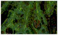
4、向步骤3所得的多重标记复合物中添加核染色剂,孵育,洗涤,封片,以显微镜成像、检测。染色情况如图2所示。荧光强度如表格1所示:4. Add a nuclear staining agent to the multiple labeling complex obtained in step 3, incubate, wash, cover the slides, image and detect with a microscope. The dyeing situation is shown in Figure 2. The fluorescence intensity is shown in Table 1:
表格1荧光强度Table 1 Fluorescence intensity
注:+表示阳性,+越多阳性越强;-表示阴性;+表示有背景,+越多背景越强;-表示无背景。Note: + means positive, the more +, the stronger the positive; - means negative; + means there is background, the more +, the stronger the background; - means no background.
人乳腺癌切片CD4,CD8,PD-1传统荧光染色测试结果如表格2所示:The results of CD4, CD8, and PD-1 traditional fluorescent staining of human breast cancer sections are shown in Table 2:
表格2荧光强度Table 2 Fluorescence Intensity
注:+表示阳性,+越多阳性越强;-表示阴性;+表示有背景,+越多背景越强;-表示无背景。Note: + means positive, the more +, the stronger the positive; - means negative; + means there is background, the more +, the stronger the background; - means no background.
相比传统荧光染色效果,增强型荧光抗体阳性定位准确,荧光信号有明显增强,荧光背景也更低。Compared with the effect of traditional fluorescent staining, the positive localization of the enhanced fluorescent antibody is accurate, the fluorescent signal is significantly enhanced, and the fluorescent background is also lower.
实施例2:Example 2:
分别制备IgG为NLRP3,荧光素为570-Cy3;IgG为ACE2,荧光素为FIT C;IgG为SMA,荧光素为Cy5的增强型荧光抗体Cy3-DGL-NLRP3、FITC-DG L-ACE2和Cy5-DGL-SMA;其余步骤和参数与实施例1相同。染色情况如图3所示,荧光强度如表格3所示:Prepare the enhanced fluorescent antibodies Cy3-DGL-NLRP3, FITC-DGL-ACE2 and Cy5 with NLRP3 as IgG and 570-Cy3 as fluorescein; ACE2 as IgG and FITC as fluorescein as FITC; SMA as SMA as IgG and Cy5 as fluorescein as Cy5 -DGL-SMA; the remaining steps and parameters are the same as in Example 1. The staining situation is shown in Figure 3, and the fluorescence intensity is shown in Table 3:
表格3荧光强度Table 3 Fluorescence Intensity
注:+表示阳性,+越多阳性越强;-表示阴性;+表示有背景,+越多背景越强;-表示无背景。Note: + means positive, the more +, the stronger the positive; - means negative; + means there is background, the more +, the stronger the background; - means no background.
NLRP3,ACE2,SMA传统荧光染色测试结果如表格4所示:NLRP3, ACE2, SMA traditional fluorescent staining test results are shown in Table 4:
表格4荧光强度Table 4 Fluorescence Intensity
注:+表示阳性,+越多阳性越强;-表示阴性;+表示有背景,+越多背景越强;-表示无背景。Note: + means positive, the more +, the stronger the positive; - means negative; + means there is background, the more +, the stronger the background; - means no background.
相比传统荧光染色效果,增强型荧光抗体阳性定位准确,荧光信号有明显增强,荧光背景也更低。Compared with the effect of traditional fluorescent staining, the positive localization of the enhanced fluorescent antibody is accurate, the fluorescent signal is significantly enhanced, and the fluorescent background is also lower.
实施例3:Example 3:
分别制备IgG为CD4,荧光素为570-Cy3;IgG为CD20,荧光素为FITC;IgG为PD-L1,荧光素为Cy5的增强型荧光抗体Cy3-DGL-CD4、FITC-DGL-CD20和Cy5-DGL-PD-L1;其余步骤和参数与实施例1相同。染色情况如图4所示,荧光强度如表格5所示:Prepare CD4 for IgG, 570-Cy3 for fluorescein; CD20 for IgG, FITC for fluorescein; PD-L1 for IgG, and Cy5 for fluorescein as enhanced fluorescent antibodies Cy3-DGL-CD4, FITC-DGL-CD20 and Cy5 -DGL-PD-L1; the remaining steps and parameters are the same as in Example 1. The staining situation is shown in Figure 4, and the fluorescence intensity is shown in Table 5:
表格5荧光强度Table 5 Fluorescence Intensity
注:+表示阳性,+越多阳性越强;-表示阴性;+表示有背景,+越多背景越强;-表示无背景。Note: + means positive, the more +, the stronger the positive; - means negative; + means there is background, the more +, the stronger the background; - means no background.
将Cy3-DGL-CD4稀释10倍进行染色测试,结果如表格6所示:Dilute Cy3-DGL-CD4 10 times for staining test, the results are shown in Table 6:
表格6荧光强度Table 6 Fluorescence Intensity
注:+表示阳性,+越多阳性越强;-表示阴性;+表示有背景,+越多背景越强;-表示无背景。Note: + means positive, the more +, the stronger the positive; - means negative; + means there is background, the more +, the stronger the background; - means no background.
将Cy3-DGL-CD4稀释15倍进行染色测试,结果如表格7所示:Dilute Cy3-DGL-CD4 15 times for staining test, the results are shown in Table 7:
表格7荧光强度Table 7 Fluorescence Intensity


注:+表示阳性,+越多阳性越强;-表示阴性;+表示有背景,+越多背景越强;-表示无背景。Note: + means positive, the more +, the stronger the positive; - means negative; + means there is background, the more +, the stronger the background; - means no background.
增强型荧光抗体不断稀释后,染色强度也能符合要求,说明增强型荧光抗体具有良好的灵敏度。After continuous dilution of the enhanced fluorescent antibody, the staining intensity can also meet the requirements, indicating that the enhanced fluorescent antibody has good sensitivity.
人肠癌切片CD4,CD20,PD-L1传统荧光染色测试结果如表格8所示:The results of CD4, CD20, and PD-L1 traditional fluorescent staining of human intestinal cancer slices are shown in Table 8:
表格8荧光强度Table 8 Fluorescence Intensity
注:+表示阳性,+越多阳性越强;-表示阴性;+表示有背景,+越多背景越强;-表示无背景。Note: + means positive, the more +, the stronger the positive; - means negative; + means there is background, the more +, the stronger the background; - means no background.
相比如图5所示的传统荧光染色,增强型荧光抗体阳性定位准确,荧光信号有明显增强。Compared with the traditional fluorescent staining shown in Figure 5, the positive localization of the enhanced fluorescent antibody is accurate, and the fluorescent signal is significantly enhanced.
对于本领域的技术人员来说,可根据以上描述的技术方案以及构思,做出其它各种相应的改变以及变形,而所有的这些改变以及变形都应该属于本发明权利要求的保护范围之内。For those skilled in the art, various other corresponding changes and modifications can be made according to the technical solutions and ideas described above, and all these changes and modifications should fall within the protection scope of the claims of the present invention.
Claims (10)
Priority Applications (1)
| Application Number | Priority Date | Filing Date | Title |
|---|---|---|---|
| CN202211711212.1ACN115975009A (en) | 2022-12-29 | 2022-12-29 | A kind of enhanced fluorescent antibody, its preparation method and application |
Applications Claiming Priority (1)
| Application Number | Priority Date | Filing Date | Title |
|---|---|---|---|
| CN202211711212.1ACN115975009A (en) | 2022-12-29 | 2022-12-29 | A kind of enhanced fluorescent antibody, its preparation method and application |
Publications (1)
| Publication Number | Publication Date |
|---|---|
| CN115975009Atrue CN115975009A (en) | 2023-04-18 |
Family
ID=85959273
Family Applications (1)
| Application Number | Title | Priority Date | Filing Date |
|---|---|---|---|
| CN202211711212.1APendingCN115975009A (en) | 2022-12-29 | 2022-12-29 | A kind of enhanced fluorescent antibody, its preparation method and application |
Country Status (1)
| Country | Link |
|---|---|
| CN (1) | CN115975009A (en) |
Citations (4)
| Publication number | Priority date | Publication date | Assignee | Title |
|---|---|---|---|---|
| US20080206183A1 (en)* | 2005-04-28 | 2008-08-28 | Central National De La Recherche Scientifique | Method of Preparing Grafted Polylysine Dendrimers |
| CN101333436A (en)* | 2008-08-06 | 2008-12-31 | 湖南大学 | Multicolor optically encoded silicon shell nanorods and preparation method thereof |
| CN101825628A (en)* | 2010-05-04 | 2010-09-08 | 武汉伊艾博科技有限公司 | Competitive immunological detection kit produced by antibody univalent polymerized marking method, use method thereof and application thereof |
| CN114397440A (en)* | 2022-01-20 | 2022-04-26 | 百盛(广州)生物制品有限公司 | Preparation and method of enzyme-labeled secondary antibody based on polylysine macromolecule (DGL) structure |
- 2022
- 2022-12-29CNCN202211711212.1Apatent/CN115975009A/enactivePending
Patent Citations (4)
| Publication number | Priority date | Publication date | Assignee | Title |
|---|---|---|---|---|
| US20080206183A1 (en)* | 2005-04-28 | 2008-08-28 | Central National De La Recherche Scientifique | Method of Preparing Grafted Polylysine Dendrimers |
| CN101333436A (en)* | 2008-08-06 | 2008-12-31 | 湖南大学 | Multicolor optically encoded silicon shell nanorods and preparation method thereof |
| CN101825628A (en)* | 2010-05-04 | 2010-09-08 | 武汉伊艾博科技有限公司 | Competitive immunological detection kit produced by antibody univalent polymerized marking method, use method thereof and application thereof |
| CN114397440A (en)* | 2022-01-20 | 2022-04-26 | 百盛(广州)生物制品有限公司 | Preparation and method of enzyme-labeled secondary antibody based on polylysine macromolecule (DGL) structure |
Similar Documents
| Publication | Publication Date | Title |
|---|---|---|
| CN110095608A (en) | Tumour excretion body nano fluorescent sensor based on Magnetic Isolation and DNA self assembly | |
| CN103267850B (en) | Probe for pathological diagnosis of tumor, and preparation method and application thereof | |
| CN106053405B (en) | A kind of super-resolution optical imaging method based on unimolecule positioning mode | |
| CN103076312B (en) | Cell fluorescent labeling method | |
| CN104316684A (en) | Quantum-dot immunofluorescence kit for detecting cervical carcinoma | |
| CN106980018B (en) | A kind of kit and its application using CD45 immunofluorescences joint CEP17 probe identification circulating tumor cells | |
| EP2728359B1 (en) | A method of sequential and multiple immunostaining for detection of various antigens in the same specimens | |
| CN110095599A (en) | The Microimmunofluorescence test method of cell-free loss | |
| CN118995884A (en) | Method for improving detection efficiency of in-situ ortho-position connection technology | |
| CN104155457B (en) | Relevant " the box-like mark of the polypeptide-protein groups " detection kit of a kind of colorectal cancer | |
| CN104345154B (en) | A kind of double-antibody sandwich test kit detecting many tumors relevant " the box-like mark of polypeptide-protein groups " | |
| CN106153876A (en) | A kind of method utilizing blood glucose meter detection by quantitative biological marker | |
| CN106834511A (en) | A kind of kit of the breast cancer detection based on liquid biopsy | |
| ES2799705T3 (en) | In-situ imaging and cyclic sample multiplexing procedures | |
| CN102313813B (en) | Integration method for enriching and detecting rare cells from biological fluid samples | |
| CN107356756A (en) | A kind of fluorescence probe and its synthetic method and the application in circulating tumor cell detection | |
| CN110361442A (en) | A kind of excretion body and the preparation method and application thereof for mass spectrum flow cytomery | |
| CN117586174B (en) | Near infrared fluorescent probe for diagnosing colorectal cancer and preparation method and application thereof | |
| CN115975009A (en) | A kind of enhanced fluorescent antibody, its preparation method and application | |
| CN103063849A (en) | A method for simultaneous detection of cancer-associated fibroblasts and expressed proteins thereof | |
| CN113912607A (en) | SNAP-tag probe and preparation method and application thereof | |
| Wei et al. | Highly sensitive fluorescent detection of EDIL3 overexpressed exosomes for the diagnosis of triple-negative breast cancer | |
| CN103229057B (en) | The method amplified for signal and reagent | |
| CN104849449B (en) | Application of the enzyme labelled antibody gold nano-probe in diaminobenzidine catalyzed coloration and dark-field imaging | |
| WO2012124763A1 (en) | Tissue evaluation method |
Legal Events
| Date | Code | Title | Description |
|---|---|---|---|
| PB01 | Publication | ||
| PB01 | Publication | ||
| SE01 | Entry into force of request for substantive examination | ||
| SE01 | Entry into force of request for substantive examination | ||
| CB02 | Change of applicant information | ||
| CB02 | Change of applicant information | Address after:No. 201, No. 145 Dongyi Road, Donghuan Street, Panyu District, Guangzhou City, Guangdong Province, 510000 Applicant after:Guangzhou ruibei Medical Technology Co.,Ltd. Address before:510000 2303, No. 13, Zexi street, Hanxi Village (Hanxi business center), Zhongcun street, Panyu District, Guangzhou City, Guangdong Province Applicant before:Guangzhou ruibei Medical Technology Co.,Ltd. |