CN115969612A - Biodegradable aqueous humor drainage device for treating glaucoma and preparation method thereof - Google Patents
Biodegradable aqueous humor drainage device for treating glaucoma and preparation method thereofDownload PDFInfo
- Publication number
- CN115969612A CN115969612ACN202211642621.0ACN202211642621ACN115969612ACN 115969612 ACN115969612 ACN 115969612ACN 202211642621 ACN202211642621 ACN 202211642621ACN 115969612 ACN115969612 ACN 115969612A
- Authority
- CN
- China
- Prior art keywords
- drainage
- aqueous humor
- drainage device
- biodegradable
- magnesium alloy
- Prior art date
- Legal status (The legal status is an assumption and is not a legal conclusion. Google has not performed a legal analysis and makes no representation as to the accuracy of the status listed.)
- Pending
Links
Images


Classifications
- A—HUMAN NECESSITIES
- A61—MEDICAL OR VETERINARY SCIENCE; HYGIENE
- A61F—FILTERS IMPLANTABLE INTO BLOOD VESSELS; PROSTHESES; DEVICES PROVIDING PATENCY TO, OR PREVENTING COLLAPSING OF, TUBULAR STRUCTURES OF THE BODY, e.g. STENTS; ORTHOPAEDIC, NURSING OR CONTRACEPTIVE DEVICES; FOMENTATION; TREATMENT OR PROTECTION OF EYES OR EARS; BANDAGES, DRESSINGS OR ABSORBENT PADS; FIRST-AID KITS
- A61F9/00—Methods or devices for treatment of the eyes; Devices for putting in contact-lenses; Devices to correct squinting; Apparatus to guide the blind; Protective devices for the eyes, carried on the body or in the hand
- A61F9/007—Methods or devices for eye surgery
- A61F9/00781—Apparatus for modifying intraocular pressure, e.g. for glaucoma treatment
- A—HUMAN NECESSITIES
- A61—MEDICAL OR VETERINARY SCIENCE; HYGIENE
- A61K—PREPARATIONS FOR MEDICAL, DENTAL OR TOILETRY PURPOSES
- A61K31/00—Medicinal preparations containing organic active ingredients
- A61K31/33—Heterocyclic compounds
- A61K31/395—Heterocyclic compounds having nitrogen as a ring hetero atom, e.g. guanethidine or rifamycins
- A61K31/40—Heterocyclic compounds having nitrogen as a ring hetero atom, e.g. guanethidine or rifamycins having five-membered rings with one nitrogen as the only ring hetero atom, e.g. sulpiride, succinimide, tolmetin, buflomedil
- A61K31/407—Heterocyclic compounds having nitrogen as a ring hetero atom, e.g. guanethidine or rifamycins having five-membered rings with one nitrogen as the only ring hetero atom, e.g. sulpiride, succinimide, tolmetin, buflomedil condensed with other heterocyclic ring systems, e.g. ketorolac, physostigmine
- A—HUMAN NECESSITIES
- A61—MEDICAL OR VETERINARY SCIENCE; HYGIENE
- A61K—PREPARATIONS FOR MEDICAL, DENTAL OR TOILETRY PURPOSES
- A61K9/00—Medicinal preparations characterised by special physical form
- A61K9/70—Web, sheet or filament bases ; Films; Fibres of the matrix type containing drug
- A61K9/7007—Drug-containing films, membranes or sheets
- A—HUMAN NECESSITIES
- A61—MEDICAL OR VETERINARY SCIENCE; HYGIENE
- A61L—METHODS OR APPARATUS FOR STERILISING MATERIALS OR OBJECTS IN GENERAL; DISINFECTION, STERILISATION OR DEODORISATION OF AIR; CHEMICAL ASPECTS OF BANDAGES, DRESSINGS, ABSORBENT PADS OR SURGICAL ARTICLES; MATERIALS FOR BANDAGES, DRESSINGS, ABSORBENT PADS OR SURGICAL ARTICLES
- A61L31/00—Materials for other surgical articles, e.g. stents, stent-grafts, shunts, surgical drapes, guide wires, materials for adhesion prevention, occluding devices, surgical gloves, tissue fixation devices
- A61L31/02—Inorganic materials
- A61L31/022—Metals or alloys
- A—HUMAN NECESSITIES
- A61—MEDICAL OR VETERINARY SCIENCE; HYGIENE
- A61L—METHODS OR APPARATUS FOR STERILISING MATERIALS OR OBJECTS IN GENERAL; DISINFECTION, STERILISATION OR DEODORISATION OF AIR; CHEMICAL ASPECTS OF BANDAGES, DRESSINGS, ABSORBENT PADS OR SURGICAL ARTICLES; MATERIALS FOR BANDAGES, DRESSINGS, ABSORBENT PADS OR SURGICAL ARTICLES
- A61L31/00—Materials for other surgical articles, e.g. stents, stent-grafts, shunts, surgical drapes, guide wires, materials for adhesion prevention, occluding devices, surgical gloves, tissue fixation devices
- A61L31/08—Materials for coatings
- A61L31/082—Inorganic materials
- A61L31/086—Phosphorus-containing materials, e.g. apatite
- A—HUMAN NECESSITIES
- A61—MEDICAL OR VETERINARY SCIENCE; HYGIENE
- A61F—FILTERS IMPLANTABLE INTO BLOOD VESSELS; PROSTHESES; DEVICES PROVIDING PATENCY TO, OR PREVENTING COLLAPSING OF, TUBULAR STRUCTURES OF THE BODY, e.g. STENTS; ORTHOPAEDIC, NURSING OR CONTRACEPTIVE DEVICES; FOMENTATION; TREATMENT OR PROTECTION OF EYES OR EARS; BANDAGES, DRESSINGS OR ABSORBENT PADS; FIRST-AID KITS
- A61F2210/00—Particular material properties of prostheses classified in groups A61F2/00 - A61F2/26 or A61F2/82 or A61F9/00 or A61F11/00 or subgroups thereof
- A61F2210/0004—Particular material properties of prostheses classified in groups A61F2/00 - A61F2/26 or A61F2/82 or A61F9/00 or A61F11/00 or subgroups thereof bioabsorbable
- A—HUMAN NECESSITIES
- A61—MEDICAL OR VETERINARY SCIENCE; HYGIENE
- A61F—FILTERS IMPLANTABLE INTO BLOOD VESSELS; PROSTHESES; DEVICES PROVIDING PATENCY TO, OR PREVENTING COLLAPSING OF, TUBULAR STRUCTURES OF THE BODY, e.g. STENTS; ORTHOPAEDIC, NURSING OR CONTRACEPTIVE DEVICES; FOMENTATION; TREATMENT OR PROTECTION OF EYES OR EARS; BANDAGES, DRESSINGS OR ABSORBENT PADS; FIRST-AID KITS
- A61F2240/00—Manufacturing or designing of prostheses classified in groups A61F2/00 - A61F2/26 or A61F2/82 or A61F9/00 or A61F11/00 or subgroups thereof
- A61F2240/001—Designing or manufacturing processes
- A—HUMAN NECESSITIES
- A61—MEDICAL OR VETERINARY SCIENCE; HYGIENE
- A61F—FILTERS IMPLANTABLE INTO BLOOD VESSELS; PROSTHESES; DEVICES PROVIDING PATENCY TO, OR PREVENTING COLLAPSING OF, TUBULAR STRUCTURES OF THE BODY, e.g. STENTS; ORTHOPAEDIC, NURSING OR CONTRACEPTIVE DEVICES; FOMENTATION; TREATMENT OR PROTECTION OF EYES OR EARS; BANDAGES, DRESSINGS OR ABSORBENT PADS; FIRST-AID KITS
- A61F2250/00—Special features of prostheses classified in groups A61F2/00 - A61F2/26 or A61F2/82 or A61F9/00 or A61F11/00 or subgroups thereof
- A61F2250/0058—Additional features; Implant or prostheses properties not otherwise provided for
- A61F2250/0067—Means for introducing or releasing pharmaceutical products into the body
- A—HUMAN NECESSITIES
- A61—MEDICAL OR VETERINARY SCIENCE; HYGIENE
- A61L—METHODS OR APPARATUS FOR STERILISING MATERIALS OR OBJECTS IN GENERAL; DISINFECTION, STERILISATION OR DEODORISATION OF AIR; CHEMICAL ASPECTS OF BANDAGES, DRESSINGS, ABSORBENT PADS OR SURGICAL ARTICLES; MATERIALS FOR BANDAGES, DRESSINGS, ABSORBENT PADS OR SURGICAL ARTICLES
- A61L2300/00—Biologically active materials used in bandages, wound dressings, absorbent pads or medical devices
- A61L2300/20—Biologically active materials used in bandages, wound dressings, absorbent pads or medical devices containing or releasing organic materials
- A61L2300/204—Biologically active materials used in bandages, wound dressings, absorbent pads or medical devices containing or releasing organic materials with nitrogen-containing functional groups, e.g. aminoxides, nitriles, guanidines
- A—HUMAN NECESSITIES
- A61—MEDICAL OR VETERINARY SCIENCE; HYGIENE
- A61L—METHODS OR APPARATUS FOR STERILISING MATERIALS OR OBJECTS IN GENERAL; DISINFECTION, STERILISATION OR DEODORISATION OF AIR; CHEMICAL ASPECTS OF BANDAGES, DRESSINGS, ABSORBENT PADS OR SURGICAL ARTICLES; MATERIALS FOR BANDAGES, DRESSINGS, ABSORBENT PADS OR SURGICAL ARTICLES
- A61L2420/00—Materials or methods for coatings medical devices
- A61L2420/02—Methods for coating medical devices
- A—HUMAN NECESSITIES
- A61—MEDICAL OR VETERINARY SCIENCE; HYGIENE
- A61L—METHODS OR APPARATUS FOR STERILISING MATERIALS OR OBJECTS IN GENERAL; DISINFECTION, STERILISATION OR DEODORISATION OF AIR; CHEMICAL ASPECTS OF BANDAGES, DRESSINGS, ABSORBENT PADS OR SURGICAL ARTICLES; MATERIALS FOR BANDAGES, DRESSINGS, ABSORBENT PADS OR SURGICAL ARTICLES
- A61L31/00—Materials for other surgical articles, e.g. stents, stent-grafts, shunts, surgical drapes, guide wires, materials for adhesion prevention, occluding devices, surgical gloves, tissue fixation devices
- A61L31/14—Materials characterised by their function or physical properties, e.g. injectable or lubricating compositions, shape-memory materials, surface modified materials
- A61L31/148—Materials at least partially resorbable by the body
- A—HUMAN NECESSITIES
- A61—MEDICAL OR VETERINARY SCIENCE; HYGIENE
- A61L—METHODS OR APPARATUS FOR STERILISING MATERIALS OR OBJECTS IN GENERAL; DISINFECTION, STERILISATION OR DEODORISATION OF AIR; CHEMICAL ASPECTS OF BANDAGES, DRESSINGS, ABSORBENT PADS OR SURGICAL ARTICLES; MATERIALS FOR BANDAGES, DRESSINGS, ABSORBENT PADS OR SURGICAL ARTICLES
- A61L31/00—Materials for other surgical articles, e.g. stents, stent-grafts, shunts, surgical drapes, guide wires, materials for adhesion prevention, occluding devices, surgical gloves, tissue fixation devices
- A61L31/14—Materials characterised by their function or physical properties, e.g. injectable or lubricating compositions, shape-memory materials, surface modified materials
- A61L31/16—Biologically active materials, e.g. therapeutic substances
- A—HUMAN NECESSITIES
- A61—MEDICAL OR VETERINARY SCIENCE; HYGIENE
- A61P—SPECIFIC THERAPEUTIC ACTIVITY OF CHEMICAL COMPOUNDS OR MEDICINAL PREPARATIONS
- A61P27/00—Drugs for disorders of the senses
- A61P27/02—Ophthalmic agents
- A61P27/06—Antiglaucoma agents or miotics
Landscapes
- Health & Medical Sciences (AREA)
- Life Sciences & Earth Sciences (AREA)
- Veterinary Medicine (AREA)
- Animal Behavior & Ethology (AREA)
- General Health & Medical Sciences (AREA)
- Public Health (AREA)
- Heart & Thoracic Surgery (AREA)
- Vascular Medicine (AREA)
- Surgery (AREA)
- Ophthalmology & Optometry (AREA)
- Epidemiology (AREA)
- Engineering & Computer Science (AREA)
- Chemical & Material Sciences (AREA)
- Nuclear Medicine, Radiotherapy & Molecular Imaging (AREA)
- Biomedical Technology (AREA)
- Medicinal Chemistry (AREA)
- Pharmacology & Pharmacy (AREA)
- Bioinformatics & Cheminformatics (AREA)
- Inorganic Chemistry (AREA)
- Prostheses (AREA)
- Chemical Kinetics & Catalysis (AREA)
- General Chemical & Material Sciences (AREA)
- Organic Chemistry (AREA)
- Molecular Biology (AREA)
- Materials For Medical Uses (AREA)
Abstract
Description
Translated fromChinese技术领域technical field
本发明属于医用植入技术领域,具体涉及一种可生物降解的用于治疗青光眼的房水引流装置及其制备方法。The invention belongs to the field of medical implant technology, and in particular relates to a biodegradable aqueous humor drainage device for treating glaucoma and a preparation method thereof.
背景技术Background technique
青光眼是一组以视野缺损,视神经凹陷性萎缩为主的特征性视神经疾病。青光眼为世界第2高危的致盲眼病,是致不可逆盲的首要原因。眼内压升高是青光眼视神经萎缩的主要危险因素,降低眼内压是目前唯一被证实有效的延缓青光眼视神经损害的方法。当药物无法控制眼内压时,手术成为治疗青光眼的主要方法,青光眼引流或滤过手术目的是将因房水改道引流到其他的途径,但手术后滤过、引流通道和周围组织的瘢痕化是手术失败的主要危险因素,目前虽然有一些抗瘢痕药物辅助使用,但是由于其无差异性的抑制细胞增殖,造成一些并发症的发生。Glaucoma is a group of characteristic optic nerve diseases mainly characterized by visual field defect and optic nerve pitting atrophy. Glaucoma is the second most dangerous blinding eye disease in the world and the leading cause of irreversible blindness. Elevated intraocular pressure is the main risk factor for glaucomatous optic atrophy, and reducing intraocular pressure is currently the only proven effective way to delay glaucomatous optic nerve damage. When the intraocular pressure cannot be controlled by drugs, surgery becomes the main method to treat glaucoma. The purpose of glaucoma drainage or filtration surgery is to divert the drainage of aqueous humor to other ways, but the scarring of filtration, drainage channels and surrounding tissues after surgery It is the main risk factor for surgical failure. Although there are some anti-scar drugs that are used as adjunctive drugs, some complications occur due to their indiscriminate inhibition of cell proliferation.
传统青光眼外引流装置,因引流管径恒定,手术早期房水引流容易过畅,引起如:浅前房等手术后并发症。而在引流装置植入后期,因异物长期刺激引起引流装置被瘢痕包裹,使得房水引流量减少,眼内压再次升高,导致手术失败。同时传统青光眼引流管前端长期位于前房中,引流管与角膜内皮持续摩擦可能造成角膜内皮细胞丢失,造成角膜失代偿,角膜雾状混浊丧失透明性,患者出现明显异物感、流泪等不适,需要将引流装置取出,导致眼压再次升高、手术失败,甚至需要行角膜内皮移植手术。Traditional glaucoma external drainage device, because the drainage tube diameter is constant, the aqueous humor drainage is easy to be smooth in the early stage of surgery, which may cause postoperative complications such as shallow anterior chamber. However, in the later stage of drainage device implantation, the drainage device was wrapped in scars due to long-term foreign body stimulation, which reduced the drainage of aqueous humor and increased intraocular pressure again, resulting in the failure of the operation. At the same time, the front end of the traditional glaucoma drainage tube is located in the anterior chamber for a long time. The continuous friction between the drainage tube and the corneal endothelium may cause the loss of corneal endothelial cells, resulting in corneal decompensation, corneal foggy opacity and loss of transparency. The drainage device needs to be taken out, causing the intraocular pressure to rise again, the operation fails, and even endothelial corneal transplantation is required.
本申请人先前申请了一专利号202123353992.0的一种可生物降解的青光眼引流装置,其用于对前房内的房水进行引流,包括引流片;引流片的插入端为斜面,引流片的表面设有引流涂层,使引流片的表面存在内在间隙,将引流装置插入前房后,房水能经引流装置的内在间隙流出;引流装置采用可生物降解材料制成,引流装置植入后能随着时间逐步降解。上述的可生物降解的青光眼引流装置可有效避免因异物刺激造成的炎症、瘢痕及瘢痕包裹,有效避免因引流装置一端长期位于前房内导致摩擦或刺激角膜引起的角膜内皮细胞减少,本引流装置完全生物降解后,可形成天然的房水滤过引流通道(从前房到结膜下),再无异物刺激,从而提高青光眼手术成功率。但是该引流装置存在如下问题:1)该引流装置的插入端为斜面设计,由于插入端较为尖锐,受外力作用时可能损伤晶状体、角膜等眼内组织,从而存在安全隐患,同时也存在与前房联接部位尺寸狭窄而导致房水引流不畅的隐患;2)其利用表面涂层中的内在间隙来引导房水流出,由于涂层中的内在间隙蜿蜒曲折、且尺寸很小,所以存在植入早期房水流动不畅、无法有效降低眼压的隐患;虽然在该引流装置上设置了羟基磷灰石涂层,但由于其所述引流片厚度较厚(0.3~0.8mm),导致其涂层厚度严重受限,最厚不能超过0.003mm,只能起到降低纯镁在房水中降解速率的作用,无法产生调控成纤维细胞增殖的作用,并且由于涂层厚度严重局限,其降低纯镁在房水中降解速率的作用也非常有限,在一些眼部物质对镁及镁合金具有较强腐蚀性的病患中无法使用,严重限制了可降解引流装置的适应症范围。The applicant previously applied for a biodegradable glaucoma drainage device with patent number 202123353992.0, which is used to drain the aqueous humor in the anterior chamber, including a drainage piece; the insertion end of the drainage piece is a slope, and the surface of the drainage piece is Drainage coating is provided, so that there is an internal gap on the surface of the drainage sheet. After the drainage device is inserted into the anterior chamber, the aqueous humor can flow out through the internal gap of the drainage device; the drainage device is made of biodegradable materials, and the drainage device can be implanted. Gradually degrades over time. The above-mentioned biodegradable glaucoma drainage device can effectively avoid inflammation, scar and scar wrapping caused by foreign body stimulation, and effectively avoid the reduction of corneal endothelial cells caused by friction or irritation of the cornea caused by one end of the drainage device being located in the anterior chamber for a long time. After complete biodegradation, a natural aqueous humor filtration and drainage channel (from the anterior chamber to the subconjunctiva) can be formed without foreign body stimulation, thereby improving the success rate of glaucoma surgery. But this drainage device has the following problems: 1) the insertion end of the drainage device is designed as a bevel, and because the insertion end is relatively sharp, it may damage intraocular tissues such as the lens and cornea when subjected to external force, thereby there is a potential safety hazard. The hidden danger of poor drainage of aqueous humor due to the narrow size of the connecting part of the room; 2) It uses the internal gap in the surface coating to guide the outflow of aqueous humor. Since the internal gap in the coating is winding and small in size, there are In the early stage of implantation, the flow of aqueous humor is not smooth, and the hidden dangers that the intraocular pressure cannot be effectively reduced; although the drainage device is equipped with a hydroxyapatite coating, the thickness of the drainage sheet is relatively thick (0.3-0.8mm), resulting in The thickness of the coating is severely limited, and the thickest cannot exceed 0.003mm. It can only reduce the degradation rate of pure magnesium in aqueous humor, but cannot regulate the proliferation of fibroblasts. The degradation rate of pure magnesium in aqueous humor is also very limited, and it cannot be used in some patients whose eye substances are highly corrosive to magnesium and magnesium alloys, which severely limits the scope of indications for degradable drainage devices.
发明内容Contents of the invention
本发明旨在至少在一定程度上解决相关技术中的技术问题之一。为此,本发明的主要目的在于提供一种可生物降解的用于治疗青光眼的房水引流装置,旨在解决现有引流装置在使用时安全性欠佳,以及引流不畅、无法有效降低眼压使疗效欠佳等问题。本发明还提供了该可生物降解的用于治疗青光眼的房水引流装置的制备方法。The present invention aims to solve one of the technical problems in the related art at least to a certain extent. For this reason, the main purpose of the present invention is to provide a biodegradable aqueous humor drainage device for the treatment of glaucoma, aiming to solve the problem of poor safety of the existing drainage device during use, poor drainage, and inability to effectively reduce eye pressure. Problems such as poor curative effect due to pressure. The invention also provides a preparation method of the biodegradable aqueous humor drainage device for treating glaucoma.
本发明的目的是通过以下技术方案实现的:The purpose of the present invention is achieved by the following technical solutions:
一种可生物降解的用于治疗青光眼的房水引流装置,用于联接前房和结膜下间隙,对前房内的房水进行引流,包括整体横截面尺寸均匀的长方体引流片,所述引流片的表面设置有引流间隙,且所述引流片采用可生物降解材料制成。A biodegradable aqueous humor drainage device for treating glaucoma, used for connecting the anterior chamber and the subconjunctival space, and draining the aqueous humor in the anterior chamber, including a rectangular parallelepiped drainage piece with uniform overall cross-sectional size, the drainage Drainage gaps are provided on the surface of the sheet, and the drainage sheet is made of biodegradable materials.
在某些具体实施例中,所述引流片在体外乳酸钠林格注射液中的降解速率0.01-0.1mm/月。In some specific embodiments, the degradation rate of the drainage sheet in sodium lactate Ringer injection in vitro is 0.01-0.1 mm/month.
在某些具体实施例中,所述可生物降解材料为纯镁,所述纯镁中镁含量大于/等于99.99%。In some specific embodiments, the biodegradable material is pure magnesium, and the magnesium content in the pure magnesium is greater than/equal to 99.99%.
在某些具体实施例中,所述可生物降解材料为镁合金,所述镁合金包括按质量百分比计的如下组分:0.01-5.0%Zn,0.0-1%Ca,0.0-1%Sr,余量为镁和不可避免的杂质元素;所述镁合金的抗拉强度大于/等于200MPa。In some specific embodiments, the biodegradable material is a magnesium alloy, and the magnesium alloy includes the following components by mass percentage: 0.01-5.0% Zn, 0.0-1% Ca, 0.0-1% Sr, The balance is magnesium and unavoidable impurity elements; the tensile strength of the magnesium alloy is greater than/equal to 200MPa.
在某些具体实施例中,所述引流间隙为贯穿于引流片长度方向且与长度方向相平行的凹槽。In some specific embodiments, the drainage gap is a groove running through the length direction of the drainage sheet and parallel to the length direction.
进一步,所述凹槽的深度0.01-0.1mm,宽度0.05-0.2mm。Further, the groove has a depth of 0.01-0.1mm and a width of 0.05-0.2mm.
在某些具体实施例中,所述引流片的外表面涂覆有可生物降解的表面涂层,所述表面涂层的厚度小于/等于0.02mm。In some specific embodiments, the outer surface of the drainage sheet is coated with a biodegradable surface coating, and the thickness of the surface coating is less than/equal to 0.02mm.
进一步,所述表面涂层成分选自羟基磷灰石、磷酸三钙或磷酸氢钙中的一种或任意两种及以上的混合物。Further, the surface coating component is selected from one of hydroxyapatite, tricalcium phosphate or calcium hydrogen phosphate, or a mixture of any two or more of them.
进一步,所述引流装置的尺寸为长1-6mm,宽0.5-4.0mm,厚度为0.1-0.5mm。Furthermore, the size of the drainage device is 1-6 mm in length, 0.5-4.0 mm in width, and 0.1-0.5 mm in thickness.
一种前述房水引流装置的制备方法,包括如下步骤:A method for preparing the aforementioned aqueous humor drainage device, comprising the steps of:
1)对纯镁或镁合金铸锭进行热塑性变形或冷塑性变形后再结晶退火;1) Perform thermoplastic deformation or cold plastic deformation on pure magnesium or magnesium alloy ingots and then crystallize and anneal;
2)将步骤1)得到的材料加工成长方体的装置材;2) Process the material obtained in step 1) into a cuboid device material;
3)采用砂纸对装置材表面进行机械打磨、抛光,去除机械加工的残留痕迹;3) Use sandpaper to mechanically grind and polish the surface of the device material to remove the residual traces of mechanical processing;
4)采用机械加工或化学刻蚀方法沿长度方向加工出贯穿与引流片长度方向的引流间隙;4) Machining or chemical etching methods along the length direction to process the drainage gap that runs through the length direction of the drainage sheet;
5)丙酮中超声清洗,然后自然风干。5) Ultrasonic cleaning in acetone, then air-dried naturally.
在某些具体实施例中,还包括在步骤5)得到的材料表面通过化学沉积或电化学沉积法制备涂层。In some specific embodiments, it also includes preparing a coating on the surface of the material obtained in step 5) by chemical deposition or electrochemical deposition.
与现有技术相比,本发明至少具有以下优点:Compared with the prior art, the present invention has at least the following advantages:
1)本发明所提供的房水引流装置,通过采用整体横截面尺寸均匀的长方体引流片,该引流片用于结膜下经巩膜隧道沟通前房后,由于插入端为平整面,即使受外力作用也不会对晶状体、角膜等眼内组织造成损伤,保证了眼部的安全性,并且形成的引流通道尺寸整体均匀,特别是与前房联接的部位尺寸不会狭窄,可以引导房水顺利地由前房流出;1) The aqueous humor drainage device provided by the present invention adopts a cuboid drainage piece with a uniform overall cross-sectional size. The drainage piece is used to communicate with the anterior chamber through the scleral tunnel under the conjunctiva. It will not cause damage to the lens, cornea and other intraocular tissues, ensuring the safety of the eye, and the overall size of the drainage channel formed is uniform, especially the size of the part connected to the anterior chamber will not be narrow, which can guide the aqueous humor to flow smoothly. Outflow from the anterior chamber;
2)该引流片的表面设置有规定尺寸的、平行贯穿引流片长度方向的凹槽作为引流间隙,在植入早期可以引导房水以最短距离由前房经巩膜下流出,迅速降低眼内压;而合理的凹槽尺寸又能避免早期房水的过量引流而导致的眼压过低问题,从而消除术后并发症隐患;2) The surface of the drainage sheet is provided with a groove of a specified size that runs parallel to the length of the drainage sheet as a drainage gap. In the early stage of implantation, it can guide the aqueous humor to flow out from the anterior chamber through the subsclera at the shortest distance, and quickly reduce the intraocular pressure. ; and a reasonable groove size can avoid the problem of low intraocular pressure caused by excessive drainage of aqueous humor in the early stage, thereby eliminating the hidden danger of postoperative complications;
3)本申请所提供的房水引流装置可以使用纯镁或镁合金来制作。当需要使用的引流装置尺寸较大时,可以使用纯镁材料以降低成本和避免合金元素带来的复杂性;当需要使用的引流装置尺寸较小时,则使用镁合金材料,利用其较高的强度来保证引流装置的力学性能。同时,根据实际需要,还可以在表面制作涂层以降低降解速率,延长引流装置在体内降解的时间。因此,该引流装置的适应病症范围大大增加,不仅适合于身体健康、自身引流通道形成能力强的患者,也适合于年老体弱、易发过敏、自身引流通道形成较慢等患者的使用。3) The aqueous humor drainage device provided in this application can be made of pure magnesium or magnesium alloy. When the size of the drainage device to be used is large, pure magnesium material can be used to reduce the cost and avoid the complexity caused by alloy elements; when the size of the drainage device to be used is small, the magnesium alloy material can be used to take advantage of its higher Strength to ensure the mechanical properties of the drainage device. At the same time, according to actual needs, coatings can also be made on the surface to reduce the degradation rate and prolong the degradation time of the drainage device in the body. Therefore, the range of applicable diseases of the drainage device is greatly increased, and it is not only suitable for patients who are in good health and have a strong ability to form self-drainage channels, but also suitable for patients who are elderly, infirm, prone to allergies, and slow to form self-drainage channels.
附图说明Description of drawings
为了更清楚地说明本发明具体实施方式,下面将对具体实施方式或现有技术描述中所需要使用的附图作简单地介绍。In order to illustrate the specific implementation of the present invention more clearly, the following will briefly introduce the drawings that are required for the specific implementation or the description of the prior art.
图1为本发明所提供的房水引流装置的植入眼球的俯视图;Fig. 1 is a top view of the implanted eyeball of the aqueous humor drainage device provided by the present invention;
图2为本发明所提供的房水引流装置的植入眼球的侧面图;Fig. 2 is a side view of the implanted eyeball of the aqueous humor drainage device provided by the present invention;
图3为本发明所提供的房水引流装置的整体结构示意图;3 is a schematic diagram of the overall structure of the aqueous humor drainage device provided by the present invention;
图4为本发明所提供的房水引流装置的侧视结构示意图;Fig. 4 is a side view structural schematic diagram of the aqueous humor drainage device provided by the present invention;
图5为本发明所提供的房水引流装置对瘢痕形成的影响;Fig. 5 is the effect of the aqueous humor drainage device provided by the present invention on scar formation;
图6为本发明所提供的房水引流装置在植入眼球后的降解情况;Fig. 6 is the degradation situation of the aqueous humor drainage device provided by the present invention after being implanted into the eyeball;
图7为本发明所提供的房水引流装置在植入眼球后HE染色情况;Fig. 7 is the HE staining situation after the aqueous humor drainage device provided by the present invention is implanted into the eyeball;
图8为本发明所提供的房水引流装置在植入眼球后的眼内压波动情况;Fig. 8 is the intraocular pressure fluctuation after the aqueous humor drainage device provided by the present invention is implanted into the eyeball;
图9为本发明所提供的房水引流装置在植入眼球后的角膜内皮细胞情况;Fig. 9 is the situation of corneal endothelial cells after the aqueous humor drainage device provided by the present invention is implanted into the eyeball;
图10为本发明所提供的房水引流装置在植入眼球中的房水引流情况;Fig. 10 is the aqueous humor drainage situation when the aqueous humor drainage device provided by the present invention is implanted in the eyeball;
其中,1.引流装置;2.瞳孔;3.结膜;4.角巩膜缘;11、引流片;12、引流间隙;13、表面涂层。Among them, 1. Drainage device; 2. Pupil; 3. Conjunctiva; 4. Corneoscleral limbus; 11. Drainage sheet; 12. Drainage gap; 13. Surface coating.
具体实施方式Detailed ways
下面将结合附图对本发明技术方案的实施例进行详细的描述。以下实施例仅用于更加清楚地说明本发明的技术方案,因此只作为示例,而不能以此来限制本发明的保护范围。Embodiments of the technical solutions of the present invention will be described in detail below in conjunction with the accompanying drawings. The following examples are only used to illustrate the technical solutions of the present invention more clearly, and therefore are only examples, rather than limiting the protection scope of the present invention.
需要注意的是,除非另有说明,本申请使用的技术术语或者科学术语应当为本发明所属领域技术人员所理解的通常意义。本申请中采用的原材料,设备或装置等等,若未进行特殊说明,均认为可以通过商业途径购买获得。It should be noted that, unless otherwise specified, the technical terms or scientific terms used in this application shall have the usual meanings understood by those skilled in the art to which the present invention belongs. The raw materials, equipment or devices used in this application, etc., are considered to be commercially available unless otherwise specified.
在本申请的描述中,需要理解的是,术语“引流装置”是指用于联接前房与结膜下间隙、引导房水从前房流出,以降低眼压、治疗青光眼的一种专用医疗产品;“引流片”是指该引流装置中用可降解镁合金制作的一种片材,是引流装置的基体部分;“引流间隙”是指在引流片表面采用机械加工或化学刻蚀方法形成的一种房水导流通道,是引流装置的一个重要结构单元;“表面涂层”是指在引流片表面制作的一种额外的覆盖层,是用于进一步改善引流装置使用效果和适应症范围的一个组成部分。In the description of this application, it should be understood that the term "drainage device" refers to a special medical product used to connect the anterior chamber and the subconjunctival space, guide the outflow of aqueous humor from the anterior chamber, reduce intraocular pressure, and treat glaucoma; "Drainage sheet" refers to a sheet made of degradable magnesium alloy in the drainage device, which is the base part of the drainage device; "drainage gap" refers to a hole formed on the surface of the drainage sheet by mechanical processing or chemical etching. The water diversion channel of the seed room is an important structural unit of the drainage device; "surface coating" refers to an additional covering layer made on the surface of the drainage sheet, which is used to further improve the use effect and indication range of the drainage device a component.
本发明所提供的一种可生物降解的用于治疗青光眼的房水引流装置,如图1、图2、图3和图4所示,用于联接前房和结膜下间隙,对前房内的房水进行引流,包括整体横截面尺寸均匀的长方体引流片11,引流片11的表面存在引流间隙12,且引流片11采用可生物降解材料制成。具体使用时,引流装置1由角巩膜缘4切口插入前房,并置于结膜3下,使得前房与结膜下连通,通过引流装置1表面的引流间隙12,能有效引流房水,以降低眼内压。A biodegradable aqueous humor drainage device for treating glaucoma provided by the present invention, as shown in Figure 1, Figure 2, Figure 3 and Figure 4, is used to connect the anterior chamber and the subconjunctival space, The aqueous humor is drained, including a rectangular
其中所述可生物降解材料为纯镁或镁合金,其中镁合金进一步为医用镁合金,该可生物降解材料在体外乳酸钠林格注射液(37℃±0.5℃,连续浸泡30天,每天更换模拟房水)中的降解速率为0.01-0.1mm/月,其中镁合金抗拉强度大于/等于200MPa。本申请中的房水引流装置所使用的可降解生物材料可以有效起到沟通前房与巩膜下、构建生理性房水引流通道并防止其闭合的效应。故当本申请中的房水引流装置完全生物降解后,可形成天然的房水引流通道(从前房到结膜下),再无异物刺激,从而提高青光眼手术成功率。还应理解的是,引流装置随时间逐步降解,有效避免术后角膜内皮细胞丢失。Wherein the biodegradable material is pure magnesium or magnesium alloy, wherein the magnesium alloy is further a medical magnesium alloy, and the biodegradable material is soaked in Sodium Lactate Ringer Injection (37°C±0.5°C) continuously for 30 days, and the analogue is changed every day. The degradation rate in aqueous humor) is 0.01-0.1mm/month, and the tensile strength of the magnesium alloy is greater than/equal to 200MPa. The degradable biomaterial used in the aqueous humor drainage device in this application can effectively communicate the anterior chamber and the subsclera, construct a physiological aqueous humor drainage channel and prevent it from closing. Therefore, when the aqueous humor drainage device in the present application is completely biodegradable, a natural aqueous humor drainage channel (from the anterior chamber to the subconjunctiva) can be formed without any stimulation from foreign objects, thereby improving the success rate of glaucoma surgery. It should also be understood that the drainage device gradually degrades over time, effectively avoiding postoperative corneal endothelial cell loss.
其中所述纯镁为按质量百分比计大于/等于99.99%的镁,所述镁合金包括按质量百分比计的如下组分:0.01-5.0%Zn,0.0-1%Ca,0.0-1%Sr,余量为镁和不可避免的杂质元素。Wherein the pure magnesium is greater than/equal to 99.99% magnesium by mass percentage, and the magnesium alloy includes the following components by mass percentage: 0.01-5.0% Zn, 0.0-1% Ca, 0.0-1% Sr, The balance is magnesium and unavoidable impurity elements.
本发明由于使用了整体横截面尺寸均匀的长方体引流片和强度更高的镁合金材料,引流装置无薄弱部位,且强度相应提高,因此手术时不需要专门制作巩膜隧道,而只需要用隧道刀在巩膜上穿刺一个切口,即可将引流装置插入眼部,实现前房与结膜下间隙的联通,引导房水流出眼部以控制眼压。该房水引流装置植入后的使用状态图如图1、图2所示,手术时间大大减少,一般为5-10分钟;比现有青光眼外装置植入手术时间缩短了0.5-1倍,明显提高了手术效率;即本申请中的房水引流装置能够大大减少因巩膜隧道与植入物嵌合欠佳引起房水外漏导致眼压过低等并发症,有显著缩短手术时间,有效避免手术过程中的麻醉失效等隐患;更重要的是,该引流装置能够提高在巩膜隧道中的固定性、减少因巩膜隧道与引流装置嵌合欠佳存在一定间隙引起房水外漏,导致眼压过低、浅前房、脉络膜脱离等并发症的发生,并明显减少手术中粘弹剂的使用,有效提高手术的安全性和有效性。另外,本发明选用的几种合金元素Zn、Ca和Sr均是具有良好生物相容性的元素,通过合理控制其含量,能够到达在提高房水引流装置强度的同时不降低引流装置安全性的目的;同时,在降解过程中,这些合金元素的溶解释放还能对调控成纤维细胞增殖产生一定的辅助作用。但是如果镁合金元素含量过高,则会显著提高镁合金引流装置的降解速率,缩短房水引流装置在体内的有效支撑时间,难以达到了良好的治疗效果。Since the present invention uses a rectangular parallelepiped drainage sheet with uniform overall cross-sectional size and higher strength magnesium alloy material, the drainage device has no weak parts, and the strength is correspondingly improved. Therefore, it is not necessary to specially make a scleral tunnel during the operation, but only a tunnel knife is required. Puncture an incision on the sclera to insert the drainage device into the eye, realize the communication between the anterior chamber and the subconjunctival space, and guide the aqueous humor to flow out of the eye to control intraocular pressure. The use state diagrams of the aqueous humor drainage device after implantation are shown in Figure 1 and Figure 2, and the operation time is greatly reduced, generally 5-10 minutes; compared with the existing glaucoma external device implantation operation time is shortened by 0.5-1 times, Significantly improved operation efficiency; that is, the aqueous humor drainage device in this application can greatly reduce complications such as low intraocular pressure caused by aqueous humor leakage caused by poor fit between the scleral tunnel and the implant, and can significantly shorten the operation time and effectively Avoid hidden dangers such as anesthesia failure during the operation; more importantly, the drainage device can improve the fixation in the scleral tunnel and reduce the leakage of aqueous humor caused by a certain gap between the scleral tunnel and the drainage device, resulting in ocular The incidence of complications such as hypotension, shallow anterior chamber, and choroidal detachment can be significantly reduced, and the use of viscoelastic agents in surgery can be significantly reduced, effectively improving the safety and effectiveness of surgery. In addition, several alloy elements Zn, Ca and Sr selected in the present invention are all elements with good biocompatibility, and by rationally controlling their content, the strength of the aqueous humor drainage device can be improved without reducing the safety of the drainage device. Objective; At the same time, during the degradation process, the dissolution and release of these alloy elements can also play an auxiliary role in regulating the proliferation of fibroblasts. However, if the content of magnesium alloy elements is too high, the degradation rate of the magnesium alloy drainage device will be significantly increased, and the effective support time of the aqueous humor drainage device in the body will be shortened, making it difficult to achieve a good therapeutic effect.
再次参考图3和图4,其中所述引流间隙12通过在引流装置表面经机械加工或化学刻蚀而形成;进一步的,其中所述引流间隙12为贯穿于引流片长度方向且与长度方向相平行的凹槽,深度0.01-0.1mm,宽度0.05-0.2mm。Referring to Fig. 3 and Fig. 4 again, wherein the
本发明通过在该引流片的表面通过机械加工或化学刻蚀方法制作了引流间隙,从而保证植入早期就能产生良好的引流效果。在引流装置刚植入眼部时,装置与巩膜直接贴合较为紧密,如果仅仅依靠涂层表面存在内在间隙来引流房水,会出现引流不畅现象,很难有效降低眼压以及缓解患者病痛;本发明特意制作的引流间隙从根本上解决了这一难题,所述引流间隙贯穿了引流装置长度方向,也就是直接沟通了前房和结膜下间隙,所以引流装置一经植入,房水就能顺利地经由引流间隙流出,有效降低眼压、缓解患者病痛。引流间隙的深度为0.01-0.1mm,宽度为0.05-0.2mm,如果过浅或过窄,很难达到有效引流效果;如果过深或过宽,则早期(通常是2周左右)房水流出过快,存在眼压过低、术后并发症增加等隐患,也对引流装置强度损失较大。In the present invention, drainage gaps are manufactured on the surface of the drainage sheet through mechanical processing or chemical etching, so as to ensure good drainage effect in the early stage of implantation. When the drainage device is first implanted in the eye, the device is closely attached to the sclera. If the aqueous humor is drained only by the internal gap on the surface of the coating, the drainage will not be smooth, and it is difficult to effectively reduce the intraocular pressure and relieve the pain of the patient. The drainage gap specially made by the present invention fundamentally solves this problem, and the drainage gap runs through the length direction of the drainage device, that is, it directly communicates with the anterior chamber and the subconjunctival space, so once the drainage device is implanted, the aqueous humor It can flow out smoothly through the drainage gap, effectively reducing intraocular pressure and relieving patients' pain. The depth of the drainage gap is 0.01-0.1mm, and the width is 0.05-0.2mm. If it is too shallow or too narrow, it is difficult to achieve an effective drainage effect; if it is too deep or too wide, the aqueous humor will flow out in the early stage (usually about 2 weeks) If it is too fast, there will be hidden dangers such as low intraocular pressure and increased postoperative complications, and it will also cause a greater loss of the strength of the drainage device.
为了降低镁合金引流装置的降解速率以及提高调控成纤维细胞增殖的效果,以提高该引流装置的适应症范围,如图4所示,还可以在所述引流片11的外表面涂覆有可生物降解的表面涂层13,所述表面涂层13的厚度小于/等于0.02mm。进一步的,该表面涂层13成分选自羟基磷灰石、磷酸三钙或磷酸氢钙中的一种或任意两种及以上的混合物。In order to reduce the degradation rate of the magnesium alloy drainage device and improve the effect of regulating the proliferation of fibroblasts, so as to improve the scope of indications of the drainage device, as shown in Figure 4, the outer surface of the
本发明通过在镁合金材料表面涂覆同样可以生物降解的涂层,不仅可以降低镁合金引流装置的降解速率,而且可促进天然巩膜房水引流通道的形成。引流装置通过手术放置于巩膜层间,镁合金引流装置上下两面分别与层间巩膜组织相接触,涂层、镁合金基体及其降解产物调控成纤维细胞增殖,分别于巩膜引流通道上下层间各形成一道非常稀薄的纤维增殖膜,待引流装置完全降解后,两层纤维增殖膜增殖与瘢痕化已停止,上下两层纤维膜无法进一步瘢痕愈合,天然房水引流通道形成,有效避免巩膜引流通道因瘢痕化发生堵塞,明显提高青光眼外引流手术的长期有效性。The invention can not only reduce the degradation rate of the magnesium alloy drainage device by coating the surface of the magnesium alloy material with a biodegradable coating, but also can promote the formation of the natural sclera aqueous humor drainage channel. The drainage device is placed between the scleral layers through surgery. The upper and lower sides of the magnesium alloy drainage device are respectively in contact with the interlayer scleral tissue. A very thin fibrous proliferation membrane is formed. After the drainage device is completely degraded, the proliferation and scarring of the two layers of fibrous proliferation membranes have stopped, and the upper and lower fibrous membranes cannot further heal the scar. Natural aqueous humor drainage channels are formed, effectively avoiding scleral drainage channels Blockage occurs due to scarring, which significantly improves the long-term effectiveness of glaucoma drainage surgery.
在本发明中表面涂层厚度最大可达0.02mm,这样就可以在很大的范围内来调整表面涂层厚度,以实现有效降低引流装置降解速率和调控成纤维细胞增殖的目的;而且本发明引入了一些有益的合金元素,所以在一些眼部物质对镁合金腐蚀性较弱的病患中,也可以不使用涂层(即涂层厚度为0);由此可见,本发明的房水引流装置适用的病患范围大大增加。In the present invention, the thickness of the surface coating can be up to 0.02mm, so that the thickness of the surface coating can be adjusted in a large range to achieve the purpose of effectively reducing the degradation rate of the drainage device and regulating the proliferation of fibroblasts; and the present invention Introduced some beneficial alloying elements, so in some patients whose eyes are less corrosive to magnesium alloys, the coating (that is, the thickness of the coating is 0); it can be seen that the aqueous humor of the present invention The range of patients for which the drainage device is applicable has greatly increased.
其中所述引流装置的尺寸为长1-6mm,宽0.5-4.0mm,厚度为0.1-0.5mm。Wherein the size of the drainage device is 1-6 mm in length, 0.5-4.0 mm in width, and 0.1-0.5 mm in thickness.
由于镁合金材料强度的提高,引流装置可以制造的更薄、体积更小,更小的房水引流装置,有助于将本引流装置应用于更多青光眼类型,如;闭角型青光眼;同时,更薄、体积更小的房水引流装置,可进一步减少因引流装置前端(前房内)与角膜内皮细胞持续摩擦导致角膜内皮丢失的风险,有效提高青光眼外引流手术的长期安全性。Due to the improvement of the strength of the magnesium alloy material, the drainage device can be made thinner, smaller, and smaller, which helps to apply the drainage device to more types of glaucoma, such as; angle-closure glaucoma; at the same time , The thinner and smaller aqueous humor drainage device can further reduce the risk of corneal endothelial loss caused by the continuous friction between the front end of the drainage device (in the anterior chamber) and corneal endothelial cells, and effectively improve the long-term safety of external glaucoma drainage surgery.
实施例1:镁合金材料的房水引流装置的制备Example 1: Preparation of aqueous humor drainage device made of magnesium alloy
本实施例中以镁合金(Mg-2Zn-0.1Ca(即含有按重量百分数计2%Zn和0.1%Ca,下同),抗拉强度为220MPa,降解速率为0.03mm/月)为例,用其制备引流装置可经过以下方法获得:Taking magnesium alloy (Mg-2Zn-0.1Ca (that is, containing 2% Zn and 0.1% Ca by weight percentage, the same below), tensile strength is 220MPa, and degradation rate is 0.03mm/month) as an example in the present embodiment, It can be used to prepare a drainage device through the following methods:
a.热挤压态镁合金;a. Hot extruded magnesium alloy;
b.材料加工成长3mm、宽2mm、厚度0.3mm的长方体装置材;b. The material is processed into a cuboid device material with a length of 3mm, a width of 2mm, and a thickness of 0.3mm;
d.装置材表面用400#、600#、800#、1200#砂纸打磨、抛光;d. The surface of the device is ground and polished with 400#, 600#, 800#, 1200# sandpaper;
e.沿长度方向在上下表面分别加工出一条贯穿全长的引流间隙,其宽度为0.3mm、深度为0.1mm,两个表面的引流间隙需错位0.2mm以上。e. Process a drainage gap running through the entire length on the upper and lower surfaces along the length direction, with a width of 0.3mm and a depth of 0.1mm, and the drainage gaps on the two surfaces must be misaligned by more than 0.2mm.
e.丙酮中超声清洗;e. Ultrasonic cleaning in acetone;
f.自然风干,获得镁合金材料引流装置。f. Naturally air-dried to obtain a magnesium alloy material drainage device.
实施例2:镁合金/HA涂层复合材料Embodiment 2: Magnesium alloy/HA coating composite material
在实施例1的基础上,将经过步骤a-f所加工的镁合金材料引流装置为基底,通过电化学沉积的方式,形成羟基磷灰石(HA)涂层,电解液为0.042mol/L(Ca(NO3)2)和0.025mol/L(NH4H2PO4)的水溶液,Ca/P比为1.68,pH为5.0,电流密度10mA/cm2,室温沉积2h,涂层厚度为0.004mm,超声清洗。制备所得的引流装置的降解速率为0.06mm/月。On the basis of Example 1, the magnesium alloy material drainage device processed through steps af is used as the substrate, and a hydroxyapatite (HA) coating is formed by electrochemical deposition, and the electrolyte is 0.042mol/L (Ca (NO3 )2 ) and 0.025mol/L (NH4 H2 PO4 ) aqueous solution, the Ca/P ratio is 1.68, the pH is 5.0, the current density is 10mA/cm2 , deposited at room temperature for 2h, and the coating thickness is 0.004mm , ultrasonic cleaning. The degradation rate of the prepared drainage device was 0.06mm/month.
实施例3:镁合金/磷酸三钙涂层材料的房水引流装置Embodiment 3: Aqueous humor drainage device of magnesium alloy/tricalcium phosphate coating material
本实施例中以镁合金(Mg-5Zn-0.5Sr,抗拉强度280MPa,降解速率为0.05mm/月)为例,用其制备引流装置可经过以下方法获得:In this embodiment, magnesium alloy (Mg-5Zn-0.5Sr, tensile strength 280MPa, degradation rate 0.05mm/month) is taken as an example, and the drainage device prepared with it can be obtained through the following methods:
a.热挤压态镁合金;a. Hot extruded magnesium alloy;
b.材料加工成长1mm、宽0.5mm、厚度0.1mm的长方体装置材;b. The material is processed into a cuboid device material with a length of 1mm, a width of 0.5mm, and a thickness of 0.1mm;
d.装置材表面用400#、600#、800#、1200#砂纸、打磨抛光;d. Use 400#, 600#, 800#, 1200# sandpaper to polish and polish the surface of the device;
e.沿长度方向在上下表面分别加工出一条贯穿全长的引流间隙,其宽度为0.1mm、深度为0.05mm,两个表面的引流间隙需错位0.2mm以上。e. Process a drain gap running through the entire length on the upper and lower surfaces along the length direction, with a width of 0.1 mm and a depth of 0.05 mm. The drain gaps on the two surfaces must be misaligned by more than 0.2 mm.
e.丙酮中超声清洗;e. Ultrasonic cleaning in acetone;
f.自然风干,获得镁合金材料引流装置;f. Naturally air-dried to obtain a magnesium alloy material drainage device;
g.以经上述步骤所加工好的镁合金材料引流装置为基底,通过化学沉积的方式,形成磷酸三钙涂层;g. Using the magnesium alloy drainage device processed through the above steps as the base, form a tricalcium phosphate coating by chemical deposition;
磷酸三钙涂层制作方法为:将经过步骤a-f所加工的镁合金材料引流装置为基底,通过化学沉积的方式,形成磷酸三钙(TCP)涂层,沉积液为0.075mol/L(Ca(NO3)2)和0.05mol/L(Na2HPO4)的水溶液,Ca/P摩尔比为1.5,60℃沉积72h,涂层厚度为0.02mm;The manufacturing method of the tricalcium phosphate coating is as follows: the magnesium alloy material drainage device processed through steps af is used as the base, and the tricalcium phosphate (TCP) coating is formed by chemical deposition, and the deposition liquid is 0.075mol/L (Ca( NO3 )2 ) and 0.05mol/L (Na2 HPO4 ) aqueous solution, the Ca/P molar ratio is 1.5, deposited at 60°C for 72 hours, and the coating thickness is 0.02mm;
i.超声清洗。i. Ultrasonic cleaning.
实施例4:镁合金/磷酸氢钙涂层材料的房水引流装置Embodiment 4: Aqueous humor drainage device of magnesium alloy/calcium hydrogen phosphate coating material
本实施例中以镁合金(Mg-4Zn-1Ca,抗拉强度260MPa,降解速率为0.075mm/月)为例,用其制备引流装置可经过以下方法获得:In this embodiment, magnesium alloy (Mg-4Zn-1Ca, tensile strength 260MPa, degradation rate 0.075mm/month) is taken as an example, and the drainage device prepared with it can be obtained by the following methods:
a.热挤压态镁合金;a. Hot extruded magnesium alloy;
b.材料加工成长3mm、宽1.5mm、厚度0.2mm的长方体装置材;b. The material is processed into a cuboid device material with a length of 3mm, a width of 1.5mm, and a thickness of 0.2mm;
d.装置材表面用400#、600#、800#、1200#砂纸、打磨抛光;d. Use 400#, 600#, 800#, 1200# sandpaper to polish and polish the surface of the device;
e.沿长度方向在上下表面分别加工出1条贯穿全长的引流间隙,其宽度为0.2mm、深度为0.05mm,两个表面的引流间隙需错位0.2mm以上。e. Process a drainage gap running through the entire length on the upper and lower surfaces along the length direction, with a width of 0.2mm and a depth of 0.05mm. The drainage gaps on the two surfaces must be misaligned by more than 0.2mm.
e.丙酮中超声清洗;e. Ultrasonic cleaning in acetone;
f.自然风干,获得镁合金材料引流装置;f. Naturally air-dried to obtain a magnesium alloy material drainage device;
g.以经上述步骤所加工好的镁合金材料引流装置为基底,通过化学沉积的方式,形成磷酸氢钙涂层;g. Using the magnesium alloy material drainage device processed through the above steps as the base, form a calcium hydrogen phosphate coating by chemical deposition;
磷酸氢钙涂层制作方法为:将经过步骤a-f所加工的镁合金材料引流装置为基底,通过电化学沉积的方式,形成磷酸氢钙(DCP)涂层,沉积液为0.032mol/L(Ca(NO3)2)和0.022mol/L(Na2HPO4)的水溶液,Ca/P摩尔比为1.45,50℃沉积10h,涂层厚度为0.01mm;The manufacturing method of the calcium hydrogen phosphate coating is as follows: the magnesium alloy material drainage device processed through the steps af is used as the base, and the calcium hydrogen phosphate (DCP) coating is formed by means of electrochemical deposition, and the deposition liquid is 0.032mol/L (Ca (NO3 )2 ) and 0.022mol/L (Na2 HPO4 ) aqueous solution, the Ca/P molar ratio is 1.45, deposited at 50°C for 10h, and the coating thickness is 0.01mm;
i.超声清洗。i. Ultrasonic cleaning.
实施例5:纯镁/(羟基磷灰石)涂层材料引流装置Embodiment 5: pure magnesium/(hydroxyapatite) coating material drainage device
本实施例中以纯镁(99.99%Mg,降解速率为0.02mm/月)为例,用其制备青光眼引流装置可经过以下方法获得:In this embodiment, pure magnesium (99.99% Mg, with a degradation rate of 0.02mm/month) is taken as an example, and the glaucoma drainage device prepared by using it can be obtained by the following methods:
a.冷挤压后再结晶退火态纯镁;a. Crystallized and annealed pure magnesium after cold extrusion;
b.材料加工成长2.8mm、宽1.5mm、厚度0.4mm的长方体装置材;b. The material is processed into a cuboid device material with a length of 2.8mm, a width of 1.5mm, and a thickness of 0.4mm;
d.装置材表面用400#、600#、800#、1200#砂纸打磨、抛光;d. The surface of the device is ground and polished with 400#, 600#, 800#, 1200# sandpaper;
e.沿长度方向在上下表面分别加工出一条贯穿全长的引流间隙,其宽度为0.2mm、深度为0.08mm,两个表面的引流间隙需错位0.2mm以上。e. Process a drainage gap running through the entire length on the upper and lower surfaces along the length direction, with a width of 0.2mm and a depth of 0.08mm. The drainage gaps on the two surfaces must be misaligned by more than 0.2mm.
e.丙酮中超声清洗;e. Ultrasonic cleaning in acetone;
f.自然风干,获得纯镁材料引流装置;f. Naturally air-dried to obtain a drainage device made of pure magnesium material;
g.以经上述步骤所加工好的纯镁材料引流装置为基底,通过化学沉积的方式,形成羟基磷灰石涂层;g. Using the pure magnesium material drainage device processed through the above steps as the base, form a hydroxyapatite coating by chemical deposition;
羟基磷灰石涂层制作方法为:将经过步骤a-f所加工的镁合金材料引流装置为基底,通过化学沉积的方式,形成涂层,沉积液为0.35mol/L(C10H12CaN2Na2O8·2H2O,EDTA-Ca)和0.45mol/L(KH2PO4)混合溶液,使用NaOH溶液调节pH=7.3,95℃沉积15h,涂层厚度为0.008mm;The manufacturing method of the hydroxyapatite coating is as follows: the magnesium alloy material drainage device processed through steps af is used as the base, and the coating is formed by chemical deposition, and the deposition liquid is 0.35mol/L (C10 H12 CaN2 Na2 O8 ·2H2 O, EDTA-Ca) and 0.45mol/L (KH2 PO4 ) mixed solution, use NaOH solution to adjust pH=7.3, deposit at 95°C for 15h, and the coating thickness is 0.008mm;
i.超声清洗。i. Ultrasonic cleaning.
测试例1:引流装置在植入体内2个月后的房水浓度情况Test Example 1: The concentration of aqueous humor after the drainage device was implanted in the body for 2 months
本申请人针对上述实施例(1、2、3和4),同时引入对比值(常规小梁切除术、医用钛合金),对其植入体内2月后的房水浓度进行测量,具体为:For the above-mentioned embodiments (1, 2, 3 and 4), the applicant simultaneously introduces contrast values (conventional trabeculectomy, medical titanium alloy) to measure the concentration of aqueous humor after 2 months of its implantation, specifically: :
a.10%水合氯醛,3ml/kg腹腔内注射麻醉;a. 10% chloral hydrate, 3ml/kg intraperitoneal injection anesthesia;
b.碘伏消毒,开睑器开睑;b. Povidone iodine disinfection, eyelid opener to open eyelids;
c.1ml空针前房穿刺,取房水0.1ml,置于离心管中;c. Puncture the anterior chamber with a 1ml empty needle, take 0.1ml of aqueous humor, and place it in a centrifuge tube;
d.立即置于-20℃冰箱冷藏;d. Immediately refrigerate at -20°C refrigerator;
e.全自动生化分析仪测量各组房水内的离子浓度。e. The automatic biochemical analyzer measures the concentration of ions in the aqueous humor of each group.
表1.手术后2月房水离子浓度比较Table 1. Comparison of aqueous humor ion concentration in two months after surgery
从表1数据可知,上述实施例1~4制备的房水引流装置植入眼部后,房水的成分与单纯小梁切除和钛合金植入组均无明显差异,表明本发明设计的房水引流装置于眼内生物降解过程中,降解产物对前房内离子浓度变化无明显影响。It can be seen from the data in Table 1 that after the aqueous humor drainage devices prepared in Examples 1 to 4 above are implanted into the eyes, there is no significant difference in the components of the aqueous humor compared with those in the simple trabeculectomy and titanium alloy implantation groups, indicating that the aqueous humor drainage devices designed in the present invention During the biodegradation process of the water drainage device in the eye, the degradation products had no obvious effect on the ion concentration changes in the anterior chamber.
测试例2:Mg-2Zn-0.1Ca/HA引流装置对瘢痕形成的影响Test Example 2: Effect of Mg-2Zn-0.1Ca/HA Drainage Device on Scar Formation
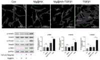
本申请人以实施例2制备所得的房水引流装置为例,对其对瘢痕形成的影响进行测试,具体为:The applicant took the aqueous humor drainage device prepared in Example 2 as an example to test its effect on scar formation, specifically:
(1)蛋白质提取与免疫蛋白印迹(Western blot)(1) Protein extraction and Western blot
a.HA涂层镁合金片,分别放置于24培养孔盘内a. HA-coated magnesium alloy sheets, respectively placed in 24 culture well plates
b.原代HTCFs培养于24孔培养盘内,每孔细胞含量为5x10⁴;b. The primary HTCFs were cultured in a 24-well culture dish, and the cell content in each well was 5x10⁴ ;
c.分别将Mg-2Zn-0.1Ca/HA浸提液、TGFβ1、HA-Mg浸提液+TGFβ1加入孔中,将24孔培养盘,置于37℃5%CO2培养箱中,培养48小时;c. Add Mg-2Zn-0.1Ca/HA extract, TGFβ1, HA-Mg extract + TGFβ1 into the wells respectively, place the 24-well culture plate in a 5% CO2 incubator at 37°C, and cultivate for 48 Hour;
e.标记1.5ml离心管,将离心管放入冰中;e. Label the 1.5ml centrifuge tube and place the centrifuge tube in ice;
f.清洗刮刀;f. Clean the scraper;
g.离心机准备:4℃,3000转10分钟。g. Centrifuge preparation: 4°C, 3000 rpm for 10 minutes.
(2)蛋白质提取;(2) protein extraction;
a.每孔冰PBS冲洗2次;a. Rinse each well twice with ice PBS;
b.每孔加入400ul裂解液(Lysis buffer),24孔盘置于冰上,刮刀小心从样品表面刮下细胞;b. Add 400ul of Lysis buffer to each well, place the 24-well plate on ice, and carefully scrape the cells from the surface of the sample with a scraper;
c.显微镜下检查,确认细胞已被完全收集;c. Check under a microscope to confirm that the cells have been completely collected;
d.将细胞悬液移液于离心管中;d. Pipette the cell suspension into a centrifuge tube;
e.吹打离心管5次,使细胞分布均匀;e. Pipette the
f.4℃,3000转离心10分钟,同时标记新的1.5ml离心管,并将离心管置于冰上;f. Centrifuge at 3000 rpm for 10 minutes at 4°C, label a new 1.5ml centrifuge tube at the same time, and place the centrifuge tube on ice;
g.将上清液移液至新离心管中,并置于冰上。g. Pipette the supernatant into a new centrifuge tube and place on ice.
(3)蛋白质浓度检测;(3) Detection of protein concentration;
a.标记96孔盘;a. Mark the 96-well plate;
b.按照BCA蛋白浓度检测试剂盒说明拟定标准蛋白曲线;b. Draw up a standard protein curve according to the instructions of the BCA protein concentration detection kit;
c.90ul BCA裂解液加入到样品孔,加入10ul样品至96孔板内相应各孔;c. Add 90ul BCA lysate to the sample wells, and add 10ul samples to the corresponding wells in the 96-well plate;
d.每孔加入100ul BCA试剂;d. Add 100ul BCA reagent to each well;
e.将96孔板,置于37℃5%CO2培养箱中30分钟(其余样品保存在-80℃);e. Place the 96-well plate in a 5% CO2 incubator at 37°C for 30 minutes (the remaining samples are stored at -80°C);
f.96孔板置于酶标仪中,在570nm吸光度下读取溶液吸光度值;f. Place the 96-well plate in a microplate reader, and read the absorbance value of the solution at 570nm absorbance;
g.以吸光度,蛋白含量,计算样本蛋白浓度;g. Calculate the sample protein concentration by absorbance and protein content;
h.RIPA溶液平衡各样本中蛋白浓度;h. RIPA solution balances the protein concentration in each sample;
I.缓冲液中加入已经配平的蛋白样品,混匀,蛋白加热器中(100℃)5min后立即置于冰中冷却,12000rpm离心5分钟,放回冰中。I. Add the balanced protein sample into the buffer solution, mix well, put it in the protein heater (100°C) for 5 minutes, then place it in ice to cool immediately, centrifuge at 12000rpm for 5 minutes, and put it back in the ice.
(4)配制电泳胶(4) Preparation of electrophoresis gel
a.70%乙醇清洗凝胶支架及玻璃夹板,组装后加入蒸馏水,测试是否漏水;a. Clean the gel bracket and glass splint with 70% ethanol, add distilled water after assembly, and test for water leakage;
b.按如下剂量配制10%的分离胶(SDS-polyacrylamide gel)2张:b. Prepare 2 sheets of 10% separating gel (SDS-polyacrylamide gel) according to the following dosage:

c.将配置好溶液充分混匀后,溶液加入到玻璃夹层支架中,至最低刻度线,分离胶上方加入蒸馏水,等待约30分钟,待其凝固;c. After fully mixing the prepared solution, add the solution to the glass interlayer bracket to the lowest scale line, add distilled water above the separation gel, and wait for about 30 minutes until it solidifies;
d.按如下剂量配制5%的浓缩胶(Stocking gel)2张d. Prepare 2 sheets of 5% stocking gel according to the following dosage

e.倒出玻璃夹层支架中蒸馏水,将配制好的Stocking胶混匀后加入到10%SDS-polyacrylamide胶的上方;e. Pour out the distilled water in the glass sandwich bracket, mix the prepared Stocking glue and add it on top of the 10% SDS-polyacrylamide glue;
f.70%乙醇清洗样品梳后,插入Stocking胶内,确认Stocking胶与样品梳间隙内无气泡残留,等待约15分钟,待其凝固;f. After cleaning the sample comb with 70% ethanol, insert it into the Stocking glue, confirm that there are no air bubbles in the gap between the Stocking glue and the sample comb, and wait for about 15 minutes for it to solidify;
(5)电泳(5) Electrophoresis
a.配制电泳缓冲液(Running buffer);a. Preparation of electrophoresis buffer (Running buffer);
b.将电泳胶置于电泳仪内;b. Place the electrophoresis gel in the electrophoresis instrument;
c.加入Running buffer到完全浸没电泳胶;c. Add Running buffer to fully submerge the electrophoresis gel;
d.移除样品梳,使用微量移液器吹出胶样残渣;d. Remove the sample comb and use a micropipette to blow out the glue-like residue;
e.在最左侧加入wb蛋白标记(Marker)6ul后,逐个在胶槽内加入各组样品蛋白;e. After adding 6ul of wb protein marker (Marker) on the far left, add each group of sample proteins in the glue tank one by one;
f.Stocking胶使用恒压120V 10分钟,SDS-polyacrylamide胶使用恒压100V约100分钟,直至溴酚蓝(Marker)跑到胶底部时停止电泳。f.Stocking gel uses a constant voltage of 120V for 10 minutes, and SDS-polyacrylamide gel uses a constant voltage of 100V for about 100 minutes, until the bromophenol blue (Marker) runs to the bottom of the gel and stops electrophoresis.
(6)转膜(6) transfer film
a.打开玻璃板,取出电泳胶,并放置于转膜缓冲液中待用;a. Open the glass plate, take out the electrophoresis gel, and place it in the transfer buffer for later use;
b.将电泳胶放置到聚偏二氟乙烯(PVDF)膜上,按海绵、滤纸、胶、PVDF膜、滤纸、海绵顺序放置,其中PVDF膜在正极,凝胶朝向负极,胶和PVDF膜之间避免气泡;封紧后放入电转槽;b. Place the electrophoretic gel on the polyvinylidene fluoride (PVDF) membrane, and place it in the order of sponge, filter paper, glue, PVDF membrane, filter paper, and sponge, wherein the PVDF membrane is on the positive electrode, the gel faces the negative electrode, and the gel and PVDF membrane Avoid air bubbles; seal tightly and put into the electric transfer tank;
c.将PVDF膜从电转槽中取出,TBST液洗脱;c. Take the PVDF membrane out of the electroporation tank and elute with TBST solution;
(7)固定及抗体孵化(7) Fixation and antibody incubation
a.配制固定缓冲液(Running buffer)以及5%BSA;a. Prepare running buffer and 5% BSA;
b.PVDF膜浸没于用TBST配制的5%脱脂牛奶中,室温下缓慢摇荡1小时,TBST洗脱奶粉;b. Immerse the PVDF membrane in 5% skimmed milk prepared with TBST, shake slowly at room temperature for 1 hour, and elute the milk powder with TBST;
c.加入1:1000稀释的α-平滑肌肌动蛋白(α-SMA)抗体,Smad-2、Smad-3抗体,4℃下放置于摇转机上过夜;c. Add 1:1000 diluted α-smooth muscle actin (α-SMA) antibody, Smad-2, Smad-3 antibody, and place on a shaker overnight at 4°C;
d.加入辣根过氧化物酶(HRP)标记的二抗,室温下1小时。d. Add horseradish peroxidase (HRP)-labeled secondary antibody for 1 hour at room temperature.
(8)发光鉴定(8) Luminescence identification
a.按照wb检测试剂盒说明,将混合液均匀滴加在PVDF膜上增强显影;a. According to the instructions of the wb detection kit, evenly drop the mixed solution on the PVDF membrane to enhance the development;
b.Western blot曝光机行条带曝光;b.Western blot exposure machine for strip exposure;
(9)结果分析(9) Analysis of results
结果如图5所示,Mg-2Zn-0.1Ca/HA降解产物通过介导TGFβ/Smad信号通路,抑制Tenon’s成纤维细胞α-SMA表达。The results are shown in Figure 5, Mg-2Zn-0.1Ca/HA degradation products inhibit the expression of Tenon's fibroblast α-SMA by mediating the TGFβ/Smad signaling pathway.
测试例3:引流装置在不同时间的降解情况Test Example 3: Degradation of the drainage device at different times
本测试例以实施例5制备所得的房水引流装置为例,对其在体内的降解情况进行测试,超声生物显微镜(UBM)检查具体为:In this test example, the aqueous humor drainage device prepared in Example 5 is taken as an example to test its degradation in vivo, and the ultrasonic biomicroscope (UBM) inspection is specifically as follows:
a.10%水合氯醛,3ml/kg腹腔内注射麻醉;a. 10% chloral hydrate, 3ml/kg intraperitoneal injection anesthesia;
b.于术后1个月,采用超声生物显微镜(SW-3200L,索维,中国)测量引流装置于沟通前房与结膜下间隙及降解情况。b. One month after the operation, the ultrasonic biomicroscope (SW-3200L, Sower, China) was used to measure the degradation of the drainage device in the anterior chamber and subconjunctival space.
其中图6为术后1月UBM图像引流装置沟通前房与结膜下间隙(新西兰白兔)。从图6可见引流装置于巩膜隧道固定良好,一端位于前房内,一端位于结膜下间隙,滤过泡隆起明显;Figure 6 shows the UBM image drainage device communicating the anterior chamber and the subconjunctival space (New Zealand white rabbit) at 1 month after operation. It can be seen from Figure 6 that the drainage device is well fixed in the scleral tunnel, one end is located in the anterior chamber, and the other end is located in the subconjunctival space, and the filtration bleb is obviously raised;
眼组织切片HE染色具体为:HE staining of eye tissue sections is as follows:
1.组织包埋与切片1. Tissue embedding and sectioning
a.耳缘静脉注射空气10ml,处死实验动物;a. Inject 10ml of air into the ear vein, and kill the experimental animals;
b.于穹窿部剪开结膜及筋膜,暴露巩膜;b. Cut the conjunctiva and fascia at the fornix to expose the sclera;
c.剪断各条肌肉,暴露视神经,球后约1mm处剪断视神经,完整取出眼球;c. Cut off the muscles, expose the optic nerve, cut off the optic nerve about 1mm behind the ball, and take out the eyeball completely;
d.固定:将新鲜组织尽快采用4%多聚甲醛固定48小时;d. Fix: fix the fresh tissue with 4% paraformaldehyde for 48 hours as soon as possible;
e.脱水:组织固定后,流水冲洗12小时,由低浓度到高浓度的乙醇逐级脱水标本,分别为50%乙醇脱水1小时、75%乙醇脱水1小时、85%乙醇脱水1小时、95%乙醇脱水0.5小时、再次95%乙醇脱水0.5小时和无水乙醇脱水0.5小时、再次无水乙醇脱水0.5小时、二甲苯透明0.5小时、再次二甲苯透明0.5小时、浸蜡0.5小时后、再次浸蜡1小时;e. Dehydration: After the tissue is fixed, rinse with running water for 12 hours, and dehydrate the specimen step by step with ethanol from low concentration to high concentration, dehydrating with 50% ethanol for 1 hour, 75% ethanol for 1 hour, 85% ethanol for 1 hour, and 95% ethanol for 1 hour. Dehydration with % ethanol for 0.5 hours, dehydration with 95% ethanol for 0.5 hours and absolute ethanol for 0.5 hours, dehydration with absolute ethanol for 0.5 hours again, xylene transparent for 0.5 hours, xylene transparent again for 0.5 hours, dipping in wax for 0.5 hours, dipping again wax for 1 hour;
f.浸蜡与包埋:将眼球组织转到预热的包埋框中,迅速倒入熔蜡,用温热的镊子调整眼球组织的位置,后放上底板,熔点为52-60℃的石蜡进行包埋,小心去除底板内的气泡,放置于冰上,使熔蜡凝固;f. Wax immersion and embedding: transfer the eyeball tissue to a preheated embedding frame, quickly pour in molten wax, adjust the position of the eyeball tissue with warm tweezers, and then put it on the bottom plate, the melting point is 52-60 ℃ Embed in paraffin, carefully remove air bubbles in the bottom plate, place on ice to solidify the melted wax;
g.切片与展片:刀片修去组织块周围多余石蜡,保留2mm左右石蜡,修整成梯形的组织块,以便于展片时蜡带分离。组织切片厚度为4μm,展片的温度一般为42-45℃,切片置于37℃干燥箱过夜备用。g. Slicing and exhibition: The blade is trimmed to remove excess paraffin around the tissue block, leaving about 2mm of paraffin, trimmed into a trapezoidal tissue block, so as to facilitate the separation of the wax band during the exhibition. The thickness of the tissue slices is 4 μm, the temperature of the slices is generally 42-45°C, and the slices are placed in a 37°C drying oven overnight for later use.
2.H&E染色2. H&E staining
a.将切片放入二甲苯中脱蜡10分钟,换用新的二甲苯再次脱蜡10分钟,无水乙醇处理5分钟,换用新的无水乙醇再次处理5分钟,95%乙醇处理3分钟,85%乙醇处理3分钟,70%乙醇处理3分钟,50%乙醇处理3分钟,自来水冲洗10分钟;a. Dewax the slices in xylene for 10 minutes, dewax with new xylene for another 10 minutes, treat with absolute ethanol for 5 minutes, replace with new absolute ethanol for another 5 minutes, and treat with 95% ethanol for 3 minutes Minutes, 85% ethanol treatment for 3 minutes, 70% ethanol treatment for 3 minutes, 50% ethanol treatment for 3 minutes, tap water for 10 minutes;
b.苏木素染色液染色5分钟后,自来水冲洗1分钟,根据细胞核染色深度使用0.5%盐酸乙醇分化2-30秒;b. After staining with hematoxylin staining solution for 5 minutes, rinse with tap water for 1 minute, and use 0.5% hydrochloric acid ethanol to differentiate for 2-30 seconds according to the staining depth of the nucleus;
c.切片分别放入30%、50%、70%、85%乙醇依次脱水;c. Put the slices into 30%, 50%, 70%, and 85% ethanol to dehydrate in turn;
d.伊红染色液复染10分钟后,95%乙醇脱水1分钟,无水乙醇脱水1分钟,二甲苯透明5分钟,中性树胶封片;d. After restaining with eosin staining solution for 10 minutes, dehydrate with 95% ethanol for 1 minute, dehydrate with absolute ethanol for 1 minute, clear with xylene for 5 minutes, and seal with neutral gum;
e.在显微镜下进行观察,照相。e. Observe and take pictures under a microscope.
图7为术后5月眼组织切片HE染色(新西兰白兔)。从图7可见引流装置完全降解,天然房水引流通道形成,滤过泡隆起明显,HE染色未见明显炎性细胞浸润。说明在房水引流通道形成的同时,眼部组织没有明显炎症反应。Figure 7 shows the HE staining of eye tissue sections (New Zealand white rabbit) at 5 months after operation. It can be seen from Figure 7 that the drainage device was completely degraded, the natural aqueous humor drainage channel was formed, the filtration bleb bulge was obvious, and there was no obvious inflammatory cell infiltration by HE staining. It shows that while the aqueous humor drainage channel is formed, there is no obvious inflammatory reaction in the eye tissue.
测试例4:引流装置对眼压的影响Test Example 4: Effect of drainage device on intraocular pressure
分别于新西兰白兔行本申请实施例1制备所得的房水引流装置植入术(Mg Plate)、小梁切除术(Trabecu l ectomy),未行手术眼(Contro l)作为空白对照组。The aqueous humor drainage device implantation (Mg Plate) and trabeculectomy (Trabeculectomy) prepared in Example 1 of the present application were performed on New Zealand white rabbits respectively, and the unoperated eyes (Control) were used as the blank control group.
每周检测眼压,奥布卡因滴眼液局部麻醉后,Tonopen眼压计测量眼压,取3次眼压值的平均值并记录。结果如图8所示,从图8可知,对其进行术后持续监测眼压5个月的过程中,术后第1个月新型镁合金引流装置组和小梁切除术眼压值均低于空白对照组,后小梁切除术组眼压水平恢复到空白对照组水平,说明其引流通道已完全愈合,而新型镁合金引流装置组眼压水平一直明显低于小梁切除术组及空白对照组,术后3月起新型镁合金引流装置组眼压进一步降低直至术后5月,说明随着引流装置的进一步生物降解,生理性引流通道逐渐扩大,房水引流量逐渐增多,巩膜房水引流通道形成并得以保持。The intraocular pressure was tested every week. After local anesthesia with ubocaine eye drops, the intraocular pressure was measured with a Tonopen tonometer, and the average value of the three intraocular pressure values was taken and recorded. The results are shown in Figure 8. It can be seen from Figure 8 that during the continuous monitoring of intraocular pressure for 5 months after operation, the intraocular pressure values of the new magnesium alloy drainage device group and the trabeculectomy group were all low at the first month after operation In the blank control group, the intraocular pressure level of the posterior trabeculectomy group returned to the level of the blank control group, indicating that the drainage channel has been completely healed, while the intraocular pressure level of the new magnesium alloy drainage device group has been significantly lower than that of the trabeculectomy group and blank control group. In the control group, the intraocular pressure of the new magnesium alloy drainage device group further decreased from 3 months after operation until 5 months after operation, indicating that with the further biodegradation of the drainage device, the physiological drainage channel gradually expanded, the aqueous humor drainage volume gradually increased, and the scleral aqueous humor drainage Flow channels are formed and maintained.
测试例5:引流装置对角膜内皮细胞代偿性的影响Test Example 5: Effect of Drainage Device on Compensation of Corneal Endothelial Cells
本申请实施例1制备所得的房水引流装置为例,测试其植入后对角膜内皮细胞代偿性的影响,本测试例对兔14和兔2的左眼均未行任何手术治疗,兔14的右眼行引流装置手术,兔2的右眼行小梁切除术,具体步骤为:The aqueous humor drainage device prepared in Example 1 of the present application was taken as an example to test its influence on corneal endothelial cell compensation after implantation. In this test example, no surgical treatment was performed on the left eyes of
a.10%水合氯醛,3ml/kg腹腔内注射麻醉;a. 10% chloral hydrate, 3ml/kg intraperitoneal injection anesthesia;
b.于术后5个月,采用角膜内皮细胞计(EM-3000,TOMEY,Japan)测量角膜内皮细胞;结果如图9所示,由图9可知,右眼均行引流装置植入术,左眼为空白对照组(未行任何手术治疗),角膜内皮细胞数量、密度及形态均未见明显差异。b. At 5 months after the operation, the corneal endothelial cells were measured with a corneal endothelial cell meter (EM-3000, TOMEY, Japan); the results are shown in Figure 9, and it can be seen from Figure 9 that the right eye was implanted with a drainage device. The left eye was the blank control group (without any surgical treatment), and there was no significant difference in the number, density and shape of corneal endothelial cells.
测试例6:引流装置对房水引流的影响Test Example 6: Effect of Drainage Device on Aqueous Aqueous Drainage
本申请实施例1制备所得的房水引流装置为例,测试其对房水引流的影响,具体为:The aqueous humor drainage device prepared in Example 1 of the present application was taken as an example, and its influence on aqueous humor drainage was tested, specifically:
分别于术后1月、2月、3月、4月、5月于实验动物前房内注射前囊膜染色剂(台盼蓝),观察是否有染色剂被引流到结膜下,并观察其弥散范围,判断有效引流区域面积,具体操作步骤如下:At 1 month, 2 months, 3 months, 4 months, and 5 months after the operation, the anterior capsule dye (trypan blue) was injected into the anterior chamber of the experimental animals to observe whether the dye was drained to the subconjunctiva, and to observe its Diffusion range, to determine the area of effective drainage area, the specific operation steps are as follows:
a.10%水合氯醛,3ml/kg腹腔内注射麻醉;a. 10% chloral hydrate, 3ml/kg intraperitoneal injection anesthesia;
b.左氧氟沙星眼液点术眼,5分钟1次,点3次;b. Levofloxacin eye drops in the eye, once every 5 minutes, 3 times;
c.碘伏消毒,铺巾,开睑器开睑;c. Disinfect with iodine, spread a towel, and open the eyelids with a lid speculum;
d.1ml空针行前房穿刺,前房内注入台盼蓝0.1ml;d. Anterior chamber puncture was performed with 1ml empty needle, and 0.1ml of trypan blue was injected into the anterior chamber;
e.10分钟后观察染色剂引流情况。e. Observe the drainage of the dye after 10 minutes.
结果如图10所示,术后房水引流情况。A和B:引流装置的植入后的状态图;C:术后1月房水引流情况;D:术后5月房水引流情况;E:术后1月单纯小梁切除后房水引流情况;F:术后5月传统引流管房水引流情况。The results are shown in Figure 10, postoperative aqueous humor drainage. A and B: The status diagram of the drainage device after implantation; C: Aqueous humor drainage at 1 month after operation; D: Aqueous humor drainage at 5 months after operation; E: Aqueous humor drainage after simple trabeculectomy at 1 month after operation Situation; F: Aqueous humor drainage with traditional drainage tube at 5 months after operation.
从图10可知,术后1月于前房内注入台盼蓝,引流装置组可见明显蓝色染色剂被引流到结膜下,而小梁切除术组(E组)结膜未见蓝色染色剂,说明房水引流通道完全愈合。术后5月于期房内注入台盼蓝,结膜下仍可见蓝色染色剂,其弥散面积明显大于引流装置术后1月以及传统引流管术后5月引流弥散面积,说明随着引流装置的降解,巩膜房水引流通道面积逐渐增加,且通道未瘢痕愈合,引流通道得以保持。It can be seen from Figure 10 that trypan blue was injected into the anterior chamber one month after the operation, and the blue dye was obviously drained to the subconjunctiva in the drainage device group, but no blue dye was seen in the conjunctiva of the trabeculectomy group (Group E) , indicating that the aqueous humor drainage channel is completely healed. Trypan blue was injected into the off-
以上实施例仅用以说明本发明的技术方案,而非对其限制;尽管参照前述各实施例对本发明进行了详细的说明,本领域的普通技术人员应当理解:其依然可以对前述各实施例所记载的技术方案进行修改,或者对其中部分或者全部技术特征进行等同替换;而这些修改或者替换,并不使相应技术方案的本质脱离本发明各实施例技术方案的范围,其均应涵盖在本发明的权利要求和说明书的范围当中。The above embodiments are only used to illustrate the technical solutions of the present invention, rather than to limit them; although the present invention has been described in detail with reference to the foregoing embodiments, those of ordinary skill in the art should understand that: it can still be applied to the foregoing embodiments Modifications are made to the technical solutions described, or equivalent replacements are made to some or all of the technical features; and these modifications or replacements do not make the essence of the corresponding technical solutions depart from the scope of the technical solutions of the various embodiments of the present invention, and all of them shall be included in Within the scope of the claims and description of the present invention.
Claims (10)
Translated fromChinesePriority Applications (5)
| Application Number | Priority Date | Filing Date | Title |
|---|---|---|---|
| CN202211642621.0ACN115969612A (en) | 2022-12-20 | 2022-12-20 | Biodegradable aqueous humor drainage device for treating glaucoma and preparation method thereof |
| LU505769ALU505769B1 (en) | 2022-12-20 | 2023-12-13 | Biodegradable aqueous humour drainage device for treating glaucoma and preparation method therefor |
| CN202311750144.4ACN117814992A (en) | 2022-12-20 | 2023-12-19 | Biodegradable aqueous humor drainage device for treating glaucoma, preparation method and application |
| PCT/CN2023/139832WO2024131780A1 (en) | 2022-12-20 | 2023-12-19 | Degradable glaucoma drainage apparatus having scarring and gradient controlling effects, and preparation method therefor |
| US19/083,513US20250221857A1 (en) | 2022-12-20 | 2025-03-19 | Apparatuses for drainage of degradable glaucoma capable of gradient control of scars and methods of making the same |
Applications Claiming Priority (1)
| Application Number | Priority Date | Filing Date | Title |
|---|---|---|---|
| CN202211642621.0ACN115969612A (en) | 2022-12-20 | 2022-12-20 | Biodegradable aqueous humor drainage device for treating glaucoma and preparation method thereof |
Publications (1)
| Publication Number | Publication Date |
|---|---|
| CN115969612Atrue CN115969612A (en) | 2023-04-18 |
Family
ID=85971610
Family Applications (2)
| Application Number | Title | Priority Date | Filing Date |
|---|---|---|---|
| CN202211642621.0APendingCN115969612A (en) | 2022-12-20 | 2022-12-20 | Biodegradable aqueous humor drainage device for treating glaucoma and preparation method thereof |
| CN202311750144.4APendingCN117814992A (en) | 2022-12-20 | 2023-12-19 | Biodegradable aqueous humor drainage device for treating glaucoma, preparation method and application |
Family Applications After (1)
| Application Number | Title | Priority Date | Filing Date |
|---|---|---|---|
| CN202311750144.4APendingCN117814992A (en) | 2022-12-20 | 2023-12-19 | Biodegradable aqueous humor drainage device for treating glaucoma, preparation method and application |
Country Status (4)
| Country | Link |
|---|---|
| US (1) | US20250221857A1 (en) |
| CN (2) | CN115969612A (en) |
| LU (1) | LU505769B1 (en) |
| WO (1) | WO2024131780A1 (en) |
Cited By (1)
| Publication number | Priority date | Publication date | Assignee | Title |
|---|---|---|---|---|
| WO2024131780A1 (en)* | 2022-12-20 | 2024-06-27 | 重庆大学 | Degradable glaucoma drainage apparatus having scarring and gradient controlling effects, and preparation method therefor |
Family Cites Families (13)
| Publication number | Priority date | Publication date | Assignee | Title |
|---|---|---|---|---|
| US6102045A (en)* | 1994-07-22 | 2000-08-15 | Premier Laser Systems, Inc. | Method and apparatus for lowering the intraocular pressure of an eye |
| US6369116B1 (en)* | 1995-06-02 | 2002-04-09 | Oculex Pharmaceuticals, Inc. | Composition and method for treating glaucoma |
| US6713081B2 (en)* | 2001-03-15 | 2004-03-30 | The United States Of America As Represented By The Department Of Health And Human Services | Ocular therapeutic agent delivery devices and methods for making and using such devices |
| US20060240073A1 (en)* | 2002-12-20 | 2006-10-26 | Life Spring Biotech Co. Ltd. | Structure of modulating intraocular pressure on glaucoma |
| US20050048099A1 (en)* | 2003-01-09 | 2005-03-03 | Allergan, Inc. | Ocular implant made by a double extrusion process |
| ES2523454T3 (en)* | 2003-06-16 | 2014-11-26 | Solx, Inc. | Referral for the treatment of glaucoma |
| US8784870B2 (en)* | 2008-07-21 | 2014-07-22 | Otonomy, Inc. | Controlled release compositions for modulating free-radical induced damage and methods of use thereof |
| CN102266282B (en)* | 2011-07-28 | 2013-02-27 | 上海交通大学 | A kind of micro/nano fiber sustained-release preparation for treating scar and preparation method thereof |
| CN105266952A (en)* | 2014-07-09 | 2016-01-27 | 首都医科大学附属北京同仁医院 | A device used in anti-glaucoma surgeries for preventing postoperative conjunctiva adhesion |
| CN109937025B (en)* | 2016-04-20 | 2022-07-29 | 多斯医学公司 | Delivery device for bioabsorbable ocular drugs |
| CA3077101A1 (en)* | 2017-06-13 | 2018-12-20 | Innfocus, Inc. | Systems, methods, and apparatus for treatment of glaucoma |
| CN114869862A (en)* | 2022-04-24 | 2022-08-09 | 温州医科大学附属眼视光医院 | Glaucoma surgery slow-release anti-scar membrane with drainage function and preparation method thereof |
| CN115969612A (en)* | 2022-12-20 | 2023-04-18 | 重庆医科大学附属第三医院(捷尔医院) | Biodegradable aqueous humor drainage device for treating glaucoma and preparation method thereof |
- 2022
- 2022-12-20CNCN202211642621.0Apatent/CN115969612A/enactivePending
- 2023
- 2023-12-13LULU505769Apatent/LU505769B1/enactiveIP Right Grant
- 2023-12-19CNCN202311750144.4Apatent/CN117814992A/enactivePending
- 2023-12-19WOPCT/CN2023/139832patent/WO2024131780A1/ennot_activeCeased
- 2025
- 2025-03-19USUS19/083,513patent/US20250221857A1/enactivePending
Cited By (1)
| Publication number | Priority date | Publication date | Assignee | Title |
|---|---|---|---|---|
| WO2024131780A1 (en)* | 2022-12-20 | 2024-06-27 | 重庆大学 | Degradable glaucoma drainage apparatus having scarring and gradient controlling effects, and preparation method therefor |
Also Published As
| Publication number | Publication date |
|---|---|
| CN117814992A (en) | 2024-04-05 |
| WO2024131780A1 (en) | 2024-06-27 |
| US20250221857A1 (en) | 2025-07-10 |
| LU505769B1 (en) | 2024-06-13 |
Similar Documents
| Publication | Publication Date | Title |
|---|---|---|
| Hicks et al. | Development and clinical assessment of an artificial cornea | |
| Bikbova et al. | Corneal changes in diabetes mellitus | |
| Stanzel et al. | Subretinal delivery of ultrathin rigid-elastic cell carriers using a metallic shooter instrument and biodegradable hydrogel encapsulation | |
| Vallabh et al. | Corneal endothelial cell loss in glaucoma and glaucoma surgery and the utility of management with descemet membrane endothelial keratoplasty (DMEK) | |
| KR101539700B1 (en) | A composition for treating diseases related to angiogenesis and cornea or conjunctiva transplant using chondrocyte-derived extracellular matrix membrane | |
| JPWO2007083685A1 (en) | Corneal endothelial preparation capable of cell proliferation in vivo | |
| LU505769B1 (en) | Biodegradable aqueous humour drainage device for treating glaucoma and preparation method therefor | |
| Andreev et al. | A new collagen scaffold for the improvement of corneal biomechanical properties in a rabbit model | |
| Yang et al. | Application of platelet‐rich fibrin grafts following pterygium excision | |
| US6376543B1 (en) | Secondary cataract inhibitor | |
| Lin et al. | Limbal stem cell dysfunction induced by severe dry eye via activation of the p38 MAPK signaling pathway | |
| KR20090005797A (en) | Antibacterial contact lens containing naringin and manufacturing method thereof | |
| CN219680933U (en) | Biodegradable aqueous humor drainage device for glaucoma treatment | |
| Li et al. | Characterization and regulation of gap junctions in porcine ciliary epithelium | |
| RU2723135C1 (en) | Method of corneal graft preparation for layer-by-layer keratoplasty | |
| RU2082364C1 (en) | Method to treat bullous keratopathy | |
| CN117982732A (en) | A reinforcement material for posterior scleral reinforcement surgery and a preparation method thereof | |
| Wei et al. | Expression and function of PDGF-alpha in columnar epithelial cells of age-related cataracts patients | |
| Treffers | Corneal endothelial wound healing | |
| CN113456675A (en) | Application of acellular SIS in inhibition of scar tissue hyperplasia after glaucoma operation | |
| Lee et al. | Clinical and histopathologic features of failed Descemet stripping automated endothelial keratoplasty grafts | |
| Pilkerton et al. | Experimental vitreous fibroplasia following perforating ocular injuries | |
| Zhang et al. | Plasma processing of artificial corneal endothelium for the treatment of bullous keratopathy: Basic research and clinical application | |
| RU2559918C1 (en) | Method of forming locomotor stump | |
| CN119548677B (en) | A collagen-based artificial cornea with a double-layer structure capable of rapid epithelialization and a preparation method thereof |
Legal Events
| Date | Code | Title | Description |
|---|---|---|---|
| PB01 | Publication | ||
| PB01 | Publication | ||
| SE01 | Entry into force of request for substantive examination | ||
| SE01 | Entry into force of request for substantive examination | ||
| WD01 | Invention patent application deemed withdrawn after publication | ||
| WD01 | Invention patent application deemed withdrawn after publication | Application publication date:20230418 |