CN115901711A - A method for characterizing the three-dimensional structural information of chloroplasts based on three-photon fluorescence microscopy - Google Patents
A method for characterizing the three-dimensional structural information of chloroplasts based on three-photon fluorescence microscopyDownload PDFInfo
- Publication number
- CN115901711A CN115901711ACN202310013337.5ACN202310013337ACN115901711ACN 115901711 ACN115901711 ACN 115901711ACN 202310013337 ACN202310013337 ACN 202310013337ACN 115901711 ACN115901711 ACN 115901711A
- Authority
- CN
- China
- Prior art keywords
- photon fluorescence
- chloroplast
- cell
- chloroplasts
- photon
- Prior art date
- Legal status (The legal status is an assumption and is not a legal conclusion. Google has not performed a legal analysis and makes no representation as to the accuracy of the status listed.)
- Granted
Links
Images


Landscapes
- Investigating, Analyzing Materials By Fluorescence Or Luminescence (AREA)
Abstract
Description
Translated fromChinese技术领域technical field
本发明属于应用光学的生物荧光成像技术领域,具体涉及一种利用三光子荧光显微术对叶绿体三维层析成像进行叶绿体三维结构表征的方法。The invention belongs to the technical field of applied optical biofluorescence imaging, and in particular relates to a method for characterization of chloroplast three-dimensional structure by using three-photon fluorescence microscopy to perform chloroplast three-dimensional tomographic imaging.
背景技术Background technique
三光子荧光显微术具有亚微米量级的空间分辨率,且成像深度大,层析效果好。一方面,由于三光子荧光显微术通常采用近红外光激发,在生物组织中具有更低的散射,激发光波长越长,抗生物组织的散射能力越强,穿透深度也越大。另一方面,三光子荧光为近似五阶非线性光学效应,荧光信号高度局限于聚焦光斑处,因此三光子荧光显微术在生物组织中具有极佳的信号背景比和高的空间分辨率。目前,三光子荧光显微术已经在活体动物脑成像、动物器官成像中实现了高空间分辨、极大深度的成像。Three-photon fluorescence microscopy has sub-micron spatial resolution, large imaging depth and good tomographic effect. On the one hand, since three-photon fluorescence microscopy is usually excited by near-infrared light, which has lower scattering in biological tissues, the longer the wavelength of the excitation light, the stronger the anti-scattering ability of biological tissues and the greater the penetration depth. On the other hand, three-photon fluorescence is an approximate fifth-order nonlinear optical effect, and the fluorescence signal is highly limited to the focused spot, so three-photon fluorescence microscopy has excellent signal-to-background ratio and high spatial resolution in biological tissues. At present, three-photon fluorescence microscopy has achieved high spatial resolution and extremely deep imaging in living animal brain imaging and animal organ imaging.
植物细胞中,叶绿体形态结构和空间分布直接决定了植物碳同化速率,并能随环境变化动态调整,从而改变光合和光保护策略。因此,叶绿体的结构表型是植物生理研究和育种中重要的特性。然而,现有的多种基于光谱技术的叶片叶绿素表征方法仅能计算叶绿素含量,无法对叶绿体结构和分布进行测量。另一方面,透射电镜和扫描电镜技术虽然能实现叶绿体三维结构的重建,但这些方法具有制样繁琐,成像深度局限等缺点。所以,开发一种具有高通量、可原位测量植物细胞叶绿体三维结构表型的方法对于后续植物光合生理研究和以叶绿体结构表型为目标的现代育种具有重要意义。In plant cells, the morphological structure and spatial distribution of chloroplasts directly determine the plant carbon assimilation rate, and can dynamically adjust with environmental changes, thereby changing photosynthesis and photoprotection strategies. Therefore, the structural phenotype of chloroplasts is an important trait in plant physiology research and breeding. However, the existing leaf chlorophyll characterization methods based on spectral techniques can only calculate the chlorophyll content, but cannot measure the structure and distribution of chloroplasts. On the other hand, although transmission electron microscopy and scanning electron microscopy can realize the reconstruction of the three-dimensional structure of chloroplasts, these methods have disadvantages such as cumbersome sample preparation and limited imaging depth. Therefore, the development of a high-throughput, in situ method for measuring the three-dimensional structural phenotype of plant cell chloroplasts is of great significance for subsequent studies on plant photosynthetic physiology and modern breeding targeting chloroplast structural phenotypes.
发明内容Contents of the invention
针对目前技术的不足,本发明提供了一种基于三光子荧光显微术表征叶绿体三维结构信息的方法。本发明可以实现完整叶片的叶绿体三维成像,进而提取叶绿体的体积、密度以及空间分布等信息。Aiming at the deficiencies of the current technology, the present invention provides a method for characterizing chloroplast three-dimensional structural information based on three-photon fluorescence microscopy. The invention can realize the three-dimensional imaging of the chloroplast of the complete leaf, and then extract information such as the volume, density and spatial distribution of the chloroplast.
一种基于三光子荧光显微术表征叶绿体三维结构信息的方法,包括如下步骤:A method for characterizing chloroplast three-dimensional structural information based on three-photon fluorescence microscopy, comprising the steps of:
(1)采用抽真空的方式,将细胞壁染料标记到叶片细胞的细胞壁上;(1) Vacuuming is used to mark the cell wall dye on the cell wall of the leaf cells;
(2)将上述叶片固定在三光子荧光显微镜的载物台上,采用飞秒激光激发,分别对叶绿体和细胞壁进行三光子荧光成像,获取叶片中同一区域的叶绿体和细胞壁的三光子层析成像图;(2) Fix the above leaves on the stage of a three-photon fluorescence microscope, and use femtosecond laser excitation to perform three-photon fluorescence imaging of chloroplasts and cell walls respectively, and obtain three-photon tomography of chloroplasts and cell walls in the same region of the leaves picture;
(3)对获取的植物叶片三维层扫图进行三维重建分析,提取叶片中叶绿体的单个体积、表面积、个数、总体积占细胞体积之比,总表面积与细胞表面积之比以及时空分布动态等结构表型信息。(3) Perform 3D reconstruction and analysis on the obtained 3D layer scan images of plant leaves, and extract the single volume, surface area, number, ratio of total volume to cell volume, ratio of total surface area to cell surface area, and temporal and spatial distribution dynamics of chloroplasts in leaves, etc. Structural phenotype information.
所述的叶绿体三光子荧光成像基于叶绿素的三光子吸收,采用1700 nm飞秒激光激发三光子荧光,标记细胞壁的染料采用碘化丙啶染料,采用1600 nm飞秒激光激发三光子荧光。The chloroplast three-photon fluorescence imaging is based on the three-photon absorption of chlorophyll, and a 1700 nm femtosecond laser is used to excite three-photon fluorescence. The dye for marking the cell wall is propidium iodide dye, and a 1600 nm femtosecond laser is used to excite three-photon fluorescence.
所述的抽真空的方式包含如下步骤:The described vacuumizing method comprises the steps:
(1)在密封盖上钻有小孔的离心管中,装入碘化丙啶染料的重水溶液;(1) Fill the heavy aqueous solution of propidium iodide dye in a centrifuge tube with a small hole drilled on the sealing cover;
(2)将叶片从植株上摘下,用剪刀将叶子剪成小片,装入上述离心管中,此时叶片半浮在液面上;(2) Remove the leaves from the plant, cut the leaves into small pieces with scissors, and put them into the above centrifuge tube. At this time, the leaves are half floating on the liquid surface;
(3)将装有叶片的离心管放入真空釜中,在真空泵的抽气下形成真空,静置3分钟,此时重水溶液中的气泡开始渗出,并伴随叶片下沉;(3) Put the centrifuge tube with blades into the vacuum kettle, form a vacuum under the pumping of the vacuum pump, and let it stand for 3 minutes. At this time, the air bubbles in the heavy aqueous solution begin to seep out, and sink with the blades;
(4)拔开真空阀,释放真空釜中的负压,摇晃离心管使液体中的气泡溢出;重复抽气-静置-恢复过程3-4次,直到叶片完全下沉且再次抽气时无更多气泡溢出。(4) Unplug the vacuum valve, release the negative pressure in the vacuum kettle, shake the centrifuge tube to make the air bubbles in the liquid overflow; repeat the pumping-standstill-recovery process 3-4 times until the blade sinks completely and pumps air again No more bubbles spilling out.
所述的三光子荧光显微镜包含重频1 MHz的飞秒脉冲激光器和扫描显微镜,其中,物镜采用60X的高倍物镜。The three-photon fluorescence microscope includes a femtosecond pulsed laser with a repetition rate of 1 MHz and a scanning microscope, wherein the objective lens adopts a 60X high-magnification objective lens.
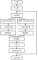
所述的提取叶片中叶绿体的体积、密度以及空间分布等信息的流程为:(1)导入三光子荧光层析成像图,(2)在细胞壁三光子荧光成像通道选择叶肉细胞胞体所在的感兴趣区域(ROI),(3)提取ROI中细胞边界及囊括在内的叶绿体边界空间坐标点云,(4)对细胞/叶绿体对象的边界点云进行平滑处理,(5)三维重建,生成水密模型,即模型表面没有孔隙和漏洞,(6)提取细胞/叶绿体对象体积、表面积、空间位置,(7)三维可视化,(8)计算单个叶绿体体积、表面积、数量,求和后计算占整个细胞的比例,分析叶绿体时空分布动态等显微结构表型特征。The process of extracting the volume, density, and spatial distribution of chloroplasts in leaves is as follows: (1) importing the three-photon fluorescence tomography image, (2) selecting the mesophyll cell body of interest in the three-photon fluorescence imaging channel of the cell wall Region (ROI), (3) Extract the cell boundary and the chloroplast boundary space coordinate point cloud included in the ROI, (4) Smooth the boundary point cloud of the cell/chloroplast object, (5) 3D reconstruction, generate a watertight model , that is, there are no pores and holes on the surface of the model, (6) extract the volume, surface area, and spatial position of the cell/chloroplast object, (7) three-dimensional visualization, (8) calculate the volume, surface area, and quantity of a single chloroplast, and calculate the percentage of the entire cell after summing Proportion, analysis of microstructural phenotypic characteristics such as chloroplast temporal and spatial distribution dynamics.
本发明具有的效益:The benefits that the present invention has:
第一,相较于之前的方法,本发明采用三光子荧光显微术,利用叶绿素的三光子荧光特性,实现了较高通量下表征叶片原位叶绿体三维结构分布特征的方法,对解析植物的光合生理过程具有重要意义。First, compared with the previous methods, the present invention uses three-photon fluorescence microscopy and utilizes the three-photon fluorescence characteristics of chlorophyll to realize a method for characterizing the three-dimensional structure and distribution characteristics of leaf in situ chloroplasts at a higher throughput, which is useful for analyzing plant photosynthetic physiological process is of great significance.
第二,得益于三光子荧光显微术的大深度成像特点,该方法可以实现完整叶片的三光子荧光成像,并进而实现完整叶片下叶肉细胞和叶绿体三维结构特征提取。Second, thanks to the large-depth imaging characteristics of three-photon fluorescence microscopy, this method can realize three-photon fluorescence imaging of complete leaves, and then realize the extraction of three-dimensional structural features of mesophyll cells and chloroplasts under complete leaves.
第三,该方法同时还可以实现植株在体上的叶绿体移动追踪,对解析叶绿体光响应对光合和光保护策略的关联具有重要意义。Third, this method can also track the movement of chloroplasts on the plant body, which is of great significance for analyzing the relationship between chloroplast light response and photosynthetic and photoprotective strategies.
附图说明Description of drawings
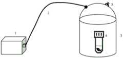
图1为本发明实现对叶片抽真空染色的装置示意图;Fig. 1 is the device schematic diagram that the present invention realizes the vacuum dyeing of blade;
图2为本发明实现叶绿体和细胞壁三光子层析成像的三光子荧光显微镜示意图;Fig. 2 is a schematic diagram of a three-photon fluorescence microscope for realizing three-photon tomography of chloroplasts and cell walls in the present invention;
图3为本发明实现叶肉细胞、叶绿体体积和表面积计算方法流程示意图;Fig. 3 is a schematic flow chart of the method for calculating the volume and surface area of mesophyll cells and chloroplasts in the present invention;
图4为本发明实现叶肉细胞、叶绿体三光子双通道成像提取特征过程示例图;Fig. 4 is an example diagram of the feature extraction process of three-photon dual-channel imaging of mesophyll cells and chloroplasts in the present invention;
图5为本发明实现叶肉细胞、叶绿体三光子双通道成像效果示例图;Fig. 5 is an example diagram of the three-photon dual-channel imaging effect of mesophyll cells and chloroplasts in the present invention;
图6为细胞和叶绿体三维重构可视化结果。Figure 6 shows the visualization results of the three-dimensional reconstruction of cells and chloroplasts.
其中:in:
1 真空泵、2 真空管、3 真空釜、4 离心管、5 真空阀、6低重频(1 MHz)高脉冲激光器、7 一号反射镜、8 二号反射镜、9 三号反射镜、10 扫描振镜、11 扫描透镜、12 套筒透镜、13长通二向色镜(700 nm)、14 物镜、15 载物台、16 长通二向色镜(635 nm)、17 带通滤光片(605-625 nm)、18 带通滤光片(650-700 nm)、19 光电倍增管、20 光电倍增管、21 计算机。1 Vacuum pump, 2 Vacuum tube, 3 Vacuum kettle, 4 Centrifuge tube, 5 Vacuum valve, 6 Low repetition frequency (1 MHz) high pulse laser, 7 No. 1 reflector, 8 No. 2 reflector, 9 No. 3 reflector, 10 Scanning Galvanometer, 11 scanning lens, 12 tube lens, 13 long-pass dichroic mirror (700 nm), 14 objective lens, 15 stage, 16 long-pass dichroic mirror (635 nm), 17 band-pass filter (605-625 nm), 18 bandpass filter (650-700 nm), 19 photomultiplier tube, 20 photomultiplier tube, 21 computer.
具体实施方式Detailed ways
以下结合附图对本发明具体实施方法作进一步清晰完整地阐述,显然,所描述的实施例仅是一部分实施例。基于本发明的实施例,本领域技术人员在没有做出创造性劳动前提下所获得的其他实施例都属于本发明保护的范围。The specific implementation method of the present invention will be further clearly and completely described below in conjunction with the accompanying drawings. Obviously, the described embodiments are only a part of the embodiments. Based on the embodiments of the present invention, other embodiments obtained by those skilled in the art without making creative efforts all fall within the protection scope of the present invention.
本实施例主要包括如下步骤:This embodiment mainly includes the following steps:
(1)如图1所示,将玉米叶片从植株上剪下,装入盖子上钻有小孔的离心管4中,向离心管中装入碘化丙啶染料的重水溶液,此时叶片悬浮在液面。在真空泵1的抽气下,带有真空阀5的真空釜3中的气体通过真空管2被抽出,重水溶液中的气泡也随即渗出,叶片开始下沉。重复操作3-4次,直到玉米叶片完全下沉。(1) As shown in Figure 1, cut the corn leaves from the plant, put them into a centrifuge tube 4 with a small hole drilled on the cover, and put a heavy aqueous solution of propidium iodide dye into the centrifuge tube, and the leaves suspended in the liquid. Under the pumping of the
(2)如图2所示,三光子显微镜系统采用低重频(1 MHz)高脉冲激光器6,扫描显微镜优化了镜组的红外透过率和反射率。激发光经过一号反色镜7进入显微镜,在显微镜中经二号反射镜8和三号反射镜9到达扫描振镜10进行角度扫描,之后经过扫描透镜11和套筒透镜12进行扩束,经过700 nm的长通二向色13后在60倍率的水镜14(UPLSAPO60XW)下方聚焦,激发样品的荧光信号。在荧光探测通道中,采用635 nm的长通二向色镜16分光,透射通道的光电倍增管20前面装有650-700 nm的带通滤光18,收集叶绿体的三光子荧光信号。反射通道中光电倍增管19的前面装有605-625 nm的带通滤光片17,用于收集碘化丙啶染料的三光子荧光信号。将标记了碘化丙啶染料的玉米叶片固定在显微镜的载物台15上,先选用1700nm 飞秒波长的激发光对叶片进行叶绿体的三光子荧光层析成像,之后再更换1600 nm 飞秒波长的激发光对叶片中的碘化丙啶染料进行三光子荧光层析成像,获取完整叶片下的三光子荧光双通道层析成像。(2) As shown in Figure 2, the three-photon microscope system uses a low repetition frequency (1 MHz) high-pulse laser 6, and the scanning microscope optimizes the infrared transmittance and reflectance of the mirror group. The excitation light enters the microscope through the No. 1
(3)在计算机21上对获取的植物叶片三维层层析成像图进行三维重建分析,图3展示了提取叶绿体三维结构信息方法的具体流程图。导入三光子层析成像图,如图4所示,选取叶肉细胞所在区域,通过细胞壁的三光子荧光层析成像勾勒出中间单颗叶肉细胞胞体所在的感兴趣区域(ROI),提取细胞边界空间坐标点云,切换到叶绿体成像通道中,提取叶绿体边界空间坐标点云。图5展示了单颗叶肉细胞胞体ROI下的叶肉细胞、叶绿体三光子双通道成像效果图。之后,利用平滑算法对细胞和叶绿体对象边界点云进行平滑处理,最终生成三角网格,对细胞和叶绿体分别重建水密的模型,经计算该叶肉细胞的体积为49328 μm3,叶肉细胞内的叶绿体一共83个,平均体积243 μm3,平均表面积206 μm2,总体积为20230 μm3,总表面积17108 μm2,叶绿体总体积与细胞体积之比0.41,叶绿体总表面积与细胞表面积之比2.39,对叶绿体空间位置进行计算,得到叶绿体中心点至细胞壁距离平均为6.34 μm,平均最近邻点距离9.29 μm。从生成的图6细胞和叶绿体三维重构可视化图中,可进一步分析单颗叶绿体在叶肉细胞中的相对空间位置,确定叶绿体主要沿着细胞边界分布。(3) Perform three-dimensional reconstruction and analysis on the obtained three-dimensional tomographic image of plant leaves on the
(4)之后,可以对保持活性的植物叶片进行三光子荧光双通道层析成像,并对叶片进行光照处理,期间重复采集同一视野的三光子荧光双通道层析成像,从而分析叶绿体在光照下的时空动态分布规律。(4) After that, three-photon fluorescence dual-channel tomography can be performed on the leaves of the plants that remain active, and the leaves can be treated with light. During this period, the three-photon fluorescence dual-channel tomography of the same field of view can be collected repeatedly, so as to analyze the chloroplasts under light. The spatiotemporal dynamic distribution law.
Claims (5)
Priority Applications (1)
| Application Number | Priority Date | Filing Date | Title |
|---|---|---|---|
| CN202310013337.5ACN115901711B (en) | 2023-01-05 | 2023-01-05 | A method for characterizing the three-dimensional structural information of chloroplasts based on three-photon fluorescence microscopy |
Applications Claiming Priority (1)
| Application Number | Priority Date | Filing Date | Title |
|---|---|---|---|
| CN202310013337.5ACN115901711B (en) | 2023-01-05 | 2023-01-05 | A method for characterizing the three-dimensional structural information of chloroplasts based on three-photon fluorescence microscopy |
Publications (2)
| Publication Number | Publication Date |
|---|---|
| CN115901711Atrue CN115901711A (en) | 2023-04-04 |
| CN115901711B CN115901711B (en) | 2023-08-25 |
Family
ID=86471284
Family Applications (1)
| Application Number | Title | Priority Date | Filing Date |
|---|---|---|---|
| CN202310013337.5AActiveCN115901711B (en) | 2023-01-05 | 2023-01-05 | A method for characterizing the three-dimensional structural information of chloroplasts based on three-photon fluorescence microscopy |
Country Status (1)
| Country | Link |
|---|---|
| CN (1) | CN115901711B (en) |
Cited By (1)
| Publication number | Priority date | Publication date | Assignee | Title |
|---|---|---|---|---|
| TWI860001B (en)* | 2023-07-26 | 2024-10-21 | 國立成功大學 | Method for measuring cellular mechanics in an in vitro fibrosis model |
Citations (11)
| Publication number | Priority date | Publication date | Assignee | Title |
|---|---|---|---|---|
| WO1997011355A1 (en)* | 1995-09-19 | 1997-03-27 | Cornell Research Foundation, Inc. | Multi-photon laser microscopy |
| WO2007040003A1 (en)* | 2005-10-03 | 2007-04-12 | Osaka University | Method of observing dynamic state of intracellular structure and observation apparatus |
| JP2010237554A (en)* | 2009-03-31 | 2010-10-21 | Japan Science & Technology Agency | Fluorescence microscope, fluorescence observation method |
| US20140371582A1 (en)* | 2013-06-13 | 2014-12-18 | Research Foundation Of The City University Of New York | Method of deep tissue imaging using multi-photon excitation of a fluorophore |
| WO2015136583A1 (en)* | 2014-03-11 | 2015-09-17 | 国立大学法人山口大学 | Two-photon-absorbing compound |
| JP2016074787A (en)* | 2014-10-03 | 2016-05-12 | 国立大学法人山口大学 | Luminescent composition that emits light by three-photon excitation |
| US20160238532A1 (en)* | 2013-06-21 | 2016-08-18 | Invenio Imaging Inc. | Multi-photon systems and methods |
| WO2018005623A1 (en)* | 2016-06-28 | 2018-01-04 | The Regents Of The University Of California | Fast two-photon imaging by diffracted swept-laser excitation |
| CN110208227A (en)* | 2019-05-14 | 2019-09-06 | 复旦大学 | A kind of list object lens mating plate micro imaging system |
| US20200063093A1 (en)* | 2017-03-10 | 2020-02-27 | Prellis Biologics, Inc. | Three-dimensional printed organs, devices, and matrices |
| CN114136930A (en)* | 2021-01-14 | 2022-03-04 | 北京林业大学 | A method for rapid identification of plant chloroplast integrity |
- 2023
- 2023-01-05CNCN202310013337.5Apatent/CN115901711B/enactiveActive
Patent Citations (11)
| Publication number | Priority date | Publication date | Assignee | Title |
|---|---|---|---|---|
| WO1997011355A1 (en)* | 1995-09-19 | 1997-03-27 | Cornell Research Foundation, Inc. | Multi-photon laser microscopy |
| WO2007040003A1 (en)* | 2005-10-03 | 2007-04-12 | Osaka University | Method of observing dynamic state of intracellular structure and observation apparatus |
| JP2010237554A (en)* | 2009-03-31 | 2010-10-21 | Japan Science & Technology Agency | Fluorescence microscope, fluorescence observation method |
| US20140371582A1 (en)* | 2013-06-13 | 2014-12-18 | Research Foundation Of The City University Of New York | Method of deep tissue imaging using multi-photon excitation of a fluorophore |
| US20160238532A1 (en)* | 2013-06-21 | 2016-08-18 | Invenio Imaging Inc. | Multi-photon systems and methods |
| WO2015136583A1 (en)* | 2014-03-11 | 2015-09-17 | 国立大学法人山口大学 | Two-photon-absorbing compound |
| JP2016074787A (en)* | 2014-10-03 | 2016-05-12 | 国立大学法人山口大学 | Luminescent composition that emits light by three-photon excitation |
| WO2018005623A1 (en)* | 2016-06-28 | 2018-01-04 | The Regents Of The University Of California | Fast two-photon imaging by diffracted swept-laser excitation |
| US20200063093A1 (en)* | 2017-03-10 | 2020-02-27 | Prellis Biologics, Inc. | Three-dimensional printed organs, devices, and matrices |
| CN110208227A (en)* | 2019-05-14 | 2019-09-06 | 复旦大学 | A kind of list object lens mating plate micro imaging system |
| CN114136930A (en)* | 2021-01-14 | 2022-03-04 | 北京林业大学 | A method for rapid identification of plant chloroplast integrity |
Non-Patent Citations (7)
Cited By (1)
| Publication number | Priority date | Publication date | Assignee | Title |
|---|---|---|---|---|
| TWI860001B (en)* | 2023-07-26 | 2024-10-21 | 國立成功大學 | Method for measuring cellular mechanics in an in vitro fibrosis model |
Also Published As
| Publication number | Publication date |
|---|---|
| CN115901711B (en) | 2023-08-25 |
Similar Documents
| Publication | Publication Date | Title |
|---|---|---|
| US11869176B2 (en) | Hyperspectral imaging system | |
| US7697576B2 (en) | Cytological analysis by raman spectroscopic imaging | |
| US20060281068A1 (en) | Cytological methods for detecting a disease condition such as malignancy by Raman spectroscopic imaging | |
| US20070178067A1 (en) | System and method for cytological analysis by raman spectroscopic imaging | |
| EP2186469B1 (en) | Method and device for the localisation of fluorophores or absorbers in surrounding medium | |
| US20160077007A1 (en) | System and method for controlling depth of imaging in tissues using fluorescence microscopy under ultraviolet excitation following staining with fluorescing agents | |
| CN115901711B (en) | A method for characterizing the three-dimensional structural information of chloroplasts based on three-photon fluorescence microscopy | |
| JP2011530082A (en) | Application of dyes for confocal imaging of cellular microstructures | |
| US20120081518A1 (en) | Method for 3-dimensional microscopic visualization of thick biological tissues | |
| Chen et al. | A high-resolution study of PM2. 5 accumulation inside leaves in leaf stomata compared with non-stomatal areas using three-dimensional X-ray microscopy | |
| Cui et al. | Drop-ANd-See: a simple, real-time, and noninvasive technique for assaying plasmodesmal permeability | |
| Indirabai et al. | Direct estimation of leaf area index of tropical forests using LiDAR point cloud | |
| JP2019528451A (en) | Device for detection of tubules from tissue biopsy material | |
| JP6284024B2 (en) | Cell viability determination system, cell viability determination method | |
| WO2003060477A2 (en) | Iterative optical based histology | |
| CN113299372A (en) | Photoacoustic pathological image processing method, storage medium and terminal device | |
| Glynn et al. | Charting the molecular landscape of neuronal organisation within the hippocampus using cryo electron tomography | |
| CN111110198A (en) | Photoacoustic wavefront shaping microscopic imaging method for biological tissue | |
| JP2024505077A (en) | Analysis of embedded tissue samples using fluorescence-based detection | |
| CN108872166A (en) | Double fluorescence labeling realizes that rape seed is unicellular and oil droplet three-dimensional visualization method | |
| Li et al. | Video-rate multimodal multiphoton imaging and three-dimensional characterization of cellular dynamics in wounded skin | |
| CN107167400B (en) | Method for detecting crude oil density in petroleum inclusion | |
| Santos et al. | Too bright for 2 dimensions: recent progress in advanced 3-dimensional microscopy of the kidney | |
| CN117045178B (en) | Image quality intelligent monitoring method and system for fluorescent endoscope imaging | |
| WO2007011571A2 (en) | Digitizing biology |
Legal Events
| Date | Code | Title | Description |
|---|---|---|---|
| PB01 | Publication | ||
| PB01 | Publication | ||
| SE01 | Entry into force of request for substantive examination | ||
| SE01 | Entry into force of request for substantive examination | ||
| GR01 | Patent grant | ||
| GR01 | Patent grant |