CN115884791A - Method of using medical ice slurry to alleviate symptoms of ocular surface discomfort - Google Patents
Method of using medical ice slurry to alleviate symptoms of ocular surface discomfortDownload PDFInfo
- Publication number
- CN115884791A CN115884791ACN202180024320.9ACN202180024320ACN115884791ACN 115884791 ACN115884791 ACN 115884791ACN 202180024320 ACN202180024320 ACN 202180024320ACN 115884791 ACN115884791 ACN 115884791A
- Authority
- CN
- China
- Prior art keywords
- eye
- cold slurry
- slurry
- cold
- cornea
- Prior art date
- Legal status (The legal status is an assumption and is not a legal conclusion. Google has not performed a legal analysis and makes no representation as to the accuracy of the status listed.)
- Pending
Links
- 239000002002slurrySubstances0.000titleclaimsabstractdescription212
- 238000000034methodMethods0.000titleclaimsabstractdescription79
- 208000024891symptomDiseases0.000titleclaimsabstractdescription16
- 210000004087corneaAnatomy0.000claimsabstractdescription57
- 230000008014freezingEffects0.000claimsabstractdescription32
- 238000007710freezingMethods0.000claimsabstractdescription32
- 230000035807sensationEffects0.000claimsabstractdescription23
- XLYOFNOQVPJJNP-UHFFFAOYSA-NwaterSubstancesOXLYOFNOQVPJJNP-UHFFFAOYSA-N0.000claimsabstractdescription18
- 231100000862numbnessToxicity0.000claimsabstractdescription14
- 230000000994depressogenic effectEffects0.000claimsabstractdescription7
- PEDCQBHIVMGVHV-UHFFFAOYSA-Nglycerol groupChemical groupOCC(O)COPEDCQBHIVMGVHV-UHFFFAOYSA-N0.000claimsdescription97
- 208000004044HypesthesiaDiseases0.000claimsdescription68
- 208000034783hypoesthesiaDiseases0.000claimsdescription68
- 238000011282treatmentMethods0.000claimsdescription38
- 230000036407painEffects0.000claimsdescription37
- 208000002193PainDiseases0.000claimsdescription36
- 230000000699topical effectEffects0.000claimsdescription35
- 239000007924injectionSubstances0.000claimsdescription34
- 238000002347injectionMethods0.000claimsdescription34
- 230000006378damageEffects0.000claimsdescription19
- 230000002035prolonged effectEffects0.000claimsdescription19
- 210000003786scleraAnatomy0.000claimsdescription19
- 208000003556Dry Eye SyndromesDiseases0.000claimsdescription17
- 206010013774Dry eyeDiseases0.000claimsdescription16
- 239000002245particleSubstances0.000claimsdescription14
- 230000003238somatosensory effectEffects0.000claimsdescription10
- 230000001681protective effectEffects0.000claimsdescription9
- 238000001356surgical procedureMethods0.000claimsdescription9
- 208000028006Corneal injuryDiseases0.000claimsdescription8
- 210000000795conjunctivaAnatomy0.000claimsdescription8
- 230000004064dysfunctionEffects0.000claimsdescription8
- 206010015958Eye painDiseases0.000claimsdescription7
- 208000027418Wounds and injuryDiseases0.000claimsdescription6
- 208000014674injuryDiseases0.000claimsdescription6
- 208000004550Postoperative PainDiseases0.000claimsdescription4
- 230000001684chronic effectEffects0.000claimsdescription4
- 208000004454HyperalgesiaDiseases0.000claimsdescription3
- 206010053552allodyniaDiseases0.000claimsdescription3
- 208000002177CataractDiseases0.000claimsdescription2
- 230000009692acute damageEffects0.000claimsdescription2
- 230000036961partial effectEffects0.000claimsdescription2
- 230000008439repair processEffects0.000claimsdescription2
- 210000001508eyeAnatomy0.000description86
- 241000283973Oryctolagus cuniculusSpecies0.000description36
- 235000011187glycerolNutrition0.000description33
- 210000005036nerveAnatomy0.000description29
- 230000000694effectsEffects0.000description28
- 239000000203mixtureSubstances0.000description22
- 241001465754MetazoaSpecies0.000description19
- 239000000243solutionSubstances0.000description18
- 230000035876healingEffects0.000description16
- 230000001886ciliary effectEffects0.000description15
- FAPWRFPIFSIZLT-UHFFFAOYSA-MSodium chlorideChemical compound[Na+].[Cl-]FAPWRFPIFSIZLT-UHFFFAOYSA-M0.000description13
- 239000006072pasteSubstances0.000description13
- 239000006196dropSubstances0.000description11
- 210000000744eyelidAnatomy0.000description11
- 230000007774longtermEffects0.000description11
- 239000012620biological materialSubstances0.000description9
- 238000002425crystallisationMethods0.000description9
- 230000008025crystallizationEffects0.000description9
- 210000001519tissueAnatomy0.000description9
- 238000009472formulationMethods0.000description8
- 239000000546pharmaceutical excipientSubstances0.000description8
- 230000004044responseEffects0.000description8
- 239000000523sampleSubstances0.000description8
- 239000011780sodium chlorideSubstances0.000description8
- 238000001816coolingMethods0.000description7
- 230000006870functionEffects0.000description7
- 230000030214innervationEffects0.000description7
- 230000001225therapeutic effectEffects0.000description7
- 206010002091AnaesthesiaDiseases0.000description6
- 230000037005anaesthesiaEffects0.000description6
- 230000003902lesionEffects0.000description6
- 239000002502liposomeSubstances0.000description6
- RYGMFSIKBFXOCR-UHFFFAOYSA-NCopperChemical compound[Cu]RYGMFSIKBFXOCR-UHFFFAOYSA-N0.000description5
- 208000023715Ocular surface diseaseDiseases0.000description5
- 210000003050axonAnatomy0.000description5
- 230000008859changeEffects0.000description5
- 229910052802copperInorganic materials0.000description5
- 239000010949copperSubstances0.000description5
- 239000010432diamondSubstances0.000description5
- 210000003560epithelium cornealAnatomy0.000description5
- GNBHRKFJIUUOQI-UHFFFAOYSA-NfluoresceinChemical compoundO1C(=O)C2=CC=CC=C2C21C1=CC=C(O)C=C1OC1=CC(O)=CC=C21GNBHRKFJIUUOQI-UHFFFAOYSA-N0.000description5
- 108091008708free nerve endingsProteins0.000description5
- 230000009467reductionEffects0.000description5
- 239000000126substanceSubstances0.000description5
- 238000004781supercoolingMethods0.000description5
- 206010073696Wallerian degenerationDiseases0.000description4
- 230000005495cold plasmaEffects0.000description4
- 210000000981epitheliumAnatomy0.000description4
- 238000012544monitoring processMethods0.000description4
- 210000003007myelin sheathAnatomy0.000description4
- 230000004224protectionEffects0.000description4
- 108020003175receptorsProteins0.000description4
- 230000001953sensory effectEffects0.000description4
- 230000007704transitionEffects0.000description4
- 230000008734wallerian degenerationEffects0.000description4
- 206010010984Corneal abrasionDiseases0.000description3
- DNIAPMSPPWPWGF-UHFFFAOYSA-NPropylene glycolChemical compoundCC(O)CODNIAPMSPPWPWGF-UHFFFAOYSA-N0.000description3
- 230000002411adverseEffects0.000description3
- 210000000133brain stemAnatomy0.000description3
- 150000001875compoundsChemical class0.000description3
- 238000010586diagramMethods0.000description3
- 230000007794irritationEffects0.000description3
- 239000010410layerSubstances0.000description3
- 150000002632lipidsChemical class0.000description3
- 238000005461lubricationMethods0.000description3
- 230000007246mechanismEffects0.000description3
- 210000000929nociceptorAnatomy0.000description3
- 108091008700nociceptorsProteins0.000description3
- 239000012811non-conductive materialSubstances0.000description3
- 229920003023plasticPolymers0.000description3
- 239000004033plasticSubstances0.000description3
- 238000010186stainingMethods0.000description3
- 230000004489tear productionEffects0.000description3
- 238000012360testing methodMethods0.000description3
- 210000003901trigeminal nerveAnatomy0.000description3
- 239000003981vehicleSubstances0.000description3
- LYCAIKOWRPUZTN-UHFFFAOYSA-NEthylene glycolChemical compoundOCCOLYCAIKOWRPUZTN-UHFFFAOYSA-N0.000description2
- 241000282412HomoSpecies0.000description2
- 206010061218InflammationDiseases0.000description2
- 206010052143Ocular discomfortDiseases0.000description2
- 206010033372Pain and discomfortDiseases0.000description2
- BGDKAVGWHJFAGW-UHFFFAOYSA-NTropicamideChemical compoundC=1C=CC=CC=1C(CO)C(=O)N(CC)CC1=CC=NC=C1BGDKAVGWHJFAGW-UHFFFAOYSA-N0.000description2
- 230000003444anaesthetic effectEffects0.000description2
- 239000003242anti bacterial agentSubstances0.000description2
- 229940088710antibiotic agentDrugs0.000description2
- 230000006907apoptotic processEffects0.000description2
- 239000000607artificial tearSubstances0.000description2
- 230000033228biological regulationEffects0.000description2
- 230000015572biosynthetic processEffects0.000description2
- 210000004556brainAnatomy0.000description2
- 229960001139cefazolinDrugs0.000description2
- MLYYVTUWGNIJIB-BXKDBHETSA-NcefazolinChemical compoundS1C(C)=NN=C1SCC1=C(C(O)=O)N2C(=O)[C@@H](NC(=O)CN3N=NN=C3)[C@H]2SC1MLYYVTUWGNIJIB-BXKDBHETSA-N0.000description2
- 239000004020conductorSubstances0.000description2
- 239000013078crystalSubstances0.000description2
- 230000007423decreaseEffects0.000description2
- 230000003247decreasing effectEffects0.000description2
- 230000007850degenerationEffects0.000description2
- 230000003292diminished effectEffects0.000description2
- LOKCTEFSRHRXRJ-UHFFFAOYSA-Idipotassium trisodium dihydrogen phosphate hydrogen phosphate dichlorideChemical compoundP(=O)(O)(O)[O-].[K+].P(=O)(O)([O-])[O-].[Na+].[Na+].[Cl-].[K+].[Cl-].[Na+]LOKCTEFSRHRXRJ-UHFFFAOYSA-I0.000description2
- 208000037265diseases, disorders, signs and symptomsDiseases0.000description2
- 239000003814drugSubstances0.000description2
- 238000011156evaluationMethods0.000description2
- 239000003889eye dropSubstances0.000description2
- 229940012356eye dropsDrugs0.000description2
- 239000012530fluidSubstances0.000description2
- 238000005755formation reactionMethods0.000description2
- 238000010438heat treatmentMethods0.000description2
- 230000004054inflammatory processEffects0.000description2
- 230000004410intraocular pressureEffects0.000description2
- 150000002500ionsChemical class0.000description2
- 206010023365keratopathyDiseases0.000description2
- 239000000314lubricantSubstances0.000description2
- 239000000463materialSubstances0.000description2
- 210000004175meibomian glandAnatomy0.000description2
- 238000002844meltingMethods0.000description2
- 230000008018meltingEffects0.000description2
- 230000002981neuropathic effectEffects0.000description2
- 230000001575pathological effectEffects0.000description2
- 230000002085persistent effectEffects0.000description2
- SONNWYBIRXJNDC-VIFPVBQESA-NphenylephrineChemical compoundCNC[C@H](O)C1=CC=CC(O)=C1SONNWYBIRXJNDC-VIFPVBQESA-N0.000description2
- 229960001802phenylephrineDrugs0.000description2
- 239000002953phosphate buffered salineSubstances0.000description2
- 230000035479physiological effects, processes and functionsEffects0.000description2
- 238000012545processingMethods0.000description2
- 230000028327secretionEffects0.000description2
- 230000011664signalingEffects0.000description2
- 239000002904solventSubstances0.000description2
- 230000000638stimulationEffects0.000description2
- 208000011580syndromic diseaseDiseases0.000description2
- 229940124597therapeutic agentDrugs0.000description2
- 229960004791tropicamideDrugs0.000description2
- 210000001170unmyelinated nerve fiberAnatomy0.000description2
- CPKVUHPKYQGHMW-UHFFFAOYSA-N1-ethenylpyrrolidin-2-one;molecular iodineChemical compoundII.C=CN1CCCC1=OCPKVUHPKYQGHMW-UHFFFAOYSA-N0.000description1
- BFUUJUGQJUTPAF-UHFFFAOYSA-N2-(3-amino-4-propoxybenzoyl)oxyethyl-diethylazanium;chlorideChemical compound[Cl-].CCCOC1=CC=C(C(=O)OCC[NH+](CC)CC)C=C1NBFUUJUGQJUTPAF-UHFFFAOYSA-N0.000description1
- -12-hydroxypropylChemical group0.000description1
- 108010078777ColistinProteins0.000description1
- 206010061788Corneal infectionDiseases0.000description1
- 229920000858CyclodextrinPolymers0.000description1
- FBPFZTCFMRRESA-FSIIMWSLSA-ND-GlucitolNatural productsOC[C@H](O)[C@H](O)[C@@H](O)[C@H](O)COFBPFZTCFMRRESA-FSIIMWSLSA-N0.000description1
- FBPFZTCFMRRESA-KVTDHHQDSA-ND-MannitolChemical compoundOC[C@@H](O)[C@@H](O)[C@H](O)[C@H](O)COFBPFZTCFMRRESA-KVTDHHQDSA-N0.000description1
- FBPFZTCFMRRESA-JGWLITMVSA-ND-glucitolChemical compoundOC[C@H](O)[C@@H](O)[C@H](O)[C@H](O)COFBPFZTCFMRRESA-JGWLITMVSA-N0.000description1
- 208000016192Demyelinating diseaseDiseases0.000description1
- 206010012305DemyelinationDiseases0.000description1
- 206010015946Eye irritationDiseases0.000description1
- 239000001116FEMA 4028Substances0.000description1
- 208000003098Ganglion CystsDiseases0.000description1
- WQZGKKKJIJFFOK-GASJEMHNSA-NGlucoseNatural productsOC[C@H]1OC(O)[C@H](O)[C@@H](O)[C@@H]1OWQZGKKKJIJFFOK-GASJEMHNSA-N0.000description1
- 208000003809Herpes Zoster OphthalmicusDiseases0.000description1
- 229920001612Hydroxyethyl starchPolymers0.000description1
- 206010020751HypersensitivityDiseases0.000description1
- PIWKPBJCKXDKJR-UHFFFAOYSA-NIsofluraneChemical compoundFC(F)OC(Cl)C(F)(F)FPIWKPBJCKXDKJR-UHFFFAOYSA-N0.000description1
- YQEZLKZALYSWHR-UHFFFAOYSA-NKetamineChemical compoundC=1C=CC=C(Cl)C=1C1(NC)CCCCC1=OYQEZLKZALYSWHR-UHFFFAOYSA-N0.000description1
- 206010024229LeprosyDiseases0.000description1
- 241000533950LeucojumSpecies0.000description1
- 229930195725MannitolNatural products0.000description1
- 206010065062Meibomian gland dysfunctionDiseases0.000description1
- 102000006386Myelin ProteinsHuman genes0.000description1
- 108010083674Myelin ProteinsProteins0.000description1
- 229930193140NeomycinNatural products0.000description1
- 208000028389Nerve injuryDiseases0.000description1
- 206010030865Ophthalmic herpes zosterDiseases0.000description1
- 239000004372Polyvinyl alcoholSubstances0.000description1
- 229920000153Povidone-iodinePolymers0.000description1
- 229910003798SPO2Inorganic materials0.000description1
- 101100478210Schizosaccharomyces pombe (strain 972 / ATCC 24843) spo2 geneProteins0.000description1
- 206010040030Sensory lossDiseases0.000description1
- CZMRCDWAGMRECN-UGDNZRGBSA-NSucroseChemical compoundO[C@H]1[C@H](O)[C@@H](CO)O[C@@]1(CO)O[C@@H]1[C@H](O)[C@@H](O)[C@H](O)[C@@H](CO)O1CZMRCDWAGMRECN-UGDNZRGBSA-N0.000description1
- 229930006000SucroseNatural products0.000description1
- 208000005400Synovial CystDiseases0.000description1
- XSQUKJJJFZCRTK-UHFFFAOYSA-NUreaChemical compoundNC(N)=OXSQUKJJJFZCRTK-UHFFFAOYSA-N0.000description1
- 206010052428WoundDiseases0.000description1
- 230000002159abnormal effectEffects0.000description1
- 230000007488abnormal functionEffects0.000description1
- 208000004064acoustic neuromaDiseases0.000description1
- 230000004913activationEffects0.000description1
- 239000000654additiveSubstances0.000description1
- 210000001789adipocyteAnatomy0.000description1
- 239000002671adjuvantSubstances0.000description1
- 210000003766afferent neuronAnatomy0.000description1
- 208000026935allergic diseaseDiseases0.000description1
- 230000004075alterationEffects0.000description1
- 210000003484anatomyAnatomy0.000description1
- 238000013459approachMethods0.000description1
- 229940021870bacitracin ophthalmic ointmentDrugs0.000description1
- 230000004888barrier functionEffects0.000description1
- 230000009286beneficial effectEffects0.000description1
- WQZGKKKJIJFFOK-VFUOTHLCSA-Nbeta-D-glucoseChemical compoundOC[C@H]1O[C@@H](O)[C@H](O)[C@@H](O)[C@@H]1OWQZGKKKJIJFFOK-VFUOTHLCSA-N0.000description1
- 229960004853betadexDrugs0.000description1
- 230000005540biological transmissionEffects0.000description1
- 230000004397blinkingEffects0.000description1
- 238000009530blood pressure measurementMethods0.000description1
- 210000004204blood vesselAnatomy0.000description1
- 210000005252bulbus oculiAnatomy0.000description1
- RMRJXGBAOAMLHD-IHFGGWKQSA-NbuprenorphineChemical compoundC([C@]12[C@H]3OC=4C(O)=CC=C(C2=4)C[C@@H]2[C@]11CC[C@]3([C@H](C1)[C@](C)(O)C(C)(C)C)OC)CN2CC1CC1RMRJXGBAOAMLHD-IHFGGWKQSA-N0.000description1
- 229960001736buprenorphineDrugs0.000description1
- 229960001889buprenorphine hydrochlorideDrugs0.000description1
- UAIXRPCCYXNJMQ-RZIPZOSSSA-Nbuprenorphine hydrochlorieChemical compound[Cl-].C([C@]12[C@H]3OC=4C(O)=CC=C(C2=4)C[C@@H]2[C@]11CC[C@]3([C@H](C1)[C@](C)(O)C(C)(C)C)OC)C[NH+]2CC1CC1UAIXRPCCYXNJMQ-RZIPZOSSSA-N0.000description1
- BPKIGYQJPYCAOW-FFJTTWKXSA-Icalcium;potassium;disodium;(2s)-2-hydroxypropanoate;dichloride;dihydroxide;hydrateChemical compoundO.[OH-].[OH-].[Na+].[Na+].[Cl-].[Cl-].[K+].[Ca+2].C[C@H](O)C([O-])=OBPKIGYQJPYCAOW-FFJTTWKXSA-I0.000description1
- 239000004202carbamideSubstances0.000description1
- 230000015556catabolic processEffects0.000description1
- 210000001159caudate nucleusAnatomy0.000description1
- 210000004027cellAnatomy0.000description1
- 230000005779cell damageEffects0.000description1
- 208000037887cell injuryDiseases0.000description1
- 238000012512characterization methodMethods0.000description1
- 239000003795chemical substances by applicationSubstances0.000description1
- 210000003161choroidAnatomy0.000description1
- 230000007012clinical effectEffects0.000description1
- 208000035850clinical syndromeDiseases0.000description1
- 230000015271coagulationEffects0.000description1
- 238000005345coagulationMethods0.000description1
- 229960003346colistinDrugs0.000description1
- 238000011284combination treatmentMethods0.000description1
- 230000001143conditioned effectEffects0.000description1
- MOVRKLZUVNCBIP-RFZYENFJSA-NcortancylChemical compoundC1CC2=CC(=O)C=C[C@]2(C)[C@@H]2[C@@H]1[C@@H]1CC[C@@](C(=O)COC(=O)C)(O)[C@@]1(C)CC2=OMOVRKLZUVNCBIP-RFZYENFJSA-N0.000description1
- 230000009089cytolysisEffects0.000description1
- 230000007547defectEffects0.000description1
- 238000006731degradation reactionMethods0.000description1
- 230000003210demyelinating effectEffects0.000description1
- 210000001787dendriteAnatomy0.000description1
- 230000002638denervationEffects0.000description1
- 230000006866deteriorationEffects0.000description1
- 238000011161developmentMethods0.000description1
- 230000018109developmental processEffects0.000description1
- 230000010339dilationEffects0.000description1
- 239000003085diluting agentSubstances0.000description1
- 201000010099diseaseDiseases0.000description1
- 208000035475disorderDiseases0.000description1
- 235000012489doughnutsNutrition0.000description1
- 238000001035dryingMethods0.000description1
- 210000002919epithelial cellAnatomy0.000description1
- 238000011067equilibrationMethods0.000description1
- 231100000013eye irritationToxicity0.000description1
- 239000012467final productSubstances0.000description1
- 230000009969flowable effectEffects0.000description1
- 230000006589gland dysfunctionEffects0.000description1
- 239000008103glucoseSubstances0.000description1
- 239000011440groutSubstances0.000description1
- VNDHXHMRJVTMTK-WZVRVNPQSA-Hhexasodium 4-[[(1S,3R,5R,6S,8R,10R,11S,13R,15R,16S,18R,20R,21S,23R,25R,26S,28R,30R,31S,33R,35R,36R,37R,38R,39R,40R,41R,42R,43R,44R,45R,46R,47R,48R,49R)-36,37,38,39,40,41,42,43,44,45,46,47,48,49-tetradecahydroxy-10-(hydroxymethyl)-15,20,25,30,35-pentakis(4-sulfonatobutoxymethyl)-2,4,7,9,12,14,17,19,22,24,27,29,32,34-tetradecaoxaoctacyclo[31.2.2.23,6.28,11.213,16.218,21.223,26.228,31]nonatetracontan-5-yl]methoxy]butane-1-sulfonateChemical compound[Na+].[Na+].[Na+].[Na+].[Na+].[Na+].OC[C@H]1O[C@@H]2O[C@H]3[C@H](O)[C@@H](O)[C@H](O[C@@H]3COCCCCS([O-])(=O)=O)O[C@H]3[C@H](O)[C@@H](O)[C@H](O[C@@H]3COCCCCS([O-])(=O)=O)O[C@H]3[C@H](O)[C@@H](O)[C@H](O[C@@H]3COCCCCS([O-])(=O)=O)O[C@H]3[C@H](O)[C@@H](O)[C@H](O[C@@H]3COCCCCS([O-])(=O)=O)O[C@H]3[C@H](O)[C@@H](O)[C@H](O[C@@H]3COCCCCS([O-])(=O)=O)O[C@H]3[C@H](O)[C@@H](O)[C@H](O[C@@H]3COCCCCS([O-])(=O)=O)O[C@H]1[C@H](O)[C@H]2OVNDHXHMRJVTMTK-WZVRVNPQSA-H0.000description1
- 230000036571hydrationEffects0.000description1
- 238000006703hydration reactionMethods0.000description1
- 229940050526hydroxyethylstarchDrugs0.000description1
- 230000009610hypersensitivityEffects0.000description1
- 239000000819hypertonic solutionSubstances0.000description1
- 229940021223hypertonic solutionDrugs0.000description1
- 230000001771impaired effectEffects0.000description1
- 238000001727in vivoMethods0.000description1
- 238000011065in-situ storageMethods0.000description1
- 230000001939inductive effectEffects0.000description1
- 208000015181infectious diseaseDiseases0.000description1
- 230000000977initiatory effectEffects0.000description1
- 238000003780insertionMethods0.000description1
- 230000037431insertionEffects0.000description1
- 238000011835investigationMethods0.000description1
- 229960002725isofluraneDrugs0.000description1
- 229960003299ketamineDrugs0.000description1
- 238000000608laser ablationMethods0.000description1
- 239000007788liquidSubstances0.000description1
- 239000003589local anesthetic agentSubstances0.000description1
- 230000005923long-lasting effectEffects0.000description1
- 229960003511macrogolDrugs0.000description1
- 238000007726management methodMethods0.000description1
- 239000000594mannitolSubstances0.000description1
- 235000010355mannitolNutrition0.000description1
- 238000004519manufacturing processMethods0.000description1
- 238000005259measurementMethods0.000description1
- 210000000412mechanoreceptorAnatomy0.000description1
- 108091008704mechanoreceptorsProteins0.000description1
- 239000002184metalSubstances0.000description1
- 229910052751metalInorganic materials0.000description1
- 238000012986modificationMethods0.000description1
- 230000004048modificationEffects0.000description1
- 210000003205muscleAnatomy0.000description1
- 210000005012myelinAnatomy0.000description1
- JORAUNFTUVJTNG-BSTBCYLQSA-Nn-[(2s)-4-amino-1-[[(2s,3r)-1-[[(2s)-4-amino-1-oxo-1-[[(3s,6s,9s,12s,15r,18s,21s)-6,9,18-tris(2-aminoethyl)-3-[(1r)-1-hydroxyethyl]-12,15-bis(2-methylpropyl)-2,5,8,11,14,17,20-heptaoxo-1,4,7,10,13,16,19-heptazacyclotricos-21-yl]amino]butan-2-yl]amino]-3-hChemical compoundCC(C)CCCCC(=O)N[C@@H](CCN)C(=O)N[C@H]([C@@H](C)O)CN[C@@H](CCN)C(=O)N[C@H]1CCNC(=O)[C@H]([C@@H](C)O)NC(=O)[C@H](CCN)NC(=O)[C@H](CCN)NC(=O)[C@H](CC(C)C)NC(=O)[C@@H](CC(C)C)NC(=O)[C@H](CCN)NC1=O.CCC(C)CCCCC(=O)N[C@@H](CCN)C(=O)N[C@H]([C@@H](C)O)CN[C@@H](CCN)C(=O)N[C@H]1CCNC(=O)[C@H]([C@@H](C)O)NC(=O)[C@H](CCN)NC(=O)[C@H](CCN)NC(=O)[C@H](CC(C)C)NC(=O)[C@@H](CC(C)C)NC(=O)[C@H](CCN)NC1=OJORAUNFTUVJTNG-BSTBCYLQSA-N0.000description1
- 229960004927neomycinDrugs0.000description1
- 230000008764nerve damageEffects0.000description1
- 210000001640nerve endingAnatomy0.000description1
- 210000004126nerve fiberAnatomy0.000description1
- 208000004296neuralgiaDiseases0.000description1
- 208000021722neuropathic painDiseases0.000description1
- 206010069732neurotrophic keratopathyDiseases0.000description1
- 238000011587new zealand white rabbitMethods0.000description1
- 230000003040nociceptive effectEffects0.000description1
- 230000037361pathwayEffects0.000description1
- 239000008188pelletSubstances0.000description1
- 230000008447perceptionEffects0.000description1
- 210000000578peripheral nerveAnatomy0.000description1
- 229920000642polymerPolymers0.000description1
- XDJYMJULXQKGMM-UHFFFAOYSA-Npolymyxin E1Natural productsCCC(C)CCCCC(=O)NC(CCN)C(=O)NC(C(C)O)C(=O)NC(CCN)C(=O)NC1CCNC(=O)C(C(C)O)NC(=O)C(CCN)NC(=O)C(CCN)NC(=O)C(CC(C)C)NC(=O)C(CC(C)C)NC(=O)C(CCN)NC1=OXDJYMJULXQKGMM-UHFFFAOYSA-N0.000description1
- KNIWPHSUTGNZST-UHFFFAOYSA-Npolymyxin E2Natural productsCC(C)CCCCC(=O)NC(CCN)C(=O)NC(C(C)O)C(=O)NC(CCN)C(=O)NC1CCNC(=O)C(C(C)O)NC(=O)C(CCN)NC(=O)C(CCN)NC(=O)C(CC(C)C)NC(=O)C(CC(C)C)NC(=O)C(CCN)NC1=OKNIWPHSUTGNZST-UHFFFAOYSA-N0.000description1
- 229920005862polyolPolymers0.000description1
- 150000003077polyolsChemical class0.000description1
- 229920002451polyvinyl alcoholPolymers0.000description1
- 238000012805post-processingMethods0.000description1
- 230000002980postoperative effectEffects0.000description1
- 229960001621povidone-iodineDrugs0.000description1
- 238000002360preparation methodMethods0.000description1
- 230000003449preventive effectEffects0.000description1
- 230000008569processEffects0.000description1
- 239000000047productSubstances0.000description1
- 230000002062proliferating effectEffects0.000description1
- 229960001371proparacaine hydrochlorideDrugs0.000description1
- 239000011241protective layerSubstances0.000description1
- 230000009979protective mechanismEffects0.000description1
- 238000011555rabbit modelMethods0.000description1
- 238000007674radiofrequency ablationMethods0.000description1
- 238000011084recoveryMethods0.000description1
- 230000002829reductive effectEffects0.000description1
- 230000011514reflexEffects0.000description1
- 208000014733refractive errorDiseases0.000description1
- 238000011160researchMethods0.000description1
- 230000029058respiratory gaseous exchangeEffects0.000description1
- 150000003839saltsChemical class0.000description1
- 239000000600sorbitolSubstances0.000description1
- 150000003431steroidsChemical class0.000description1
- 230000004936stimulating effectEffects0.000description1
- 238000003860storageMethods0.000description1
- 239000005720sucroseSubstances0.000description1
- 235000000346sugarNutrition0.000description1
- 150000005846sugar alcoholsChemical class0.000description1
- 150000008163sugarsChemical class0.000description1
- 239000004094surface-active agentSubstances0.000description1
- 230000008961swellingEffects0.000description1
- 239000006188syrupSubstances0.000description1
- 235000020357syrupNutrition0.000description1
- 238000002560therapeutic procedureMethods0.000description1
- 229940034610toothpasteDrugs0.000description1
- 239000000606toothpasteSubstances0.000description1
- 231100000331toxicToxicity0.000description1
- 230000002588toxic effectEffects0.000description1
- 231100000419toxicityToxicity0.000description1
- 230000001988toxicityEffects0.000description1
- 230000001052transient effectEffects0.000description1
- 230000008733traumaEffects0.000description1
- 210000000427trigeminal ganglionAnatomy0.000description1
- 206010044652trigeminal neuralgiaDiseases0.000description1
- 210000000836trigeminal nucleiAnatomy0.000description1
- 230000001228trophic effectEffects0.000description1
- 238000010792warmingMethods0.000description1
- 230000003442weekly effectEffects0.000description1
- BPICBUSOMSTKRF-UHFFFAOYSA-NxylazineChemical compoundCC1=CC=CC(C)=C1NC1=NCCCS1BPICBUSOMSTKRF-UHFFFAOYSA-N0.000description1
- 229960001600xylazineDrugs0.000description1
Images


Classifications
- A—HUMAN NECESSITIES
- A61—MEDICAL OR VETERINARY SCIENCE; HYGIENE
- A61B—DIAGNOSIS; SURGERY; IDENTIFICATION
- A61B18/00—Surgical instruments, devices or methods for transferring non-mechanical forms of energy to or from the body
- A61B18/02—Surgical instruments, devices or methods for transferring non-mechanical forms of energy to or from the body by cooling, e.g. cryogenic techniques
- A—HUMAN NECESSITIES
- A61—MEDICAL OR VETERINARY SCIENCE; HYGIENE
- A61F—FILTERS IMPLANTABLE INTO BLOOD VESSELS; PROSTHESES; DEVICES PROVIDING PATENCY TO, OR PREVENTING COLLAPSING OF, TUBULAR STRUCTURES OF THE BODY, e.g. STENTS; ORTHOPAEDIC, NURSING OR CONTRACEPTIVE DEVICES; FOMENTATION; TREATMENT OR PROTECTION OF EYES OR EARS; BANDAGES, DRESSINGS OR ABSORBENT PADS; FIRST-AID KITS
- A61F7/00—Heating or cooling appliances for medical or therapeutic treatment of the human body
- A61F7/02—Compresses or poultices for effecting heating or cooling
- A—HUMAN NECESSITIES
- A61—MEDICAL OR VETERINARY SCIENCE; HYGIENE
- A61F—FILTERS IMPLANTABLE INTO BLOOD VESSELS; PROSTHESES; DEVICES PROVIDING PATENCY TO, OR PREVENTING COLLAPSING OF, TUBULAR STRUCTURES OF THE BODY, e.g. STENTS; ORTHOPAEDIC, NURSING OR CONTRACEPTIVE DEVICES; FOMENTATION; TREATMENT OR PROTECTION OF EYES OR EARS; BANDAGES, DRESSINGS OR ABSORBENT PADS; FIRST-AID KITS
- A61F7/00—Heating or cooling appliances for medical or therapeutic treatment of the human body
- A61F7/10—Cooling bags, e.g. ice-bags
- A—HUMAN NECESSITIES
- A61—MEDICAL OR VETERINARY SCIENCE; HYGIENE
- A61F—FILTERS IMPLANTABLE INTO BLOOD VESSELS; PROSTHESES; DEVICES PROVIDING PATENCY TO, OR PREVENTING COLLAPSING OF, TUBULAR STRUCTURES OF THE BODY, e.g. STENTS; ORTHOPAEDIC, NURSING OR CONTRACEPTIVE DEVICES; FOMENTATION; TREATMENT OR PROTECTION OF EYES OR EARS; BANDAGES, DRESSINGS OR ABSORBENT PADS; FIRST-AID KITS
- A61F9/00—Methods or devices for treatment of the eyes; Devices for putting in contact-lenses; Devices to correct squinting; Apparatus to guide the blind; Protective devices for the eyes, carried on the body or in the hand
- A61F9/0008—Introducing ophthalmic products into the ocular cavity or retaining products therein
- A—HUMAN NECESSITIES
- A61—MEDICAL OR VETERINARY SCIENCE; HYGIENE
- A61K—PREPARATIONS FOR MEDICAL, DENTAL OR TOILETRY PURPOSES
- A61K47/00—Medicinal preparations characterised by the non-active ingredients used, e.g. carriers or inert additives; Targeting or modifying agents chemically bound to the active ingredient
- A61K47/02—Inorganic compounds
- A—HUMAN NECESSITIES
- A61—MEDICAL OR VETERINARY SCIENCE; HYGIENE
- A61K—PREPARATIONS FOR MEDICAL, DENTAL OR TOILETRY PURPOSES
- A61K47/00—Medicinal preparations characterised by the non-active ingredients used, e.g. carriers or inert additives; Targeting or modifying agents chemically bound to the active ingredient
- A61K47/06—Organic compounds, e.g. natural or synthetic hydrocarbons, polyolefins, mineral oil, petrolatum or ozokerite
- A61K47/08—Organic compounds, e.g. natural or synthetic hydrocarbons, polyolefins, mineral oil, petrolatum or ozokerite containing oxygen, e.g. ethers, acetals, ketones, quinones, aldehydes, peroxides
- A61K47/10—Alcohols; Phenols; Salts thereof, e.g. glycerol; Polyethylene glycols [PEG]; Poloxamers; PEG/POE alkyl ethers
- A—HUMAN NECESSITIES
- A61—MEDICAL OR VETERINARY SCIENCE; HYGIENE
- A61K—PREPARATIONS FOR MEDICAL, DENTAL OR TOILETRY PURPOSES
- A61K9/00—Medicinal preparations characterised by special physical form
- A61K9/0012—Galenical forms characterised by the site of application
- A61K9/0048—Eye, e.g. artificial tears
- A—HUMAN NECESSITIES
- A61—MEDICAL OR VETERINARY SCIENCE; HYGIENE
- A61K—PREPARATIONS FOR MEDICAL, DENTAL OR TOILETRY PURPOSES
- A61K9/00—Medicinal preparations characterised by special physical form
- A61K9/10—Dispersions; Emulsions
- A—HUMAN NECESSITIES
- A61—MEDICAL OR VETERINARY SCIENCE; HYGIENE
- A61M—DEVICES FOR INTRODUCING MEDIA INTO, OR ONTO, THE BODY; DEVICES FOR TRANSDUCING BODY MEDIA OR FOR TAKING MEDIA FROM THE BODY; DEVICES FOR PRODUCING OR ENDING SLEEP OR STUPOR
- A61M19/00—Local anaesthesia; Hypothermia
- G—PHYSICS
- G02—OPTICS
- G02C—SPECTACLES; SUNGLASSES OR GOGGLES INSOFAR AS THEY HAVE THE SAME FEATURES AS SPECTACLES; CONTACT LENSES
- G02C5/00—Constructions of non-optical parts
- G02C5/001—Constructions of non-optical parts specially adapted for particular purposes, not otherwise provided for or not fully classifiable according to technical characteristics, e.g. therapeutic glasses
- A—HUMAN NECESSITIES
- A61—MEDICAL OR VETERINARY SCIENCE; HYGIENE
- A61B—DIAGNOSIS; SURGERY; IDENTIFICATION
- A61B18/00—Surgical instruments, devices or methods for transferring non-mechanical forms of energy to or from the body
- A61B2018/00315—Surgical instruments, devices or methods for transferring non-mechanical forms of energy to or from the body for treatment of particular body parts
- A61B2018/00321—Head or parts thereof
- A61B2018/00327—Ear, nose or throat
- A—HUMAN NECESSITIES
- A61—MEDICAL OR VETERINARY SCIENCE; HYGIENE
- A61F—FILTERS IMPLANTABLE INTO BLOOD VESSELS; PROSTHESES; DEVICES PROVIDING PATENCY TO, OR PREVENTING COLLAPSING OF, TUBULAR STRUCTURES OF THE BODY, e.g. STENTS; ORTHOPAEDIC, NURSING OR CONTRACEPTIVE DEVICES; FOMENTATION; TREATMENT OR PROTECTION OF EYES OR EARS; BANDAGES, DRESSINGS OR ABSORBENT PADS; FIRST-AID KITS
- A61F7/00—Heating or cooling appliances for medical or therapeutic treatment of the human body
- A61F2007/0001—Body part
- A61F2007/0002—Head or parts thereof
- A61F2007/0004—Eyes or part of the face surrounding the eyes
- A—HUMAN NECESSITIES
- A61—MEDICAL OR VETERINARY SCIENCE; HYGIENE
- A61F—FILTERS IMPLANTABLE INTO BLOOD VESSELS; PROSTHESES; DEVICES PROVIDING PATENCY TO, OR PREVENTING COLLAPSING OF, TUBULAR STRUCTURES OF THE BODY, e.g. STENTS; ORTHOPAEDIC, NURSING OR CONTRACEPTIVE DEVICES; FOMENTATION; TREATMENT OR PROTECTION OF EYES OR EARS; BANDAGES, DRESSINGS OR ABSORBENT PADS; FIRST-AID KITS
- A61F7/00—Heating or cooling appliances for medical or therapeutic treatment of the human body
- A61F2007/0054—Heating or cooling appliances for medical or therapeutic treatment of the human body with a closed fluid circuit, e.g. hot water
- A61F2007/0056—Heating or cooling appliances for medical or therapeutic treatment of the human body with a closed fluid circuit, e.g. hot water for cooling
- A—HUMAN NECESSITIES
- A61—MEDICAL OR VETERINARY SCIENCE; HYGIENE
- A61F—FILTERS IMPLANTABLE INTO BLOOD VESSELS; PROSTHESES; DEVICES PROVIDING PATENCY TO, OR PREVENTING COLLAPSING OF, TUBULAR STRUCTURES OF THE BODY, e.g. STENTS; ORTHOPAEDIC, NURSING OR CONTRACEPTIVE DEVICES; FOMENTATION; TREATMENT OR PROTECTION OF EYES OR EARS; BANDAGES, DRESSINGS OR ABSORBENT PADS; FIRST-AID KITS
- A61F7/00—Heating or cooling appliances for medical or therapeutic treatment of the human body
- A61F2007/0059—Heating or cooling appliances for medical or therapeutic treatment of the human body with an open fluid circuit
- A61F2007/0063—Heating or cooling appliances for medical or therapeutic treatment of the human body with an open fluid circuit for cooling
- A—HUMAN NECESSITIES
- A61—MEDICAL OR VETERINARY SCIENCE; HYGIENE
- A61F—FILTERS IMPLANTABLE INTO BLOOD VESSELS; PROSTHESES; DEVICES PROVIDING PATENCY TO, OR PREVENTING COLLAPSING OF, TUBULAR STRUCTURES OF THE BODY, e.g. STENTS; ORTHOPAEDIC, NURSING OR CONTRACEPTIVE DEVICES; FOMENTATION; TREATMENT OR PROTECTION OF EYES OR EARS; BANDAGES, DRESSINGS OR ABSORBENT PADS; FIRST-AID KITS
- A61F7/00—Heating or cooling appliances for medical or therapeutic treatment of the human body
- A61F7/02—Compresses or poultices for effecting heating or cooling
- A61F2007/0282—Compresses or poultices for effecting heating or cooling for particular medical treatments or effects
- A61F2007/0285—Local anaesthetic effect
- A—HUMAN NECESSITIES
- A61—MEDICAL OR VETERINARY SCIENCE; HYGIENE
- A61M—DEVICES FOR INTRODUCING MEDIA INTO, OR ONTO, THE BODY; DEVICES FOR TRANSDUCING BODY MEDIA OR FOR TAKING MEDIA FROM THE BODY; DEVICES FOR PRODUCING OR ENDING SLEEP OR STUPOR
- A61M2205/00—General characteristics of the apparatus
- A61M2205/36—General characteristics of the apparatus related to heating or cooling
- A61M2205/3606—General characteristics of the apparatus related to heating or cooling cooled
- A—HUMAN NECESSITIES
- A61—MEDICAL OR VETERINARY SCIENCE; HYGIENE
- A61M—DEVICES FOR INTRODUCING MEDIA INTO, OR ONTO, THE BODY; DEVICES FOR TRANSDUCING BODY MEDIA OR FOR TAKING MEDIA FROM THE BODY; DEVICES FOR PRODUCING OR ENDING SLEEP OR STUPOR
- A61M2210/00—Anatomical parts of the body
- A61M2210/06—Head
- A61M2210/0612—Eyes
Landscapes
- Health & Medical Sciences (AREA)
- Life Sciences & Earth Sciences (AREA)
- Veterinary Medicine (AREA)
- Public Health (AREA)
- General Health & Medical Sciences (AREA)
- Animal Behavior & Ethology (AREA)
- Chemical & Material Sciences (AREA)
- Engineering & Computer Science (AREA)
- Medicinal Chemistry (AREA)
- Pharmacology & Pharmacy (AREA)
- Epidemiology (AREA)
- Heart & Thoracic Surgery (AREA)
- Biomedical Technology (AREA)
- Physics & Mathematics (AREA)
- Ophthalmology & Optometry (AREA)
- Vascular Medicine (AREA)
- Surgery (AREA)
- Nuclear Medicine, Radiotherapy & Molecular Imaging (AREA)
- Anesthesiology (AREA)
- Chemical Kinetics & Catalysis (AREA)
- General Chemical & Material Sciences (AREA)
- Oil, Petroleum & Natural Gas (AREA)
- Thermal Sciences (AREA)
- Dispersion Chemistry (AREA)
- Otolaryngology (AREA)
- Medical Informatics (AREA)
- Molecular Biology (AREA)
- General Physics & Mathematics (AREA)
- Optics & Photonics (AREA)
- Hematology (AREA)
- Inorganic Chemistry (AREA)
- Medicinal Preparation (AREA)
- Pharmaceuticals Containing Other Organic And Inorganic Compounds (AREA)
- Acyclic And Carbocyclic Compounds In Medicinal Compositions (AREA)
Abstract
Translated fromChinese
Description
Translated fromChinese技术领域technical field
本发明总体上涉及用于产生和施用生物材料例如冷浆(cold slurry)的设备、系统和方法。更具体地,本发明涉及通过向对象施用冷浆来以安全且有效的方式引起眼部感觉减退而用于治疗眼表不适的系统和方法。The present invention generally relates to apparatus, systems and methods for producing and applying biological material, such as cold slurry. More specifically, the present invention relates to systems and methods for treating ocular surface discomfort by administering cold slurries to a subject to induce ocular hypoesthesia in a safe and effective manner.
背景技术Background technique
眼的角膜是透明的无血管组织,其水平测量为约11至12mm且垂直测量为9至11mm。Sridhar,M.S.,Anatomy of cornea and ocular surface.Indian Journal ofOphthalmology,66(2),190-194(2018年2月)。其位于眼的最外表面,定位于瞳孔和虹膜的前面,以便在光进入时折射光。The cornea of the eye is a transparent avascular tissue measuring approximately 11 to 12 mm horizontally and 9 to 11 mm vertically. Sridhar, M.S., Anatomy of cornea and ocular surface. Indian Journal of Ophthalmology, 66(2), 190-194 (February 2018). It is located on the outermost surface of the eye, positioned in front of the pupil and iris to refract light as it enters.
角膜的神经支配始于脑干,在那里,大感觉根从脑桥分支并附着至位于髓质的外侧部分的三叉神经尾核(trigeminal nucleus caudalis)。从三叉神经尾核,三叉神经分支成三个分支,其中一个是眼分支。该根再分成三个分支,其中一个延伸,称为鼻睫神经。这种纯感觉神经沿着眶腔的上部行进,并为角膜贡献较小的分支。来自该神经的两种分支称为睫状短神经和睫状长神经。睫状短神经通过感觉根进入睫状神经节,然后离开该核以刺穿巩膜并进入脉络膜周围空间,在那里,其可进入角膜。Belmonte,C.,Tervo,T.T.,&Gallar,J.(2011).CHAPTER 16-Sensory Innervation of the Eye.Adler's Physiology of theEye(第11版,第363至384页).Elsevier Inc。Innervation of the cornea begins in the brainstem where the large sensory roots branch from the pons and attach to the trigeminal nucleus caudalis located in the lateral part of the medulla. From the caudate nucleus of the trigeminal nerve, the trigeminal nerve branches into three branches, one of which is the ocular branch. This root divides into three branches, one of which extends, called the nasociliary nerve. This purely sensory nerve runs along the upper part of the orbital cavity and contributes a smaller branch to the cornea. The two branches from this nerve are called the short ciliary nerve and the long ciliary nerve. The short ciliary nerve enters the ciliary ganglion through the sensory root, then exits the nucleus to pierce the sclera and enter the perichoroidal space where it can enter the cornea. Belmonte, C., Tervo, T.T., & Gallar, J. (2011). CHAPTER 16-Sensory Innervation of the Eye. Adler's Physiology of the Eye (11th ed., pp. 363-384). Elsevier Inc.
脉络膜周围空间位于巩膜(眼球的最外层)与脉络膜(负责为眼结构提供营养的富含血管的层)之间。约有8至10根睫状短神经刺穿巩膜,但这些神经一旦进入脉络膜周围空间内就分支成约15至20个分支。关于睫状长神经,有超过50个分支刺穿巩膜并在脉络膜周围空间内再次划分。在角膜缘(limbus),即巩膜与角膜的交界处,神经失去其髓鞘并继续作为游离神经末梢。神经从角膜收集感觉信号并将它们向后发送至脑干。Belmonte,C.,Tervo,T.T.,&Gallar,J.(2011).CHAPTER 16-Sensory Innervation of the Eye.Adler'sPhysiology of the Eye(第11版,第363至384页).Elsevier Inc。并非角膜神经支配的所有细节都完全充分了解,并且其可因患者而有所变化。其他神经纤维或神经支配路径中的一些正常解剖变化可能有一些贡献。The perichoroidal space is located between the sclera (the outermost layer of the eyeball) and the choroid (the blood-vessel-rich layer responsible for nourishing the eye's structures). About 8 to 10 short ciliary nerves penetrate the sclera, but these nerves branch into about 15 to 20 branches once inside the perichoroidal space. Regarding the long ciliary nerve, there are more than 50 branches that pierce the sclera and divide again within the perichoroidal space. At the limbus, the junction of the sclera and the cornea, the nerve loses its myelin sheath and continues as a free nerve ending. Nerves collect sensory signals from the cornea and send them back to the brainstem. Belmonte, C., Tervo, T.T., & Gallar, J. (2011). CHAPTER 16-Sensory Innervation of the Eye. Adler's Physiology of the Eye (11th ed., pp. 363-384). Elsevier Inc. Not all details of the innervation of the cornea are fully understood and can vary from patient to patient. Some normal anatomical changes in other nerve fibers or innervation pathways may have some contribution.
游离神经末梢位于角膜上皮(保护角膜结构的前层)下方并且经常导致疼痛的眼部感觉。当患者受到这些症状的困扰时,该病症称为干眼综合征(dry eye syndrome,DES),也称为眼表疾病(ocular surface disease,OSD)。该病症的原因是多方面的。一个重要原因是水性泪液产生的量不足,导致眼缺乏水合作用和润滑作用。眼表疾病的另一些原因可包括睑板腺功能障碍或角膜上皮的损伤。Free nerve endings lie beneath the corneal epithelium (the front layer that protects the corneal structure) and often cause painful ocular sensations. When patients are bothered by these symptoms, the condition is called dry eye syndrome (DES), also known as ocular surface disease (OSD). The causes of this condition are multifactorial. A significant cause is insufficient production of aqueous tears, resulting in a lack of hydration and lubrication of the eye. Other causes of ocular surface disease may include meibomian gland dysfunction or damage to the corneal epithelium.
这些“干眼引起的角膜传入神经元性质和角膜输入的中枢处理的改变可对流泪和眼部疼痛二者的调节具有显著影响。(dry eye-induced alterations to the propertiesof corneal afferent neurons and the central processing of corneal input mayhave significant consequences for both the regulation of tearing and ocularpain.)”Mcmonnies,C.W.,The potential role of neuropathic mechanisms in dry eyesyndromes,Journal of Optometry,10,5-13(2017)。重要的是,一些患者即使他们的眼表恢复至临床上正常的外观但仍继续具有眼表疼痛。这种情况提出了临床挑战,因为其原因被认为是在刺激神经的最初损伤之后长期持续存在的角膜神经支配的体感功能障碍。These "dry eye-induced alterations to the properties of corneal afferent neurons and the central processing of corneal input can have significant effects on the regulation of both tearing and ocular pain." processing of corneal input may have significant consequences for both the regulation of tearing and ocular pain.)” Mcmonnies, C.W., The potential role of neuropathic mechanisms in dry eye syndromes, Journal of Optometry, 10, 5-13 (2017). Importantly, some patients continue to have ocular surface pain even though their ocular surface returns to a clinically normal appearance. This condition presents a clinical challenge, as its cause is believed to be somatosensory dysfunction of corneal innervation that persists long after the initial injury of the stimulating nerve.
角膜不适的另一些缘由可包括潜在地在光性屈光性角膜切削术之后的术后疼痛,光性屈光性角膜切削术是一种用于治疗屈光不正的手术,其需要在施加准分子激光消融之前移出角膜上皮。另一些外科手术也可导致角膜不适,包括不一定涉及移出上皮但在手术期间上皮经受轻度至中度干燥的手术。在眼创伤(例如,角膜擦伤)和激光原位角膜磨镶术(laser in-situ keratomileusis,LASIK)手术之后,患者也可经历眼部不适。Other causes of corneal discomfort may include potentially postoperative pain following photorefractive keratectomy, a procedure used to treat refractive errors that requires The corneal epithelium was removed prior to molecular laser ablation. Other surgical procedures can also cause corneal discomfort, including procedures that do not necessarily involve removal of the epithelium but where the epithelium undergoes mild to moderate drying during the procedure. Patients may also experience ocular discomfort following ocular trauma (eg, corneal abrasions) and laser in-situ keratomileusis (LASIK) procedures.
存在三种不同类型的使角膜受神经支配的伤害感受器(nociceptive receptor)。百分之二十的角膜伤害感受器是Aδ机械感受器,它们负责快速传导由眼表的恶化引起的尖锐、疼痛刺激。百分之七十的角膜伤害感受器是多觉型感受器,其受角膜神经损伤刺激并引起神经性疼痛和“反射性撕裂”。Levitt,A.E.,et al.,Chronic dry eye symptoms afterLASIK:parallels and lessons to be learned from other persistent post-operative pain disorders,Molecular Pain,11:21(2015)。最后百分之十的角膜伤害感受器是C-纤维冷感受器,其在维持基础泪液分泌方面起着至关重要的作用。这些感受器对角膜组织内的温度变化高度敏感,并且LASIK手术可导致泪膜表面的泪液蒸发,使温度每秒降低约0.3度,从而影响C-纤维信号。(Levitt et al.,2015)。There are three different types of nociceptive receptors that innervate the cornea. Twenty percent of the corneal nociceptors are Aδ mechanoreceptors, which are responsible for the rapid conduction of sharp, painful stimuli caused by deterioration of the ocular surface. Seventy percent of corneal nociceptors are polysensory receptors that are stimulated by corneal nerve damage and cause neuropathic pain and "reflex tearing." Levitt, A.E., et al., Chronic dry eye symptoms after LASIK: parallels and lessons to be learned from other persistent post-operative pain disorders, Molecular Pain, 11:21 (2015). The last ten percent of corneal nociceptors are C-fiber cold receptors, which play a crucial role in maintaining basal tear secretion. These receptors are highly sensitive to temperature changes within the corneal tissue, and LASIK surgery can cause tear fluid to evaporate from the surface of the tear film, reducing the temperature by about 0.3 degrees per second, affecting C-fiber signaling. (Levitt et al., 2015).
许多机制,包括干燥、先前的手术、眼睑腺功能障碍或先前的化学刺激,可导致OSD的临床综合征,显著的是眼部刺激的迹象和特征为干燥、灼热或不适的症状。即使在已经解决针对该机制的最初损害,即眼的正常润滑作用已经恢复之后,患者仍可报道眼表不适的显著症状,尽管他们的眼表只具有最小的疾病迹象,这表明致超敏或异常性疼痛的组分。事实上,参考文献指出“仅眼表的状态不足以理解干眼并且在评价患有干眼的患者时必须考虑角膜体感功能......(the status of the ocular surface alone is not sufficientto understand dry eye and that corneal somatosensory function…must beconsidered when evaluating a patient with dry eye)。”Spierer O,Felix ER,McClel-lan AL,et al.Corneal mechanical thresholds negatively associate withdry eye and ocular pain symptoms.Invest Ophthalmol Vis Sci.57:617–625(2016)。这种情况使得治疗医师面临一种困境——患者具有残留的疼痛和不适,但眼表表现正常(角膜体感功能障碍)。如所预期的,旨在改善眼表的附加润滑作用和其他治疗不再对这些患者具有任何帮助。A number of mechanisms, including dryness, previous surgery, eyelid gland dysfunction, or previous chemical irritation, can lead to the clinical syndrome of OSD, marked by signs of ocular irritation and symptoms characterized by dryness, burning, or discomfort. Even after the initial damage to this mechanism has resolved, i.e., the normal lubrication of the eye has been restored, patients can still report significant symptoms of ocular surface discomfort even though their ocular surfaces have only minimal signs of disease, suggesting hypersensitivity or Components of allodynia. In fact, the reference states that "the status of the ocular surface alone is not sufficient to understand dry eye and that corneal somatosensory function must be considered in the evaluation of patients with dry eye...(the status of the ocular surface alone is not sufficient to understand dry eye and that corneal somatosensory function…must be considered when evaluating a patient with dry eye).” Spierer O, Felix ER, McClel-lan AL, et al. Corneal mechanical thresholds negatively associate with dry eye and ocular pain Vesmoltoms. Invest Ophthalmic .57:617–625 (2016). This situation presents the treating physician with a dilemma in which the ocular surface appears normal in the patient with residual pain and discomfort (corneal somatosensory dysfunction). As expected, additional lubrication and other treatments aimed at improving the ocular surface no longer helped these patients.
目前与干眼综合征/眼表疾病、PRK或LASIK手术或角膜体感功能障碍相关的疼痛的治疗方法是有限的、具有短暂的价值或与不利副作用相关。干眼综合征最常见的是用热敷、非处方人工泪液(over-the-counter artificial tear)或处方眼滴剂来治疗,旨在改善泪液产生或降低炎症。医师也可以推荐表面眼部润滑剂,其是可清除眼睑下碎屑的卫生制品。这些方法通过软化睑酯(来自睑板腺的油性、富含脂质的分泌物)来起作用,以帮助将泪液产生分散到角膜上。这些治疗的局限性在于短期缓解和需要连续应用。润滑剂或人工泪液可缓解刺激,但实际上并不能解决干眼的原因,而且还可导致增加眼睑下聚集的碎屑。Shen Lee,B.,et al.,Managing dry eye disease and facilitating realisticpatient expectations:A review and appraisal of current therapies,ClinicalOphthalmology,14 119–126(January 2020)。Current treatments for pain associated with dry eye syndrome/ocular surface disease, PRK or LASIK surgery, or corneal somatosensory dysfunction are limited, of transient value, or associated with adverse side effects. Dry eye syndrome is most commonly treated with warm compresses, over-the-counter artificial tears, or prescription eye drops aimed at improving tear production or reducing inflammation. Physicians may also recommend topical eye lubricants, which are hygienic products that remove debris from under the eyelids. These methods work by softening the meibomian glands, the oily, lipid-rich secretion from the meibomian glands, to help distribute tear production across the cornea. The limitations of these treatments are short-term relief and the need for continuous application. Lubricants or artificial tears may relieve irritation but do not actually address the cause of dry eye and can also lead to increased debris that collects under the eyelids. Shen Lee, B., et al., Managing dry eye disease and facilitating realistic patient expectations: A review and appreciation of current therapies, Clinical Ophthalmology, 14 119–126 (January 2020).
关于用于光性屈光性角膜切削术和LASIK眼手术的术后疼痛管理,表面NSAID和软绷带型接触镜是最常见的治疗。NSAID药物阻止前列腺素(在角膜组织损伤的情况下产生的参与炎症的激素样物质)的产生。Pathak,A.K.,&Karacal,H.,(2019).Pain reductionafter photoablation.EyeWiki by the American Academy of Ophthalmology。表面NSAID带有角膜损伤,例如糜烂、缺损、角膜上皮愈合延迟或角膜融解(可导致视力丧失)的风险。对于软绷带型接触镜,这种方法可刺激上皮细胞的再生长,并且作为用于抗生素或表面NSAID的递送系统。然而,绷带型接触镜可促进细菌生长,并且通常无法有效缓解疼痛。Shetty,R.,et al.,Pain management after photorefractive keratectomy,Journal ofCataract Refract Surgery,45(7):972-976(2019)。With regard to postoperative pain management for photorefractive keratectomy and LASIK eye surgery, topical NSAIDs and soft bandage-type contact lenses are the most common treatments. NSAID drugs block the production of prostaglandins (hormone-like substances involved in inflammation that are produced in the event of corneal tissue damage). Pathak, A.K., & Karacal, H., (2019). Pain reduction after photoablation. EyeWiki by the American Academy of Ophthalmology. Topical NSAIDs carry a risk of corneal damage such as erosions, defects, delayed healing of the corneal epithelium, or corneal melting which can lead to vision loss. For soft bandage contact lenses, this approach can stimulate epithelial cell regrowth and serve as a delivery system for antibiotics or topical NSAIDs. However, bandage contact lenses can promote bacterial growth and are often not effective in relieving pain. Shetty, R., et al., Pain management after photorefractive keratectomy, Journal of Cataract Refract Surgery, 45(7):972-976 (2019).
急性眼部疼痛也可用表面眼用麻醉滴剂,例如盐酸丙美卡因(ProparacaineHydrochloride)和盐酸丁卡因(Tetracaine Hydrochloride)来治疗。这些水溶液作为用于疼痛的短期治疗、或当测量眼内压、去除异物(foreign body)、舒缓角膜中的缝时给予,或者作为用于眼科手术的术前麻醉剂而给予。表面麻醉剂可阻断角膜神经发送疼痛刺激持续约15至20分钟/剂量。在这种短期疼痛缓解的情况下,患者需要连续施加,但长期使用可最终导致角膜毒性。对角膜的毒性作用包括损伤基质角膜细胞,其为在愈合角膜创伤中起重要作用的细胞。如果上皮细胞不能穿过角膜而迁移,则上皮将最终开始脱落并导致角膜上皮长期不愈合。Acute eye pain can also be treated with topical ophthalmic anesthetic drops such as Proparacaine Hydrochloride and Tetracaine Hydrochloride. These aqueous solutions are given as short-term treatments for pain, or when measuring intraocular pressure, removing foreign bodies, soothing seams in the cornea, or as preoperative anesthetics for ophthalmic surgery. A topical anesthetic blocks the corneal nerves that send painful stimuli for about 15 to 20 minutes per dose. In the case of such short-term pain relief, patients require continuous application, but long-term use can eventually lead to corneal toxicity. Toxic effects on the cornea include damage to stromal keratocytes, cells that play an important role in healing corneal wounds. If the epithelial cells are unable to migrate across the cornea, the epithelium will eventually begin to shed off and cause chronic non-healing of the corneal epithelium.
然而,保持对疼痛的一些感知对健康角膜的正常功能至关重要。神经性角膜病变(也称为神经营养性角膜炎)是这样的综合征,其中由于角膜和结膜的病理性感觉缺乏,眼表经历从泪膜异常进展为上皮病变到最终基质裂解的综合征。对于真正的神经性角膜病变,由于三叉神经的病理性破坏,眼必须具有角膜和结膜的感觉缺乏,这可由于以下而发生:旨在治疗三叉神经痛的手术、听神经瘤(acoustic neuromata)的手术或者感染例如眼带状疱疹(herpes zoster ophthalmicus)或麻风(leprosy)。其他形式的神经性角膜病变由于滥用表面麻醉剂而发生。在兔模型中,在兔中的三叉神经节受控热凝固之后,已经显示出角膜上皮中的典型营养变化。发现这种去神经支配显著影响上皮的增殖活性,并且有丝分裂稀少。However, maintaining some perception of pain is critical to the proper function of a healthy cornea. Neurokeratopathy (also known as neurotrophic keratitis) is a syndrome in which the ocular surface undergoes an abnormal progression from tear film to epitheliopathy to eventual stromal lysis due to pathological sensory deficits in the cornea and conjunctiva. For true neuropathic keratopathy, the eye must have anesthesia of the cornea and conjunctiva due to pathological destruction of the trigeminal nerve, which can occur due to: surgery aimed at treating trigeminal neuralgia, surgery for acoustic neuromas Or an infection such as herpes zoster ophthalmicus or leprosy. Other forms of neurokeratopathy occur as a result of the abuse of topical anesthetics. In a rabbit model, typical trophic changes in the corneal epithelium have been shown following controlled thermal coagulation of the trigeminal ganglion in rabbits. This denervation was found to significantly affect the proliferative activity of the epithelium and was mitotically sparse.
如上所述,角膜对疼痛或扰动极为敏感。具有许多导致轻度至重度角膜疼痛和不适的人临床病症,所有这些都可潜在地通过开发用于角膜疼痛的安全且有效的治疗来解决。表面用麻木滴剂的现状是仅使角膜麻木持续数分钟,并且长期使用与严重的并发症(包括角膜感染和角膜融解)相关。此外,用于治疗眼部疼痛的传统方法导致眼的感觉完全麻醉,这在长期情况下可能是非常有问题的,因为具有发展神经性角膜病变的风险。此外,在慢性发炎和疼痛的眼中,角膜体感功能障碍变成了疼痛综合征的主要特征。总之,有许多患者患有使人衰弱的眼表不适,其可与活动性角膜病变相关或者在原始损伤之后可持续很长时间而没有可检出的持续病变。显然,对于开发部分阻断角膜感觉并显著降低患者不适的长效安全的角膜麻醉治疗存在大量未满足的临床需求。As mentioned above, the cornea is extremely sensitive to pain or disturbance. There are many human clinical conditions that cause mild to severe corneal pain and discomfort, all of which can potentially be addressed by the development of safe and effective treatments for corneal pain. The status quo with topical numbing drops only numbs the cornea for a few minutes, and long-term use is associated with serious complications, including corneal infection and corneal melting. Furthermore, traditional methods for treating ocular pain result in complete anesthesia of the eye, which can be very problematic in the long-term because of the risk of developing neurokeratopathy. Furthermore, in chronically inflamed and painful eyes, corneal somatosensory dysfunction becomes a major feature of the pain syndrome. In conclusion, there are many patients with debilitating ocular surface discomfort that can be associated with active keratopathy or persist long after the original injury without detectable persistent lesions. Clearly, there is a substantial unmet clinical need to develop long-lasting and safe corneal anesthesia treatments that partially block corneal sensation and significantly reduce patient discomfort.
发明内容Contents of the invention
在一个方面中,本发明提供了减轻眼表不适的症状的方法,所述方法包括:邻近患者眼的角膜缘经表面施加冷浆,其中所述冷浆包含水和冰点降低剂,其中所述冷浆的表面施加被配置成使眼的角膜在一段时间内产生一定程度的麻木,并且其中在该段时间之后眼的眼部感觉恢复。In one aspect, the present invention provides a method of alleviating symptoms of ocular surface discomfort, the method comprising: applying a cold slurry adjacent to the limbal surface of a patient's eye, wherein the cold slurry comprises water and a freezing point depressant, wherein the The topical application of the cold slurry is configured to cause some degree of numbness to the cornea of the eye for a period of time, and wherein the ocular sensation of the eye returns after that period of time.
在一些实施方案中,将冷浆施加至角膜缘的后部。In some embodiments, the cold slurry is applied to the posterior portion of the limbus.
在一些实施方案中,在第一天表面施加之后未在任何一天再次表面施加冷浆的情况下,所述一段时间为超过约2天。In some embodiments, the period of time is greater than about 2 days without resurface application of cold slurry on any day after the first day of surface application.
在一些实施方案中,在第一天表面施加之后未在任何一天再次表面施加冷浆的情况下,所述一段时间为超过约7天。In some embodiments, the period of time is greater than about 7 days without resurface application of cold slurry on any day after the first day of surface application.
在一些实施方案中,冰点降低剂是甘油。In some embodiments, the freezing point depressant is glycerin.
在一些实施方案中,眼的眼部感觉在表面施加冷浆之后约21天之后恢复。In some embodiments, ocular sensation in the eye returns after about 21 days following topical application of the cold slurry.
在一些实施方案中,在冷浆的表面施加期间,使患者眼的巩膜冷却至约-6℃至约4℃的温度。In some embodiments, the sclera of the patient's eye is cooled to a temperature of about -6°C to about 4°C during the topical application of the cold slurry.
在一些实施方案中,将冷浆表面施加约5分钟至约15分钟。In some embodiments, the cold slurry surface is applied for about 5 minutes to about 15 minutes.
在一些实施方案中,以约每90秒再次表面施加另外量的冷浆。In some embodiments, an additional amount of cold slurry is resurfaced approximately every 90 seconds.
在一些实施方案中,所述方法还包括在表面施加冷浆之前将接触镜放置在患者的眼上。In some embodiments, the method further includes placing a contact lens on the patient's eye prior to topically applying the cold slurry.
在一些实施方案中,冷浆被配置成糊剂稠度。In some embodiments, the cold paste is formulated to a paste consistency.
在另一个方面中,本发明提供了减轻眼表不适的症状的方法,所述方法包括:将保护性覆盖物放置在患者眼的角膜上;以及将冷浆表面施加于患者眼的球结膜,其中冷浆的表面施加使患者眼的疼痛长时间减轻,并且其中在疼痛的长时间减轻期间,患者眼的角膜的部分感觉被维持。In another aspect, the present invention provides a method of alleviating symptoms of ocular surface discomfort, the method comprising: placing a protective covering over the cornea of the patient's eye; and topically applying cold slurry to the bulbar conjunctiva of the patient's eye, wherein the topical application of the cold slurry provides prolonged pain relief to the patient's eye, and wherein during the prolonged pain relief, partial sensation to the cornea of the patient's eye is maintained.
在一些实施方案中,将冷浆施加至角膜缘的后部。In some embodiments, the cold slurry is applied to the posterior portion of the limbus.
在一些实施方案中,将冷浆施加在保护性覆盖物之上。In some embodiments, cold grout is applied over the protective covering.
在一些实施方案中,在第一天表面施加之后未在任何一天再次表面施加冷浆的情况下,疼痛的长时间减轻持续超过约7天。In some embodiments, the prolonged reduction in pain persists for more than about 7 days without retopical application of the cold slurry on any day after the first day of topical application.
在一些实施方案中,在第一天表面施加之后未在任何一天再次表面施加冷浆的情况下,疼痛的长时间减轻持续超过约2天。In some embodiments, the prolonged reduction in pain persists for more than about 2 days without retopical application of the cold slurry on any day after the first day of topical application.
在一些实施方案中,在第一天表面施加之后未在任何一天再次表面施加冷浆的情况下,疼痛的长时间减轻持续超过约14天。In some embodiments, the prolonged reduction in pain persists for more than about 14 days without retopical application of the cold slurry on any day after the first day of topical application.
在一些实施方案中,所述症状是由干眼综合征或角膜体感功能障碍引起的。In some embodiments, the symptoms are caused by dry eye syndrome or corneal somatosensory dysfunction.
在一些实施方案中,在冷浆的表面施加期间,使患者眼的巩膜冷却至约-6℃至约4℃的温度。In some embodiments, the sclera of the patient's eye is cooled to a temperature of about -6°C to about 4°C during the topical application of the cold slurry.
在一些实施方案中,保护性覆盖物是接触镜,并且其中接触镜防止眼的角膜冷冻。In some embodiments, the protective covering is a contact lens, and wherein the contact lens prevents freezing of the cornea of the eye.
在另一个方面中,本发明提供了减轻眼表不适的症状的方法,所述方法包括:向患者的眼施用冷浆,其中所述冷浆包含水和一定百分比的冰颗粒,其中冷浆的施用引起眼的长时间感觉减退,其中在该长时间感觉减退之后眼的眼部感觉恢复,并且其中冷浆的施用不对眼的角膜造成永久性损伤。In another aspect, the present invention provides a method of alleviating symptoms of ocular surface discomfort, the method comprising: administering a cold slurry to the eye of a patient, wherein the cold slurry comprises water and a percentage of ice particles, wherein the cold slurry contains The administration causes prolonged hypoesthesia in the eye, wherein ocular sensation in the eye returns after the prolonged hypoesthesia, and wherein application of the cold slurry does not cause permanent damage to the cornea of the eye.
在一些实施方案中,所述方法还包括治疗选自以下的病症:干眼综合征、慢性眼痛、术后疼痛、光性屈光性角膜切削术后疼痛、LASIK后疼痛、白内障手术后疼痛和开放性球损伤修复后疼痛、角膜损伤后疼痛、角膜体感功能障碍、异常性疼痛以及由急性损伤引起的疼痛。In some embodiments, the method further comprises treating a condition selected from dry eye syndrome, chronic eye pain, post-operative pain, post-photorefractive keratectomy pain, post-LASIK pain, post-cataract surgery pain and pain after repair of open bulb injury, pain after corneal injury, corneal somatosensory dysfunction, allodynia, and pain resulting from acute injury.
在一些实施方案中,冷浆通过注射来施用。In some embodiments, the cold slurry is administered by injection.
在一些实施方案中,将冷浆注射到结膜下空间中。In some embodiments, the cold slurry is injected into the subconjunctival space.
在一些实施方案中,冷浆通过表面施加来施用。In some embodiments, the cold slurry is applied by surface application.
在一些实施方案中,冰粒的百分比为约20%至40%。In some embodiments, the percentage of ice particles is about 20% to 40%.
在一些实施方案中,冷浆的温度为约-20℃至约-5℃。In some embodiments, the temperature of the cold slurry is from about -20°C to about -5°C.
在另一个方面中,本发明提供了减轻眼表不适的症状的方法,所述方法包括:在患者眼的眼表上或其附近经表面施加冷浆,其中冷浆的表面施加使眼的角膜发生长时间感觉减退,其中在该长时间感觉减退之后眼的眼部感觉恢复,并且其中冷浆的表面施加不对眼的角膜造成永久性损伤。In another aspect, the present invention provides a method of alleviating symptoms of ocular surface discomfort, the method comprising: applying a cold slurry superficially on or near the ocular surface of a patient's eye, wherein the surface application of the cold slurry causes the cornea of the eye to Prolonged hypoesthesia occurs where ocular sensation of the eye returns after the prolonged hypoesthesia and where the superficial application of the cold slurry does not cause permanent damage to the cornea of the eye.
在一些实施方案中,在角膜缘附近施加冷浆。In some embodiments, the cold slurry is applied near the limbus.
在一些实施方案中,在单次冷浆表面施加治疗之后,长时间感觉减退持续超过约1天。In some embodiments, the prolonged hypoesthesia persists for more than about 1 day following a single cold slurry topical application treatment.
在一些实施方案中,眼的眼部感觉在表面施加冷浆之后约30天内恢复。In some embodiments, ocular sensation in the eye returns within about 30 days after topical application of the cold slurry.
在一些实施方案中,将冷浆表面施加约5分钟至约15分钟。In some embodiments, the cold slurry surface is applied for about 5 minutes to about 15 minutes.
在一些实施方案中,所述方法还包括在表面施加冷浆之前将接触镜放置在患者的眼上,并且其中接触镜防止眼的角膜冷冻。In some embodiments, the method further comprises placing a contact lens on the patient's eye prior to surface applying the cold slurry, and wherein the contact lens prevents freezing of the cornea of the eye.
附图简述Brief description of the drawings
以下附图描绘了本发明的一些举例说明性实施方案。The following figures depict some illustrative embodiments of the invention.
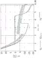
图1描绘了液态水、包含10%甘油(体积/体积(volume by volume,v/v))的溶液和包含20%甘油(v/v)的溶液的冰点降低图。Figure 1 depicts a graph of the freezing point depression of liquid water, a solution comprising 10% glycerol (volume by volume (v/v)) and a solution comprising 20% glycerol (v/v).
图2是示出了可形成可注射冷浆的示例性生物材料的组分以体积和重量计降解的表。Figure 2 is a table showing the degradation by volume and weight of components of an exemplary biomaterial that can form an injectable cold slurry.
图3是示出了结晶设定点为-5.5℃和-8.1℃的冷浆的含冰量的表征的图。Figure 3 is a graph showing the characterization of the ice content of cold slurries with crystallization set points of -5.5°C and -8.1°C.
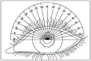
图4示出了人眼的图,其示出了眼的不同区域(4A)和用于解剖学参考的度数(4B)。Figure 4 shows a diagram of a human eye showing the different regions of the eye (4A) and powers for anatomical reference (4B).
图5是示出了在向眼施用表面施加冷浆(实线)和注射冷浆(虚线)之后兔中的实时巩膜温度监测的图。Figure 5 is a graph showing real-time scleral temperature monitoring in rabbits following application of cold slurry (solid line) and injection of cold slurry (dashed line) to the ocular application surface.
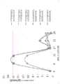
图6是示出了在角膜暴露的情况下向眼施用注射冷浆(菱形)、表面施加冷浆(三角形)和室温下表面施加浆料(方形)之后兔眼随时间的感觉减退的图。Figure 6 is a graph showing hypoesthesia in rabbit eyes over time following application of cold slush injection (diamonds), superficial application of cold slurry (triangles), and superficial application of slurry at room temperature (squares) to the eye with the cornea exposed.
图7是示出了在角膜未暴露的情况下向眼施用表面施加冷浆之后兔眼随时间的感觉减退的图。Figure 7 is a graph showing hypoesthesia in rabbit eyes over time following application of cold slurry to an ocular application surface without cornea exposure.
图8是经荧光素染色的兔角膜的图像,其示出了在有意的8mm角膜擦伤(8A)(作为对照组)之后和在表面施加冷浆(8B)之后随时间的角膜愈合。Figure 8 is an image of fluorescein-stained rabbit cornea showing corneal healing over time after intentional 8mm corneal abrasion (8A) as a control group and after superficial application of cold slurry (8B).
图9是示出了在其中先表面施加冷浆并随后注射冷浆的组合处理之后6只兔的眼随时间的感觉减退的图。在三只兔(用菱形、方形和三角形显示)中,注射的浆料不包含脂质体,而在另外三只兔(用“X”、星形和圆圈显示)中,注射的浆料包含脂质体。Figure 9 is a graph showing the hypoesthesia in the eyes of 6 rabbits over time following a combination treatment in which cold slurries were applied topically and then injected. In three rabbits (shown with diamonds, squares and triangles), the injected slurry did not contain liposomes, while in the other three rabbits (shown with "X", stars and circles) the injected slurry contained Liposomes.
具体实施方式Detailed ways
本公开内容涉及用生物材料例如冷浆来治疗眼表不适的设备、装置、系统和方法。在一些实施方案中,生物材料是冷浆(例如,冰浆),其可通过表面施加或通过注射到人患者或对象(例如,不是患者的人或非人动物)的眼中来递送,用于预防或治疗目的,以减轻眼部不适。本文中公开的系统和方法提供了出乎意料长期的眼感觉减退。感觉减退可引起长期角膜麻木,然后在施加冷浆治疗之后的数天或数周内恢复眼部感觉,而不对角膜造成永久性损伤或不破坏角膜愈合的进程。The present disclosure relates to devices, devices, systems and methods for treating ocular surface discomfort with biological materials such as cold slurries. In some embodiments, the biological material is a cold slurry (e.g., ice slurry) that can be delivered by topical application or by injection into the eye of a human patient or subject (e.g., a human or non-human animal that is not a patient) for use in For preventive or therapeutic purposes, to relieve eye discomfort. The systems and methods disclosed herein provide unexpectedly long-term ocular hypoesthesia. Hypoesthesia can cause long-term corneal numbness, followed by restoration of ocular sensation days or weeks after application of the cold slurry treatment, without permanent damage to the cornea or disruption of the corneal healing process.
在一些实施方案中,可表面施加冷浆以实现期望的治疗作用,例如通过长期的角膜麻木来改善或治疗眼表不适。在一些实施方案中,治疗有效的冷浆完全由水和赋形剂材料(即,不含活性药物化合物的材料)构成。在另一些实施方案中,冷浆还包含已知的活性药物化合物。在一些实施方案中,在表面施加浆料之前,将保护层如接触镜施加至角膜。在一些实施方案中,通过将塑料或其他非导热材料、窥器插入对象的眼中来保护眼睑免受表面施加浆料的影响。In some embodiments, the cold slurry may be applied topically to achieve a desired therapeutic effect, such as amelioration or treatment of ocular surface discomfort through long-term corneal numbness. In some embodiments, a therapeutically effective cold slurry consists entirely of water and excipient materials (ie, materials that do not contain the active pharmaceutical compound). In other embodiments, the cold syrup also contains known active pharmaceutical compounds. In some embodiments, a protective layer, such as a contact lens, is applied to the cornea prior to surface application of the slurry. In some embodiments, the eyelid is protected from the surface applied slurry by inserting a plastic or other non-thermally conductive material, a speculum, into the subject's eye.
在一些实施方案中,可改变将浆料施加至对象的眼的时间长度以引起更大或更温和的感觉减退。在一些实施方案中,改变施加或注射至对象的眼中的浆料的温度以引起更大或更温和的感觉减退。在一些实施方案中,感觉减退随着时间而降低至不明显的点。在另一些实施方案中,在对象的眼可特别敏感的情况下,可引起更大的感觉减退以使对象的眼中更多神经麻木。In some embodiments, the length of time the slurry is applied to the subject's eye can be varied to induce greater or milder hypoesthesia. In some embodiments, the temperature of the slurry applied or injected into the subject's eye is varied to induce greater or milder hypoesthesia. In some embodiments, the hypoesthesia decreases over time to the point of being insignificant. In other embodiments, where the subject's eye may be particularly sensitive, greater hypoesthesia may be induced to numb more nerves in the subject's eye.
在一些实施方案中,在临床护理点接收包含生物材料的容器(例如,小瓶、注射器)。生物材料可以以结晶(或部分结晶)状态接收。在一些实施方案中,待通过表面施加或注射而施用于人患者或对象(例如不是患者的人或非人动物)的最终产品是由水的无菌冰粒以及不同量的赋形剂或添加剂(例如冰点降低剂)构成的冷浆。例如,冷浆中冰粒的百分比可占按浆料的重量计小于约10%、按重量计约10%至约20%、按重量计约20%至约30%、按重量计约30%至约40%、按重量计约40%至约60%、按重量计大于约60%,等等。将控制冰粒的尺寸以允许通过多种尺寸(例如,针规格尺寸为约7至约43)的容器的流动性,如美国申请No.15/505,042(公开No.US2017/0274011)中所述并通过引用并入本文。此外,可使用另一些方法来调节冰粒的尺寸以允许通过多种尺寸的容器的流动性。在一些实施方案中,大多数冰粒的直径小于注射用腔或容器的内径的约一半。例如,冰粒的直径可为约1.5mm或更小以用于3mm导管。In some embodiments, the container (eg, vial, syringe) comprising the biological material is received at the point of clinical care. The biological material may be received in a crystalline (or partially crystalline) state. In some embodiments, the final product to be administered to a human patient or subject (e.g., a human or non-human animal that is not a patient) by topical application or injection is composed of sterile ice pellets of water and varying amounts of excipients or additives (such as freezing point depressant) composed of cold slurry. For example, the percentage of ice particles in the cold slurry can be less than about 10% by weight of the slurry, about 10% to about 20% by weight, about 20% to about 30% by weight, about 30% by weight to about 40%, about 40% to about 60% by weight, greater than about 60% by weight, and the like. The size of the ice particles will be controlled to allow flow through containers of various sizes (e.g., pin gauge sizes from about 7 to about 43), as described in U.S. Application No. 15/505,042 (Publication No. US2017/0274011) and incorporated herein by reference. Additionally, other methods can be used to adjust the size of the ice particles to allow flow through containers of various sizes. In some embodiments, the diameter of the majority of the ice particles is less than about half the inner diameter of the injection cavity or container. For example, the diameter of the ice particles may be about 1.5mm or less for use with a 3mm conduit.
有可用于制备冷浆的多种技术。本公开内容不限于任何特定的方法或技术。There are various techniques that can be used to prepare cold stock. This disclosure is not limited to any particular methodology or technique.
在一些实施方案中,冷浆中可包含一种或更多种赋形剂。赋形剂是本身不是治疗剂而是用作用于将治疗剂递送至对象或患者的稀释剂、佐剂和/或载剂的任何物质,和/或是添加至组合物以改善其处理特性、稳定性特性或储存特性的物质。赋形剂可占冷浆的小于约10%体积/体积(v/v)、浆料的约10%v/v至约20%v/v、约20%v/v至约30%v/v、约30%v/v至40%v/v、以及大于约40%v/v。添加的多种赋形剂可用于改变冷浆的相变温度(例如,降低冰点)、改变冷浆的含冰百分比、改变冷浆的黏度、防止冰粒聚集、防止形成树状冰(即,具有多分支“树样”形成物的晶体,例如在雪花中见到的那些)、保持冰粒分离、提高流体相的导热性或改善冷浆的整体预防性、治疗性或美学效力。In some embodiments, one or more excipients may be included in the cold slurry. An excipient is any substance that is not a therapeutic agent per se but acts as a diluent, adjuvant and/or vehicle for delivering a therapeutic agent to a subject or patient, and/or is added to a composition to improve its handling properties, Substances with stability or storage properties. The excipient may comprise less than about 10% volume/volume (v/v) of the cold slurry, about 10% v/v to about 20% v/v, about 20% v/v to about 30% v/v of the slurry v, about 30% v/v to 40% v/v, and greater than about 40% v/v. A variety of excipients can be added to change the phase transition temperature of the cold slurry (e.g., lower the freezing point), change the ice percentage of the cold slurry, change the viscosity of the cold slurry, prevent ice particles from agglomerating, and prevent dendrite formation (i.e., Crystals with multi-branched "tree-like" formations, such as those seen in snowflakes), keep ice particles separate, increase thermal conductivity of the fluid phase, or improve the overall preventative, therapeutic or aesthetic efficacy of cold slurries.
可添加一种或更多种冰点降低剂作为赋形剂以形成冰点低于0℃的冷浆。降低浆料的冰点以使其维持流动性并保持可注射性,同时仍包含有效百分比的冰粒。合适的冰点降低剂包括盐类(例如,氯化钠、磺丁基醚倍他环糊精钠(betadex sulfobutyl ethersodium))、离子、乳酸盐林格溶液(Lactated Ringer’s solution)、糖类(例如,葡萄糖、山梨糖醇、甘露糖醇、羟乙基淀粉、蔗糖、(2-羟丙基)-β-环糊精、或其组合)、生物相容性表面活性剂例如甘油(也称为丙三醇)、其他多元醇(例如聚乙烯醇、聚乙二醇300、聚乙二醇400、丙二醇)、其他糖醇或脲,等等。其他示例性冰点降低剂在美国申请No.15/505,042(公开No.US2017/0274011)中公开并将其整体并入本文。在另一些实施方案中,形成了具有牙膏的稠度并具有最佳用于表面施加的稠度的浆料糊剂。One or more freezing point depressants may be added as excipients to form a cold slurry with a freezing point below 0°C. The freezing point of the slurry is lowered to maintain fluidity and maintain injectability while still containing an effective percentage of ice particles. Suitable freezing point depressants include salts (e.g., sodium chloride, betadex sulfobutyl ethersodium), ions, Lactated Ringer's solution, sugars (e.g., , glucose, sorbitol, mannitol, hydroxyethyl starch, sucrose, (2-hydroxypropyl)-β-cyclodextrin, or combinations thereof), biocompatible surfactants such as glycerol (also known as glycerol), other polyols (e.g. polyvinyl alcohol, macrogol 300, macrogol 400, propylene glycol), other sugar alcohols or urea, etc. Other exemplary freezing point depressants are disclosed in US Application No. 15/505,042 (Publication No. US2017/0274011 ), which is incorporated herein in its entirety. In other embodiments, a paste paste is formed having the consistency of toothpaste and having a consistency optimal for surface application.
冰点降低剂的浓度将决定冷浆的冰粒百分比及其流动性和可注射性。冰点降低的程度可使用下式来计算,如美国申请No.15/505,042(公开No.US2017/0274011,其并入本文)中所述的:The concentration of freezing point depressant will determine the ice particle percentage of the cold slurry and its flowability and injectability. The degree of freezing point depression can be calculated using the following formula, as described in U.S. Application No. 15/505,042 (Publication No. US2017/0274011, which is incorporated herein):
ΔTF=KFbi其中ΔTF是冰点降低(如由TF(纯溶剂)-TF(溶液)定义),KF是冰点降低常数(cryoscopic constant),b是质量摩尔浓度,并且i是表示每个单独的溶质分子的离子颗粒数的van’t Hoff因子。也可使用计算冰点降低的其他方法,如美国申请No.15/505,042(公开No.US2017/0274011)中所公开的。ΔTF =KF bi where ΔTF is the freezing point depression (as defined by TF (pure solvent) - TF (solution) ), KF is the freezing point depression constant (cryoscopic constant), b is the molar concentration, and i is The van't Hoff factor expressing the number of ion particles per individual solute molecule. Other methods of calculating freezing point depression can also be used, as disclosed in US Application No. 15/505,042 (Publication No. US2017/0274011).
参照图1,其是示出了纯水T1、水与10%(v/v)甘油的混合物T2以及水与20%(v/v)甘油的混合物T3的冰点降低图。在该图中,所有物质均放置在恒定温度为-20℃的冷冻器中。使用放置在每种物质中的温度计测量温度。该图显示,水与甘油的混合物与纯水具有不同的冰点,这意味着溶液可冷却至低于0℃并且仅部分结晶。该图显示,冷却导致纯水T1在0℃的平衡冰点下结晶。这由纯水保持在约0℃的温度下的时间(约1.3小时至约4.4小时)来指示,该时间在纯水T1通过为约-6℃的过冷却点之后立即开始。对于纯溶剂而言,具有结晶的平衡窗口(即,图1中纯水T1的“平线”部分)是典型的。对于10%甘油溶液T2,冷却导致溶液在约2.2小时之后在约-3℃的初始冰点下开始结晶,并且在约6小时之后随着溶液的温度进一步下降至约-8℃而继续结晶。初始结晶在10%甘油溶液T2通过为约-8℃的过冷却点(这可因样品而变化,例如,过冷却点为约-15℃至约-3℃)之后立即发生,其在大约2.2小时处示出。对于溶液(即,不纯的混合物)而言,10%甘油溶液T2具有下降的结晶温度窗口是典型的。类似地,对于20%甘油溶液T3,冷却导致溶液在约3.5小时之后在约-7℃的初始冰点下开始结晶(在初始过冷却点之后,初始过冷却点可因样品而变化,例如,为约-25℃至约-5℃),并且在约6小时之后随着溶液的温度进一步下降至约-11℃而继续结晶,并且在此之后在超过6.5小时后继续下降。初始结晶在20%甘油溶液T3通过为约-14℃的过冷却点之后立即出现,其在大约3.5小时处示出。类似于10%甘油溶液T2的迹线,20%甘油溶液T3的下降的结晶温度窗口对于溶液而言是典型的。Referring to FIG. 1 , it is a graph showing the freezing point depressions of pure water T1 , a mixture of water and 10% (v/v) glycerol T2 , and a mixture of water and 20% (v/v) glycerin T3 . In this figure, everything was placed in a freezer at a constant temperature of -20°C. Measure the temperature using a thermometer placed in each substance. The graph shows that a mixture of water and glycerol has a different freezing point than pure water, meaning that the solution can be cooled below 0°C and only partially crystallize. The figure shows that cooling results in the crystallization of pure water T1 at the equilibrium freezing point of 0°C. This is indicated by the time (about 1.3 hours to about 4.4 hours) that the pure water is kept at a temperature of about 0°C, which begins immediately after the pure water T1 passes the supercooling point of about -6°C. For pure solvents, an equilibrium window with crystallization (ie, the "flat line" portion of pure water T1 in Figure 1) is typical. For 10% glycerol solution T2, cooling caused the solution to begin to crystallize after about 2.2 hours at an initial freezing point of about -3°C, and continued to crystallize after about 6 hours as the temperature of the solution dropped further to about -8°C. Initial crystallization occurs immediately after the 10% glycerol solution T2 passes a supercooling point of about -8°C (this may vary from sample to sample, e.g., a supercooling point of about -15°C to about -3°C), which occurs at about 2.2 Hours are shown. It is typical for solutions (ie, impure mixtures) that the 10% glycerol solution T2 has a decreasing crystallization temperature window. Similarly, for 20% glycerol solution T3, cooling caused the solution to begin to crystallize after about 3.5 hours at an initial freezing point of about -7°C (after the initial supercooling point, which may vary from sample to sample, e.g., about -25°C to about -5°C), and crystallization continued after about 6 hours as the temperature of the solution dropped further to about -11°C, and thereafter continued to drop over 6.5 hours. Initial crystallization occurs immediately after the 20% glycerol solution T3 passes the supercooling point of about -14°C, which is shown at about 3.5 hours. Similar to the trace for 10% glycerol solution T2, the decreasing crystallization temperature window for 20% glycerol solution T3 is typical for solutions.
参照图2,该图表示出了可形成冷浆的示例性生物材料的组分。该图表显示,可针对特定温度计算示例性生物材料的含冰百分比。示例性浆料在-10℃下包含按质量计(重量/重量;w/w)30%的冰。该示例性浆料具有80mL的盐水(0.9%NaCl)和20mL的甘油(即,丙三醇)。以重量计,这样的浆料具有约79.6g的纯水、约0.72g的氯化钠和约25.2g的甘油(大约20%v/v)。在另一些实施方案中,通过调节甘油与盐水的相对体积,浆料可包含更高或更低百分比的甘油。例如,其他合适的浆料包含约10%甘油(v/v)、约10%至约20%甘油、约30%甘油或多于约30%甘油。如果将活性药物化合物添加至浆料,则可相应地调节盐水的浓度以维持赋形剂例如甘油的期望浓度。含冰百分比将根据生物材料的组成而变化。Referring to Figure 2, this chart shows the composition of exemplary biomaterials from which a cold slurry can be formed. This graph shows that the percent ice content of exemplary biomaterials can be calculated for a particular temperature. An exemplary slurry contains 30% by mass (weight/weight; w/w) ice at -10°C. This exemplary slurry has 80 mL of saline (0.9% NaCl) and 20 mL of glycerin (ie, glycerol). By weight, such a slurry has about 79.6 g of pure water, about 0.72 g of sodium chloride, and about 25.2 g of glycerin (about 20% v/v). In other embodiments, the slurry may contain a higher or lower percentage of glycerol by adjusting the relative volumes of glycerol and saline. For example, other suitable slurries comprise about 10% glycerol (v/v), about 10% to about 20% glycerol, about 30% glycerol, or more than about 30% glycerol. If an active pharmaceutical compound is added to the slurry, the concentration of saline can be adjusted accordingly to maintain the desired concentration of excipients such as glycerol. The ice percentage will vary depending on the composition of the biomaterial.
参照图3,不同的浆料组合物(批次)根据其温度谱和含冰量来表征。将不同浆料批次放入加热至40℃并带有测量浆料温度随时间的变化的热电偶线的铜板上。绘制的数据示出了三个不同浆料批次随时间的温度变化。对于每种浆料,在两个不同位置处测量温度:嵌入铜板内部(迹线AC、BC和CC)和暴露于板外部的铜板的中间(迹线AM、BM和CM)。温度迹线示出了三个单独产生的浆料批次:具有15%甘油的浆料组合物(具有-8.1℃的温度设定点)由迹线AC和AM表示和二者均具有10%甘油的两个不同浆料批次(具有-5.5℃的温度设定点)由迹线BC和BM以及迹线CC和CM表示。当将浆料首次引入到铜板中时,嵌入板内部的热电偶线(迹线AC、BC和CC)最初测量加热板的温热温度(例如,迹线AC在时间点0处为31℃),并随后由于引入的浆料的冷却效应而在较低温度下达到平衡(例如,迹线AC在大约2分钟处为22℃)。在另一方面,对于位于板中间的热电偶线,当将浆料首次引入到铜板中时,由于所述线是暴露的,浆料立即接触热电偶线。这由于结晶的浆料与线接触而导致中间位置的初始负温度读数(例如,迹线AM在时间点0处为-5℃),随后随着浆料在加热板上开始熔化,在较温热的温度下达到平衡(例如,迹线AM在大约4分钟处为18℃)。暴露于板外部的热电偶线(迹线AM、BM和CM)可被用于检测结晶的浆料开始熔化期间的相变。该图显示,具有10%甘油的两种浆料组合物在类似的时间点(迹线BM在大约4分钟处,以及迹线CM在大约2.7分钟处)达到其相变,这与15%甘油浆料的相变不同(迹线AM的相变出现在大约0.2分钟处)。该图还显示,依赖于热电偶的位置(中间/底部),具有相同组成(10%甘油:迹线BC和BM以及迹线CC和CM)的两个浆料批次以类似的时间框和在约15℃至19℃的类似温度下达到平衡(如通过两个热电偶线位置测量的)。在另一方面,依赖于热电偶的位置(中间/底部),具有不同组成(15%甘油:迹线AC和AM)的浆料具有与其他两种浆料不同的温度谱,在约19℃至22℃的温度下更快地达到平衡。因此,图3表明具有不同组成的浆料具有不同的温度谱,并且具有相同组成的浆料(例如,由BC和BM表示的浆料以及由CC和CM表示的浆料具有类似的温度谱,其与由AC和AM表示的浆料不同)中存在批次间一致性。Referring to Figure 3, different slurry compositions (batches) were characterized according to their temperature profile and ice content. The different slurry batches were placed on copper plates heated to 40°C with thermocouple wires to measure the slurry temperature as a function of time. The plotted data shows the temperature change over time for three different slurry batches. For each paste, the temperature was measured at two different locations: embedded inside the copper board (traces AC , BC and CC ) and in the middle of the copper board exposed to the outside of the board (traces AM , BM and CM ). The temperature traces show three separately produced slurry batches: the slurry composition with 15% glycerin (with a temperature set point of -8.1°C) is represented by traces AC and AM and both have Two different slurry batches of 10% glycerol (with a temperature set point of -5.5°C) are represented by tracesBC andBM and tracesCC andCM . When the paste is first introduced into the copper plate, thermocouple wires embedded inside the plate (traces AC , BC , andCC ) initially measure the warming temperature of the heated plate (e.g., trace AC at
参照图4A,示出了眼的图,其描绘了巩膜区2、巩膜区3、角膜1和角膜缘(角膜1和巩膜区2之间的虚线)。图4A转载自Andreoli CM,Gardiner MF.Open globe injuries:Emergent evaluation and initial management.In:UpToDate,Post TW(Ed),UpToDate,Waltham,MA。图4B示出了具有叠加量角器的眼的图,其示出了相对于眼的以度(°)计的角度。在该图中,90°表示沿着眼的最高位置。Referring to Fig. 4A, a diagram of an eye is shown depicting
在一些实施方案中,本文中所述的冷浆可表面施加,或作为替代地,注射,以实现减轻眼表不适的长期感觉减退。感觉减退是指眼部不适或疼痛的减轻但没有完全阻断眼部感觉。因此,感觉减退与麻醉不同,麻醉的特征在于眼部感觉的更深度的阻断。感觉减退可包括导致疼痛响应降低的角膜麻木,同时维持眼的其他正常功能,包括正常的愈合过程。在另一方面,由于丧失了所有的角膜感觉,麻醉可导致眼的异常功能。角膜感觉对于眼的正常功能(包括启动保护机制,例如眨眼和产生泪液)是重要的。In some embodiments, the cold slurries described herein may be applied topically, or alternatively, injected, to achieve long-term hypoesthesia that relieves ocular surface discomfort. Hypoesthesia is a reduction in ocular discomfort or pain without complete blockage of ocular sensation. Hypoesthesia is thus distinct from anesthesia, which is characterized by a deeper block of ocular sensation. Hypoesthesia can include corneal numbness resulting in reduced pain response, while maintaining other normal functions of the eye, including normal healing processes. On the other hand, anesthesia can lead to abnormal function of the eye due to loss of all corneal sensation. Corneal sensation is important for the normal function of the eye, including the activation of protective mechanisms such as blinking and tear production.
一种方法是将冷浆的滴剂施加至眼表,其体积可为1至100微升而不等,优选地为约10至80微升而不等。作为滴剂,制剂可被直接施用于眼表。或者,可先刮角膜,并随后可施用滴剂。在一些实施方案中,表面施加的冷浆具有更易流动的糊剂稠度,并且作为治疗可将大量的冷浆表面施加(3至50ml)至眼表。One method is to apply drops of cold slurry to the ocular surface, which may vary in volume from 1 to 100 microliters, preferably from about 10 to 80 microliters. As drops, the formulation can be applied directly to the ocular surface. Alternatively, the cornea may be scraped first, and then the drops may be applied. In some embodiments, the topically applied cold paste has a more flowable paste consistency, and a large amount of the cold paste (3 to 50 ml) may be topically applied (3 to 50 ml) to the ocular surface as a treatment.
在一些实施方案中,将如本文中所述的冷浆表面施加于角膜缘的后部(例如,在图4A中标示为巩膜区2的区域)持续约1分钟至约20分钟。在一些实施方案中,施加冷浆持续约5分钟至约10分钟。在一些实施方案中,每1至10秒将冷浆施用于每只眼中,持续1至20分钟。这种治疗可在短时间(例如,5至20分钟)内重复数次。在一些实施方案中,将冷浆表面施加于角膜缘的后部持续约10分钟,每90秒再施加新鲜浆料直至达到10分钟。In some embodiments, a cold slurry surface as described herein is applied to the posterior portion of the limbus (eg, the area designated as
在一些实施方案中,在表面施加期间,保护敏感的眼部结构免于遇到冷浆以限制潜在的副作用。对角膜表面的保护可限制由冷冻角膜组织引起的一些或全部角膜细胞损伤或屈光性变化。对睑结膜和眼睑的保护可防止与治疗作用无关的发红、肿胀和炎症。通过选择性地将冰施加至球结膜之上角膜缘后部的眼表面(对应于称为区域2的前部解剖学区域,图4A),可使对角膜的潜在不利作用最小化。In some embodiments, during topical application, the sensitive ocular structures are protected from encountering the cold slurry to limit potential side effects. Protection of the corneal surface may limit some or all of the corneal cell damage or refractive changes caused by freezing the corneal tissue. Protection of the palpebral conjunctiva and eyelids prevents redness, swelling and inflammation unrelated to the therapeutic effect. Potential adverse effects on the cornea can be minimized by selectively applying ice to the ocular surface posterior to the limbus above the bulbar conjunctiva (corresponding to the anterior anatomical region referred to as
在一些实施方案中,可施加保护性接触镜或其他保护性覆盖物以位于角膜上来保护角膜免受损伤。在一些实施方案中,角膜覆盖物完全阻止冷浆直接接触角膜表面。在一些实施方案中,在冷浆表面施加期间,使用眼睑窥器来保持眼睑打开。在一些实施方案中,窥器由不导热材料例如塑料或本领域已知的另一些非传导性材料制成。不导热材料可用于窥器,以防止眼睑(内部和外部)在冷浆治疗期间冷冻,冷冻可导致眼睑受损。在一些实施方案中,将冷浆仅施加于巩膜并且保护角膜免于冷冻。在一些实施方案中,防止角膜冷冻将确保角膜更快愈合。In some embodiments, a protective contact lens or other protective covering may be applied to sit over the cornea to protect the cornea from damage. In some embodiments, the corneal onlay completely prevents the cold slurry from directly contacting the corneal surface. In some embodiments, an eyelid speculum is used to hold the eyelid open during topical application of the cold slurry. In some embodiments, the speculum is made of a thermally non-conductive material such as plastic or another non-conductive material known in the art. Thermally non-conductive material may be used for the speculum to prevent the eyelids (inner and outer) from freezing during cold slurry treatment, which can lead to eyelid damage. In some embodiments, the cold slurry is applied to the sclera only and the cornea is protected from freezing. In some embodiments, preventing corneal freezing will ensure faster corneal healing.
可使用装置来控制暴露于冷浆的区域仅是球结膜/巩膜,使得冷浆不能物理接触与所期望临床作用无关的邻近组织或使其冷冻。在一些实施方案中,冷浆制剂根本不与眼表直接接触。冷浆制剂可被包含在导热材料,例如,小的金属或聚合物圆环(donut)或另一些保护性环内,从而提供阻止制剂与眼表直接接触的屏障,但允许发生必要的冷却。在一些实施方案中,所述装置仅将冷却引导至具有潜在治疗效应的区域并防止装置/冷却接触和影响邻近组织,以使不期望的副作用(例如,由于直接向眼施加高渗溶液而对眼的潜在刺激)最小化。Devices can be used to control the area exposed to the cold plasma to be only the bulbar conjunctiva/sclera so that the cold plasma cannot physically contact or freeze adjacent tissues not relevant to the desired clinical effect. In some embodiments, the cold slurry formulation does not come into direct contact with the ocular surface at all. The cold slurry formulation may be contained within a thermally conductive material, eg, a small metal or polymer donut or some other protective ring, thereby providing a barrier preventing direct contact of the formulation with the ocular surface, but allowing the necessary cooling to occur. In some embodiments, the device directs cooling only to areas of potential therapeutic effect and prevents the device/cooling from contacting and affecting adjacent tissues, so that undesired side effects (e.g., adverse effects due to direct application of a hypertonic solution to the eye) eye irritation potential) is minimized.
在一些实施方案中,本文中所述的冷浆作为结膜下推注以约每2分钟注射。每次注射可提供约0.5至1.5ml的冷冻浆料,并且在期望治疗的持续时间内每约2分钟重复,或者总共约10分钟。在一些实施方案中,将冷浆注射在更加紧邻睫状神经的轴突。睫状神经位于眼的约0°和180°处(图4B)。In some embodiments, the cold slurries described herein are injected as a subconjunctival bolus about every 2 minutes. Each injection may provide about 0.5 to 1.5 ml of frozen slurry and is repeated every about 2 minutes for the duration of the desired treatment, or about 10 minutes in total. In some embodiments, the cold plasma is injected more proximal to the axon of the ciliary nerve. The ciliary nerve is located at approximately 0° and 180° of the eye (Fig. 4B).
在一些实施方案中,使用标准注射器来注射浆料。或者,可使用已调节浆料用于注射的注射器。在一些实施方案中,注射器可具有约18G至约25G的针。In some embodiments, the slurry is injected using a standard syringe. Alternatively, a syringe in which the slurry has been conditioned for injection can be used. In some embodiments, the syringe may have a needle of about 18G to about 25G.
在一些实施方案中,在治疗(例如,冷浆注射或表面施加)期间进行对眼表的实时温度感测。在一些实施方案中,施加冷浆以使组织(例如,角膜表面、结膜或眼的任何其他部分)冷却至小于约0℃、小于约-1℃、小于约-2℃、小于约-3℃、小于约-4℃或小于约-5℃。在一些实施方案中,施加冷浆持续超过约一分钟,优选持续约2分钟至约10分钟。可改变冷却组织的温度和施加浆料的时间长度以改变对象经历的感觉减退。In some embodiments, real-time temperature sensing of the ocular surface is performed during therapy (eg, cold slurry injection or topical application). In some embodiments, the cold slurry is applied to cool the tissue (e.g., corneal surface, conjunctiva, or any other part of the eye) to less than about 0°C, less than about -1°C, less than about -2°C, less than about -3°C , less than about -4°C, or less than about -5°C. In some embodiments, the cold slurry is applied for more than about one minute, preferably for about 2 minutes to about 10 minutes. The temperature at which the tissue is cooled and the length of time the slurry is applied can be varied to alter the hypoesthesia experienced by the subject.
在一些实施方案中,将冷浆随时间周期性地再施用于对象的眼以维持治疗效果。具有用于表面施用和/或注射的可能频率的范围。例如,可以以下列任一种来施用治疗:每隔一周一次;每月一次;每隔一个月一次;每三个月一次,等等。In some embodiments, the cold slurry is reapplied to the subject's eye periodically over time to maintain the therapeutic effect. There is a range of possible frequencies for topical application and/or injection. For example, treatment can be administered any of the following: every other week; monthly; every other month; every three months, etc.
在一些实施方案中,冷浆用作安全的角膜麻木治疗来治疗角膜不适或疼痛。多种冷浆制剂可与本文中所述的方法一起使用,包括上述的那些。参照图5至图9描述了冷浆的另一些具体实施方案。“ECT-4143”是包含15%甘油、30%L-α-磷脂酰胆碱脂质体和0.9%盐水(或磷酸缓冲盐水)的浆料制剂。在一些实施方案中,在约-25℃至10℃的温度(浆料的温度)下向眼施用(表面或通过注射)ECT-4143。在一些实施方案中,在约-18℃的温度(浆料的温度,例如在下文参照图5至图9描述的实施方案中)下向眼施用(表面或通过注射)ECT-4143。每约90秒施用ECT-4143,每次施加约2至3ml,直至达到10分钟的总处理时间。In some embodiments, cold slurries are used as a safe corneal numb treatment to treat corneal discomfort or pain. A variety of cold paste formulations can be used with the methods described herein, including those described above. Other specific embodiments of the cold slurry are described with reference to FIGS. 5 to 9 . "ECT-4143" is a slurry formulation comprising 15% glycerol, 30% L-α-phosphatidylcholine liposomes, and 0.9% saline (or phosphate-buffered saline). In some embodiments, ECT-4143 is administered (topically or by injection) to the eye at a temperature (temperature of the slurry) of about -25°C to 10°C. In some embodiments, ECT-4143 is administered (topically or by injection) to the eye at a temperature of about -18°C (the temperature of the slurry, eg, in the embodiments described below with reference to Figures 5-9). ECT-4143 was administered approximately 2 to 3 ml per application approximately every 90 seconds until a total treatment time of 10 minutes was reached.
“ECT-1719”是包含15%甘油和0.9%盐水(或磷酸缓冲盐水)的浆料制剂。在一些实施方案中,在约-20℃至-5℃、或约-15℃至约-10℃的温度(浆料的温度)下向眼施用(表面或通过注射)ECT-1719。在一些实施方案中,在约-11℃的温度(浆料的温度,例如在下文参照图5至图9描述的实施方案中)下向眼施用(表面或通过注射)ECT-1719。在一些实施方案中,ECT-1719以每次注射0.7ml体积来注射,四次总注射组成2.8ml总注射体积。每约120秒施用ECT-1719,直至达到10分钟的总处理。"ECT-1719" is a slurry formulation containing 15% glycerol and 0.9% saline (or phosphate buffered saline). In some embodiments, ECT-1719 is administered (topically or by injection) to the eye at a temperature (temperature of the slurry) of about -20°C to -5°C, or about -15°C to about -10°C. In some embodiments, ECT-1719 is administered (topically or by injection) to the eye at a temperature of about -11°C (the temperature of the slurry, eg, in the embodiments described below with reference to Figures 5-9). In some embodiments, ECT-1719 is injected in a volume of 0.7 ml per injection, with four total injections making up a total injection volume of 2.8 ml. ECT-1719 was administered approximately every 120 seconds until a total treatment of 10 minutes was achieved.
参照图5,在向眼进行以下施用之后在兔中进行实时巩膜温度监测:在一只兔中表面施加冷浆(ECT-4143,实线)和在第二只兔中注射冷浆(ECT-1719,虚线)。通过用在其远端包含温度探针的25g针插入眼球筋膜囊下(subtenon)空间来实现温度监测。如在图5可以看出的,接受注射的冷浆(ECT-1719)的兔的巩膜温度在整个操作期间(从冷浆注射之后约0秒至冷浆注射之后约463秒)在约0℃和约8℃之间波动。图中在约463秒处的锐线(sharpline)表示7.5分钟之后研究结束并从眼部组织移除温度探针。对于记录温度期间(在表面施加之后约0秒至在表面施加之后约600秒)的大部分时间,接受表面施加的冷浆(ECT-4143)的兔的巩膜温度低于接受注射的冷浆的兔的巩膜温度,在约-6℃至约4℃之间波动。在表面施加之后,巩膜的温度从施加时(图5中约0秒)的约4℃持续下降至约120秒之后的约0℃。在使巩膜冷却的初始时期之后,从表面施加之后约120秒至表面施加之后约520秒,温度在约0℃至-5℃保持相对稳定。另外,对于在表面施加之后约220秒至约520秒的持续时间,巩膜温度显示出非常小的可变性,在约-2℃至约-3℃保持稳定。在表面施加之后约620秒之后,处理结束并移除温度探针,显示测量温度急剧升高,如图5中所示。Referring to Figure 5, real-time sclera temperature monitoring was performed in rabbits after the following administrations to the eye: surface application of cold slurry in one rabbit (ECT-4143, solid line) and injection of cold slurry in a second rabbit (ECT-4143, solid line). 1719, dotted line). Temperature monitoring was achieved by insertion into the subtenon space with a 25g needle containing a temperature probe at its distal end. As can be seen in Figure 5, the sclera temperature of the rabbits receiving the injected cold slurry (ECT-1719) was around 0°C throughout the procedure (from about 0 seconds after the cold slurry injection to about 463 seconds after the cold slurry injection). and fluctuate between about 8°C. The sharp line in the graph at about 463 seconds indicates that 7.5 minutes later the study ended and the temperature probe was removed from the ocular tissue. For most of the time during which the temperature was recorded (from about 0 seconds after surface application to about 600 seconds after surface application), the sclera temperature of the rabbits receiving the surface-applied cold slurry (ECT-4143) was lower than that of the injected cold slurry. The temperature of the sclera in rabbits fluctuates between about -6°C and about 4°C. After surface application, the temperature of the sclera continued to drop from about 4°C at the time of application (about 0 seconds in Figure 5) to about 0°C after about 120 seconds. After the initial period of allowing the sclera to cool, from about 120 seconds after surface application to about 520 seconds after surface application, the temperature remained relatively constant at about 0°C to -5°C. Additionally, the sclera temperature exhibited very little variability for a duration of about 220 seconds to about 520 seconds after topical application, remaining stable at about -2°C to about -3°C. After about 620 seconds after surface application, the treatment ended and the temperature probe was removed, showing a sharp increase in the measured temperature, as shown in FIG. 5 .
使用单丝/触觉计测量冷浆处理之后的感觉减退作用作为对眼的触觉刺激的响应。以6cm细丝长度开始并以0.5cm的增量递减,在每个长度下对眼探测3次,直至引起眨眼响应。细丝随着其缩短变得更硬,因此在探测进入眼时可对眼应用更大的压力。每个时间点的感觉减退基于单丝的给定长度。在每个时间点下,记录的特定单丝长度是不存在眨眼响应时的最短长度(最高压力)。例如,从最长的6cm单丝开始,如果兔在探测时不眨眼,则使用下一个5.5cm长的单丝探测眼。如果兔再次不眨眼,则使用下一个5cm的单丝长度。现在,如果兔眨眼了,则记录之前的5.5cm长度,因为这是无眨眼响应时的最短长度(反映了一定程度的感觉减退)。感觉减退的最深水平是当用最短的细丝长度(例如,0.5cm)探测时兔不眨眼。感觉减退为零度(没有疼痛阻断/没有麻木)是当用最长的细丝长度(例如,6cm)探测时兔眨眼。可将细丝长度转换为压力(g/mm2),使得6cm细丝产生0.4g/mm2压力(最低压力),而0.5cm细丝产生15.9g/mm2压力(最高压力)。因此,所记录的压力是对应于不存在眨眼响应下的最短细丝长度的压力。Hypoesthesia after cold slurry treatment was measured as a response to tactile stimulation of the eye using a monofilament/tactilemeter. Eyes were probed 3 times at each length starting with a 6 cm filament length and decremented in 0.5 cm increments until a blink response was elicited. The filament becomes stiffer as it shortens, so more pressure can be applied to the eye when probing into the eye. Hypoesthesia at each time point is based on a given length of monofilament. At each time point, the specific filament length recorded was the shortest length (highest pressure) in the absence of a blink response. For example, start with the longest 6 cm monofilament and if the rabbit does not blink when probing, use the next 5.5 cm long monofilament to probe the eye. If the rabbit does not blink again, the next 5 cm length of monofilament is used. Now, if the rabbit blinked, the previous length of 5.5 cm was recorded as this was the shortest length without a blink response (reflecting some degree of hypoesthesia). The deepest level of hypoesthesia is when the rabbit does not blink when probed with the shortest filament length (eg, 0.5 cm). Hypoesthesia is zero (no pain block/no numbness) when the rabbit blinks when probed with the longest filament length (eg, 6 cm). Filament length can be converted to pressure (g/mm2 ), such that a 6 cm filament produces a pressure of 0.4 g/mm2 (lowest pressure) and a 0.5 cm filament produces a pressure of 15.9 g/mm2 (highest pressure). Therefore, the recorded pressure is the pressure corresponding to the shortest filament length in the absence of a blink response.
参照图6,在角膜暴露的情况下向眼施用注射冷浆(ECT-1719,该组中有3只兔,用菱形示出)、表面施加冷浆(ECT-4143,该组中有3只兔,用三角形示出)和室温下表面施加浆料(ECT-4143,该组中有1只兔,用方形示出)之后,在兔中随时间测量的感觉减退效果(如本文中所述使用触觉刺激测量的角膜麻木程度)。在图6中,感觉减退的程度作为所记录压力(即,基于其中缺乏眨眼响应的最短单丝)相对于最高可能压力(即,使用0.5cm的最短单丝,对应于15.9g/mm2压力)的百分比显示。示出了在冷浆施用之后第1、7、14和28天的感觉减退程度。对于注射冷浆(ECT-1719,用菱形示出),第1天的感觉减退效果为约20%并持续逐渐减弱,到第14天达到基线水平(误差棒与0%重叠)。对于表面施加的冷浆(在18℃下施加的ECT-4143,用三角形示出),第1天的感觉减退效果为100%(可测量的最大角膜麻木),其随后继续下降,在第28天逐渐减弱,在此期间疼痛响应恢复至基线水平(误差棒与0%重叠)。对于在室温下表面施加浆料(ECT-4143,用方形示出),在处理之后的任何时间点都没有观察到感觉减退效果。因此,图6表明了表面施加的冷浆的出乎意料的强烈感觉减退效果,这导致长期感觉减退(几乎1个月)。注射冷浆导致中度感觉减退,其持续时间也比预期的长(例如,约1周至2周)。重要的是,对于表面和注射方法二者,冷浆处理产生正常化至基线水平而不引起任何永久性麻木作用的长期感觉减退。Referring to Figure 6, cold slurry was administered to the eye with the cornea exposed (ECT-1719, there were 3 rabbits in the group, shown with diamonds), cold slurry was applied topically (ECT-4143, there were 3 rabbits in the group rabbits, shown with triangles) and the hypoesthesia effect measured over time in rabbits after surface application of the slurry (ECT-4143, 1 rabbit in the group, shown with squares) at room temperature (as described herein Degree of corneal numbness measured using tactile stimuli). In Figure 6, the degree of hypoesthesia is plotted as recorded pressure (i.e., based on the shortest monofilament in which the blink response is lacking) relative to the highest possible pressure (i.e., using the shortest monofilament of 0.5 cm, corresponding to a pressure of 15.9 g/mm2 ) displayed as a percentage. The degree of hypoesthesia at
图7示出了除了角膜未暴露(用接触镜保护)之外类似于图6的在向眼施用表面施加的冷浆(6只兔,ECT-4143)之后在兔中随时间的感觉减退效果。以与如上关于图6所述的相同方式测量感觉减退效果。示出了在用表面施加的冷浆处理之后的第1、7、14、21和28天的感觉减退效果。第1天的感觉减退效果为约50%并缓慢地逐渐减弱,到第21天达到基线水平(误差棒与0%重叠)。因此,图7表明了表面施加的冷浆(在角膜保护的情况下)的出乎意料的中度至强度的感觉减退效果,这导致长期感觉减退(约3周)而不引起永久性角膜麻木或任何角膜损伤。Figure 7 shows the hypoesthesia effect in rabbits over time after application of topically applied cold slurry (6 rabbits, ECT-4143) to the eye similar to Figure 6 except that the cornea is not exposed (protected with a contact lens) . The hypoesthesia effect was measured in the same manner as described above with respect to FIG. 6 . The hypoesthesia effect is shown at 1, 7, 14, 21 and 28 days after treatment with the topically applied cold slurry. The hypoesthesia effect on
参照图8,使用荧光素染色的兔角膜的代表性图像示出了在对角膜和眼睑施加保护的情况下向两个对照组施加有意的8mm角膜擦伤(图8A)之后以及在处理组中表面施加冷浆(图8B)之后随时间的角膜愈合。通过测量损伤随时间的尺寸来确定损伤的进展。如在图8A中可以看出的,对照组(3只兔)中的角膜愈合在角膜擦伤之后的前24小时中以1.31mm2/小时的平均愈合速率发生,并在角膜擦伤之后24小时至60小时以0.62mm2/小时的平均愈合速率发生。出乎意料地,如图8B中所示,与对照组相比,接受表面施加的冷浆(ECT-4143)的兔角膜愈合没有受损。在该组(3只兔)中,冷浆处理之后的角膜愈合在冷浆处理之后的前24小时中以1.09mm2/小时的平均愈合速率发生,并在冷浆处理之后24小时至60小时以0.63mm2/小时的平均愈合速率发生。Referring to Figure 8, representative images of rabbit corneas stained with fluorescein are shown after applying intentional 8mm corneal abrasions (Figure 8A) to the two control groups with protection applied to the cornea and eyelids and in the treatment group Corneal healing over time after topical application of cold slurry (FIG. 8B). Lesion progression was determined by measuring the size of the lesion over time. As can be seen in FIG. 8A , corneal healing in the control group (3 rabbits) occurred at an average healing rate of 1.31 mm2 /hour in the first 24 hours after corneal abrasion, and 24 hours after corneal abrasion. Hours to 60 hours occurred at an average healing rate of 0.62 mm2 /hour. Unexpectedly, as shown in Figure 8B, corneal healing was not impaired in rabbits receiving topically applied cold slurry (ECT-4143) compared to controls. In this group (3 rabbits), corneal healing after cold slurry treatment occurred at an average healing rate of 1.09 mm2 /hour in the first 24 hours after cold slurry treatment, and between 24 hours and 60 hours after cold slurry treatment Healing occurred at an average rate of 0.63 mm2 /hour.
参照图9,附图示出了在先表面施加冷浆并随后注射冷浆的组合处理之后6只兔中随时间的感觉减退效果。在3只兔中(用菱形、方形和三角形示出),表面施加的冷浆是不包含脂质体的ECT-1719,其后是注射相同的冷浆制剂(ECT-1719)。在另外3只兔(用“X”、星形和圆圈示出)中,表面施加的冷浆还是ECT-1719(不包含脂质体),其后是注射包含脂质体的冷浆(ECT-4143)。感觉减退效果作为兔不产生眨眼的最大压力(如本文参照图6和图7所述)显示。如图9中所示,无论组合处理(脂质体或非脂质体注射)如何,感觉减退效果在处理之后继续提高,很可能在第4天至第11天的某天达到峰值。感觉减退效果逐渐减弱在约第17天时恢复至基线水平。令人惊讶的是,在约第22天自发地出现第二个不太明显的感觉减退期,其在第26天逐渐减弱回到基线水平。Referring to Figure 9, the figure shows the hypoesthesia effect over time in 6 rabbits following the combined treatment of first topical application of cold slurries followed by injection of cold slurries. In 3 rabbits (shown with diamonds, squares and triangles), the topically applied cold slurry was ECT-1719 without liposomes, followed by injection of the same cold slurry formulation (ECT-1719). In another 3 rabbits (shown with "X", star and circle), the topically applied cold slurry was again ECT-1719 (without liposomes), followed by injection of liposome-containing cold slurry (ECT-1719 -4143). The hypoesthesia effect was shown as the maximum pressure at which the rabbit did not produce a blink (as described herein with reference to Figures 6 and 7). As shown in Figure 9, regardless of the combined treatment (liposomal or non-liposomal injection), the hypoesthesia effect continued to increase after treatment, most likely peaking sometime between
本文中所述的数据支持冷浆(表面和注射)作为长期且安全的角膜麻木治疗,其产生感觉减退而无永久性的角膜麻木或损伤。The data described herein support cold slurries (topical and injected) as a long-term and safe treatment for corneal numbness that produces hypoesthesia without permanent corneal numbness or damage.
不受任何理论的束缚,基本前提是冷浆的施加通过引起神经上髓鞘的变性来使疼痛刺激的信号传导中止。髓磷脂是富含脂质的脂肪物质,其允许电刺激以快速且有效的方式沿着神经轴突向下行进。在游离末梢和神经的有髓部分二者上施用冷浆的情况下,低温将使脂肪细胞的脂质组分冷冻或结晶,引起凋亡并使髓鞘变性,这一过程称为沃勒变性(Wallerian degeneration)。该过程将显著减少睫状神经将疼痛刺激从角膜传导至脑干。由于眼表上远端神经末梢的庞大体积,并非所有的周围神经都受到影响,因此并非来自眼表的所有感觉都被消除,从而诱导相对感觉减退而不是完全麻醉。此外,该作用在大约4至8周之后消退,此时眼部感觉完全恢复。引起沃勒变性的另一些选项包括射频消融术(radiofrequency ablation)和冷冻神经裂解(cryoneurolysis)(在接近-80℃的温度下冷冻),但这些操作存在损伤周围组织和结构的风险。此外,包含冰晶的非活性载剂将不伤害眼的其他组分,使其可合理地应用于治疗导致眼表疼痛的神经。这种方法保留了视力和眼表的正常功能。Without being bound by any theory, the basic premise is that the application of cold slurries halts the signaling of pain stimuli by causing degeneration of the myelin sheath on the nerves. Myelin is a lipid-rich fatty substance that allows electrical stimulation to travel down nerve axons in a fast and efficient manner. In the case of cold slurries applied both to free extremities and to the myelinated portion of the nerve, the low temperature will freeze or crystallize the lipid components of the adipocytes, causing apoptosis and degeneration of the myelin sheath, a process called Wallerian degeneration (Wallerian degeneration). This procedure significantly reduces the transmission of painful stimuli from the cornea to the brainstem by the ciliary nerve. Due to the bulky volume of distal nerve endings on the ocular surface, not all peripheral nerves are affected and therefore not all sensation from the ocular surface is eliminated, thereby inducing relative hypoesthesia rather than complete anesthesia. Furthermore, the effect subsides after about 4 to 8 weeks, at which point ocular sensation is fully restored. Other options for causing Wallerian degeneration include radiofrequency ablation and cryoneurolysis (freezing at temperatures approaching -80°C), but these procedures run the risk of damaging surrounding tissues and structures. Furthermore, an inactive vehicle comprising ice crystals will not harm other components of the eye, making it a reasonable application for the treatment of nerves that cause pain on the ocular surface. This approach preserves vision and the normal function of the ocular surface.
不受特定理论的束缚,注射到角膜缘周围的结膜下空间中使得冷浆分布在睫状神经的游离神经末梢周围。存在两条主要的具有分支到每只眼的角膜中的游离神经末梢的睫状神经。每条睫状神经沿其位于角膜中游离神经末梢的下游的轴突髓鞘化。冷浆的注射向下游扩散至睫状神经轴突被髓鞘化的地方。随着冷浆扩散至睫状神经的轴突,其引起髓鞘的结晶和凋亡,从而使睫状神经脱髓鞘。脱髓鞘阻止神经向脑传递疼痛信号。或者,冷浆可引起神经的沃勒变性,并且类似地阻止疼痛信号被传递到脑。Without being bound by a particular theory, the injection into the subconjunctival space around the limbus causes the cold slurry to distribute around the free nerve endings of the ciliary nerve. There are two major ciliary nerves with free nerve endings branching into the cornea of each eye. Each ciliary nerve is myelinated along its axon downstream of the free nerve endings in the cornea. The injection of cold plasma spreads downstream to where the axons of the ciliary nerve are myelinated. As the cold slurry spreads to the axons of the ciliary nerve, it causes crystallization and apoptosis of the myelin sheath, thereby demyelinating the ciliary nerve. Demyelination prevents nerves from sending pain signals to the brain. Alternatively, cold slurries can cause Wallerian degeneration of the nerves and similarly prevent pain signals from being transmitted to the brain.
表面施加的和/或注射的冷浆优于其他施用方法,因为其不损伤角膜表面。Topically applied and/or injected cold slurries are preferred over other methods of application because they do not damage the corneal surface.
本文中公开的系统和方法的范围不限于本文中所述的具体实施方案。实际上,根据在先描述,除了所述的那些之外的对装置、系统和方法的多种修改对本领域技术人员而言将变得明显。The scope of the systems and methods disclosed herein is not limited to the specific embodiments described herein. Indeed, various modifications to the devices, systems and methods, in addition to those described, will become apparent to those skilled in the art from the foregoing description.
实施例1-用于角膜麻木的冷浆处理的体内测试Example 1 - In vivo testing of cold pulp treatment for corneal numbness
在本实施例中描述的研究结果可在图5至图9中看见。进行了动物研究的临床前测试以确定治疗的效力,包括探究递送治疗的最佳方式、治疗作用的持续时间以及任何潜在的副作用。对于眼科探究,新西兰白兔(New Zealand white rabbit)是理想的模型,因为其角膜和角膜神经支配系统与人非常类似,并且其是文献中用于角膜研究的标准公认的模型。The results of the studies described in this example can be seen in Figures 5-9. Preclinical testing with animal studies is performed to determine the efficacy of the treatment, including exploring the best way to deliver the treatment, the duration of the therapeutic effect, and any potential side effects. For ophthalmic investigations, the New Zealand white rabbit is an ideal model because its cornea and corneal innervation are very similar to humans, and it is the standard accepted model in the literature for corneal studies.
操作准备operational preparation
给予动物前驱麻醉剂(兔甲苯噻嗪(Xylazine)1.1mg/kg IM,盐酸丁丙诺啡(Buprenorphine)0.01至0.05mg/kg IM)和术前抗生素(头孢唑啉(Cefazolin)25至50mg/kgIM)。然后将动物麻醉(兔氯胺酮(Ketamine)33mg/kg IM)。将动物放在加热垫上并监测体征。向待研究的眼施用两滴盐酸丙美卡因0.5%和5%去氧肾上腺素/0.5%托吡卡胺(tropicamide)(扩张滴剂)。对动物进行吸入麻醉(浓度为1.5%至2%的异氟醚)并补充O2。Animals were given preanesthesia (rabbit Xylazine 1.1 mg/kg IM, buprenorphine hydrochloride (Buprenorphine) 0.01 to 0.05 mg/kg IM) and preoperative antibiotics (cefazolin (Cefazolin) 25 to 50 mg/kg IM ). Animals were then anesthetized (Rabbit Ketamine 33 mg/kg IM). Place the animal on a heating pad and monitor for signs. Two drops of proparacaine hydrochloride 0.5% and 5% phenylephrine/0.5% tropicamide (dilation drops) were administered to the eye to be studied. Animals were anesthetized by inhalation (1.5% to 2% isoflurane) and supplemented withO2 .
研究操作research operations
以通常的无菌方式准备和覆盖动物,包括将聚维酮-碘滴剂滴注至眼表上。放置窥器,并进行浆料的表面或结膜下注射。Animals were prepared and covered in the usual aseptic manner, including instillation of povidone-iodine drops onto the ocular surface. A speculum is placed, and a superficial or subconjunctival injection of the slurry is performed.
注射施用Injection administration
为了评估ECT-1719的感觉减退潜能,将约0.7mL的冷浆注射到角膜缘周围的结膜下空间中。注射的冷浆在角膜缘周围360度均匀分布,部分原因是来自角膜的压力、注射力和存在的天然潜在空间。注射操作每120秒重复,总共10分钟。To assess the hypoesthesia potential of ECT-1719, approximately 0.7 mL of cold slurry was injected into the subconjunctival space around the limbus. The injected cold slurry is evenly distributed 360 degrees around the limbus, due in part to pressure from the cornea, the force of the injection and the natural potential space that exists. The injection operation was repeated every 120 seconds for a total of 10 minutes.
对照动物接受无菌盐水(对照)处理或载剂对照(未冷却的浆料)处理。在手术结束时,仔细检查眼,移除窥器,取下覆盖物,并将眼用无菌盐水洗涤。有一个另外的对照组,其将常规的麻醉剂滴剂施加至角膜。所有手术仅在左眼上进行(出于控制目的),并且持续约10分钟。Control animals received either sterile saline (control) treatment or vehicle control (uncooled slurry) treatment. At the conclusion of the procedure, the eye is carefully inspected, the speculum is removed, the covering is removed, and the eye is washed with sterile saline. There was an additional control group, which had conventional anesthetic drops applied to the cornea. All procedures were performed on the left eye only (for control purposes) and lasted approximately 10 minutes.
上述外科操作通常在人中进行,根据情况注射多种不同的药剂(例如类固醇、抗生素等)。The above-mentioned surgical operations are usually performed in humans, injecting a variety of different agents (eg, steroids, antibiotics, etc.) depending on the situation.
表面施用surface application
为了评价ECT-4143的感觉减退潜能,将浆料表面施加于眼表(角膜缘的后部)。将角膜用接触镜保护,并将眼睑用塑料窥器保护。每约90秒表面施加约2至3mL的冷浆(每次施加约2至3ml,直至达到10分钟的总处理时间)。在手术结束时,仔细检查眼,移除窥器,取下覆盖物,并将眼用无菌盐水洗涤。To evaluate the hypoesthesia potential of ECT-4143, the slurry was topically applied to the ocular surface (posterior portion of the limbus). The cornea is protected with a contact lens and the eyelids are protected with a plastic speculum. Approximately 2 to 3 mL of cold slurry was applied to the surface every approximately 90 seconds (approximately 2 to 3 ml per application until a total treatment time of 10 minutes was reached). At the conclusion of the procedure, the eye is carefully inspected, the speculum is removed, the covering is removed, and the eye is washed with sterile saline.
用于存活动物的后期操作For post-processing of surviving animals
向进行了手术的眼施加新霉素(Neomycin)/多黏菌素/杆菌肽眼用软膏剂,以及术后施加数滴乙酸泼尼松(Prednisone Acetate)。将动物从手术台上移出并放置在加热垫上。在等待恢复的同时监测动物的体征(例如,心率、呼吸、SPO2)。继续监测动物直至恢复肌肉控制。将动物送回至其各自的原始笼。Neomycin/Colistin/Bacitracin Ophthalmic Ointment was applied to the operated eye and a few drops of Prednisone Acetate were applied postoperatively. Remove the animal from the operating table and place on a heating pad. Animals were monitored for signs (eg, heart rate, respiration,SPO2 ) while awaiting recovery. Continue to monitor the animal until muscle control is restored. Animals were returned to their respective home cages.
术后动物监测Postoperative Animal Monitoring
使动物在手术之后一天接受全面的眼检查,并随后每周一次,包括测量角膜感觉。还进行了眼内压测量,因为可在已经受这种治疗的动物中观察到眼内压的有益降低。此外,进行了使用荧光素染色的裂隙灯检查和扩张眼底检查(即,将眼用5%去氧肾上腺素、0.5%托吡卡胺扩张)。将动物放在限制性笼中持续数秒钟,同时滴注滴眼剂。Animals received a comprehensive eye examination one day after surgery and weekly thereafter, including measurement of corneal sensation. Intraocular pressure measurements were also performed since a beneficial reduction in intraocular pressure was observed in animals that had received this treatment. In addition, a slit-lamp examination using fluorescein staining and a dilated fundus examination (ie, the eye was dilated with 5% phenylephrine, 0.5% tropicamide) were performed. The animal is placed in a restraint cage for several seconds while eye drops are instilled.
施用的效果The effect of application
使用多种技术检查了施用冷浆的效果。The effect of applying cold slurry was examined using various techniques.
使用触觉计测试了冷浆的麻木作用。从具有一定刚度的装置中延伸出细丝。用冷浆处理的动物能够比对照组中动物耐受更大的来自触觉计的力。这可通过当用触觉计细丝戳入眼中时动物是否退缩来证明。该测试在研究过程中进行了多次,以确定麻木作用的长度。The numbing effect of the cold pulp was tested using a tactometer. A filament extends from a rigid device. Animals treated with cold slurry were able to tolerate greater force from the tactometer than animals in the control group. This was evidenced by whether the animal flinched when poked into the eye with a tactilemeter filament. The test was performed several times during the study to determine the length of the numbing effect.
还检查了冷浆对眼的愈合能力的影响。用环钻和角膜刷在角膜上产生上皮缺损。用荧光素染色验证损伤并用拍照记录。使用荧光素染色并测量损伤的尺寸测量了损伤的进展。冷浆不影响眼的愈合能力。The effect of the cold slurry on the healing ability of the eye was also examined. Create an epithelial defect on the cornea with a trephine and a corneal brush. Lesions were verified by fluorescein staining and photographed. Lesion progression was measured using fluorescein staining and measuring the size of the lesions. The cold paste does not affect the healing ability of the eye.
Claims (31)
Translated fromChineseApplications Claiming Priority (3)
| Application Number | Priority Date | Filing Date | Title |
|---|---|---|---|
| US202063000922P | 2020-03-27 | 2020-03-27 | |
| US63/000,922 | 2020-03-27 | ||
| PCT/US2021/024514WO2021195582A1 (en) | 2020-03-27 | 2021-03-26 | Methods of alleviating symptoms of ocular surface discomfort using medical ice slurry |
Publications (1)
| Publication Number | Publication Date |
|---|---|
| CN115884791Atrue CN115884791A (en) | 2023-03-31 |
Family
ID=77890737
Family Applications (1)
| Application Number | Title | Priority Date | Filing Date |
|---|---|---|---|
| CN202180024320.9APendingCN115884791A (en) | 2020-03-27 | 2021-03-26 | Method of using medical ice slurry to alleviate symptoms of ocular surface discomfort |
Country Status (7)
| Country | Link |
|---|---|
| US (5) | US11399882B2 (en) |
| EP (1) | EP4126055A4 (en) |
| JP (1) | JP2023520775A (en) |
| CN (1) | CN115884791A (en) |
| AU (1) | AU2021242350A1 (en) |
| CA (1) | CA3173472A1 (en) |
| WO (1) | WO2021195582A1 (en) |
Families Citing this family (5)
| Publication number | Priority date | Publication date | Assignee | Title |
|---|---|---|---|---|
| US11504322B2 (en) | 2014-08-28 | 2022-11-22 | The General Hospital Corporation | Injectable slurries and methods of manufacturing the same |
| DK3185876T3 (en) | 2014-08-28 | 2022-10-31 | Massachusetts Gen Hospital | COMPOSITIONS AND METHODS FOR THE TREATMENT OF NEUROLOGICAL DISORDERS |
| CN115884791A (en)* | 2020-03-27 | 2023-03-31 | 视酷医疗有限公司 | Method of using medical ice slurry to alleviate symptoms of ocular surface discomfort |
| JP2025504589A (en) | 2022-02-07 | 2025-02-12 | モスコヴィッツ、マーティン・ジェイ | Cryosurgery devices and materials therefor, and methods of use thereof |
| WO2024163973A1 (en)* | 2023-02-03 | 2024-08-08 | Sidoti Charles | Formulations, devices, and methods for alleviating symptoms of ocular surface discomfort |
Citations (4)
| Publication number | Priority date | Publication date | Assignee | Title |
|---|---|---|---|---|
| US20170274011A1 (en)* | 2014-08-28 | 2017-09-28 | The General Hospital Corporation | Injectable slurries and methods of manufacturing and using the same |
| US20190053939A1 (en)* | 2016-02-26 | 2019-02-21 | The General Hospital Corporation | Medical ice slurry production and delivery systems and methods |
| CN110809457A (en)* | 2017-03-01 | 2020-02-18 | 米拉基创新智库有限公司 | Cryotherapy |
| US20200054483A1 (en)* | 2018-08-19 | 2020-02-20 | Recensmedical, Inc. | Device for cooling anesthesia by chilled fluidic cooling medium |
Family Cites Families (86)
| Publication number | Priority date | Publication date | Assignee | Title |
|---|---|---|---|---|
| US2101628A (en) | 1935-02-18 | 1937-12-07 | Valerie B Padelford | Ice bag |
| US2313282A (en) | 1941-12-03 | 1943-03-09 | Paul F Tunke | Therapeutic device |
| US2796903A (en) | 1954-08-19 | 1957-06-25 | Walter T Anderson | Self-sealing ice bag |
| US3762419A (en) | 1971-12-13 | 1973-10-02 | C Walters | Eye device |
| US4068918A (en) | 1975-07-21 | 1978-01-17 | Holcombe Cressie E Jun | Eye cooler for sunbathers |
| US4243041A (en) | 1979-04-05 | 1981-01-06 | Paul Malcolm D | Cold-pack goggles |
| US5190033A (en) | 1991-06-10 | 1993-03-02 | Johnson Linda J | Ice peas cold/hot therapeutic pack |
| US5514094A (en)* | 1994-02-14 | 1996-05-07 | The Anello Corporation | Cooling system for ocular infusion solution |
| DE29712559U1 (en) | 1997-07-16 | 1997-09-18 | Goldmann, Bernd, 90559 Burgthann | Ice flow |
| US6241711B1 (en) | 1998-01-28 | 2001-06-05 | Skinvestment, Llc | Therapeutic face and eye masque |
| US6409746B1 (en) | 1999-10-07 | 2002-06-25 | Kao Corporation | Eye pillow |
| AU2350300A (en) | 1999-11-30 | 2001-06-12 | Procter & Gamble Company, The | Cooling pad |
| US6824556B1 (en) | 2001-10-12 | 2004-11-30 | Carol Stonebrook Lachance | Therapeutic pad system for applying a coolant to an affected body part |
| US20040249427A1 (en) | 2003-06-06 | 2004-12-09 | Yunes Nabilsi | Medical cooler device |
| WO2005049071A2 (en)* | 2003-11-14 | 2005-06-02 | Tissue Engineering Refraction, Inc. | Epithelium treatment methods and devices for treating the epithelium |
| US20050120734A1 (en) | 2003-12-04 | 2005-06-09 | Innercool Therapies, Inc. | Induction of hypothermia by infusion of saline slush |
| US8262715B2 (en) | 2004-11-23 | 2012-09-11 | Eye Delivery System, Llc | Medical device and method for temperature control and treatment of the eye and surrounding tissues via magnetic drug therapy |
| US20070282282A1 (en)* | 2004-11-23 | 2007-12-06 | Wong Edward K Jr | Medical device and method for temperature control and treatment of the eye and surrounding tissues |
| KR20060084878A (en) | 2005-01-21 | 2006-07-26 | 이영미 | Cold and hot combined health eye patch |
| JP2007229175A (en) | 2006-02-28 | 2007-09-13 | Kobayashi Pharmaceut Co Ltd | Cooling sheet for eye |
| KR200422938Y1 (en) | 2006-05-03 | 2006-08-02 | 황덕형 | Poultice combined eye patch |
| FR2909545B1 (en) | 2006-12-07 | 2009-02-27 | Anne Marie Caselli | REFRESHING COVER FOR CRYOTHERAPEUTIC APPLICATIONS. |
| US20080148769A1 (en) | 2006-12-26 | 2008-06-26 | Muriel Higgins | Therapy pad used on a head |
| US7930772B2 (en) | 2007-02-05 | 2011-04-26 | Pedro Javier Fontanez | Blind head cooling helmet |
| WO2009009540A1 (en) | 2007-07-09 | 2009-01-15 | Velomedix, Inc. | Hypothermia devices and methods |
| KR20100002818U (en) | 2008-09-01 | 2010-03-10 | 김동휘 | Cold pack |
| US9144513B2 (en) | 2008-11-19 | 2015-09-29 | Suzanne Paulson | Stye care kit and method |
| KR20100009316U (en) | 2009-03-13 | 2010-09-27 | 박재진 | Eye bandage inside the heating or cooling pack |
| KR20100107157A (en) | 2009-03-25 | 2010-10-05 | 변응호 | Band type ice patch |
| US20100312317A1 (en) | 2009-06-06 | 2010-12-09 | Carol Baltazar | Hot/cold temperature treatment apparatus |
| US20110307041A1 (en) | 2010-06-09 | 2011-12-15 | FFPCo, LLC | Cooling eye mask |
| GB2492311B (en) | 2011-05-20 | 2015-01-28 | Dhayan Tomas Ishigaki | Periorbital edema reduction |
| KR20130007077A (en) | 2011-06-29 | 2013-01-18 | 문종원 | Cool & hot fomentation eye-bandage |
| KR101107942B1 (en) | 2011-07-13 | 2012-01-25 | 김형만 | Sleep Headband |
| US20140025144A1 (en) | 2012-07-20 | 2014-01-23 | Cindy Robin Ragan | Icy eyewear |
| KR101347094B1 (en) | 2013-02-27 | 2014-01-03 | 연세대학교 원주산학협력단 | Eyeball cooling module, eyeball cooling apparatus, eyeball cooling eyepatch, eyeball cooling glasses and eyeball cooling goggle having the eyeball cooling module |
| DE202013102909U1 (en) | 2013-07-03 | 2013-09-13 | Cheng-Chuan YANG | Apparatus for the heat and cold treatment of the eyes |
| WO2015011455A1 (en) | 2013-07-24 | 2015-01-29 | Susan Grant | Medical device |
| KR101486160B1 (en) | 2013-08-27 | 2015-01-23 | 안갑선 | eye patch with self cooling pouch |
| CN203861547U (en) | 2014-03-31 | 2014-10-08 | 深圳市倍轻松科技股份有限公司 | Eye massager |
| CN203861426U (en) | 2014-03-31 | 2014-10-08 | 深圳市倍轻松科技股份有限公司 | Eye massager |
| KR101463888B1 (en) | 2014-07-16 | 2014-11-20 | 정민시 | eyeglasses-type eyemask |
| US20190328753A1 (en) | 2015-03-03 | 2019-10-31 | Richard W. Yee | Compositions and methods for treating ocular diseases |
| KR101544610B1 (en) | 2015-03-17 | 2015-08-13 | 주식회사 서준 | eye-glass type eyemask |
| EP4450102A3 (en) | 2015-03-26 | 2025-02-26 | The Regents Of The University Of Michigan | Applicator for cryoanesthesia and analgesia |
| US10201471B2 (en) | 2015-04-16 | 2019-02-12 | Cheng-Chuan YANG | Massage device |
| GB2537833A (en) | 2015-04-27 | 2016-11-02 | Yang Cheng-Chuan | Massage device |
| DE202015102909U1 (en) | 2015-06-05 | 2015-07-20 | Cheng-Chuan YANG | massager |
| US9999539B2 (en) | 2016-02-03 | 2018-06-19 | Robert W. Johnson | Thermal eye pod assembly |
| US20170231816A1 (en) | 2016-02-16 | 2017-08-17 | Edwin Ryan | Ophthalmic injection device and method |
| CN107280854A (en) | 2016-04-11 | 2017-10-24 | 天津康盟生物科技发展有限公司 | A kind of Medical-use cold compress eyeshade |
| US11696850B2 (en) | 2016-04-13 | 2023-07-11 | Umay Care Holdings Inc. | Eye thermal compress |
| US20170368298A1 (en) | 2016-06-28 | 2017-12-28 | Dinka Bojanova | Face plunge device and methods for triggering the central nervous system and promoting human health and beauty benefits using same |
| KR101827457B1 (en) | 2016-07-18 | 2018-02-08 | 하태석 | Eye mask for ice and hot fomentation with excellent improvement effectveness of blood circulation |
| CN106214325B (en) | 2016-07-29 | 2018-11-30 | 虞典 | A kind of cold compress eyeshade |
| CN107773350A (en) | 2016-08-28 | 2018-03-09 | 陈莉 | Eyeshade |
| KR101851179B1 (en) | 2016-09-02 | 2018-04-24 | (주)모다슨 | Cool and warm massaging apparatus for eye |
| CN106344397A (en) | 2016-10-31 | 2017-01-25 | 广东溢泓源科技有限公司 | Black eye removing cosmetic instrument |
| CN106691685A (en) | 2016-11-11 | 2017-05-24 | 苏怀 | Eye patch for cold/hot compress |
| US10835415B2 (en) | 2017-02-02 | 2020-11-17 | Robert W. Johnson | Therapeutic eye pod system |
| KR20180134019A (en) | 2017-06-08 | 2018-12-18 | 주식회사 휴니랩 | Facial cooling mask with temperature control function |
| US20200163797A1 (en) | 2017-07-28 | 2020-05-28 | The Regents Of The University Of Michigan | Ocular surface pain mitigation methods and devices for practicing the same |
| CN107736962B (en) | 2017-09-07 | 2019-11-01 | 浙江赫德科技有限公司 | A kind of Ophthalmic eye shield heating-cooling device |
| CN108272549A (en) | 2018-01-25 | 2018-07-13 | 殷凤芳 | Multifunctional facial cold and hot compress bag |
| CN108524098A (en) | 2018-02-08 | 2018-09-14 | 南京石用软石医疗器械有限公司 | A kind of powder Medical-use cold compress eyeshade of jade containing soft rock |
| US10369056B2 (en) | 2018-03-08 | 2019-08-06 | Suzanne Paulson | System for treatment of eye conditions |
| CN108814802A (en) | 2018-05-07 | 2018-11-16 | 朱宝荣 | A kind of cold and hot coating apparatus of open eye |
| CN109077909A (en) | 2018-06-05 | 2018-12-25 | 宁波富爵电子科技有限公司 | A kind of eye massage device with cold/hot dressing function |
| CN108743030B (en) | 2018-06-29 | 2019-09-20 | 中国人民解放军陆军军医大学第一附属医院 | Eye mask ice pack |
| CN108815584B (en) | 2018-07-31 | 2020-10-23 | 嫦娥创新(武汉)生物科技有限公司 | Medical cold gel with medical and American composite functions and preparation method thereof |
| CN109077947A (en) | 2018-08-30 | 2018-12-25 | 湖北舒邦药业有限公司 | A kind of medical ophthalmology cold compress dressing and preparation method thereof |
| KR102735829B1 (en) | 2018-12-07 | 2024-11-29 | 주식회사 리센스메디컬 | Cooling device and cooling method |
| US20200206023A1 (en) | 2018-12-28 | 2020-07-02 | Verily Life Sciences Llc | Ophthalmic device for treating dry eye |
| US10933111B2 (en) | 2019-01-08 | 2021-03-02 | Boston Biotechnology US CORP | Treating dry eye disorders |
| CN110025463B (en) | 2019-05-15 | 2024-04-02 | 月立集团有限公司 | A multifunctional eye protection device |
| CN110215335B (en) | 2019-06-24 | 2021-09-24 | 襄阳市中心医院 | An ophthalmic ice pack detumescence device |
| US20220273569A1 (en) | 2019-07-24 | 2022-09-01 | The General Hospital Corporation | Methods of creating a substance with different freezing points by encapsulation |
| CN110279517A (en) | 2019-07-25 | 2019-09-27 | 首都医科大学附属北京儿童医院 | A kind of Multifunctional cold hot compress elastic force Medical children eyeshade |
| CN110393623A (en) | 2019-07-25 | 2019-11-01 | 北京爽视医疗器械有限公司 | Eye ice Shu Yi device |
| CN110433272A (en) | 2019-08-20 | 2019-11-12 | 广州市金熊大健康科技有限公司 | A kind of pharmaceutical composition and preparation method thereof for cold-hot compression eye sticker |
| CN110393624A (en) | 2019-08-28 | 2019-11-01 | 深圳市龙华区中心医院 | Eye Ice Care Device |
| CN110755193A (en) | 2019-10-29 | 2020-02-07 | 深圳市倍力健科技有限公司 | Eye cold and hot compress instrument universal for eyes and heads and adopting water bag applying mode |
| CN110882103A (en) | 2019-12-17 | 2020-03-17 | 青岛大学附属医院 | Cold compress bag for eyelid postoperative care |
| CN111297557A (en) | 2019-12-30 | 2020-06-19 | 江苏纽唯盛机电有限公司 | Cold and hot fluid massage eye mask |
| CN111096846B (en) | 2019-12-30 | 2025-05-13 | 中国人民解放军联勤保障部队第九二〇医院 | An on-site eye rescue device for preventing eye contents from falling out and infection |
| CN115884791A (en)* | 2020-03-27 | 2023-03-31 | 视酷医疗有限公司 | Method of using medical ice slurry to alleviate symptoms of ocular surface discomfort |
- 2021
- 2021-03-26CNCN202180024320.9Apatent/CN115884791A/enactivePending
- 2021-03-26AUAU2021242350Apatent/AU2021242350A1/enactivePending
- 2021-03-26USUS17/439,749patent/US11399882B2/enactiveActive
- 2021-03-26EPEP21775461.3Apatent/EP4126055A4/enactivePending
- 2021-03-26CACA3173472Apatent/CA3173472A1/enactivePending
- 2021-03-26JPJP2022558188Apatent/JP2023520775A/enactivePending
- 2021-03-26WOPCT/US2021/024514patent/WO2021195582A1/ennot_activeCeased
- 2022
- 2022-06-14USUS17/839,674patent/US11653969B2/enactiveActive
- 2023
- 2023-04-12USUS18/299,444patent/US20240081884A1/ennot_activeAbandoned
- 2024
- 2024-10-04USUS18/907,345patent/US20250064500A1/enactivePending
- 2024-11-14USUS18/948,163patent/US20250064501A1/enactivePending
Patent Citations (4)
| Publication number | Priority date | Publication date | Assignee | Title |
|---|---|---|---|---|
| US20170274011A1 (en)* | 2014-08-28 | 2017-09-28 | The General Hospital Corporation | Injectable slurries and methods of manufacturing and using the same |
| US20190053939A1 (en)* | 2016-02-26 | 2019-02-21 | The General Hospital Corporation | Medical ice slurry production and delivery systems and methods |
| CN110809457A (en)* | 2017-03-01 | 2020-02-18 | 米拉基创新智库有限公司 | Cryotherapy |
| US20200054483A1 (en)* | 2018-08-19 | 2020-02-20 | Recensmedical, Inc. | Device for cooling anesthesia by chilled fluidic cooling medium |
Also Published As
| Publication number | Publication date |
|---|---|
| EP4126055A1 (en) | 2023-02-08 |
| US11653969B2 (en) | 2023-05-23 |
| AU2021242350A8 (en) | 2022-10-27 |
| US20240081884A1 (en) | 2024-03-14 |
| EP4126055A4 (en) | 2024-05-01 |
| CA3173472A1 (en) | 2021-09-30 |
| US11399882B2 (en) | 2022-08-02 |
| US20220079648A1 (en) | 2022-03-17 |
| WO2021195582A1 (en) | 2021-09-30 |
| US20250064500A1 (en) | 2025-02-27 |
| JP2023520775A (en) | 2023-05-19 |
| AU2021242350A1 (en) | 2022-10-06 |
| US20220379071A1 (en) | 2022-12-01 |
| US20250064501A1 (en) | 2025-02-27 |
Similar Documents
| Publication | Publication Date | Title |
|---|---|---|
| CN115884791A (en) | Method of using medical ice slurry to alleviate symptoms of ocular surface discomfort | |
| Hoeh et al. | Initial clinical experience with the CyPass micro-stent: safety and surgical outcomes of a novel supraciliary microstent | |
| McDonald et al. | Conductive keratoplasty for the correction of low to moderate hyperopia: US clinical trial 1-year results on 355 eyes | |
| Park et al. | Clinical effects and safety of 3% diquafosol ophthalmic solution for patients with dry eye after cataract surgery: a randomized controlled trial | |
| Gallar et al. | Recovery of corneal sensitivity to mechanical and chemical stimulation after laser in situ keratomileusis | |
| WO2008014036A1 (en) | Aqueous gel formulation and method for inducing topical anesthesia | |
| ES2949382T3 (en) | Multikinase inhibitors and uses in ocular fibrosis | |
| McDonald | Conductive keratoplasty: a radiofrequency-based technique for the correction of hyperopia | |
| TW201906880A (en) | Thermally sensitive polymer and its use | |
| Fili et al. | PreserFlo™ microshunt versus trabeculectomy in patients with moderate to advanced open-angle glaucoma: 12-month follow-up of a single-center prospective study | |
| Lin et al. | Two-year results of conductive keratoplasty for the correction of low to moderate hyperopia | |
| BELLOWS | Cyclocryotherapy for glaucoma | |
| Scelfo et al. | Ocular surface disease in glaucoma patients | |
| Chung et al. | Bilateral cataract surgery: a controlled clinical trial | |
| Zeng et al. | Application of a cold patch for relieving pain after transepithelial photorefractive keratectomy | |
| Rojo-Arnao et al. | Adjunctive bevacizumab in patients undergoing Ahmed valve implantation: a pilot study | |
| TW202515576A (en) | Methods for treating ocular surface pain | |
| González et al. | Neurotrophic keratitis after transscleral diode laser cyclophotocoagulation | |
| CN112022799A (en) | High-permeability local anesthetic gel and preparation method thereof | |
| WO2023192691A2 (en) | Methods and formulations for intranasal delivery of insulin in the treatment of diabetic eye disease | |
| US20250177199A1 (en) | Formulations, devices, and methods for alleviating symptoms of ocular surface discomfort | |
| Chalam et al. | Efficacy and safety of ‘dropless vitrectomy surgery’and comparison of outcomes to standard of care topical therapy | |
| KR100938233B1 (en) | Prostaglandin-based eye drop composition and preparation method thereof | |
| JP2007500250A5 (en) | ||
| Dogan et al. | Effects of intravitreal injection on the ocular surface and corneal endothelium in patients with neovascular age-related macular degeneration |
Legal Events
| Date | Code | Title | Description |
|---|---|---|---|
| PB01 | Publication | ||
| PB01 | Publication | ||
| SE01 | Entry into force of request for substantive examination | ||
| SE01 | Entry into force of request for substantive examination | ||
| REG | Reference to a national code | Ref country code:HK Ref legal event code:DE Ref document number:40090894 Country of ref document:HK |