CN115671028A - PLGA-based local ophthalmic sustained-release composite preparation and preparation method and application thereof - Google Patents
PLGA-based local ophthalmic sustained-release composite preparation and preparation method and application thereofDownload PDFInfo
- Publication number
- CN115671028A CN115671028ACN202211413007.7ACN202211413007ACN115671028ACN 115671028 ACN115671028 ACN 115671028ACN 202211413007 ACN202211413007 ACN 202211413007ACN 115671028 ACN115671028 ACN 115671028A
- Authority
- CN
- China
- Prior art keywords
- plga
- drug
- preparation
- sustained
- release composite
- Prior art date
- Legal status (The legal status is an assumption and is not a legal conclusion. Google has not performed a legal analysis and makes no representation as to the accuracy of the status listed.)
- Granted
Links
- 238000002360preparation methodMethods0.000titleclaimsabstractdescription62
- 229920001606poly(lactic acid-co-glycolic acid)Polymers0.000titleclaimsabstractdescription37
- 238000013268sustained releaseMethods0.000titleclaimsabstractdescription32
- 239000012730sustained-release formSubstances0.000titleclaimsabstractdescription32
- 239000002131composite materialSubstances0.000titleclaimsabstractdescription19
- 239000003814drugSubstances0.000claimsabstractdescription109
- 238000000034methodMethods0.000claimsabstractdescription27
- 239000002105nanoparticleSubstances0.000claimsabstractdescription19
- 238000009987spinningMethods0.000claimsabstractdescription16
- 239000000835fiberSubstances0.000claimsabstractdescription10
- IJGRMHOSHXDMSA-UHFFFAOYSA-NAtomic nitrogenChemical compoundN#NIJGRMHOSHXDMSA-UHFFFAOYSA-N0.000claimsabstractdescription9
- 238000002156mixingMethods0.000claimsabstractdescription7
- 229910052757nitrogenInorganic materials0.000claimsabstractdescription5
- 230000004048modificationEffects0.000claimsabstractdescription4
- 238000012986modificationMethods0.000claimsabstractdescription4
- 229920000642polymerPolymers0.000claimsabstractdescription4
- 238000001179sorption measurementMethods0.000claimsabstractdescription4
- 238000001035dryingMethods0.000claimsabstractdescription3
- 238000010041electrostatic spinningMethods0.000claimsabstract2
- 229940079593drugDrugs0.000claimsdescription98
- 230000000699topical effectEffects0.000claimsdescription21
- 239000000243solutionSubstances0.000claimsdescription20
- WEVYAHXRMPXWCK-UHFFFAOYSA-NAcetonitrileChemical groupCC#NWEVYAHXRMPXWCK-UHFFFAOYSA-N0.000claimsdescription18
- 239000012074organic phaseSubstances0.000claimsdescription18
- 238000000502dialysisMethods0.000claimsdescription13
- 239000008346aqueous phaseSubstances0.000claimsdescription11
- 239000000203mixtureSubstances0.000claimsdescription11
- XLYOFNOQVPJJNP-UHFFFAOYSA-NwaterSubstancesOXLYOFNOQVPJJNP-UHFFFAOYSA-N0.000claimsdescription11
- 238000001523electrospinningMethods0.000claimsdescription10
- 239000002904solventSubstances0.000claimsdescription10
- 239000004372Polyvinyl alcoholSubstances0.000claimsdescription7
- 229920002451polyvinyl alcoholPolymers0.000claimsdescription7
- 238000002791soakingMethods0.000claimsdescription7
- YMWUJEATGCHHMB-UHFFFAOYSA-NDichloromethaneChemical compoundClCClYMWUJEATGCHHMB-UHFFFAOYSA-N0.000claimsdescription6
- OKKJLVBELUTLKV-UHFFFAOYSA-NMethanolChemical compoundOCOKKJLVBELUTLKV-UHFFFAOYSA-N0.000claimsdescription6
- ZMXDDKWLCZADIW-UHFFFAOYSA-NN,N-DimethylformamideChemical compoundCN(C)C=OZMXDDKWLCZADIW-UHFFFAOYSA-N0.000claimsdescription6
- 239000012071phaseSubstances0.000claimsdescription6
- 239000008363phosphate bufferSubstances0.000claimsdescription6
- 238000011065in-situ storageMethods0.000claimsdescription5
- HEDRZPFGACZZDS-UHFFFAOYSA-NChloroformChemical compoundClC(Cl)ClHEDRZPFGACZZDS-UHFFFAOYSA-N0.000claimsdescription4
- IAZDPXIOMUYVGZ-UHFFFAOYSA-NDimethylsulphoxideChemical compoundCS(C)=OIAZDPXIOMUYVGZ-UHFFFAOYSA-N0.000claimsdescription4
- AEMRFAOFKBGASW-UHFFFAOYSA-NGlycolic acidChemical compoundOCC(O)=OAEMRFAOFKBGASW-UHFFFAOYSA-N0.000claimsdescription4
- WYURNTSHIVDZCO-UHFFFAOYSA-NTetrahydrofuranChemical compoundC1CCOC1WYURNTSHIVDZCO-UHFFFAOYSA-N0.000claimsdescription4
- 239000007943implantSubstances0.000claimsdescription4
- JVTAAEKCZFNVCJ-UHFFFAOYSA-Nlactic acidChemical compoundCC(O)C(O)=OJVTAAEKCZFNVCJ-UHFFFAOYSA-N0.000claimsdescription4
- 206010036346Posterior capsule opacificationDiseases0.000claimsdescription2
- 239000007983Tris bufferSubstances0.000claimsdescription2
- 239000008367deionised waterSubstances0.000claimsdescription2
- 229910021641deionized waterInorganic materials0.000claimsdescription2
- 235000014655lactic acidNutrition0.000claimsdescription2
- 239000004310lactic acidSubstances0.000claimsdescription2
- 239000011550stock solutionSubstances0.000claimsdescription2
- YLQBMQCUIZJEEH-UHFFFAOYSA-NtetrahydrofuranNatural productsC=1C=COC=1YLQBMQCUIZJEEH-UHFFFAOYSA-N0.000claimsdescription2
- LENZDBCJOHFCAS-UHFFFAOYSA-NtrisChemical compoundOCC(N)(CO)COLENZDBCJOHFCAS-UHFFFAOYSA-N0.000claimsdescription2
- 238000009472formulationMethods0.000claims5
- 150000001875compoundsChemical class0.000abstractdescription20
- 230000000694effectsEffects0.000abstractdescription11
- 239000000890drug combinationSubstances0.000abstractdescription9
- 238000005538encapsulationMethods0.000abstractdescription9
- 229960004857mitomycinDrugs0.000description21
- 239000002245particleSubstances0.000description19
- AOJJSUZBOXZQNB-TZSSRYMLSA-NDoxorubicinChemical compoundO([C@H]1C[C@@](O)(CC=2C(O)=C3C(=O)C=4C=CC=C(C=4C(=O)C3=C(O)C=21)OC)C(=O)CO)[C@H]1C[C@H](N)[C@H](O)[C@H](C)O1AOJJSUZBOXZQNB-TZSSRYMLSA-N0.000description18
- NWIBSHFKIJFRCO-WUDYKRTCSA-NMytomycinChemical compoundC1N2C(C(C(C)=C(N)C3=O)=O)=C3[C@@H](COC(N)=O)[C@@]2(OC)[C@@H]2[C@H]1N2NWIBSHFKIJFRCO-WUDYKRTCSA-N0.000description18
- 238000012377drug deliveryMethods0.000description11
- 101001007419Homo sapiens Lens epithelial cell protein LEP503Proteins0.000description9
- 238000010586diagramMethods0.000description9
- 229960004679doxorubicinDrugs0.000description9
- 102000055233human LENEPHuman genes0.000description9
- 210000001542lens epithelial cellAnatomy0.000description9
- 230000002195synergetic effectEffects0.000description8
- 238000009826distributionMethods0.000description7
- 239000000126substanceSubstances0.000description7
- 230000008569processEffects0.000description6
- 208000002177CataractDiseases0.000description5
- 239000000843powderSubstances0.000description5
- 239000000047productSubstances0.000description5
- 238000001356surgical procedureMethods0.000description5
- 239000000725suspensionSubstances0.000description5
- 239000003792electrolyteSubstances0.000description4
- 238000000921elemental analysisMethods0.000description4
- 238000002513implantationMethods0.000description4
- 238000011068loading methodMethods0.000description4
- 238000003756stirringMethods0.000description4
- 230000005684electric fieldEffects0.000description3
- 238000001125extrusionMethods0.000description3
- 238000010438heat treatmentMethods0.000description3
- 229940113601irrigation solutionDrugs0.000description3
- 238000001000micrographMethods0.000description3
- 239000011148porous materialSubstances0.000description3
- 230000035899viabilityEffects0.000description3
- 239000002699waste materialSubstances0.000description3
- 208000034530PLAA-associated neurodevelopmental diseaseDiseases0.000description2
- 238000002835absorbanceMethods0.000description2
- 230000003833cell viabilityEffects0.000description2
- 238000012512characterization methodMethods0.000description2
- 231100000135cytotoxicityToxicity0.000description2
- 230000003013cytotoxicityEffects0.000description2
- 238000013461designMethods0.000description2
- 239000002552dosage formSubstances0.000description2
- 238000002296dynamic light scatteringMethods0.000description2
- 206010014801endophthalmitisDiseases0.000description2
- 238000005516engineering processMethods0.000description2
- 238000013401experimental designMethods0.000description2
- 230000002401inhibitory effectEffects0.000description2
- 230000005764inhibitory processEffects0.000description2
- 238000011031large-scale manufacturing processMethods0.000description2
- 239000007788liquidSubstances0.000description2
- 230000007774longtermEffects0.000description2
- 238000004519manufacturing processMethods0.000description2
- 239000012528membraneSubstances0.000description2
- 238000000465mouldingMethods0.000description2
- 239000003960organic solventSubstances0.000description2
- 238000013379physicochemical characterizationMethods0.000description2
- 230000002265preventionEffects0.000description2
- 230000002441reversible effectEffects0.000description2
- 238000001878scanning electron micrographMethods0.000description2
- 238000000807solvent castingMethods0.000description2
- 230000004083survival effectEffects0.000description2
- 229910021642ultra pure waterInorganic materials0.000description2
- 239000012498ultrapure waterSubstances0.000description2
- XBBVURRQGJPTHH-UHFFFAOYSA-N2-hydroxyacetic acid;2-hydroxypropanoic acidChemical compoundOCC(O)=O.CC(O)C(O)=OXBBVURRQGJPTHH-UHFFFAOYSA-N0.000description1
- 102000004190EnzymesHuman genes0.000description1
- 108090000790EnzymesProteins0.000description1
- 206010061218InflammationDiseases0.000description1
- 208000035965Postoperative ComplicationsDiseases0.000description1
- 230000006978adaptationEffects0.000description1
- 238000013459approachMethods0.000description1
- 210000001742aqueous humorAnatomy0.000description1
- 230000004888barrier functionEffects0.000description1
- 230000009286beneficial effectEffects0.000description1
- 230000015572biosynthetic processEffects0.000description1
- 210000004155blood-retinal barrierAnatomy0.000description1
- 230000004378blood-retinal barrierEffects0.000description1
- 239000007853buffer solutionSubstances0.000description1
- 239000002775capsuleSubstances0.000description1
- 230000015556catabolic processEffects0.000description1
- 230000008859changeEffects0.000description1
- 239000003153chemical reaction reagentSubstances0.000description1
- 238000003776cleavage reactionMethods0.000description1
- 238000005345coagulationMethods0.000description1
- 230000015271coagulationEffects0.000description1
- 229940000425combination drugDrugs0.000description1
- 238000002648combination therapyMethods0.000description1
- 238000013270controlled releaseMethods0.000description1
- 230000001186cumulative effectEffects0.000description1
- 230000007547defectEffects0.000description1
- 239000007857degradation productSubstances0.000description1
- 238000006731degradation reactionMethods0.000description1
- 230000001419dependent effectEffects0.000description1
- LOKCTEFSRHRXRJ-UHFFFAOYSA-Idipotassium trisodium dihydrogen phosphate hydrogen phosphate dichlorideChemical groupP(=O)(O)(O)[O-].[K+].P(=O)(O)([O-])[O-].[Na+].[Na+].[Cl-].[K+].[Cl-].[Na+]LOKCTEFSRHRXRJ-UHFFFAOYSA-I0.000description1
- 239000003937drug carrierSubstances0.000description1
- 230000000857drug effectEffects0.000description1
- 230000007613environmental effectEffects0.000description1
- 150000002148estersChemical class0.000description1
- 239000003889eye dropSubstances0.000description1
- 229940012356eye dropsDrugs0.000description1
- 229920001002functional polymerPolymers0.000description1
- 230000009477glass transitionEffects0.000description1
- 238000000703high-speed centrifugationMethods0.000description1
- 230000004054inflammatory processEffects0.000description1
- 239000007924injectionSubstances0.000description1
- 238000002347injectionMethods0.000description1
- 230000002262irrigationEffects0.000description1
- 238000003973irrigationMethods0.000description1
- 238000012417linear regressionMethods0.000description1
- 239000003509long acting drugSubstances0.000description1
- 238000012423maintenanceMethods0.000description1
- 230000014759maintenance of locationEffects0.000description1
- 239000000463materialSubstances0.000description1
- 239000011159matrix materialSubstances0.000description1
- 230000002503metabolic effectEffects0.000description1
- 229910021421monocrystalline siliconInorganic materials0.000description1
- 239000000178monomerSubstances0.000description1
- CZFNISFYDPIDNM-UHFFFAOYSA-Nn,n-dimethylformamide;oxolaneChemical compoundCN(C)C=O.C1CCOC1CZFNISFYDPIDNM-UHFFFAOYSA-N0.000description1
- QJGQUHMNIGDVPM-UHFFFAOYSA-Nnitrogen groupChemical group[N]QJGQUHMNIGDVPM-UHFFFAOYSA-N0.000description1
- 231100000252nontoxicToxicity0.000description1
- 230000003000nontoxic effectEffects0.000description1
- 239000004745nonwoven fabricSubstances0.000description1
- 238000005457optimizationMethods0.000description1
- 239000000546pharmaceutical excipientSubstances0.000description1
- 229940124531pharmaceutical excipientDrugs0.000description1
- 238000005191phase separationMethods0.000description1
- 239000002953phosphate buffered salineSubstances0.000description1
- 229920005594polymer fiberPolymers0.000description1
- 239000002861polymer materialSubstances0.000description1
- 238000010094polymer processingMethods0.000description1
- 238000001556precipitationMethods0.000description1
- 238000003672processing methodMethods0.000description1
- 102000004169proteins and genesHuman genes0.000description1
- 108090000623proteins and genesProteins0.000description1
- 238000000611regression analysisMethods0.000description1
- 238000005185salting outMethods0.000description1
- 150000003839saltsChemical class0.000description1
- 238000004626scanning electron microscopyMethods0.000description1
- 230000007017scissionEffects0.000description1
- 238000000935solvent evaporationMethods0.000description1
- 238000001694spray dryingMethods0.000description1
- 238000005507sprayingMethods0.000description1
- 238000003860storageMethods0.000description1
- 239000006228supernatantSubstances0.000description1
- 239000013589supplementSubstances0.000description1
- 230000002459sustained effectEffects0.000description1
- 239000011885synergistic combinationSubstances0.000description1
- 238000001308synthesis methodMethods0.000description1
- 238000003786synthesis reactionMethods0.000description1
- 230000009885systemic effectEffects0.000description1
- 230000001225therapeutic effectEffects0.000description1
- 231100000331toxicToxicity0.000description1
- 230000002588toxic effectEffects0.000description1
- 238000012546transferMethods0.000description1
- 230000004304visual acuityEffects0.000description1
- 210000004127vitreous bodyAnatomy0.000description1
Images


Classifications
- Y—GENERAL TAGGING OF NEW TECHNOLOGICAL DEVELOPMENTS; GENERAL TAGGING OF CROSS-SECTIONAL TECHNOLOGIES SPANNING OVER SEVERAL SECTIONS OF THE IPC; TECHNICAL SUBJECTS COVERED BY FORMER USPC CROSS-REFERENCE ART COLLECTIONS [XRACs] AND DIGESTS
- Y02—TECHNOLOGIES OR APPLICATIONS FOR MITIGATION OR ADAPTATION AGAINST CLIMATE CHANGE
- Y02A—TECHNOLOGIES FOR ADAPTATION TO CLIMATE CHANGE
- Y02A50/00—TECHNOLOGIES FOR ADAPTATION TO CLIMATE CHANGE in human health protection, e.g. against extreme weather
- Y02A50/30—Against vector-borne diseases, e.g. mosquito-borne, fly-borne, tick-borne or waterborne diseases whose impact is exacerbated by climate change
Landscapes
- Pharmaceuticals Containing Other Organic And Inorganic Compounds (AREA)
- Medicinal Preparation (AREA)
Abstract
Description
Translated fromChinese技术领域technical field
本申请涉及药物递送技术领域,具体而言,涉及一种基于PLGA的局部眼用缓释复合制剂及其制备方法和应用。The present application relates to the technical field of drug delivery, in particular, to a PLGA-based topical ophthalmic sustained-release compound preparation and its preparation method and application.
背景技术Background technique
聚乳酸-羟基乙酸共聚物(PLGA)在制药和医用工程材料领域应用广泛,是美国食品药品监督管理局认证的化学合成功能高分子材料。PLGA不仅具有良好的生物相容性与可降解性,而且易于注射,能延长药物释放达数月之久,已正式作为药用辅料收录进美国药典。PLGA由乳酸(PLA)和羟基乙酸(PGA)两种单体随机聚合而成,并能通过酯键断裂水解成PLA与PGA。PLGA的性质在很大程度上取决于PLA和PGA混合摩尔比,主要理化参数有初始分子量、结晶度、乳酸-羟基乙酸组成比(L/G)、亲水/疏水性、玻璃化转变温度及降解速率等。由于PLGA化学结构具有高度可变性,故PLGA缓释剂型能包封不同性质的药物,其药物递送装置形式可根据临床需求加工制造成任意几何形状与尺寸。Polylactic-co-glycolic acid (PLGA) is widely used in the field of pharmaceutical and medical engineering materials, and is a chemically synthesized functional polymer material certified by the US Food and Drug Administration. PLGA not only has good biocompatibility and degradability, but also is easy to inject and can prolong drug release for several months. It has been officially included in the United States Pharmacopoeia as a pharmaceutical excipient. PLGA is randomly polymerized from two monomers, lactic acid (PLA) and glycolic acid (PGA), and can be hydrolyzed into PLA and PGA by cleavage of ester bonds. The properties of PLGA depend to a large extent on the mixed molar ratio of PLA and PGA. The main physical and chemical parameters are initial molecular weight, crystallinity, lactic acid-glycolic acid composition ratio (L/G), hydrophilicity/hydrophobicity, glass transition temperature and degradation rate, etc. Due to the highly variable chemical structure of PLGA, PLGA sustained-release dosage forms can encapsulate drugs with different properties, and the form of drug delivery devices can be processed into arbitrary geometric shapes and sizes according to clinical needs.
目前,PLGA药物递送系统主要为微纳颗粒和微观植入装置这两种剂性。对于PLGA微纳颗粒,常用合成方法有乳化-溶剂蒸发法、相分离(凝聚)法、喷雾-干燥法、纳米沉淀法与盐析法,通过调整加工工艺参数,实现微米或纳米粒径大小颗粒的制备。其次,植入式装置用于长效给药,通常以毫米或厘米作为规格单位。植入式缓释药物系统采用溶剂浇铸模压法、挤压法或静电纺丝3种加工方法:(1)溶剂浇铸模压法:将PLGA与药物在有机溶剂中混合,在60℃温度下浇铸溶剂,使其完全蒸发,形成药物-PLGA复合材料。然后在80℃温度和25000psi压力下,将浇铸的PLGA复合材料压缩成密度为1g/cc的几何形状;(2)挤压法:采用加热元件和挤出螺杆剪切应力作用将PLGA-药物混合物加热到半液态。用螺杆将混合物从模具中挤出,冷却固化后,切割成不同长度大小的植入装置;(3)静电纺丝技术是一种简单、有效获得无纺布的高聚物处理方法。PLGA纺丝液在静电力推动下从喷丝口喷出。在一定范围值的电场强度下,产生PLGA射流并瞬时拉伸,经溶剂蒸发,PLGA高分子纤维堆积固化在收集装置上。静电纺丝可制得在纳米到微米尺度的的PLGA纤维,宏观形状与尺寸多样(例如,套管,支架等)。At present, PLGA drug delivery systems are mainly in two dosage forms: micro-nano particles and micro-implanted devices. For PLGA micro-nano particles, commonly used synthesis methods include emulsification-solvent evaporation method, phase separation (coagulation) method, spray-drying method, nano-precipitation method and salting-out method. preparation. Second, implantable devices are used for long-acting drug delivery, usually in millimeters or centimeters. The implantable sustained-release drug system adopts three processing methods: solvent casting molding method, extrusion method or electrospinning: (1) Solvent casting molding method: mix PLGA and drug in an organic solvent, and cast the solvent at a temperature of 60°C , so that it is completely evaporated to form a drug-PLGA composite. The cast PLGA composite was then compressed into a geometric shape with a density of 1 g/cc at a temperature of 80°C and a pressure of 25,000 psi; (2) Extrusion method: the PLGA-drug mixture was compressed using a heating element and extrusion screw shear stress Heat until semi-liquid. The mixture is extruded from the mold with a screw, cooled and solidified, and cut into implant devices of different lengths and sizes; (3) Electrospinning technology is a simple and effective polymer processing method for obtaining non-woven fabrics. The PLGA spinning solution is ejected from the spinneret under the promotion of electrostatic force. Under a certain range of electric field strength, PLGA jets are generated and stretched instantaneously. After the solvent evaporates, PLGA polymer fibers accumulate and solidify on the collecting device. Electrospinning can produce PLGA fibers in the nanometer to micrometer scale, with various macroscopic shapes and sizes (eg, sleeves, scaffolds, etc.).
现有技术在眼部给药系统中存在的缺陷:目前市场上以滴眼液和注射剂型为主,然而由于眼生理结构与动态清除双重屏障的存在,如何维持眼内局部(如前房水、玻璃体)有效药物生物利用度,改善患者依从性,尤其是针对药物联合治疗方案,依然面临巨大挑战。理想的眼局部递药系统不仅在单次给药后较长时间内能保持有效的药物浓度,而且为药物联合使用能提供便捷、安全且侵入性小的给药策略。然而,现有PLGA药物递送系统仍无法满足眼内长效给药的需求,其局限性主要体现在性能和制备2个方面:首先,在性能方面,PLGA微纳颗粒及其释放药物被血-房水、血-视网膜屏障以及代谢酶和药物外排泵快速清除,故不能在眼内长期滞留;PLGA基质可能会由于制备工艺条件不同,导致药物突释,不仅造成经济成本浪费,而且会引发药物系统性毒副作用,降低有效使用期;植入后药物控释库缺少术后可逆移除的功能,故安全性不可控;在制备方面,传统PLGA药物载体制备过程复杂(见上述讨论),其溶剂选择、搅拌速度以及温度等工艺参数显著影响药物的稳定型、载药能力、粒径分布和释药速率等;PLGA固有亲水性降解产物可能引发炎症反应;此外,产品批次质量差异较大,不易于工业化生产。Defects in the prior art in the ocular drug delivery system: eye drops and injections are currently the main forms on the market, but due to the existence of eye physiological structure and dynamic clearance double barriers, how to maintain local (such as anterior humor) in the eye? , Vitreous body) Effective drug bioavailability and improving patient compliance, especially for drug combination therapy, still face huge challenges. An ideal ocular topical drug delivery system can not only maintain the effective drug concentration for a long time after a single dose, but also provide a convenient, safe and less invasive drug delivery strategy for drug combination. However, the existing PLGA drug delivery system still cannot meet the needs of long-acting intraocular drug delivery, and its limitations are mainly reflected in two aspects: performance and preparation: first, in terms of performance, PLGA micronanoparticles and their released drugs are absorbed by the blood- Aqueous humor, blood-retinal barrier, metabolic enzymes and drug efflux pump are quickly cleared, so they cannot stay in the eye for a long time; PLGA matrix may cause drug burst release due to different preparation process conditions, which not only causes waste of economic costs, but also causes Drug systemic toxic and side effects reduce the effective period of use; the drug controlled release library after implantation lacks the function of reversible removal after surgery, so the safety is uncontrollable; in terms of preparation, the preparation process of traditional PLGA drug carriers is complicated (see the above discussion), The process parameters such as solvent selection, stirring speed and temperature significantly affect the stability, drug loading capacity, particle size distribution and drug release rate of the drug; PLGA inherently hydrophilic degradation products may cause inflammation; in addition, product batch quality differences Larger, not easy to industrialized production.
发明内容Contents of the invention
本申请的目的在于提供一种基于PLGA的局部眼用缓释复合制剂,此复合制剂安全无毒、可用于递送药物联合,并协调药物释放,具有药效持续,减少给药频次的潜能。The purpose of this application is to provide a PLGA-based topical sustained-release ophthalmic compound preparation, which is safe and non-toxic, can be used to deliver drug combination, coordinate drug release, have the potential of sustained drug effect and reduce the frequency of administration.
本申请的另一目的在于提供一种基于PLGA的局部眼用缓释复合制剂。该制备方法简单、稳定且高效,适用于集成规模化生产。Another object of the present application is to provide a PLGA-based topical ophthalmic sustained-release compound preparation. The preparation method is simple, stable and efficient, and is suitable for integrated large-scale production.
本申请的再一目的在于提供一种基于PLGA的局部眼用缓释复合制剂在制备局部眼用植入剂中的应用。Another object of the present application is to provide an application of a PLGA-based topical ophthalmic sustained-release compound preparation in the preparation of topical ophthalmic implants.
本申请解决其技术问题是采用以下技术方案来实现的。The application solves the technical problem by adopting the following technical solutions.
一方面,本申请实施例提供一种基于PLGA的局部眼用缓释复合制剂的制备方法,包括以下步骤:On the one hand, the embodiment of the present application provides a preparation method of a PLGA-based topical ophthalmic sustained-release compound preparation, comprising the following steps:
将基于PLGA的高聚物纺丝液加热搅拌后,经静电纺丝和纤维表面改性后,获得生物功能性PLGA微型套管;After heating and stirring the PLGA-based polymer spinning solution, after electrospinning and fiber surface modification, biofunctional PLGA micro-sleeves are obtained;
将上述PLGA微型套管与药物混合孵育,通过吸附组装,并用氮气流干燥后,获得所述基于PLGA的局部眼用缓释复合制剂。The PLGA micro-cannula was mixed and incubated with the drug, assembled by adsorption, and dried with a nitrogen flow to obtain the PLGA-based topical ophthalmic sustained-release compound preparation.
另一方面,本申请实施例提供一种采用上述的制备方法制备而成的基于PLGA的局部眼用缓释复合制剂的理化表征。On the other hand, the embodiment of the present application provides a physical and chemical characterization of a PLGA-based topical ophthalmic sustained-release composite preparation prepared by the above-mentioned preparation method.
相对于现有技术,本申请的实施例至少具有如下优点或有益效果:Compared with the prior art, the embodiments of the present application have at least the following advantages or beneficial effects:
1、适配灵活:PLGA复合药物缓释库的套管规格可个性化制备,适用于不同IOL襻尺寸与结构设计。1. Flexible adaptation: The specifications of the cannula for the PLGA composite drug sustained-release library can be individually prepared, which is suitable for different IOL loop sizes and structural designs.
2、改善患者治疗体验:PLGA复合药物缓释库的套管可在白内障手术中一次性植入,且具有安全可逆性,不影响视觉视力。2. Improve the patient's treatment experience: the cannula of the PLGA compound drug slow-release library can be implanted in one time during cataract surgery, and it is safe and reversible without affecting visual acuity.
3、二-梯度长效维持眼内药物浓度:套管作为纳米药物贮库,能延长在眼内的滞留;其次,微流控优化PLGA纳米药物具有协调长效药物释放的功能。3. Two-gradient long-term maintenance of intraocular drug concentration: the cannula, as a nano-drug reservoir, can prolong the retention in the eye; secondly, microfluidic optimization of PLGA nano-drugs has the function of coordinating long-term drug release.
4、提高联合用药治疗效果:通过微流控工艺参数调控以及溶剂的选择,按协同比例高效包载协同药物联合,在低给药剂量条件下满足治疗需求。4. Improving the therapeutic effect of combined drugs: Through the control of microfluidic process parameters and the selection of solvents, the combination of synergistic drugs can be efficiently loaded according to the synergistic ratio, and the treatment needs can be met under the condition of low dosage.
5、可规模化生产:工艺制备简单、重复性强,批次间制备PLGA纳米粒子结构稳定,粒径分布均一。5. Scale production: the process is simple and repeatable, and the structure of PLGA nanoparticles prepared between batches is stable and the particle size distribution is uniform.
综上,本产品针对白内障术后并发症(如后囊膜浑浊,眼内炎等),设计并开发可植入长效缓释药物递送系统。产品制备采用静电纺丝联合微流控的组合技术,制备用于眼内植入的功能性PLGA药物递送系统。该方法工艺简单、高效,适用于集成规模化生产。In summary, this product designs and develops an implantable long-acting sustained-release drug delivery system for complications after cataract surgery (such as posterior capsule opacity, endophthalmitis, etc.). The product preparation adopts the combined technology of electrospinning and microfluidics to prepare a functional PLGA drug delivery system for intraocular implantation. The method has simple process and high efficiency, and is suitable for integrated large-scale production.
附图说明Description of drawings
为了更清楚地说明本申请实施例的技术方案,下面将对实施例中所需要使用的附图作简单地介绍,应当理解,以下附图仅示出了本申请的某些实施例,因此不应被看作是对范围的限定,对于本领域普通技术人员来讲,在不付出创造性劳动的前提下,还可以根据这些附图获得其他相关的附图。In order to more clearly illustrate the technical solutions of the embodiments of the present application, the following will briefly introduce the accompanying drawings used in the embodiments. It should be understood that the following drawings only show some embodiments of the present application, so It should be regarded as a limitation on the scope, and those skilled in the art can also obtain other related drawings based on these drawings without creative work.
实验例1中采用酯封端PLGA,分子量76~115kDa,L/G比例75/25。In Experimental Example 1, ester-terminated PLGA was used, with a molecular weight of 76-115 kDa and an L/G ratio of 75/25.
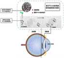
图1为本申请PLGA的局部眼用缓释复合制剂联合人工晶状体植入示意图;Figure 1 is a schematic diagram of the local ophthalmic sustained-release compound preparation of the present application PLGA combined with intraocular lens implantation;
图2为本申请PLGA的局部眼用缓释复合制剂的制备工艺流程图;Fig. 2 is the preparation process flow chart of the local ophthalmic slow-release composite preparation of the present application PLGA;
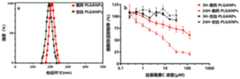
图3为透析时长去除PLGA纳米粒子(PLGANPs)制剂中残余乙腈的影响;Figure 3 is the effect of dialysis time to remove residual acetonitrile in the PLGA nanoparticles (PLGANPs) preparation;
图4为本申请PLGA微型套管的结构图,其中a)和b)扫描电镜内部纤维结构及尺寸,c)为宏观形貌与套管直径尺寸的图相;Fig. 4 is the structural diagram of the PLGA micro sleeve of the present application, wherein a) and b) the internal fiber structure and size of the scanning electron microscope, and c) is the picture of the macroscopic appearance and the diameter of the sleeve;
图5为基于试验设计预测模型的二维等高线图。微流控制备工艺中总流量(TFR)与水相/有机相流速比(FRR)对载药PLGA纳米粒子的a)粒径尺寸,b)多分散系数,c)阿霉素包封率以及d)丝裂霉素C包封率的影响,颜色梯度从蓝到红色表示理化参数值从小逐渐变大;Figure 5 is a two-dimensional contour map based on the DOE prediction model. Effects of total flow rate (TFR) and aqueous/organic phase flow rate ratio (FRR) on a) particle size, b) polydispersity coefficient, c) doxorubicin encapsulation efficiency and d) The effect of mitomycin C encapsulation efficiency, the color gradient from blue to red indicates that the physical and chemical parameters gradually increase from small to large;
图6表示联合药物组合阿霉素-丝裂霉素C协同杀伤人晶状体上皮细胞;其中a)阿霉素-丝裂霉素C在3:1摩尔比的联合指数曲线,b)单药游离阿霉素、游离丝裂霉素C、联药阿霉素-丝裂霉素C的中值效应曲线,c)MTT法检测单药或联合药物组会抑制人晶状体上皮细胞存活的效果,d)药物处理人晶状体上皮细胞的半抑制浓度(IC50);Fig. 6 shows that the combined drug combination doxorubicin-mitomycin C synergistically kills human lens epithelial cells; wherein a) the combined exponential curve of doxorubicin-mitomycin C at a molar ratio of 3:1, b) the single-drug free Median effect curves of doxorubicin, free mitomycin C, and combined drug doxorubicin-mitomycin C, c) MTT method to detect the effect of single drug or combined drug group on inhibiting the survival of human lens epithelial cells, d ) the half-inhibitory concentration (IC50 ) of drug treatment of human lens epithelial cells;
图7表示空白(无载药)PLGANPs与原位共载阿霉素-丝裂霉素CPLGANPs(DM-PLGANPs)的理化表征。其中,a)空白PLGANPs(无药物载入,黑色曲线)与DM-PLGANPs(红色曲线)粒径尺寸分布图,b)空白PLGANPs(无药物载入,黑色曲线)与载药DM-PLGANPs(红色曲线)对人晶状体上皮细胞的细胞毒性;Figure 7 shows the physicochemical characterization of blank (no drug-loaded) PLGANPs and in situ co-loaded doxorubicin-mitomycin CPLGANPs (DM-PLGANPs). Among them, a) particle size distribution diagram of blank PLGANPs (no drug loading, black curve) and DM-PLGANPs (red curve), b) blank PLGANPs (no drug loading, black curve) and drug-loaded DM-PLGANPs (red curve) Curve) to the cytotoxicity of human lens epithelial cells;
图8为DM-PLGANPs在37℃、复方电解质眼内冲洗液中7天的稳定性;Figure 8 shows the stability of DM-PLGANPs at 37°C in compound electrolyte ocular irrigation solution for 7 days;
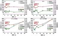
图9为DM-PLGANPs悬液与冷冻干燥粉末分别在4℃和25℃条件下贮藏28天的稳定性,a)、b)DM-PLGANPs悬液的在不同时间点的粒径尺寸与多分散系数,c)、d)DM-PLGANPs冷冻干燥粉在不同时间点的粒径尺寸与多分散系数;Figure 9 shows the stability of DM-PLGANPs suspension and freeze-dried powder stored at 4°C and 25°C for 28 days, respectively, the particle size and polydispersity of a) and b) DM-PLGANPs suspension at different time points Coefficient, c), d) particle size and polydispersity coefficient of DM-PLGANPs freeze-dried powder at different time points;
图10为DM-PLGANPs的表面形貌扫描电镜图及其能谱仪元素分析图,a)DM-PLGANPs形貌扫描电镜图,b)DM-PLGANPs能谱仪元素分析图;Figure 10 is the scanning electron microscope image of the surface morphology of DM-PLGANPs and its elemental analysis diagram of the energy spectrometer, a) the scanning electron microscope image of the DM-PLGANPs morphology, b) the elemental analysis diagram of the DM-PLGANPs energy spectrometer;
图11为透析法检测DM-PLGANPs药物释放速率,等浓度游离联合药物(黑色曲线)作为对照组。Figure 11 is the dialysis method to detect the drug release rate of DM-PLGANPs, and the equal concentration of free combined drug (black curve) is used as the control group.
具体实施方式Detailed ways
为使本申请实施例的目的、技术方案和优点更加清楚,下面将对本申请实施例中的技术方案进行清楚、完整地描述。实施例中未注明具体条件者,按照常规条件或制造商建议的条件进行。所用试剂或仪器未注明生产厂商者,均为可以通过市售购买获得的常规产品。In order to make the purpose, technical solutions and advantages of the embodiments of the present application clearer, the technical solutions in the embodiments of the present application will be clearly and completely described below. Those who do not indicate the specific conditions in the examples are carried out according to the conventional conditions or the conditions suggested by the manufacturer. The reagents or instruments used were not indicated by the manufacturer, and they were all conventional products that could be purchased from the market.
需要说明的是,在不冲突的情况下,本申请中的实施例及实施例中的特征可以相互组合。下面将参考具体实施例来详细说明本申请。It should be noted that, in the case of no conflict, the embodiments in the present application and the features in the embodiments can be combined with each other. The present application will be described in detail below with reference to specific examples.
一种基于PLGA的局部眼用缓释复合制剂的制备方法,其特征在于,包括以下步骤:A preparation method of a PLGA-based topical ophthalmic sustained-release compound preparation, characterized in that it comprises the following steps:
将基于PLGA的高聚物纺丝液加热搅拌后,经静电纺丝和纤维表面改性后,获得生物功能性PLGA微型套管;After heating and stirring the PLGA-based polymer spinning solution, after electrospinning and fiber surface modification, biofunctional PLGA micro-sleeves are obtained;
将上述PLGA微型套管与药物混合孵育,通过吸附组装,并用氮气流干燥后,获得所述基于PLGA的局部眼用缓释复合制剂。The PLGA micro-cannula was mixed and incubated with the drug, assembled by adsorption, and dried with a nitrogen flow to obtain the PLGA-based topical ophthalmic sustained-release compound preparation.
在本申请的一些实施例中,上述药物包括载药PLGA纳米粒子(PLGANPs)和游离药物;所述载药PLGANPs的制备方法为:In some embodiments of the present application, the above-mentioned drugs include drug-loaded PLGA nanoparticles (PLGANPs) and free drugs; the preparation method of the drug-loaded PLGANPs is:
将聚乙烯醇溶解在水相中,将PLGA溶解于有机相中,将药物溶于上述水相或有机相中;将上述水相和有机相混合,原位包载药物,制备得到载药PLGANPs。Dissolving polyvinyl alcohol in the water phase, dissolving PLGA in the organic phase, dissolving the drug in the above water phase or organic phase; mixing the above water phase and organic phase, and encapsulating the drug in situ to prepare drug-loaded PLGANPs .
在本申请的一些实施例中,上述水相和有机相在分岔混频结构微流通道中以小于1微秒流速快速均匀混合,制备得到载药PLGANPs。In some embodiments of the present application, the above-mentioned aqueous phase and organic phase are rapidly and uniformly mixed in a bifurcated frequency mixing structure microfluidic channel at a flow rate of less than 1 microsecond to prepare drug-loaded PLGANPs.
在本申请的一些实施例中,上述水相为磷酸盐缓冲液(pH7.4)、Tris缓冲液(pH7.4)或纯水;所述有机相为乙腈、甲醇或二甲基亚砜;所述聚乙烯醇的浓度为0~2%(w/v),所述PLGA的分子量为30~150kDa,L/G比例为50/50~90/10;所述药物为用于白内障术后并发症防治的药物,所述水相和有机相的流速比(FRR)为(1~5):1,总流量(TFR)为1~20mL/min,最终制备载药PLGA纳米粒子总产量体积(TV)为2~20mL。In some embodiments of the present application, the above-mentioned aqueous phase is phosphate buffer (pH7.4), Tris buffer (pH7.4) or pure water; the organic phase is acetonitrile, methanol or dimethyl sulfoxide; The concentration of the polyvinyl alcohol is 0-2% (w/v), the molecular weight of the PLGA is 30-150kDa, and the L/G ratio is 50/50-90/10; the medicine is used for cataract surgery For the prevention and treatment of complications, the flow rate ratio (FRR) of the aqueous phase and the organic phase is (1-5): 1, the total flow rate (TFR) is 1-20mL/min, and the total output volume of the drug-loaded PLGA nanoparticles is finally prepared (TV) is 2-20 mL.
在本申请的一些实施例中,上述组装成为载药PLGANPs后还包括将其在室温进行透析,所述透析处理的截留分子量为10~300kDa,透析液为磷酸盐缓冲液,透析温度为25℃,透析时间为1h~18h。透析时长对PLGANPs中残余乙腈去除的影响如图3所示。In some embodiments of the present application, the above-mentioned assembly into drug-loaded PLGANPs further includes dialysis at room temperature, the molecular weight cut-off of the dialysis treatment is 10-300kDa, the dialysate is phosphate buffered saline, and the dialysate temperature is 25°C , Dialysis time is 1h ~ 18h. The effect of dialysis time on the removal of residual acetonitrile in PLGANPs is shown in Figure 3.
在本申请的一些实施例中,上述PLGA微型套管的制备步骤具体为:将PLGA溶于溶剂中,得到PLGA纺丝液,在设定电场范围内,PLGA纺丝液通过喷丝头喷出,喷射过程中溶剂蒸发或固化,经过拉长拉细后,纤维直径分布100~1000nm,宏观套管直径为0.5mm~2mm。静电纺丝工艺影响PLGA纤维套管的化学性质、表面性能及其形貌结构。通过调节PLGA纺丝液浓度、流速,电场强度等参数,可制备出载药PLGA套管。In some embodiments of the present application, the preparation steps of the above-mentioned PLGA micro-sleeve are as follows: dissolving PLGA in a solvent to obtain a PLGA spinning solution, and within the set electric field range, the PLGA spinning solution is sprayed out through a spinneret , The solvent evaporates or solidifies during the spraying process. After elongation and thinning, the fiber diameter distribution is 100-1000nm, and the diameter of the macroscopic sleeve is 0.5mm-2mm. The electrospinning process affects the chemical properties, surface properties and morphology of PLGA fiber sleeves. The drug-loaded PLGA sleeve can be prepared by adjusting the parameters such as the concentration, flow rate, and electric field strength of the PLGA spinning solution.
在本申请的一些实施例中,在静电纺丝步骤的基本参数为:PLGA纺丝液溶剂为N,N-二甲基甲酰胺、四氢呋喃、氯仿或二氯甲烷中的一种或几种的任意比例混合溶剂;所述PLGA纺丝液配置浓度0.1~0.4g/mL,纺丝原液流速为0.4~5mL/h,电压为5~20kV,接收距离为5~20cm。In some embodiments of the present application, the basic parameters in the electrospinning step are: the PLGA spinning liquid solvent is one or more of N,N-dimethylformamide, tetrahydrofuran, chloroform or dichloromethane The solvent is mixed in any proportion; the concentration of the PLGA spinning solution is 0.1-0.4g/mL, the flow rate of the spinning stock solution is 0.4-5mL/h, the voltage is 5-20kV, and the receiving distance is 5-20cm.
进一步,上述混合浸泡步骤中药物与PLGA微型套管的质量比为(0.5~5):100,浸泡溶液pH为3~7.4,浸泡时间为20min~16h,用去离子水冲洗并干燥。Further, in the mixing and soaking step above, the mass ratio of the drug to the PLGA micro-tube is (0.5-5):100, the pH of the soaking solution is 3-7.4, the soaking time is 20min-16h, rinsed with deionized water and dried.
一种基于PLGA的局部眼用缓释复合制剂,采用上述的制备方法制备而成。A PLGA-based local ophthalmic slow-release compound preparation is prepared by the above-mentioned preparation method.
一种基于PLGA的局部眼用缓释复合制剂在制备局部眼用植入剂中的应用。在白内障术后并发症(后发性白内障、眼内炎等)防治中的应用具体分为两个方面:1、与人工晶状体联合使用,适配于各种襻结构;2、在白内障术中植入前囊袋作为药物贮库。Application of a PLGA-based local ophthalmic sustained-release compound preparation in the preparation of local ophthalmic implants. The application in the prevention and treatment of cataract postoperative complications (post-cataract, endophthalmitis, etc.) is specifically divided into two aspects: 1. It is used in combination with intraocular lenses and is suitable for various loop structures; 2. In cataract surgery The pre-implantation pouch acts as a drug depot.
以下结合实施例对本申请的特征和性能作进一步的详细描述。The characteristics and performance of the present application will be described in further detail below in conjunction with the examples.
实施例1Example 1
一种基于PLGA的局部眼用缓释复合制剂,其产品结构示意图与制备流程图如图1和图2所示,其制备方法包括以下步骤:A local ophthalmic sustained-release compound preparation based on PLGA, its product structure schematic diagram and preparation flow chart as shown in Figure 1 and Figure 2, its preparation method comprises the following steps:
(1)静电纺丝制备PLGA微型套管(1) Preparation of PLGA microcasings by electrospinning
首先将PLGA溶解在配比为1:1的N,N-二甲基甲酰胺-四氢呋喃,得到PLGA纺丝液。调节电压(最大20kV),使PLGA纺丝溶液的表面张力与带电液滴在喷丝头末端处于平衡。使射流从锥表面喷出速度为3.6mL/h,形成纤维毡(网或者膜)。喷丝头与接地模具之间的距离为10cm,使用不同规格尺寸旋转滚筒模具收集、干燥后获得具有不同尺寸的PLGA微型套管。本实施例制备得到的PLGA套管如图4所示,其中图4a和图4b为套管微观内部结构与孔径尺寸的电镜扫描图,图4c为不同直径尺寸(0.5~2mm)的套管图。Firstly, PLGA was dissolved in N,N-dimethylformamide-tetrahydrofuran with a ratio of 1:1 to obtain PLGA spinning solution. Adjust the voltage (maximum 20kV) to balance the surface tension of the PLGA spinning solution with the charged droplets at the end of the spinneret. The jet flow is ejected from the cone surface at a speed of 3.6 mL/h to form a fiber mat (net or film). The distance between the spinneret and the grounded mold was 10 cm, and PLGA micro-sleeves with different sizes were obtained after collection and drying by rotating drum molds of different sizes. The PLGA bushing prepared in this example is shown in Figure 4, where Figures 4a and 4b are scanning electron microscope images of the microscopic internal structure and pore size of the bushing, and Figure 4c is a map of bushings with different diameters (0.5-2 mm) .
(2)载药PLGA纳米粒子的合成(2) Synthesis of drug-loaded PLGA nanoparticles
将50mg PLGA(L/G=75/25)溶解在5mL乙腈中,得到浓度为1%w/vPLGA有机相;将50mg聚乙烯醇(PVA)和协同药物组合阿霉素-丝裂霉素C溶解在10mL磷酸盐缓冲液中)混合,得到浓度为0.5%w/vPVA、0.2mM阿霉素、0.6mM丝裂霉素C水相;将上述采用0.22μm孔径水系PES滤膜过滤水相溶液,分别使用5mL与10mL的针管旋接在有机相与水相的微流芯片(Precision Nanosystems,Inc)接口处,并组装在
(3)制备基于PLGA的局部眼用缓释复合制剂(3) Preparation of local ophthalmic sustained-release compound preparation based on PLGA
将制备得到的载药PLGA纳米粒子与PLGA微型套管按照1:100的质量比,浸泡在酸盐缓冲液,共同混合孵育16h,得到本实施例的局部眼用缓释复合制剂。The prepared drug-loaded PLGA nanoparticles and PLGA micro-cannula were soaked in salt buffer solution at a mass ratio of 1:100, mixed together and incubated for 16 hours to obtain the topical ophthalmic sustained-release composite preparation of this embodiment.
实施例2Example 2
一种基于PLGA的局部眼用缓释复合制剂,为获得高效包载协同联合用药PLGANPs的制备方法如下:A local ophthalmic sustained-release compound preparation based on PLGA, in order to obtain high-efficiency entrapped synergistic drug combination PLGANPs, the preparation method is as follows:
采用2因素3水平试验设计(表1),在低、中、高水平调节微流控制备工艺参数总流量(TFR)与有机相/水相比(FRR),制备共载阿霉素-丝裂霉素C协同联合用药DM-PLGANPs,对DM-PLGANPs的粒径、多分散系数、包封率等进行理化表征。如图5所示,为基于DoE预测模型的二维等高线图。预测模型使用多元线性回归和偏最小平方回归分析分析TFR、FFR和L/G对载药DM-PLGANPs的粒径、多分散系数、药物包封率的影响。微流控制备参数TFR与FRR能显著影响PLGANPs的理化特征。其中FRR对于粒径尺寸与多分散系数的影响较大,即FRR趋近于5,粒径尺寸越大,多分散系数越小。FRR增大也能提高阿霉素与丝裂霉素C的药物包封率。微流控制备DM-PLGANPs具体步骤与实施例1相同。Using 2 factors and 3 levels of experimental design (Table 1), adjust the microfluidic preparation process parameters total flow rate (TFR) and organic phase/water ratio (FRR) at low, medium and high levels to prepare co-loaded doxorubicin-silk The DM-PLGANPs were co-administered with split mycin C, and the particle size, polydispersity coefficient and encapsulation efficiency of DM-PLGANPs were characterized physically and chemically. As shown in Figure 5, it is a two-dimensional contour map based on the DoE prediction model. The prediction model used multiple linear regression and partial least square regression analysis to analyze the effects of TFR, FFR and L/G on the particle size, polydispersity coefficient and drug encapsulation efficiency of drug-loaded DM-PLGANPs. The microfluidic preparation parameters TFR and FRR can significantly affect the physical and chemical characteristics of PLGANPs. Among them, FRR has a great influence on particle size and polydispersity coefficient, that is, FRR approaches 5, and the larger the particle size, the smaller the polydispersity coefficient. Increased FRR can also increase the drug encapsulation efficiency of doxorubicin and mitomycin C. The specific steps of microfluidic preparation of DM-PLGANPs are the same as in Example 1.
表1.试验设计参数Table 1. Experimental Design Parameters


在本实施例2中采用PLGA为酯封端,分子量76~115kDa,L/G比例为50/50~85/15。水相为磷酸盐缓冲液(pH7.4),含有0.2mM阿霉素与0.6mM丝裂霉素C以及0.5%(w/v)PVA;有机相为乙腈,含1%(w/v)PLGA。微流控制备参数包括TFR范围在5~15mL/min和FRR范围在1:1~5:1。In Example 2, PLGA is used as the ester end cap, the molecular weight is 76-115 kDa, and the L/G ratio is 50/50-85/15. The aqueous phase is phosphate buffer (pH7.4), containing 0.2mM doxorubicin and 0.6mM mitomycin C and 0.5% (w/v) PVA; the organic phase is acetonitrile, containing 1% (w/v) PLGA. Microfluidic preparation parameters include TFR ranging from 5 to 15 mL/min and FRR ranging from 1:1 to 5:1.
将DM-PLGANPs在超纯水中稀释至0.1w/v(PLGANPs占溶剂的质量百分比),并通过孔径为0.45μm滤膜过滤。用动态光散射法测定DM-PLGANPs粒径尺寸、多分散系数。使用超高速离心法,在4℃、200000×g,离心30分钟,收集含有游离阿霉素-丝裂霉素C的上清液,分别在360nm和480nm处对丝裂霉素C和阿霉素的吸光度进行检测,采用以下公式计算药物包封率和载药量:Dilute DM-PLGANPs in ultrapure water to 0.1w/v (the mass percentage of PLGANPs in the solvent), and filter through a filter membrane with a pore size of 0.45 μm. The particle size and polydispersity coefficient of DM-PLGANPs were determined by dynamic light scattering. Using ultra-high-speed centrifugation, centrifuge at 4°C, 200,000×g for 30 minutes, collect the supernatant containing free doxorubicin-mitomycin C, and detect mitomycin C and doxorubicin at 360nm and 480nm, respectively. The absorbance of the protein was detected, and the drug encapsulation efficiency and drug loading were calculated using the following formula:


实验例Experimental example
(1)原位共包载阿霉素-丝裂霉素C协同联合药物(1) In situ co-encapsulation of doxorubicin-mitomycin C synergistic combination drug
采用MTT法检测联合阿霉素-丝裂霉素C对人晶状体上皮细胞(HLE-B3)抑制存活的协同作用,其结果如图6所示。阿霉素-丝裂霉素C的药物联合指数小于1,表明1:3药物浓度摩尔比例具有协同抑制人晶状体上皮细胞活力的作用。协同药物联合的半抑制浓度(IC50)比游离单药低2-4倍,表明阿霉素-丝裂霉素C在1:3比例下能强效抑制人晶状体上皮细胞活力。图7和表2为本实验例制备空白(无载药)PLGANPs与原位共载协同阿霉素-丝裂霉素C药物联合的PLGANPs(DM-PLGANPs)理化表征。其中图7a为DM-PLGANPs与空白PLGANPs的代表性粒径分布图,图7b为空白PLGANPs与载药DM-PLGANPs处理人晶状体上皮细胞3h和24h后,不同浓度对细胞活力抑制的影响。空白PLGANPs组对人晶状体上皮细胞的活力无显著影响,而载药DM-PLGANPs呈时间与浓度依赖性的细胞活力抑制,即治疗时间越长、药物浓度越高,细胞毒性越大。MTT method was used to detect the synergistic effect of combined doxorubicin-mitomycin C on the inhibition of survival of human lens epithelial cells (HLE-B3), and the results are shown in FIG. 6 . The drug combination index of doxorubicin-mitomycin C is less than 1, indicating that the molar ratio of 1:3 drug concentration has a synergistic effect on inhibiting the viability of human lens epithelial cells. The half-inhibitory concentration (IC50) of the synergistic drug combination is 2-4 times lower than that of the free single drug, indicating that doxorubicin-mitomycin C can potently inhibit the viability of human lens epithelial cells at a ratio of 1:3. Figure 7 and Table 2 show the physical and chemical characterization of the blank (no drug-loaded) PLGANPs prepared in this experimental example and the PLGANPs (DM-PLGANPs) co-loaded in situ with a combination of doxorubicin-mitomycin C drugs. Figure 7a is the representative particle size distribution diagram of DM-PLGANPs and blank PLGANPs, and Figure 7b is the effect of different concentrations on the inhibition of cell viability after the blank PLGANPs and drug-loaded DM-PLGANPs treated human lens epithelial cells for 3h and 24h. The blank PLGANPs group had no significant effect on the viability of human lens epithelial cells, while the drug-loaded DM-PLGANPs inhibited cell viability in a time- and concentration-dependent manner, that is, the longer the treatment time and the higher the drug concentration, the greater the cytotoxicity.
表2 PLGANPs的理化表征Table 2 Physicochemical characterization of PLGANPs

从表2和图7中可以看出相比空白PLGANPs(193nm),共载药的DM-PLGANPs粒径大小呈略微增加(224nm)。然而,通过微流控制备出的纳米粒子的多分散系数相似,大约为0.1,表明PLGANPs颗粒分布均匀。DM-PLGANPs能按药物联合协同比例(例如,1:3)包封阿霉素-丝裂霉素C,其包封率分别83.88%和76.77%,阿霉素-丝裂霉素C载药量分别为1.85%和3.47%。It can be seen from Table 2 and Figure 7 that the particle size of the co-loaded DM-PLGANPs was slightly increased (224nm) compared to the blank PLGANPs (193nm). However, the polydispersity coefficients of nanoparticles prepared by microfluidics were similar, about 0.1, indicating that the PLGANPs particles were uniformly distributed. DM-PLGANPs can encapsulate doxorubicin-mitomycin C according to the drug combination synergistic ratio (for example, 1:3), and the encapsulation efficiencies are 83.88% and 76.77%, respectively. The amounts are 1.85% and 3.47%, respectively.
(2)载药PLGANPs的稳定性(2) Stability of drug-loaded PLGANPs
将DM-PLGANPs放入复方电解质眼内冲洗液中,37℃条件下贮藏7天。随后在七天内按照时间点取0.2mL载药PLGANPs,并在超纯水中稀释至0.1w/v,通过孔径为0.45μm滤膜过滤。用动态光散射法测定载药PLGANPs的粒径尺寸和多分散系数。其结果如图8所示。从图8中可以看出在模拟眼内环境条件下,载药PLGANPs的粒径与多分散系数无显著变化。The DM-PLGANPs were put into the compound electrolyte intraocular irrigation solution and stored at 37°C for 7 days. Then take 0.2mL of drug-loaded PLGANPs according to the time points within seven days, dilute to 0.1w/v in ultrapure water, and filter through a filter membrane with a pore size of 0.45μm. The particle size and polydispersity coefficient of drug-loaded PLGANPs were determined by dynamic light scattering. The result is shown in Figure 8. It can be seen from Figure 8 that the particle size and polydispersity coefficient of the drug-loaded PLGANPs did not change significantly under the simulated intraocular environmental conditions.
载药PLGANPs可制备成混悬液与冻干粉末,并在4℃和25℃条件下分别贮藏28天。结果如图9所示,图9a和图9b所示载药PLGANPs悬液的粒径尺寸、多分散系数;图9c、图9d表示载药PLGANPs冻干粉末形式的粒径尺寸、多分散系数;从图9中可以看出在载药PLGANPs混悬液与冻干粉末贮藏条件下28天内,载药PLGANPs的粒径与多分散系数无显著变化,稳定性良好。Drug-loaded PLGANPs can be prepared into suspension and freeze-dried powder, and stored at 4°C and 25°C for 28 days, respectively. The results are shown in Figure 9, the particle size and polydispersity coefficient of the drug-loaded PLGANPs suspension shown in Figure 9a and Figure 9b; Figure 9c and Figure 9d show the particle size and polydispersity coefficient of the drug-loaded PLGANPs freeze-dried powder form; It can be seen from Figure 9 that the particle size and polydispersity coefficient of drug-loaded PLGANPs have no significant changes within 28 days under the storage conditions of drug-loaded PLGANPs suspension and freeze-dried powder, and the stability is good.
(3)载药PLGANPs形貌及其包封药物的分布(3) Morphology of drug-loaded PLGANPs and distribution of encapsulated drugs
使用扫描电镜观察PLGANPs的形貌特征。将载药PLGANPs滴加至1mm2单晶硅片上方,随后在40℃烘箱中烘干2h,在特高分辨率场发射扫描电镜(Verios G4,FEI)10kV加速电压下进行拍摄。载药PLGANPs的表面形貌扫描电镜图及其能谱仪元素分析图如图10所示;其中图10a)DM-PLGANPs的形貌扫描电镜图;图10b为包载药物(阿霉素、丝裂霉素C)能谱仪元素分析图。从图10中可以看出载药PLGANPs具有球状形貌,其氮元素信号的分布表明药物被包埋在载药PLGANPs中。The morphology characteristics of PLGANPs were observed by scanning electron microscopy. Drug-loaded PLGANPs were added dropwise onto a 1 mm2 single crystal silicon wafer, then dried in an oven at 40 °C for 2 h, and photographed under an ultra-high resolution field emission scanning electron microscope (Verios G4, FEI) at an accelerating voltage of 10 kV. The scanning electron micrograph of the surface morphology of drug-loaded PLGANPs and its elemental analysis diagram of energy spectrometer are shown in Figure 10; where Figure 10a) is the scanning electron micrograph of the morphology of DM-PLGANPs; Split mycin C) Energy spectrometer elemental analysis diagram. It can be seen from Figure 10 that the drug-loaded PLGANPs have a spherical morphology, and the distribution of nitrogen signal indicates that the drug is embedded in the drug-loaded PLGANPs.
(4)载药PLGANPs的累计药物释放百分比(4) Cumulative drug release percentage of drug-loaded PLGANPs
将1mL纯化载药PLGANPs转移到截留分子量为300kDa的透析袋中,在10mL复方电解质眼内冲洗液中检测7天内药物释放。在避光、恒温37℃,100rpm转速条件下,从透析袋外取0.5mL溶液,同时补充相同体积的新鲜复方电解质眼内冲洗液。分别在360nm和480nm处对丝裂霉素C和阿霉素的吸光度进行检测。结果如图11所示。从图11中可以看出载药DM-PLGANPs能协调持续释放联合药物阿霉素与丝裂霉素C长达7天,其释放比例为1:3的药物联合比例,而游离药物在1天内已完成100%药物释放。
以上所描述的实施例是本申请一部分实施例,而不是全部的实施例。本申请的实施例的详细描述并非旨在限制要求保护的本申请的范围,而是仅仅表示本申请的选定实施例。基于本申请中的实施例,本领域普通技术人员在没有作出创造性劳动前提下所获得的所有其他实施例,都属于本申请保护的范围。The embodiments described above are some of the embodiments of the present application, but not all of them. The detailed description of the embodiments of the application is not intended to limit the scope of the claimed application, but merely represents selected embodiments of the application. Based on the embodiments in this application, all other embodiments obtained by persons of ordinary skill in the art without creative efforts fall within the protection scope of this application.
Claims (10)
Priority Applications (1)
| Application Number | Priority Date | Filing Date | Title |
|---|---|---|---|
| CN202211413007.7ACN115671028B (en) | 2022-11-11 | 2022-11-11 | A kind of PLGA-based topical ophthalmic sustained-release compound preparation and its preparation method and application |
Applications Claiming Priority (1)
| Application Number | Priority Date | Filing Date | Title |
|---|---|---|---|
| CN202211413007.7ACN115671028B (en) | 2022-11-11 | 2022-11-11 | A kind of PLGA-based topical ophthalmic sustained-release compound preparation and its preparation method and application |
Publications (2)
| Publication Number | Publication Date |
|---|---|
| CN115671028Atrue CN115671028A (en) | 2023-02-03 |
| CN115671028B CN115671028B (en) | 2023-07-14 |
Family
ID=85051877
Family Applications (1)
| Application Number | Title | Priority Date | Filing Date |
|---|---|---|---|
| CN202211413007.7AActiveCN115671028B (en) | 2022-11-11 | 2022-11-11 | A kind of PLGA-based topical ophthalmic sustained-release compound preparation and its preparation method and application |
Country Status (1)
| Country | Link |
|---|---|
| CN (1) | CN115671028B (en) |
Citations (11)
| Publication number | Priority date | Publication date | Assignee | Title |
|---|---|---|---|---|
| US20080033351A1 (en)* | 2006-08-04 | 2008-02-07 | Allergan, Inc. | Ocular implant delivery assemblies with distal caps |
| US20120202743A1 (en)* | 2009-04-29 | 2012-08-09 | Ipsen Pharma S.A.S. | SUSTAINED RELEASE FORMULATIONS COMPRISING GnRH ANALOGUES |
| US20130018448A1 (en)* | 2011-07-12 | 2013-01-17 | Boston Scientific Scimed, Inc. | Drug elution medical device |
| CN104800885A (en)* | 2015-05-13 | 2015-07-29 | 中山大学 | Preparation method and application of bioactive bracket with chemotactic function |
| CN204840394U (en)* | 2015-05-13 | 2015-12-09 | 中山大学 | Preparation and application of biological activity support with chemotactic function |
| CN106421912A (en)* | 2016-10-13 | 2017-02-22 | 中山大学 | Preparation and application of matrix acellular nerve scaffold |
| CN206934376U (en)* | 2016-10-13 | 2018-01-30 | 中山大学 | A kind of matrixing Acellular nerve support |
| CN107789669A (en)* | 2017-10-31 | 2018-03-13 | 无锡中科光远生物材料有限公司 | A kind of three-dimensional shell material for CO2 laser weld |
| CN109758197A (en)* | 2019-03-11 | 2019-05-17 | 宁波光远致信生物科技有限公司 | A kind of tissue repair casing and its preparation method and application |
| CN109938875A (en)* | 2019-03-07 | 2019-06-28 | 宁波光远致信生物科技有限公司 | A kind of nerve prosthesis and its preparation method and application |
| CN112107742A (en)* | 2020-10-22 | 2020-12-22 | 南京佑羲医药科技有限公司 | Long-acting intelligent implantable drug carrying device and manufacturing method thereof |
- 2022
- 2022-11-11CNCN202211413007.7Apatent/CN115671028B/enactiveActive
Patent Citations (11)
| Publication number | Priority date | Publication date | Assignee | Title |
|---|---|---|---|---|
| US20080033351A1 (en)* | 2006-08-04 | 2008-02-07 | Allergan, Inc. | Ocular implant delivery assemblies with distal caps |
| US20120202743A1 (en)* | 2009-04-29 | 2012-08-09 | Ipsen Pharma S.A.S. | SUSTAINED RELEASE FORMULATIONS COMPRISING GnRH ANALOGUES |
| US20130018448A1 (en)* | 2011-07-12 | 2013-01-17 | Boston Scientific Scimed, Inc. | Drug elution medical device |
| CN104800885A (en)* | 2015-05-13 | 2015-07-29 | 中山大学 | Preparation method and application of bioactive bracket with chemotactic function |
| CN204840394U (en)* | 2015-05-13 | 2015-12-09 | 中山大学 | Preparation and application of biological activity support with chemotactic function |
| CN106421912A (en)* | 2016-10-13 | 2017-02-22 | 中山大学 | Preparation and application of matrix acellular nerve scaffold |
| CN206934376U (en)* | 2016-10-13 | 2018-01-30 | 中山大学 | A kind of matrixing Acellular nerve support |
| CN107789669A (en)* | 2017-10-31 | 2018-03-13 | 无锡中科光远生物材料有限公司 | A kind of three-dimensional shell material for CO2 laser weld |
| CN109938875A (en)* | 2019-03-07 | 2019-06-28 | 宁波光远致信生物科技有限公司 | A kind of nerve prosthesis and its preparation method and application |
| CN109758197A (en)* | 2019-03-11 | 2019-05-17 | 宁波光远致信生物科技有限公司 | A kind of tissue repair casing and its preparation method and application |
| CN112107742A (en)* | 2020-10-22 | 2020-12-22 | 南京佑羲医药科技有限公司 | Long-acting intelligent implantable drug carrying device and manufacturing method thereof |
Also Published As
| Publication number | Publication date |
|---|---|
| CN115671028B (en) | 2023-07-14 |
Similar Documents
| Publication | Publication Date | Title |
|---|---|---|
| AU2019250153B2 (en) | Methods and biocompatible compositions to achieve sustained drug release in the eye | |
| JP2975332B2 (en) | Biodegradable eye implant | |
| Goyal et al. | Current nanotechnological strategies for treating glaucoma | |
| WO2008157614A9 (en) | Sustained intraocular delivery of drugs from biodegradable polymeric microparticles | |
| PT109154B (en) | NON-INVASIVE EYE INSERT TECHNOLOGY FOR CONTROLLED DRUG RELEASE | |
| CN102233129A (en) | Long-acting sustained release preparation for preventing or treating retinal damage, and preparation method thereof | |
| CN113041215A (en) | Eye surface in-situ medicine and preparation method thereof | |
| EP3536352A1 (en) | Keratoconjunctival cover sheet and method for producing keratoconjunctival cover sheet | |
| Kaushal et al. | Nanocarriers based ocular therapeutics: updates, challenges and future prospectives | |
| CN116473915B (en) | ROS responsive micelle-gel complex for treating cornea neovascularization | |
| Meshram et al. | Ocular in Situ gels: Development, evaluation and advancements | |
| Sonawane et al. | PLGA: a wow smart biodegradable polymer in drug delivery system | |
| CN115671028B (en) | A kind of PLGA-based topical ophthalmic sustained-release compound preparation and its preparation method and application | |
| CN116687840B (en) | Gel preparation for treating eye symptoms and preparation method thereof | |
| Felt et al. | Polymeric systems for ophthalmic drug delivery | |
| CN114533703B (en) | Tripterine composite film and preparation method and application thereof | |
| CN115444818A (en) | Eye drop solubilizing auxiliary material based on silk fibroin nanofiber and preparation method of eye drop medicament containing auxiliary material | |
| WO2021237096A1 (en) | Durable implants and microparticles for long-term ocular therapy | |
| CN116370698B (en) | A kind of hydrogel scleral plug and its preparation method and application | |
| CN1813752A (en) | Fluorouracial nano particle formulation and its preparing method | |
| Ramesh | Ocular barriers and ocular drug delivery: Bridging the gap using nanomicelles as drug carriers | |
| HK40003833A (en) | Methods and biocompatible compositions to achieve sustained drug release in the eye | |
| Jiang | Sustained Release of Anti-VEGF from Injectable Devices for Treating Wet Age-Related Macular Degeneration | |
| CN113350268A (en) | Medicine sustained-release gel for subconjunctival implantation of eye and preparation method thereof | |
| CN1491102A (en) | Novel Ophthalmic Compositions |
Legal Events
| Date | Code | Title | Description |
|---|---|---|---|
| PB01 | Publication | ||
| PB01 | Publication | ||
| SE01 | Entry into force of request for substantive examination | ||
| SE01 | Entry into force of request for substantive examination | ||
| GR01 | Patent grant | ||
| GR01 | Patent grant |