CN115590614A - Intravascular plaque removal system - Google Patents
Intravascular plaque removal systemDownload PDFInfo
- Publication number
- CN115590614A CN115590614ACN202110773417.1ACN202110773417ACN115590614ACN 115590614 ACN115590614 ACN 115590614ACN 202110773417 ACN202110773417 ACN 202110773417ACN 115590614 ACN115590614 ACN 115590614A
- Authority
- CN
- China
- Prior art keywords
- balloon
- plaque
- carotid artery
- catheter
- blood vessel
- Prior art date
- Legal status (The legal status is an assumption and is not a legal conclusion. Google has not performed a legal analysis and makes no representation as to the accuracy of the status listed.)
- Pending
Links
- 210000004204blood vesselAnatomy0.000claimsabstractdescription41
- 230000017531blood circulationEffects0.000claimsabstractdescription26
- 210000004369bloodAnatomy0.000claimsabstractdescription20
- 239000008280bloodSubstances0.000claimsabstractdescription20
- 238000005286illuminationMethods0.000claimsabstractdescription8
- 238000003780insertionMethods0.000claimsabstract2
- 230000037431insertionEffects0.000claimsabstract2
- 210000001168carotid artery commonAnatomy0.000claimsdescription18
- 210000004004carotid artery internalAnatomy0.000claimsdescription17
- 210000001715carotid arteryAnatomy0.000claimsdescription14
- 238000000608laser ablationMethods0.000claimsdescription13
- 238000000034methodMethods0.000claimsdescription13
- 210000000269carotid artery externalAnatomy0.000claimsdescription12
- 208000006170carotid stenosisDiseases0.000claimsdescription11
- 239000000835fiberSubstances0.000claimsdescription10
- 238000002583angiographyMethods0.000claimsdescription7
- 238000011049fillingMethods0.000claimsdescription5
- 239000012530fluidSubstances0.000claimsdescription5
- 230000008321arterial blood flowEffects0.000claimsdescription4
- 238000002679ablationMethods0.000claimsdescription3
- 229940113601irrigation solutionDrugs0.000claims1
- 238000013172carotid endarterectomyMethods0.000description15
- 210000001105femoral arteryAnatomy0.000description15
- 230000003902lesionEffects0.000description14
- 230000005540biological transmissionEffects0.000description12
- 230000006378damageEffects0.000description8
- 238000010586diagramMethods0.000description8
- 239000013307optical fiberSubstances0.000description8
- 210000001519tissueAnatomy0.000description8
- 238000001356surgical procedureMethods0.000description7
- 238000011010flushing procedureMethods0.000description6
- 230000002792vascularEffects0.000description6
- 210000001367arteryAnatomy0.000description5
- 230000008901benefitEffects0.000description5
- 230000000903blocking effectEffects0.000description5
- 210000004556brainAnatomy0.000description5
- 238000002513implantationMethods0.000description5
- 238000002271resectionMethods0.000description5
- 208000031481Pathologic ConstrictionDiseases0.000description4
- 230000000694effectsEffects0.000description4
- 238000002594fluoroscopyMethods0.000description4
- 230000036262stenosisEffects0.000description4
- 208000037804stenosisDiseases0.000description4
- 230000000007visual effectEffects0.000description4
- 230000002146bilateral effectEffects0.000description3
- 230000007547defectEffects0.000description3
- 230000007774longtermEffects0.000description3
- 229910001000nickel titaniumInorganic materials0.000description3
- 210000005259peripheral bloodAnatomy0.000description3
- 239000011886peripheral bloodSubstances0.000description3
- 210000001685thyroid glandAnatomy0.000description3
- 238000012800visualizationMethods0.000description3
- 206010061660Artery dissectionDiseases0.000description2
- 206010007687Carotid artery stenosisDiseases0.000description2
- 208000032843HemorrhageDiseases0.000description2
- 230000000702anti-platelet effectEffects0.000description2
- 239000003146anticoagulant agentSubstances0.000description2
- 210000000709aortaAnatomy0.000description2
- 208000034158bleedingDiseases0.000description2
- 230000000740bleeding effectEffects0.000description2
- 230000036770blood supplyEffects0.000description2
- 230000000747cardiac effectEffects0.000description2
- 210000001326carotid sinusAnatomy0.000description2
- 239000002872contrast mediaSubstances0.000description2
- 210000004351coronary vesselAnatomy0.000description2
- 230000009977dual effectEffects0.000description2
- 238000013171endarterectomyMethods0.000description2
- 238000002695general anesthesiaMethods0.000description2
- 239000007943implantSubstances0.000description2
- 238000000338in vitroMethods0.000description2
- 230000002262irrigationEffects0.000description2
- 238000003973irrigationMethods0.000description2
- 239000007788liquidSubstances0.000description2
- 210000003141lower extremityAnatomy0.000description2
- 230000010412perfusionEffects0.000description2
- 238000000053physical methodMethods0.000description2
- 238000000926separation methodMethods0.000description2
- 230000003685thermal hair damageEffects0.000description2
- XLYOFNOQVPJJNP-UHFFFAOYSA-NwaterSubstancesOXLYOFNOQVPJJNP-UHFFFAOYSA-N0.000description2
- 206010003497AsphyxiaDiseases0.000description1
- 206010048964Carotid artery occlusionDiseases0.000description1
- 208000019743Cranial nerve injuryDiseases0.000description1
- 206010013952DysphoniaDiseases0.000description1
- 208000005189EmbolismDiseases0.000description1
- 208000010496Heart ArrestDiseases0.000description1
- 208000010473HoarsenessDiseases0.000description1
- 206010020772HypertensionDiseases0.000description1
- 208000032382Ischaemic strokeDiseases0.000description1
- 208000028389Nerve injuryDiseases0.000description1
- 239000004677NylonSubstances0.000description1
- 206010040026Sensory disturbanceDiseases0.000description1
- 208000002847Surgical WoundDiseases0.000description1
- 208000027418Wounds and injuryDiseases0.000description1
- 230000002776aggregationEffects0.000description1
- 238000004220aggregationMethods0.000description1
- 230000003872anastomosisEffects0.000description1
- 210000003484anatomyAnatomy0.000description1
- 230000036471bradycardiaEffects0.000description1
- 230000002612cardiopulmonary effectEffects0.000description1
- 210000004720cerebrumAnatomy0.000description1
- 230000008859changeEffects0.000description1
- 238000001514detection methodMethods0.000description1
- 210000002249digestive systemAnatomy0.000description1
- 239000003814drugSubstances0.000description1
- 229940079593drugDrugs0.000description1
- 238000001839endoscopyMethods0.000description1
- 239000012634fragmentSubstances0.000description1
- 230000036541healthEffects0.000description1
- 208000011316hemodynamic instabilityDiseases0.000description1
- 210000003090iliac arteryAnatomy0.000description1
- 238000003384imaging methodMethods0.000description1
- 208000015181infectious diseaseDiseases0.000description1
- 208000014674injuryDiseases0.000description1
- 208000019073internal carotid artery stenosisDiseases0.000description1
- 238000001361intraarterial administrationMethods0.000description1
- 238000007917intracranial administrationMethods0.000description1
- 210000000629knee jointAnatomy0.000description1
- 239000000463materialSubstances0.000description1
- 239000012528membraneSubstances0.000description1
- 210000003975mesenteric arteryAnatomy0.000description1
- 229910052751metalInorganic materials0.000description1
- 239000002184metalSubstances0.000description1
- 238000001531micro-dissectionMethods0.000description1
- 230000008764nerve damageEffects0.000description1
- 210000000653nervous systemAnatomy0.000description1
- HLXZNVUGXRDIFK-UHFFFAOYSA-Nnickel titaniumChemical compound[Ti].[Ti].[Ti].[Ti].[Ti].[Ti].[Ti].[Ti].[Ti].[Ti].[Ti].[Ni].[Ni].[Ni].[Ni].[Ni].[Ni].[Ni].[Ni].[Ni].[Ni].[Ni].[Ni].[Ni].[Ni]HLXZNVUGXRDIFK-UHFFFAOYSA-N0.000description1
- 229920001778nylonPolymers0.000description1
- 230000035515penetrationEffects0.000description1
- 230000007505plaque formationEffects0.000description1
- 230000008569processEffects0.000description1
- 230000011514reflexEffects0.000description1
- 238000010992refluxMethods0.000description1
- 208000037803restenosisDiseases0.000description1
- 230000037390scarringEffects0.000description1
- 208000010110spontaneous platelet aggregationDiseases0.000description1
- 239000010935stainless steelSubstances0.000description1
- 229910001220stainless steelInorganic materials0.000description1
- 238000004659sterilization and disinfectionMethods0.000description1
- 238000003860storageMethods0.000description1
- 230000001225therapeutic effectEffects0.000description1
- 238000002560therapeutic procedureMethods0.000description1
- 210000000689upper legAnatomy0.000description1
- 238000011179visual inspectionMethods0.000description1
Images


Classifications
- A—HUMAN NECESSITIES
- A61—MEDICAL OR VETERINARY SCIENCE; HYGIENE
- A61B—DIAGNOSIS; SURGERY; IDENTIFICATION
- A61B18/00—Surgical instruments, devices or methods for transferring non-mechanical forms of energy to or from the body
- A61B18/18—Surgical instruments, devices or methods for transferring non-mechanical forms of energy to or from the body by applying electromagnetic radiation, e.g. microwaves
- A61B18/20—Surgical instruments, devices or methods for transferring non-mechanical forms of energy to or from the body by applying electromagnetic radiation, e.g. microwaves using laser
- A61B18/22—Surgical instruments, devices or methods for transferring non-mechanical forms of energy to or from the body by applying electromagnetic radiation, e.g. microwaves using laser the beam being directed along or through a flexible conduit, e.g. an optical fibre; Couplings or hand-pieces therefor
- A61B18/24—Surgical instruments, devices or methods for transferring non-mechanical forms of energy to or from the body by applying electromagnetic radiation, e.g. microwaves using laser the beam being directed along or through a flexible conduit, e.g. an optical fibre; Couplings or hand-pieces therefor with a catheter
- A61B18/245—Surgical instruments, devices or methods for transferring non-mechanical forms of energy to or from the body by applying electromagnetic radiation, e.g. microwaves using laser the beam being directed along or through a flexible conduit, e.g. an optical fibre; Couplings or hand-pieces therefor with a catheter for removing obstructions in blood vessels or calculi
- A—HUMAN NECESSITIES
- A61—MEDICAL OR VETERINARY SCIENCE; HYGIENE
- A61B—DIAGNOSIS; SURGERY; IDENTIFICATION
- A61B1/00—Instruments for performing medical examinations of the interior of cavities or tubes of the body by visual or photographical inspection, e.g. endoscopes; Illuminating arrangements therefor
- A61B1/00147—Holding or positioning arrangements
- A61B1/00154—Holding or positioning arrangements using guiding arrangements for insertion
- A—HUMAN NECESSITIES
- A61—MEDICAL OR VETERINARY SCIENCE; HYGIENE
- A61B—DIAGNOSIS; SURGERY; IDENTIFICATION
- A61B1/00—Instruments for performing medical examinations of the interior of cavities or tubes of the body by visual or photographical inspection, e.g. endoscopes; Illuminating arrangements therefor
- A61B1/04—Instruments for performing medical examinations of the interior of cavities or tubes of the body by visual or photographical inspection, e.g. endoscopes; Illuminating arrangements therefor combined with photographic or television appliances
- A61B1/05—Instruments for performing medical examinations of the interior of cavities or tubes of the body by visual or photographical inspection, e.g. endoscopes; Illuminating arrangements therefor combined with photographic or television appliances characterised by the image sensor, e.g. camera, being in the distal end portion
- A—HUMAN NECESSITIES
- A61—MEDICAL OR VETERINARY SCIENCE; HYGIENE
- A61B—DIAGNOSIS; SURGERY; IDENTIFICATION
- A61B1/00—Instruments for performing medical examinations of the interior of cavities or tubes of the body by visual or photographical inspection, e.g. endoscopes; Illuminating arrangements therefor
- A61B1/06—Instruments for performing medical examinations of the interior of cavities or tubes of the body by visual or photographical inspection, e.g. endoscopes; Illuminating arrangements therefor with illuminating arrangements
- A61B1/0661—Endoscope light sources
- A61B1/0684—Endoscope light sources using light emitting diodes [LED]
- A—HUMAN NECESSITIES
- A61—MEDICAL OR VETERINARY SCIENCE; HYGIENE
- A61B—DIAGNOSIS; SURGERY; IDENTIFICATION
- A61B1/00—Instruments for performing medical examinations of the interior of cavities or tubes of the body by visual or photographical inspection, e.g. endoscopes; Illuminating arrangements therefor
- A61B1/313—Instruments for performing medical examinations of the interior of cavities or tubes of the body by visual or photographical inspection, e.g. endoscopes; Illuminating arrangements therefor for introducing through surgical openings, e.g. laparoscopes
- A61B1/3137—Instruments for performing medical examinations of the interior of cavities or tubes of the body by visual or photographical inspection, e.g. endoscopes; Illuminating arrangements therefor for introducing through surgical openings, e.g. laparoscopes for examination of the interior of blood vessels
- A—HUMAN NECESSITIES
- A61—MEDICAL OR VETERINARY SCIENCE; HYGIENE
- A61B—DIAGNOSIS; SURGERY; IDENTIFICATION
- A61B17/00—Surgical instruments, devices or methods
- A61B17/22—Implements for squeezing-off ulcers or the like on inner organs of the body; Implements for scraping-out cavities of body organs, e.g. bones; for invasive removal or destruction of calculus using mechanical vibrations; for removing obstructions in blood vessels, not otherwise provided for
- A—HUMAN NECESSITIES
- A61—MEDICAL OR VETERINARY SCIENCE; HYGIENE
- A61B—DIAGNOSIS; SURGERY; IDENTIFICATION
- A61B17/00—Surgical instruments, devices or methods
- A61B17/32—Surgical cutting instruments
- A61B17/3205—Excision instruments
- A61B17/3207—Atherectomy devices working by cutting or abrading; Similar devices specially adapted for non-vascular obstructions
- A—HUMAN NECESSITIES
- A61—MEDICAL OR VETERINARY SCIENCE; HYGIENE
- A61B—DIAGNOSIS; SURGERY; IDENTIFICATION
- A61B18/00—Surgical instruments, devices or methods for transferring non-mechanical forms of energy to or from the body
- A61B18/18—Surgical instruments, devices or methods for transferring non-mechanical forms of energy to or from the body by applying electromagnetic radiation, e.g. microwaves
- A61B18/20—Surgical instruments, devices or methods for transferring non-mechanical forms of energy to or from the body by applying electromagnetic radiation, e.g. microwaves using laser
- A61B18/22—Surgical instruments, devices or methods for transferring non-mechanical forms of energy to or from the body by applying electromagnetic radiation, e.g. microwaves using laser the beam being directed along or through a flexible conduit, e.g. an optical fibre; Couplings or hand-pieces therefor
- A61B18/24—Surgical instruments, devices or methods for transferring non-mechanical forms of energy to or from the body by applying electromagnetic radiation, e.g. microwaves using laser the beam being directed along or through a flexible conduit, e.g. an optical fibre; Couplings or hand-pieces therefor with a catheter
- A—HUMAN NECESSITIES
- A61—MEDICAL OR VETERINARY SCIENCE; HYGIENE
- A61B—DIAGNOSIS; SURGERY; IDENTIFICATION
- A61B34/00—Computer-aided surgery; Manipulators or robots specially adapted for use in surgery
- A61B34/20—Surgical navigation systems; Devices for tracking or guiding surgical instruments, e.g. for frameless stereotaxis
- A—HUMAN NECESSITIES
- A61—MEDICAL OR VETERINARY SCIENCE; HYGIENE
- A61B—DIAGNOSIS; SURGERY; IDENTIFICATION
- A61B90/00—Instruments, implements or accessories specially adapted for surgery or diagnosis and not covered by any of the groups A61B1/00 - A61B50/00, e.g. for luxation treatment or for protecting wound edges
- A61B90/30—Devices for illuminating a surgical field, the devices having an interrelation with other surgical devices or with a surgical procedure
- A—HUMAN NECESSITIES
- A61—MEDICAL OR VETERINARY SCIENCE; HYGIENE
- A61B—DIAGNOSIS; SURGERY; IDENTIFICATION
- A61B90/00—Instruments, implements or accessories specially adapted for surgery or diagnosis and not covered by any of the groups A61B1/00 - A61B50/00, e.g. for luxation treatment or for protecting wound edges
- A61B90/36—Image-producing devices or illumination devices not otherwise provided for
- A61B90/361—Image-producing devices, e.g. surgical cameras
- A—HUMAN NECESSITIES
- A61—MEDICAL OR VETERINARY SCIENCE; HYGIENE
- A61B—DIAGNOSIS; SURGERY; IDENTIFICATION
- A61B90/00—Instruments, implements or accessories specially adapted for surgery or diagnosis and not covered by any of the groups A61B1/00 - A61B50/00, e.g. for luxation treatment or for protecting wound edges
- A61B90/36—Image-producing devices or illumination devices not otherwise provided for
- A61B90/37—Surgical systems with images on a monitor during operation
- A—HUMAN NECESSITIES
- A61—MEDICAL OR VETERINARY SCIENCE; HYGIENE
- A61M—DEVICES FOR INTRODUCING MEDIA INTO, OR ONTO, THE BODY; DEVICES FOR TRANSDUCING BODY MEDIA OR FOR TAKING MEDIA FROM THE BODY; DEVICES FOR PRODUCING OR ENDING SLEEP OR STUPOR
- A61M25/00—Catheters; Hollow probes
- A61M25/10—Balloon catheters
- A61M25/1011—Multiple balloon catheters
- A—HUMAN NECESSITIES
- A61—MEDICAL OR VETERINARY SCIENCE; HYGIENE
- A61B—DIAGNOSIS; SURGERY; IDENTIFICATION
- A61B1/00—Instruments for performing medical examinations of the interior of cavities or tubes of the body by visual or photographical inspection, e.g. endoscopes; Illuminating arrangements therefor
- A61B1/012—Instruments for performing medical examinations of the interior of cavities or tubes of the body by visual or photographical inspection, e.g. endoscopes; Illuminating arrangements therefor characterised by internal passages or accessories therefor
- A61B1/015—Control of fluid supply or evacuation
- A—HUMAN NECESSITIES
- A61—MEDICAL OR VETERINARY SCIENCE; HYGIENE
- A61B—DIAGNOSIS; SURGERY; IDENTIFICATION
- A61B1/00—Instruments for performing medical examinations of the interior of cavities or tubes of the body by visual or photographical inspection, e.g. endoscopes; Illuminating arrangements therefor
- A61B1/012—Instruments for performing medical examinations of the interior of cavities or tubes of the body by visual or photographical inspection, e.g. endoscopes; Illuminating arrangements therefor characterised by internal passages or accessories therefor
- A61B1/018—Instruments for performing medical examinations of the interior of cavities or tubes of the body by visual or photographical inspection, e.g. endoscopes; Illuminating arrangements therefor characterised by internal passages or accessories therefor for receiving instruments
- A—HUMAN NECESSITIES
- A61—MEDICAL OR VETERINARY SCIENCE; HYGIENE
- A61B—DIAGNOSIS; SURGERY; IDENTIFICATION
- A61B17/00—Surgical instruments, devices or methods
- A61B17/12—Surgical instruments, devices or methods for ligaturing or otherwise compressing tubular parts of the body, e.g. blood vessels or umbilical cord
- A61B17/12022—Occluding by internal devices, e.g. balloons or releasable wires
- A61B17/12027—Type of occlusion
- A61B17/1204—Type of occlusion temporary occlusion
- A61B17/12045—Type of occlusion temporary occlusion double occlusion, e.g. during anastomosis
- A—HUMAN NECESSITIES
- A61—MEDICAL OR VETERINARY SCIENCE; HYGIENE
- A61B—DIAGNOSIS; SURGERY; IDENTIFICATION
- A61B17/00—Surgical instruments, devices or methods
- A61B17/22—Implements for squeezing-off ulcers or the like on inner organs of the body; Implements for scraping-out cavities of body organs, e.g. bones; for invasive removal or destruction of calculus using mechanical vibrations; for removing obstructions in blood vessels, not otherwise provided for
- A61B2017/22038—Implements for squeezing-off ulcers or the like on inner organs of the body; Implements for scraping-out cavities of body organs, e.g. bones; for invasive removal or destruction of calculus using mechanical vibrations; for removing obstructions in blood vessels, not otherwise provided for with a guide wire
- A—HUMAN NECESSITIES
- A61—MEDICAL OR VETERINARY SCIENCE; HYGIENE
- A61B—DIAGNOSIS; SURGERY; IDENTIFICATION
- A61B17/00—Surgical instruments, devices or methods
- A61B17/22—Implements for squeezing-off ulcers or the like on inner organs of the body; Implements for scraping-out cavities of body organs, e.g. bones; for invasive removal or destruction of calculus using mechanical vibrations; for removing obstructions in blood vessels, not otherwise provided for
- A61B2017/22051—Implements for squeezing-off ulcers or the like on inner organs of the body; Implements for scraping-out cavities of body organs, e.g. bones; for invasive removal or destruction of calculus using mechanical vibrations; for removing obstructions in blood vessels, not otherwise provided for with an inflatable part, e.g. balloon, for positioning, blocking, or immobilisation
- A61B2017/22055—Implements for squeezing-off ulcers or the like on inner organs of the body; Implements for scraping-out cavities of body organs, e.g. bones; for invasive removal or destruction of calculus using mechanical vibrations; for removing obstructions in blood vessels, not otherwise provided for with an inflatable part, e.g. balloon, for positioning, blocking, or immobilisation with three or more balloons
- A—HUMAN NECESSITIES
- A61—MEDICAL OR VETERINARY SCIENCE; HYGIENE
- A61B—DIAGNOSIS; SURGERY; IDENTIFICATION
- A61B17/00—Surgical instruments, devices or methods
- A61B17/22—Implements for squeezing-off ulcers or the like on inner organs of the body; Implements for scraping-out cavities of body organs, e.g. bones; for invasive removal or destruction of calculus using mechanical vibrations; for removing obstructions in blood vessels, not otherwise provided for
- A61B2017/22051—Implements for squeezing-off ulcers or the like on inner organs of the body; Implements for scraping-out cavities of body organs, e.g. bones; for invasive removal or destruction of calculus using mechanical vibrations; for removing obstructions in blood vessels, not otherwise provided for with an inflatable part, e.g. balloon, for positioning, blocking, or immobilisation
- A61B2017/22065—Functions of balloons
- A61B2017/22067—Blocking; Occlusion
- A—HUMAN NECESSITIES
- A61—MEDICAL OR VETERINARY SCIENCE; HYGIENE
- A61B—DIAGNOSIS; SURGERY; IDENTIFICATION
- A61B17/00—Surgical instruments, devices or methods
- A61B17/22—Implements for squeezing-off ulcers or the like on inner organs of the body; Implements for scraping-out cavities of body organs, e.g. bones; for invasive removal or destruction of calculus using mechanical vibrations; for removing obstructions in blood vessels, not otherwise provided for
- A61B2017/22079—Implements for squeezing-off ulcers or the like on inner organs of the body; Implements for scraping-out cavities of body organs, e.g. bones; for invasive removal or destruction of calculus using mechanical vibrations; for removing obstructions in blood vessels, not otherwise provided for with suction of debris
- A—HUMAN NECESSITIES
- A61—MEDICAL OR VETERINARY SCIENCE; HYGIENE
- A61B—DIAGNOSIS; SURGERY; IDENTIFICATION
- A61B17/00—Surgical instruments, devices or methods
- A61B17/22—Implements for squeezing-off ulcers or the like on inner organs of the body; Implements for scraping-out cavities of body organs, e.g. bones; for invasive removal or destruction of calculus using mechanical vibrations; for removing obstructions in blood vessels, not otherwise provided for
- A61B2017/22081—Treatment of vulnerable plaque
- A—HUMAN NECESSITIES
- A61—MEDICAL OR VETERINARY SCIENCE; HYGIENE
- A61B—DIAGNOSIS; SURGERY; IDENTIFICATION
- A61B17/00—Surgical instruments, devices or methods
- A61B17/22—Implements for squeezing-off ulcers or the like on inner organs of the body; Implements for scraping-out cavities of body organs, e.g. bones; for invasive removal or destruction of calculus using mechanical vibrations; for removing obstructions in blood vessels, not otherwise provided for
- A61B2017/22082—Implements for squeezing-off ulcers or the like on inner organs of the body; Implements for scraping-out cavities of body organs, e.g. bones; for invasive removal or destruction of calculus using mechanical vibrations; for removing obstructions in blood vessels, not otherwise provided for after introduction of a substance
- A—HUMAN NECESSITIES
- A61—MEDICAL OR VETERINARY SCIENCE; HYGIENE
- A61B—DIAGNOSIS; SURGERY; IDENTIFICATION
- A61B17/00—Surgical instruments, devices or methods
- A61B17/32—Surgical cutting instruments
- A61B17/3205—Excision instruments
- A61B17/3207—Atherectomy devices working by cutting or abrading; Similar devices specially adapted for non-vascular obstructions
- A61B2017/320741—Atherectomy devices working by cutting or abrading; Similar devices specially adapted for non-vascular obstructions for stripping the intima or the internal plaque from a blood vessel, e.g. for endarterectomy
- A—HUMAN NECESSITIES
- A61—MEDICAL OR VETERINARY SCIENCE; HYGIENE
- A61B—DIAGNOSIS; SURGERY; IDENTIFICATION
- A61B18/00—Surgical instruments, devices or methods for transferring non-mechanical forms of energy to or from the body
- A61B2018/00053—Mechanical features of the instrument of device
- A61B2018/00214—Expandable means emitting energy, e.g. by elements carried thereon
- A61B2018/0022—Balloons
- A61B2018/0025—Multiple balloons
- A—HUMAN NECESSITIES
- A61—MEDICAL OR VETERINARY SCIENCE; HYGIENE
- A61B—DIAGNOSIS; SURGERY; IDENTIFICATION
- A61B18/00—Surgical instruments, devices or methods for transferring non-mechanical forms of energy to or from the body
- A61B2018/00315—Surgical instruments, devices or methods for transferring non-mechanical forms of energy to or from the body for treatment of particular body parts
- A61B2018/00345—Vascular system
- A61B2018/00404—Blood vessels other than those in or around the heart
- A61B2018/00422—Angioplasty
- A—HUMAN NECESSITIES
- A61—MEDICAL OR VETERINARY SCIENCE; HYGIENE
- A61B—DIAGNOSIS; SURGERY; IDENTIFICATION
- A61B18/00—Surgical instruments, devices or methods for transferring non-mechanical forms of energy to or from the body
- A61B2018/00571—Surgical instruments, devices or methods for transferring non-mechanical forms of energy to or from the body for achieving a particular surgical effect
- A61B2018/00601—Cutting
- A—HUMAN NECESSITIES
- A61—MEDICAL OR VETERINARY SCIENCE; HYGIENE
- A61B—DIAGNOSIS; SURGERY; IDENTIFICATION
- A61B18/00—Surgical instruments, devices or methods for transferring non-mechanical forms of energy to or from the body
- A61B2018/00982—Surgical instruments, devices or methods for transferring non-mechanical forms of energy to or from the body combined with or comprising means for visual or photographic inspections inside the body, e.g. endoscopes
- A—HUMAN NECESSITIES
- A61—MEDICAL OR VETERINARY SCIENCE; HYGIENE
- A61B—DIAGNOSIS; SURGERY; IDENTIFICATION
- A61B34/00—Computer-aided surgery; Manipulators or robots specially adapted for use in surgery
- A61B34/20—Surgical navigation systems; Devices for tracking or guiding surgical instruments, e.g. for frameless stereotaxis
- A61B2034/2046—Tracking techniques
- A61B2034/2065—Tracking using image or pattern recognition
- A—HUMAN NECESSITIES
- A61—MEDICAL OR VETERINARY SCIENCE; HYGIENE
- A61B—DIAGNOSIS; SURGERY; IDENTIFICATION
- A61B90/00—Instruments, implements or accessories specially adapted for surgery or diagnosis and not covered by any of the groups A61B1/00 - A61B50/00, e.g. for luxation treatment or for protecting wound edges
- A61B90/30—Devices for illuminating a surgical field, the devices having an interrelation with other surgical devices or with a surgical procedure
- A61B2090/306—Devices for illuminating a surgical field, the devices having an interrelation with other surgical devices or with a surgical procedure using optical fibres
- A—HUMAN NECESSITIES
- A61—MEDICAL OR VETERINARY SCIENCE; HYGIENE
- A61B—DIAGNOSIS; SURGERY; IDENTIFICATION
- A61B90/00—Instruments, implements or accessories specially adapted for surgery or diagnosis and not covered by any of the groups A61B1/00 - A61B50/00, e.g. for luxation treatment or for protecting wound edges
- A61B90/36—Image-producing devices or illumination devices not otherwise provided for
- A61B90/361—Image-producing devices, e.g. surgical cameras
- A61B2090/3614—Image-producing devices, e.g. surgical cameras using optical fibre
- A—HUMAN NECESSITIES
- A61—MEDICAL OR VETERINARY SCIENCE; HYGIENE
- A61B—DIAGNOSIS; SURGERY; IDENTIFICATION
- A61B90/00—Instruments, implements or accessories specially adapted for surgery or diagnosis and not covered by any of the groups A61B1/00 - A61B50/00, e.g. for luxation treatment or for protecting wound edges
- A61B90/36—Image-producing devices or illumination devices not otherwise provided for
- A61B90/37—Surgical systems with images on a monitor during operation
- A61B2090/376—Surgical systems with images on a monitor during operation using X-rays, e.g. fluoroscopy
- A—HUMAN NECESSITIES
- A61—MEDICAL OR VETERINARY SCIENCE; HYGIENE
- A61M—DEVICES FOR INTRODUCING MEDIA INTO, OR ONTO, THE BODY; DEVICES FOR TRANSDUCING BODY MEDIA OR FOR TAKING MEDIA FROM THE BODY; DEVICES FOR PRODUCING OR ENDING SLEEP OR STUPOR
- A61M25/00—Catheters; Hollow probes
- A61M25/10—Balloon catheters
- A61M2025/1043—Balloon catheters with special features or adapted for special applications
- A61M2025/1097—Balloon catheters with special features or adapted for special applications with perfusion means for enabling blood circulation only while the balloon is in an inflated state, e.g. temporary by-pass within balloon
Landscapes
- Health & Medical Sciences (AREA)
- Life Sciences & Earth Sciences (AREA)
- Surgery (AREA)
- Engineering & Computer Science (AREA)
- Heart & Thoracic Surgery (AREA)
- Veterinary Medicine (AREA)
- Public Health (AREA)
- Nuclear Medicine, Radiotherapy & Molecular Imaging (AREA)
- Biomedical Technology (AREA)
- Animal Behavior & Ethology (AREA)
- General Health & Medical Sciences (AREA)
- Medical Informatics (AREA)
- Molecular Biology (AREA)
- Physics & Mathematics (AREA)
- Optics & Photonics (AREA)
- Pathology (AREA)
- Biophysics (AREA)
- Radiology & Medical Imaging (AREA)
- Vascular Medicine (AREA)
- Oral & Maxillofacial Surgery (AREA)
- Electromagnetism (AREA)
- Otolaryngology (AREA)
- Microelectronics & Electronic Packaging (AREA)
- Pulmonology (AREA)
- Child & Adolescent Psychology (AREA)
- Anesthesiology (AREA)
- Hematology (AREA)
- Gynecology & Obstetrics (AREA)
- Robotics (AREA)
- Orthopedic Medicine & Surgery (AREA)
- Endoscopes (AREA)
- Surgical Instruments (AREA)
Abstract
Description
Translated fromChinese技术领域technical field
本发明涉及一种血管内斑块切除系统,具体而言涉及动脉内的斑块切除系统。The present invention relates to an intravascular atherectomy system, in particular to an intra-arterial atherectomy system.
背景技术Background technique
颈动脉斑块的发病率较高,数据表明我国颈动脉斑块的患者达到2亿人,60岁以上人群颈动脉斑块的检出率接近100%。颈动脉斑块形成达到一定程度可以导致颈动脉狭窄或不稳定斑块脱落,进而导致缺血性卒中发生,严重威胁人群的健康。The incidence of carotid plaque is relatively high. Data show that there are 200 million patients with carotid plaque in my country, and the detection rate of carotid plaque in people over 60 years old is close to 100%. Carotid artery plaque formation to a certain extent can lead to carotid artery stenosis or unstable plaque detachment, leading to ischemic stroke, which seriously threatens the health of the population.
目前针对颈动脉斑块导致的颈动脉狭窄或闭塞的主要手术方式包括颈动脉内膜剥脱术(CEA)和颈动脉球囊扩张支架植入术(CAS),前者具有斑块切除比较彻底的优势,后者具有微创,不需要开放式手术的优势。但是,二者均存在一定的问题。At present, the main surgical methods for carotid artery stenosis or occlusion caused by carotid artery plaque include carotid endarterectomy (CEA) and carotid artery balloon stent implantation (CAS). The former has the advantage of more thorough plaque resection , the latter has the advantage of being minimally invasive and does not require open surgery. However, both have certain problems.
CEA本身特有的主要问题:1、心肺功能较差者不能耐受全身麻醉下的这类开放性手术;2、发生心脏事件相对较高;3、血管缝合口致命性出血,导致窒息和心脏骤停;4、切口感染的可能;5、颈动脉窦神经损伤及颅神经损伤的缺陷,严重者造成术后难以控制的高血压、声音嘶哑、颈部皮肤感觉障碍等;6、手术切口较大,瘢痕形成,影响外观的缺陷。The main problems unique to CEA itself: 1. People with poor cardiopulmonary function cannot tolerate this kind of open surgery under general anesthesia; 2. The occurrence of cardiac events is relatively high; 3. Fatal bleeding at the suture site of blood vessels, leading to asphyxia and
CAS本身特有的问题:1、支架植入后需要长期口服双联抗血小板聚集药物,增加了神经系统和消化系统出血的风险;2、斑块未被清除,而是靠球囊扩张挤压出血管通道,支架植入维持狭窄部位的形态并保持通畅,但远期存在支架内再狭窄或闭塞问题;3、围术期斑块脱落、栓子栓塞事件相对较高,尤见于不稳定斑块的患者;4、对于颈动脉窦反射比较敏感的患者,支架植入术后常出现较长时间的心率减慢,血流动力学不稳定的情况;5、支架本身为金属永久性植入物。Problems unique to CAS itself: 1. Long-term oral administration of dual anti-platelet aggregation drugs is required after stent implantation, which increases the risk of bleeding in the nervous system and digestive system; 2. The plaque is not removed, but is extruded by balloon expansion Vascular access, stent implantation maintains the shape of the stenosis and keeps it open, but in the long-term there is restenosis or occlusion in the stent; 3. Plaque shedding and embolism events are relatively high in the perioperative period, especially in
因此,需要一种新型的血管内斑块切除系统。Therefore, there is a need for a novel intravascular atherectomy system.
发明内容Contents of the invention
本发明的目的在于提供一种血管内斑块切除系统,既能获得CEA开放式可视环境下彻底切除斑块的效果,可以完全剥除病变部位的内膜和斑块;又能媲美CAS微创手术的特点,同时规避CEA和CAS本身固有的缺陷。The purpose of the present invention is to provide an intravascular plaque excision system, which can not only obtain the effect of thorough plaque excision under the open visual environment of CEA, but also can completely strip off the intima and plaque of the diseased part; The characteristics of invasive surgery, while avoiding the inherent defects of CEA and CAS itself.
根据本发明的一个方面,提供一种血管内斑块切除系统,包括:适于插入血管中的球囊导管系统,包括导引导管和设置在远端的第一球囊、第二球囊和第三球囊,所述第一球囊、第二球囊和第三球囊适于被充盈以在血管中阻断血流,所述第二球囊包括血液转流单元;内镜装置,包括内镜连接管和设置在连接管远端的照明单元和图像采集单元,适于通过所述导引导管插入血管中进行照明和图像采集;和内膜剥离装置,包括设置在近端的操作单元、在远端的剥离单元和连接操作单元与剥离单元的剥离连接管,适于通过导引导管进入血管中进行血管中斑块的剥离操作。According to one aspect of the present invention, an intravascular atherectomy system is provided, comprising: a balloon catheter system suitable for being inserted into a blood vessel, including a guiding catheter and a first balloon, a second balloon and a a third balloon, the first balloon, the second balloon and the third balloon are adapted to be inflated to block blood flow in the blood vessel, the second balloon includes a blood diversion unit; an endoscopic device, It includes an endoscope connecting tube and an illumination unit and an image acquisition unit arranged at the far end of the connecting tube, suitable for being inserted into blood vessels through the guiding catheter for illumination and image acquisition; The unit, the stripping unit at the distal end and the stripping connection tube connecting the operating unit and the stripping unit are suitable for entering the blood vessel through the guiding catheter to perform the stripping operation of the plaque in the blood vessel.
优选的,所述的血管内斑块切除系统,还包括:Preferably, the intravascular plaque excision system further includes:
激光装置,包括激光发生器和激光传导光纤,所述激光传输光纤适于通过导引导管进行血管中,将激光发生器产生的激光传导到选定位置对血管中的斑块进行激光消融。The laser device includes a laser generator and a laser transmission fiber. The laser transmission fiber is suitable for passing through a guide catheter into a blood vessel, and conducts the laser generated by the laser generator to a selected position for laser ablation of the plaque in the blood vessel.
优选的,所述的血管内斑块切除系统还包括:Preferably, the intravascular atherectomy system also includes:
数字减影血管造影机,能够对血管成像以确定血管中产生斑块的位置。A digital subtraction angiography machine that images blood vessels to determine where plaque has developed in them.
优选的,所述的血管内斑块切除系统还包括:Preferably, the intravascular atherectomy system also includes:
控制装置,根据预设的指令控制球囊进行充盈和激光发生器产生激光。The control device controls the balloon to be inflated and the laser generator to generate laser light according to preset instructions.
优选的,所述的血管内斑块切除系统适于对颈动脉内的斑块进行切除。Preferably, the intravascular plaque excision system is suitable for excision of plaque in the carotid artery.
优选的,所述第一球囊适于放置在患侧颈外动脉开口处,所述第二球囊适于放置在患侧颈总动脉的斑块的远心端,所述第三球囊适于放置在患侧颈总动脉内的斑块的近心端,使得所述第一、第二和第三球囊充盈时在斑块周围的血管内形成无血环境。Preferably, the first balloon is adapted to be placed at the opening of the external carotid artery on the affected side, the second balloon is adapted to be placed on the distal end of the plaque of the common carotid artery on the affected side, and the third balloon It is suitable for being placed at the proximal end of the plaque in the common carotid artery of the affected side, so that when the first, second and third balloons are inflated, a bloodless environment is formed in the blood vessel around the plaque.
优选的,所述第二球囊的血液转流单元与动脉血流联通以在打开状态下将动脉血流输送到第二球囊的远心端。Preferably, the blood flow unit of the second balloon communicates with the arterial blood flow to deliver the arterial blood flow to the distal end of the second balloon in an open state.
优选的,所述球囊导管系统包括第一球囊导管、第二球囊导管和第三球囊导管。Preferably, the balloon catheter system comprises a first balloon catheter, a second balloon catheter and a third balloon catheter.
优选的,所述第三球囊导管包括所述导引导管。Preferably, said third balloon catheter comprises said guiding catheter.
优选的,所述内镜装置具有导管,所述导管包括容纳照明单元和图像采集单元的第一腔道,和适于激光传输光纤和/或灌洗液通过的第二腔道,所述内镜装置的远端设有血管壁保护装置。Preferably, the endoscopic device has a catheter, and the catheter includes a first lumen for accommodating an illumination unit and an image acquisition unit, and a second lumen for passing a laser transmission optical fiber and/or lavage fluid, and the inner lumen The distal end of the mirror device is provided with a vessel wall protection device.
优选的,所述内镜还包括第三通道,用作工作通道。Preferably, the endoscope further includes a third channel used as a working channel.
根据本发明另一个方面,提供一种操作上述任一项所述的血管内斑块切除系统的方法,包括:According to another aspect of the present invention, there is provided a method of operating the intravascular atherectomy system described in any one of the above, comprising:
将所述血管内斑块切除系统的第一球囊、第二球囊和第三球囊插入颈动脉,使得所述第一球囊放置在患侧颈外动脉开口处,所述第二球囊放置在患侧颈总动脉的斑块的远心端,所述第三球囊放置在患侧颈总动脉内的斑块的近心端;Inserting the first balloon, the second balloon and the third balloon of the intravascular atherectomy system into the carotid artery, so that the first balloon is placed at the opening of the external carotid artery on the affected side, and the second balloon The balloon is placed at the distal end of the plaque in the affected common carotid artery, and the third balloon is placed at the proximal end of the plaque in the affected common carotid artery;
依次充盈所述第二球囊、第一球囊和第三球囊;和sequentially inflating said second balloon, first balloon and third balloon; and
第二球囊导管有血液返流后,打开所述第二球囊中设置的血液转流单元。After the second balloon catheter has blood backflow, the blood diversion unit provided in the second balloon is opened.
优选的,所述方法还包括:在充盈所述第一球囊、第二球囊和第三球囊后操作所述内膜剥离装置。Preferably, the method further comprises: operating the intimal dissection device after inflating the first balloon, the second balloon and the third balloon.
优选的,所述方法还包括:操作所述激光装置以将激光引导到颈动脉内的斑块。Advantageously, the method further comprises operating the laser device to direct laser light to plaque within the carotid artery.
优选的,所述方法还包括:将灌洗液引导到颈动脉内。Preferably, the method further comprises: directing lavage fluid into the carotid artery.
血管内斑块切除系统主要用于冠状动脉领域以及外周血管领域。但是这两个领域使用的血管内斑块切除系统均是在X光透视下进行,无法直视手术区域,容易造成血管穿透。并且其只能对凸向血管腔的斑块进行部分消融,无法做到对增厚的内膜与斑块的完全消融,斑块仍然会再生长,造成术后再狭窄或闭塞,而且非直视下容易造成血管壁损伤。Endovascular atherectomy systems are mainly used in the field of coronary arteries as well as in the field of peripheral blood vessels. However, the intravascular atherectomy systems used in these two fields are all performed under X-ray fluoroscopy, which cannot directly view the surgical area, which is likely to cause blood vessel penetration. Moreover, it can only partially ablate the plaque protruding to the vascular lumen, and cannot completely ablate the thickened intima and plaque. It is easy to cause damage to the vessel wall under visual inspection.
根据本发明一种实施方式的血管内斑块切除系统实现了血管内镜可视环境下手术,达到CEA同样的手术即期结果,同时确保血管壁免于激光损伤。本发明的血管内斑块切除系统属于微创介入手术系统,无需颈部切开皮肤和分离组织;同时具备CEA和CAS的优势,成功规避了二者的劣势;能够实现体内无永久植入物,减少或不需要长期双联抗血小板聚集治疗。另外还便于补救技术的实施,例如可以经球囊导管释放颈动脉支架或覆膜支架。The intravascular plaque excision system according to an embodiment of the present invention realizes the operation under the visual environment of the endoscope, achieves the same immediate result of the operation as CEA, and at the same time ensures that the blood vessel wall is free from laser damage. The intravascular plaque excision system of the present invention belongs to the minimally invasive interventional surgery system, without neck incision and tissue separation; it has the advantages of CEA and CAS at the same time, and successfully avoids the disadvantages of the two; it can realize no permanent implant in the body , reducing or not requiring long-term dual antiplatelet aggregation therapy. It also facilitates the implementation of salvage techniques, such as the release of carotid stents or stent grafts via balloon catheters.
采用本发明所述的血管内斑块切除系统切除颈动脉斑块,主要包括以下步骤:首次经股动脉入路,将改良的软性电子内镜作为颈动脉腔内照明和录像采集系统,在可视环境下通过光纤激光传输系统,定向清除凸向管腔的斑块,采用物理方法剥离病变处内膜,在血管壁保护装置下进一步以激光消除分离的内膜组织,从而彻底清除内膜和斑块,达到与CEA相同的效果,同时确保血管壁免于激光损伤。Using the intravascular plaque excision system of the present invention to remove carotid plaque mainly includes the following steps: using the improved flexible electronic endoscope as the carotid intraluminal lighting and video acquisition system through the femoral artery approach for the first time. Under the visual environment, through the optical fiber laser transmission system, the plaque protruding to the lumen is directional removed, and the intima of the lesion is peeled off by physical methods, and the separated intima tissue is further eliminated by laser under the protection device of the vessel wall, so as to completely remove the intima and plaque, achieving the same effect as CEA while ensuring that the vessel wall is protected from laser damage.
本发明的这种新型的血管内斑块切除系统具有独特的创新性:This novel intravascular atherectomy system of the present invention has unique innovations:
1)内镜系统:首次将高分辨率(4K)的软性电子内镜应用于颈动脉斑块腔内消融术。该内镜具备头端在同一平面内向左或向右转向最大至90度的特点,通过旋转内镜导管和改变内镜头端指向,可以实现对颈动脉壁和斑块的全角度观察,为内膜斑块剥离操作提供精细、超高清的影像。通过该内镜获得的即时影像效果堪比显微镜下开放手术(CEA)。而其最大的优点就是不需要开放式切开颈动脉壁,就可以动脉腔内获得全角度视野。该内镜的最大创新之处在于内镜装置(6F和12F)的远端设有血管壁保护装置,可以最大限度保护血管壁,免受激光的误伤以及提高工作效率。1) Endoscopic system: For the first time, a high-resolution (4K) flexible electronic endoscope is applied to endovascular ablation of carotid artery plaque. The endoscope has the feature that the head end can turn left or right up to 90 degrees in the same plane. By rotating the endoscope catheter and changing the direction of the endoscope end, it can realize the full-angle observation of the carotid artery wall and plaque, which is an endoscopic Membrane plaque stripping provides detailed, ultra-high-definition imaging. The instant image obtained through this endoscope is comparable to microscopic open surgery (CEA). The biggest advantage is that it can obtain a full-angle view in the arterial lumen without opening the carotid artery wall. The most innovative feature of this endoscope is that the distal end of the endoscope device (6F and 12F) is equipped with a vessel wall protection device, which can protect the vessel wall to the maximum extent, avoid accidental laser injury and improve work efficiency.
2)球囊阻断及转流单元:血管内镜需要在无血的水域环境下才可获得清晰的图像。通过三球囊分体式阻断颈总动脉正向血流、颈外动脉、甲状腺上动脉及颈内动脉的返流,通过内镜的冲洗系统就可以实现以斑块为中心的无血环境。在水域环境下,术者通过超高清内镜观察对斑块进行精准激光消融。因为第二球囊导管的引入,其在阻断颈内动脉的反向血流并与第一和第三球囊导管共同实现病变处血管内一个无血环境的同时,更重要的是提供了患侧的血液转流功能(即转流单元),保证患侧远端的大脑血供,使得手术时间相对充足,不受限制,其不同于CEA时常用的转流管。CEA时常用的转流管两端分别包括阻断颈总近端的球囊和阻断颈内动脉近端的球囊,这两个球囊是通过切开颈总动脉和颈内动脉置入的,在本发明的系统中不适用。2) Balloon occlusion and bypass unit: A clear image can only be obtained by endoscopy in a bloodless water environment. By blocking the forward blood flow of the common carotid artery, external carotid artery, superior thyroid artery, and internal carotid artery with three balloons, a bloodless environment centered on plaque can be realized through the endoscopic flushing system. In the water environment, the surgeon performs precise laser ablation of plaques through ultra-high-definition endoscopic observation. Because of the introduction of the second balloon catheter, it blocks the reverse blood flow of the internal carotid artery and realizes a bloodless environment in the blood vessel of the lesion together with the first and third balloon catheters, and more importantly, provides The blood bypass function of the affected side (ie, the bypass unit) ensures the blood supply to the remote brain of the affected side, making the operation time relatively sufficient and unlimited, which is different from the bypass tube commonly used in CEA. The two ends of the shunt tube commonly used in CEA include a balloon for blocking the proximal end of the common carotid artery and a balloon for blocking the proximal end of the internal carotid artery. These two balloons are inserted by cutting the common carotid artery and the internal carotid artery. Yes, it is not applicable in the system of the present invention.
3)全视野可视+解剖分离+激光消融:本发明的系统可以实现腔内斑块切除效果媲美传统CEA。以往的用于心脏冠脉的腔内激光消融术是在DSA下(非直视下),沿微导丝推进激光消融导管,对斑块进行部分消融,仅能大部分消除斑块,无法实现斑块完整地与中膜分离,也就无法达到解剖学水平上的清除斑块。而外周血管管径通常比颈动脉小,通常只能实现单球囊近段阻断。由于远端侧枝吻合,很难实现斑块处绝对无血,这就影响了内镜观察。因此,目前临床上在下肢动脉斑块激光消融时,仍然类似于心脏冠脉斑块激光消融,很难实现内镜下完全可视,因而无法达到解剖学水平的切除斑块。而本发明的系统用于颈动脉斑块切除时,在DSA+内镜+三球囊阻断+转流+机械剥离+血管壁保护+激光消融的组合配合下,完全实现了颈动脉斑块腔内可视环境下解剖学水平切除。3) Full field of view visualization + anatomical separation + laser ablation: the system of the present invention can achieve intracavitary plaque excision effect comparable to that of traditional CEA. In the past, intracavitary laser ablation for cardiac coronary arteries was performed under DSA (non-direct vision), and the laser ablation catheter was advanced along the micro guide wire to partially ablate the plaque, which could only mostly eliminate the plaque and could not be achieved. The plaque is completely separated from the media, and it is impossible to remove the plaque at the anatomical level. However, the diameter of peripheral blood vessels is usually smaller than that of carotid arteries, and usually only a single balloon proximal occlusion can be achieved. Due to the anastomosis of distal collaterals, it is difficult to achieve absolute bloodlessness in the plaque, which affects endoscopic observation. Therefore, the current clinical practice of laser ablation of lower extremity arterial plaques is still similar to laser ablation of coronary plaques in the heart. It is difficult to achieve full visualization under the endoscope, so it is impossible to achieve anatomical removal of plaques. However, when the system of the present invention is used for carotid plaque resection, under the combination of DSA + endoscope + three-balloon occlusion + diversion + mechanical stripping + vessel wall protection + laser ablation, the carotid plaque cavity can be completely realized. Anatomical level resection under internal visualization.
本发明系统用于颈动脉斑块切除术的操作步骤:患者平卧于DSA床板,全身麻醉,常规消毒铺巾,取双侧股动脉穿刺点,切开皮肤并以改良Seldinger技术穿刺双侧股动脉,分别置入15F动脉鞘,在DSA透视下,经右侧股动脉鞘(15F)将第三球囊(高顺应性)导管在导丝引导下置于患侧颈总动脉,再经左侧股动脉鞘(15F)将第一球囊(高顺应性)导管在导丝引导下置于患侧颈外动脉,然后经左侧股动脉鞘(15F)将第二球囊(高顺应性)导管在导丝引导下置于患侧颈内动脉。先后充盈第二、第一、第三球囊,经第三球囊导管注射造影剂,确认各个球囊阻断确切,最后打开第二球囊导管尾端Y阀的侧翼,见血液返流后将其与左侧股动脉鞘连通管连接,使左侧股动脉鞘内的动脉血经第二球囊导管流入患侧颈内动脉。再将内镜系统经第三球囊导管,在DSA透视下缓慢引入颈总动脉,斑块的近心端。经内镜冲洗以斑块为中心的术野,在内镜下再次确认各个球囊阻断确切。之后可经12F内镜的工作腔道置入内膜剥离装置,机械剥离斑块近心端,使其与中膜分离,对内镜头端的血管壁保护装置内的斑块组织进行激光消融,不断冲洗,使碎屑从12F内镜的工作腔道流出。不断向远心端剥离斑块,继之激光消融、冲洗,直至内镜下斑块完全切除,充分冲洗,确保术野干净无碎屑。结束手术时,保持第三球囊导管尾端开放,先泄掉第二球囊随即再次充盈,然后泄掉第一球囊并撤出,适当抽吸第三球囊导管,之后泄掉第三球囊,最后泄掉并撤出第二球囊。经第三球囊导管再次造影确认颈内动脉及颅内动脉通畅后结束手术。The operation steps of the system of the present invention for carotid plaque resection: the patient lies supine on the DSA bed, general anesthesia, routine disinfection and draping, take bilateral femoral artery puncture points, incise the skin and puncture the bilateral thighs with the improved Seldinger technique Arteries were inserted into the 15F arterial sheath respectively. Under DSA fluoroscopy, the third balloon (high compliance) catheter was placed in the common carotid artery of the affected side through the right femoral artery sheath (15F) under the guidance of the guide wire, and then passed through the left The lateral femoral artery sheath (15F) placed the first balloon (high compliance) catheter under the guidance of the guide wire in the external carotid artery of the affected side, and then the second balloon (high compliance) was placed through the left femoral artery sheath (15F). ) catheter was placed in the affected internal carotid artery under the guidance of the guide wire. Inflate the second, first, and third balloons successively, inject contrast agent through the third balloon catheter, confirm that each balloon is blocked accurately, and finally open the flank of the Y valve at the tail end of the second balloon catheter, see after blood reflux Connect it with the left femoral artery sheath connecting tube, so that the arterial blood in the left femoral artery sheath flows into the affected internal carotid artery through the second balloon catheter. Then, the endoscopic system was slowly introduced into the common carotid artery, the proximal end of the plaque, through the third balloon catheter under DSA perspective. The surgical field centered on the plaque was flushed through the endoscope, and the exact blockage of each balloon was reconfirmed under the endoscope. Afterwards, an intima stripping device can be inserted through the working channel of the 12F endoscope, and the proximal end of the plaque is mechanically stripped to separate it from the media. Flush to make debris flow out of the working lumen of the 12F endoscope. The plaque was continuously stripped to the distal end, followed by laser ablation and flushing, until the plaque was completely removed under the endoscope, and adequate flushing was performed to ensure that the surgical field was clean and free of debris. At the end of the operation, keep the tail end of the third balloon catheter open, first deflate the second balloon and then inflate again, then deflate the first balloon and withdraw it, properly suck the third balloon catheter, and then deflate the third balloon. balloon, and finally deflate and withdraw the second balloon. After the third balloon catheter re-angiography confirmed the patency of the internal carotid artery and intracranial artery, the operation ended.
附图说明Description of drawings
通过以下详细的描述并结合附图将更充分地理解本发明,其中相似的元件以相似的方式编号,其中:The present invention will be more fully understood from the following detailed description when taken in conjunction with the accompanying drawings, in which like elements are numbered in a like manner, in which:
图1是根据本发明一实施方式的血管内斑块切除系统结构示意图;Fig. 1 is a schematic structural diagram of an intravascular plaque excision system according to an embodiment of the present invention;
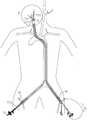
图2是根据本发明一实施方式的血管内斑块切除系统在人体内工作的示意图;Fig. 2 is a schematic diagram of the intravascular atherectomy system working in the human body according to an embodiment of the present invention;
图3是图2中A处的放大图;Fig. 3 is the enlarged view of place A in Fig. 2;
图4是根据本发明一种实施方式的血管内斑块切除系统的实验治疗效果图;Fig. 4 is an experimental treatment effect diagram of an intravascular plaque excision system according to an embodiment of the present invention;
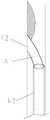
图5是根据本发明的一种实施方式的血管内斑块切除系统使用的经导管纤维剥离子的示意图;5 is a schematic diagram of a transcatheter fiber stripper used in an intravascular atherectomy system according to an embodiment of the present invention;
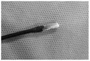
图6、图7和图8是本发明的一种实施方式的血管内斑块切除系统使用的血管壁保护装置的示意图。Fig. 6, Fig. 7 and Fig. 8 are schematic diagrams of the vessel wall protection device used in the intravascular atherectomy system according to an embodiment of the present invention.
具体实施方式detailed description
下面通过实施例,并结合附图,对本发明的技术方案作进一步详细的说明,但本发明不限于下面的实施例。The technical solution of the present invention will be further described in detail through the following examples in conjunction with the accompanying drawings, but the present invention is not limited to the following examples.
图1是根据本发明一实施方式的血管内斑块切除系统的结构示意图,用于进行血管内的激光斑块切除。Fig. 1 is a schematic structural diagram of an intravascular atherectomy system according to an embodiment of the present invention, which is used for intravascular laser atherectomy.
如图1所示,本发明公开一种血管内斑块切除系统10,可以包括内镜装置12、球囊导管系统13、内膜剥离装置14,还可以包括用于激光发生及传输的激光装置15。As shown in Figure 1, the present invention discloses an
球囊导管系统13可以通过鞘管16放进人体的血管内,可以用于阻断血流(131),阻断血流兼转流功能(132),也可以用作阻断血流兼导引导管(133)。根据本发明的一种实施方式,球囊导管系统13可以包括三个球囊,例如包括导引导管和设置在远端的第一球囊131、第二球囊132和第三球囊133。如下所述,第一球囊、第二球囊和第三球囊适于被充盈以在血管中阻断血流,第二球囊还包括血液转流单元。第一球囊131、第二球囊132和第三球囊133可以分别具有相应的导引导管并构成第一球囊导管、第二球囊导管和第三球囊导管。The balloon catheter system 13 can be put into the blood vessel of the human body through the
在操作时,例如,球囊导管系统13可以用于引导内镜装置12、内膜剥离装置14和激光装置15进入体内。在一种实施方式中,所述内镜装置12通过第三球囊导管进入人体内。In operation, for example, balloon catheter system 13 may be used to guide
所述内镜装置12可以是改良的软性电子内镜,能够对血管内,如颈动脉腔内进行照明和录像采集。通过球囊131、132、133阻断血流和内镜装置的引入,实现了血管内无血可视环境下的手术,达到与CEA同样的手术即期结果。The
根据本发明的优先实施方式,第二球囊导管可为双腔导管,一个腔道与球囊相通,通过该腔道充盈造影剂即可充盈球囊;另一腔道为主通道,其内可插入微导管、微导丝等器械,也可以注入液体或转流来自股动脉的动脉血,以保证球囊阻断处远端血管床内的正向血流。因为第二球囊导管的引入,其在阻断颈内动脉的反向血流并与第一和第三球囊导管共同实现病变处血管内一个无血环境的同时,更重要的是提供了患侧的血液转流功能,保证患侧远端的大脑血供,使得手术时间相对充足,不受限制。According to a preferred embodiment of the present invention, the second balloon catheter can be a double-lumen catheter, one lumen communicates with the balloon, through which the contrast medium can be filled to inflate the balloon; the other lumen is the main channel, and the inner Instruments such as microcatheters and microguidewires can be inserted, and fluid can be injected or arterial blood from the femoral artery can be diverted to ensure positive blood flow in the distal vascular bed where the balloon is blocked. Because of the introduction of the second balloon catheter, it blocks the reverse blood flow of the internal carotid artery and realizes a bloodless environment in the blood vessel of the lesion together with the first and third balloon catheters, and more importantly, provides The blood circulation function of the affected side ensures the blood supply to the remote brain of the affected side, so that the operation time is relatively sufficient and unrestricted.
内膜剥离装置14例如可以是一种内镜下手术剥离内膜的显微器械,如经导管显微剥离子。参见图4,该经导管显微剥离子由镍钛合金丝制成,一体化成型,分为远近端两个部分。远端为剥离铲41,所述剥离铲的铲面42由直径1mm至1.5mm的镍钛合金丝以15-45度成角切割而成,如以15度、30度或45度等多个成角切割而成,切割面周围锐利缘予以打磨钝化。近端为操纵杆43,剥离铲41与操纵杆43根据需要,沿着铲面42方向折成5-45度的成角,如折成5度、15度、30度或45度的成角,铲端至转角点的距离有5mm、10mm、15mm、20mm等不同长度。或者,内膜剥离装置14可以是经导管显微组织捕获器,由储存网、捕获环、输送杆三部分组成,前者由尼龙或其他膜性材料制成,网孔直径50-100微米,后二者由镍钛合金制成,捕获环与输送杆成角135度。内膜剥离装置14也可以是其它合适的能够剥离内膜和斑块的器械。The intima stripping device 14 may be, for example, a microscopic instrument for stripping the intima under an endoscope, such as a transcatheter microdissector. Referring to FIG. 4 , the transcatheter microdissection tool is made of nickel-titanium alloy wire, integrally formed, and divided into two parts, the distal and proximal ends. The far end is a stripping
所述内膜剥离装置14可以剥离病变处的内膜和斑块17,能够将病变处的斑块17完全剥离下来。本发明所述的斑块剥离或内膜剥离是指从解剖结构上进行剥离,即从斑块处内膜下与中膜之间潜在间隙进行剥离,采用物理方法实现,不会对血管壁造成热损伤。The intima stripping device 14 can strip the intima and
根据本发明的优选实施方式,激光装置15包括激光发生器以及激光传输光纤151。所述激光发生器用于发射激光,并对发出的激光进行控制。所述激光传输光纤151将激光引导至已剥离的斑块区域,利用激光对斑块进行消融。所述激光光纤151通过内镜装置12的光纤腔室(兼冲洗通道)到达已剥离斑块区域。According to a preferred embodiment of the present invention, the laser device 15 includes a laser generator and a laser transmission fiber 151 . The laser generator is used to emit laser light and control the emitted laser light. The laser transmission fiber 151 guides the laser to the stripped plaque area, and uses the laser to ablate the plaque. The laser optical fiber 151 reaches the stripped plaque area through the optical fiber chamber (and flushing channel) of the
根据本发明一实施方式,所述内镜装置12具有导管,导管包括至少两个腔室,其中一个腔室内设有激光传输光纤,同时也可以供灌洗液体通过。这样灌洗液体一方面可以对消融的斑块碎屑进行冲洗,同时也可以对激光传输光纤151及工作区域血管内进行降温,减少对血管壁的热损伤。所述内镜装置12的另外一个腔室内设有用于照明的光源和录像采集装置。According to an embodiment of the present invention, the
优选的,所述内镜装置12为直径6F的双腔系统(或者为直径12F的三腔系统,比6F的多出一个工作通道,可以通过该工作通道进行输送剥离装置,也可抽吸或通过捕获装置取出斑块碎屑),一个腔道为内镜照明和摄像系统,另一腔道为光纤进入兼冲洗通道。头端5cm为软性结构,可于体外操作改变镜头的指向。所述内镜装置12的远端还可以设有血管壁保护装置。例如,参见图6-8,该血管壁保护装置为镶嵌于内镜头段外围的1/2-2/3周径的薄壁不锈钢管,工作时该装置可以将分离后斑块与血管壁隔离开来,为光纤传输激光作用于靶点提供最佳的保护,避免激光对血管壁的热损伤。光纤头端位于保护装置以内,距离保护装置头端1mm。激光传输光纤151的远端位于血管壁保护装置内,这样激光传输光纤151在进行消融的时候不会对血管壁造成损伤。其中F为导管尺寸相关的单位(French的缩写),1F约为0.33mm。Preferably, the
根据本发明一实施方式,所述血管内斑块切除系统10还可以包括数字减影血管造影机,所述数字减影血管造影机(DSA)能够对血管进行成像,确定病变区(斑块)的位置和血管的管径,可根据管径选用不同规格的球囊导管。在DSA透视下经导丝引入第三、第二和第一球囊导管。球囊导管到位后,透视下监测球囊的充盈程度,造影判断球囊是否完全阻断血流。再经第三球囊导管引入内镜装置12。According to an embodiment of the present invention, the
根据本发明一实施方式,所述球囊导管系统13可以是包括3根球囊导管,即第一球囊导管(2F)、第二球囊导管(4F)和第三球囊导管(10F或12F)。所述球囊导管系统13也可以是整合的球囊导管系统,所谓整合的球囊导管系统是指将第一球囊导管和第二球囊导管的导管部分通过一定的工艺,嵌合于第三球囊导管的导管壁内,或完全无缝包被于第三球囊导管的球囊中,优点是第三球囊对颈总动脉血流的阻断效果更确切。According to one embodiment of the present invention, the balloon catheter system 13 may include three balloon catheters, namely the first balloon catheter (2F), the second balloon catheter (4F) and the third balloon catheter (10F or 12F). The balloon catheter system 13 can also be an integrated balloon catheter system. The so-called integrated balloon catheter system refers to that the catheter part of the first balloon catheter and the second balloon catheter are fitted into the first balloon catheter through a certain process. In the catheter wall of the three-balloon catheter, or completely seamlessly wrapped in the balloon of the third balloon catheter, the advantage is that the third balloon has a more accurate blocking effect on the blood flow of the common carotid artery.
如图1、图2和图3所示,所述球囊导管前端部设置有可膨胀的球囊,所述球囊可以是高顺应性球囊。3根球囊导管通过鞘鞘管16放进人体的血管内后,第一球囊131置于患侧颈外动脉开口处,第二球囊132置于颈内动脉狭窄处的远心端,以及第三球囊133置于患侧颈总动脉处,即斑块的近心端。其中颈外动脉开口处的球囊131可以阻断颈外动脉和甲状腺上动脉的反向血流,颈内动脉狭窄处的远心端处的球囊132可以阻断颈内动脉的反向血流,颈总动脉处的球囊133可以阻断颈总动脉血流,通过充盈三个球囊131、132、133,实现病变处血管腔内无血环境。其中所述颈总动脉处的球囊133的导管可用作导引导管,用于引导内镜装置12、内膜剥离装置14和激光装置15进入体内的病变区域,也可以用作抽吸导管,用于对碎屑进行抽吸。As shown in FIG. 1 , FIG. 2 and FIG. 3 , an inflatable balloon is provided at the front end of the balloon catheter, and the balloon may be a highly compliant balloon. After the three balloon catheters are put into the blood vessels of the human body through the
根据本发明一实施方式,所述血管内斑块切除系统10还可以包括持续冲洗与回抽装置,其中冲洗可以通过内镜装置12(6F内镜)的其中一个腔室向体内递送灌洗液体,回抽可以通过第三球囊导管对体内的碎屑进行抽吸(或者经过12F内镜的工作通道进行抽吸)。According to an embodiment of the present invention, the
图2是根据本发明一实施方式的血管内斑块切除系统在人体内工作的示意图。如图2所示,根据本发明其中一种实施方式的血管内斑块切除系统10在使用时,首先在数字减影血管造影机辅助下,经双侧股动脉穿刺,分别置入动脉鞘管16,在持续加压冲洗条件下经左侧股动脉鞘管引入第一和第二球囊导管,将第一球囊和第二球囊分别置于患侧颈外动脉开口处和颈内动脉狭窄处的远心端。经右侧股动脉鞘管16引入第三球囊导管,将第三球囊置于患侧颈总动脉,即斑块的近心端。优选的,在充盈之前将左侧动脉鞘管的侧管与球囊导管132尾端的Y型阀侧翼连结并打开三通开关,使左侧股动脉的血流经球囊导管132流向患侧远心端的颈内动脉。双侧股动脉鞘管均为15F动脉鞘,长20cm,其主通道可以进入15F以内的导管或导丝,其侧管带有三通开关,打开三通开关后,股动脉的血液可以从侧管流出,通过与连结在第二球囊导管尾端的Y型阀的侧翼,可将左侧股动脉的血流部分导向第二球囊导管,从而实现了通过充盈第二球囊导管头端的球囊132阻断颈内动脉反向血流的同时,又保证了颈内动脉的正向血流,为手术侧大脑提供有效的灌注。因而,手术操作可以从容进行,几乎不受时间限制。首先充盈球囊132以阻断颈内动脉血流,保证患侧大脑半球的有效灌注。再充盈颈外动脉的球囊131,阻断颈外动脉和甲状腺上动脉的反向血流,最后充盈置于患侧颈总动脉的球囊133,实现病变处血管腔内无血环境。Fig. 2 is a schematic diagram of the operation of the intravascular atherectomy system in a human body according to an embodiment of the present invention. As shown in Figure 2, when the
接着将内膜剥离装置14通过第三球囊导管送入病变区域,将病变区域的内膜和斑块进行剥离。剥离完以后,打开激光发生器,通过激光传输光纤151发射激光对剥离下来的内膜和斑块进行消融。对于斑块体积较大者,也可以腔内先激光消融,再机械剥离,之后再对剥离组织进行消融,产生的较大组织碎片可通过经导管显微组织捕获器予以取出,直至彻底清除病变处的内膜和斑块。在内镜装置12的持续灌洗和第三球囊导管的抽吸下,完成颈动脉内膜剥脱术,实现病变处增厚的内膜和斑块组织完全切除,达到解剖层次的清除病变的目的。Next, the intima stripping device 14 is sent into the lesion area through the third balloon catheter, and the intima and plaque in the lesion area are stripped. After the stripping is completed, the laser generator is turned on, and laser light is emitted through the laser transmission fiber 151 to ablate the stripped intima and plaque. For larger plaques, intracavitary laser ablation can be performed first, followed by mechanical stripping, and then the stripped tissue can be ablated, and the resulting larger tissue fragments can be removed with a transcatheter microscopic tissue trap until the lesion is completely removed intima and plaques. Under the continuous lavage of the
与第二球囊导管的血液转流功能相关的部件可以共同称为血液转流单元。例如,血液转流单元可以包括第二球囊导管的主通道或主通道内的导管、适当的控制阀(例如上述三通阀和Y型阀等)等。主通道或主通道内的导管在股动脉处引入血液,并在超过第二球囊的远端处将血液引出,控制阀等装置用来控制血液的转流。如图2中B所示的方向即为本发明一种实施方式的血液转流方向。Components related to the blood diversion function of the second balloon catheter may be collectively referred to as a blood diversion unit. For example, the blood diversion unit may include the main channel of the second balloon catheter or a catheter in the main channel, appropriate control valves (such as the above-mentioned three-way valve and Y-valve, etc.), and the like. The main channel or a catheter within the main channel introduces blood at the femoral artery and draws it out beyond the distal end of the second balloon, and devices such as control valves are used to control the flow of blood. The direction shown by B in FIG. 2 is the blood diversion direction in an embodiment of the present invention.
本发明通过采用转流的方式使得血流重新回到脑部,可以在病变区血管被阻断,但是脑部血流不停的情况下实现颈动脉斑块的剥离手术。In the present invention, the blood flow returns to the brain by means of diversion, so that the carotid artery plaque stripping operation can be realized under the condition that the blood vessels in the lesion area are blocked but the blood flow in the brain does not stop.
图3是采用本发明一种实施方式的血管内斑块切除系统的离体实验治疗效果图。如图3所示,采用本发明的血管内斑块切除系统10,能够在与传统经皮颈动脉支架植入术相媲美的微创条件下,实现了与CEA同样的手术结果,即内膜、斑块的完全切除。在内镜头端血管壁保护装置的保护下,斑块可以被激光彻底消融,而血管壁免受损伤。图3展示了体外模拟颈动脉斑块狭窄或闭塞状态下,在装有血管壁保护装置的内镜下进行激光消融,彻底切除斑块组织后血管壁的完整无损状态(模型是利用猪主动脉血管,嵌合手术获得的人体颈动脉斑块制成)。该试验证实了在血管壁保护装置下,激光切除颈动脉斑块是安全可行的。Fig. 3 is an in vitro experimental therapeutic effect diagram using the intravascular plaque excision system according to an embodiment of the present invention. As shown in Figure 3, the
需要注意的是,本发明的血管内斑块切除系统10不限于用在颈动脉斑块的切除手术,也可以用于外周血管领域,例如主动脉、肠系膜动脉、髂动脉、下肢膝关节水平以上的较大管径血管内的动脉硬化斑块,以及其他适合的领域内的手术。It should be noted that the
本发明的实施方式并不限于上述实施例所述,在不偏离本发明的精神和范围的情况下,本领域普通技术人员可以在形式和细节上对本发明做出各种改变和改进,而这些均被认为落入了本发明的保护范围。The embodiments of the present invention are not limited to the above-mentioned embodiments. Without departing from the spirit and scope of the present invention, those skilled in the art can make various changes and improvements to the present invention in form and details, and these All are considered to fall into the protection scope of the present invention.
Claims (15)
Priority Applications (4)
| Application Number | Priority Date | Filing Date | Title |
|---|---|---|---|
| CN202110773417.1ACN115590614A (en) | 2021-07-08 | 2021-07-08 | Intravascular plaque removal system |
| US18/577,475US20240238044A1 (en) | 2021-07-08 | 2022-07-08 | Endovascular plaque excision system and excision method |
| PCT/CN2022/104692WO2023280315A1 (en) | 2021-07-08 | 2022-07-08 | Endovascular plaque excision system and excision method |
| CN202280048160.6ACN117651532A (en) | 2021-07-08 | 2022-07-08 | Intravascular plaque excision system and excision method |
Applications Claiming Priority (1)
| Application Number | Priority Date | Filing Date | Title |
|---|---|---|---|
| CN202110773417.1ACN115590614A (en) | 2021-07-08 | 2021-07-08 | Intravascular plaque removal system |
Publications (1)
| Publication Number | Publication Date |
|---|---|
| CN115590614Atrue CN115590614A (en) | 2023-01-13 |
Family
ID=84801331
Family Applications (2)
| Application Number | Title | Priority Date | Filing Date |
|---|---|---|---|
| CN202110773417.1APendingCN115590614A (en) | 2021-07-08 | 2021-07-08 | Intravascular plaque removal system |
| CN202280048160.6APendingCN117651532A (en) | 2021-07-08 | 2022-07-08 | Intravascular plaque excision system and excision method |
Family Applications After (1)
| Application Number | Title | Priority Date | Filing Date |
|---|---|---|---|
| CN202280048160.6APendingCN117651532A (en) | 2021-07-08 | 2022-07-08 | Intravascular plaque excision system and excision method |
Country Status (3)
| Country | Link |
|---|---|
| US (1) | US20240238044A1 (en) |
| CN (2) | CN115590614A (en) |
| WO (1) | WO2023280315A1 (en) |
Citations (8)
| Publication number | Priority date | Publication date | Assignee | Title |
|---|---|---|---|---|
| US20010049517A1 (en)* | 1997-03-06 | 2001-12-06 | Gholam-Reza Zadno-Azizi | Method for containing and removing occlusions in the carotid arteries |
| CN201219927Y (en)* | 2008-06-30 | 2009-04-15 | 孙英信 | Laser cleaning instrument for atheromatous plaque |
| US20100081873A1 (en)* | 2008-09-30 | 2010-04-01 | AiHeart Medical Technologies, Inc. | Systems and methods for optical viewing and therapeutic intervention in blood vessels |
| JP2011087971A (en)* | 1999-06-14 | 2011-05-06 | Gore Enterprise Holdings Inc | Method and low profile apparatus for reducing embolization during treatment of carotid artery disease |
| CN108156810A (en)* | 2015-06-30 | 2018-06-12 | 科塞特·李&哈里森有限责任公司 | Intravascular catheter with multiple functions |
| CN110693543A (en)* | 2019-11-18 | 2020-01-17 | 中国医学科学院北京协和医院 | Aorta flow-switching device for in-situ windowing |
| CN113365689A (en)* | 2019-01-31 | 2021-09-07 | 马宝海德医疗有限责任公司 | Carotid stent implantation system and method |
| CN218980187U (en)* | 2022-11-09 | 2023-05-09 | 浙江大学 | Combined three-balloon blood flow blocking catheter |
Family Cites Families (5)
| Publication number | Priority date | Publication date | Assignee | Title |
|---|---|---|---|---|
| US6482172B1 (en)* | 2000-02-09 | 2002-11-19 | Jeffrey J. Thramann | Flow-by channel catheter and method of use |
| CN2761149Y (en)* | 2005-01-25 | 2006-03-01 | 董国祥 | Carotid shunt tube |
| CN202236818U (en)* | 2011-10-18 | 2012-05-30 | 复旦大学附属中山医院 | Intracarotid bypass block catheter with double saccules |
| CN211609888U (en)* | 2019-11-18 | 2020-10-02 | 中国医学科学院北京协和医院 | Aorta flow-switching device for in-situ windowing |
| CN212346665U (en)* | 2020-08-18 | 2021-01-15 | 上海市浦东医院(复旦大学附属浦东医院) | A kind of scalpel for dissection and excision of blood vessel intimal |
- 2021
- 2021-07-08CNCN202110773417.1Apatent/CN115590614A/enactivePending
- 2022
- 2022-07-08CNCN202280048160.6Apatent/CN117651532A/enactivePending
- 2022-07-08USUS18/577,475patent/US20240238044A1/enactivePending
- 2022-07-08WOPCT/CN2022/104692patent/WO2023280315A1/ennot_activeCeased
Patent Citations (8)
| Publication number | Priority date | Publication date | Assignee | Title |
|---|---|---|---|---|
| US20010049517A1 (en)* | 1997-03-06 | 2001-12-06 | Gholam-Reza Zadno-Azizi | Method for containing and removing occlusions in the carotid arteries |
| JP2011087971A (en)* | 1999-06-14 | 2011-05-06 | Gore Enterprise Holdings Inc | Method and low profile apparatus for reducing embolization during treatment of carotid artery disease |
| CN201219927Y (en)* | 2008-06-30 | 2009-04-15 | 孙英信 | Laser cleaning instrument for atheromatous plaque |
| US20100081873A1 (en)* | 2008-09-30 | 2010-04-01 | AiHeart Medical Technologies, Inc. | Systems and methods for optical viewing and therapeutic intervention in blood vessels |
| CN108156810A (en)* | 2015-06-30 | 2018-06-12 | 科塞特·李&哈里森有限责任公司 | Intravascular catheter with multiple functions |
| CN113365689A (en)* | 2019-01-31 | 2021-09-07 | 马宝海德医疗有限责任公司 | Carotid stent implantation system and method |
| CN110693543A (en)* | 2019-11-18 | 2020-01-17 | 中国医学科学院北京协和医院 | Aorta flow-switching device for in-situ windowing |
| CN218980187U (en)* | 2022-11-09 | 2023-05-09 | 浙江大学 | Combined three-balloon blood flow blocking catheter |
Also Published As
| Publication number | Publication date |
|---|---|
| CN117651532A (en) | 2024-03-05 |
| US20240238044A1 (en) | 2024-07-18 |
| WO2023280315A1 (en) | 2023-01-12 |
Similar Documents
| Publication | Publication Date | Title |
|---|---|---|
| AU763132B2 (en) | Kit for endovascular venous surgery | |
| US7108677B2 (en) | Embolization protection system for vascular procedures | |
| US6309345B1 (en) | Minimally invasive surgery device | |
| US8435225B2 (en) | Embolization protection system for vascular procedures | |
| US20080097470A1 (en) | Systems for performing gynecological procedures with mechanical distension | |
| IL143534A (en) | Endovascular system for the treatment of stenoses of the carotid and catheter for this system | |
| JPH0576537A (en) | Atherectomy instrument | |
| KR20120012796A (en) | Guide system with suction force | |
| JP2008093450A (en) | Flexible and rigid catheter ablation balloon | |
| JP2022518178A (en) | Observation and treatment of cerebrovascular lesions | |
| CN216933371U (en) | Blood vessel thrombus taking catheter | |
| KR20190008367A (en) | Apparatus and Process for Surgery of Narrow Body Lumen | |
| CN118615014A (en) | An integrated laparoscopic direct vision peripheral arterial plaque intracavitary stripping device | |
| JP2003504144A (en) | Device to remove stones from patient's vasculature | |
| CN115590614A (en) | Intravascular plaque removal system | |
| JP2020163121A (en) | Treatment method | |
| Otani et al. | Flexible ultrathin endoscope integrated with irrigation suction apparatus for assisting microneurosurgery | |
| EP4218625B1 (en) | A dual catheter arrangement and system for reperfusion of an ischemic tissue region via a coronary vessel | |
| US11547475B2 (en) | System for its use in the treatment of vascular stenosis and occlusions | |
| Berci et al. | Combined fluoroendoscopic removal of retained biliary stones | |
| Rossi et al. | Percutaneous cholangioscopy | |
| Nahlieli | Endoscopic Transoral Removal of Distal and Proximal Stones from the Parotid Duct | |
| JP2005046419A (en) | catheter | |
| JP3191543U (en) | Vascular catheter system and CTO lesion penetration method | |
| Nahlieli | Salivary gland endoscopy and minimally invasive surgery |
Legal Events
| Date | Code | Title | Description |
|---|---|---|---|
| PB01 | Publication | ||
| PB01 | Publication | ||
| SE01 | Entry into force of request for substantive examination | ||
| SE01 | Entry into force of request for substantive examination |