CN115553880A - A positioning method and system for a puncture needle point - Google Patents
A positioning method and system for a puncture needle pointDownload PDFInfo
- Publication number
- CN115553880A CN115553880ACN202210318962.6ACN202210318962ACN115553880ACN 115553880 ACN115553880 ACN 115553880ACN 202210318962 ACN202210318962 ACN 202210318962ACN 115553880 ACN115553880 ACN 115553880A
- Authority
- CN
- China
- Prior art keywords
- image
- dimensional
- target
- ultrasonic
- module
- Prior art date
- Legal status (The legal status is an assumption and is not a legal conclusion. Google has not performed a legal analysis and makes no representation as to the accuracy of the status listed.)
- Granted
Links
- 238000000034methodMethods0.000titleclaimsabstractdescription29
- 238000002604ultrasonographyMethods0.000claimsdescription102
- 230000003902lesionEffects0.000claimsdescription49
- 230000005540biological transmissionEffects0.000claimsdescription6
- 206010054107NoduleDiseases0.000description8
- 239000000463materialSubstances0.000description7
- 238000005516engineering processMethods0.000description5
- 230000008569processEffects0.000description5
- 238000012360testing methodMethods0.000description5
- 238000010586diagramMethods0.000description4
- 230000003211malignant effectEffects0.000description4
- 230000003340mental effectEffects0.000description4
- 230000001575pathological effectEffects0.000description4
- 230000004044responseEffects0.000description4
- 208000027418Wounds and injuryDiseases0.000description3
- 230000002308calcificationEffects0.000description3
- 230000008859changeEffects0.000description3
- 230000006378damageEffects0.000description3
- 208000014674injuryDiseases0.000description3
- 208000009453Thyroid NoduleDiseases0.000description2
- 230000009286beneficial effectEffects0.000description2
- 230000000694effectsEffects0.000description2
- 210000000056organAnatomy0.000description2
- 208000024770Thyroid neoplasmDiseases0.000description1
- 238000004458analytical methodMethods0.000description1
- 210000004204blood vesselAnatomy0.000description1
- 238000004364calculation methodMethods0.000description1
- 238000006243chemical reactionMethods0.000description1
- 238000002591computed tomographyMethods0.000description1
- 239000002872contrast mediaSubstances0.000description1
- 238000001514detection methodMethods0.000description1
- 238000011161developmentMethods0.000description1
- 201000010099diseaseDiseases0.000description1
- 208000037265diseases, disorders, signs and symptomsDiseases0.000description1
- 238000002592echocardiographyMethods0.000description1
- 230000005251gamma rayEffects0.000description1
- 238000012986modificationMethods0.000description1
- 230000004048modificationEffects0.000description1
- 238000012544monitoring processMethods0.000description1
- 210000005036nerveAnatomy0.000description1
- 238000012545processingMethods0.000description1
- 230000000750progressive effectEffects0.000description1
- 230000005855radiationEffects0.000description1
- 239000004576sandSubstances0.000description1
- 230000003068static effectEffects0.000description1
- 239000000126substanceSubstances0.000description1
- 210000001685thyroid glandAnatomy0.000description1
- 238000013334tissue modelMethods0.000description1
- 238000003325tomographyMethods0.000description1
- 235000012431wafersNutrition0.000description1
- 239000002699waste materialSubstances0.000description1
Images


Classifications
- A—HUMAN NECESSITIES
- A61—MEDICAL OR VETERINARY SCIENCE; HYGIENE
- A61B—DIAGNOSIS; SURGERY; IDENTIFICATION
- A61B17/00—Surgical instruments, devices or methods
- A61B17/34—Trocars; Puncturing needles
- A61B17/3403—Needle locating or guiding means
- A—HUMAN NECESSITIES
- A61—MEDICAL OR VETERINARY SCIENCE; HYGIENE
- A61B—DIAGNOSIS; SURGERY; IDENTIFICATION
- A61B10/00—Instruments for taking body samples for diagnostic purposes; Other methods or instruments for diagnosis, e.g. for vaccination diagnosis, sex determination or ovulation-period determination; Throat striking implements
- A61B10/02—Instruments for taking cell samples or for biopsy
- A61B10/0233—Pointed or sharp biopsy instruments
- A61B10/0266—Pointed or sharp biopsy instruments means for severing sample
- A—HUMAN NECESSITIES
- A61—MEDICAL OR VETERINARY SCIENCE; HYGIENE
- A61B—DIAGNOSIS; SURGERY; IDENTIFICATION
- A61B17/00—Surgical instruments, devices or methods
- A61B17/34—Trocars; Puncturing needles
- A61B17/3417—Details of tips or shafts, e.g. grooves, expandable, bendable; Multiple coaxial sliding cannulas, e.g. for dilating
- A—HUMAN NECESSITIES
- A61—MEDICAL OR VETERINARY SCIENCE; HYGIENE
- A61B—DIAGNOSIS; SURGERY; IDENTIFICATION
- A61B34/00—Computer-aided surgery; Manipulators or robots specially adapted for use in surgery
- A61B34/10—Computer-aided planning, simulation or modelling of surgical operations
- A—HUMAN NECESSITIES
- A61—MEDICAL OR VETERINARY SCIENCE; HYGIENE
- A61B—DIAGNOSIS; SURGERY; IDENTIFICATION
- A61B34/00—Computer-aided surgery; Manipulators or robots specially adapted for use in surgery
- A61B34/20—Surgical navigation systems; Devices for tracking or guiding surgical instruments, e.g. for frameless stereotaxis
- A—HUMAN NECESSITIES
- A61—MEDICAL OR VETERINARY SCIENCE; HYGIENE
- A61B—DIAGNOSIS; SURGERY; IDENTIFICATION
- A61B17/00—Surgical instruments, devices or methods
- A61B17/34—Trocars; Puncturing needles
- A61B17/3403—Needle locating or guiding means
- A61B2017/3413—Needle locating or guiding means guided by ultrasound
- A—HUMAN NECESSITIES
- A61—MEDICAL OR VETERINARY SCIENCE; HYGIENE
- A61B—DIAGNOSIS; SURGERY; IDENTIFICATION
- A61B34/00—Computer-aided surgery; Manipulators or robots specially adapted for use in surgery
- A61B34/10—Computer-aided planning, simulation or modelling of surgical operations
- A61B2034/101—Computer-aided simulation of surgical operations
- A61B2034/105—Modelling of the patient, e.g. for ligaments or bones
- A—HUMAN NECESSITIES
- A61—MEDICAL OR VETERINARY SCIENCE; HYGIENE
- A61B—DIAGNOSIS; SURGERY; IDENTIFICATION
- A61B34/00—Computer-aided surgery; Manipulators or robots specially adapted for use in surgery
- A61B34/20—Surgical navigation systems; Devices for tracking or guiding surgical instruments, e.g. for frameless stereotaxis
- A61B2034/2046—Tracking techniques
- A61B2034/2063—Acoustic tracking systems, e.g. using ultrasound
- A—HUMAN NECESSITIES
- A61—MEDICAL OR VETERINARY SCIENCE; HYGIENE
- A61B—DIAGNOSIS; SURGERY; IDENTIFICATION
- A61B34/00—Computer-aided surgery; Manipulators or robots specially adapted for use in surgery
- A61B34/20—Surgical navigation systems; Devices for tracking or guiding surgical instruments, e.g. for frameless stereotaxis
- A61B2034/2046—Tracking techniques
- A61B2034/2065—Tracking using image or pattern recognition
Landscapes
- Health & Medical Sciences (AREA)
- Life Sciences & Earth Sciences (AREA)
- Surgery (AREA)
- Engineering & Computer Science (AREA)
- Medical Informatics (AREA)
- Biomedical Technology (AREA)
- Heart & Thoracic Surgery (AREA)
- Molecular Biology (AREA)
- Animal Behavior & Ethology (AREA)
- General Health & Medical Sciences (AREA)
- Public Health (AREA)
- Veterinary Medicine (AREA)
- Nuclear Medicine, Radiotherapy & Molecular Imaging (AREA)
- Pathology (AREA)
- Robotics (AREA)
- Ultra Sonic Daignosis Equipment (AREA)
Abstract
Description
Translated fromChinese技术领域technical field
本公开涉及医疗技术领域,具体而言,涉及一种穿刺针尖的定位方法和系统。The present disclosure relates to the field of medical technology, in particular, to a positioning method and system for a puncture needle point.
背景技术Background technique
随着科学技术的发展,已使得超声能够清晰的显示人体内部的组织结构,因而超声引导穿刺技术应运而生。这种技术就是在实时超声影像的监视和引导下,通过穿刺针对体内病变或目标进行穿刺的临床技术。具体做法是在超声的引导下,避开重要脏器及较大的血管神经,将穿刺针穿入病变组织内进行治疗,或通过吸出、切割出少量细胞或组织进行病理检查,以确定组织病变的性质。With the development of science and technology, ultrasound has been able to clearly display the tissue structure inside the human body, so ultrasound-guided puncture technology came into being. This technology is a clinical technique of puncturing lesions or targets in the body through puncture under the monitoring and guidance of real-time ultrasound images. The specific method is to avoid important organs and large blood vessels and nerves under the guidance of ultrasound, and to penetrate the diseased tissue with a puncture needle for treatment, or to aspirate or cut out a small amount of cells or tissues for pathological examination to determine the tissue lesion. nature.
但是,在超声引导下,对甲状腺结节进行穿刺时,可能存在多个结节,每个结节的性质可能不同,例如,良性结节、恶性结节,需要从中选择出恶行结节进行穿刺。但是,采用超声引导实时返回的是二维图像,无法了解穿刺针头进入体内的深度。不少病例因为取材原因,未能得出相应的病理结果,需要再次穿刺,从而使患者在经济和精神上承担了很大的负担,造成了医疗资源的浪费。However, under the guidance of ultrasound, when puncturing thyroid nodules, there may be multiple nodules, and the nature of each nodule may be different, for example, benign nodules and malignant nodules. puncture. However, what is returned in real time using ultrasound guidance is a two-dimensional image, and it is impossible to understand the depth of the puncture needle into the body. Many cases failed to obtain the corresponding pathological results due to material collection, and needed to be punctured again, which made the patients bear a great financial and mental burden and caused a waste of medical resources.
因此,本公开提供了一种穿刺针尖的定位方法,以解决上述技术问题之一。Therefore, the present disclosure provides a positioning method of a puncture needle point to solve one of the above technical problems.
发明内容Contents of the invention
本公开的目的在于提供一种穿刺针尖的定位方法和系统,能够解决上述提到的至少一个技术问题。具体方案如下:The purpose of the present disclosure is to provide a positioning method and system for a puncture needle point, which can solve at least one of the above-mentioned technical problems. The specific plan is as follows:
根据本公开的具体实施方式,第一方面,本公开提供一种穿刺针尖的定位方法,包括:According to a specific embodiment of the present disclosure, in a first aspect, the present disclosure provides a method for positioning a puncture needle point, including:
获取患者身体的一组切片图像,其中,所述一组切片图像中包括所述患者身体的体表轮廓的图像和所述体表轮廓内的至少一个目标患处的图像;Acquiring a group of slice images of the patient's body, wherein the set of slice images includes an image of the body surface contour of the patient's body and an image of at least one target lesion within the body surface contour;
基于所述一组切片图像生成三维空间中所述患者身体的三维身体模型;generating a three-dimensional body model of the patient's body in three-dimensional space based on the set of sliced images;
对所述三维身体模型中的所述至少一个目标患处标记一个穿刺目标,并确定所述穿刺目标的表面的目标三维位置;marking a puncture target on the at least one target lesion in the three-dimensional body model, and determining a target three-dimensional position of a surface of the puncture target;
通过换能器中的各个超声模块分别向所述患者身体的内部发射超声波,接收所述超声波的回波信息,并分别获取各个超声模块从发射至接收的传播时间段,其中,所述换能器固定放置于所述患者身体的体表处,且能够探测到所述目标三维位置;Each ultrasonic module in the transducer transmits ultrasonic waves to the inside of the patient's body, receives the echo information of the ultrasonic waves, and obtains the propagation time period of each ultrasonic module from transmission to reception, wherein the transducer The device is fixedly placed on the body surface of the patient's body, and can detect the three-dimensional position of the target;
根据各个超声模块的回波信息和传播时间段获取超声图像和第一超声模块探测到的第一距离,其中,所述超声图像中包括所述穿刺目标的图像和穿刺针的针尖的图像,所述第一距离是指所述换能器中的第一超声模块与所述针尖的距离;Obtain an ultrasonic image and a first distance detected by the first ultrasonic module according to the echo information and propagation time period of each ultrasonic module, wherein the ultrasonic image includes an image of the puncture target and an image of the needle tip of the puncture needle, so The first distance refers to the distance between the first ultrasonic module in the transducer and the needle tip;
基于所述超声图像与多个二维模型图像分别进行匹配,获取一匹配的二维模型图像和所述匹配的二维模型图像所在的匹配平面,其中,所述二维模型图像是指经过所述目标三维位置的任一角度平面上所述三维身体模型的投影图像;Based on the matching of the ultrasonic image and a plurality of two-dimensional model images respectively, a matched two-dimensional model image and a matching plane where the matched two-dimensional model image is obtained, wherein the two-dimensional model image refers to the A projection image of the three-dimensional body model on any angle plane of the three-dimensional position of the target;
根据各个超声模块的回波信息和传播时间段分析所述匹配的二维模型图像和所述超声图像,获取第二超声模块探测到的第二距离,其中,所述第二距离是指所述换能器中的第二超声模块与所述穿刺目标的距离;Analyze the matched two-dimensional model image and the ultrasonic image according to the echo information and propagation time period of each ultrasonic module, and obtain the second distance detected by the second ultrasonic module, wherein the second distance refers to the the distance between the second ultrasonic module in the transducer and the puncture target;
基于所述匹配平面、所述目标三维位置和所述第二距离确定所述第二超声模块的第一三维位置;determining a first three-dimensional position of the second ultrasound module based on the matching plane, the target three-dimensional position and the second distance;
基于所述匹配平面、所述第二超声模块的第一三维位置和第二距离以及所述第一超声模块与所述第二超声模块的预设模块位置关系确定所述针尖在所述三维身体模型中的第二三维位置;Determine the position of the needle tip on the three-dimensional body based on the matching plane, the first three-dimensional position and the second distance of the second ultrasound module, and the preset module position relationship between the first ultrasound module and the second ultrasound module. the second 3D position in the model;
基于所述目标三维位置和所述第二三维位置获取所述穿刺目标与所述针尖的三维位置关系信息。The three-dimensional positional relationship information between the puncture target and the needle tip is acquired based on the target three-dimensional position and the second three-dimensional position.
根据本公开的具体实施方式,第二方面,本公开提供一种穿刺针尖的定位系统,包括:According to a specific embodiment of the present disclosure, in a second aspect, the present disclosure provides a positioning system for a puncture needle point, including:
扫描装置,配置为获取患者身体的一组切片图像,其中,所述一组切片图像中包括所述患者身体的体表轮廓的图像和所述体表轮廓内的至少一个目标患处的图像;A scanning device configured to acquire a set of slice images of the patient's body, wherein the set of slice images includes an image of the body surface contour of the patient's body and an image of at least one target lesion within the body surface contour;
定位装置,包括换能器,所述换能器包括多个超声模块,a positioning device comprising a transducer comprising a plurality of ultrasound modules,
所述定位装置,配置为:The positioning device is configured as:
基于所述一组切片图像生成三维空间中所述患者身体的三维身体模型;generating a three-dimensional body model of the patient's body in three-dimensional space based on the set of sliced images;
对所述三维身体模型中的所述至少一个目标患处标记一个穿刺目标,并确定所述穿刺目标的表面的目标三维位置;marking a puncture target on the at least one target lesion in the three-dimensional body model, and determining a target three-dimensional position of a surface of the puncture target;
通过换能器中的各个超声模块分别向所述患者身体的内部发射超声波,接收所述超声波的回波信息,并分别获取各个超声模块从发射至接收的传播时间段,其中,所述换能器固定放置于所述患者身体的体表处,且能够探测到所述目标三维位置;Each ultrasonic module in the transducer transmits ultrasonic waves to the inside of the patient's body, receives the echo information of the ultrasonic waves, and obtains the propagation time period of each ultrasonic module from transmission to reception, wherein the transducer The device is fixedly placed on the body surface of the patient's body, and can detect the three-dimensional position of the target;
根据各个超声模块的回波信息和传播时间段获取超声图像和第一超声模块探测到的第一距离,其中,所述超声图像中包括所述穿刺目标的图像和穿刺针的针尖的图像,所述第一距离是指所述换能器中的第一超声模块与所述针尖的距离;Obtain an ultrasonic image and a first distance detected by the first ultrasonic module according to the echo information and propagation time period of each ultrasonic module, wherein the ultrasonic image includes an image of the puncture target and an image of the needle tip of the puncture needle, so The first distance refers to the distance between the first ultrasonic module in the transducer and the needle tip;
基于所述超声图像与多个二维模型图像分别进行匹配,获取一匹配的二维模型图像和所述匹配的二维模型图像所在的匹配平面,其中,所述二维模型图像是指经过所述目标三维位置的任一角度平面上所述三维身体模型的投影图像;Based on the matching of the ultrasonic image and a plurality of two-dimensional model images respectively, a matched two-dimensional model image and a matching plane where the matched two-dimensional model image is obtained, wherein the two-dimensional model image refers to the A projection image of the three-dimensional body model on any angle plane of the three-dimensional position of the target;
根据各个超声模块的回波信息和传播时间段分析所述匹配的二维模型图像和所述超声图像,获取第二超声模块探测到的第二距离,其中,所述第二距离是指所述换能器中的第二超声模块与所述穿刺目标的距离;Analyze the matched two-dimensional model image and the ultrasonic image according to the echo information and propagation time period of each ultrasonic module, and obtain the second distance detected by the second ultrasonic module, wherein the second distance refers to the the distance between the second ultrasonic module in the transducer and the puncture target;
基于所述匹配平面、所述目标三维位置和所述第二距离确定所述第二超声模块的第一三维位置;determining a first three-dimensional position of the second ultrasound module based on the matching plane, the target three-dimensional position and the second distance;
基于所述匹配平面、所述第二超声模块的第一三维位置和第二距离以及所述第一超声模块与所述第二超声模块的预设模块位置关系确定所述针尖在所述三维身体模型中的第二三维位置;Determine the position of the needle tip on the three-dimensional body based on the matching plane, the first three-dimensional position and the second distance of the second ultrasound module, and the preset module position relationship between the first ultrasound module and the second ultrasound module. the second 3D position in the model;
基于所述目标三维位置和所述第二三维位置获取所述穿刺目标与所述针尖的三维位置关系信息。The three-dimensional positional relationship information between the puncture target and the needle tip is acquired based on the target three-dimensional position and the second three-dimensional position.
本公开实施例的上述方案与现有技术相比,至少具有以下有益效果:Compared with the prior art, the above solutions of the embodiments of the present disclosure have at least the following beneficial effects:
本公开提供了一种穿刺针尖的定位方法和系统。本公开利用三维空间中所述患者身体的三维身体模型确定穿刺目标位置,然后利用换能器中每个超声模块在阵列中的排列位置与超声图像中的区域图像的位置具有一一对应的关系,分析超声图像,并利用超声模块探测的距离值,确定与穿刺目标相关的第一超声模块的三维位置,及与针尖相关的第二超声模块的三维位置,进而确定针尖的三维位置,最终为操作者提供所述穿刺目标与所述针尖的三维位置关系信息,使操作者在穿刺的过程中能够全方位的了解针尖与穿刺目标的位置关系,及时调整穿刺路径,准确获取检材,避免了多次穿刺给患者带来的经济损失和精神伤害,提高了医疗资源的利用率。The present disclosure provides a method and system for positioning a puncture needle tip. The disclosure utilizes the three-dimensional body model of the patient's body in three-dimensional space to determine the puncture target position, and then utilizes the one-to-one correspondence relationship between the arrangement position of each ultrasonic module in the array and the position of the region image in the ultrasonic image , analyze the ultrasonic image, and use the distance value detected by the ultrasonic module to determine the three-dimensional position of the first ultrasonic module related to the puncture target, and the three-dimensional position of the second ultrasonic module related to the needle tip, and then determine the three-dimensional position of the needle tip. Finally, The operator provides the three-dimensional positional relationship information between the puncture target and the needle tip, so that the operator can fully understand the positional relationship between the needle tip and the puncture target during the puncture process, adjust the puncture path in time, and obtain the test material accurately, avoiding the The economic losses and mental injuries caused by multiple punctures have increased the utilization rate of medical resources.
附图说明Description of drawings
图1示出了根据本公开实施例的穿刺针尖的定位方法的流程图;Fig. 1 shows a flowchart of a positioning method of a puncture needle point according to an embodiment of the present disclosure;
图2示出了根据本公开实施例的一组切片图像依切片顺序堆叠于所述三维空间中的示意图;Fig. 2 shows a schematic diagram of a group of slice images stacked in the three-dimensional space according to slice order according to an embodiment of the present disclosure;
图3示出了根据本公开实施例的相控阵换能器发送和接收回波信息的示意图;Fig. 3 shows a schematic diagram of sending and receiving echo information by a phased array transducer according to an embodiment of the present disclosure;
图4示出了根据本公开实施例的第一超声模块、第二超声模块、穿刺目标于针尖的位置关系示意图;Fig. 4 shows a schematic diagram of the positional relationship between the first ultrasound module, the second ultrasound module, the puncture target and the needle tip according to an embodiment of the present disclosure;
图5示出了根据本公开实施例的穿刺针尖的定位系统的系统框图。Fig. 5 shows a system block diagram of a puncture needle point positioning system according to an embodiment of the present disclosure.
具体实施方式detailed description
为了使本公开的目的、技术方案和优点更加清楚,下面将结合附图对本公开作进一步地详细描述,显然,所描述的实施例仅仅是本公开一部分实施例,而不是全部的实施例。基于本公开中的实施例,本领域普通技术人员在没有做出创造性劳动前提下所获得的所有其它实施例,都属于本公开保护的范围。In order to make the purpose, technical solutions and advantages of the present disclosure clearer, the present disclosure will be further described in detail below in conjunction with the accompanying drawings. Apparently, the described embodiments are only some of the embodiments of the present disclosure, not all of them. Based on the embodiments in the present disclosure, all other embodiments obtained by persons of ordinary skill in the art without creative efforts fall within the protection scope of the present disclosure.
在本公开实施例中使用的术语是仅仅出于描述特定实施例的目的,而非旨在限制本公开。在本公开实施例和所附权利要求书中所使用的单数形式的“一种”、“所述”和“该”也旨在包括多数形式,除非上下文清楚地表示其他含义,“多种”一般包含至少两种。Terms used in the embodiments of the present disclosure are for the purpose of describing specific embodiments only, and are not intended to limit the present disclosure. The singular forms "a", "said" and "the" used in the embodiments of this disclosure and the appended claims are also intended to include plural forms, unless the context clearly indicates otherwise, "a plurality" Generally contain at least two.
应当理解,本文中使用的术语“和/或”仅仅是一种描述关联对象的关联关系,表示可以存在三种关系,例如,A和/或B,可以表示:单独存在A,同时存在A和B,单独存在B这三种情况。另外,本文中字符“/”,一般表示前后关联对象是一种“或”的关系。It should be understood that the term "and/or" used herein is only an association relationship describing associated objects, which means that there may be three relationships, for example, A and/or B, which may mean that A exists alone, and A and B exist simultaneously. B, there are three situations of B alone. In addition, the character "/" in this article generally indicates that the contextual objects are an "or" relationship.
应当理解,尽管在本公开实施例中可能采用术语第一、第二、第三等来描述,但这些描述不应限于这些术语。这些术语仅用来将描述区分开。例如,在不脱离本公开实施例范围的情况下,第一也可以被称为第二,类似地,第二也可以被称为第一。It should be understood that although the terms first, second, third, etc. may be used for description in the embodiments of the present disclosure, these descriptions should not be limited to these terms. These terms are used only to differentiate the descriptions. For example, without departing from the scope of the embodiments of the present disclosure, the first may also be referred to as the second, and similarly, the second may also be referred to as the first.
取决于语境,如在此所使用的词语“如果”、“若”可以被解释成为“在……时”或“当……时”或“响应于确定”或“响应于检测”。类似地,取决于语境,短语“如果确定”或“如果检测(陈述的条件或事件)”可以被解释成为“当确定时”或“响应于确定”或“当检测(陈述的条件或事件)时”或“响应于检测(陈述的条件或事件)”。Depending on the context, the words "if", "if" as used herein may be interpreted as "at" or "when" or "in response to determining" or "in response to detecting". Similarly, depending on the context, the phrases "if determined" or "if detected (the stated condition or event)" could be interpreted as "when determined" or "in response to the determination" or "when detected (the stated condition or event) )" or "in response to detection of (a stated condition or event)".
还需要说明的是,术语“包括”、“包含”或者其任何其他变体意在涵盖非排他性的包含,从而使得包括一系列要素的商品或者装置不仅包括那些要素,而且还包括没有明确列出的其他要素,或者是还包括为这种商品或者装置所固有的要素。在没有更多限制的情况下,由语句“包括一个……”限定的要素,并不排除在包括所述要素的商品或者装置中还存在另外的相同要素。It should also be noted that the term "comprises", "comprises" or any other variation thereof is intended to cover a non-exclusive inclusion such that an article or arrangement comprising a list of elements includes not only those elements but also includes items not expressly listed. other elements of the product, or elements inherent in the product or device. Without further limitations, an element defined by the phrase "comprising a ..." does not exclude the presence of additional identical elements in an article or device comprising said element.
特别需要说明的是,在说明书中存在的符号和/或数字,如果在附图说明中未被标记的,均不是附图标记。In particular, it should be noted that if the symbols and/or numbers in the description are not marked in the description of the drawings, they are not reference signs.
下面结合附图详细说明本公开的可选实施例。Optional embodiments of the present disclosure will be described in detail below in conjunction with the accompanying drawings.
实施例1Example 1
对本公开提供的实施例,即一种穿刺针尖的定位方法的实施例。The embodiment provided by the present disclosure is an embodiment of a positioning method for a puncture needle tip.
下面结合图1对本公开实施例进行详细说明。The embodiment of the present disclosure will be described in detail below with reference to FIG. 1 .
步骤S101,获取患者身体的一组切片图像。Step S101, acquiring a group of sliced images of a patient's body.
一组切片图像,是利用断层扫描(英文全称Computed Tomography,简称CT)机的精确准直的X射线、γ射线或超声波,与高灵敏度的探测器相配合围绕患者身体的目标患处进行一定厚度的分层,并作断层扫描,获得的一组层叠的图像。CT机基于每层的CT机坐标进行扫描生成一张对应层的切片图像。每层的CT机坐标是一个二维坐标,CT机从每层的CT机坐标的预设原点位置开始进行对应层面的扫描,每层的切片图像的左上角的第一个像素点的位置对应着预设原点位置。A set of sliced images is to use the precisely collimated X-rays, γ-rays or ultrasonic waves from a tomography (Computed Tomography, CT for short) machine to cooperate with a high-sensitivity detector to scan a certain thickness around the target affected part of the patient's body. Layered and tomographically scanned to obtain a set of stacked images. The CT machine scans based on the CT machine coordinates of each layer to generate a slice image of the corresponding layer. The CT machine coordinates of each layer are two-dimensional coordinates. The CT machine scans the corresponding layer from the preset origin position of the CT machine coordinates of each layer. The position of the first pixel point in the upper left corner of the slice image of each layer corresponds to to the preset origin position.
CT机具有扫描时间快,图像清晰的特点,可用于多种疾病的检查;根据射线不同,CT机包括:X射线CT机(简称X-CT机)、γ射线CT机(简称γ-CT机)和超声波CT机(简称声波CT机)。CT machine has the characteristics of fast scanning time and clear image, and can be used for the examination of various diseases; according to different radiation, CT machine includes: X-ray CT machine (referred to as X-CT machine), γ-ray CT machine (abbreviated as γ-CT machine) ) and ultrasonic CT machine (sonic CT machine for short).
其中,所述一组切片图像中包括围绕所述患者身体的体表轮廓的图像和所述体表轮廓内的至少一个目标患处的图像。例如,目标患处为患者身体内甲状腺的结节。根据性质划分,结节分为良性结节和恶性结节,通常需要通过切取、钳取或穿刺的方法从患者身体内获取病变组织,进行病理学检查,确定结节的性质。也可以在扫描前对目标患注入造影剂,以便增强患者身体内目标患处的图像效果。Wherein, the group of slice images includes images of body surface contours surrounding the patient's body and images of at least one target lesion within the body surface contours. For example, the target lesion is a nodule of the thyroid in the patient's body. According to the nature, nodules are divided into benign nodules and malignant nodules. It is usually necessary to obtain diseased tissue from the patient's body by cutting, forceps or puncture, and perform pathological examination to determine the nature of the nodules. It is also possible to inject a contrast agent into the target patient before scanning, so as to enhance the image effect of the target lesion in the patient's body.
步骤S102,基于所述一组切片图像生成三维空间中所述患者身体的三维身体模型。Step S102, generating a three-dimensional body model of the patient's body in three-dimensional space based on the group of sliced images.
三维空间,是指能够被感知到的空间,通常采用三个坐标轴(比如,X轴、Y轴和Z轴)描述三维空间,其中点的位置由三个坐标轴上的点表示。在现实生活中肉眼可见的物体均可用三维空间描述。A three-dimensional space refers to a space that can be perceived. Usually, three coordinate axes (for example, X axis, Y axis, and Z axis) are used to describe the three-dimensional space, and the positions of points are represented by points on the three coordinate axes. Objects visible to the naked eye in real life can be described in three-dimensional space.
所述患者身体的三维身体模型,是在三维空间中生成的患者身体的虚拟三维模型,也就是通过三维空间描述患者身体。由于所述一组切片图像中包括所述患者身体的体表轮廓的图像和所述体表轮廓内的至少一个目标患处的图像,因此,在该三维身体模型中包括虚拟的目标患处和患者身体的体表。本公开实施例的目的就是通过三维空间描述出穿刺目标和穿刺针的针尖的位置关系,使穿刺的操作者能够准确完成穿刺。The three-dimensional body model of the patient's body is a virtual three-dimensional model of the patient's body generated in three-dimensional space, that is, the patient's body is described in three-dimensional space. Since the group of slice images includes the image of the body surface contour of the patient's body and the image of at least one target lesion within the body surface contour, the virtual target lesion and the patient's body are included in the three-dimensional body model body surface. The purpose of the embodiments of the present disclosure is to describe the positional relationship between the puncture target and the needle tip of the puncture needle through three-dimensional space, so that the puncture operator can accurately complete the puncture.
在一些具体实施例中,所述基于所述一组切片图像生成三维空间中所述患者身体的三维身体模型,包括以下步骤:In some specific embodiments, said generating a three-dimensional body model of said patient's body in three-dimensional space based on said set of slice images comprises the following steps:
步骤S102-1,将每个切片图像中的同一图像位置分别标记为对应切片图像的二维原点位置,并在每个切片图像中标记多个二维特征位置。Step S102-1, mark the same image position in each slice image as the two-dimensional origin position of the corresponding slice image, and mark multiple two-dimensional feature positions in each slice image.
由于每个切片图像是一个二维图像,如果在每个切片图像中建立一个二维坐标,则在该二维坐标中存在一个原点位置。本公开实施例在每个切片图像中的同一位置设置为对应切片图像的二维原点位置。例如,由于每层的切片图像的左上角的第一个像素点的位置对应着每层的CT机坐标的预设原点位置,因此,将每个切片图像的左上角的第一个像素点的位置标记为对应切片图像的二维原点位置,使在每个切片图像中建立的二维坐标与断层扫描的CT机坐标保持一致,避免了两个坐标间的转换,提高了数据处理的效率。当然也可以在每个切片图像中的其他同一位置作为二维原点位置,本公开实施例不做限制。例如,以每个切片图像的第5行第6列的像素点作为二维原点位置。Since each slice image is a two-dimensional image, if a two-dimensional coordinate is established in each slice image, there is an origin position in the two-dimensional coordinate. In the embodiment of the present disclosure, the same position in each slice image is set as the two-dimensional origin position of the corresponding slice image. For example, since the position of the first pixel in the upper left corner of the slice image of each layer corresponds to the preset origin position of the CT machine coordinates of each layer, the position of the first pixel in the upper left corner of each slice image The position is marked as the two-dimensional origin position of the corresponding slice image, so that the two-dimensional coordinates established in each slice image are consistent with the CT machine coordinates of the tomographic scan, avoiding the conversion between the two coordinates, and improving the efficiency of data processing. Of course, another same position in each slice image may also be used as the two-dimensional origin position, which is not limited in this embodiment of the present disclosure. For example, the pixel point in row 5 and column 6 of each slice image is used as the two-dimensional origin position.
所述多个二维特征位置分别标记于目标患处图像的轮廓处和体表轮廓图像的体表轮廓处。标记的二维特征位置越多,生成的三维身体模型的颗粒度越小,三维身体模型越接近于真实的患者身体的情况,定位穿刺目标越准确。The multiple two-dimensional feature positions are respectively marked on the contour of the target lesion image and the body surface contour of the body surface contour image. The more marked two-dimensional feature positions, the smaller the granularity of the generated three-dimensional body model, the closer the three-dimensional body model is to the real condition of the patient's body, and the more accurate the positioning of the puncture target.
步骤S102-2,将所述一组切片图像依切片顺序堆叠于所述三维空间中。Step S102-2, stacking the group of slice images in the three-dimensional space in slice order.
其中,每个切片图像以其二维原点位置为交点垂直于预设坐标轴,且每个切片图像的二维原点位置在预设坐标轴上以预设层距为间隔。所述预设层距是CT机进行扫描前设定的层与层之间的距离,本具体实施例在建立三维身体模型时利用预设层距,能够使生成的三维身体模型更真实的虚拟出患者身体。预设层距越小,生成的三维身体模型更接近于虚拟出患者身体的情况。Wherein, each slice image is perpendicular to the preset coordinate axis with its two-dimensional origin position as the intersection point, and the two-dimensional origin position of each slice image is separated by a preset layer distance on the preset coordinate axis. The preset layer distance is the distance between the layers set before the CT machine scans. In this specific embodiment, the preset layer distance is used when the three-dimensional body model is established, so that the generated three-dimensional body model can be more realistic and virtual. out of the patient's body. The smaller the preset layer distance, the closer the generated 3D body model is to virtualizing the patient's body.
例如,如图2所示,三维空间以x轴、y轴和z轴表示,为说明方便,以每个切片图像的左下角为二维原点位置,二维原点位置与y轴相交且垂直于y轴,两两切片图像平行,且间隔预设层距,图中的切片图像自下而上堆叠,最底下的切片图像为第一个切片图像,第一个切片图像位于xz平面上。For example, as shown in Figure 2, the three-dimensional space is represented by the x-axis, y-axis, and z-axis. For the convenience of illustration, the lower left corner of each slice image is taken as the two-dimensional origin position, and the two-dimensional origin position intersects the y-axis and is perpendicular to On the y-axis, two slice images are parallel and separated by a preset layer distance. The slice images in the figure are stacked from bottom to top. The bottom slice image is the first slice image, and the first slice image is located on the xz plane.
步骤S102-3,将所述三维空间中每个切片图像中的二维特征位置基于预设层距转变为三维特征位置。Step S102-3, converting the 2D feature position in each slice image in the 3D space into a 3D feature position based on the preset layer distance.
三维特征位置与每个切片图像中的二维特征位置相对应,用于表示切片图像中的二维特征位置在三维空间中的位置。例如,如图2所示,预设层距为5毫米,自下而上堆叠的第六个切片图像的二维特征位置为(20,60),也就是其在xz平面上的投影为(20,60),其在y轴上的高度为预设层距与切片图像堆叠层数的积,即5毫米×6层=30毫米,则与第六个切片图像的二维特征位置(20,60)对应的三维特征位置为(20,30,60)。The three-dimensional feature position corresponds to the two-dimensional feature position in each slice image, and is used to represent the position of the two-dimensional feature position in the slice image in three-dimensional space. For example, as shown in Figure 2, the preset layer distance is 5 mm, and the two-dimensional feature position of the sixth slice image stacked from bottom to top is (20, 60), that is, its projection on the xz plane is ( 20, 60), its height on the y-axis is the product of the preset layer distance and the number of stacked slice images, that is, 5 mm × 6 layers = 30 mm, and the two-dimensional feature position of the sixth slice image (20 , 60) the corresponding three-dimensional feature position is (20, 30, 60).
步骤S102-4,基于所述三维特征位置拟合生成所述三维身体模型。Step S102-4, generating the 3D body model based on the 3D feature position fitting.
拟合时将属于相同组织的三维特征位置生成三角形网格,通过三角形网格拟合成对应组织的三维组织模型。由于三维特征位置定位于目标患处的轮廓和体表轮廓,生成的三维身体模型包括三维体表模型和三维患处模型,真实的虚拟出当前患者身体体表和目标患处的情况。When fitting, the three-dimensional feature positions belonging to the same tissue are generated into a triangular mesh, and the three-dimensional tissue model of the corresponding tissue is fitted through the triangular mesh. Since the three-dimensional feature position is positioned on the contour and body surface contour of the target lesion, the generated three-dimensional body model includes a three-dimensional body surface model and a three-dimensional lesion model, and truly virtualizes the current condition of the patient's body surface and the target lesion.
步骤S103,对所述三维身体模型中的所述至少一个目标患处标记一个穿刺目标,并获取所述穿刺目标的表面的目标三维位置。Step S103, marking a puncture target on the at least one target affected part in the three-dimensional body model, and acquiring a target three-dimensional position of a surface of the puncture target.
由于三维身体模型能够真实的描述当前患者身体的情况,当将三维身体模型以三维图像的形式显示于显示器中时,操作者能够清楚的观察到目标患处的情况。通常患者身体的器官内存在一个目标患处,也可能存在多个目标患处,而多个目标患处中包括良性目标患处和/或恶性目标患处。例如,甲状腺结节可能包括多个结节,其中既有良性结节,也有恶性结节。穿刺的目的就是从可疑的目标患处中提取少量的检材,进行病理检查,以确定目标患处的性质。Since the three-dimensional body model can truly describe the current condition of the patient's body, when the three-dimensional body model is displayed on the display in the form of a three-dimensional image, the operator can clearly observe the condition of the target affected area. Usually, there is one target lesion in the organ of the patient's body, and there may be multiple target lesion, and the multiple target lesion includes benign target lesion and/or malignant target lesion. For example, a thyroid nodule may include multiple nodules, both benign and malignant. The purpose of puncture is to extract a small amount of material from the suspicious target lesion for pathological examination to determine the nature of the target lesion.
穿刺目标,就是操作者确定的可疑的目标患处,也就是操作者准备提取检材的目标患处。The puncture target is the suspicious target lesion determined by the operator, that is, the target lesion for which the operator intends to extract the test material.
由于目标患处体积较小,可以将穿刺目标内的任一三维位置确定为目标三维位置。可选的,在穿刺目标的表面确定目标三维位置,能够减少定位时的误差。Since the volume of the target affected area is small, any three-dimensional position within the puncture target can be determined as the target three-dimensional position. Optionally, determining the three-dimensional position of the target on the surface of the puncture target can reduce errors during positioning.
在另一些具体实施例中,所述对所述三维身体模型中的所述至少一个目标患处标记一个穿刺目标,并确定所述穿刺目标的表面的目标三维位置,包括以下步骤:In some other specific embodiments, the marking a puncture target on the at least one target affected part in the three-dimensional body model, and determining the target three-dimensional position of the surface of the puncture target comprises the following steps:
步骤S103-1,分析所述三维身体模型中的所述至少一个目标患处的形状和/或大小,确定一个穿刺目标。Step S103-1, analyzing the shape and/or size of the at least one target lesion in the three-dimensional body model to determine a puncture target.
其中,所述穿刺目标包括:纵横比大于1的目标患处、沙粒样钙化严重的目标患处或体积最小的目标患处。Wherein, the puncture target includes: a target lesion with an aspect ratio greater than 1, a target lesion with severe sand-like calcification, or a target lesion with the smallest volume.
当在三维身体模型中显示的目标患处,其纵横比大于1大于1时,能够确定其为可疑的目标患处;When the aspect ratio of the target lesion displayed in the three-dimensional body model is greater than 1, it can be determined as a suspicious target lesion;
当在三维身体模型中的目标患处以沙粒样显示,且在切片图像中显示钙化特征时,能够确定其为可疑的目标患处;When the target lesion in the three-dimensional body model is displayed as sand grains and calcification features are displayed in the slice image, it can be determined as a suspicious target lesion;
当在三维身体模型中显示多个目标患处时,确定最小的目标患处为可疑的目标患处。When a plurality of target affected areas are displayed in the three-dimensional body model, the smallest target affected area is determined as a suspicious target affected area.
步骤S103-2,在所述三维身体模型中标记所述穿刺目标,并确定所述穿刺目标的表面的目标三维位置。Step S103-2, marking the puncture target in the three-dimensional body model, and determining the target three-dimensional position of the surface of the puncture target.
当确定所述穿刺目标的目标三维位置时,可以确定穿刺目标中易于提取检材的位置为目标三维位置。例如,如果穿刺目标为一头大一头小的形状,可以将目标三维位置确定在大头的部位,以便能够获得更多的目标检材。当然,确定穿刺目标的目标三维位置,本具体实施例不做限制。When determining the target three-dimensional position of the puncture target, a position in the puncture target that is easy to extract the test material may be determined as the target three-dimensional position. For example, if the puncture target is in the shape of a large end and a small end, the three-dimensional position of the target can be determined at the part of the large end, so that more target samples can be obtained. Certainly, the determination of the target three-dimensional position of the puncture target is not limited in this embodiment.
步骤S104,通过换能器中的各个超声模块分别向所述患者身体的内部发射超声波,接收所述超声波的回波信息,并分别获取各个超声模块从发射至接收的传播时间段。Step S104, transmit ultrasonic waves to the inside of the patient's body through each ultrasonic module in the transducer, receive the echo information of the ultrasonic waves, and obtain the propagation time period of each ultrasonic module from transmission to reception.
超声引导穿刺技术的主要设备为B超机。如图3所示,B超机的换能器固定放置于所述患者身体的体表,通过换能器中设置的多个超声模块(比如压电晶片)依次发射超声波,通过反馈的回波信息,生成二维的超声图像,操作者观看超声图像进行穿刺操作。The main equipment of ultrasound-guided puncture technology is B-ultrasound machine. As shown in Figure 3, the transducer of the B-ultrasound machine is fixedly placed on the body surface of the patient's body, and the ultrasonic waves are transmitted sequentially through a plurality of ultrasonic modules (such as piezoelectric wafers) arranged in the transducer, and the feedback echoes information to generate a two-dimensional ultrasound image, and the operator watches the ultrasound image to perform the puncture operation.
换能器包括相控阵换能器、凸阵换能器和线阵换能器。Transducers include phased array transducers, convex array transducers and linear array transducers.
相控阵换能器中的多个超声模块以阵列的方式排列,能够获得一个平面的回波信息;Multiple ultrasonic modules in the phased array transducer are arranged in an array to obtain echo information of a plane;
凸阵换能器中的多个超声模块以突出阵列的方式排列,能够获得一个比相控阵换能器面积更大的平面的回波信息;The multiple ultrasonic modules in the convex array transducer are arranged in a protruding array, which can obtain the echo information of a plane with a larger area than the phased array transducer;
线阵换能器中的多个超声模块以线性方式排列,能够获得一个线型的回波信息。Multiple ultrasonic modules in a linear array transducer are arranged in a linear manner, and a linear echo information can be obtained.
优选的,本公开实施例采用相控阵换能器。Preferably, the embodiments of the present disclosure use phased array transducers.
其中,所述换能器固定放置于所述患者身体的体表处,且能够探测到所述目标三维位置。Wherein, the transducer is fixedly placed on the body surface of the patient, and can detect the three-dimensional position of the target.
所述传播时间段是指一个超声模块自发射超声波至接收到回波信息的时间段。The propagation time period refers to a time period from when an ultrasonic module transmits ultrasonic waves to when it receives echo information.
步骤S105,根据各个超声模块的回波信息和传播时间段获取超声图像和第一超声模块探测到的第一距离。Step S105, acquiring an ultrasonic image and a first distance detected by the first ultrasonic module according to the echo information and propagation time period of each ultrasonic module.
其中,所述超声图像中包括所述穿刺目标的图像和穿刺针的针尖的图像,所述第一距离是指所述换能器中的第一超声模块与所述针尖的距离。Wherein, the ultrasonic image includes an image of the puncture target and an image of a needle tip of a puncture needle, and the first distance refers to a distance between the first ultrasonic module in the transducer and the needle tip.
在另一些具体实施例中,所述根据各个超声模块的回波信息和传播时间段获取超声图像和第一超声模块探测到的第一距离,包括以下步骤:In some other specific embodiments, the acquiring the ultrasonic image and the first distance detected by the first ultrasonic module according to the echo information and the propagation time period of each ultrasonic module includes the following steps:
步骤S105-1,基于各个超声模块的回波信息在对应图像位置进行图像拟合,生成所述超声图像。Step S105-1, performing image fitting at corresponding image positions based on the echo information of each ultrasound module to generate the ultrasound image.
每个超声模块在阵列中的排列位置与超声图像中的区域图像的位置具有一一对应的关系,以黑白超声图像为例,以灰度值表示对应位置的超声模块接收的回波信息的强弱,回波信息越强,灰度值越高,然后根据超声模块的排列位置所有位置的灰度值拟合成超声图像;而彩色超声图像,则是以RGB值表示对应位置的超声模块接收的回波信息的强弱,回波信息越强,RGB值越高,然后根据超声模块的排列位置将所有位置的RGB值拟合成超声图像。关于将灰度值或RGB值拟合成超声图像的过程本实施例不做详述,可参照现有技术中各种实现方式实施。The arrangement position of each ultrasonic module in the array has a one-to-one correspondence with the position of the regional image in the ultrasonic image. Taking the black and white ultrasonic image as an example, the intensity of the echo information received by the ultrasonic module at the corresponding position is represented by the gray value. Weak, the stronger the echo information, the higher the gray value, and then fit the ultrasonic image according to the gray value of all positions in the arrangement position of the ultrasonic module; while the color ultrasonic image is received by the ultrasonic module at the corresponding position with RGB values The strength of the echo information, the stronger the echo information, the higher the RGB value, and then according to the arrangement position of the ultrasound module, the RGB values of all positions are fitted into an ultrasound image. The process of fitting the gray value or RGB value to the ultrasonic image will not be described in detail in this embodiment, and various implementations in the prior art may be referred to for implementation.
步骤S105-2,确定满足预设穿刺针回波信息条件的回波信息在所述超声图像中表征的穿刺针图像。Step S105-2, determining the image of the puncture needle represented by the echo information satisfying the preset echo information condition of the puncture needle in the ultrasonic image.
当超声波发射后,由于不同类型的物体对于超声波会产生不同的反射、折射、衍射、散射等传播规律,因而身体内不同类型的组织或物体反馈的回波信息不同,如图3所示,当超声波与穿刺针相遇时,反馈的回波信息强;当超声波与目标患处相遇时,反馈的回波信息相对于穿刺针来说较弱;而对于目标患处来说,也因其内部的组织不同,也存在回波信息强弱之分。After the ultrasonic waves are emitted, different types of objects will produce different propagation rules such as reflection, refraction, diffraction, and scattering for the ultrasonic waves, so the echo information fed back by different types of tissues or objects in the body is different, as shown in Figure 3. When the ultrasound meets the puncture needle, the feedback echo information is strong; when the ultrasound meets the target lesion, the feedback echo information is weaker than that of the puncture needle; and for the target lesion, because of the different internal tissues , there is also a difference between the strength of the echo information.
所述预设穿刺针回波信息条件,包括穿刺针的回波信息的适用范围。所述满足预设穿刺针回波信息条件的回波信息,可以理解为,回波信息满足穿刺针的回波信息的适用范围。The preset echo information condition of the puncture needle includes the applicable range of the echo information of the puncture needle. The echo information satisfying the preset echo information condition of the puncture needle can be understood as the echo information meeting the scope of application of the echo information of the puncture needle.
由于超声图像是由回波信息拟合而成的,当接收到穿刺针的回波信息时,在超声图像中能够显示所述穿刺针图像。Since the ultrasonic image is fitted by echo information, when the echo information of the puncture needle is received, the image of the puncture needle can be displayed in the ultrasonic image.
步骤S105-3,基于预设针尖特征图像对所述超声图像中的穿刺针图像进行针尖位置分析,获取所述针尖在所述超声图像中的针尖图像位置。Step S105-3, analyzing the needle point position of the puncture needle image in the ultrasound image based on the preset needle point feature image, and acquiring the needle point image position of the needle point in the ultrasound image.
如图3所示,针尖构造为圆锥型;而有的针尖构造为斜切口的形状。针尖具有特定的形状特征,且针尖在穿刺时是以直线运动的,而其他物质处于相对静止状态。基于上述针尖的特征,在一些具体实施例中,基于预设针尖特征图像对所述穿刺针图像进行匹配识别,确定所述穿刺针图像中的针尖图像;基于所述针尖图像在所述超声图像中的位置确定所述针尖图像位置。在另一些具体实施例中,获取当前穿刺针图像的前一穿刺针图像;对所述前一穿刺针图像与所述当前穿刺针图像进行图像对比,从所述当前穿刺针图像中获取与所述前一穿刺针图像的位置变化图像,基于预设针尖特征图像对所述位置变化图像进行匹配识别,确定所述位置变化图像中的针尖图像;基于所述针尖图像在所述超声图像中的位置确定所述针尖图像位置。As shown in FIG. 3 , the needle point is constructed in a conical shape; and some needle points are constructed in the shape of an oblique cut. The needle tip has specific shape characteristics, and the needle tip moves in a straight line when piercing, while other substances are in a relatively static state. Based on the characteristics of the above-mentioned needle tip, in some specific embodiments, the image of the puncture needle is matched and identified based on the preset feature image of the needle tip, and the image of the needle tip in the image of the puncture needle is determined; based on the image of the needle tip in the ultrasonic image The position in determines the tip image position. In some other specific embodiments, the previous puncture needle image of the current puncture needle image is obtained; image comparison is performed between the previous puncture needle image and the current puncture needle image, and the current puncture needle image is obtained from the current puncture needle image. The position change image of the previous puncture needle image, matching and identifying the position change image based on the preset needle tip feature image, and determining the needle tip image in the position change image; based on the needle tip image in the ultrasonic image position determines the tip image position.
步骤S105-4,确定所述针尖图像位置对应的针尖回波信息,以及接收所述针尖回波信息的所述第一超声模块。Step S105-4, determining needle tip echo information corresponding to the needle tip image position, and the first ultrasound module receiving the needle tip echo information.
由于针尖图像位置处于超声图像中,而超声图像中的每个区域图像是基于超声模块接收的回波信息生成的,也就是超声图像中的每个区域图像与超声模块具有一一对应的关系。因此,所述针尖图像位置处于哪个区域图像中,便能够获得与该区域图像对应的回波信息,进而能够获得接收该回波信息的超声模块。Since the position of the needle tip image is in the ultrasonic image, and each regional image in the ultrasonic image is generated based on the echo information received by the ultrasonic module, that is, each regional image in the ultrasonic image has a one-to-one correspondence with the ultrasonic module. Therefore, in which area image the needle tip image position is in, the echo information corresponding to the area image can be obtained, and then the ultrasound module receiving the echo information can be obtained.
步骤S105-5,基于所述第一超声模块的传播时间段和所述超声波的预设传播速度获取所述第一距离。Step S105-5, acquiring the first distance based on the propagation time period of the first ultrasonic module and the preset propagation velocity of the ultrasonic waves.
本具体实施例,通过满足预设穿刺针回波信息条件的回波信息确定超声图像中的穿刺针图像,提高了图像识别的准确性。进而,获取所述针尖在所述超声图像中的针尖图像位置,提高了在超声图像中确定针尖图像位置的准确性。利用超声图像中的每个区域图像与超声模块具有一一对应的关系,解决了针尖图像位置对应第一超声模块的问题,从而能够通过第一超声模块的传播时间段和超声波的预设传播速度获取所述第一距离。In this specific embodiment, the image of the puncture needle in the ultrasonic image is determined by the echo information satisfying the preset echo information condition of the puncture needle, which improves the accuracy of image recognition. Furthermore, acquiring the needle tip image position of the needle tip in the ultrasound image improves the accuracy of determining the needle tip image position in the ultrasound image. Utilizing the one-to-one correspondence between each region image in the ultrasound image and the ultrasound module, the problem that the position of the needle tip image corresponds to the first ultrasound module is solved, so that the propagation time period of the first ultrasound module and the preset propagation speed of the ultrasound can be passed Get the first distance.
步骤S106,基于所述超声图像与多个二维模型图像分别进行匹配,获取一匹配的二维模型图像和所述匹配的二维模型图像所在的匹配平面。Step S106, based on matching the ultrasonic image with multiple 2D model images respectively, acquiring a matched 2D model image and a matching plane where the matched 2D model image is located.
其中,所述二维模型图像是指经过所述目标三维位置的任一角度平面上所述三维身体模型的投影图像。Wherein, the two-dimensional model image refers to a projected image of the three-dimensional body model on any angle plane passing through the target three-dimensional position.
由于超声模块是垂直于超声模块平面发射超声波和接收回波信息的,因而,获取的匹配平面必定平行于所述多个超声模块所在的平面。Since the ultrasound module transmits ultrasound and receives echo information perpendicular to the plane of the ultrasound module, the obtained matching plane must be parallel to the plane where the multiple ultrasound modules are located.
在匹配前,可以对所述超声图像进行去杂,获取所述超声图像中与目标患处相关的目标轮廓图像,使去杂后的目标轮廓图像的显示方式更接近于二维模型图像。从而提高匹配的准确性。基于所述目标轮廓图像与多个二维模型图像分别进行匹配,获取一匹配的二维模型图像和所述匹配的二维模型图像所在的匹配平面。Before the matching, the ultrasonic image may be decluttered, and a target contour image related to the target lesion in the ultrasonic image may be obtained, so that the display mode of the decluttered target contour image is closer to a two-dimensional model image. Thereby improving the matching accuracy. Based on the target contour image being matched with multiple 2D model images respectively, a matched 2D model image and a matching plane where the matched 2D model image is located are acquired.
步骤S107,根据各个超声模块的回波信息和传播时间段分析所述匹配的二维模型图像和所述超声图像,获取第二超声模块探测到的第二距离。Step S107 , analyzing the matched two-dimensional model image and the ultrasonic image according to the echo information and propagation time period of each ultrasonic module, and acquiring a second distance detected by the second ultrasonic module.
其中,所述第二距离是指所述换能器中的第二超声模块与所述穿刺目标的距离。Wherein, the second distance refers to the distance between the second ultrasonic module in the transducer and the puncture target.
在另一些具体实施例中,所述根据各个超声模块的回波信息和传播时间段分析所述匹配的二维模型图像和所述超声图像,获取第二超声模块探测到的第二距离,包括以下步骤:In some other specific embodiments, the analyzing the matched two-dimensional model image and the ultrasonic image according to the echo information and propagation time period of each ultrasonic module, and acquiring the second distance detected by the second ultrasonic module includes The following steps:
步骤S107-1,基于所述匹配的二维模型图像中标记的所述穿刺目标的图像确定所述超声图像中穿刺目标的目标图像位置。Step S107-1, determining the target image position of the puncture target in the ultrasound image based on the image of the puncture target marked in the matched two-dimensional model image.
由于匹配的二维模型图像与超声图像具有匹配关系,因而匹配的二维模型图像中标记的所述穿刺目标的图像,在超声图像具有与之匹配的图像,也就是超声图像中的穿刺目标的图像,进而在超声图像中能够获取该图像的目标图像位置。Since the matched two-dimensional model image has a matching relationship with the ultrasonic image, the image of the puncture target marked in the matched two-dimensional model image has a matching image in the ultrasonic image, that is, the image of the puncture target in the ultrasonic image image, and then the target image position of the image can be obtained in the ultrasound image.
步骤S107-2,确定所述目标图像位置对应的目标回波信息。Step S107-2, determining target echo information corresponding to the position of the target image.
步骤S107-3,从各个超声模块中确定接收所述目标回波信息的所述第二超声模块。Step S107-3, determining the second ultrasound module receiving the target echo information from all the ultrasound modules.
由于每个超声模块在阵列中的排列位置与超声图像中的区域图像的位置具有一一对应的关系,因而,通过目标图像位置所在的区域图像便可获得对应的目标回波信息,进而通过目标回波信息获取接受该目标回波信息的第二超声模块。Since the arrangement position of each ultrasonic module in the array has a one-to-one correspondence with the position of the regional image in the ultrasonic image, the corresponding target echo information can be obtained through the regional image where the target image position is located, and then through the target The echo information is obtained from the second ultrasound module that receives the echo information of the target.
步骤S107-4,基于所述第二超声模块的传播时间段和所述超声波的预设传播速度获取所述第二距离。Step S107-4, acquiring the second distance based on the propagation time period of the second ultrasonic module and the preset propagation velocity of the ultrasonic waves.
本具体实施例,通过匹配的二维模型图像中标记的所述穿刺目标的图像确定所述超声图像中穿刺目标的目标图像位置,提高了在超声图像中确定目标图像位置的准确性。利用超声图像中的每个区域图像与超声模块具有一一对应的关系,解决了目标图像位置对应的第二超声模块的问题,从而能够通过第二超声模块的传播时间段和超声波的预设传播速度获取所述第二距离。In this specific embodiment, the target image position of the puncture target in the ultrasound image is determined by the image of the puncture target marked in the matched two-dimensional model image, which improves the accuracy of determining the target image position in the ultrasound image. Utilizing the one-to-one correspondence between each area image in the ultrasonic image and the ultrasonic module, the problem of the second ultrasonic module corresponding to the target image position is solved, so that the propagation time period of the second ultrasonic module and the preset propagation of ultrasonic waves can be passed Velocity obtains the second distance.
步骤S108,基于所述匹配平面、所述目标三维位置和所述第二距离确定所述第二超声模块的第一三维位置。Step S108, determining a first three-dimensional position of the second ultrasound module based on the matching plane, the three-dimensional position of the target and the second distance.
第一三维位置,是指第二超声模块在所述三维空间中的位置。The first three-dimensional position refers to the position of the second ultrasound module in the three-dimensional space.
在另一些具体实施例中,所述基于所述匹配平面、所述目标三维位置和所述第二距离确定所述第二超声模块的第一三维位置,包括以下步骤:In other specific embodiments, the determining the first three-dimensional position of the second ultrasound module based on the matching plane, the three-dimensional position of the target and the second distance includes the following steps:
步骤S108-1,基于所述目标三维位置获取垂直于所述匹配平面的第一直线。Step S108-1, obtaining a first straight line perpendicular to the matching plane based on the three-dimensional position of the target.
由于所述匹配平面平行于所述多个超声模块所在的平面,因而,通过目标三维位置垂直于所述匹配平面的第一直线必定垂直于所述多个超声模块中的一个超声模块。Since the matching plane is parallel to the plane where the multiple ultrasonic modules are located, the first straight line passing through the target three-dimensional position and perpendicular to the matching plane must be perpendicular to one of the multiple ultrasonic modules.
例如,如图4所示,穿刺目标T的目标三维位置W4,第一直线L1通过目标三维位置W4垂直于匹配平面P1,且垂直于第二超声模块M2。For example, as shown in FIG. 4 , the target three-dimensional position W4 of the puncture target T, the first straight line L1 passing through the target three-dimensional position W4 is perpendicular to the matching plane P1 and perpendicular to the second ultrasound module M2.
步骤S108-2,以所述第二距离为线段的长度,且以所述目标三维位置为线段的端点,延所述第一直线分别计算获得所述目标三维位置两侧的线段的第一端点三维位置和第二端点三维位置。Step S108-2, taking the second distance as the length of the line segment, and taking the target three-dimensional position as the end point of the line segment, respectively calculating along the first straight line to obtain the first distance of the line segment on both sides of the target three-dimensional position. The 3D position of the endpoint and the 3D position of the second endpoint.
例如,如图4所示,以第二距离d2为线段的长度,且以所述目标三维位置W4为线段的端点,延所述第一直线分别计算获得所述目标三维位置两侧的线段的第一端点三维位置W1和第二端点三维位置为W5。For example, as shown in Figure 4, the second distance d2 is used as the length of the line segment, and the target three-dimensional position W4 is used as the end point of the line segment, and the line segments on both sides of the target three-dimensional position are obtained by calculating respectively along the first straight line The three-dimensional position of the first endpoint of W1 and the three-dimensional position of the second endpoint of W5.
步骤S108-3,计算获得第一最短距离和第二最短距离。Step S108-3, calculating and obtaining the first shortest distance and the second shortest distance.
其中,所述第一最短距离为所述第一端点三维位置至所述三维身体模型的体表的最短距离,所述第二最短距离为所述第二端点三维位置至所述三维身体模型的体表的最短距离。Wherein, the first shortest distance is the shortest distance from the three-dimensional position of the first end point to the body surface of the three-dimensional body model, and the second shortest distance is the shortest distance from the three-dimensional position of the second end point to the three-dimensional body model The shortest distance from the body surface.
步骤S108-4,确定所述第一最短距离和所述第二最短距离中的最小距离所对应的端点三维位置为所述第一三维位置。Step S108-4, determining the three-dimensional position of the end point corresponding to the minimum distance among the first shortest distance and the second shortest distance as the first three-dimensional position.
为了获得清晰的超声图像,操作者在距离目标患处较近的身体一侧的体表固定设置换能器。因此,本具体实施例,利用最短距离法确定第一三维位置。例如,如图4所示,第一端点三维位置W1比第二端点三维位置为W5距离体表更近,因而,确定第一端点三维位置W1为第一三维位置,也就意味着,第二超声模块M2位于第一三维位置W1。In order to obtain a clear ultrasound image, the operator fixedly sets the transducer on the body surface on the side of the body that is closer to the target lesion. Therefore, in this specific embodiment, the shortest distance method is used to determine the first three-dimensional position. For example, as shown in Figure 4, the three-dimensional position W1 of the first end point is closer to the body surface than the three-dimensional position W5 of the second end point. Therefore, determining the three-dimensional position W1 of the first end point as the first three-dimensional position means that The second ultrasound module M2 is located at the first three-dimensional position W1.
本具体实施例,解决了第一超声模块在三维空间中的位置问题,进而为确定针尖的位置信息提供了计算基础。This specific embodiment solves the problem of the position of the first ultrasound module in the three-dimensional space, thereby providing a calculation basis for determining the position information of the needle tip.
步骤S109,基于所述匹配平面、所述第二超声模块的第一三维位置和第二距离以及所述第一超声模块与所述第二超声模块的预设模块位置关系确定所述针尖在所述三维身体模型中的第二三维位置。Step S109, based on the matching plane, the first three-dimensional position and the second distance of the second ultrasound module, and the preset module position relationship between the first ultrasound module and the second ultrasound module, determine the position of the needle tip at the A second three-dimensional position in the three-dimensional body model.
所述第二三维位置,是指针尖在所述三维空间中的位置,具体至穿刺操作,可以理解为,所述针尖在所述三维身体模型中的三维位置。The second three-dimensional position is the position of the pointer tip in the three-dimensional space, specifically for the puncturing operation, it can be understood as the three-dimensional position of the needle tip in the three-dimensional body model.
由于超声模块是以阵列的形式排列的,每个超声模块的位置相对固定。Since the ultrasonic modules are arranged in an array, the position of each ultrasonic module is relatively fixed.
预设模块位置关系,是指所述第一超声模块与所述第二超声模块在超声模块所在平面中的相对位置关系。例如,在超声模块所在平面的二维坐标中,第一超声模块与所述第二超声模块在x轴坐标上的距离,及第一超声模块与所述第二超声模块在y轴坐标上的距离,均可用于描述第一超声模块与所述第二超声模块的相对位置关系。The preset module positional relationship refers to the relative positional relationship between the first ultrasonic module and the second ultrasonic module in the plane where the ultrasonic modules are located. For example, in the two-dimensional coordinates of the plane where the ultrasonic module is located, the distance between the first ultrasonic module and the second ultrasonic module on the x-axis coordinate, and the distance between the first ultrasonic module and the second ultrasonic module on the y-axis coordinate Both distances can be used to describe the relative positional relationship between the first ultrasonic module and the second ultrasonic module.
在另一些具体实施例中,所述基于所述匹配平面、所述第二超声模块的第一三维位置和第二距离以及所述第一超声模块与所述第二超声模块的预设模块位置关系确定所述针尖在所述三维身体模型中的第二三维位置,包括以下步骤:In some other specific embodiments, the said matching plane, the first three-dimensional position and the second distance of the second ultrasonic module, and the preset module positions of the first ultrasonic module and the second ultrasonic module Determining a second three-dimensional position of the needle tip in the three-dimensional body model, comprising the following steps:
步骤S109-1,在所述第一三维位置生成平行于所述匹配平面的模块平面。Step S109-1, generating a module plane parallel to the matching plane at the first three-dimensional position.
本具体实施例,将所述模块平面确定为超声模块所在的平面。In this specific embodiment, the module plane is determined as the plane where the ultrasound module is located.
例如,如图4所示,在所述第一三维位置W1生成平行于所述匹配平面的模块平面P2。For example, as shown in FIG. 4, a module plane P2 parallel to the matching plane is generated at the first three-dimensional position W1.
步骤S109-2,基于所述第一三维位置和所述预设模块位置关系在所述模块平面上计算获得所述第一超声模块的第三三维位置。Step S109-2, calculating and obtaining a third three-dimensional position of the first ultrasound module on the module plane based on the first three-dimensional position and the preset module position relationship.
第三三维位置,是指第一超声模块在所述三维空间中的位置。The third three-dimensional position refers to the position of the first ultrasound module in the three-dimensional space.
关于计算获得所述第一超声模块的第三三维位置的过程本实施例不做详述,可参照现有技术中各种实现方式实施。例如,如图4所示,第一超声模块M1,第三三维位置W3。The process of calculating and obtaining the third three-dimensional position of the first ultrasound module is not described in detail in this embodiment, and may be implemented with reference to various implementation manners in the prior art. For example, as shown in FIG. 4 , the first ultrasound module M1 and the third three-dimensional position W3.
步骤S109-3,基于所述第三三维位置获取垂直于所述模块平面的第二直线。Step S109-3, obtaining a second straight line perpendicular to the module plane based on the third three-dimensional position.
例如,如图4所示,经过第三三维位置W3,第二直线L2垂直于模块平面P2。For example, as shown in FIG. 4 , passing through the third three-dimensional position W3, the second straight line L2 is perpendicular to the module plane P2.
步骤S109-4,以所述第一距离为线段的长度,且以所述第三三维位置为线段的出发端点,延所述第二直线计算获得所述第二三维位置。Step S109-4, taking the first distance as the length of the line segment and the third 3D position as the starting point of the line segment, and calculating along the second straight line to obtain the second 3D position.
由于模块平面已经确定,所述三维身体模型所在的方向已经确定,从而能够确定所述针尖在所述三维身体模型中的第二三维位置。例如,如图4所示,第一距离d1,以所述第三三维位置W3为线段的出发端点,向着三维身体模型的方向,延所述第二直线L2计算获得所述针尖Z在所述三维身体模型中的第二三维位置W2。Since the module plane has been determined, the direction in which the three-dimensional body model is located has been determined, so that the second three-dimensional position of the needle tip in the three-dimensional body model can be determined. For example, as shown in FIG. 4 , the first distance d1 is calculated along the second straight line L2 to obtain the needle point Z at the A second three-dimensional position W2 in the three-dimensional body model.
本具体实施例,解决了针尖在三维空间中的位置问题,从而能够为操作者提供针尖准确的位置信息,有利于操作者能够准确的完成穿刺操作。This specific embodiment solves the problem of the position of the needle tip in three-dimensional space, thereby providing the operator with accurate position information of the needle tip, which is beneficial for the operator to accurately complete the puncturing operation.
步骤S110,基于所述目标三维位置和所述第二三维位置获取所述穿刺目标与所述针尖的三维位置关系信息。Step S110, acquiring the three-dimensional positional relationship information between the puncture target and the needle point based on the target three-dimensional position and the second three-dimensional position.
所述穿刺目标与所述针尖的三维位置关系信息包括以下至少一种:The three-dimensional positional relationship information between the puncture target and the needle tip includes at least one of the following:
所述穿刺目标与所述针尖间的距离;the distance between the puncture target and the needle tip;
所述穿刺目标与所述针尖间的横坐标距离;The abscissa distance between the puncture target and the needle tip;
所述穿刺目标与所述针尖间的纵坐标距离;The ordinate distance between the puncture target and the needle tip;
所述穿刺目标与所述针尖间的竖坐标距离。The vertical coordinate distance between the puncture target and the needle tip.
本公开实施例,利用三维空间中所述患者身体的三维身体模型确定穿刺目标位置,然后利用换能器中每个超声模块在阵列中的排列位置与超声图像中的区域图像的位置具有一一对应的关系,分析超声图像,并利用超声模块探测的距离值,确定与穿刺目标相关的第一超声模块的三维位置,及与针尖相关的第二超声模块的三维位置,进而确定针尖的三维位置,最终为操作者提供所述穿刺目标与所述针尖的三维位置关系信息,使操作者在穿刺的过程中能够全方位的了解针尖与穿刺目标的位置关系,及时调整穿刺路径,准确获取检材,避免了多次穿刺给患者带来的经济损失和精神伤害,提高了医疗资源的利用率。In the embodiment of the present disclosure, the puncture target position is determined by using the three-dimensional body model of the patient's body in the three-dimensional space, and then the relationship between the arrangement position of each ultrasonic module in the transducer and the position of the region image in the ultrasonic image is used. Corresponding relationship, analyze the ultrasonic image, and use the distance value detected by the ultrasonic module to determine the three-dimensional position of the first ultrasonic module related to the puncture target, and the three-dimensional position of the second ultrasonic module related to the needle tip, and then determine the three-dimensional position of the needle tip , and finally provide the operator with information on the three-dimensional positional relationship between the puncture target and the needle tip, so that the operator can fully understand the positional relationship between the needle tip and the puncture target during the puncture process, adjust the puncture path in time, and accurately obtain test materials , avoiding the economic loss and mental injury caused by multiple punctures to patients, and improving the utilization rate of medical resources.
实施例2Example 2
本公开还提供了与上述实施例承接的装置实施例,用于实现如上实施例所述的方法步骤,基于相同的名称含义的解释与如上实施例相同,具有与如上实施例相同的技术效果,此处不再赘述。The present disclosure also provides a device embodiment inherited from the above embodiment, which is used to implement the method steps described in the above embodiment. The explanation based on the same name and meaning is the same as the above embodiment, and has the same technical effect as the above embodiment. I won't repeat them here.
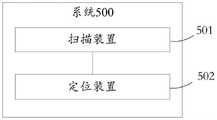
如图5所示,本公开提供一种穿刺针尖的定位系统500,包括:As shown in FIG. 5 , the present disclosure provides a puncture needle point positioning system 500, including:
扫描装置501,配置为获取患者身体的一组切片图像,其中,所述一组切片图像中包括所述患者身体的体表轮廓的图像和所述体表轮廓内的至少一个目标患处的图像;The
定位装置502,包括换能器,所述换能器包括多个超声模块,The
所述定位装置502,配置为:The
基于所述一组切片图像生成三维空间中所述患者身体的三维身体模型;generating a three-dimensional body model of the patient's body in three-dimensional space based on the set of sliced images;
对所述三维身体模型中的所述至少一个目标患处标记一个穿刺目标,并确定所述穿刺目标的表面的目标三维位置;marking a puncture target on the at least one target lesion in the three-dimensional body model, and determining a target three-dimensional position of a surface of the puncture target;
通过换能器中的各个超声模块分别向所述患者身体的内部发射超声波,接收所述超声波的回波信息,并分别获取各个超声模块从发射至接收的传播时间段,其中,所述换能器固定放置于所述患者身体的体表处,且能够探测到所述目标三维位置;Each ultrasonic module in the transducer transmits ultrasonic waves to the inside of the patient's body, receives the echo information of the ultrasonic waves, and obtains the propagation time period of each ultrasonic module from transmission to reception, wherein the transducer The device is fixedly placed on the body surface of the patient's body, and can detect the three-dimensional position of the target;
根据各个超声模块的回波信息和传播时间段获取超声图像和第一超声模块探测到的第一距离,其中,所述超声图像中包括所述穿刺目标的图像和穿刺针的针尖的图像,所述第一距离是指所述换能器中的第一超声模块与所述针尖的距离;Obtain an ultrasonic image and a first distance detected by the first ultrasonic module according to the echo information and propagation time period of each ultrasonic module, wherein the ultrasonic image includes an image of the puncture target and an image of the needle tip of the puncture needle, so The first distance refers to the distance between the first ultrasonic module in the transducer and the needle tip;
基于所述超声图像与多个二维模型图像分别进行匹配,获取一匹配的二维模型图像和所述匹配的二维模型图像所在的匹配平面,其中,所述二维模型图像是指经过所述目标三维位置的任一角度平面上所述三维身体模型的投影图像;Based on the matching of the ultrasonic image and a plurality of two-dimensional model images respectively, a matched two-dimensional model image and a matching plane where the matched two-dimensional model image is obtained, wherein the two-dimensional model image refers to the A projection image of the three-dimensional body model on any angle plane of the three-dimensional position of the target;
根据各个超声模块的回波信息和传播时间段分析所述匹配的二维模型图像和所述超声图像,获取第二超声模块探测到的第二距离,其中,所述第二距离是指所述换能器中的第二超声模块与所述穿刺目标的距离;Analyze the matched two-dimensional model image and the ultrasonic image according to the echo information and propagation time period of each ultrasonic module, and obtain the second distance detected by the second ultrasonic module, wherein the second distance refers to the the distance between the second ultrasonic module in the transducer and the puncture target;
基于所述匹配平面、所述目标三维位置和所述第二距离确定所述第二超声模块的第一三维位置;determining a first three-dimensional position of the second ultrasound module based on the matching plane, the target three-dimensional position and the second distance;
基于所述匹配平面、所述第二超声模块的第一三维位置和第二距离以及所述第一超声模块与所述第二超声模块的预设模块位置关系确定所述针尖在所述三维身体模型中的第二三维位置;Determine the position of the needle tip on the three-dimensional body based on the matching plane, the first three-dimensional position and the second distance of the second ultrasound module, and the preset module position relationship between the first ultrasound module and the second ultrasound module. the second 3D position in the model;
基于所述目标三维位置和所述第二三维位置获取所述穿刺目标与所述针尖的三维位置关系信息。The three-dimensional positional relationship information between the puncture target and the needle tip is acquired based on the target three-dimensional position and the second three-dimensional position.
可选的,所述定位装置502配置为所述根据各个超声模块的回波信息和传播时间段获取超声图像和第一超声模块探测到的第一距离,包括:Optionally, the
基于各个超声模块的回波信息在对应图像位置进行图像拟合,生成所述超声图像;performing image fitting at corresponding image positions based on the echo information of each ultrasound module to generate the ultrasound image;
确定满足预设穿刺针回波信息条件的回波信息在所述超声图像中表征的穿刺针图像;determining the puncture needle image represented in the ultrasonic image by the echo information meeting the preset puncture needle echo information condition;
基于预设针尖特征图像对所述超声图像中的穿刺针图像进行针尖位置分析,获取所述针尖在所述超声图像中的针尖图像位置;Performing a needle point position analysis on the puncture needle image in the ultrasonic image based on the preset needle point characteristic image, and acquiring the needle point image position of the needle point in the ultrasonic image;
确定所述针尖图像位置对应的针尖回波信息,以及接收所述针尖回波信息的所述第一超声模块;determining needle tip echo information corresponding to the needle tip image position, and the first ultrasound module receiving the needle tip echo information;
基于所述第一超声模块的传播时间段和所述超声波的预设传播速度获取所述第一距离。The first distance is acquired based on a propagation time period of the first ultrasonic module and a preset propagation speed of the ultrasonic waves.
可选的,所述定位装置502配置为所述根据各个超声模块的回波信息和传播时间段分析所述匹配的二维模型图像和所述超声图像,获取第二超声模块探测到的第二距离,包括:Optionally, the
基于所述匹配的二维模型图像中标记的所述穿刺目标的图像确定所述超声图像中穿刺目标的目标图像位置;determining a target image position of the puncture target in the ultrasound image based on the image of the puncture target marked in the matched two-dimensional model image;
确定所述目标图像位置对应的目标回波信息;determining target echo information corresponding to the target image position;
从各个超声模块中确定接收所述目标回波信息的所述第二超声模块;determining the second ultrasound module receiving the target echo information from each ultrasound module;
基于所述第二超声模块的传播时间段和所述超声波的预设传播速度获取所述第二距离。The second distance is acquired based on a propagation time period of the second ultrasonic module and a preset propagation speed of the ultrasonic waves.
可选的,所述定位装置502配置为所述基于所述匹配平面、所述目标三维位置和所述第一距离确定所述第二超声模块的第一三维位置,包括:Optionally, the
基于所述目标三维位置获取垂直于所述匹配平面的第一直线;obtaining a first straight line perpendicular to the matching plane based on the three-dimensional position of the target;
以所述第二距离为线段的长度,且以所述目标三维位置为线段的端点,延所述第一直线分别计算获得所述目标三维位置两侧的线段的第一端点三维位置和第二端点三维位置;Taking the second distance as the length of the line segment, and taking the target three-dimensional position as the end point of the line segment, respectively calculating and obtaining the three-dimensional positions of the first end points of the line segments on both sides of the target three-dimensional position along the first straight line The three-dimensional position of the second endpoint;
计算获得第一最短距离和第二最短距离,其中,所述第一最短距离为所述第一端点三维位置至所述三维身体模型的体表的最短距离,所述第二最短距离为所述第二端点三维位置至所述三维身体模型的体表的最短距离;Calculate and obtain a first shortest distance and a second shortest distance, wherein the first shortest distance is the shortest distance from the three-dimensional position of the first endpoint to the body surface of the three-dimensional body model, and the second shortest distance is the shortest distance from the three-dimensional position of the first endpoint to the body surface of the three-dimensional body model The shortest distance from the three-dimensional position of the second end point to the body surface of the three-dimensional body model;
确定所述第一最短距离和所述第二最短距离中的最小距离所对应的端点三维位置为所述第一三维位置。Determine the three-dimensional position of the end point corresponding to the minimum distance among the first shortest distance and the second shortest distance as the first three-dimensional position.
可选的,所述定位装置502配置为所述基于所述匹配平面、所述第二超声模块的第一三维位置和第二距离以及所述第一超声模块与所述第二超声模块的预设模块位置关系确定所述针尖在所述三维身体模型中的第二三维位置,包括:Optionally, the
在所述第一三维位置生成平行于所述匹配平面的模块平面;generating a module plane parallel to the matching plane at the first three-dimensional location;
基于所述第一三维位置和所述预设模块位置关系在所述模块平面上计算获得所述第一超声模块的第三三维位置;calculating and obtaining a third three-dimensional position of the first ultrasound module on the module plane based on the first three-dimensional position and the preset module position relationship;
基于所述第三三维位置获取垂直于所述模块平面的第二直线;obtaining a second straight line perpendicular to the module plane based on the third three-dimensional position;
以所述第一距离为线段的长度,且以所述第三三维位置为线段的出发端点,延所述第二直线计算获得所述第二三维位置。The second three-dimensional position is obtained by calculating along the second straight line by using the first distance as the length of the line segment and the third three-dimensional position as the starting point of the line segment.
可选的,所述定位装置502配置为所述基于所述一组切片图像生成三维空间中所述患者身体的三维身体模型,包括:Optionally, the
将每个切片图像中的同一图像位置分别标记为对应切片图像的二维原点位置,并在每个切片图像中标记多个二维特征位置,所述多个二维特征位置分别标记于目标患处图像的轮廓处和体表轮廓图像的体表轮廓处;Marking the same image position in each slice image as the two-dimensional origin position of the corresponding slice image, and marking multiple two-dimensional feature positions in each slice image, the multiple two-dimensional feature positions are respectively marked on the target lesion The contour of the image and the body surface contour of the body surface contour image;
将所述一组切片图像依切片顺序堆叠于所述三维空间中,其中,每个切片图像以其二维原点位置为交点垂直于预设坐标轴,且每个切片图像的二维原点位置在预设坐标轴上以预设层距为间隔;Stacking the group of sliced images in the three-dimensional space according to slice order, wherein each sliced image is perpendicular to the preset coordinate axis with its two-dimensional origin position as the intersection point, and the two-dimensional origin position of each sliced image is at The preset layer distance is used as the interval on the preset coordinate axis;
将所述三维空间中每个切片图像中的二维特征位置基于预设层距转变为三维特征位置;converting the two-dimensional feature position in each slice image in the three-dimensional space into a three-dimensional feature position based on a preset layer distance;
基于所述三维特征位置拟合生成所述三维身体模型。The three-dimensional body model is generated based on the three-dimensional feature position fitting.
可选的,所述定位装置502配置为所述对所述三维身体模型中的所述至少一个目标患处标记一个穿刺目标,并确定所述穿刺目标的表面的目标三维位置,包括:Optionally, the
分析所述三维身体模型中的所述至少一个目标患处的形状和/或大小,确定一个穿刺目标,其中,所述穿刺目标包括:纵横比大于1的目标患处、沙粒样钙化严重的目标患处或体积最小的目标患处;Analyzing the shape and/or size of the at least one target lesion in the three-dimensional body model to determine a puncture target, wherein the puncture target includes: a target lesion with an aspect ratio greater than 1, and a target lesion with severe sand-like calcification Or the target lesion with the smallest volume;
在所述三维身体模型中标记所述穿刺目标,并确定所述穿刺目标的表面的目标三维位置。The puncture target is marked in the three-dimensional body model, and a target three-dimensional position of a surface of the puncture target is determined.
本公开实施例,利用三维空间中所述患者身体的三维身体模型确定穿刺目标位置,然后利用换能器中每个超声模块在阵列中的排列位置与超声图像中的区域图像的位置具有一一对应的关系,分析超声图像,并利用超声模块探测的距离值,确定与穿刺目标相关的第一超声模块的三维位置,及与针尖相关的第二超声模块的三维位置,进而确定针尖的三维位置,最终为操作者提供所述穿刺目标与所述针尖的三维位置关系信息,使操作者在穿刺的过程中能够全方位的了解针尖与穿刺目标的位置关系,及时调整穿刺路径,准确获取检材,避免了多次穿刺给患者带来的经济损失和精神伤害,提高了医疗资源的利用率。In the embodiment of the present disclosure, the puncture target position is determined by using the three-dimensional body model of the patient's body in the three-dimensional space, and then the relationship between the arrangement position of each ultrasonic module in the transducer and the position of the region image in the ultrasonic image is used. Corresponding relationship, analyze the ultrasonic image, and use the distance value detected by the ultrasonic module to determine the three-dimensional position of the first ultrasonic module related to the puncture target, and the three-dimensional position of the second ultrasonic module related to the needle tip, and then determine the three-dimensional position of the needle tip , and finally provide the operator with information on the three-dimensional positional relationship between the puncture target and the needle tip, so that the operator can fully understand the positional relationship between the needle tip and the puncture target during the puncture process, adjust the puncture path in time, and accurately obtain test materials , avoiding the economic loss and mental injury caused by multiple punctures to patients, and improving the utilization rate of medical resources.
最后应说明的是:本说明书中各个实施例采用递进的方式描述,每个实施例重点说明的都是与其他实施例的不同之处,各个实施例之间相同相似部分互相参见即可。对于实施例公开的系统或装置而言,由于其与实施例公开的方法相对应,所以描述比较简单,相关之处参见方法部分说明即可。Finally, it should be noted that each embodiment in this specification is described in a progressive manner, each embodiment focuses on the difference from other embodiments, and the same and similar parts of each embodiment can be referred to each other. As for the system or device disclosed in the embodiment, since it corresponds to the method disclosed in the embodiment, the description is relatively simple, and for relevant details, please refer to the description of the method part.
以上实施例仅用以说明本公开的技术方案,而非对其限制;尽管参照前述实施例对本公开进行了详细的说明,本领域的普通技术人员应当理解:其依然可以对前述各实施例所记载的技术方案进行修改,或者对其中部分技术特征进行等同替换;而这些修改或者替换,并不使相应技术方案的本质脱离本公开各实施例技术方案的精神和范围。The above embodiments are only used to illustrate the technical solutions of the present disclosure, rather than to limit them; although the present disclosure has been described in detail with reference to the foregoing embodiments, those of ordinary skill in the art should understand that: it can still be described in the foregoing embodiments The recorded technical solutions are modified, or some of the technical features are replaced equivalently; and these modifications or replacements do not make the essence of the corresponding technical solutions deviate from the spirit and scope of the technical solutions of the embodiments of the present disclosure.
Claims (10)
Priority Applications (1)
| Application Number | Priority Date | Filing Date | Title |
|---|---|---|---|
| CN202210318962.6ACN115553880B (en) | 2022-03-29 | 2022-03-29 | Positioning method and system for puncture needle tip |
Applications Claiming Priority (1)
| Application Number | Priority Date | Filing Date | Title |
|---|---|---|---|
| CN202210318962.6ACN115553880B (en) | 2022-03-29 | 2022-03-29 | Positioning method and system for puncture needle tip |
Publications (2)
| Publication Number | Publication Date |
|---|---|
| CN115553880Atrue CN115553880A (en) | 2023-01-03 |
| CN115553880B CN115553880B (en) | 2024-08-16 |
Family
ID=84737647
Family Applications (1)
| Application Number | Title | Priority Date | Filing Date |
|---|---|---|---|
| CN202210318962.6AActiveCN115553880B (en) | 2022-03-29 | 2022-03-29 | Positioning method and system for puncture needle tip |
Country Status (1)
| Country | Link |
|---|---|
| CN (1) | CN115553880B (en) |
Cited By (3)
| Publication number | Priority date | Publication date | Assignee | Title |
|---|---|---|---|---|
| CN117503344A (en)* | 2023-12-12 | 2024-02-06 | 中国人民解放军总医院第一医学中心 | Multiple puncture needle power confirmation methods, devices, electronic equipment and storage media |
| CN118845094A (en)* | 2024-09-25 | 2024-10-29 | 卡本(深圳)医疗器械有限公司 | Three-dimensional display method, device, computer equipment and medium for puncture needle tract |
| CN120451149A (en)* | 2025-07-09 | 2025-08-08 | 中国人民解放军总医院第一医学中心 | Ultrasonic blood vessel intelligent analysis method and system |
Citations (4)
| Publication number | Priority date | Publication date | Assignee | Title |
|---|---|---|---|---|
| US20160081653A1 (en)* | 2013-06-11 | 2016-03-24 | Kabushiki Kaisha Toshiba | Ultrasound diagnosis apparatus and medical image diagnosis apparatus |
| CN110279467A (en)* | 2019-06-19 | 2019-09-27 | 天津大学 | Ultrasound image under optical alignment and information fusion method in the art of puncture biopsy needle |
| CN110537961A (en)* | 2019-08-01 | 2019-12-06 | 中国人民解放军总医院 | A minimally invasive intervention guidance system and method for fusion of CT and ultrasound images |
| CN112533540A (en)* | 2018-08-13 | 2021-03-19 | 深圳迈瑞生物医疗电子股份有限公司 | Ultrasonic imaging method, ultrasonic imaging device and puncture navigation system |
- 2022
- 2022-03-29CNCN202210318962.6Apatent/CN115553880B/enactiveActive
Patent Citations (4)
| Publication number | Priority date | Publication date | Assignee | Title |
|---|---|---|---|---|
| US20160081653A1 (en)* | 2013-06-11 | 2016-03-24 | Kabushiki Kaisha Toshiba | Ultrasound diagnosis apparatus and medical image diagnosis apparatus |
| CN112533540A (en)* | 2018-08-13 | 2021-03-19 | 深圳迈瑞生物医疗电子股份有限公司 | Ultrasonic imaging method, ultrasonic imaging device and puncture navigation system |
| CN110279467A (en)* | 2019-06-19 | 2019-09-27 | 天津大学 | Ultrasound image under optical alignment and information fusion method in the art of puncture biopsy needle |
| CN110537961A (en)* | 2019-08-01 | 2019-12-06 | 中国人民解放军总医院 | A minimally invasive intervention guidance system and method for fusion of CT and ultrasound images |
Non-Patent Citations (1)
| Title |
|---|
| 鲁通;徐静;任贺;王旸;梁萍;陈恳;杨向东;胡晨明;: "介入性超声计算机辅助导航系统的初步实现及实验研究", 中国医学影像技术, no. 07, 20 July 2007 (2007-07-20)* |
Cited By (3)
| Publication number | Priority date | Publication date | Assignee | Title |
|---|---|---|---|---|
| CN117503344A (en)* | 2023-12-12 | 2024-02-06 | 中国人民解放军总医院第一医学中心 | Multiple puncture needle power confirmation methods, devices, electronic equipment and storage media |
| CN118845094A (en)* | 2024-09-25 | 2024-10-29 | 卡本(深圳)医疗器械有限公司 | Three-dimensional display method, device, computer equipment and medium for puncture needle tract |
| CN120451149A (en)* | 2025-07-09 | 2025-08-08 | 中国人民解放军总医院第一医学中心 | Ultrasonic blood vessel intelligent analysis method and system |
Also Published As
| Publication number | Publication date |
|---|---|
| CN115553880B (en) | 2024-08-16 |
Similar Documents
| Publication | Publication Date | Title |
|---|---|---|
| CN115553880A (en) | A positioning method and system for a puncture needle point | |
| JP5143333B2 (en) | System and method for performing image processing for observing abnormal parts in different types of images | |
| JP6430498B2 (en) | System and method for mapping of ultrasonic shear wave elastography measurements | |
| CN102551800B (en) | For showing the method and system of ultrasound data | |
| JP5511641B2 (en) | Ultrasonic probe, position display device, and ultrasonic diagnostic device | |
| CN109310399B (en) | Medical ultrasonic image processing apparatus | |
| US20100286519A1 (en) | Ultrasound system and method to automatically identify and treat adipose tissue | |
| EP3742973B1 (en) | Device and method for obtaining anatomical measurements from an ultrasound image | |
| US10368841B2 (en) | Ultrasound diagnostic apparatus | |
| EP1775601B1 (en) | System and method for forming 3-dimensional images using multiple sectional plane images | |
| WO2014200099A1 (en) | Ultrasonic diagnostic device | |
| CN105007825B (en) | Ultrasonic diagnostic device and medical image processing device | |
| KR20140020486A (en) | Method and apparatus for analyzing elastography of tissue using ultrasound | |
| CN109640831A (en) | Supersonic diagnostic appts | |
| EP3017766B1 (en) | Ultrasound imaging apparatus and control method thereof | |
| CN105142528B (en) | Diagnostic ultrasound equipment | |
| JP2019502445A (en) | Apparatus and method for characterizing a subject's tissue | |
| US8636662B2 (en) | Method and system for displaying system parameter information | |
| US20120078101A1 (en) | Ultrasound system for displaying slice of object and method thereof | |
| US20150105658A1 (en) | Ultrasonic imaging apparatus and control method thereof | |
| CN116058875A (en) | Ultrasonic imaging method and ultrasonic imaging system | |
| KR20180087698A (en) | Ultrasound diagnostic apparatus for displaying shear wave data of the object and method for operating the same | |
| CN116158784A (en) | Ultrasonic diagnostic device, ultrasonic image analysis device and control method thereof | |
| JP7336766B2 (en) | Ultrasonic diagnostic device, ultrasonic diagnostic method and ultrasonic diagnostic program | |
| EP3939496A1 (en) | Method and system for context-aware photoacoustic imaging |
Legal Events
| Date | Code | Title | Description |
|---|---|---|---|
| PB01 | Publication | ||
| PB01 | Publication | ||
| SE01 | Entry into force of request for substantive examination | ||
| SE01 | Entry into force of request for substantive examination | ||
| GR01 | Patent grant | ||
| GR01 | Patent grant |