CN115429202A - A traction capsule endoscope - Google Patents
A traction capsule endoscopeDownload PDFInfo
- Publication number
- CN115429202A CN115429202ACN202211178279.3ACN202211178279ACN115429202ACN 115429202 ACN115429202 ACN 115429202ACN 202211178279 ACN202211178279 ACN 202211178279ACN 115429202 ACN115429202 ACN 115429202A
- Authority
- CN
- China
- Prior art keywords
- capsule endoscope
- pull
- housing
- behind
- wire
- Prior art date
- Legal status (The legal status is an assumption and is not a legal conclusion. Google has not performed a legal analysis and makes no representation as to the accuracy of the status listed.)
- Pending
Links
Images


Classifications
- A—HUMAN NECESSITIES
- A61—MEDICAL OR VETERINARY SCIENCE; HYGIENE
- A61B—DIAGNOSIS; SURGERY; IDENTIFICATION
- A61B1/00—Instruments for performing medical examinations of the interior of cavities or tubes of the body by visual or photographical inspection, e.g. endoscopes; Illuminating arrangements therefor
- A61B1/04—Instruments for performing medical examinations of the interior of cavities or tubes of the body by visual or photographical inspection, e.g. endoscopes; Illuminating arrangements therefor combined with photographic or television appliances
- A61B1/041—Capsule endoscopes for imaging
- A—HUMAN NECESSITIES
- A61—MEDICAL OR VETERINARY SCIENCE; HYGIENE
- A61B—DIAGNOSIS; SURGERY; IDENTIFICATION
- A61B1/00—Instruments for performing medical examinations of the interior of cavities or tubes of the body by visual or photographical inspection, e.g. endoscopes; Illuminating arrangements therefor
- A61B1/00002—Operational features of endoscopes
- A61B1/00011—Operational features of endoscopes characterised by signal transmission
- A61B1/00018—Operational features of endoscopes characterised by signal transmission using electrical cables
- A—HUMAN NECESSITIES
- A61—MEDICAL OR VETERINARY SCIENCE; HYGIENE
- A61B—DIAGNOSIS; SURGERY; IDENTIFICATION
- A61B1/00—Instruments for performing medical examinations of the interior of cavities or tubes of the body by visual or photographical inspection, e.g. endoscopes; Illuminating arrangements therefor
- A61B1/00002—Operational features of endoscopes
- A61B1/00025—Operational features of endoscopes characterised by power management
- A61B1/00027—Operational features of endoscopes characterised by power management characterised by power supply
- A61B1/00029—Operational features of endoscopes characterised by power management characterised by power supply externally powered, e.g. wireless
- A—HUMAN NECESSITIES
- A61—MEDICAL OR VETERINARY SCIENCE; HYGIENE
- A61B—DIAGNOSIS; SURGERY; IDENTIFICATION
- A61B1/00—Instruments for performing medical examinations of the interior of cavities or tubes of the body by visual or photographical inspection, e.g. endoscopes; Illuminating arrangements therefor
- A61B1/04—Instruments for performing medical examinations of the interior of cavities or tubes of the body by visual or photographical inspection, e.g. endoscopes; Illuminating arrangements therefor combined with photographic or television appliances
- A61B1/045—Control thereof
- A—HUMAN NECESSITIES
- A61—MEDICAL OR VETERINARY SCIENCE; HYGIENE
- A61B—DIAGNOSIS; SURGERY; IDENTIFICATION
- A61B1/00—Instruments for performing medical examinations of the interior of cavities or tubes of the body by visual or photographical inspection, e.g. endoscopes; Illuminating arrangements therefor
- A61B1/273—Instruments for performing medical examinations of the interior of cavities or tubes of the body by visual or photographical inspection, e.g. endoscopes; Illuminating arrangements therefor for the upper alimentary canal, e.g. oesophagoscopes, gastroscopes
- A61B1/2733—Oesophagoscopes
Landscapes
- Health & Medical Sciences (AREA)
- Life Sciences & Earth Sciences (AREA)
- Surgery (AREA)
- Engineering & Computer Science (AREA)
- Biomedical Technology (AREA)
- Molecular Biology (AREA)
- Pathology (AREA)
- Radiology & Medical Imaging (AREA)
- Nuclear Medicine, Radiotherapy & Molecular Imaging (AREA)
- Biophysics (AREA)
- Physics & Mathematics (AREA)
- Heart & Thoracic Surgery (AREA)
- Medical Informatics (AREA)
- Optics & Photonics (AREA)
- Animal Behavior & Ethology (AREA)
- General Health & Medical Sciences (AREA)
- Public Health (AREA)
- Veterinary Medicine (AREA)
- Gastroenterology & Hepatology (AREA)
- Computer Networks & Wireless Communication (AREA)
- Endoscopes (AREA)
Abstract
Translated fromChinese
Description
Translated fromChinese技术领域technical field
本发明涉及医疗器械领域,具体涉及到一种可用于消化道检查的胶囊内窥镜。The invention relates to the field of medical devices, in particular to a capsule endoscope that can be used for digestive tract inspection.
背景技术Background technique
胶囊内窥镜正在逐渐应用于消化道疾病的临床诊断,受检者吞服胶囊内窥镜,通过胶囊内窥镜的摄像组件近距离拍摄体腔组织的状况,并将图像数据无线传输至外部图像接收装置并显示,便于医生进行临床诊断,从而克服了传统插入式内镜检查耐受性差的缺陷。Capsule endoscopes are gradually being used in the clinical diagnosis of gastrointestinal diseases. The subject swallows the capsule endoscope, and uses the camera component of the capsule endoscope to take a close-up shot of the condition of the body cavity tissue, and wirelessly transmits the image data to an external image The receiving device and display are convenient for doctors to make clinical diagnosis, thereby overcoming the defect of poor tolerance of traditional insertion endoscopy.
虽然胶囊内窥镜具有检查方便、无创伤、无痛苦、无交叉污染、不影响受检者正常工作等优点,对受检者具有更好的受检体验,但相较于传统的机械插入式内窥镜检查,胶囊内窥镜的成本较高,大部分受检者难以承受较高的检查费用,且胶囊内窥镜由于在狭小的空间内容置了照明装置、摄像系统、内置磁体、无线传输单元和电池单元等电子部件,加大了设计复杂度和设计成本,且现有的胶囊内窥镜外形整体偏大,部分受检者难以吞咽。Although the capsule endoscope has the advantages of convenient inspection, no trauma, no pain, no cross-contamination, and does not affect the normal work of the examinee, it has a better inspection experience for the examinee, but compared with the traditional mechanical insertion For endoscopic inspection, the cost of capsule endoscope is relatively high, and most of the examinees cannot bear the high inspection fee, and capsule endoscope is equipped with lighting device, camera system, built-in magnet, wireless Electronic components such as the transmission unit and battery unit increase the design complexity and design cost, and the overall shape of the existing capsule endoscope is too large, which is difficult for some subjects to swallow.
另外,现有的胶囊内窥镜多数采用无线传输方案,客观上存在信号传输不稳定、胶囊内窥镜内置电池供电不足的风险。In addition, most of the existing capsule endoscopes use wireless transmission solutions, objectively there are risks of unstable signal transmission and insufficient power supply of the built-in battery of the capsule endoscope.
胶囊内窥镜在针对食道部分检查时,由于胶囊内窥镜通过食道的时间通常只有不足一秒钟或更短时间,且行进速度较快,能够拍摄的图像非常有限,且图像质量模糊,从而导致无法对食道进行很好的检查。When the capsule endoscope inspects the esophagus, because the time for the capsule endoscope to pass through the esophagus is usually less than one second or less, and the traveling speed is relatively fast, the images that can be captured are very limited and the image quality is blurred. This prevents a good examination of the esophagus.
中国专利公开号CN109924937A公开一种内窥镜装置及内窥检测方法,该技术方案通过气源控制牵引管对胶囊尾部的夹紧度,再通过拉动牵引管控制胶囊内窥镜在受检者体内的位置,该装置中的牵引管结构复杂,成本较高,且存在脱离胶囊内窥镜的风险,从而不能实现检查食道的目的。Chinese Patent Publication No. CN109924937A discloses an endoscope device and an endoscope detection method. The technical solution controls the clamping degree of the traction tube to the tail of the capsule through the air source, and then controls the capsule endoscope in the subject by pulling the traction tube. The position of the traction tube in this device is complicated, the cost is high, and there is a risk of detaching from the capsule endoscope, so that the purpose of examining the esophagus cannot be achieved.
因此,有必要开发一种针对食道部位的结构简单、成本较小、信号传输可靠稳定、且可自主控制胶囊内窥镜的行进速度,便于拍摄足够图像,方便医生分析病变部位的胶囊内窥镜。Therefore, it is necessary to develop a capsule endoscope for the esophagus that has a simple structure, low cost, reliable and stable signal transmission, and can independently control the travel speed of the capsule endoscope, which is convenient for taking enough images and convenient for doctors to analyze the lesion. .
发明内容Contents of the invention
为了解决现有技术的不足,本发明提供一种牵引式胶囊内窥镜,结构简单、组装便捷、成本较低,特别适用于大规模筛查,且采用外部电源供电和有线信号传输,通过牵引线控制胶囊内窥镜在待检者体内的行进速度,大幅提高检测精度。In order to solve the deficiencies of the prior art, the present invention provides a traction capsule endoscope, which has simple structure, convenient assembly, and low cost, and is especially suitable for large-scale screening. The speed of the capsule endoscope in the body of the subject is controlled by wire, which greatly improves the detection accuracy.
本发明提供一种牵引式胶囊内窥镜,包括摄像单元、透明前盖、PCBA板、外壳及照明单元,进一步还包括设置在外壳端部的卡扣,所述PCBA板通过卡扣与外壳固定连接,所述外壳设置第一连接部,所述透明前盖设置第二连接部,所述第一连接部与第二连接部相互套接,所述外壳的尾部设置牵引线,所述牵引线内置至少两根导线,所述两根导线电连接PCBA板。The invention provides a traction capsule endoscope, which includes a camera unit, a transparent front cover, a PCBA board, a housing and an illumination unit, and further includes a buckle arranged at the end of the housing, and the PCBA board is fixed to the housing through the buckle connection, the housing is provided with a first connection part, the transparent front cover is provided with a second connection part, the first connection part and the second connection part are nested with each other, and the tail of the housing is provided with a pull wire, and the pull wire At least two wires are built in, and the two wires are electrically connected to the PCBA board.
进一步的,所述透明前盖和外壳通过激光焊接或胶水任一种方式固定。Further, the transparent front cover and the shell are fixed by laser welding or glue.
进一步的,所述牵引式胶囊内窥镜的直径是8-12mm,长度是15-25mm。Further, the traction capsule endoscope has a diameter of 8-12mm and a length of 15-25mm.
进一步的,所述牵引式胶囊内窥镜的直径是10mm,长度是18mm。Further, the traction capsule endoscope has a diameter of 10mm and a length of 18mm.
进一步的,所述牵引式胶囊内窥镜形状为葫芦形、水滴形、纺锤形的任一种形状。Further, the shape of the traction capsule endoscope is any one of a gourd shape, a drop shape, and a spindle shape.
进一步的,所述外壳的内侧壁设置向内凸伸的筋位,所述筋位为条状且沿外壳的内壁从外壳的第一连接部的端面往下延伸,所述筋位与外壳一体成型。Further, the inner side wall of the housing is provided with inwardly protruding ribs, the ribs are strip-shaped and extend downward from the end face of the first connecting part of the housing along the inner wall of the housing, and the ribs are integrated with the housing forming.
进一步的,所述外壳的第一连接部的端面设置卡扣,所述卡扣数量至少为两个,且卡扣数量与筋位数量相同,所述卡扣设置在筋位的正上方。Further, the end surface of the first connecting part of the housing is provided with buckles, the number of the buckles is at least two, and the number of buckles is the same as the number of ribs, and the buckles are arranged directly above the ribs.
进一步的,所述导线与PCBA板采用激光焊接或锡焊的任一种方式固定连接。Further, the wires are fixedly connected to the PCBA board by laser welding or soldering.
进一步的,所述牵引线的外套材料是硅胶、热塑性聚氨酯弹性体橡胶、合成橡胶的任一种。Further, the sheath material of the pulling wire is any one of silica gel, thermoplastic polyurethane elastomer rubber, and synthetic rubber.
进一步的,所述外套的硬度是30-80A。Further, the hardness of the jacket is 30-80A.
进一步的,所述外套的硬度是50A。Further, the hardness of the jacket is 50A.
进一步的,所述牵引线的直径是0.8-2mm,Further, the diameter of the pulling wire is 0.8-2mm,
进一步的,所述牵引线的直径是1.2mm。Further, the diameter of the pulling wire is 1.2mm.
进一步的,所述牵引式胶囊内窥镜还包括内置于外壳内的磁铁。Further, the traction capsule endoscope also includes a magnet built in the shell.
采用本发明的牵引式胶囊内窥镜,弥补了现有技术的不足,本发明的牵引式胶囊内窥镜结构简单、组装方便、成本低适用于大规模筛查,且胶囊体积较小,容易吞咽,采用有线方式供电的牵引式胶囊内窥镜工作时间更长,信号传输稳定,通过牵引线控制胶囊内窥镜在待检者体内的行进速度及姿态,提高了检测精度。The traction capsule endoscope of the present invention makes up for the deficiencies of the prior art. The traction capsule endoscope of the present invention is simple in structure, convenient to assemble, low in cost and suitable for large-scale screening, and the capsule volume is small, easy Swallowing, the traction capsule endoscope powered by wired mode has a longer working time and stable signal transmission. The speed and posture of the capsule endoscope in the body of the subject are controlled by the traction wire, which improves the detection accuracy.
附图说明Description of drawings
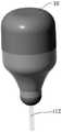
图1:本发明第一实施例的牵引式胶囊内窥镜结构示意图。Fig. 1: Schematic diagram of the structure of the traction capsule endoscope according to the first embodiment of the present invention.
图2:第一实施例的牵引式胶囊内窥镜的实物示意图。Fig. 2: The physical schematic diagram of the traction capsule endoscope of the first embodiment.
图3:第一实施例的牵引式胶囊内窥镜的PCBA板布局示意图。Fig. 3: Schematic diagram of the layout of the PCBA board of the traction capsule endoscope of the first embodiment.
图4:第一实施例的牵引式胶囊内窥镜的外壳示意图。Fig. 4: Schematic diagram of the casing of the traction capsule endoscope of the first embodiment.
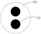
图5:第一实施例的牵引式胶囊内窥镜的牵引线剖面图。Fig. 5: A sectional view of the pulling line of the pulling capsule endoscope of the first embodiment.
图6:第一实施例的牵引式胶囊内窥镜的射频与电源信号处理电路局部示意图。Fig. 6: A partial schematic diagram of the radio frequency and power signal processing circuit of the traction capsule endoscope of the first embodiment.
图7:本发明第二实施例的牵引式胶囊内窥镜结构示意图。Fig. 7: Schematic diagram of the structure of the traction capsule endoscope according to the second embodiment of the present invention.
各序号及对应的名称分别为: 胶囊内窥镜10,摄像单元101,固定槽102,导线103,筋位104,透明前盖105,卡扣106,卡槽107,PCBA板108,第二连接部109,第一连接部110,外壳111,牵引线112,照明单元113,绝缘外套114,导线115,磁铁116,胶囊射频传输端201,第一电感202,第一电容203,第一电源线连接器204,第二电源线连接器205,第二电感206,第二电容207,接收器射频传输端208。The serial numbers and corresponding names are:
具体实施方式detailed description
为了使本发明所要解决的技术问题、技术方案及有益效果更加清楚明白,以下结合附图及实施例,对本发明进行进一步详细说明,应当理解,此处所描述的具体实施例仅用以解释本发明,并不用于限定本发明。In order to make the technical problems, technical solutions and beneficial effects to be solved by the present invention clearer, the present invention will be described in further detail below in conjunction with the accompanying drawings and embodiments. It should be understood that the specific embodiments described here are only used to explain the present invention , and are not intended to limit the present invention.
请参考图1本发明第一实施例的牵引式胶囊内窥镜结构示意图及图2本发明牵引式胶囊内窥镜的实物示意图,本发明的牵引式胶囊内窥镜10包括摄像单元101,固定槽102,导线103,筋位104,透明前盖105,卡扣106,卡槽107,PCBA板108,第二连接部109,第一连接部110,外壳111及牵引线112,所述外壳111的端部设置卡扣106,PCBA板108通过卡扣106与外壳111固定连接,所述透明前盖105和外壳111通过激光焊接或胶水固定,所述外壳111设置第一连接部110,所述透明前盖105设置第二连接部109,其余各部件的相互连接关系属于本领域普通技术人员的常规知识,本发明的创新不在于此,因此,此处仅进行简要说明。Please refer to FIG. 1 for a structural schematic diagram of the towed capsule endoscope according to the first embodiment of the present invention and FIG. 2 for a physical schematic diagram of the towed capsule endoscope of the present invention. The
进一步的,所述胶囊内窥镜的直径为8-12mm,长度为15-25mm,优选的,Further, the capsule endoscope has a diameter of 8-12mm and a length of 15-25mm, preferably,
胶囊内窥镜的直径是10mm,长度是18mm。The diameter of the capsule endoscope is 10mm and the length is 18mm.
进一步的,所述胶囊内窥镜形状为葫芦形、水滴形、纺锤形的任一种形状,也可以为其他适宜进行吞服的形状,其前端稍大,尾部稍小,方便从受检者体内拽拉出体外,实验数据表明,本发明的胶囊内窥镜直径越小且尾部越细长,其与牵引线112连接的尾部形状更接近水滴状,直径从小到大平缓过渡,更便于吞咽和从食管的狭窄处拉出。Further, the shape of the capsule endoscope is any one of gourd-shaped, drop-shaped, and spindle-shaped, or other shapes suitable for swallowing. Pulled out of the body, the experimental data show that the smaller the diameter of the capsule endoscope of the present invention and the more slender the tail, the shape of the tail connected to the pulling
参考图3本发明牵引式胶囊内窥镜的PCBA板布局示意图及图4本发明牵引式胶囊内窥镜的外壳示意图,所述摄像单元101、照明单元113固定设置在PCBA板108中心位置,且与PCBA板108电连接,所述透明前盖105的表面为光学镜面,所述照明单元113的LED灯光透过透明前盖105照亮受检者体内组织以便光学镜头透过透明前盖105获取受检者体内组织的图像,所述外壳111的前部设置向内或向外凸伸的凸台而形成的第一台阶面和第一连接部110,所述透明前盖105设置向外或向内凸伸的凸台而形成的第二台阶面和第二连接部109,所述第一连接部110与所述第二连接部109相互套接,形成固定PCBA板108的固定槽102。Referring to Fig. 3 schematic diagram of the layout of the PCBA board of the towed capsule endoscope of the present invention and Fig. 4 a schematic diagram of the shell of the towed capsule endoscope of the present invention, the
进一步的所述外壳111的内侧壁设置若干向内凸伸的筋位104,所述筋位104为条状且沿外壳111的内壁从外壳111的第一连接部110的端面往下延伸,筋位104与外壳111为一体成型结构,所述外壳111的第一连接部111的端面设置有若干卡扣106,所述卡扣数量至少为两个,且卡扣106数量与筋位104数量保持一致,所述卡扣106设置在筋位104的正上方,所述PCBA板108设置与卡扣106相适应的卡槽107,所述PCBA板108的卡槽107通过所述卡扣106将PCBA板108固定在外壳111的第一连接部111的端面,使所述PCBA板108在胶囊内窥镜的Z轴方向和径向方向上定位。Furthermore, the inner side wall of the
参考图5本发明牵引式胶囊内窥镜的牵引线剖面图,所述牵引线112内置至少两条导线,所述导线为摄像单元101及照明单元113供电和/或传输信号,牵引线112采用一体成型或胶水固定方式设置在外壳111尾部,牵引线112一端与PCBA板108固定焊接,另一端与外部电源电连接,当受检者吞服胶囊内窥镜后,操作者通过牵引线112控制胶囊内窥镜的行进速度及运动状态,进而延长胶囊内窥镜在待检部位的停留时间,拍摄所需要的部位图像,提高检测精度,防止出现漏检。Referring to Fig. 5, the traction line sectional view of the traction capsule endoscope of the present invention, the
所述两条导线与PCBA板的固定连接采用两个焊点进行激光焊接或锡焊的任一种方式,当外壳111与牵引线112一体成型时,牵引线112的一端的长度延伸到外壳111外部,方便牵引线112与PCBA板108焊接,当牵引线112与PCBA板108焊接后,将PCBA板108通过外壳111的第一连接部110端部的卡扣106固定在外壳111的第一连接部110的端面,同时延伸到外壳111外部的牵引线112的长度会内缩至外壳111内部,此时牵引线112受到PCBA板108的挤压,同时PCBA板108受到牵引线112向上的反弹力,由于外壳111的第一连接部110端面卡扣106的限位,PCBA板108在外壳111的轴向方向上定位,从而PCBA板108不会沿外壳111轴向方向向上弹起,从而方便透明前盖105的装配。The fixed connection of the two wires and the PCBA board adopts any mode of laser welding or soldering of two solder joints. When the
进一步的,所述牵引线提供内置两根导线实现电源供电和射频信号同时传输,大幅度减小了牵引线的外径,从而方便受检者吞咽,提升了受检者的舒适度。Further, the traction wire provides two built-in wires to realize the simultaneous transmission of power supply and radio frequency signal, which greatly reduces the outer diameter of the traction wire, thereby making it easier for the examinee to swallow and improving the comfort of the examinee.
进一步,所述牵引线采用生物兼容性柔性材料作为绝缘外套,例如硅胶、热塑性聚氨酯弹性体橡胶、合成橡胶的任一种或其组合,对食道黏膜刺激较小,受检者在检查过程中降低不适感。所述绝缘外套的硬度为30-80A,硬度越软越易吞咽,且对食道黏膜的刺激越小,本发明的绝缘外套材料优选硅胶材料,优选的,绝缘外套的硬度是50A。Further, the traction wire uses a biocompatible flexible material as an insulating jacket, such as any one of silica gel, thermoplastic polyurethane elastomer rubber, synthetic rubber or a combination thereof, which is less irritating to the esophageal mucosa, and the subject’s lowering of the Discomfort. The hardness of the insulating jacket is 30-80A. The softer the hardness, the easier it is to swallow, and the less irritated the esophageal mucosa. The material of the insulating jacket of the present invention is preferably a silicone material. Preferably, the hardness of the insulating jacket is 50A.
进一步的,所述牵引线的直径为0.8-2mm,优选直径为1.2mm。Further, the diameter of the pulling wire is 0.8-2mm, preferably 1.2mm.
请参考图6本发明牵引式胶囊内窥镜的射频与电源信号处理电路局部示意图,本发明的牵引式胶囊内窥镜通过牵引线内置的两条导线进行电源和射频信号的传输,电源线上供电的直流分量和传输射频的信号通过L型电路进行分离,所述L型电路包含电容和电感,具体的,该电路包括胶囊射频传输端201、第一电感202、第一电容203、第一电源线连接器204、第二电源线连接器205、第二电感206、第二电容207及接收器射频传输端208,其中,第一电感202及第一电容203接入胶囊射频传输端201,所述第二电感206及第二电容207接入接收器射频传输端208,所述第一电源线连接器204设置于靠近胶囊射频传输端201一侧,第二电源线连接器205设置于靠近接收器射频传输端208一侧。Please refer to Figure 6 for a partial schematic diagram of the radio frequency and power signal processing circuit of the traction capsule endoscope of the present invention. The traction capsule endoscope of the present invention transmits power and radio frequency signals through two wires built into the traction wire. The DC component of the power supply and the signal of the transmitted radio frequency are separated through an L-shaped circuit. The L-shaped circuit includes a capacitor and an inductor. Specifically, the circuit includes a capsule
电路具体工作原理如下:L型电路中的第一电容203及第二电容207对高频射频信号导通但对低频直流信号隔离,第一电感202及第二电感206对低频直流信号导通但对高频射频信号隔离,因此,射频信号传输路径依次经过胶囊射频传输端201、第一电容203、第一电源线连接器204、第二电源线连接器205及第二电容207;电源直流分量的传输路径依次经过第一电感202、第一电源线连接器204、第二电源线连接器205及第二电感206。The specific working principle of the circuit is as follows: the
请参考图7本发明第二实施例的牵引式胶囊内窥镜结构示意图,所述第二第二实施例的牵引式胶囊内窥镜在第一实施例的基础上增加磁铁116,利用外部磁铁(未示出)控制胶囊内窥镜在受检者体内的运动方向和速率,达到更精确的检查效果。Please refer to FIG. 7 for a schematic structural diagram of the traction capsule endoscope of the second embodiment of the present invention. The traction capsule endoscope of the second second embodiment adds a
本发明的牵引式胶囊内窥镜在传统的无线胶囊内窥镜基础上删减了无线传输单元和电池单元,采用有线的方式提供电源和数据传输,减少了胶囊内窥镜内核的内部空间,从而胶囊内窥镜的体积大幅度减小。The traction capsule endoscope of the present invention deletes the wireless transmission unit and the battery unit on the basis of the traditional wireless capsule endoscope, adopts a wired method to provide power and data transmission, and reduces the internal space of the capsule endoscope core, Therefore, the volume of the capsule endoscope is greatly reduced.
如图7所示在本发明的实施例中可以在所述胶囊内窥镜内核中增加永磁铁,利用外部磁铁来控制胶囊内窥镜在受检者体内的运动方向。As shown in FIG. 7 , in the embodiment of the present invention, a permanent magnet can be added to the inner core of the capsule endoscope, and an external magnet can be used to control the movement direction of the capsule endoscope in the subject.
以上所述仅为本发明的较佳实施例而已,并不用以限制本发明,凡在本发明的精神和原则之内所作的任何修改、等同替换和改进等,均应包含在本发明的保护范围之内。The above descriptions are only preferred embodiments of the present invention, and are not intended to limit the present invention. Any modifications, equivalent replacements and improvements made within the spirit and principles of the present invention should be included in the protection of the present invention. within range.
Claims (14)
Priority Applications (1)
| Application Number | Priority Date | Filing Date | Title |
|---|---|---|---|
| CN202211178279.3ACN115429202A (en) | 2022-09-27 | 2022-09-27 | A traction capsule endoscope |
Applications Claiming Priority (1)
| Application Number | Priority Date | Filing Date | Title |
|---|---|---|---|
| CN202211178279.3ACN115429202A (en) | 2022-09-27 | 2022-09-27 | A traction capsule endoscope |
Publications (1)
| Publication Number | Publication Date |
|---|---|
| CN115429202Atrue CN115429202A (en) | 2022-12-06 |
Family
ID=84249784
Family Applications (1)
| Application Number | Title | Priority Date | Filing Date |
|---|---|---|---|
| CN202211178279.3APendingCN115429202A (en) | 2022-09-27 | 2022-09-27 | A traction capsule endoscope |
Country Status (1)
| Country | Link |
|---|---|
| CN (1) | CN115429202A (en) |
Cited By (1)
| Publication number | Priority date | Publication date | Assignee | Title |
|---|---|---|---|---|
| WO2025007903A1 (en)* | 2023-07-03 | 2025-01-09 | 上海安翰医疗技术有限公司 | Capsule endoscope |
Citations (4)
| Publication number | Priority date | Publication date | Assignee | Title |
|---|---|---|---|---|
| US20080154093A1 (en)* | 2006-12-26 | 2008-06-26 | Korea Electronics Technology Institute | Capsule Type Endoscope with an Insertion Tube |
| CN104840176A (en)* | 2014-02-19 | 2015-08-19 | 光峰科技股份有限公司 | Digestive tract inspection system and control method thereof |
| CN111374630A (en)* | 2018-12-28 | 2020-07-07 | 王子华 | Magnetic superfine swallowable endoscope |
| CN211093933U (en)* | 2019-09-23 | 2020-07-28 | 安翰科技(武汉)股份有限公司 | Capsule endoscope |
- 2022
- 2022-09-27CNCN202211178279.3Apatent/CN115429202A/enactivePending
Patent Citations (4)
| Publication number | Priority date | Publication date | Assignee | Title |
|---|---|---|---|---|
| US20080154093A1 (en)* | 2006-12-26 | 2008-06-26 | Korea Electronics Technology Institute | Capsule Type Endoscope with an Insertion Tube |
| CN104840176A (en)* | 2014-02-19 | 2015-08-19 | 光峰科技股份有限公司 | Digestive tract inspection system and control method thereof |
| CN111374630A (en)* | 2018-12-28 | 2020-07-07 | 王子华 | Magnetic superfine swallowable endoscope |
| CN211093933U (en)* | 2019-09-23 | 2020-07-28 | 安翰科技(武汉)股份有限公司 | Capsule endoscope |
Cited By (1)
| Publication number | Priority date | Publication date | Assignee | Title |
|---|---|---|---|---|
| WO2025007903A1 (en)* | 2023-07-03 | 2025-01-09 | 上海安翰医疗技术有限公司 | Capsule endoscope |
Similar Documents
| Publication | Publication Date | Title |
|---|---|---|
| JP4751772B2 (en) | In-vivo sensing device comprising a circuit board having a hard area and a soft area | |
| JP5469867B2 (en) | Endoscope with imaging catheter assembly and method for constructing an endoscope | |
| US20090105538A1 (en) | Endoscope System | |
| US10517468B2 (en) | Capsule medical device having positioning member with abutment surfaces | |
| JP2013230383A (en) | Detachable imaging device | |
| JP2009240634A (en) | Endoscope apparatus | |
| CN105361841B (en) | Wireless capsule endoscope system for gastrointestinal tract diagnosis and treatment | |
| CN205322282U (en) | A wireless capsule endoscope system for intestines and stomach are diagnose | |
| KR200446281Y1 (en) | Capsule Endoscope with Flexible Electronic Circuit Board | |
| CN211723078U (en) | Capsule endoscope | |
| CN115429202A (en) | A traction capsule endoscope | |
| CN114145694A (en) | A detachable capsule endoscope | |
| JP5248911B2 (en) | Capsule medical device | |
| CN103356154A (en) | Multifunctional capsule endoscope system suitable for interior of alimentary canal | |
| CN102641124A (en) | Capsule endoscopy | |
| CN202589487U (en) | Capsule endoscopy | |
| CN114886366A (en) | Capsule endoscope and endoscope device provided with same | |
| CN113854938A (en) | Disposable Magnetron Wired Transmission Capsule Endoscopy System | |
| CN211749715U (en) | A self-guided endoscopic system | |
| KR102084222B1 (en) | Capsule endoscopy | |
| Brown et al. | Analysis of current and future technologies of capsule endoscopy: A mini review | |
| JP2004065574A (en) | Method for assembling capsule type endoscope, and capsule type endoscope | |
| CN115429203A (en) | Reusable capsule endoscope and assembling method thereof | |
| CN216090425U (en) | Wireless remote camera device capable of being coupled to hard esophagoscope | |
| KR101171747B1 (en) | Capsuled diagnosing device with temperature sensor |
Legal Events
| Date | Code | Title | Description |
|---|---|---|---|
| PB01 | Publication | ||
| PB01 | Publication | ||
| SE01 | Entry into force of request for substantive examination | ||
| SE01 | Entry into force of request for substantive examination |