CN115424319A - Strabismus recognition system based on deep learning - Google Patents
Strabismus recognition system based on deep learningDownload PDFInfo
- Publication number
- CN115424319A CN115424319ACN202210984726.8ACN202210984726ACN115424319ACN 115424319 ACN115424319 ACN 115424319ACN 202210984726 ACN202210984726 ACN 202210984726ACN 115424319 ACN115424319 ACN 115424319A
- Authority
- CN
- China
- Prior art keywords
- strabismus
- image
- pupil
- center point
- deep learning
- Prior art date
- Legal status (The legal status is an assumption and is not a legal conclusion. Google has not performed a legal analysis and makes no representation as to the accuracy of the status listed.)
- Pending
Links
Images


Classifications
- G—PHYSICS
- G06—COMPUTING OR CALCULATING; COUNTING
- G06V—IMAGE OR VIDEO RECOGNITION OR UNDERSTANDING
- G06V40/00—Recognition of biometric, human-related or animal-related patterns in image or video data
- G06V40/10—Human or animal bodies, e.g. vehicle occupants or pedestrians; Body parts, e.g. hands
- G06V40/16—Human faces, e.g. facial parts, sketches or expressions
- G06V40/168—Feature extraction; Face representation
- G06V40/171—Local features and components; Facial parts ; Occluding parts, e.g. glasses; Geometrical relationships
- G—PHYSICS
- G06—COMPUTING OR CALCULATING; COUNTING
- G06N—COMPUTING ARRANGEMENTS BASED ON SPECIFIC COMPUTATIONAL MODELS
- G06N3/00—Computing arrangements based on biological models
- G06N3/02—Neural networks
- G06N3/08—Learning methods
- G—PHYSICS
- G06—COMPUTING OR CALCULATING; COUNTING
- G06V—IMAGE OR VIDEO RECOGNITION OR UNDERSTANDING
- G06V10/00—Arrangements for image or video recognition or understanding
- G06V10/20—Image preprocessing
- G06V10/25—Determination of region of interest [ROI] or a volume of interest [VOI]
- G—PHYSICS
- G06—COMPUTING OR CALCULATING; COUNTING
- G06V—IMAGE OR VIDEO RECOGNITION OR UNDERSTANDING
- G06V10/00—Arrangements for image or video recognition or understanding
- G06V10/70—Arrangements for image or video recognition or understanding using pattern recognition or machine learning
- G06V10/82—Arrangements for image or video recognition or understanding using pattern recognition or machine learning using neural networks
- G—PHYSICS
- G06—COMPUTING OR CALCULATING; COUNTING
- G06V—IMAGE OR VIDEO RECOGNITION OR UNDERSTANDING
- G06V40/00—Recognition of biometric, human-related or animal-related patterns in image or video data
- G06V40/10—Human or animal bodies, e.g. vehicle occupants or pedestrians; Body parts, e.g. hands
- G06V40/16—Human faces, e.g. facial parts, sketches or expressions
- G06V40/161—Detection; Localisation; Normalisation
- G06V40/166—Detection; Localisation; Normalisation using acquisition arrangements
Landscapes
- Engineering & Computer Science (AREA)
- Theoretical Computer Science (AREA)
- Health & Medical Sciences (AREA)
- Physics & Mathematics (AREA)
- General Physics & Mathematics (AREA)
- General Health & Medical Sciences (AREA)
- Multimedia (AREA)
- Oral & Maxillofacial Surgery (AREA)
- Evolutionary Computation (AREA)
- Artificial Intelligence (AREA)
- Computer Vision & Pattern Recognition (AREA)
- Computing Systems (AREA)
- Human Computer Interaction (AREA)
- Software Systems (AREA)
- Medical Informatics (AREA)
- Databases & Information Systems (AREA)
- Life Sciences & Earth Sciences (AREA)
- Biomedical Technology (AREA)
- Biophysics (AREA)
- Computational Linguistics (AREA)
- Data Mining & Analysis (AREA)
- Molecular Biology (AREA)
- General Engineering & Computer Science (AREA)
- Mathematical Physics (AREA)
- Image Analysis (AREA)
Abstract
Translated fromChinese
Description
Translated fromChinese技术领域technical field
本发明具体涉及一种基于深度学习的斜视识别系统。The present invention specifically relates to a strabismus recognition system based on deep learning.
背景技术Background technique
通常人工实施对受试者斜视的检测。在这种情况下,受过训练的临床医师使用“赫斯伯格测试法(Hirschberg test)”对受试者的眼睛进行评估,“赫斯伯格测试法”是用于检查眼睛的瞳孔在聚焦和注视到置于其正前方的目标上时是否对称的一项公认的、但是粗略的测试法。Detection of a subject's strabismus is usually performed manually. In this case, the subject's eyes were assessed by a trained clinician using the "Hirschberg test," which is used to check whether the pupils of the eyes are in focus. An accepted, but crude test of symmetry when looking at an object directly in front of it.
从广义上而言,赫斯伯格测试法通过下述来实行,即在受试者的眼睛处照射来自位于眼睛正前方的光源的光,让患者聚焦于光或恰好紧靠其的对象,以及观察在两只眼睛中光的反射点(reflection)和瞳孔的中心之间的距离在大小和方向上是否显现得相同。Broadly speaking, the Hessberg test is performed by shining light at the subject's eye from a light source located directly in front of the eye and having the patient focus on the light or an object that happens to be in close proximity to it, And observe whether the distance between the reflection point of the light and the center of the pupil appears to be the same in size and direction in both eyes.
而人工进行斜视测试,其准确性较大程度上依赖于医生的经验,因此其对专业医生的资源需求较高,不能很好地满足庞大的斜视患者人群。对此,中国专利CN 104661580 A公开了斜视检测,其具体公开了所述至少一个图像使用包括光源和从光源偏移的目标的设备来获得,所述方法对于受试者的左眼和右眼中的每一个而言包括:从图像确定反射偏移距离,其是眼睛的参考点和光源在眼睛上的反射点之间的距离;针对相应的左眼或右眼确定反射偏移距离和参考反射偏移距离之间的差异,其中参考反射偏移距离基于光源、目标和受试者的头部的相对位置来确定;以及确定所述差异是否小于差异阈值以便确定受试者是否具有斜视,该专利公开了可通过计算机图像处理技术和分析技术来使该过程自动化完成。However, the accuracy of manual strabismus testing largely depends on the experience of doctors. Therefore, it requires a high resource requirement for professional doctors and cannot satisfy the huge crowd of strabismus patients. In this regard, Chinese patent CN 104661580 A discloses strabismus detection, which specifically discloses that the at least one image is obtained using a device including a light source and a target offset from the light source, and the method is for the left and right eyes of the subject For each of , including: determining the reflection offset distance from the image, which is the distance between the reference point of the eye and the reflection point of the light source on the eye; determining the reflection offset distance and the reference reflection for the corresponding left or right eye a difference between the offset distances, wherein the reference reflection offset distance is determined based on the relative positions of the light source, the target, and the subject's head; and determining whether the difference is less than a difference threshold in order to determine whether the subject has strabismus, the The patent discloses that this process can be automated through computerized image processing and analysis techniques.
但是目前基于图像来确定偏移距离的方式多是基于传统的单目相机,采用单目相机无法获得真实的偏移距离,只能得到像素距离来做一定的估算,这样极大的增加了判定的难度,也导致了斜视判定时可能会存在偏差的问题出现。However, at present, the method of determining the offset distance based on images is mostly based on the traditional monocular camera. The real offset distance cannot be obtained by using the monocular camera, and only the pixel distance can be obtained for a certain estimation, which greatly increases the judgment The difficulty also leads to the problem that there may be deviations in the judgment of strabismus.
发明内容Contents of the invention
针对现有技术存在的不足,本发明的目的在于提供一种基于深度学习的斜视识别系统。In view of the deficiencies in the prior art, the purpose of the present invention is to provide a squint recognition system based on deep learning.
为实现上述目的,本发明提供了如下技术方案:To achieve the above object, the present invention provides the following technical solutions:
一种基于深度学习的斜视识别系统,其包括:A strabismus recognition system based on deep learning, which includes:
两个拍摄装置,用于获取患者的人脸图像;Two photographing devices are used to obtain the face image of the patient;
坐标转换模型,基于两个拍摄装置的内参矩阵及外参矩阵,用于图像坐标点输入转换为三维坐标输出;The coordinate transformation model, based on the internal reference matrix and external reference matrix of the two shooting devices, is used to convert image coordinate point input into three-dimensional coordinate output;
眼部感兴趣区域提取单元,对拍摄装置获取的人脸图像进行人脸关键点检测,并将人脸图像转换为特征点式图像,并提取包含眼部感兴趣区域的图像;The eye region of interest extraction unit detects the key points of the face on the face image acquired by the shooting device, converts the face image into a feature point image, and extracts an image containing the eye region of interest;
虹膜分割单元,由基于改进的U-Net的网络结构训练生成,用于输入眼部感兴趣区域的图像,输出虹膜图像;The iris segmentation unit is generated by the network structure training based on the improved U-Net, which is used to input the image of the eye region of interest and output the iris image;
中心点检测单元,由基于改进的ResNet网络结构训练生成,用于输入虹膜图像,输出瞳孔中心点和反射光中心点的坐标;The center point detection unit is generated by training based on the improved ResNet network structure, which is used to input the iris image and output the coordinates of the center point of the pupil and the center point of the reflected light;
输出单元,将中心点检测单元获取的两个拍摄装置拍摄的人脸图像的瞳孔中心点和反射光中心点,利用坐标转换模型获得瞳孔中心点和反射光中心点在三维空间中的坐标,并利用该坐标计算眼睛的瞳孔和反射点的距离和方向,并输出该结果。The output unit is used to obtain the pupil center point and the reflected light center point of the face images taken by the two shooting devices obtained by the center point detection unit, and use the coordinate transformation model to obtain the coordinates of the pupil center point and the reflected light center point in three-dimensional space, and Use this coordinate to calculate the distance and direction of the pupil of the eye and the reflection point, and output the result.
所述坐标转换模型的内参矩阵通过网格标定法获取,借助标准的棋盘网格作为标定板,每个相机拍下若干张各个角度的网格图像,将图像输入到标定算法就获得相机的内参矩阵和畸变系数。The internal reference matrix of the coordinate transformation model is obtained through the grid calibration method. With the help of a standard checkerboard grid as a calibration board, each camera takes several grid images from various angles, and the images are input into the calibration algorithm to obtain the internal reference of the camera. matrix and distortion coefficients.
所述坐标转换模型的外参矩阵获取方法如下,通过两个拍摄装置同时拍摄同一个标定板画面获取外参矩阵,以第一个拍摄装置的原点作为世界坐标系的原点位置,获取第一个拍摄装置的外参矩阵,同时拍摄的同一个标定板画面图像计算得到第一个拍摄装置到第二个拍摄装置的偏移和旋转矩阵,求得的第一个拍摄装置到第二个拍摄装置的偏移和旋转矩阵就是第二个拍摄装置的外参矩阵。The extrinsic parameter matrix acquisition method of the coordinate transformation model is as follows, the extrinsic parameter matrix is obtained by taking pictures of the same calibration plate at the same time by two photographing devices, and the origin of the first photographing device is used as the origin position of the world coordinate system to obtain the first The extrinsic parameter matrix of the shooting device, the offset and rotation matrix from the first shooting device to the second shooting device is calculated by calculating the image of the same calibration board shot at the same time, and the obtained first shooting device to the second shooting device The offset and rotation matrix of is the extrinsic matrix of the second camera.
眼部感兴趣区域提取单元基于下述步骤进行,The eye region-of-interest extracting unit is based on the following steps,
一、通过人脸检测获取目标人脸图像;1. Obtain the target face image through face detection;
二、对目标人脸图像进行特征点检测;2. Perform feature point detection on the target face image;
三、对完成特征点检测的人脸图像进行对齐处理,使人脸图像变化成特征点形式图像,并且将其对齐到基准人脸上;3. Align the face image that has completed the feature point detection, so that the face image is changed into a feature point image, and aligned to the reference face;
四、根据眼睛的关键点进一步截取出感兴趣的眼部区域。Fourth, according to the key points of the eyes, the eye area of interest is further intercepted.
所述基于改进的U-Net的网络结构包括:The network structure based on the improved U-Net includes:
位于左侧的编码器,由两个3x3的卷积层+激活层,再加上一个2x2的下采样层组成一个下采样模块;The encoder on the left consists of two 3x3 convolutional layers + activation layers, plus a 2x2 downsampling layer to form a downsampling module;
位于右侧的解码器,由一个上采样的卷积层+特征拼接+两个3x3的卷积层和激活层反复构成;The decoder on the right is composed of an upsampled convolutional layer + feature splicing + two 3x3 convolutional layers and activation layers repeatedly;
不同膨胀系数的膨胀卷积,设置在编码器的底部,用于在不缩小特征图分辨率的情况下增加网络模型的感受野。Dilated convolutions with different expansion coefficients, set at the bottom of the encoder, are used to increase the receptive field of the network model without reducing the resolution of the feature map.
所述基于改进的ResNet的网络结构在残差连接中引入空洞卷积替代池化层。The improved ResNet-based network structure introduces dilated convolutions instead of pooling layers in residual connections.
所述基于改进的ResNet的网络结构包含12个串接的残差模块,每个残差模块由2个卷积层构成,每个卷积层由批量归一化和激活层修正线性单元激活,其中第9个和第10个残差模块采用系数为3的空洞卷积,第8个和第11个残差模块采用系数为2的空洞卷积,其他残差模块的残差连接采用普通的卷积层;第3个和第6个残差模块对特征图进行了系数为2的下采样,并把第5个、第9个和第12个残差模块的输出引出,用反卷积进行放大因子分别为2、4和4的上采样,使从3个分支引出的特征图与输入图像大小一致后进行通道连接实现多尺度融合,连接后的特征图再经过一个卷积层得到瞳孔的特征图和反射点的特征图。The network structure based on the improved ResNet includes 12 serially connected residual modules, each residual module is composed of 2 convolutional layers, and each convolutional layer is activated by batch normalization and activation layer correction linear unit, The 9th and 10th residual modules use hole convolution with a coefficient of 3, the 8th and 11th residual modules use hole convolution with a coefficient of 2, and the residual connections of other residual modules use ordinary Convolutional layer; the 3rd and 6th residual modules downsample the feature map with a factor of 2, and the output of the 5th, 9th and 12th residual modules is drawn out, and deconvolution is used Perform upsampling with magnification factors of 2, 4, and 4, so that the feature maps drawn from the three branches are of the same size as the input image, and then channel connection is performed to achieve multi-scale fusion. The connected feature maps pass through a convolutional layer to obtain pupils The feature map of and the feature map of the reflection point.
基于瞳孔的特征图获取瞳孔分割的损失函数DSC,

通过反向传播算法进行不断迭代优化卷积神经网络的预测精度。Continuously iteratively optimize the prediction accuracy of the convolutional neural network through the backpropagation algorithm.
本发明的有益效果:采用双拍摄装置,通过拍摄装置的标定得到其内外参数矩阵,从而能算出图像中的点到真实世界点的映射关系,进而得到真实世界的距离。得到瞳孔中心点和反射中心点在现实世界中的距离和偏移的角度,更有利于判定患者是否患有斜视和斜视的种类,且其准确性更高。The beneficial effects of the present invention are as follows: the dual camera is used, and the internal and external parameter matrix is obtained through the calibration of the camera, so that the mapping relationship between the points in the image and the points in the real world can be calculated, and then the distance in the real world can be obtained. Obtaining the distance and offset angle between the pupil center point and the reflection center point in the real world is more conducive to determining whether the patient suffers from strabismus and the type of strabismus, and its accuracy is higher.
附图说明Description of drawings
图1为本发明的拍摄装置的标定原理示意图。FIG. 1 is a schematic diagram of the calibration principle of the photographing device of the present invention.
图2为传统的U-Net网络结构图。Figure 2 is a traditional U-Net network structure diagram.
图3为改进的U-Net的网络结构图。Figure 3 is a network structure diagram of the improved U-Net.
图4为基于改进的ResNet的网络结构图。Figure 4 is a network structure diagram based on the improved ResNet.
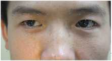
图5为脸部关键点检测示意图。Figure 5 is a schematic diagram of facial key point detection.
具体实施方式detailed description
下面将结合本发明实施例中的附图,对本发明实施例中的技术方案进行清楚、完整地描述,显然,所描述的实施例仅仅是本发明的一部分实施例,而不是全部的实施例。基于本发明中的实施例,本领域普通技术人员在没有作出创造性劳动前提下所获得的所有其他实施例,都属于本发明保护的范围。The following will clearly and completely describe the technical solutions in the embodiments of the present invention with reference to the accompanying drawings in the embodiments of the present invention. Obviously, the described embodiments are only some of the embodiments of the present invention, not all of them. Based on the embodiments of the present invention, all other embodiments obtained by persons of ordinary skill in the art without creative efforts fall within the protection scope of the present invention.
需要说明,本发明实施例中所有方向性指示(诸如上、下、左、右、前、后……)仅用于解释在某一特定姿态(如附图所示)下各部件之间的相对位置关系、运动情况等,如果该特定姿态发生改变时,则该方向性指示也相应地随之改变。It should be noted that all directional indications (such as up, down, left, right, front, back...) in the embodiments of the present invention are only used to explain the relationship between the components in a certain posture (as shown in the accompanying drawings). Relative positional relationship, movement conditions, etc., if the specific posture changes, the directional indication will also change accordingly.
在本发明中,除非另有明确的规定和限定,术语“连接”、“固定”等应做广义理解,例如,“固定”可以是固定连接,也可以是可拆卸连接,或成一体;可以是机械连接,也可以是连接;可以是直接相连,也可以通过中间媒介间接相连,可以是两个元件内部的连通或两个元件的相互作用关系,除非另有明确的限定。对于本领域的普通技术人员而言,可以根据具体情况理解上述术语在本发明中的具体含义。In the present invention, unless otherwise specified and limited, the terms "connection" and "fixation" should be understood in a broad sense, for example, "fixation" can be a fixed connection, a detachable connection, or an integral body; It may be a mechanical connection or a connection; it may be a direct connection or an indirect connection through an intermediary, and it may be an internal communication between two elements or an interaction relationship between two elements, unless otherwise clearly defined. Those of ordinary skill in the art can understand the specific meanings of the above terms in the present invention according to specific situations.
如图所示,本发明提供了一种基于深度学习的斜视识别系统,其包括:As shown in the figure, the present invention provides a squint recognition system based on deep learning, which includes:
两个拍摄装置,用于获取患者的人脸图像,拍摄装置为相机,设置在患者正前方,分别获取各自的人脸图像;Two photographing devices are used to acquire the face image of the patient. The photographing device is a camera, which is set directly in front of the patient, and respectively acquires the respective face images;
坐标转换模型,基于两个拍摄装置的内参矩阵及外参矩阵,用于图像坐标点输入转换为三维坐标输出,其中相机的内参矩阵计算采用业内成熟的网格标定法,借助标准的棋盘网格作为标定板,每个相机拍下若干张各个角度的网格图像,将图像输入到标定算法就可以得到相机的内参和畸变系数。而相机的外参矩阵则通过两个相机同时拍摄同一个标定板画面可以求得,假定相机A和相机B,以相机A的相机原点作为世界坐标系的原点位置,故可以得到相机A的外参矩阵,同时拍摄的同一个标定板画面图像计算得到相机A到相机B的偏移和旋转矩阵,求得的相机A到相机B的偏移和旋转矩阵就是相机B的外参矩阵。至此,相机的标定过程完毕,通过两个相机的内参和外参矩阵系数,并且已知同一个点在两个相机拍摄画面中的位置,可以求得该点在3维空间中的坐标位置;The coordinate transformation model, based on the internal reference matrix and external reference matrix of two shooting devices, is used to convert the input of image coordinate points into three-dimensional coordinate output. The calculation of the internal reference matrix of the camera adopts the mature grid calibration method in the industry, with the help of a standard checkerboard grid As a calibration board, each camera takes several grid images from various angles, and input the images into the calibration algorithm to obtain the internal parameters and distortion coefficients of the camera. The extrinsic parameter matrix of the camera can be obtained by shooting the same calibration board picture with two cameras at the same time. Assume that camera A and camera B take the camera origin of camera A as the origin of the world coordinate system, so the extrinsic parameter matrix of camera A can be obtained The reference matrix, the offset and rotation matrix from camera A to camera B is calculated by taking the same calibration plate image at the same time, and the obtained offset and rotation matrix from camera A to camera B is the extrinsic parameter matrix of camera B. At this point, the calibration process of the camera is completed, and the coordinate position of the point in the 3-dimensional space can be obtained through the internal reference and external reference matrix coefficients of the two cameras, and knowing the position of the same point in the pictures taken by the two cameras;
眼部感兴趣区域提取单元,对拍摄装置获取的人脸图像进行人脸关键点检测,并将人脸图像转换为特征点式图像,并提取包含眼部感兴趣区域的图像;The eye region of interest extraction unit detects the key points of the face on the face image acquired by the shooting device, converts the face image into a feature point image, and extracts an image containing the eye region of interest;
为了减少图像的其他区域对虹膜分割的干扰,我们首先需要定位出人脸区域,再根据人脸的各个关键点的位置定位眼睛所在的感兴趣区域。在这采用了开源数据工具库Dlib来完成人脸的检测对齐和眼部感兴趣区域的提取。Dlib人脸识别技术的主要过程是:首先,通过人脸检测获取目标人脸图像;然后,对目标人脸图像进行特征点检测;接下来,对完成特征点检测的人脸图像进行对齐处理,使人脸图像变化成特征点形式图像,并且将其对齐到基准人脸上,起到摆正和校正人脸的作用。In order to reduce the interference of other areas of the image on iris segmentation, we first need to locate the face area, and then locate the area of interest where the eyes are located according to the positions of each key point of the face. Here, the open source data tool library Dlib is used to complete the detection and alignment of faces and the extraction of eye regions of interest. The main process of Dlib face recognition technology is: first, obtain the target face image through face detection; then, perform feature point detection on the target face image; next, perform alignment processing on the face images that have completed feature point detection, Change the face image into an image in the form of feature points, and align it to the reference face to straighten and correct the face.
在得到了人脸的关键点之后,我们根据眼睛的关键点进一步截取出感兴趣的眼部区域,作为后面进行斜视判定的基础。After obtaining the key points of the face, we further intercepted the eye area of interest according to the key points of the eyes, as the basis for subsequent strabismus judgment.
虹膜分割单元,由基于改进的U-Net的网络结构训练生成,用于输入眼部感兴趣区域的图像,输出虹膜图像;The iris segmentation unit is generated by the network structure training based on the improved U-Net, which is used to input the image of the eye region of interest and output the iris image;
为了计算瞳孔的中心点,需要首先对虹膜进行提取,这里采取了基于语义分割的提取方法。语义分割通过对每个像素进行密集的预测、推断标签来实现细粒度的推理,从而使每个像素都被标记为其封闭对象区域的类别。随着深度学习的复兴和不断发展,目前基于深度卷积神经网络的语义分割模型越来越获得了学术界和工业界的青睐。本申请采用的虹膜语义分割模型也是基于卷积神经网络构建。In order to calculate the center point of the pupil, the iris needs to be extracted first, and the extraction method based on semantic segmentation is adopted here. Semantic segmentation enables fine-grained reasoning by densely predicting, inferring labels for each pixel such that each pixel is labeled with the category of its enclosing object region. With the revival and continuous development of deep learning, the current semantic segmentation model based on deep convolutional neural network is more and more favored by academia and industry. The iris semantic segmentation model used in this application is also constructed based on a convolutional neural network.
深度神经网络模型的精准预测来自于大量的训练,本申请中虹膜分割模型的训练数据采用了英国剑桥大学开源的基于计算机仿真生成的人眼图像nityEyes。该训练数据将人眼区域的3D模型与实时渲染框架相结合,该3D模型源自于高分辨率3D面部扫描,并结合了复杂眼球材料和结构的实时近似,以及用于眼睑动画的解剖学程序几何方法,有很高的相似性且可以无限制的增加数据量,这位模型的训练提供了坚实的基础。The accurate prediction of the deep neural network model comes from a large amount of training. The training data of the iris segmentation model in this application uses nityEyes, an open-source computer simulation-based human eye image generated by the University of Cambridge. The training data combines a 3D model of the human eye region derived from high-resolution 3D facial scans with a real-time rendering framework, combined with a real-time approximation of the complex eye material and structure, and anatomy for eyelid animation The procedural geometry method, which has high similarity and can increase the amount of data without limit, provides a solid foundation for the training of this model.
UnityEyes数据集的每张图片中,都有与之对应的元数据标记。为了下一步的卷积神经网络的训练,将预标注中的虹膜的关键点围起来填充成为掩码图。Each image in the UnityEyes dataset has a corresponding metadata tag. In order to train the convolutional neural network in the next step, the key points of the iris in the pre-marking are surrounded and filled into a mask image.
深度神经网络凭借着其自动学习的能力,近年来在各个领域取得了许多应用成果,U-Net是深度神经网络在医疗图像分割领域广为应用的典型例子。U-Net是Ronneberger等于2015年提出的一种面向生物影像的图像分割网络。在U-Net出现之前,大部分的学者都是采用传统的诸如阈值分割等方法或机器学习结合图像特征来进行分割,但脑、胸腺、角膜内皮细胞等组织的特征繁多,种类不一,采用人工提取特征的手段无疑会带来特征不足、特征偏差等问题。针对这些问题,深度神经网络可以有效的自动提取特征,从而实现更高效率的医疗图像分割。With its automatic learning ability, deep neural network has achieved many application results in various fields in recent years. U-Net is a typical example of the wide application of deep neural network in the field of medical image segmentation. U-Net is an image segmentation network for biological images proposed by Ronneberger et al. in 2015. Before the appearance of U-Net, most scholars used traditional methods such as threshold segmentation or machine learning combined with image features to segment, but the characteristics of brain, thymus, corneal endothelial cells and other tissues are various and different. The method of manual feature extraction will undoubtedly bring about problems such as insufficient features and feature deviations. To address these problems, deep neural networks can effectively and automatically extract features, thereby achieving more efficient medical image segmentation.
本申请的虹膜分割模型的构建采用了基于U-Net的升级结构。传统的U-Net的结构如图2所示。U-Net的网络结构前半部分为特征提取,后半部分是上采样,这种结构又被称为编码器-解码器结构,由于网络的整体结构是一个大些的英文字母U,所以叫做U-Net。左半部分的编码器部分,由两个3x3的卷积层+激活层(ReLU)再加上一个2x2的下采样层(MaxPooling)组成一个下采样的模块;右半部分的解码器部分,由一个上采样的卷积层(去卷积层)+特征拼接(Concat)+两个3x3的卷积层和激活层(ReLU)反复构成。本申请在传统的U-Net结构上,在编码器的最底部连续加入了由不同膨胀系数的膨胀卷积,膨胀卷积能在不缩小特征图分辨率的情况下增加网络模型的感受野,这样一方面可以让每个卷积输出都包含较大范围的信息,减少下采样层保留小目标的可见性,另一方面更大的感受野也能更好把握图像的整体特征,有助于分割的准确率。The construction of the iris segmentation model of the present application adopts the upgrade structure based on U-Net. The structure of traditional U-Net is shown in Figure 2. The first half of U-Net's network structure is feature extraction, and the second half is upsampling. This structure is also called an encoder-decoder structure. Since the overall structure of the network is a larger English letter U, it is called U. -Net. The encoder part of the left half consists of two 3x3 convolutional layers + activation layers (ReLU) plus a 2x2 downsampling layer (MaxPooling) to form a downsampling module; the decoder part of the right half consists of An upsampled convolutional layer (deconvolutional layer) + feature splicing (Concat) + two 3x3 convolutional layers and activation layers (ReLU) are repeatedly formed. Based on the traditional U-Net structure, this application continuously adds dilated convolutions with different expansion coefficients at the bottom of the encoder. The dilated convolutions can increase the receptive field of the network model without reducing the resolution of the feature map. In this way, on the one hand, each convolution output can contain a larger range of information, and the downsampling layer can be reduced to preserve the visibility of small targets. On the other hand, a larger receptive field can also better grasp the overall characteristics of the image, which helps Segmentation accuracy.
中心点检测单元,由基于改进的ResNet网络结构训练生成,用于输入虹膜图像,输出瞳孔中心点和反射光中心点的坐标;The center point detection unit is generated by training based on the improved ResNet network structure, which is used to input the iris image and output the coordinates of the center point of the pupil and the center point of the reflected light;
斜视的自动判定需要知道瞳孔的中心点和光源反射中心点的位置。本发明采用了专门设计的检测瞳孔的中心点和光源反射中心点的卷积神经网络。整个卷积神经网络的设计采用了改进的ResNet作为骨干网,去掉了ResNet中的所有池化层,在残差模块的残差连接(Skip Connection)中引入空洞卷积(Dilated/Atrous Convolution)作为池化层的替代,并从网络的不同深度引入多尺度的特征图进行多尺度融合以保留全局特征和局部特征。ResNet的残差连接可以在训练时加速收敛,本网络中包含12个残差模块,网络的输入为512x512大小的RGB三通道图像,首先对图像进行归一化(式1)后得到输入张量,The automatic determination of strabismus needs to know the position of the center point of the pupil and the center point of light source reflection. The present invention uses a specially designed convolutional neural network to detect the central point of the pupil and the central point of light source reflection. The design of the entire convolutional neural network uses the improved ResNet as the backbone network, removes all pooling layers in ResNet, and introduces Dilated/Atrous Convolution in the residual connection (Skip Connection) of the residual module as The pooling layer is replaced, and multi-scale feature maps are introduced from different depths of the network for multi-scale fusion to preserve global features and local features. The residual connection of ResNet can speed up the convergence during training. This network contains 12 residual modules. The input of the network is an RGB three-channel image of 512x512 size. First, the image is normalized (Formula 1) to obtain the input tensor ,

输入张量经过一个卷积层处理后进入由12个残差模块串接的深度网络,每个残差模块由2个卷积层构成,每个卷积层后由批量归一化(Batch Normalization)和激活层修正线性单元(ReLU)激活。其中第9个和第10个残差模块采用系数为3的空洞卷积,第8个和第11个残差模块采用系数为2的空洞卷积,其他残差模块的残差连接采用普通的卷积层处理;网络的第3个和第6个残差模块后对特征图进行了系数为2的下采样以得到不同尺度的特征,并把第5个、第9个和最后一个残差模块的输出引出,用反卷积进行放大因子分别为2、4和4的上采样,使从3个分支引出的特征图与输入图像大小一致后进行通道连接实现多尺度融合,连接后的特征图再经过一个卷积层得到瞳孔的特征图和反射点的特征图。其中瞳孔特征图用于计算瞳孔分割的损失函数,反射点特征图用于计算反射点定位的损失函数,两者损失函数有所不同,瞳孔分割采用了基于Dice系数的损失函数DSC(式2),而反射点预测图则是类似于骨骼关键点检测,运用了MSE作为损失函数(式3),整个卷积网络的损失函数由瞳孔分割的损失和反射点定位的损失经过一定的比例alpha协调相加得到(式4)。整个网络通过反向传播算法进行不断迭代优化网络的预测精度。After being processed by a convolutional layer, the input tensor enters a deep network connected in series by 12 residual modules. Each residual module consists of 2 convolutional layers, and each convolutional layer is followed by batch normalization (Batch Normalization). ) and activation layer Rectified Linear Unit (ReLU) activations. The 9th and 10th residual modules use hole convolution with a coefficient of 3, the 8th and 11th residual modules use hole convolution with a coefficient of 2, and the residual connections of other residual modules use ordinary Convolutional layer processing; after the third and sixth residual modules of the network, the feature map is down-sampled with a coefficient of 2 to obtain features of different scales, and the fifth, ninth and last residuals The output of the module is derived, and the upsampling with deconvolution factors of 2, 4 and 4 is used to make the feature maps derived from the three branches consistent with the size of the input image, and then the channel connection is performed to achieve multi-scale fusion. The connected features The graph then passes through a convolutional layer to obtain the feature map of the pupil and the feature map of the reflection point. Among them, the pupil feature map is used to calculate the loss function of pupil segmentation, and the reflection point feature map is used to calculate the loss function of reflection point positioning. The loss functions of the two are different. The pupil segmentation uses the loss function DSC based on the Dice coefficient (Formula 2) , while the reflection point prediction map is similar to bone key point detection, using MSE as the loss function (Equation 3), the loss function of the entire convolutional network is coordinated by a certain ratio of alpha between the loss of pupil segmentation and the loss of reflection point positioning Adding to get (Equation 4). The entire network continuously iteratively optimizes the prediction accuracy of the network through the backpropagation algorithm.


Loss=DSC+αMSE (式4)Loss=DSC+αMSE (Formula 4)
输出单元,将中心点检测单元获取的两个拍摄装置拍摄的人脸图像的瞳孔中心点和反射光中心点,利用坐标转换模型获得瞳孔中心点和反射光中心点在三维空间中的坐标,并利用该坐标计算眼睛的瞳孔和反射点的距离和方向,并输出该结果。The output unit is used to obtain the pupil center point and the reflected light center point of the face images taken by the two shooting devices obtained by the center point detection unit, and use the coordinate transformation model to obtain the coordinates of the pupil center point and the reflected light center point in three-dimensional space, and Use this coordinate to calculate the distance and direction of the pupil of the eye and the reflection point, and output the result.
通过卷积神经网络计算分别得到两个相机视角下的瞳孔中心点和反射光中心点,通过相机内外参数计算得到这两个点的3维坐标。得到3维的坐标点之后,通过分别计算眼睛的瞳孔和反射点的距离和方向,以此来判断出是否存在斜视以及斜视的种类。若瞳孔中心基本和反射中心一致并距离小于1cm,则判断为无斜视。若瞳孔中心在反射光点的内侧并且水平距离大于1cm,则表示该眼存在内斜视。若瞳孔中心在反射光点的外侧并且水平距离大于1cm,则表示该眼存在外斜视。若瞳孔中心在反射光点的上侧并且垂直距离大于1cm,则表示该眼存在上斜视。若瞳孔中心在反射光点的下侧并且垂直距离大于1cm,则表示该眼存在下斜视。并且通过水平和垂直方向的结合,可以判定出上外,上内,下外,下内这4种组合斜视。除此之外,通过求解瞳孔中心与反射光点的水平距离和虹膜中心到虹膜边缘距离的比值关系可以得到患者斜视的严重程度,若瞳孔中心与反射光点的水平距离和虹膜中心到虹膜边缘距离的比值小于1/3则视为轻微斜视,大于1/3视为严重斜视。The center point of the pupil and the center point of the reflected light under the perspective of the two cameras are obtained through the calculation of the convolutional neural network, and the 3D coordinates of these two points are obtained through the calculation of the internal and external parameters of the camera. After the 3D coordinate points are obtained, the distance and direction of the pupil of the eye and the reflection point are calculated respectively to determine whether there is strabismus and the type of strabismus. If the pupil center is basically consistent with the reflection center and the distance is less than 1cm, it is judged as non-strabismus. If the pupil center is inside the reflected light point and the horizontal distance is greater than 1cm, it means that the eye has esotropia. If the pupil center is outside the reflected light point and the horizontal distance is greater than 1 cm, it means that the eye has exotropia. If the center of the pupil is on the upper side of the reflected light point and the vertical distance is greater than 1cm, it means that the eye has upward strabismus. If the center of the pupil is on the lower side of the reflected light point and the vertical distance is greater than 1 cm, it means that the eye has hypotropia. And through the combination of horizontal and vertical directions, it is possible to determine the 4 combinations of upper-outside, upper-inside, lower-outside, and lower-inside strabismus. In addition, the severity of strabismus can be obtained by solving the ratio relationship between the horizontal distance between the pupil center and the reflected light point and the distance from the iris center to the iris edge. If the horizontal distance between the pupil center and the reflected light point and the distance from the iris center to the iris edge A distance ratio of less than 1/3 is considered mild strabismus, and greater than 1/3 is considered severe strabismus.
实施例不应视为对本发明的限制,但任何基于本发明的精神所作的改进,都应在本发明的保护范围之内。The embodiments should not be regarded as limiting the present invention, but any improvement based on the spirit of the present invention should be within the protection scope of the present invention.
Claims (9)
Translated fromChinese

Priority Applications (1)
| Application Number | Priority Date | Filing Date | Title |
|---|---|---|---|
| CN202210984726.8ACN115424319A (en) | 2022-08-16 | 2022-08-16 | Strabismus recognition system based on deep learning |
Applications Claiming Priority (1)
| Application Number | Priority Date | Filing Date | Title |
|---|---|---|---|
| CN202210984726.8ACN115424319A (en) | 2022-08-16 | 2022-08-16 | Strabismus recognition system based on deep learning |
Publications (1)
| Publication Number | Publication Date |
|---|---|
| CN115424319Atrue CN115424319A (en) | 2022-12-02 |
Family
ID=84199332
Family Applications (1)
| Application Number | Title | Priority Date | Filing Date |
|---|---|---|---|
| CN202210984726.8APendingCN115424319A (en) | 2022-08-16 | 2022-08-16 | Strabismus recognition system based on deep learning |
Country Status (1)
| Country | Link |
|---|---|
| CN (1) | CN115424319A (en) |
Cited By (1)
| Publication number | Priority date | Publication date | Assignee | Title |
|---|---|---|---|---|
| CN116385806A (en)* | 2023-05-29 | 2023-07-04 | 四川大学华西医院 | Classification method, system, device and storage medium for eye image strabismus types |
Citations (9)
| Publication number | Priority date | Publication date | Assignee | Title |
|---|---|---|---|---|
| CN102961117A (en)* | 2012-11-06 | 2013-03-13 | 温州医学院 | Strabismus diagnosis device based on mobile platform |
| CN105094337A (en)* | 2015-08-19 | 2015-11-25 | 华南理工大学 | Three-dimensional gaze estimation method based on irises and pupils |
| CN110575132A (en)* | 2019-07-25 | 2019-12-17 | 北京爱诺斯科技有限公司 | Method for calculating degree of strabismus based on eccentric photography |
| CN111528786A (en)* | 2020-04-24 | 2020-08-14 | 杭州电子科技大学 | System and method for detecting head position of strabismus compensation |
| CN112989939A (en)* | 2021-02-08 | 2021-06-18 | 佛山青藤信息科技有限公司 | Strabismus detection system based on vision |
| CN113065398A (en)* | 2021-03-04 | 2021-07-02 | 武汉大学 | Method and system for monitoring nystagmus |
| CN113143199A (en)* | 2021-04-30 | 2021-07-23 | 上海青研科技有限公司 | Strabismus inspection apparatus |
| CN114511894A (en)* | 2020-10-28 | 2022-05-17 | 北京京东方光电科技有限公司 | Acquiring system and method of pupil center coordinates |
| CN114820513A (en)* | 2022-04-25 | 2022-07-29 | 深圳市迪佳医疗智能科技有限公司 | Vision detection method |
- 2022
- 2022-08-16CNCN202210984726.8Apatent/CN115424319A/enactivePending
Patent Citations (9)
| Publication number | Priority date | Publication date | Assignee | Title |
|---|---|---|---|---|
| CN102961117A (en)* | 2012-11-06 | 2013-03-13 | 温州医学院 | Strabismus diagnosis device based on mobile platform |
| CN105094337A (en)* | 2015-08-19 | 2015-11-25 | 华南理工大学 | Three-dimensional gaze estimation method based on irises and pupils |
| CN110575132A (en)* | 2019-07-25 | 2019-12-17 | 北京爱诺斯科技有限公司 | Method for calculating degree of strabismus based on eccentric photography |
| CN111528786A (en)* | 2020-04-24 | 2020-08-14 | 杭州电子科技大学 | System and method for detecting head position of strabismus compensation |
| CN114511894A (en)* | 2020-10-28 | 2022-05-17 | 北京京东方光电科技有限公司 | Acquiring system and method of pupil center coordinates |
| CN112989939A (en)* | 2021-02-08 | 2021-06-18 | 佛山青藤信息科技有限公司 | Strabismus detection system based on vision |
| CN113065398A (en)* | 2021-03-04 | 2021-07-02 | 武汉大学 | Method and system for monitoring nystagmus |
| CN113143199A (en)* | 2021-04-30 | 2021-07-23 | 上海青研科技有限公司 | Strabismus inspection apparatus |
| CN114820513A (en)* | 2022-04-25 | 2022-07-29 | 深圳市迪佳医疗智能科技有限公司 | Vision detection method |
Cited By (2)
| Publication number | Priority date | Publication date | Assignee | Title |
|---|---|---|---|---|
| CN116385806A (en)* | 2023-05-29 | 2023-07-04 | 四川大学华西医院 | Classification method, system, device and storage medium for eye image strabismus types |
| CN116385806B (en)* | 2023-05-29 | 2023-09-08 | 四川大学华西医院 | Method, system, equipment and storage medium for classifying strabismus type of eye image |
Similar Documents
| Publication | Publication Date | Title |
|---|---|---|
| CN112022346B (en) | A control method of an automatic venipuncture and identification integrated robot | |
| CN112308932B (en) | Gaze detection method, device, equipment and storage medium | |
| CN109858540B (en) | Medical image recognition system and method based on multi-mode fusion | |
| CN111402254B (en) | CT image lung nodule high-performance automatic detection method and device | |
| CN113469942B (en) | CT image lesion detection method | |
| CN110736747B (en) | Method and system for positioning under cell liquid-based smear mirror | |
| CN112750531A (en) | Automatic inspection system, method, equipment and medium for traditional Chinese medicine | |
| CN110164550B (en) | A method for auxiliary diagnosis of congenital heart disease based on multi-view synergistic relationship | |
| CN110729045A (en) | A Tongue Image Segmentation Method Based on Context-Aware Residual Networks | |
| US11723630B2 (en) | Method for positioning key features of a lens based on ocular B-mode ultrasound images | |
| CN110561399A (en) | Auxiliary shooting device for dyskinesia condition analysis, control method and device | |
| CN119650104B (en) | Interactive system for improving doctor-patient communication experience based on augmented reality | |
| CN114332278B (en) | A deep learning-based OCTA image motion correction method | |
| CN118982492B (en) | A jitter distortion correction image processing system for ophthalmic OCT | |
| CN118512278A (en) | An AI modeling method and device for tooth 3D printing | |
| CN116152073B (en) | Improved multi-scale fundus image stitching method based on Loftr algorithm | |
| CN112669959A (en) | Vitiligo state of illness automatic assessment method based on image | |
| CN117974740A (en) | Acupoint positioning method and robot based on aggregation type window self-attention mechanism | |
| CN112102327A (en) | Image processing method and device and computer readable storage medium | |
| CN112651400A (en) | Stereoscopic endoscope auxiliary detection method, system, device and storage medium | |
| CN112686865A (en) | 3D view auxiliary detection method, system, device and storage medium | |
| CN115424319A (en) | Strabismus recognition system based on deep learning | |
| CN114283178A (en) | Image registration method and device, computer equipment and storage medium | |
| CN110378333A (en) | Method for positioning fovea centralis of SD-OCT image | |
| CN114332858A (en) | Focus detection method and device and focus detection model acquisition method |
Legal Events
| Date | Code | Title | Description |
|---|---|---|---|
| PB01 | Publication | ||
| PB01 | Publication | ||
| SE01 | Entry into force of request for substantive examination | ||
| SE01 | Entry into force of request for substantive examination |