CN115362253A - Method for enhancing T cells using Venetork - Google Patents
Method for enhancing T cells using VenetorkDownload PDFInfo
- Publication number
- CN115362253A CN115362253ACN202180026678.5ACN202180026678ACN115362253ACN 115362253 ACN115362253 ACN 115362253ACN 202180026678 ACN202180026678 ACN 202180026678ACN 115362253 ACN115362253 ACN 115362253A
- Authority
- CN
- China
- Prior art keywords
- cells
- cell
- dnt
- enhanced
- venetoclax
- Prior art date
- Legal status (The legal status is an assumption and is not a legal conclusion. Google has not performed a legal analysis and makes no representation as to the accuracy of the status listed.)
- Pending
Links
- 210000001744T-lymphocyteAnatomy0.000titleclaimsabstractdescription267
- 238000000034methodMethods0.000titleclaimsabstractdescription65
- 230000002708enhancing effectEffects0.000titleclaimsdescription8
- 206010028980NeoplasmDiseases0.000claimsabstractdescription64
- 201000011510cancerDiseases0.000claimsabstractdescription50
- 230000001404mediated effectEffects0.000claimsabstractdescription15
- 230000000259anti-tumor effectEffects0.000claimsabstractdescription14
- 230000010782T cell mediated cytotoxicityEffects0.000claimsabstractdescription12
- 208000031261Acute myeloid leukaemiaDiseases0.000claimsdescription87
- 208000033776Myeloid Acute LeukemiaDiseases0.000claimsdescription83
- 239000003642reactive oxygen metaboliteSubstances0.000claimsdescription55
- 230000001965increasing effectEffects0.000claimsdescription47
- 239000012636effectorSubstances0.000claimsdescription34
- 230000014509gene expressionEffects0.000claimsdescription30
- 239000000203mixtureSubstances0.000claimsdescription24
- 230000001413cellular effectEffects0.000claimsdescription21
- 101001109501Homo sapiens NKG2-D type II integral membrane proteinProteins0.000claimsdescription17
- 102100022680NKG2-D type II integral membrane proteinHuman genes0.000claimsdescription17
- 208000032839leukemiaDiseases0.000claimsdescription16
- 101001057504Homo sapiens Interferon-stimulated gene 20 kDa proteinProteins0.000claimsdescription15
- 101001055144Homo sapiens Interleukin-2 receptor subunit alphaProteins0.000claimsdescription15
- 102100026878Interleukin-2 receptor subunit alphaHuman genes0.000claimsdescription15
- 102100038077CD226 antigenHuman genes0.000claimsdescription14
- 101000884298Homo sapiens CD226 antigenProteins0.000claimsdescription14
- 102100025137Early activation antigen CD69Human genes0.000claimsdescription13
- 101000934374Homo sapiens Early activation antigen CD69Proteins0.000claimsdescription13
- 230000035755proliferationEffects0.000claimsdescription13
- 101000588302Homo sapiens Nuclear factor erythroid 2-related factor 2Proteins0.000claimsdescription12
- 102100031701Nuclear factor erythroid 2-related factor 2Human genes0.000claimsdescription12
- 230000004614tumor growthEffects0.000claimsdescription12
- 239000008194pharmaceutical compositionSubstances0.000claimsdescription8
- 210000003515double negative t cellAnatomy0.000claimsdescription7
- 239000003937drug carrierSubstances0.000claimsdescription5
- 230000001225therapeutic effectEffects0.000claimsdescription5
- 210000004475gamma-delta t lymphocyteAnatomy0.000claimsdescription2
- LQBVNQSMGBZMKD-UHFFFAOYSA-NvenetoclaxChemical compoundC=1C=C(Cl)C=CC=1C=1CC(C)(C)CCC=1CN(CC1)CCN1C(C=C1OC=2C=C3C=CNC3=NC=2)=CC=C1C(=O)NS(=O)(=O)C(C=C1[N+]([O-])=O)=CC=C1NCC1CCOCC1LQBVNQSMGBZMKD-UHFFFAOYSA-N0.000abstractdescription152
- 229960001183venetoclaxDrugs0.000abstractdescription152
- 238000011282treatmentMethods0.000abstractdescription42
- 210000004027cellAnatomy0.000description201
- 230000003013cytotoxicityEffects0.000description36
- 231100000135cytotoxicityToxicity0.000description36
- 230000000694effectsEffects0.000description20
- 238000000338in vitroMethods0.000description18
- 239000003814drugSubstances0.000description17
- 241000699670Mus sp.Species0.000description16
- 230000000719anti-leukaemic effectEffects0.000description13
- 229940079593drugDrugs0.000description13
- NMUSYJAQQFHJEW-KVTDHHQDSA-N5-azacytidineChemical compoundO=C1N=C(N)N=CN1[C@H]1[C@H](O)[C@H](O)[C@@H](CO)O1NMUSYJAQQFHJEW-KVTDHHQDSA-N0.000description11
- 210000001185bone marrowAnatomy0.000description11
- 238000002474experimental methodMethods0.000description11
- 238000001727in vivoMethods0.000description11
- 229960002756azacitidineDrugs0.000description10
- 238000000684flow cytometryMethods0.000description9
- 230000002147killing effectEffects0.000description9
- PWKSKIMOESPYIA-BYPYZUCNSA-NL-N-acetyl-CysteineChemical compoundCC(=O)N[C@@H](CS)C(O)=OPWKSKIMOESPYIA-BYPYZUCNSA-N0.000description8
- 229960004308acetylcysteineDrugs0.000description8
- 238000011467adoptive cell therapyMethods0.000description8
- 201000004428dysembryoplastic neuroepithelial tumorDiseases0.000description8
- 230000002829reductive effectEffects0.000description8
- 239000000523sampleSubstances0.000description8
- 230000006044T cell activationEffects0.000description7
- 208000037265diseases, disorders, signs and symptomsDiseases0.000description7
- 231100000673dose–response relationshipToxicity0.000description7
- 230000027721electron transport chainEffects0.000description7
- 238000001802infusionMethods0.000description7
- 230000035806respiratory chainEffects0.000description7
- 230000035899viabilityEffects0.000description7
- 102100021569Apoptosis regulator Bcl-2Human genes0.000description6
- 101000971171Homo sapiens Apoptosis regulator Bcl-2Proteins0.000description6
- 102100036011T-cell surface glycoprotein CD4Human genes0.000description6
- 230000000735allogeneic effectEffects0.000description6
- 150000001875compoundsChemical class0.000description6
- 201000010099diseaseDiseases0.000description6
- 230000034659glycolysisEffects0.000description6
- 238000002347injectionMethods0.000description6
- 239000007924injectionSubstances0.000description6
- 238000004519manufacturing processMethods0.000description6
- 239000003981vehicleSubstances0.000description6
- UHDGCWIWMRVCDJ-CCXZUQQUSA-NCytarabineChemical compoundO=C1N=C(N)C=CN1[C@H]1[C@@H](O)[C@H](O)[C@@H](CO)O1UHDGCWIWMRVCDJ-CCXZUQQUSA-N0.000description5
- 241000699666Mus <mouse, genus>Species0.000description5
- 230000004913activationEffects0.000description5
- 229960000684cytarabineDrugs0.000description5
- 230000001472cytotoxic effectEffects0.000description5
- 230000007246mechanismEffects0.000description5
- 210000003071memory t lymphocyteAnatomy0.000description5
- 230000002438mitochondrial effectEffects0.000description5
- 210000003819peripheral blood mononuclear cellAnatomy0.000description5
- 230000009467reductionEffects0.000description5
- 210000004881tumor cellAnatomy0.000description5
- STQGQHZAVUOBTE-UHFFFAOYSA-N7-Cyan-hept-2t-en-4,6-diinsaeureNatural productsC1=2C(O)=C3C(=O)C=4C(OC)=CC=CC=4C(=O)C3=C(O)C=2CC(O)(C(C)=O)CC1OC1CC(N)C(O)C(C)O1STQGQHZAVUOBTE-UHFFFAOYSA-N0.000description4
- 229930182536AntimycinNatural products0.000description4
- 102100026596Bcl-2-like protein 1Human genes0.000description4
- 229940123711Bcl2 inhibitorDrugs0.000description4
- 102100039441Cytochrome b-c1 complex subunit 2, mitochondrialHuman genes0.000description4
- 102100030878Cytochrome c oxidase subunit 1Human genes0.000description4
- 101000746756Homo sapiens Cytochrome b-c1 complex subunit 2, mitochondrialProteins0.000description4
- 101000919849Homo sapiens Cytochrome c oxidase subunit 1Proteins0.000description4
- 101000934338Homo sapiens Myeloid cell surface antigen CD33Proteins0.000description4
- 102100025243Myeloid cell surface antigen CD33Human genes0.000description4
- 108010090931Proto-Oncogene Proteins c-bcl-2Proteins0.000description4
- 102000013535Proto-Oncogene Proteins c-bcl-2Human genes0.000description4
- 230000002159abnormal effectEffects0.000description4
- CQIUKKVOEOPUDV-IYSWYEEDSA-NantimycinChemical compoundOC1=C(C(O)=O)C(=O)C(C)=C2[C@H](C)[C@@H](C)OC=C21CQIUKKVOEOPUDV-IYSWYEEDSA-N0.000description4
- 230000030833cell deathEffects0.000description4
- STQGQHZAVUOBTE-VGBVRHCVSA-NdaunorubicinChemical compoundO([C@H]1C[C@@](O)(CC=2C(O)=C3C(=O)C=4C=CC=C(C=4C(=O)C3=C(O)C=21)OC)C(C)=O)[C@H]1C[C@H](N)[C@H](O)[C@H](C)O1STQGQHZAVUOBTE-VGBVRHCVSA-N0.000description4
- 229960000975daunorubicinDrugs0.000description4
- 230000001976improved effectEffects0.000description4
- 210000000265leukocyteAnatomy0.000description4
- 239000013610patient sampleSubstances0.000description4
- 108090000623proteins and genesProteins0.000description4
- 238000010186stainingMethods0.000description4
- 210000001519tissueAnatomy0.000description4
- HPLNQCPCUACXLM-PGUFJCEWSA-NABT-737Chemical compoundC([C@@H](CCN(C)C)NC=1C(=CC(=CC=1)S(=O)(=O)NC(=O)C=1C=CC(=CC=1)N1CCN(CC=2C(=CC=CC=2)C=2C=CC(Cl)=CC=2)CC1)[N+]([O-])=O)SC1=CC=CC=C1HPLNQCPCUACXLM-PGUFJCEWSA-N0.000description3
- 239000012664BCL-2-inhibitorSubstances0.000description3
- 101001023513Homo sapiens NADH dehydrogenase [ubiquinone] 1 alpha subcomplex subunit 9, mitochondrialProteins0.000description3
- 101000738771Homo sapiens Receptor-type tyrosine-protein phosphatase CProteins0.000description3
- 241001465754MetazoaSpecies0.000description3
- 102100035383NADH dehydrogenase [ubiquinone] 1 alpha subcomplex subunit 9, mitochondrialHuman genes0.000description3
- 102100037422Receptor-type tyrosine-protein phosphatase CHuman genes0.000description3
- 210000000662T-lymphocyte subsetAnatomy0.000description3
- 230000006907apoptotic processEffects0.000description3
- 238000003556assayMethods0.000description3
- 210000004369bloodAnatomy0.000description3
- 239000008280bloodSubstances0.000description3
- 210000000601blood cellAnatomy0.000description3
- 238000002659cell therapyMethods0.000description3
- 230000005757colony formationEffects0.000description3
- 230000001419dependent effectEffects0.000description3
- 230000006870functionEffects0.000description3
- 239000000499gelSubstances0.000description3
- 230000012010growthEffects0.000description3
- 238000003119immunoblotMethods0.000description3
- 230000005764inhibitory processEffects0.000description3
- 230000036284oxygen consumptionEffects0.000description3
- 102000004169proteins and genesHuman genes0.000description3
- 230000005855radiationEffects0.000description3
- 102000004121Annexin A5Human genes0.000description2
- 108090000672Annexin A5Proteins0.000description2
- 208000010839B-cell chronic lymphocytic leukemiaDiseases0.000description2
- 210000001266CD8-positive T-lymphocyteAnatomy0.000description2
- 231100000023Cell-mediated cytotoxicityToxicity0.000description2
- 206010057250Cell-mediated cytotoxicityDiseases0.000description2
- 102000001398GranzymeHuman genes0.000description2
- 108060005986GranzymeProteins0.000description2
- WZUVPPKBWHMQCE-UHFFFAOYSA-NHaematoxylinChemical compoundC12=CC(O)=C(O)C=C2CC2(O)C1C1=CC=C(O)C(O)=C1OC2WZUVPPKBWHMQCE-UHFFFAOYSA-N0.000description2
- 102100031573Hematopoietic progenitor cell antigen CD34Human genes0.000description2
- 241000282412HomoSpecies0.000description2
- 101000777663Homo sapiens Hematopoietic progenitor cell antigen CD34Proteins0.000description2
- 241000124008MammaliaSpecies0.000description2
- 238000011579SCID mouse modelMethods0.000description2
- 230000002424anti-apoptotic effectEffects0.000description2
- 238000002617apheresisMethods0.000description2
- 230000010261cell growthEffects0.000description2
- 230000003833cell viabilityEffects0.000description2
- 230000005890cell-mediated cytotoxicityEffects0.000description2
- 238000002512chemotherapyMethods0.000description2
- 230000001447compensatory effectEffects0.000description2
- 230000003247decreasing effectEffects0.000description2
- 230000000779depleting effectEffects0.000description2
- 238000007877drug screeningMethods0.000description2
- 238000007878drug screening assayMethods0.000description2
- 238000009472formulationMethods0.000description2
- 239000001963growth mediumSubstances0.000description2
- 238000009169immunotherapyMethods0.000description2
- 230000004048modificationEffects0.000description2
- 238000012986modificationMethods0.000description2
- 230000010627oxidative phosphorylationEffects0.000description2
- 238000002360preparation methodMethods0.000description2
- 238000004393prognosisMethods0.000description2
- 239000012268protein inhibitorSubstances0.000description2
- 229940121649protein inhibitorDrugs0.000description2
- 239000002516radical scavengerSubstances0.000description2
- 230000001105regulatory effectEffects0.000description2
- 230000004044responseEffects0.000description2
- 230000035945sensitivityEffects0.000description2
- 230000008093supporting effectEffects0.000description2
- 238000002560therapeutic procedureMethods0.000description2
- 230000001988toxicityEffects0.000description2
- 231100000419toxicityToxicity0.000description2
- 238000001262western blotMethods0.000description2
- CVCLJVVBHYOXDC-IAZSKANUSA-N(2z)-2-[(5z)-5-[(3,5-dimethyl-1h-pyrrol-2-yl)methylidene]-4-methoxypyrrol-2-ylidene]indoleChemical compoundCOC1=C\C(=C/2N=C3C=CC=CC3=C\2)N\C1=C/C=1NC(C)=CC=1CCVCLJVVBHYOXDC-IAZSKANUSA-N0.000description1
- UHDGCWIWMRVCDJ-UHFFFAOYSA-N1-beta-D-Xylofuranosyl-NH-CytosineNatural productsO=C1N=C(N)C=CN1C1C(O)C(O)C(CO)O1UHDGCWIWMRVCDJ-UHFFFAOYSA-N0.000description1
- NMUSYJAQQFHJEW-UHFFFAOYSA-N5-AzacytidineNatural productsO=C1N=C(N)N=CN1C1C(O)C(O)C(CO)O1NMUSYJAQQFHJEW-UHFFFAOYSA-N0.000description1
- XAUDJQYHKZQPEU-KVQBGUIXSA-N5-aza-2'-deoxycytidineChemical compoundO=C1N=C(N)N=CN1[C@@H]1O[C@H](CO)[C@@H](O)C1XAUDJQYHKZQPEU-KVQBGUIXSA-N0.000description1
- 208000004736B-Cell LeukemiaDiseases0.000description1
- 229940122035Bcl-XL inhibitorDrugs0.000description1
- 229940123606Bcl-w inhibitorDrugs0.000description1
- 102000017420CD3 protein, epsilon/gamma/delta subunitHuman genes0.000description1
- 108050005493CD3 protein, epsilon/gamma/delta subunitProteins0.000description1
- UHDGCWIWMRVCDJ-PSQAKQOGSA-NCytidineNatural productsO=C1N=C(N)C=CN1[C@@H]1[C@@H](O)[C@@H](O)[C@H](CO)O1UHDGCWIWMRVCDJ-PSQAKQOGSA-N0.000description1
- 102000015782Electron Transport Complex IIIHuman genes0.000description1
- 108010024882Electron Transport Complex IIIProteins0.000description1
- 101000979735Homo sapiens NADH dehydrogenase [ubiquinone] 1 beta subcomplex subunit 8, mitochondrialProteins0.000description1
- 101000685323Homo sapiens Succinate dehydrogenase [ubiquinone] flavoprotein subunit, mitochondrialProteins0.000description1
- 208000031422Lymphocytic Chronic B-Cell LeukemiaDiseases0.000description1
- 208000028018Lymphocytic leukaemiaDiseases0.000description1
- 201000003793Myelodysplastic syndromeDiseases0.000description1
- 102100024975NADH dehydrogenase [ubiquinone] 1 beta subcomplex subunit 8, mitochondrialHuman genes0.000description1
- 238000011529RT qPCRMethods0.000description1
- 238000000692Student's t-testMethods0.000description1
- 102100023155Succinate dehydrogenase [ubiquinone] flavoprotein subunit, mitochondrialHuman genes0.000description1
- 101710119418Superoxide dismutase [Mn]Proteins0.000description1
- 101710202572Superoxide dismutase [Mn], mitochondrialProteins0.000description1
- 102100032891Superoxide dismutase [Mn], mitochondrialHuman genes0.000description1
- 230000033540T cell apoptotic processEffects0.000description1
- 230000005867T cell responseEffects0.000description1
- 108091023040Transcription factorProteins0.000description1
- 102000040945Transcription factorHuman genes0.000description1
- 102000004243TubulinHuman genes0.000description1
- 108090000704TubulinProteins0.000description1
- 230000003213activating effectEffects0.000description1
- 238000000540analysis of varianceMethods0.000description1
- 230000001093anti-cancerEffects0.000description1
- 230000000118anti-neoplastic effectEffects0.000description1
- 239000003963antioxidant agentSubstances0.000description1
- 230000003078antioxidant effectEffects0.000description1
- 238000013459approachMethods0.000description1
- 230000002238attenuated effectEffects0.000description1
- 230000009286beneficial effectEffects0.000description1
- 230000008901benefitEffects0.000description1
- 230000015572biosynthetic processEffects0.000description1
- 210000002798bone marrow cellAnatomy0.000description1
- 238000010322bone marrow transplantationMethods0.000description1
- 238000002619cancer immunotherapyMethods0.000description1
- 238000004113cell cultureMethods0.000description1
- 239000006143cell culture mediumSubstances0.000description1
- 230000024245cell differentiationEffects0.000description1
- 230000032823cell divisionEffects0.000description1
- 230000003915cell functionEffects0.000description1
- 230000006037cell lysisEffects0.000description1
- 230000004663cell proliferationEffects0.000description1
- 230000008859changeEffects0.000description1
- 239000003795chemical substances by applicationSubstances0.000description1
- 208000032852chronic lymphocytic leukemiaDiseases0.000description1
- 238000011278co-treatmentMethods0.000description1
- 238000002648combination therapyMethods0.000description1
- UHDGCWIWMRVCDJ-ZAKLUEHWSA-NcytidineChemical compoundO=C1N=C(N)C=CN1[C@H]1[C@H](O)[C@@H](O)[C@H](CO)O1UHDGCWIWMRVCDJ-ZAKLUEHWSA-N0.000description1
- 230000001086cytosolic effectEffects0.000description1
- 239000000824cytostatic agentSubstances0.000description1
- 230000001085cytostatic effectEffects0.000description1
- 231100000433cytotoxicToxicity0.000description1
- 238000002784cytotoxicity assayMethods0.000description1
- 231100000263cytotoxicity testToxicity0.000description1
- 229960003603decitabineDrugs0.000description1
- 208000035475disorderDiseases0.000description1
- 210000003162effector t lymphocyteAnatomy0.000description1
- YQGOJNYOYNNSMM-UHFFFAOYSA-NeosinChemical compound[Na+].OC(=O)C1=CC=CC=C1C1=C2C=C(Br)C(=O)C(Br)=C2OC2=C(Br)C(O)=C(Br)C=C21YQGOJNYOYNNSMM-UHFFFAOYSA-N0.000description1
- 230000002414glycolytic effectEffects0.000description1
- 210000002443helper t lymphocyteAnatomy0.000description1
- 208000019691hematopoietic and lymphoid cell neoplasmDiseases0.000description1
- 230000007062hydrolysisEffects0.000description1
- 238000006460hydrolysis reactionMethods0.000description1
- 230000036039immunityEffects0.000description1
- 230000003308immunostimulating effectEffects0.000description1
- 238000011534incubationMethods0.000description1
- 239000000411inducerSubstances0.000description1
- 230000006698inductionEffects0.000description1
- 230000001939inductive effectEffects0.000description1
- 239000004615ingredientSubstances0.000description1
- 239000003112inhibitorSubstances0.000description1
- 230000002401inhibitory effectEffects0.000description1
- 238000011081inoculationMethods0.000description1
- 238000001990intravenous administrationMethods0.000description1
- 238000010253intravenous injectionMethods0.000description1
- 231100000518lethalToxicity0.000description1
- 230000001665lethal effectEffects0.000description1
- 230000000670limiting effectEffects0.000description1
- 210000004185liverAnatomy0.000description1
- 230000004807localizationEffects0.000description1
- 210000004072lungAnatomy0.000description1
- 208000003747lymphoid leukemiaDiseases0.000description1
- 239000002609mediumSubstances0.000description1
- 201000001441melanomaDiseases0.000description1
- 230000037353metabolic pathwayEffects0.000description1
- 210000003470mitochondriaAnatomy0.000description1
- 208000025113myeloid leukemiaDiseases0.000description1
- 230000002071myeloproliferative effectEffects0.000description1
- 210000000581natural killer T-cellAnatomy0.000description1
- 230000017074necrotic cell deathEffects0.000description1
- 230000030648nucleus localizationEffects0.000description1
- 229950006584obatoclaxDrugs0.000description1
- 238000001543one-way ANOVAMethods0.000description1
- 210000000056organAnatomy0.000description1
- 230000037361pathwayEffects0.000description1
- 210000005259peripheral bloodAnatomy0.000description1
- 239000011886peripheral bloodSubstances0.000description1
- 230000002688persistenceEffects0.000description1
- 230000001766physiological effectEffects0.000description1
- 238000002203pretreatmentMethods0.000description1
- 230000000750progressive effectEffects0.000description1
- 230000002062proliferating effectEffects0.000description1
- 239000002718pyrimidine nucleosideSubstances0.000description1
- 108020003175receptorsProteins0.000description1
- 102000005962receptorsHuman genes0.000description1
- 230000000284resting effectEffects0.000description1
- 230000019491signal transductionEffects0.000description1
- 238000009097single-agent therapyMethods0.000description1
- 238000002415sodium dodecyl sulfate polyacrylamide gel electrophoresisMethods0.000description1
- 238000007619statistical methodMethods0.000description1
- 238000007920subcutaneous administrationMethods0.000description1
- 239000000126substanceSubstances0.000description1
- 208000011580syndromic diseaseDiseases0.000description1
- 230000002195synergetic effectEffects0.000description1
- 230000000451tissue damageEffects0.000description1
- 231100000827tissue damageToxicity0.000description1
- 238000002054transplantationMethods0.000description1
- 230000005740tumor formationEffects0.000description1
- 238000013414tumor xenograft modelMethods0.000description1
- 230000004222uncontrolled growthEffects0.000description1
- 230000003827upregulationEffects0.000description1
- 210000003462veinAnatomy0.000description1
Images


Classifications
- A—HUMAN NECESSITIES
- A61—MEDICAL OR VETERINARY SCIENCE; HYGIENE
- A61K—PREPARATIONS FOR MEDICAL, DENTAL OR TOILETRY PURPOSES
- A61K31/00—Medicinal preparations containing organic active ingredients
- A61K31/63—Compounds containing para-N-benzenesulfonyl-N-groups, e.g. sulfanilamide, p-nitrobenzenesulfonyl hydrazide
- A61K31/635—Compounds containing para-N-benzenesulfonyl-N-groups, e.g. sulfanilamide, p-nitrobenzenesulfonyl hydrazide having a heterocyclic ring, e.g. sulfadiazine
- A—HUMAN NECESSITIES
- A61—MEDICAL OR VETERINARY SCIENCE; HYGIENE
- A61K—PREPARATIONS FOR MEDICAL, DENTAL OR TOILETRY PURPOSES
- A61K40/00—Cellular immunotherapy
- A61K40/10—Cellular immunotherapy characterised by the cell type used
- A61K40/11—T-cells, e.g. tumour infiltrating lymphocytes [TIL] or regulatory T [Treg] cells; Lymphokine-activated killer [LAK] cells
- A—HUMAN NECESSITIES
- A61—MEDICAL OR VETERINARY SCIENCE; HYGIENE
- A61K—PREPARATIONS FOR MEDICAL, DENTAL OR TOILETRY PURPOSES
- A61K40/00—Cellular immunotherapy
- A61K40/40—Cellular immunotherapy characterised by antigens that are targeted or presented by cells of the immune system
- A61K40/41—Vertebrate antigens
- A61K40/42—Cancer antigens
- A61K40/428—Undefined tumor antigens, e.g. tumor lysate or antigens targeted by cells isolated from tumor
- A—HUMAN NECESSITIES
- A61—MEDICAL OR VETERINARY SCIENCE; HYGIENE
- A61P—SPECIFIC THERAPEUTIC ACTIVITY OF CHEMICAL COMPOUNDS OR MEDICINAL PREPARATIONS
- A61P35/00—Antineoplastic agents
- A—HUMAN NECESSITIES
- A61—MEDICAL OR VETERINARY SCIENCE; HYGIENE
- A61P—SPECIFIC THERAPEUTIC ACTIVITY OF CHEMICAL COMPOUNDS OR MEDICINAL PREPARATIONS
- A61P35/00—Antineoplastic agents
- A61P35/02—Antineoplastic agents specific for leukemia
- C—CHEMISTRY; METALLURGY
- C12—BIOCHEMISTRY; BEER; SPIRITS; WINE; VINEGAR; MICROBIOLOGY; ENZYMOLOGY; MUTATION OR GENETIC ENGINEERING
- C12N—MICROORGANISMS OR ENZYMES; COMPOSITIONS THEREOF; PROPAGATING, PRESERVING, OR MAINTAINING MICROORGANISMS; MUTATION OR GENETIC ENGINEERING; CULTURE MEDIA
- C12N5/00—Undifferentiated human, animal or plant cells, e.g. cell lines; Tissues; Cultivation or maintenance thereof; Culture media therefor
- C12N5/06—Animal cells or tissues; Human cells or tissues
- C12N5/0602—Vertebrate cells
- C12N5/0634—Cells from the blood or the immune system
- C12N5/0636—T lymphocytes
- A—HUMAN NECESSITIES
- A61—MEDICAL OR VETERINARY SCIENCE; HYGIENE
- A61K—PREPARATIONS FOR MEDICAL, DENTAL OR TOILETRY PURPOSES
- A61K2239/00—Indexing codes associated with cellular immunotherapy of group A61K40/00
- A61K2239/31—Indexing codes associated with cellular immunotherapy of group A61K40/00 characterized by the route of administration
- A—HUMAN NECESSITIES
- A61—MEDICAL OR VETERINARY SCIENCE; HYGIENE
- A61K—PREPARATIONS FOR MEDICAL, DENTAL OR TOILETRY PURPOSES
- A61K2239/00—Indexing codes associated with cellular immunotherapy of group A61K40/00
- A61K2239/38—Indexing codes associated with cellular immunotherapy of group A61K40/00 characterised by the dose, timing or administration schedule
- A—HUMAN NECESSITIES
- A61—MEDICAL OR VETERINARY SCIENCE; HYGIENE
- A61K—PREPARATIONS FOR MEDICAL, DENTAL OR TOILETRY PURPOSES
- A61K2239/00—Indexing codes associated with cellular immunotherapy of group A61K40/00
- A61K2239/46—Indexing codes associated with cellular immunotherapy of group A61K40/00 characterised by the cancer treated
- A61K2239/48—Blood cells, e.g. leukemia or lymphoma
- C—CHEMISTRY; METALLURGY
- C12—BIOCHEMISTRY; BEER; SPIRITS; WINE; VINEGAR; MICROBIOLOGY; ENZYMOLOGY; MUTATION OR GENETIC ENGINEERING
- C12N—MICROORGANISMS OR ENZYMES; COMPOSITIONS THEREOF; PROPAGATING, PRESERVING, OR MAINTAINING MICROORGANISMS; MUTATION OR GENETIC ENGINEERING; CULTURE MEDIA
- C12N2501/00—Active agents used in cell culture processes, e.g. differentation
- C12N2501/40—Regulators of development
- C12N2501/48—Regulators of apoptosis
Landscapes
- Health & Medical Sciences (AREA)
- Life Sciences & Earth Sciences (AREA)
- General Health & Medical Sciences (AREA)
- Chemical & Material Sciences (AREA)
- Animal Behavior & Ethology (AREA)
- Public Health (AREA)
- Veterinary Medicine (AREA)
- Engineering & Computer Science (AREA)
- Organic Chemistry (AREA)
- Epidemiology (AREA)
- Biomedical Technology (AREA)
- Medicinal Chemistry (AREA)
- Pharmacology & Pharmacy (AREA)
- Immunology (AREA)
- Biotechnology (AREA)
- Zoology (AREA)
- Bioinformatics & Cheminformatics (AREA)
- Wood Science & Technology (AREA)
- Genetics & Genomics (AREA)
- Chemical Kinetics & Catalysis (AREA)
- Nuclear Medicine, Radiotherapy & Molecular Imaging (AREA)
- General Chemical & Material Sciences (AREA)
- Hematology (AREA)
- Cell Biology (AREA)
- Biochemistry (AREA)
- General Engineering & Computer Science (AREA)
- Microbiology (AREA)
- Oncology (AREA)
- Medicines Containing Material From Animals Or Micro-Organisms (AREA)
- Micro-Organisms Or Cultivation Processes Thereof (AREA)
- Developmental Biology & Embryology (AREA)
- Virology (AREA)
- Pharmaceuticals Containing Other Organic And Inorganic Compounds (AREA)
Abstract
Translated fromChinese
Description
Translated fromChinese相关申请related application
本申请要求于2020年2月7日提交的美国临时申请号62/971,534的的优先权,其全部内容通过引用并入本申请。This application claims priority to U.S. Provisional Application No. 62/971,534, filed February 7, 2020, the entire contents of which are incorporated herein by reference.
技术领域technical field
本公开涉及用于治疗癌症的免疫疗法,更具体地,涉及使用维奈托克(Venetoclax)增强T细胞以治疗癌症。The present disclosure relates to immunotherapy for the treatment of cancer, and more particularly, to the use of Venetoclax to enhance T cells for the treatment of cancer.
背景技术Background technique
过继性细胞疗法(ACT)显著改善了某些癌症类型(如B细胞白血病和黑色素瘤(1、2))患者的预后。虽然这些成功案例证明了ACT的效力,但在其它的癌症类型上尚未获得类似的临床成效。举例而言,急性髓性白血病(AML)在患者内部和患者之间均表现为高度异质性的疾病,尽管人们正在研究各种ACT方法以试图改善罹患这种高致死性疾病患者的预后,但针对AML的ACT尚未取得临床上的成功(3)。因此,仍然需要用于治疗癌症的改进的ACT疗法。Adoptive cell therapy (ACT) has significantly improved outcomes for patients with certain cancer types such as B-cell leukemia and melanoma (1, 2). While these success stories demonstrate the efficacy of ACT, similar clinical outcomes have not been achieved in other cancer types. For example, acute myeloid leukemia (AML) is a highly heterogeneous disease both within and between patients, although various ACT approaches are being investigated in an attempt to improve outcomes for patients with this highly lethal disease, However, ACT for AML has not been clinically successful (3). Therefore, there remains a need for improved ACT therapies for the treatment of cancer.
一种形式的ACT是使用特殊的T细胞亚群,其被定义为CD4-和CD8-双阴性T(DNT)细胞。在临床前模型中,不同于许多其他T细胞疗法,从健康志愿者体扩增的同种异体DNT细胞的输注不会对正常细胞引起同种异体反应性,并且对受体的免疫排斥产生抗性,这支持其被用作“现货”ACT的潜力(3-6)。然而,DNT细胞的抗癌作用并不完全(5、6),因此,能够进一步增强DNT细胞抗肿瘤活性的方法可让患者收获更好的预后。One form of ACT uses a specific subset of T cells, defined as CD4- and CD8- double-negative T (DNT) cells. In preclinical models, unlike many other T cell therapies, infusion of allogeneic DNT cells expanded from healthy volunteers did not induce alloreactivity to normal cells and immune rejection of the recipient produced resistance, which supports its potential to be used as a "off-the-shelf" ACT (3-6). However, the anticancer effect of DNT cells is not complete (5, 6), therefore, methods that can further enhance the antitumor activity of DNT cells can lead to better prognosis for patients.
发明概述Summary of the invention
一方面,已确定维奈托克通过增加T细胞介导的细胞毒性来增强T细胞治疗效果。In one aspect, it has been established that venetoclax enhances T cell therapy by increasing T cell mediated cytotoxicity.
用被批准用于各种临床用途的269种药物的库中的化合物对T细胞进行预处理,随后,将经化合物处理的细胞用作针对人AML细胞系的效应物。令人惊讶的是,Bcl-2抑制剂维奈托克最大程度地增加了T细胞的细胞毒性。(图1)。T cells were pretreated with compounds from a library of 269 drugs approved for various clinical uses, and compound-treated cells were subsequently used as effectors against human AML cell lines. Surprisingly, the Bcl-2 inhibitor venetoclax maximized T cell cytotoxicity. (figure 1).
如实施例中所述,经维奈托克预处理的T细胞在体外对AML表现出增强的T细胞介导的细胞毒性。此外,经维奈托克处理的T细胞在异种移植模型中表现出增加的抗肿瘤活性。维奈托克增强了T细胞的细胞毒性,而其他Bcl-2家族蛋白抑制剂却不能。与未经处理的T细胞相比,经维奈托克处理的T细胞具有T细胞活化标志物CD25和CD69的更高表达,以及效应分子NKG2D和DNAM-1的更高表达。与未经处理的细胞相比,经维奈托克处理的T细胞还表现出增加水平的活性氧类(ROS)。还证明治疗相关浓度的维奈托克增加T细胞效应功能,而不会降低T细胞活力。此外,从接受维奈托克的患者体内分离出的T细胞显示ROS水平增加。As described in the Examples, T cells pretreated with venetoclax exhibited enhanced T cell-mediated cytotoxicity against AML in vitro. Furthermore, venetoclax-treated T cells exhibited increased antitumor activity in xenograft models. Venetoclax enhanced T cell cytotoxicity while other Bcl-2 family protein inhibitors did not. Compared with untreated T cells, venetoclax-treated T cells had higher expression of T cell activation markers CD25 and CD69, and higher expression of effector molecules NKG2D and DNAM-1. T cells treated with venetoclax also exhibited increased levels of reactive oxygen species (ROS) compared to untreated cells. It was also demonstrated that therapeutically relevant concentrations of venetoclax increased T cell effector function without reducing T cell viability. In addition, T cells isolated from patients receiving venetoclax showed increased levels of ROS.
因此,在一个实施方案中,提供了一种增强T细胞疗效的方法,所述方法包括使T细胞与维奈托克接触以生成功能增强型T细胞。Accordingly, in one embodiment, there is provided a method of enhancing the efficacy of T cells, the method comprising contacting the T cells with venetoclax to generate function-enhanced T cells.
如本文所述使用维奈托克对T细胞进行预处理生成增强型T细胞,所述增强型T细胞具有使所述细胞更有效地治疗癌症的若干特征。例如,在一个实施方案中,使用维奈托克增加T细胞介导的细胞毒性。在一个实施方案中,使用维奈托克增加T细胞介导的抗肿瘤活性。在一个实施方案中,使所述T细胞与维奈托克接触增加效应记忆状态的T细胞的相对比例。Pretreatment of T cells with venetoclax as described herein generates enhanced T cells with several characteristics that make the cells more effective in treating cancer. For example, in one embodiment, T cell mediated cytotoxicity is increased with venetoclax. In one embodiment, the use of venetoclax increases T cell-mediated anti-tumor activity. In one embodiment, contacting the T cells with venetoclax increases the relative proportion of T cells that are effector memory states.
在一个实施方案中,所述T细胞为常规T细胞(CD4+或CD8+)。在一个实施方案中,所述T细胞为非常规T细胞,例如双阴性T细胞(CD4-、CD8-)。In one embodiment, the T cells are conventional T cells (CD4+ or CD8+ ). In one embodiment, the T cells are unconventional T cells, such as double negative T cells (CD4− , CD8− ).
在一个实施方案中,所述方法包括使所述T细胞与浓度为至少50nM的维奈托克接触。在一个实施方案中,所述方法包括使所述T细胞与浓度为至少100nM、至少200nM、至少300nM或至少400nM的维奈托克接触,可选地,所述方法包括使所述T细胞与浓度为约100nM至1约μM的维奈托克接触。In one embodiment, the method comprises contacting the T cells with venetoclax at a concentration of at least 50 nM. In one embodiment, the method comprises contacting the T cells with venetoclax at a concentration of at least 100 nM, at least 200 nM, at least 300 nM or at least 400 nM, optionally the method comprises contacting the T cells with Exposure to venetoclax at a concentration of about 100 nM to about 1 μM.
在一个实施方案中,所述方法包括使所述T细胞与维奈托克接触至少约30分钟、至少约45分钟或至少约60分钟。在一个实施方案中,所述方法包括使所述T细胞与维奈托克接触至少1小时、至少1.5小时、至少2小时或至少4小时。在一个实施方案中,所述方法包括使所述T细胞与维奈托克接触至少6小时、至少8小时或至少12小时,可选地,约1小时至约7日。在一个实施方案中,所述方法包括使所述T细胞与维奈托克接触至少1小时且少于约14日、10日、9日、8日、7日、6日或5日。在一个实施方案中,所述方法包括使所述T细胞与维奈托克接触一段时间,相对于未接触维奈托克的对照细胞,所述时间足以增加T细胞对CD25、CD69、NKG2D、DNAM-1和NRF2中一种或更多种的表达水平。在一个实施方案中,所述方法包括使所述T细胞与维奈托克接触一段时间,相对于未接触维奈托克的对照细胞,所述时间足以增加细胞活性氧类(ROS)的水平。在一个实施方案中,所述T细胞是体外的。在另一个实施方案中,所述T细胞是体内或离体的。In one embodiment, the method comprises contacting the T cells with venetoclax for at least about 30 minutes, at least about 45 minutes, or at least about 60 minutes. In one embodiment, the method comprises contacting the T cells with venetoclax for at least 1 hour, at least 1.5 hours, at least 2 hours, or at least 4 hours. In one embodiment, the method comprises contacting the T cells with venetoclax for at least 6 hours, at least 8 hours, or at least 12 hours, alternatively, for about 1 hour to about 7 days. In one embodiment, the method comprises contacting the T cells with venetoclax for at least 1 hour and for less than about 14 days, 10 days, 9 days, 8 days, 7 days, 6 days, or 5 days. In one embodiment, the method comprises contacting the T cells with venetoclax for a period of time sufficient to increase the T cell response to CD25, CD69, NKG2D, Expression levels of one or more of DNAM-1 and NRF2. In one embodiment, the method comprises contacting the T cells with venetoclax for a period of time sufficient to increase the level of cellular reactive oxygen species (ROS) relative to control cells not exposed to venetoclax . In one embodiment, the T cells are in vitro. In another embodiment, the T cells are in vivo or ex vivo.
本文所述的增强型T细胞易于与未经维奈托克预处理的T细胞区分。在一个实施方案中,使所述T细胞与维奈托克接触增加CD25、CD69、NKG2D、DNAM-1和NRF2中的一种或更多种的表达水平。在一个实施方案中,使所述T细胞与维奈托克接触增加细胞活性氧类(ROS)的水平。The enhanced T cells described herein are readily distinguishable from T cells not pretreated with venetoclax. In one embodiment, contacting the T cell with venetoclax increases the expression level of one or more of CD25, CD69, NKG2D, DNAM-1 and NRF2. In one embodiment, contacting the T cells with venetoclax increases cellular reactive oxygen species (ROS) levels.
本文还提供了采用本文所述方法生成的增强型T细胞群。在一个实施方案中,与未接触维奈托克的对照T细胞相比,所述增强型T细胞呈现出CD25、CD69、NKG2D、DNAM-1和NRF2中的一种或更多种的增加的表达水平。在一个实施方案中,与未接触维奈托克的对照T细胞相比,所述增强型T细胞呈现出增加水平的细胞活性氧类(ROS)。Also provided herein are enhanced T cell populations generated using the methods described herein. In one embodiment, the enhanced T cells exhibit an increased expression of one or more of CD25, CD69, NKG2D, DNAM-1, and NRF2 compared to control T cells not exposed to venetoclax. The expression level. In one embodiment, the enhanced T cells exhibit increased levels of cellular reactive oxygen species (ROS) compared to control T cells not exposed to venetoclax.
在一个实施方案中,与未接触维奈托克的对照T细胞群中效应记忆状态的T细胞与幼稚状态的T细胞的比例相比,所述增强型T细胞群中效应记忆状态的T细胞与幼稚状态的T细胞的比例增加。In one embodiment, the T cells in the effector memory state in the enhanced T cell population are compared to the ratio of T cells in the effector memory state to T cells in the naive state in the control T cell population not exposed to venetoclax. The ratio of T cells to the naive state is increased.
在一个实施方案中,提供了一种组合物,所述组合物包含T细胞和维奈托克。还提供了一种药物组合物,所述药物组合物包含如本文所述用维奈托克处理的增强型T细胞。In one embodiment, a composition comprising T cells and venetoclax is provided. Also provided is a pharmaceutical composition comprising enhanced T cells treated with venetoclax as described herein.
还提供了如本文所述的增强型T细胞、组合物和/或T细胞与维奈托克的组合用于治疗有此需要的受试者的癌症的用途。在一个实施方案中,提供了一种治疗有此需要的受试者的癌症的方法,所述方法包括向所述受试者施用本文所述的增强型T细胞、组合物和/或T细胞与维奈托克的组合。在一个实施方案中,所述癌症是白血病,可选地,是急性髓性白血病(AML)。Also provided is the use of enhanced T cells, compositions and/or combinations of T cells and venetoclax as described herein for the treatment of cancer in a subject in need thereof. In one embodiment, there is provided a method of treating cancer in a subject in need thereof comprising administering to said subject an enhanced T cell, composition and/or T cell described herein Combination with venetoclax. In one embodiment, the cancer is leukemia, optionally acute myeloid leukemia (AML).
根据以下详细描述,本公开的其他特征和优点将变得显而易见。然而,应当理解,在示出本公开的优选实施方案的同时,详细描述和具体实施例仅用于示例说明,因为在本公开的精神和范围内做出的各种改变和修改通过该详细描述对于本领域技术人员而言将变得显而易见。Other features and advantages of the present disclosure will become apparent from the following detailed description. It should be understood, however, that the detailed description and the specific examples, while indicating the preferred embodiment of the present disclosure, are given by way of illustration only, since various changes and modifications may be made within the spirit and scope of the present disclosure. It will become apparent to those skilled in the art.
附图说明Description of drawings
现在将结合附图描述本公开的一个或更多个实施方案,在附图中:One or more embodiments of the disclosure will now be described with reference to the accompanying drawings, in which:
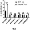
图1.药物筛选试验确定维奈托克是增强T细胞抗AML的细胞毒性的首选药物。药物筛选试验示意图,进行该试验以鉴定可与DNT细胞联合使用以产生协同抗肿瘤活性的临床批准药物。用269种不同的临床批准药物以400nM处理DNT细胞过夜。随后,洗涤经化合物处理的细胞,然后与AML细胞一起培养两小时。点状图示出了相对于未经处理的DNT细胞,DNT细胞介导的针对AML细胞的细胞毒性程度的变化。Figure 1. Drug screening assays identified venetoclax as the drug of choice for enhancing T cell cytotoxicity against AML. Schematic of the drug screening assay performed to identify clinically approved drugs that can be used in combination with DNT cells to produce synergistic antitumor activity. DNT cells were treated overnight at 400 nM with 269 different clinically approved drugs. Subsequently, the compound-treated cells were washed and then incubated with AML cells for two hours. Dot plots show changes in the degree of DNT cell-mediated cytotoxicity against AML cells relative to untreated DNT cells.
图2.维奈托克在体外增强T细胞介导的针对AML的细胞毒性。(A)为了确认药物筛选的结果,使用未经处理的DNT细胞或经不同浓度的Ven(50nM、100nM、200nM、400nM)预处理过夜的DNT细胞针对AML细胞系OCI-AML2、OCI-AML3和KG1a进行体外杀伤试验。数据代表四个生物学重复。(B)使用经400nM Ven预处理的DNT细胞作为效应物针对原发性AML患者样本(n=17)进行体外细胞毒性试验。(C)为了测定经Ven处理或未经Ven处理的DNT细胞针对白血病起始细胞的活性,在基于甲基纤维素的集落形成试验中,以每毫升103个细胞接种未经处理的AML或用未经处理的DNT细胞或经Ven处理的DNT细胞处理的AML,并于10日后测定形成的集落数。该实验使用OCI-AML2和KG1a以及患者样本140372、100857、110162和141065进行。(D)从DNT细胞移除药物后,经维奈托克处理后所增加的、针对三种AML细胞系OCI-AML2、OCI-AML3和KG1a的效应活性保持至少4日。该实验使用来自两个不同供体(UPN119和UPN38)的DNT细胞进行。(E)AML对DNT细胞的敏感性与经Ven处理后DNT细胞介导的细胞毒性增加程度之间的相关性。(F)从11个供体扩增的DNT未经处理或用400nM维奈托克处理18小时。随后,将其与OCI-AML2以1:1、2:1或4:1的DNT:AML比例进行培养,并通过膜联蛋白V染色和流式细胞术测定AML细胞的活力。每一配对符号表示来自单个供体的DNT。Figure 2. Venetoclax enhances T cell-mediated cytotoxicity against AML in vitro. (A) In order to confirm the results of drug screening, the AML cell lines OCI-AML2, OCI-AML3 and KG1a was subjected to an in vitro killing assay. Data are representative of four biological replicates. (B) In vitro cytotoxicity assays were performed on primary AML patient samples (n=17) using DNT cells pretreated with 400 nM Ven as effectors. (C) To determine the activity of Ven-treated or Ven-untreated DNT cells against leukemia-initiating cells,untreated AML or AML was treated with untreated DNT cells or Ven-treated DNT cells, and the number of colonies formed was measured 10 days later. The experiment was performed using OCI-AML2 and KG1a and
图3.在异种移植模型中,用Ven预处理DNT细胞增加其抗肿瘤活性。为了确定经Ven预处理的DNT细胞是否在异种移植模型中诱导更高的抗白血病活性,在肿瘤大小达到100mm3(箭头所示)时,皮下移植2x106OCI-AML2细胞的NOD/SCID小鼠通过静脉输注PBS(●)、2x107未经处理的DNT细胞(■)或2x107经Ven处理的DNT细胞(▲)。监测肿瘤体积直到PBS处理组达到人道终点(A),并且在白血病接种后第20日测量肿瘤重量(B)。所示结果代表了使用来自三个不同供体的DNT细胞进行的三个独立实验。(C)用PBS、DNT细胞或VenDNT细胞处理全身输注KG1a的NSG小鼠。比较各组间KG1a的骨髓移植情况。与PBS和DNT细胞处理组相比,经VenDNT处理的小鼠显示明显更低的KG1a移植水平,进一步支持了VenDNT细胞即使针对以另外方式具有抗性的那些亦能表现出优异的抗白血病活性。(D)将原发性AML细胞(ID:130607),其未经处理或用DNT或经Ven处理的DNT以2:1的DNT:AML比例处理2小时,经股骨内注入NOD/SCID小鼠中(每只小鼠1.6x106个细胞;n=每组6只)。注射后6周,通过流式细胞术测定每组的骨髓中AML移植(人CD45+CD33+细胞)的百分比。(E)向接受亚致死辐射的NSG小鼠静脉注射原发性AML细胞(n=4;2–5x106/小鼠)。两周后,通过三次输注(媒介物对照或每次输注1.5–2x107个DNT细胞或经Ven处理的DNT细胞)处理小鼠,每次间隔3-4日。AML注射后5周,通过流式细胞术测定原发性AML细胞(人CD45低CD33+,表达或不表达CD34)的骨髓移植情况。(左)用CD45和CD33染色的各组的BM细胞的代表性等高线图。(右)使用四种不同的原发性AML患者样本进行的患者来源的异种移植实验的汇总结果。横杠表示归一化到媒介物对照组的BM AML移植水平的平均值,每个符号表示单独的小鼠,误差条表示SD。数据表示相对于PBS组,骨髓白血病水平的平均值±SEM下降。采用学生t检验或单因素方差分析(one-wayANOVA)进行统计。*p<0.05;**p<0.01;***p<0.001;****p<0.0001。Figure 3. Pretreatment of DNT cells with Ven increases their antitumor activity in a xenograft model. To determine whether Ven-pretreated DNT cells induced higher anti-leukemic activity in a xenograft model, NOD/SCID mice were subcutaneously implanted with2x106 OCI-AML2 cells when the tumor size reached 100mm (arrowhead) PBS (•),2x107 untreated DNT cells (■) or2x107 Ven-treated DNT cells (▲) were infused intravenously. Tumor volume was monitored until the PBS-treated group reached the humane endpoint (A), and tumor weight was measured on
图4(A).维奈托克增强CD4+或CD8+常规T细胞的抗白血病活性。未经处理或经不同浓度Ven(25nM、50nM、100nM、200nM或400nM)处理的离体扩增的Tconv细胞用作针对AML细胞系(OCI-AML2、OCI-AML3和KG1a)的效应细胞。所示结果代表四个生物学重复。图4(B).维奈托克快速、直接增加T细胞针对AML的细胞毒性。未经处理或用维奈托克(100nM和400nM)分别处理4h、18h和3日的DNT(上图)和Tconv细胞(下图)。随后,测定其对OCI-AML2的细胞毒性。数据表示来自四个不同供体T细胞的结果的平均值±SEM。图4(C).未经处理或用维奈托克(100nM或400nM)处理4小时的DNT和Tconv细胞。随后,测定其活力。数据表示来自四个不同供体T细胞的结果的平均值±SEM。Figure 4(A). Venetoclax enhances the anti-leukemic activity of CD4+ or CD8+ conventional T cells. Ex vivo expanded Tconv cells untreated or treated with different concentrations of Ven (25nM, 50nM, 100nM, 200nM or 400nM) were used as effector cells against AML cell lines (OCI-AML2, OCI-AML3 and KG1a). Results shown are representative of four biological replicates. Figure 4(B). Venetoclax rapidly and directly increases T cell cytotoxicity against AML. DNT (top panel) and Tconv cells (bottom panel) were untreated or treated with venetoclax (100nM and 400nM) for 4h, 18h and 3 days, respectively. Subsequently, its cytotoxicity against OCI-AML2 was determined. Data represent mean ± SEM of results from four different donor T cells. Figure 4(C). DNT and Tconv cells untreated or treated with venetoclax (100 nM or 400 nM) for 4 hours. Subsequently, its viability was measured. Data represent mean ± SEM of results from four different donor T cells.
图5.维奈托克增强DNT细胞的抗白血病活性,而奥巴克拉(Obatoclax)和ABT-737则不能。(A)DNT细胞分别用不同浓度的奥巴克拉、ABT-737或维奈托克预处理过夜,然后,用作针对OCI-AML2的效应细胞。(B)结果示出了与未经处理的DNT诱导的杀伤程度相比,DNT介导的细胞毒性的变化率。(C)通过蛋白印迹测定Bcl-xL和Bcl-2在由三个供体(UPN38、UPN108和UPN134)离体扩增的DNT细胞以及AML细胞系OCI-AML2、TEX、NB4和K562上的表达。采用微管蛋白作为上样对照。Figure 5. Venetoclax enhances the anti-leukemic activity of DNT cells while Obatoclax and ABT-737 do not. (A) DNT cells were pretreated overnight with different concentrations of obacla, ABT-737 or venetoclax, and then used as effector cells against OCI-AML2. (B) Results showing the rate of change in DNT-mediated cytotoxicity compared to the degree of killing induced by untreated DNT. (C) The expression of Bcl-xL and Bcl-2 in DNT cells ex vivo expanded from three donors (UPN38, UPN108 and UPN134) and AML cell lines OCI-AML2, TEX, NB4 and K562 were determined by Western blot . Tubulin was used as a loading control.
图6.Ven增加DNT细胞上活化标志物和效应分子的表达。离体扩增的DNT细胞未经处理或用400nM Ven处理,并针对T细胞(A)活化标志物CD25和CD69以及(B)效应分子(NKG2D和DNAM-1)的表达进行染色。每对点代表Ven处理前后来自同一供体的DNT细胞。该实验使用来自四个(A)或六个(B)不同供体的DNT细胞进行。(C)用不同浓度的Ven处理的DNT细胞中颗粒酶B的表达。所示结果代表两个生物学重复。(D)在经Ven处理的CD8+T细胞上也观察到CD25、NKG2D和DNAM-1表达的剂量依赖性增加。Figure 6. Ven increases the expression of activation markers and effector molecules on DNT cells. Ex vivo expanded DNT cells were untreated or treated with 400 nM Ven and stained for expression of T cell (A) activation markers CD25 and CD69 and (B) effector molecules (NKG2D and DNAM-1). Each pair of points represents DNT cells from the same donor before and after Ven treatment. The experiments were performed using DNT cells from four (A) or six (B) different donors. (C) Expression of granzyme B in DNT cells treated with different concentrations of Ven. Results shown are representative of two biological replicates. (D) A dose-dependent increase in the expression of CD25, NKG2D, and DNAM-1 was also observed on Ven-treated CD8+ T cells.
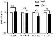
图7.Ven增加DNT细胞中的细胞ROS水平并增强其细胞毒活性。(A)通过CellROXTM染色检测的、经不同浓度Ven处理的DNT细胞(左)或CD8+T细胞(右)的细胞ROS水平。(B)(左)经qPCR测定的、由细胞ROS水平调节的转录因子Nrf2的相对表达。(右)经400nM Ven处理或未经400nM Ven处理的DNT的细胞质和细胞核部分的Nrf2蛋白质免疫印迹试验,旨在确定Nrf2蛋白的位置。使用来自三个不同供体(UPN38、UPN108和UPN134)的DNT生成数据。(C)为了确定经Ven处理的DNT中ROS水平增加的功能相关性,在不同浓度的ROS清除剂N-乙酰半胱氨酸(NAC)的存在下,经400nM Ven处理的DNT中的ROS水平,并在体外杀伤试验中将这些细胞用作针对AML的效应细胞。所示结果代表了三个独立实验。(D)为了确定经Ven处理的DNT中ROS的产生来源,对未经处理的DNT或经400nM处理的DNT进行非变性凝胶和免疫印迹试验,以检测电子传递链超级复合物亚基的组分(NDUFA9、UQCRC2和MTCO1)。所示结果代表了使用来源于两个不同供体的DNT进行的三个独立实验。(e和f)对于DNT细胞(E)和CD8+Tconv细胞(F),Ven增加了效应记忆阶段的细胞比例,同时降低了中央型记忆T细胞的频率。(G)Ven对DNT细胞的糖酵解、糖酵解能力和基础耗氧率无显著影响。(H-K)DNT(H和I)或Tconv细胞(J和K)分别用0nM、100nM或400nM的维奈托克处理4小时、18小时和2日。细胞用CellROX(H和J)或MitoSOX(I和K)染色。通过流式细胞术测量细胞或线粒体(mt)ROS的MFI。数据表示来自四个不同供体T细胞的结果的平均值±SEM。(L)用含或不含2mM NAC的400nM维奈托克处理DNT18小时。流式直方图示出了经流式细胞术测量的细胞ROS水平。经流式细胞术测量CD25和CD69的MFI。实验重复三次进行,所示数据代表使用来自两个供体的DNT进行的两个独立实验。(M)用400nM维奈托克处理DNT细胞18小时。处理后,分离线粒体,通过SDS-PAGE凝胶法和使用抗NDUFB8(复合物I)、SDHA(复合物II)、UQCRC2(复合物III)、MTCO1(复合物IV)的抗体的免疫印迹法测量呼吸链复合物亚基的水平。Figure 7. Ven increases cellular ROS levels and enhances their cytotoxic activity in DNT cells. (A) Cellular ROS levels of DNT cells (left) or CD8+ T cells (right) treated with different concentrations of Ven, detected by CellROXTM staining. (B) (Left) Relative expression of transcription factor Nrf2 regulated by cellular ROS levels as determined by qPCR. (Right) Nrf2 western blot of cytoplasmic and nuclear fractions of DNT treated with or without 400 nM Ven to determine the localization of Nrf2 protein. Data were generated using DNTs from three different donors (UPN38, UPN108 and UPN134). (C) To determine the functional relevance of increased ROS levels in Ven-treated DNT, ROS levels in 400 nM Ven-treated DNT in the presence of different concentrations of the ROS scavenger N-acetylcysteine (NAC) , and used these cells as effector cells against AML in in vitro killing assays. Results shown are representative of three independent experiments. (D) To determine the source of ROS generation in Ven-treated DNT, non-denaturing gel and immunoblot assays were performed on untreated DNT or 400 nM-treated DNT to detect the subunit group of the electron transport chain supercomplex points (NDUFA9, UQCRC2 and MTCO1). Results shown are representative of three independent experiments performed using DNT derived from two different donors. (e and f) For DNT cells (E) and CD8+ Tconv cells (F), Ven increased the proportion of cells in the effector memory phase while decreasing the frequency of central memory T cells. (G) Ven had no significant effect on glycolysis, glycolysis capacity and basal oxygen consumption rate of DNT cells. (HK) DNT (H and I) or Tconv cells (J and K) were treated with OnM, 100 nM or 400 nM venetoclax for 4 hours, 18 hours and 2 days, respectively. Cells were stained with CellROX (H and J) or MitoSOX (I and K). MFI of cellular or mitochondrial (mt) ROS was measured by flow cytometry. Data represent mean ± SEM of results from four different donor T cells. (L) DNTs were treated with 400 nM venetoclax with or without 2 mM NAC for 18 hours. Flow histograms showing cellular ROS levels measured by flow cytometry. MFI of CD25 and CD69 was measured by flow cytometry. Experiments were performed in triplicate and data shown are representative of two independent experiments using DNT from two donors. (M) DNT cells were treated with 400 nM venetoclax for 18 hours. After treatment, mitochondria were isolated and measured by SDS-PAGE gel method and immunoblotting using antibodies against NDUFB8 (complex I), SDHA (complex II), UQCRC2 (complex III), MTCO1 (complex IV) Levels of respiratory chain complex subunits.
图8.在接受Ven+Aza处理的患者中,与细胞毒活性相关的T细胞亚群的比例有所增加。分别于Ven+Aza处理前及处理的第4日采集患者外周血样本,并采用流式细胞术测定不同T细胞亚群的频率、效应分子表达及细胞ROS的水平。(A)比较Ven+Aza处理前后获得的样本之间的CD8+和DNT细胞的频率。(B-E)比较CD8+T(b和c)和DNT(D和E)细胞群中效应记忆T细胞亚群(CD45RA-CD62L-)的频率、NKG2D的表达水平及细胞ROS水平。所示图表是来自四位患者的样本结果的汇总。Figure 8. The proportion of T cell subsets associated with cytotoxic activity was increased in patients treated with Ven+Aza. Peripheral blood samples were collected before Ven+Aza treatment and on the 4th day of treatment, and flow cytometry was used to determine the frequency of different T cell subsets, the expression of effector molecules and the level of cellular ROS. (A) Comparison of the frequency of CD8+ and DNT cells between samples obtained before and after Ven+Aza treatment. (BE) Comparison of frequency of effector memory T cell subsets (CD45RA- CD62L-), expression levels of NKG2D, and cellular ROS levels in CD8+ T (b and c) and DNT (D and E) cell populations. The graph shown is a summary of sample results from four patients.
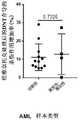
图9.可以看出,经Ven处理和未经其处理的DNT细胞对自体和同种异体PBMC的杀伤程度不显著。Figure 9. It can be seen that the degree of killing of autologous and allogeneic PBMCs by Ven-treated and untreated DNT cells is not significant.
图10.维奈托克在增强其针对AML的细胞毒性的同时,不会杀伤DNT。(A)通过膜联蛋白V染色法和流式细胞术测定经400nM维奈托克处理18小时的DNT和OCI-AML2细胞的活力。(B和C)用浓度递增的维奈托克处理DNT 18小时。随后,测定其对OCI-AML2和两种原发性AML细胞(090765和110162)的活力(B)和细胞毒性(C)。采用ANOVA进行统计。****p<0.0001。Figure 10. While venetoclax enhances its cytotoxicity against AML, it does not kill DNT. (A) The viability of DNT and OCI-AML2 cells treated with 400 nM venetoclax for 18 hours was determined by Annexin V staining and flow cytometry. (B and C) DNT were treated with increasing concentrations of venetoclax for 18 hours. Subsequently, their viability (B) and cytotoxicity (C) against OCI-AML2 and two primary AML cells (090765 and 110162) were determined. ANOVA was used for statistics. ****p<0.0001.
图11.对于诊断性和复发性/难治性AML样本,维奈托克对DNT介导的细胞毒性具有类似的作用。将经400nM维奈托克处理或未经处理的DNT与诊断性(n=12)或复发性/难治性(n=4)原发性AML样本以2:1的比例共培养2小时。确定了对于每种患者样本类型,经维奈托克处理后,DNT介导的细胞毒性的增加。Figure 11. Venetoclax has similar effects on DNT-mediated cytotoxicity in diagnostic and relapsed/refractory AML samples. 400 nM venetoclax-treated or untreated DNT were co-cultured with diagnostic (n = 12) or relapsed/refractory (n = 4) primary AML samples at a ratio of 2:1 for 2 hours. The increase in DNT-mediated cytotoxicity following venetoclax treatment was determined for each patient sample type.
图12.在存在维奈托克的情况下,DNT诱导优异的抗白血病活性。(A)在DNT存在或不存在的情况下,KG1a和OCI-AML2细胞未经处理或使用维奈托克(100nM)处理。(B)在存在或不存在维奈托克(100nM)的情况下,经DNT导致的AML计数减少的百分比。Figure 12. DNT induces excellent antileukemic activity in the presence of venetoclax. (A) KG1a and OCI-AML2 cells were untreated or treated with venetoclax (100 nM) in the presence or absence of DNT. (B) Percent reduction in AML counts by DNT in the presence or absence of venetoclax (100 nM).
图13.经Ven处理的DNT在不增加骨髓中T细胞移植的情况下诱导AML总数更大地降低。向接受亚致死辐射(250cGy)的NSG小鼠静脉注射KG1a细胞(2x10⁶个细胞/小鼠)或原发性AML细胞。两周后,通过三次输注(媒介物对照(PBS)或每次输注1.5-2x107个DNT细胞或经Ven处理的DNT细胞)处理小鼠,每次间隔3-4日。注射AML后五周,通过用抗人CD45、CD3、CD33和CD34抗体对骨髓细胞进行染色和流式细胞术分析,测定骨髓中的AML细胞计数(A)和T细胞频率(B)。Figure 13. Ven-treated DNT induces a greater reduction in the total number of AML without increasing T cell engraftment in the bone marrow. NSG mice receiving sublethal radiation (250cGy) were injected intravenously with KG1a cells (2x10⁶ cells/mouse) or primary AML cells. Two weeks later, mice were treated with three infusions (vehicle control (PBS) or 1.5-2 x10⁷ DNT cells or Ven-treated DNT cells per infusion) spaced 3-4 days apart. Five weeks after AML injection, AML cell counts (A) and T cell frequencies (B) in bone marrow were determined by staining and flow cytometry analysis of bone marrow cells with antibodies against human CD45, CD3, CD33, and CD34.
图14.未经处理的DNT和经维奈托克处理的DNT不引起组织损伤。向接受亚致死辐射(250cGy)的NSG小鼠静脉注射KG1a细胞(2x10⁶个细胞/小鼠)。两周后,通过三次输注(媒介物对照(PBS)或每次输注1.5-2x107个DNT细胞或经Ven处理的DNT细胞)处理小鼠,每次间隔3-4日。第35日,肝脏(顶部)和肺(底部)组织用苏木精和伊红(H&E)染色(放大50倍)。PV-门静脉;ALV-肺泡;BR-细支气管。Figure 14. Untreated DNT and DNT treated with venetoclax did not cause tissue damage. NSG mice receiving sublethal radiation (250cGy) were injected intravenously with KG1a cells (2x10⁶ cells/mouse). Two weeks later, mice were treated with three infusions (vehicle control (PBS) or 1.5-2 x10⁷ DNT cells or Ven-treated DNT cells per infusion) spaced 3-4 days apart. On
图15.其他已知的ROS诱导试剂对DNT活力、ROS水平和抗AML的细胞毒性的影响。用浓度递增的阿糖胞苷(0–3μM)、抗霉素(0–250nM)或柔红霉素(0–10μM)处理DNT 18小时。随后,测定DNT中的细胞ROS水平(A)、DNT活力(B)和抗OCI-AML2的细胞毒性(C)。Figure 15. Effect of other known ROS-inducing agents on DNT activity, ROS levels and cytotoxicity against AML. DNTs were treated with increasing concentrations of cytarabine (0–3 μM), antimycin (0–250 nM) or daunorubicin (0–10 μM) for 18 hours. Subsequently, cellular ROS levels in DNT (A), DNT viability (B) and cytotoxicity against OCI-AML2 (C) were determined.
图16.维奈托克不影响电子传递链(ETC)复合物亚基的表达。将蛋白的相对水平归一化至上样对照MnSOD,并相对于对照的值(一般设定为1.0)表示。显示了代表性免疫印迹。数据表示为三次独立实验的平均值±SD。Figure 16. Venetoclax does not affect the expression of electron transport chain (ETC) complex subunits. Relative levels of protein were normalized to the loading control MnSOD and expressed relative to the value of the control (generally set at 1.0). Representative immunoblots are shown. Data are expressed as mean ± SD of three independent experiments.
具体实施方案specific implementation plan
已经证明用维奈托克预处理T细胞在体外和体内增加T细胞介导的细胞毒性和抗肿瘤活性。因此,预期与维奈托克和相关组合物接触的T细胞以及T细胞和维奈托克的组合均可用于治疗癌症受试者。Pretreatment of T cells with venetoclax has been shown to increase T cell-mediated cytotoxicity and antitumor activity in vitro and in vivo. Accordingly, T cells contacted with venetoclax and related compositions, as well as combinations of T cells and venetoclax, are contemplated to be useful in treating cancer subjects.
I.增强T细胞及T细胞群的方法I. Methods of Enhancing T Cells and T Cell Populations
在一个实施方案中,提供了一种增强T细胞疗效的方法,该方法包括使T细胞与维奈托克接触以生成增强型T细胞。In one embodiment, there is provided a method of enhancing the therapeutic effect of T cells, the method comprising contacting the T cells with venetoclax to generate enhanced T cells.
本文使用的术语“维奈托克”或“Ven”是指一种能够结合并抑制Bcl-2的分子。在一个实施方案中,维奈托克是药物VenclextaTM或药物VenclyxtoTM。The term "Venetoclax" or "Ven" as used herein refers to a molecule capable of binding and inhibiting Bcl-2. In one embodiment, venetoclax is the drug Venclexta™ or the drug Venclyxto™ .
在一个实施方案中,该方法还包括使癌细胞与氮杂胞苷接触,或将氮杂胞苷与本文所述的增强型T细胞联合施用或使用。本文使用的术语“氮杂胞苷”或“阿扎胞苷”或“5-氮杂胞苷”是指一种化合物,其是具有抗肿瘤活性的胞苷的嘧啶核苷类似物。氮杂胞苷的正确化学名称包括4-氨基-1-β-D-呋喃核糖基-1,3,5-三嗪-2(1H)-酮或4-氨基-1-[3,4-二羟基-5-(羟甲基)草脲胺-2-基]-1,3,5-三嗪-2-酮。In one embodiment, the method further comprises contacting the cancer cell with azacitidine, or administering or using azacitidine in combination with the enhanced T cells described herein. The term "azacytidine" or "azacitidine" or "5-azacytidine" as used herein refers to a compound which is a pyrimidine nucleoside analog of cytidine which has antineoplastic activity. The correct chemical names for azacytidine include 4-amino-1-β-D-ribofuranosyl-1,3,5-triazin-2(1H)-one or 4-amino-1-[3,4- Dihydroxy-5-(hydroxymethyl)oxuron-2-yl]-1,3,5-triazin-2-one.
本文使用的术语“T细胞”包括胸腺细胞、未成熟T淋巴细胞、成熟T淋巴细胞、静止T淋巴细胞或活化T淋巴细胞。T细胞可以是辅助性T细胞(Th),例如辅助性T细胞1(Th1)或辅助性T细胞2(Th2)细胞。T细胞可以由本领域技术人员获得。T细胞可以是常规T细胞(Tconv)或非常规T细胞,例如双阴性T细胞(DNT)、γ-δT细胞或NKT细胞。在一个实施方案中,T细胞是活化的T细胞。在一个实施方案中,T细胞是已经在离体或体外扩增和/或活化的细胞。The term "T cell" as used herein includes thymocytes, immature T lymphocytes, mature T lymphocytes, resting T lymphocytes or activated T lymphocytes. The T cells may be T helper cells (Th), such as T helper 1 (Th1) or T helper 2 (Th2) cells. T cells can be obtained by those skilled in the art. T cells may be conventional T cells (Tconv ) or unconventional T cells, such as double negative T cells (DNT), γ-δ T cells or NKT cells. In one embodiment, the T cells are activated T cells. In one embodiment, T cells are cells that have been expanded and/or activated ex vivo or in vitro.
T细胞可以容易地从例如诸如血液样本或细胞培养物等生物源中获得和/或分离。对于治疗应用,T细胞可以是自体T细胞或同种异体T细胞。在一个实施方案中,T细胞是从受试者(例如罹患癌症或疑似罹患癌症的受试者)获得的自体T细胞。在另一个实施方案中,T细胞是同种异体T细胞,例如从一个或更多个未患癌症的受试者获得的T细胞。在一个实施方案中,T细胞从一个或更多个健康供体获得。T cells can be readily obtained and/or isolated from, for example, biological sources such as blood samples or cell cultures. For therapeutic applications, the T cells can be autologous or allogeneic T cells. In one embodiment, the T cells are autologous T cells obtained from a subject (eg, a subject having or suspected of having cancer). In another embodiment, the T cells are allogeneic T cells, eg, T cells obtained from one or more cancer-free subjects. In one embodiment, T cells are obtained from one or more healthy donors.
可使用CD4和CD8耗竭抗体混合物进行富集来获得DNT。在一个实施方案中,DNT不表达CD4和CD8。在一个实施方案中,DNT具有表型CD3+、γδ-TCR+或αβ-TcR+、CD4-、CD8-、α-Gal-、CTLA4-。在一个实施方案中,DNT具有表型CD3+、γδ-TCR+或αβ-TcR+。在一个实施方案中,DNT可以从包含外周血单核细胞(PBMC)的样本中获得。在一个实施方案中,样本是血液样本。在一个实施方案中,样本是单采血液(apheresis)样本,或富集白细胞单采产物,例如白细胞单采物(leukopak)。在一个实施方案中,样本是骨髓样本。DNTs can be obtained by enrichment using CD4 and CD8 depleting antibody cocktails. In one embodiment, the DNT does not express CD4 and CD8. In one embodiment, the DNT has the phenotype CD3+, γδ-TCR+ or αβ-TcR+, CD4-, CD8-, α-Gal-, CTLA4-. In one embodiment, the DNT has the phenotype CD3+, γδ-TCR+ or αβ-TcR+. In one embodiment, DNT can be obtained from a sample comprising peripheral blood mononuclear cells (PBMC). In one embodiment, the sample is a blood sample. In one embodiment, the sample is an apheresis sample, or an enriched leukocyte apheresis product, such as a leukopak. In one embodiment, the sample is a bone marrow sample.
在一个实施方案中,T细胞在与维奈托克接触前进行体外或离体扩增。分离和扩增DNT的示例性方法在美国专利第6,953,576号“调节肿瘤免疫的方法”、PCT公开号WO2007/056854“扩增双阴性T细胞的方法”和PCT公开号WO2016/023134“用于治疗癌症的免疫疗法”中均有所描述,所有这些文献的全部内容通过引用并入本文。In one embodiment, the T cells are expanded in vitro or ex vivo prior to contacting with venetoclax. Exemplary methods of isolating and expanding DNTs are described in US Patent No. 6,953,576 "Methods of Modulating Tumor Immunity", PCT Publication No. WO2007/056854 "Methods of Expanding Double Negative T Cells" and PCT Publication No. WO2016/023134 "Used in Therapeutic Immunotherapy of Cancer", all of which are incorporated herein by reference in their entirety.
本文使用的术语“增强型T细胞”是指与未接触过维奈托克的对照T细胞相比,在接触维奈托克后表现出增加的细胞毒性和/或抗肿瘤活性的单个T细胞或T细胞群。可选地,增强型T细胞可以是DNT或常规T细胞(Tconv)。在一个实施方案中,可根据生理活性和/或基因表达区分增强型T细胞与其他T细胞和/或对照T细胞。例如,在一个实施方案中,与未接触维奈托克的对照T细胞相比,增强型T细胞中,CD25、CD69、NKG2D、DNAM-1和NRF2中一种或更多种的表达水平呈现出增加。在一个实施方案中,与未接触维奈托克的对照T细胞相比,增强型T细胞中,选自CD25、CD69、NKG2D、DNAM-1和NRF2的2、3、4或5种基因的表达水平呈现出增加。As used herein, the term "enhanced T cell" refers to a single T cell that exhibits increased cytotoxicity and/or anti-tumor activity after exposure to venetoclax compared to control T cells that have not been exposed to venetoclax or T cell populations. Alternatively, enhanced T cells may be DNT or conventional T cells (Tconv ). In one embodiment, enhancing T cells can be distinguished from other T cells and/or control T cells based on physiological activity and/or gene expression. For example, in one embodiment, the expression levels of one or more of CD25, CD69, NKG2D, DNAM-1, and NRF2 in the enhanced T cells exhibit out increase. In one embodiment, the expression of 2, 3, 4 or 5 genes selected from CD25, CD69, NKG2D, DNAM-1 and NRF2 in the enhanced T cells compared to control T cells not exposed to venetoclax Expression levels appear to be increased.
本文使用的术语“接触(contacted或contacting)”是指任何将T细胞暴露于维奈托克以生成增强型T细胞的方法。“接触”包括“孵育”和“暴露”,其并未暗示有任何具体时间或温度要求,除非另有说明。在一个实施方案中,T细胞在体外与维奈托克接触,例如将维奈托克与培养基组合并于培养基中暴露或孵育T细胞。T细胞可以通过体外孵育与维奈托克“接触”,也可以通过向受试者给药或联合给药使T细胞在体内与维奈托克“接触”。As used herein, the term "contacted or contacting" refers to any method of exposing T cells to venetoclax to generate enhanced T cells. "Contacting" includes "incubating" and "exposing", and does not imply any specific time or temperature requirement unless otherwise stated. In one embodiment, T cells are contacted with venetoclax in vitro, eg, combining venetoclax with culture medium and exposing or incubating the T cells in the culture medium. T cells can be "contacted" with venetoclax by incubation in vitro, or can be "contacted" with venetoclax in vivo by administering or co-administering T cells to a subject.
在一个实施方案中,T细胞在体外、离体或体内与浓度为至少25nM、50nM或100nM的维奈托克接触。在一个实施方案中,T细胞与浓度为至少100nM、至少200nM、至少300nM或至少400nM的维奈托克接触。在一个实施方案中,T细胞与浓度为约10nM至10μM,可选地为约50nM至500nM、约50nM至800nM或约100nM至约1μM的维奈托克接触。In one embodiment, the T cells are contacted with venetoclax at a concentration of at least 25 nM, 50 nM or 100 nM in vitro, ex vivo or in vivo. In one embodiment, the T cells are contacted with venetoclax at a concentration of at least 100 nM, at least 200 nM, at least 300 nM, or at least 400 nM. In one embodiment, T cells are contacted with venetoclax at a concentration of about 10 nM to 10 μM, alternatively about 50 nM to 500 nM, about 50 nM to 800 nM, or about 100 nM to about 1 μM.
在另一个实施方案中,T细胞与维奈托克接触至少约30分钟、45分钟、60分钟或90分钟。在一个实施方案中,T细胞与维奈托克接触至少约1小时、2小时、4小时、6小时、12小时、18小时、24小时、36小时或48小时。在一个实施方案中,T细胞与维奈托克接触约1小时至14日,可选地2小时至30日、约4小时至14日、约4小时至6日、约4小时至48小时或约6小时至约24小时。在一个实施方案中,T细胞与维奈托克接触少于约14日、10日、9日、8日、7日、6日或5日。In another embodiment, the T cells are contacted with venetoclax for at least about 30 minutes, 45 minutes, 60 minutes, or 90 minutes. In one embodiment, the T cells are contacted with venetoclax for at least about 1 hour, 2 hours, 4 hours, 6 hours, 12 hours, 18 hours, 24 hours, 36 hours, or 48 hours. In one embodiment, T cells are contacted with venetoclax for about 1 hour to 14 days, alternatively 2 hours to 30 days, about 4 hours to 14 days, about 4 hours to 6 days, about 4 hours to 48 hours or about 6 hours to about 24 hours. In one embodiment, the T cells are contacted with venetoclax for less than about 14 days, 10 days, 9 days, 8 days, 7 days, 6 days, or 5 days.
在一个实施方案中,T细胞与足够浓度的维奈托克接触足够的时间,以增加CD25、CD69、NKG2D、DNAM-1和NRF2中一种或更多种的表达。在一个实施方案中,T细胞与足够浓度的维奈托克接触足够的时间,以增加细胞ROS水平。In one embodiment, the T cells are contacted with a sufficient concentration of venetoclax for a sufficient time to increase the expression of one or more of CD25, CD69, NKG2D, DNAM-1 and NRF2. In one embodiment, the T cells are contacted with a sufficient concentration of venetoclax for a sufficient time to increase cellular ROS levels.
在一些实施方案中,T细胞与维奈托克接触以成为增强型T细胞,可移除部分或全部维奈托克,或分离增强型T细胞以降低浓度或减少细胞外维奈托克。In some embodiments, T cells are contacted with venetoclax to become enhanced T cells, some or all of the venetoclax can be removed, or the enhanced T cells can be isolated to reduce the concentration or reduce extracellular venetoclax.
如本文所述,使T细胞与维奈托克接触生成增强型T细胞群,该增强型T细胞群具有使它们特别适用于治疗癌症的若干特征。例如,在一个实施方案中,维奈托克增加T细胞介导的抗肿瘤活性。在一个实施方案中,维奈托克增加T细胞介导的细胞毒性。As described herein, contacting T cells with venetoclax generates enhanced T cell populations that have several characteristics that make them particularly useful in the treatment of cancer. For example, in one embodiment, venetoclax increases T cell-mediated anti-tumor activity. In one embodiment, venetoclax increases T cell mediated cytotoxicity.
本文使用的术语“抗肿瘤活性”是指杀伤肿瘤细胞和/或抑制肿瘤生长的任何活性。在一个实施方案中,“抗肿瘤活性”包括减少肿瘤细胞的集落形成。The term "anti-tumor activity" as used herein refers to any activity that kills tumor cells and/or inhibits tumor growth. In one embodiment, "anti-tumor activity" includes reducing colony formation of tumor cells.
本文使用的术语“细胞毒性”是指引起细胞死亡、导致细胞成为细胞抑制性的和/或阻止细胞增殖的特质。The term "cytotoxicity" as used herein refers to the property of causing cell death, causing cells to become cytostatic and/or preventing cell proliferation.
II.产品、组合物和试剂盒II. Products, Compositions and Kits
另一方面,提供了根据本文所述的方法生成的增强型T细胞群。还提供了包含如本文所述的增强型T细胞的组合物。例如,在一个实施方案中,增强型T细胞,可选地与药学上可接受的载体一起,包含在药物组合物中。In another aspect, a population of enhanced T cells generated according to the methods described herein is provided. Compositions comprising enhanced T cells as described herein are also provided. For example, in one embodiment, enhanced T cells, optionally together with a pharmaceutically acceptable carrier, are included in a pharmaceutical composition.
在另一个实施方案中,提供了一种组合物,该组合物包含T细胞和维奈托克。在一个实施方案中,所述组合物还包含细胞培养基。In another embodiment, a composition comprising T cells and venetoclax is provided. In one embodiment, the composition further comprises a cell culture medium.
还提供了一种试剂盒,该试剂盒包含T细胞和维奈托克。在一个实施方案中,试剂盒还包括用于执行本文所述方法的使用说明,例如用于生成增强型T细胞、用于治疗癌症或用于减缓肿瘤生长或增殖的使用说明。在一个实施方案中,T细胞和维奈托克在不同的容器中。在一个实施方案中,T细胞和维奈托克在同一容器中,可选地作为含有药学上可接受的载体的组合物。Also provided is a kit comprising T cells and venetoclax. In one embodiment, the kit further comprises instructions for performing the methods described herein, eg, for generating enhanced T cells, for treating cancer, or for slowing tumor growth or proliferation. In one embodiment, the T cells and venetoclax are in separate containers. In one embodiment, the T cells and venetoclax are in the same container, optionally as a composition with a pharmaceutically acceptable carrier.
还提供了本文所述的产品、组合物或试剂盒用于治疗癌症或制备用于治疗癌症的药物的用途。Also provided is the use of a product, composition or kit described herein for the treatment of cancer or for the preparation of a medicament for the treatment of cancer.
III.治疗癌症和减缓肿瘤生长和增殖的方法与用途III. Methods and Uses of Treating Cancer and Slowing Tumor Growth and Proliferation
与未经维奈托克处理的T细胞相比,通过本文所述方法生成的增强型T细胞在体外增加了针对AML细胞的细胞毒性。如实施例2所示,与用对照T细胞处理的AML细胞相比,经增强型T细胞处理的AML细胞表现出对AML细胞的更加特异性的杀伤和更少的集落形成。此外,实施例3证明增强型T细胞在肿瘤异种移植模型中具有更强的抗肿瘤活性。Enhanced T cells generated by the methods described herein have increased cytotoxicity against AML cells in vitro compared to T cells not treated with venetoclax. As shown in Example 2, AML cells treated with enhanced T cells exhibited more specific killing of AML cells and less colony formation than AML cells treated with control T cells. In addition, Example 3 demonstrates that enhanced T cells have stronger anti-tumor activity in tumor xenograft models.
因此,在一个实施方案中,提供了一种治疗有此需要的受试者的癌症的方法。在一个实施方案中,该方法包括向受试者施用有效量的增强型T细胞。在一个实施方案中,如本文所述,通过使T细胞与维奈托克接触来生成增强型T细胞。在一个实施方案中,方法包括向受试者施用T细胞和维奈托克,该T细胞和维奈托克可选地与药学上可接受的载体组合成组合物,其中T细胞通过在体内与维奈托克接触而增强。Accordingly, in one embodiment, there is provided a method of treating cancer in a subject in need thereof. In one embodiment, the method comprises administering to the subject an effective amount of enhanced T cells. In one embodiment, enhanced T cells are generated by contacting the T cells with venetoclax as described herein. In one embodiment, the method comprises administering to the subject T cells and venetoclax, optionally in combination with a pharmaceutically acceptable carrier, wherein the T cells pass through in vivo Enhanced by contact with venetoclax.
还提供了一种减缓肿瘤生长和/或增殖的方法。在一个实施方案中,方法包括使肿瘤与有效量的增强型T细胞接触。在一个实施方案中,如本文所述,通过使T细胞与维奈托克接触来生成增强型T细胞。Also provided is a method of slowing tumor growth and/or proliferation. In one embodiment, the method comprises contacting the tumor with an effective amount of enhanced T cells. In one embodiment, enhanced T cells are generated by contacting the T cells with venetoclax as described herein.
还提供了如本文所述的增强型T细胞、组合物和/或试剂盒用于治疗有此需要的受试者的癌症的用途。在一个实施方案中,根据本文所述的方法生成增强型T细胞。在一个实施方案中,增强型T细胞、组合物和/或试剂盒用于制造用于治疗癌症的药物。在一个实施方案中,用途包括向受试者使用或施用增强型T细胞。在另一个实施方案中,该用途包括在同一时间或不同时间向受试者使用或施用维奈托克和T细胞。Also provided are uses of enhanced T cells, compositions and/or kits as described herein for treating cancer in a subject in need thereof. In one embodiment, enhanced T cells are generated according to the methods described herein. In one embodiment, the enhanced T cells, compositions and/or kits are used in the manufacture of a medicament for the treatment of cancer. In one embodiment, the use comprises using or administering enhanced T cells to a subject. In another embodiment, the use comprises using or administering venetoclax and T cells to the subject at the same time or at different times.
还提供了减缓肿瘤生长和增殖的用途。在一个实施方案中,本文所述的增强型T细胞、组合物和/或试剂盒用于减缓肿瘤生长和增殖。在一个实施方案中,增强型T细胞、组合物和/或试剂盒用于制造减缓肿瘤生长和增殖的药物。在一个实施方案中,增强型T细胞和/或组合物用于制造减缓肿瘤生长和增殖的药物。在一个实施方案中,T细胞和维奈托克用于制造减缓肿瘤生长和增殖的药物。Also provided is the use of slowing tumor growth and proliferation. In one embodiment, the enhanced T cells, compositions and/or kits described herein are used to slow tumor growth and proliferation. In one embodiment, enhanced T cells, compositions and/or kits are used in the manufacture of a medicament to slow tumor growth and proliferation. In one embodiment, the enhanced T cells and/or compositions are used in the manufacture of a medicament to slow tumor growth and proliferation. In one embodiment, T cells and venetoclax are used to make a drug that slows tumor growth and proliferation.
本文使用的术语“癌症”是指由不受控制的异常生长的细胞(其可扩散至邻近组织或身体其他部位)导致的一系列疾病中的一种。在一个实施方案中,癌症是白血病,例如急性髓性白血病(AML)。The term "cancer" as used herein refers to one of a series of diseases caused by the uncontrolled growth of abnormal cells that can spread to adjacent tissues or other parts of the body. In one embodiment, the cancer is leukemia, such as acute myeloid leukemia (AML).
术语“癌细胞”是指以不受控制的异常生长和侵袭另一组织能力为特征的细胞或来源于这种细胞的细胞。例如,癌细胞包括从癌症患者获得的原发性癌细胞或来源于这种细胞的细胞系。在一个实施方案中,癌细胞是白血病细胞,例如AML细胞。The term "cancer cell" refers to a cell or a cell derived from such a cell characterized by uncontrolled abnormal growth and the ability to invade another tissue. For example, cancer cells include primary cancer cells obtained from cancer patients or cell lines derived from such cells. In one embodiment, the cancer cells are leukemia cells, such as AML cells.
本文使用的术语“白血病”是指涉及在造血组织、其他器官,通常情况下在血液中发现的异常白细胞数量增加的进行性增生的任何疾病。“白血病细胞”是指特征在于细胞增加的异常增生的白细胞。白血病细胞可以从诊断为罹患白血病的受试者获得。As used herein, the term "leukemia" refers to any disease involving progressive proliferation of increased numbers of abnormal white blood cells found in blood-forming tissues, other organs, and usually blood. "Leukemia cell" refers to an abnormally proliferating white blood cell characterized by an increase in cells. Leukemic cells can be obtained from a subject diagnosed with leukemia.
术语“急性髓性白血病(acute myeloid leukemia)”或“急性骨髓性白血病(acutemyelogenous leukemia)”(“AML”)是指髓性血细胞癌症,其以快速生长的异常白细胞在骨髓里聚集并干扰正常血细胞的生成为特征。骨髓增生异常综合征或骨髓增生综合征等白血病前期病况也可能发展为AML。The term "acute myeloid leukemia" or "acutemyelogenous leukemia" ("AML") refers to a cancer of myeloid blood cells in which rapidly growing abnormal white blood cells accumulate in the bone marrow and interfere with normal blood cells The generation is characteristic. Preleukemic conditions such as myelodysplastic syndrome or myeloproliferative syndrome may also develop into AML.
术语“肿瘤”是指癌细胞的集合。在一个实施方案中,肿瘤是白血病肿瘤,例如AML细胞。在一个实施方案中,肿瘤是血液肿瘤。The term "tumor" refers to a collection of cancer cells. In one embodiment, the tumor is a leukemia tumor, such as AML cells. In one embodiment, the tumor is a hematological tumor.
本文使用的术语“受试者”包括动物界的所有成员,包括哺乳动物,适当情况下其指人类。可选地,术语“受试者”包括已诊断出罹患癌症或处于缓解期的哺乳动物。在一个实施方案中,受试者已经接受治疗或同时正在接受化疗(可选地使用阿糖胞苷和/或氮杂胞苷)。The term "subject" as used herein includes all members of the animal kingdom, including mammals, which refers to humans where appropriate. Alternatively, the term "subject" includes mammals who have been diagnosed with cancer or are in remission. In one embodiment, the subject has been treated or is concurrently receiving chemotherapy (optionally with cytarabine and/or azacitidine).
在一个实施方案中,本文所述的方法和用途涉及施用或使用有效量的增强型T细胞,或有效量的T细胞和维奈托克。In one embodiment, the methods and uses described herein involve administering or using an effective amount of enhanced T cells, or an effective amount of T cells and venetoclax.
本文使用的短语“有效量”或“治疗有效量”是指对于实现预期效果所需的剂量和时间段有效的量。例如,在治疗癌症的情况下,与未经治疗产生的反应相比,有效量是例如诱导缓解、减少肿瘤负荷和/或防止肿瘤扩散或癌细胞生长的量。在一个实施方案中,维奈托克的有效量是指增加T细胞介导的抗肿瘤活性和/或增加T细胞介导的细胞毒性的量。在一个实施方案中,增强型T细胞的有效量是指在体外或体内具有足以抗癌症和/或肿瘤细胞的细胞毒性的量。As used herein, the phrase "effective amount" or "therapeutically effective amount" refers to an amount effective, at dosages and for periods of time required, to achieve the desired effect. For example, in the case of treating cancer, an effective amount is, for example, that induces remission, reduces tumor burden and/or prevents tumor spread or cancer cell growth compared to the response produced without treatment. In one embodiment, the effective amount of venetoclax refers to an amount that increases T cell-mediated anti-tumor activity and/or increases T cell-mediated cytotoxicity. In one embodiment, the effective amount of enhanced T cells refers to the amount that has sufficient cytotoxicity against cancer and/or tumor cells in vitro or in vivo.
有效量会因动物的疾病状态、年龄、性别和体重等因素而异。与这样的量相对应的给定剂量的量会因各种因素而异,例如药物制剂、给药途径、疾病或疾患的类型、接受治疗的受试者或宿主的身份等,但通常仍可由本领域技术人员确定。在一个实施方案中,以注射方式向受试者施用增强型T细胞或T细胞和维奈托克。在一个实施方案中,注射为静脉注射。在一个实施方案中,注射为皮下注射,可选地在肿瘤部位。An effective amount will vary depending on factors such as the disease state, age, sex, and weight of the animal. The amount corresponding to such an amount for a given dosage will vary depending on various factors, such as the pharmaceutical formulation, the route of administration, the type of disease or disorder, the identity of the subject or host being treated, etc., but can generally be determined by Determined by those skilled in the art. In one embodiment, the boosted T cells or T cells and venetoclax are administered to the subject by injection. In one embodiment, the injection is intravenous. In one embodiment, the injection is subcutaneous, optionally at the tumor site.
在一个实施方案中,增强型T细胞或T细胞与维奈托克的组合可用于在体外、离体或体内减缓癌细胞的生长或增殖。本文使用的“减缓癌细胞的生长或增殖”是指减少由于细胞生长或细胞分裂而导致的来源于癌细胞的细胞的数量,其包括细胞死亡。本文使用的术语“细胞死亡”包括杀伤细胞的所有形式,包括细胞裂解、坏死和/或凋亡。在一个实施方案中,增强型T细胞或T细胞与维奈托克的组合可用于在体外、离体或体内杀伤癌细胞。In one embodiment, enhanced T cells or a combination of T cells and venetoclax are used to slow the growth or proliferation of cancer cells in vitro, ex vivo, or in vivo. As used herein, "slowing down the growth or proliferation of cancer cells" refers to reducing the number of cells derived from cancer cells due to cell growth or cell division, which includes cell death. As used herein, the term "cell death" includes all forms of killing cells, including cell lysis, necrosis and/or apoptosis. In one embodiment, enhanced T cells or a combination of T cells and venetoclax can be used to kill cancer cells in vitro, ex vivo or in vivo.
在一个实施方案中,可以使用本领域已知的药学上可接受的配方来配制增强型T细胞或T细胞和/或维奈托克,以供受试者使用,或制备后施用于受试者。例如,在《雷明顿制药学》(2003年第20版)和1999年出版的《美国药典:国家处方集》(USP 24NF19)中描述了用于选择和制备合适制剂的常规规程和成分。术语“药学上可接受的”是指与动物,特别是人类的治疗相容。In one embodiment, enhanced T cells or T cells and/or venetoclax can be prepared using pharmaceutically acceptable formulations known in the art for use by subjects, or administered to subjects after preparation By. General procedures and ingredients for selecting and preparing suitable formulations are described, for example, in Remington's Pharmaceutical Sciences (20th Edition, 2003) and in the United States Pharmacopoeia: National Formulary, published in 1999 (USP 24NF19). The term "pharmaceutically acceptable" means compatible with the treatment of animals, especially humans.
在一个实施方案中,同时向受试者施用T细胞和维奈托克,可选地,其作为包含T细胞和维奈托克的组合物,或作为两个单独的剂量施用。在一个实施方案中,在不同时间对受试者使用或施用T细胞和维奈托克。例如,在一个实施方案中,在施用维奈托克之前或之后使用或施用T细胞。在一个实施方案中,在施用维奈托克之前或之后使用或施用T细胞,使用或施用的间隔时间小于约1分钟、2分钟、5分钟、10分钟、30分钟、45分钟、1小时、1.5小时、2小时、3小时、4小时、5小时、8小时、10小时、12小时、16小时或24小时。在一个实施方案中,在施用维奈托克之前或之后使用或施用T细胞,使用或施用的间隔时间小于约1日、2日、3日、4日、5日、6日或7日。In one embodiment, the T cells and venetoclax are administered to the subject simultaneously, optionally as a composition comprising the T cells and venetoclax, or as two separate doses. In one embodiment, the T cells and venetoclax are administered or administered to the subject at different times. For example, in one embodiment, T cells are used or administered before or after administration of venetoclax. In one embodiment, the T cells are used or administered before or after venetoclax is administered at intervals of less than about 1 minute, 2 minutes, 5 minutes, 10 minutes, 30 minutes, 45 minutes, 1 hour, 1.5 hours, 2 hours, 3 hours, 4 hours, 5 hours, 8 hours, 10 hours, 12 hours, 16 hours or 24 hours. In one embodiment, the T cells are used or administered before or after the administration of venetoclax with an interval of less than about 1 day, 2 days, 3 days, 4 days, 5 days, 6 days, or 7 days.
在一个实施方案中,使用或施用维奈托克,以使其在受试者体内的浓度达到至少25nM、50nM或100nM。在一个实施方案中,使用或施用维奈托克,以使其在受试者体内的浓度达到至少100nM、至少200nM、至少300nM或至少400nM。在一个实施方案中,在施用或使用外源性T细胞,可选地在施用或使用DNT的同时,确立了维奈托克的浓度为至少25nM、50nM、200nM、300nM或400nM。In one embodiment, venetoclax is used or administered such that its concentration in the subject is at least 25 nM, 50 nM or 100 nM. In one embodiment, venetoclax is used or administered such that its concentration in the subject is at least 100 nM, at least 200 nM, at least 300 nM, or at least 400 nM. In one embodiment, a concentration of venetoclax of at least 25 nM, 50 nM, 200 nM, 300 nM or 400 nM is established while administering or using exogenous T cells, optionally simultaneously with administering or using DNT.
在一个实施方案中,维奈托克每日使用或施用的剂量为50mg至800mg,可选地为100mg至600mg。例如,在一个实施方案中,与T细胞联合使用或施用,将维奈托克对受试者使用或施用,进而通过维奈托克增强在体内T细胞。In one embodiment, venetoclax is used or administered daily in a dose of 50 mg to 800 mg, alternatively 100 mg to 600 mg. For example, in one embodiment, venetoclax is used or administered to a subject in combination with T cells, thereby enhancing T cells in vivo by venetoclax.
以下非限制性实施例用于说明本公开。The following non-limiting examples serve to illustrate the present disclosure.
实施例1:维奈托克增加T细胞介导的细胞毒性的效力Example 1: Potency of venetoclax to increase T cell-mediated cytotoxicity
为了鉴定增加T细胞介导的抗AML的细胞毒性效力的分子,使用体外扩增的DNT细胞作为抗白血病T细胞的替代物,并用批准用于各种临床用途的269种药物的化合物库进行预处理。随后,将经化合物处理的细胞用作抗人AML细胞系OCI-AML2的效应物。Bcl-2抑制剂(Ven)最大程度地增加了DNT细胞的细胞毒性(图1)。To identify molecules that increase the potency of T cell-mediated cytotoxicity against AML, in vitro expanded DNT cells were used as a surrogate for anti-leukemic T cells and pretreated with a compound library of 269 drugs approved for various clinical uses. deal with. Subsequently, compound-treated cells were used as effectors against the human AML cell line OCI-AML2. Bcl-2 inhibitor (Ven) maximized the cytotoxicity of DNT cells (Fig. 1).
Ven主要用于治疗慢性淋巴细胞白血病(CLL)和小淋巴细胞白血病,其中Ven抑制抗细胞凋亡分子Bcl-2的活性,促进恶性肿瘤细胞的凋亡。对于复发性/难治性CLL患者,Ven作为单一疗法的总应答率为64.8–79.4%(9)。最近,Ven已被FDA批准与去甲基化药物氮杂胞苷或地西他滨一起用于AML患者的治疗,因为这些药物显著改善了不适合其他常规治疗的未接受治疗的
实施例2:Ven预处理以剂量依赖性方式增加DNT细胞对三种不同AML细胞系的细胞毒性Example 2: Ven pretreatment increases the cytotoxicity of DNT cells to three different AML cell lines in a dose-dependent manner
为了确认药物筛选的结果,用不同浓度的Ven预处理DNT细胞。用Ven预处理以剂量依赖性方式增加了DNT细胞对三种不同AML细胞系AML2-OCI、AML3-OCI和KG1a的细胞毒性(图2A)。与未经处理的DNT相比,经Ven处理的DNT对17个原发性AML样本中的16个样本也显示出较高的细胞毒性(图2B)。值得注意的是,经Ven处理的DNT有效地杀伤了四个对DNT具有抗性的样本(090271、080043、290985和150099)。经Ven处理的DNT在杀伤确诊阶段以及诱导化疗后复发性/难治性患者的AML细胞方面同样有效(图11)。此外,经维奈托克处理的DNT细胞还更有效地减少了AML细胞系AML2-OCI和KG1a以及原发性AML胚细胞(blast)的集落形成,证明了其对白血病起始细胞的作用(图2C)(9–12)。To confirm the results of drug screening, DNT cells were pretreated with different concentrations of Ven. Pretreatment with Ven increased the cytotoxicity of DNT cells against three different AML cell lines AML2-OCI, AML3-OCI and KG1a in a dose-dependent manner (Fig. 2A). Ven-treated DNT also showed higher cytotoxicity against 16 out of 17 primary AML samples compared with untreated DNT (Fig. 2B). Notably, Ven-treated DNT effectively killed four DNT-resistant samples (090271, 080043, 290985, and 150099). Ven-treated DNT was also effective in killing AML cells at the confirmed stage as well as relapsed/refractory patients after induction chemotherapy (Fig. 11). In addition, venetoclax-treated DNT cells also more effectively reduced colony formation in AML cell lines AML2-OCI and KG1a, as well as primary AML blasts, demonstrating its effect on leukemia-initiating cells ( Figure 2C) (9–12).
如图2A所示,用不同浓度的Ven预处理的DNT细胞对三种AML细胞系OCI-AML2、OCI-AML3和KG1a的细胞毒性呈剂量依赖性增加。从DNT细胞中移除药物后,经维奈托克处理增加的这种效应活性至少保持了4日(图2D)。观察到AML对DNT细胞的敏感性与经Ven处理后的DNT细胞介导的细胞毒性增加程度之间呈现明显的负相关(图2E)。维奈托克增加了DNT介导的对AML细胞的杀伤作用(图2A),但没有降低DNT的活力(图10)。由图可知,经维奈托克处理的DNT(经Ven处理的DNT)的抗白血病活性在来自所有十一个测试DNT供体的DNT中均有增加,平均增加了61.25±31%(图2F)。As shown in Figure 2A, DNT cells pretreated with different concentrations of Ven showed a dose-dependent increase in the cytotoxicity of three AML cell lines OCI-AML2, OCI-AML3 and KG1a. This effector activity increased by venetoclax treatment was maintained for at least 4 days after drug removal from DNT cells (Fig. 2D). A clear negative correlation was observed between the sensitivity of AML to DNT cells and the degree of increased cytotoxicity mediated by DNT cells after Ven treatment (Fig. 2E). Venetoclax increased DNT-mediated killing of AML cells (Figure 2A), but did not decrease DNT viability (Figure 10). As can be seen from the figure, the anti-leukemic activity of venetoclax-treated DNT (Ven-treated DNT) was increased in DNT from all eleven tested DNT donors, with an average increase of 61.25 ± 31% (Fig. 2F ).
为了测定维奈托克存在时DNT的抗白血病活性,用维奈托克、DNT或两者处理KG1a和OCI-AML2。用DNT和维奈托克两者处理AML细胞比单独使用任一种处理产生的活AML细胞数量更少(图12)。To determine the antileukemic activity of DNT in the presence of venetoclax, KG1a and OCI-AML2 were treated with venetoclax, DNT or both. Treatment of AML cells with both DNT and venetoclax resulted in lower numbers of viable AML cells than either treatment alone (Figure 12).
实施例3:相比未经处理的DNT细胞,经Ven处理的DNT细胞诱导明显更大程度的肿瘤体积和肿瘤重量减少Example 3: Ven-treated DNT cells induce a significantly greater reduction in tumor volume and tumor weight than untreated DNT cells
接下来,采用异种移植模型在体内研究使用Ven对扩增T细胞的离体处理是否能提高其疗效。免疫缺陷小鼠皮下接种人白血病细胞。肿瘤形成后(尺寸>100mm3),给这些小鼠静脉输注单剂量的非药物处理或经Ven处理的DNT细胞,并监测肿瘤生长情况。虽然如前所述,DNT细胞处理有效地靶向白血病(4–6),但用VenDNT细胞处理进一步减小肿瘤体积(第20日,DNT组为26.15%±5.724%,VenDNT处理组为52.23%±8.468%;图3A)。同样,与用PBS或DNT细胞处理的小鼠相比,用VenDNT细胞处理的小鼠的肿瘤重量显著降低(图3B)。这些数据表明,经Ven处理的DNT细胞可以在体内更有效地靶向AML细胞。鉴于AML主要存在于骨髓(BM)中,接下来研究了VenDNT细胞是否能更有效地靶向骨髓移植的AML。此前的报道显示KG1a在异种移植模型中对DNT细胞处理具有高度的耐药性(6)。尽管DNT细胞的作用很小,但与PBS和DNT细胞处理组相比,VenDNT细胞处理组小鼠的KG1a移植水平显著下降,进一步支持了VenDNT细胞即使针对以另外方式耐药的细胞亦能表现出优异的抗白血病活性(图3C)。Next, a xenograft model was used to investigate in vivo whether ex vivo treatment of expanded T cells with Ven could enhance their efficacy. Immunodeficient mice were inoculated subcutaneously with human leukemia cells. After tumor formation (>100mm3 in size), these mice were given a single dose intravenously of non-drug-treated or Ven-treated DNT cells and monitored for tumor growth. Although DNT cell treatment effectively targeted leukemia (4–6) as previously described, treatment with VenDNT cells further reduced tumor volume (26.15% ± 5.724% in the DNT group and 52.23% in the VenDNT-treated group on
用经Ven处理的DNT离体处理的原发性AML样本移植的量少于单独使用DNT处理的相同细胞的量(图3D)。进一步研究了经Ven处理的DNT对原发性AML样本移植的影响。小鼠经静脉注射原发性AML细胞后,用DNT或经Ven处理的DNT进行处理。与用媒介物对照或DNT处理的小鼠相比,用经Ven处理的DNT处理小鼠减少了AML移植和计数(图3E和图13A)。在DNT组和经Ven处理的DNT组中检测到T细胞频率相似(图13B),这表明经Ven处理的DNT具有更高的抗白血病活性,这得益于DNT在功能上的提升,而不是其在持续性或增殖能力上的提升。重要的是,从这些处理中没有观察到明显的毒性(图14)。Primary AML samples treated ex vivo with Ven-treated DNT engrafted less than the same cells treated with DNT alone (Fig. 3D). The effect of Ven-treated DNT on transplantation of primary AML samples was further investigated. Mice were treated with DNT or Ven-treated DNT after intravenous injection of primary AML cells. Treatment of mice with Ven-treated DNT reduced AML engraftment and enumeration compared to vehicle control or DNT-treated mice (Figure 3E and Figure 13A). Similar frequencies of T cells were detected in the DNT group and the Ven-treated DNT group (Fig. 13B), suggesting that Ven-treated DNT had higher anti-leukemic activity due to the functional enhancement of DNT rather than An increase in its persistence or ability to proliferate. Importantly, no overt toxicity was observed from these treatments (Figure 14).
实施例4:Ven增加常规T细胞的细胞毒性Example 4: Ven increases the cytotoxicity of conventional T cells
虽然用Ven处理DNT细胞以增强其抗白血病活性可能有利于DNT疗法,但由于临床上Tconv细胞更为广泛地作为癌症免疫疗法使用,因此进行实验以确定Ven是否对CD4+或CD8+常规T(Tconv)细胞的抗白血病活性有作用。为此,在与AML细胞系共培养之前,用不同浓度的Ven预处理多克隆活化的Tconv细胞。与在DNT细胞中观察到的相似,观察到Tconv细胞对各种AML细胞系的细胞毒性显著增强(图4)。这些数据表明,Ven可以提高Tconv和DNT细胞的抗白血病活性,并支持Ven与过继T细胞疗法联合使用以进一步增强治疗效果。Although treating DNT cells with Ven to enhance their anti-leukemic activity might be beneficial for DNT therapy, since Tconv cells are more widely used clinically as cancer immunotherapy, experiments were performed to determine whether Ven is effective against CD4+ or CD8+ conventional T cells. The anti-leukemic activity of (Tconv ) cells contributes. To this end, polyclonal activated Tconv cells were pretreated with different concentrations of Ven before co-cultivation with AML cell lines. Similar to that observed in DNT cells, a significantly enhanced cytotoxicity of Tconv cells against various AML cell lines was observed (Fig. 4). These data suggest that Ven can enhance the anti-leukemic activity of Tconv and DNT cells and support the combination of Ven with adoptive T cell therapy to further enhance the therapeutic effect.
实施例5:与其他Bcl-2抑制剂相比,Ven独特地增加了DNT细胞毒性Example 5: Ven Uniquely Increases DNT Cytotoxicity Compared to Other Bcl-2 Inhibitors
众所周知,Bcl-2保护细胞免于凋亡,可以预期抑制该通路会导致T细胞凋亡加剧,并减弱T细胞功能。鉴于Ven能增加T细胞介导的细胞毒性这一意外发现,接下来使用Bcl-2家族蛋白泛抑制剂奥巴克拉以及Bcl-2、Bcl-xL和Bcl-w抑制剂ABT-737确定对其他抗细胞凋亡Bcl-2家族蛋白的抑制是否会有类似的效果。与Ven相反,这些Bcl-2家族蛋白抑制剂诱导DNT细胞死亡或抑制其细胞毒性(图5A和图5B)。Bcl-xL在DNT细胞上的表达水平相比其在AML细胞上的表达水平相对更高(图5C),这表明DNT细胞可以通过Bcl-xL的补偿活性对Bcl-2抑制产生抗性。Bcl-2 is known to protect cells from apoptosis, and inhibition of this pathway can be expected to lead to increased T cell apoptosis and attenuated T cell function. Given the unexpected finding that Ven increases T cell-mediated cytotoxicity, we next determined the efficacy of other Bcl-2 family protein pan-inhibitor obacla and the Bcl-2, Bcl-xL, and Bcl-w inhibitor ABT-737. Whether inhibition of anti-apoptotic Bcl-2 family proteins would have a similar effect. In contrast to Ven, these Bcl-2 family protein inhibitors induced DNT cell death or suppressed their cytotoxicity (Fig. 5A and Fig. 5B). The expression level of Bcl-xL on DNT cells was relatively higher than that on AML cells (Fig. 5C), suggesting that DNT cells can develop resistance to Bcl-2 inhibition through the compensatory activity of Bcl-xL.
实施例6:Ven处理增加DNT效应分子与活化标志物的表达和ROS水平Example 6: Ven treatment increases expression of DNT effector molecules and activation markers and ROS levels
为了阐明Ven介导T细胞的增加的细胞毒性的潜在机制,比较了经Ven处理或未经Ven处理的DNT细胞上T细胞活化标志物和效应分子的表达。Ven处理导致DNT细胞上活化标志物CD69和CD25(图6A)以及效应分子NKG2D和DNAM-1的表达更高(图6B)。经Ven处理的DNT细胞也比媒介物处理的细胞表达更高水平的颗粒酶B(图6C)。同样,在经Ven处理的CD8+(图6D)Tconv细胞上也观察到CD25、NKG2D和DNAM-1表达的剂量依赖性增加。用维奈托克处理DNT和Tconv细胞,最少4小时,最多3日,增加了T细胞对AML的细胞毒性(图4B),同时增加了T细胞活化标志物(CD69和CD25;图6A)和活化受体(NKG2D和DNAM-1;图6B)的表达,而没有改变T细胞的活力(图4C)。因此,维奈托克直接活化效应T细胞以增加其细胞毒性,而不消耗幼稚或抑制性T细胞亚群。To elucidate the underlying mechanism by which Ven mediates the increased cytotoxicity of T cells, the expression of T cell activation markers and effector molecules on Ven-treated or non-Ven-treated DNT cells was compared. Ven treatment resulted in higher expression of activation markers CD69 and CD25 (Fig. 6A) and effector molecules NKG2D and DNAM-1 (Fig. 6B) on DNT cells. Ven-treated DNT cells also expressed higher levels of granzyme B than vehicle-treated cells (Fig. 6C). Likewise, a dose-dependent increase in the expression of CD25, NKG2D, and DNAM-1 was also observed on Ven-treated CD8+ (Fig. 6D) Tconv cells. Treatment of DNT and Tconv cells with venetoclax for a minimum of 4 hours and a maximum of 3 days increased T cell cytotoxicity against AML (Fig. 4B), while increasing T cell activation markers (CD69 and CD25; Fig. 6A) and activating receptors (NKG2D and DNAM-1; Figure 6B), without altering T cell viability (Figure 4C). Thus, venetoclax directly activates effector T cells to increase their cytotoxicity without depleting naive or suppressor T cell subsets.
最近的研究报道,维奈托克增加ROS水平,而ROS在T细胞活化信号级联放大中起重要作用(9、13–15)。然而,之前未曾报道过Ven是否增加T细胞中的ROS水平以及增强T细胞活化。为了确定ROS在维奈托克介导的T细胞活化和增强中的介入,测定了经浓度递增的维奈托克处理的DNT细胞和CD8+Tconv细胞中细胞与线粒体ROS的水平。维奈托克以剂量依赖性方式增加DNT和CD8+Tconv细胞中的细胞ROS(图7A)。尽管抗氧化剂Nrf2的表达和核定位出现补偿性升高,但仍观察到ROS水平增加(图7B)。Recent studies have reported that venetoclax increases ROS levels, which play an important role in T cell activation signaling cascades (9, 13–15). However, it has not been reported before whether Ven increases ROS levels in T cells as well as enhances T cell activation. To determine the involvement of ROS in venetoclax-mediated T cell activation and enhancement, cellular and mitochondrial ROS levels were measured in DNT cells and CD8+ Tconv cells treated with increasing concentrations of venetoclax. Venetoclax increased cellular ROS in DNT and CD8+ Tconv cells in a dose-dependent manner (Fig. 7A). Increased ROS levels were observed despite a compensatory increase in the expression and nuclear localization of the antioxidant Nrf2 (Fig. 7B).
为了确定经维奈托克处理的DNT细胞中较高ROS水平的功能相关性,我们用维奈托克和浓度递增的N-乙酰半胱氨酸(NAC)共同处理DNT细胞。用维奈托克和NAC处理降低了细胞ROS水平,并消除了维奈托克对DNT细胞介导的抗AML细胞毒性的影响(图7C),从而证明了维奈托克-DNT细胞中ROS水平升高的功能相关性。如图7L所示,用维奈托克和浓度递增的ROS清除剂N-乙酰半胱氨酸(NAC)共同处理DNT消除了维奈托克诱导的ROS生成并阻断了活化标志物的上调。To determine the functional relevance of higher ROS levels in venetoclax-treated DNT cells, we co-treated DNT cells with venetoclax and increasing concentrations of N-acetylcysteine (NAC). Treatment with venetoclax and NAC reduced cellular ROS levels and abolished the effect of venetoclax on DNT cell-mediated anti-AML cytotoxicity (Fig. 7C), thus demonstrating that ROS in venetoclax-DNT cells Functional relevance of elevated levels. As shown in Figure 7L, co-treatment of DNT with venetoclax and increasing concentrations of the ROS scavenger N-acetylcysteine (NAC) abolished venetoclax-induced ROS generation and blocked the upregulation of activation markers .
维奈托克增加了恶性肿瘤细胞中ROS的生成(9、21),并且ROS在T细胞活化和分化中起着重要作用(15、22–24)。为了进一步了解维奈托克活化T细胞的机制,我们测量了经维奈托克处理的DNT和Tconv细胞中的ROS生成。维奈托克以与增加的T细胞效应功能相关的浓度和时间增加DNT和Tconv细胞中细胞ROS与线粒体ROS(图7H–K)。Venetoclax increases ROS production in malignant tumor cells (9, 21), and ROS play an important role in T cell activation and differentiation (15, 22–24). To further understand the mechanism by which venetoclax activates T cells, we measured ROS generation in venetoclax-treated DNT and Tconv cells. Venetoclax increased cellular and mitochondrial ROS in DNT and Tconv cells at concentrations and times associated with increased T cell effector function (Fig. 7H–K).
为了解Ven增加DNT中的线粒体ROS的机制,我们测量了呼吸链蛋白的水平。在电子传递链(ETC)复合物I、II和IV、亚基NDUFA9、UQCRC2和MTCO1中均未观察到变化。ROS的生成受呼吸链超级复合物的调节,呼吸链超级复合物是含呼吸链复合物I、III和IV的高级四级结构。呼吸链超级复合物的减少可能与线粒体ROS的较高产生有关(16、17)。如通过非变性凝胶测量的,维奈托克减少了DNT细胞中呼吸链超级复合物的形成(图7D)。To understand the mechanism by which Ven increases mitochondrial ROS in DNT, we measured the levels of respiratory chain proteins. No changes were observed in electron transport chain (ETC) complexes I, II and IV, subunits NDUFA9, UQCRC2 and MTCO1. ROS generation is regulated by respiratory chain supercomplexes, which are higher-order quaternary structures containing respiratory chain complexes I, III, and IV. A reduction in respiratory chain supercomplexes may be associated with higher production of mitochondrial ROS (16, 17). Venetoclax reduced the formation of respiratory chain supercomplexes in DNT cells as measured by non-denaturing gels (Fig. 7D).
为了确定其他ROS诱导剂对DNT介导的细胞毒性的影响,用浓度递增的阿糖胞苷、柔红霉素和抗霉素对DNT进行了处理。在用阿糖胞苷和抗霉素处理的DNT中观察到ROS水平以剂量依赖性方式增加,而活力没有或几乎没有损失(图15A和图15B)。经柔红霉素处理的DNT具有较低的ROS水平,其活力也大幅下降(图15A和图15B)。与维奈托克处理不同,尽管细胞ROS水平增加,但阿糖胞苷和抗霉素并没有增强DNT的细胞毒性,而柔红霉素降低了DNT介导的抗AML的细胞毒性(图15C)。这些数据表明,DNT介导的细胞毒性的ROS依赖性增加是维奈托克独有的。To determine the effect of other ROS inducers on DNT-mediated cytotoxicity, DNTs were treated with increasing concentrations of cytarabine, daunorubicin, and antimycin. A dose-dependent increase in ROS levels with little or no loss of viability was observed in DNTs treated with cytarabine and antimycin (Fig. 15A and Fig. 15B). DNT treated with daunorubicin had a lower ROS level and its activity was also greatly reduced (Fig. 15A and Fig. 15B). Unlike venetoclax treatment, cytarabine and antimycin did not enhance DNT cytotoxicity despite increased cellular ROS levels, whereas daunorubicin reduced DNT-mediated cytotoxicity against AML (Fig. 15C ). These data suggest that the ROS-dependent increase in DNT-mediated cytotoxicity is unique to venetoclax.
有趣的是,对于DNT细胞(图7E)和CD8+Tconv细胞(图7F),Ven还增加了效应记忆阶段细胞的比例,同时降低了中央型记忆T细胞的频率。由于效应记忆T细胞优先依赖于糖酵解作用,而中央型记忆T细胞依赖于氧化磷酸化作用,并且已证明Ven抑制AML细胞上的氧化磷酸化作用,因此比较了DNT和VenDNT细胞的糖酵解水平、糖酵解能力、耗氧率(OCR)。然而,Ven对DNT细胞的糖酵解、糖酵解能力和基础耗氧率没有显著影响,这表明Ven使DNT细胞不依赖于其代谢途径而偏向效应记忆表型(图7G)。为了解维奈托克增加ROS产生的机制,我们测量了呼吸链蛋白的水平。在电子传递链(ETC)复合物I、III和IV的亚基NDUFA9、UQCRC2和MTCO1中均未观察到变化(图7M和图16)。Interestingly, for both DNT cells (Fig. 7E) and CD8+ Tconv cells (Fig. 7F), Ven also increased the proportion of effector memory phase cells while decreasing the frequency of central type memory T cells. Since effector memory T cells are preferentially dependent on glycolysis, whereas central memory T cells are dependent on oxidative phosphorylation, and Ven has been shown to inhibit oxidative phosphorylation on AML cells, glycolysis in DNT and VenDNT cells was compared Hydrolysis level, glycolysis capacity, oxygen consumption rate (OCR). However, Ven had no significant effect on glycolysis, glycolytic capacity, and basal oxygen consumption rate of DNT cells, suggesting that Ven biased DNT cells toward an effector memory phenotype independent of their metabolic pathways (Fig. 7G). To understand the mechanism by which venetoclax increases ROS production, we measured the levels of respiratory chain proteins. No changes were observed in subunits NDUFA9, UQCRC2, and MTCO1 of electron transport chain (ETC) complexes I, III, and IV (Fig. 7M and Fig. 16).
总的来说,这些结果表明Ven活化T细胞并使其偏向更多的效应表型。Collectively, these results suggest that Ven activates T cells and biases them towards a more effector phenotype.
实施例7:Ven处理增加了T细胞群中细胞毒性CD8+和DNT细胞的比例Example 7: Ven Treatment Increases the Proportion of Cytotoxic CD8+ and DNT Cells in the T Cell Population
最近的报道表明,对未接受治疗的AML患者进行Ven和Aza联合治疗显著改善临床结果,且治疗相关毒性较低(9和19)。为了测定维奈托克是否能增加患者的T细胞效应活性,我们检测了在经维奈托克和氮杂胞苷处理前和处理后第4日AML患者的T细胞。与处理前水平相比,我们观察到经维奈托克和氮杂胞苷处理后CD8+和DNT细胞的比例增加(图8A)。与我们在体外的研究结果一致,所有患者中处于效应记忆/效应状态的CD8+T细胞的比例增加(图8B)。此外,经处理后,患者CD8+T细胞上的NKG2D表达和细胞ROS水平均有所增加(图8C)。同样,观察到DNT细胞中效应记忆/效应亚群的频率增加(图8D),并且,DNT细胞也表现出较高的NKG2D表达水平和细胞ROS水平(图8E)。Recent reports have demonstrated that combination therapy with Ven and Aza in untreated AML patients significantly improved clinical outcomes with less treatment-related toxicity (9 and 19). To determine whether venetoclax increases T cell effector activity in patients, we examined T cells from AML patients before and on
实施例8:Ven选择性地增加DNT细胞对AML的细胞毒活性Example 8: Ven selectively increases the cytotoxic activity of DNT cells against AML
为了测定Ven是否增加DNT细胞对正常血细胞的细胞毒性,以健康供体的自体和同种异体PBMC为靶点。虽然观察到对OCI-AML2具有较强的细胞毒性,但观察到经Ven处理和未经其处理的DNT细胞对自体和同种异体PBMC的杀伤程度并不明显(图9),这表明Ven选择性地增加DNT细胞对AML的细胞毒活性。To determine whether Ven increases the cytotoxicity of DNT cells against normal blood cells, autologous and allogeneic PBMC from healthy donors were targeted. Although strong cytotoxicity against OCI-AML2 was observed, the degree of killing of autologous and allogeneic PBMC by Ven-treated and untreated DNT cells was not significantly observed (Fig. 9), suggesting that Ven selects Sexually increased the cytotoxic activity of DNT cells against AML.
虽然结合了当前认为是优选实施例的内容来描述本公开,但应当理解,本公开并不限于所公开的实施例。相反,本公开旨在涵盖所附权利要求的精神和范围内包括的各种修改和等同布置。While the present disclosure has been described in connection with what are presently considered to be the preferred embodiments, it is to be understood that the disclosure is not limited to the disclosed embodiments. On the contrary, the present disclosure is intended to cover various modifications and equivalent arrangements included within the spirit and scope of the appended claims.
如分别具体地指出每个单独的出版物、专利或专利申请的全部内容均通过引用以其整体而并入本文一样,所有出版物、专利和专利申请的全部内容均通过引用并入本文。All publications, patents, and patent applications are herein incorporated by reference in their entirety as if each individual publication, patent, or patent application was specifically incorporated by reference in its entirety.
参考文献references
1.Park JH,Riviere I,Gonen M,Wang X,Senechal B,Curran KJ,et al.Long-Term Follow-up of CD19 CAR Therapy in Acute Lymphoblastic Leukemia.N Engl JMed.2018;378(5):449-59.1. Park JH, Riviere I, Gonen M, Wang X, Senechal B, Curran KJ, et al. Long-Term Follow-up of CD19 CAR Therapy in Acute Lymphoblastic Leukemia. N Engl JMed. 2018; 378(5): 449 -59.
2.Rosenberg SA,Restifo NP.Adoptive cell transfer as personalizedimmunotherapy for human cancer.Science.2015;348(6230):62-8.2. Rosenberg SA, Restifo NP. Adaptive cell transfer as personalized immunotherapy for human cancer. Science. 2015; 348(6230):62-8.
3.Lee JB,Chen B,Vasic D,Law AD,Zhang L.Cellular immunotherapy foracute myeloid leukemia:How specific should it be?Blood Rev.2019;35:18-31.3. Lee JB, Chen B, Vasic D, Law AD, Zhang L. Cellular immunotherapy for acute myeloid leukemia: How specific should it be? Blood Rev. 2019; 35:18-31.
4.Lee JB,Kang H,Fang L,D'Souza C,Adeyi O,Zhang L.DevelopingAllogeneic Double-Negative T Cells as a Novel Off-the-Shelf Adoptive CellularTherapy for Cancer.Clin Cancer Res.2019.4. Lee JB, Kang H, Fang L, D'Souza C, Adeyi O, Zhang L. Developing Allogeneic Double-Negative T Cells as a Novel Off-the-Shelf Adoptive Cellular Therapy for Cancer. Clin Cancer Res. 2019.
5.Lee J,Minden MD,Chen WC,Streck E,Chen B,Kang H,et al.AllogeneicHuman Double Negative T Cells as a Novel Immunotherapy for Acute MyeloidLeukemia and Its Underlying Mechanisms.Clin Cancer Res.2018;24(2):370-82.5. Lee J, Minden MD, Chen WC, Streck E, Chen B, Kang H, et al. Allogeneic Human Double Negative T Cells as a Novel Immunotherapy for Acute Myeloid Leukemia and Its Underlying Mechanisms. Clin Cancer Res. 2018; 24(2) :370-82.
6.Chen B,Lee JB,Kang H,Minden MD,Zhang L.Targeting chemotherapy-resistant leukemia by combining DNT cellular therapy with conventionalchemotherapy.J Exp Clin Cancer Res.2018;37(1):88.6. Chen B, Lee JB, Kang H, Minden MD, Zhang L. Targeting chemotherapy-resistant leukemia by combining DNT cellular therapy with conventional chemotherapy. J Exp Clin Cancer Res. 2018; 37(1): 88.
7.Li Q,Cheng L,Shen K,Jin H,Li H,Cheng Y,et al.Efficacy and Safety ofBcl-2 Inhibitor Venetoclax in Hematological Malignancy:A Systematic Reviewand Meta-Analysis of Clinical Trials.Front Pharmacol.2019;10:697.7. Li Q, Cheng L, Shen K, Jin H, Li H, Cheng Y, et al. Efficacy and Safety of Bcl-2 Inhibitor Venetoclax in Hematological Malignancy: A Systematic Review and Meta-Analysis of Clinical Trials. Front Pharmacol. 2019; 10:697.
8.Pollyea DA,Stevens BM,Jones CL,Winters A,Pei S,Minhajuddin M,etal.Venetoclax with azacitidine disrupts energy metabolism and targetsleukemia stem cells in patients with acute myeloid leukemia.Nat Med.2018;24(12):1859-66.8. Pollyea DA, Stevens BM, Jones CL, Winters A, Pei S, Minhajuddin M, et al. Venetoclax with azacitidine disrupts energy metabolism and targets leukemia stem cells in patients with acute myeloid leukemia. Nat Med. 2018; 24(12): 1859 -66.
9.Jones CL,Stevens BM,D'Alessandro A,Reisz JA,Culp-Hill R,Nemkov T,etal.Inhibition of Amino Acid Metabolism Selectively Targets Human LeukemiaStem Cells.Cancer Cell.2018;34(5):724-40 e4.9. Jones CL, Stevens BM, D'Alessandro A, Reisz JA, Culp-Hill R, Nemkov T, et al. Inhibition of Amino Acid Metabolism Selectively Targets Human LeukemiaStem Cells. Cancer Cell. 2018; 34(5): 724-40 e4.
10.Tettamanti S,Marin V,Pizzitola I,Magnani CF,Giordano Attianese GM,Cribioli E,et al.Targeting of acute myeloid leukaemia by cytokine-inducedkiller cells redirected with a novel CD123-specific chimeric antigenreceptor.Br J Haematol.2013;161(3):389-401.10.Tettamanti S,Marin V,Pizzitola I,Magnani CF,Giordano Attianese GM,Cribioli E,et al.Targeting of acute myeloid leukaemia by cytokine-inducedkiller cells redirected with a novel CD123-specific chimeric antigenreceptor.Br J Haematol.2013; 161(3):389-401.
11.Jin L,Lee EM,Ramshaw HS,Busfield SJ,Peoppl AG,Wilkinson L,etal.Monoclonal antibody-mediated targeting of CD123,IL-3 receptor alpha chain,eliminates human acute myeloid leukemic stem cells.Cell Stem Cell.2009;5(1):31-42.11. Jin L, Lee EM, Ramshaw HS, Busfield SJ, Peoppl AG, Wilkinson L, et al. Monoclonal antibody-mediated targeting of CD123, IL-3 receptor alpha chain, eliminates human acute myeloid leukemic stem cells. Cell Stem Cell. 2009 ;5(1):31-42.
12.Dick JE.Acute myeloid leukemia stem cells.Ann N Y Acad Sci.2005;1044:1-5.12. Dick JE. Acute myeloid leukemia stem cells. Ann N Y Acad Sci. 2005; 1044:1-5.
13.Chamoto K,Chowdhury PS,Kumar A,Sonomura K,Matsuda F,Fagarasan S,etal.Mitochondrial activation chemicals synergize with surface receptor PD-1blockade for T cell-dependent antitumor activity.Proc Natl Acad Sci U SA.2017;114(5):E761-E70.13. Chamoto K, Chowdhury PS, Kumar A, Sonomura K, Matsuda F, Fagarasan S, et al. Mitochondrial activation chemicals synergize with surface receptor PD-1blockade for T cell-dependent antitumor activity. Proc Natl Acad Sci U SA. 2017; 114 (5):E761-E70.
14.Klein Geltink RI,O'Sullivan D,Pearce EL.Caught in the cROSsfire:GSH Controls T Cell Metabolic Reprogramming.Immunity.2017;46(4):525-7.14. Klein Geltink RI, O'Sullivan D, Pearce EL. Caught in the cROSsfire: GSH Controls T Cell Metabolic Reprogramming. Immunity. 2017; 46(4):525-7.
15.Franchina DG,Dostert C,Brenner D.Reactive Oxygen Species:Involvement in T Cell Signaling and Metabolism.Trends Immunol.2018;39(6):489-502.15. Franchina DG, Dostert C, Brenner D. Reactive Oxygen Species: Involvement in T Cell Signaling and Metabolism. Trends Immunol. 2018; 39(6): 489-502.
16.Lopez-Fabuel I,Le Douce J,Logan A,James AM,Bonvento G,Murphy MP,etal.Complex I assembly into supercomplexes determines differentialmitochondrial ROS production in neurons and astrocytes.Proc Natl Acad Sci U SA.2016;113(46):13063-8.16. Lopez-Fabuel I, Le Douce J, Logan A, James AM, Bonvento G, Murphy MP, et al. Complex I assembly into supercomplexes determines differential mitochondrial ROS production in neurons and astrocytes. Proc Natl Acad Sci U SA. 2016; 113( 46):13063-8.
17.Hou T,Zhang R,Jian C,Ding W,Wang Y,Ling S,et al.NDUFAB1 conferscardio-protection by enhancing mitochondrial bioenergetics throughcoordination of respiratory complex and supercomplex assembly.Cell Res.2019;29(9):754-66.17. Hou T, Zhang R, Jian C, Ding W, Wang Y, Ling S, et al. NDUFAB1 confers cardio-protection by enhancing mitochondrial bioenergetics through coordination of respiratory complex and supercomplex assembly. Cell Res. 2019; 29(9): 754 -66.
18.Roulois,D.et al.DNA-Demethylating Agents Target Colorectal CancerCells by Inducing Viral Mimicry by Endogenous Transcripts.Cell 162,961-973,doi:10.1016/j.cell.2015.07.056(2015).18. Roulois, D. et al. DNA-Demethylating Agents Target Colorectal Cancer Cells by Inducing Viral Mimicry by Endogenous Transcripts. Cell 162, 961-973, doi:10.1016/j.cell.2015.07.056 (2015).
19.DiNardo,C.D.et al.Venetoclax combined with decitabine orazacytidine in treatment-naive,elderly patients with acute myeloidleukemia.Blood 133,7-17,doi:10.1182/blood-2018-08-868752(2019).19. DiNardo, C.D. et al. Venetoclax combined with decitabine orazacytidine in treatment-naive, elderly patients with acute myeloidleukemia. Blood 133, 7-17, doi: 10.1182/blood-2018-08-868752 (2019).
20.Liu,M.et al.Dual Inhibition of DNA and Histone MethyltransferasesIncreases Viral Mimicry in Ovarian Cancer Cells.Cancer Res.78,5754-5766,doi:10.1158/0008-5472.CAN-17-3953(2018).20. Liu, M. et al. Dual Inhibition of DNA and Histone Methyltransferases Increases Viral Mimicry in Ovarian Cancer Cells. Cancer Res. 78, 5754-5766, doi: 10.1158/0008-5472. CAN-17-3953 (2018).
21.Nguyen LXT,Troadec E,Kalvala A,et al.The Bcl-2 inhibitorvenetoclax inhibits Nrf2 antioxidant pathway activation induced byhypomethylating agents in AML.J Cell Physiol.2019;234(8):14040-14049.21. Nguyen LXT, Troadec E, Kalvala A, et al. The Bcl-2 inhibitor venetoclax inhibits Nrf2 antioxidant pathway activation induced by hypomethylating agents in AML. J Cell Physiol. 2019; 234(8): 14040-14049.
22.Belikov AV,Schraven B,Simeoni L.T cells and reactive oxygenspecies.J Biomed Sci.2015;22:85.22. Belikov AV, Schraven B, Simeoni L. T cells and reactive oxygen species. J Biomed Sci. 2015; 22:85.
23.Pilipow K,Scamardella E,Puccio S,et al.Antioxidant metabolismregulates CD8+T memory stem cell formation and antitumor immunity.JCIInsight.2018;3(18).23. Pilipow K, Scamardella E, Puccio S, et al. Antioxidant metabolism regulates CD8+T memory stem cell formation and antitumor immunity. JCI Insight. 2018; 3(18).
24.Mak TW,Grusdat M,Duncan GS,et al.Glutathione Primes T CellMetabolism for Inflammation.Immunity.2017;46(4):675-689.24. Mak TW, Grusdat M, Duncan GS, et al. Glutathione Primes T Cell Metabolism for Inflammation. Immunity. 2017; 46(4):675-689.
Claims (40)
Applications Claiming Priority (3)
| Application Number | Priority Date | Filing Date | Title |
|---|---|---|---|
| US202062971534P | 2020-02-07 | 2020-02-07 | |
| US62/971,534 | 2020-02-07 | ||
| PCT/CA2021/050138WO2021155479A1 (en) | 2020-02-07 | 2021-02-08 | Methods for enhancing t cells using venetoclax |
Publications (1)
| Publication Number | Publication Date |
|---|---|
| CN115362253Atrue CN115362253A (en) | 2022-11-18 |
Family
ID=77199122
Family Applications (1)
| Application Number | Title | Priority Date | Filing Date |
|---|---|---|---|
| CN202180026678.5APendingCN115362253A (en) | 2020-02-07 | 2021-02-08 | Method for enhancing T cells using Venetork |
Country Status (5)
| Country | Link |
|---|---|
| US (1) | US20230059785A1 (en) |
| EP (1) | EP4100513A4 (en) |
| CN (1) | CN115362253A (en) |
| CA (1) | CA3167134A1 (en) |
| WO (1) | WO2021155479A1 (en) |
Families Citing this family (2)
| Publication number | Priority date | Publication date | Assignee | Title |
|---|---|---|---|---|
| EP4054652A4 (en)* | 2019-11-07 | 2023-11-22 | Icahn School of Medicine at Mount Sinai | Synthetic modified rna and uses thereof |
| GB2606175A (en)* | 2021-04-28 | 2022-11-02 | Stina Linnea Wickstroem | Methods and uses |
Citations (2)
| Publication number | Priority date | Publication date | Assignee | Title |
|---|---|---|---|---|
| CN107073039A (en)* | 2014-08-15 | 2017-08-18 | 大学健康网络 | Immunotherapy for treatment of cancer |
| US20190336496A1 (en)* | 2018-02-16 | 2019-11-07 | Abbvie Inc. | Selective bcl-2 inhibitors in combination with an anti-pd-1 or an anti-pd-l1 antibody for the treatment of cancers |
Family Cites Families (2)
| Publication number | Priority date | Publication date | Assignee | Title |
|---|---|---|---|---|
| SI3179991T1 (en)* | 2014-08-11 | 2022-04-29 | Acerta Pharma B.V. | Therapeutic combinations of a btk inhibitor and a bcl-2 inhibitor |
| US20220249637A1 (en)* | 2019-06-12 | 2022-08-11 | Juno Therapeutics, Inc. | Combination therapy of a cell-mediated cytotoxic therapy and an inhibitor of a prosurvival bcl2 family protein |
- 2021
- 2021-02-08CNCN202180026678.5Apatent/CN115362253A/enactivePending
- 2021-02-08WOPCT/CA2021/050138patent/WO2021155479A1/ennot_activeCeased
- 2021-02-08CACA3167134Apatent/CA3167134A1/enactivePending
- 2021-02-08EPEP21750194.9Apatent/EP4100513A4/enactivePending
- 2021-02-08USUS17/797,630patent/US20230059785A1/enactivePending
Patent Citations (2)
| Publication number | Priority date | Publication date | Assignee | Title |
|---|---|---|---|---|
| CN107073039A (en)* | 2014-08-15 | 2017-08-18 | 大学健康网络 | Immunotherapy for treatment of cancer |
| US20190336496A1 (en)* | 2018-02-16 | 2019-11-07 | Abbvie Inc. | Selective bcl-2 inhibitors in combination with an anti-pd-1 or an anti-pd-l1 antibody for the treatment of cancers |
Non-Patent Citations (3)
| Title |
|---|
| DHAKAL P等: "Treatment Strategies for Therapy-related Acute Myeloid Leukemia", 《CLIN LYMPHOMA MYELOMA LEUK》, vol. 20, no. 03, 24 December 2019 (2019-12-24), pages 147 - 155* |
| LEE JB等: "Cellular immunotherapy for acute myeloid leukemia: How specific should it be?", 《BLOOD REV》, vol. 35, 23 February 2019 (2019-02-23), pages 18 - 31, XP085658910, DOI: 10.1016/j.blre.2019.02.001* |
| 赵爽;赵宏伟;马威;: "维奈托克对急性髓系白血病肿瘤标志基因和炎症因子的影响", 医药导报, no. 01, 1 January 2018 (2018-01-01), pages 31 - 34* |
Also Published As
| Publication number | Publication date |
|---|---|
| US20230059785A1 (en) | 2023-02-23 |
| CA3167134A1 (en) | 2021-08-12 |
| EP4100513A1 (en) | 2022-12-14 |
| EP4100513A4 (en) | 2024-03-13 |
| WO2021155479A1 (en) | 2021-08-12 |
Similar Documents
| Publication | Publication Date | Title |
|---|---|---|
| Ma et al. | The oncogenic microRNA miR-21 promotes regulated necrosis in mice | |
| Blazar et al. | Phase 1/2 randomized, placebo-control trial of palifermin to prevent graft-versus-host disease (GVHD) after allogeneic hematopoietic stem cell transplantation (HSCT) | |
| Yuan et al. | Doxorubicin-polyglycerol-nanodiamond conjugate is a cytostatic agent that evades chemoresistance and reverses cancer-induced immunosuppression in triple-negative breast cancer | |
| EP3442541B1 (en) | Composition for use in treating cancer | |
| KR20180087238A (en) | New Therapeutic Strategy for Hematologic Cancer | |
| KR20180041229A (en) | Methods for stem cell transplantation | |
| Shen et al. | Bruton’s tyrosine kinase inhibitors in the treatment of primary central nervous system lymphoma: a mini-review | |
| JP6242071B2 (en) | Immune exhausted CD8 + T cell function improving agent, cancer therapeutic agent and preventive or therapeutic agent for metabolic syndrome | |
| CN115362253A (en) | Method for enhancing T cells using Venetork | |
| US7825088B2 (en) | Methods for the treatment of multiple myeloma | |
| Avraham et al. | Synergism between immunostimulation and prevention of surgery-induced immune suppression: an approach to reduce post-operative tumor progression | |
| Wu et al. | Deleting the mitochondrial respiration negative regulator MCJ enhances the efficacy of CD8+ T cell adoptive therapies in pre-clinical studies | |
| Shakhar et al. | Amelioration of operation-induced suppression of marginating pulmonary NK activity using poly IC: a potential approach to reduce postoperative metastasis | |
| Mawad et al. | Strategies to reduce relapse after allogeneic hematopoietic cell transplantation in acute myeloid leukemia | |
| ES2789574T3 (en) | Modulation of myeloid-derived suppressor cell inflammasome activation for the treatment of GvHD or tumor | |
| JP2013513655A (en) | Methods, pharmaceutical compositions and kits for use in the treatment of adult T-cell leukemia / lymphoma | |
| Takei et al. | Suppression of multiple anti‐apoptotic BCL2 family proteins recapitulates the effects of JAK2 inhibitors in JAK2V617F driven myeloproliferative neoplasms | |
| ES2331800T3 (en) | USE OF THYROSINE-KINASE INHIBITORS FOR THE TREATMENT OF DIABETES. | |
| Clarke et al. | Allogeneic blood transfusion reduces murine pulmonary natural killer (NK) activity and enhances lung metastasis of a syngeneic tumour | |
| CN104434908A (en) | Application of kaempferol in medicines for inhibiting rejection reaction of receptor on organ transplantation | |
| US9546354B2 (en) | Z cells activated by zinc finger-like protein and uses thereof in cancer treatment | |
| Jung et al. | Alpha-melanocyte stimulating hormone preserves islet graft survival through down-regulation of Toll-like receptors | |
| EP3641799A1 (en) | Nk-92 cells and il-15 agonist combination therapy | |
| US20240335465A1 (en) | A non-canonical signaling activity of cgamp triggers dna damage response signaling | |
| Mielcarek et al. | Denileukin diftitox as prophylaxis against graft-versus-host disease in the canine hematopoietic cell transplantation model |
Legal Events
| Date | Code | Title | Description |
|---|---|---|---|
| PB01 | Publication | ||
| PB01 | Publication | ||
| SE01 | Entry into force of request for substantive examination | ||
| SE01 | Entry into force of request for substantive examination |