CN115088010A - Image enhancement based on fiber optic shape sensing - Google Patents
Image enhancement based on fiber optic shape sensingDownload PDFInfo
- Publication number
- CN115088010A CN115088010ACN202180014342.7ACN202180014342ACN115088010ACN 115088010 ACN115088010 ACN 115088010ACN 202180014342 ACN202180014342 ACN 202180014342ACN 115088010 ACN115088010 ACN 115088010A
- Authority
- CN
- China
- Prior art keywords
- imaging
- shape sensing
- processor unit
- optical shape
- data
- Prior art date
- Legal status (The legal status is an assumption and is not a legal conclusion. Google has not performed a legal analysis and makes no representation as to the accuracy of the status listed.)
- Pending
Links
Images


Classifications
- G—PHYSICS
- G06—COMPUTING OR CALCULATING; COUNTING
- G06T—IMAGE DATA PROCESSING OR GENERATION, IN GENERAL
- G06T5/00—Image enhancement or restoration
- G06T5/70—Denoising; Smoothing
- G—PHYSICS
- G06—COMPUTING OR CALCULATING; COUNTING
- G06T—IMAGE DATA PROCESSING OR GENERATION, IN GENERAL
- G06T7/00—Image analysis
- G06T7/0002—Inspection of images, e.g. flaw detection
- G06T7/0012—Biomedical image inspection
- G—PHYSICS
- G06—COMPUTING OR CALCULATING; COUNTING
- G06T—IMAGE DATA PROCESSING OR GENERATION, IN GENERAL
- G06T5/00—Image enhancement or restoration
- G06T5/50—Image enhancement or restoration using two or more images, e.g. averaging or subtraction
- G—PHYSICS
- G06—COMPUTING OR CALCULATING; COUNTING
- G06T—IMAGE DATA PROCESSING OR GENERATION, IN GENERAL
- G06T5/00—Image enhancement or restoration
- G06T5/73—Deblurring; Sharpening
- G—PHYSICS
- G06—COMPUTING OR CALCULATING; COUNTING
- G06V—IMAGE OR VIDEO RECOGNITION OR UNDERSTANDING
- G06V10/00—Arrangements for image or video recognition or understanding
- G06V10/20—Image preprocessing
- G06V10/25—Determination of region of interest [ROI] or a volume of interest [VOI]
- G—PHYSICS
- G06—COMPUTING OR CALCULATING; COUNTING
- G06T—IMAGE DATA PROCESSING OR GENERATION, IN GENERAL
- G06T2207/00—Indexing scheme for image analysis or image enhancement
- G06T2207/10—Image acquisition modality
- G06T2207/10116—X-ray image
- G—PHYSICS
- G06—COMPUTING OR CALCULATING; COUNTING
- G06T—IMAGE DATA PROCESSING OR GENERATION, IN GENERAL
- G06T2207/00—Indexing scheme for image analysis or image enhancement
- G06T2207/20—Special algorithmic details
- G06T2207/20172—Image enhancement details
- G—PHYSICS
- G06—COMPUTING OR CALCULATING; COUNTING
- G06T—IMAGE DATA PROCESSING OR GENERATION, IN GENERAL
- G06T2207/00—Indexing scheme for image analysis or image enhancement
- G06T2207/20—Special algorithmic details
- G06T2207/20172—Image enhancement details
- G06T2207/20182—Noise reduction or smoothing in the temporal domain; Spatio-temporal filtering
- G—PHYSICS
- G06—COMPUTING OR CALCULATING; COUNTING
- G06T—IMAGE DATA PROCESSING OR GENERATION, IN GENERAL
- G06T2207/00—Indexing scheme for image analysis or image enhancement
- G06T2207/20—Special algorithmic details
- G06T2207/20172—Image enhancement details
- G06T2207/20201—Motion blur correction
- G—PHYSICS
- G06—COMPUTING OR CALCULATING; COUNTING
- G06T—IMAGE DATA PROCESSING OR GENERATION, IN GENERAL
- G06T2207/00—Indexing scheme for image analysis or image enhancement
- G06T2207/30—Subject of image; Context of image processing
- G06T2207/30004—Biomedical image processing
- G06T2207/30021—Catheter; Guide wire
- G—PHYSICS
- G06—COMPUTING OR CALCULATING; COUNTING
- G06T—IMAGE DATA PROCESSING OR GENERATION, IN GENERAL
- G06T2207/00—Indexing scheme for image analysis or image enhancement
- G06T2207/30—Subject of image; Context of image processing
- G06T2207/30004—Biomedical image processing
- G06T2207/30101—Blood vessel; Artery; Vein; Vascular
- G—PHYSICS
- G06—COMPUTING OR CALCULATING; COUNTING
- G06T—IMAGE DATA PROCESSING OR GENERATION, IN GENERAL
- G06T2207/00—Indexing scheme for image analysis or image enhancement
- G06T2207/30—Subject of image; Context of image processing
- G06T2207/30204—Marker
Landscapes
- Engineering & Computer Science (AREA)
- Physics & Mathematics (AREA)
- General Physics & Mathematics (AREA)
- Theoretical Computer Science (AREA)
- General Health & Medical Sciences (AREA)
- Health & Medical Sciences (AREA)
- Multimedia (AREA)
- Medical Informatics (AREA)
- Nuclear Medicine, Radiotherapy & Molecular Imaging (AREA)
- Radiology & Medical Imaging (AREA)
- Quality & Reliability (AREA)
- Computer Vision & Pattern Recognition (AREA)
- Apparatus For Radiation Diagnosis (AREA)
- Ultra Sonic Daignosis Equipment (AREA)
- Magnetic Resonance Imaging Apparatus (AREA)
Abstract
Description
Translated fromChinese技术领域technical field
本发明涉及用于在医学成像系统的介入期间增强视图的系统和方法。The present invention relates to systems and methods for enhancing views during interventions in medical imaging systems.
背景技术Background technique
可以在X射线引导下进行微创介入。为了最大限度地减少辐射的量,医师会在图像质量上做出妥协。X射线图像中的低信噪比对象,如支架支柱和钙化,可以使用图像集成来增强,如StentBoost:后者是关于通过显示支架支柱的更精细细节来增强图像中的支架,同时淡化背景噪声和解剖结构。这仅在图像可以正确叠加时才有效。这意味着标记物是可识别的,并且设备的运动没有平面外叠加。Minimally invasive interventions can be performed under X-ray guidance. To minimize the amount of radiation, physicians compromise on image quality. Low signal-to-noise ratio objects in X-ray images, such as stent struts and calcifications, can be enhanced using image integration, such as StentBoost: the latter is about enhancing stents in images by revealing finer details of the stent struts while attenuating background noise and anatomy. This only works if the images can be stacked correctly. This means that the markers are identifiable and there is no out-of-plane superimposition of the motion of the device.
发明内容SUMMARY OF THE INVENTION
在介入期间具有用于图像增强的改进技术将是有利的。It would be advantageous to have improved techniques for image enhancement during interventions.
本发明的目的利用独立权利要求的主题来解决,其中,在从属权利要求中并入了另外的实施例。The object of the invention is solved with the subject-matter of the independent claims, wherein further embodiments are incorporated in the dependent claims.
应当注意,本发明的以下描述的方面和示例也适用于设备、方法,以及计算机程序单元和计算机可读介质。It should be noted that the aspects and examples of the invention described below also apply to apparatus, methods, as well as computer program elements and computer readable media.
在第一个方面中,提供了一种图像处理系统,包括:处理器单元,其被布置为接收与成像系统相关联的成像数据以及与和所述成像系统配准的光学形状感测系统相关联的光学形状感测数据,使得所述光学形状感测数据可以被定位在所述成像系统中;其中,所述处理器单元被配置为基于所述成像数据和/或所述光学形状感测数据在所述成像数据中定义感兴趣区域,并且还被配置为使用所述光学形状感测数据作为所述感兴趣区域内的标记物,使得处理器单元基于接收到的光学形状感测数据来对所述感兴趣区域的成像数据应用图像增强。In a first aspect, there is provided an image processing system comprising: a processor unit arranged to receive imaging data associated with the imaging system and associated with an optical shape sensing system registered with the imaging system coupled optical shape sensing data such that the optical shape sensing data can be located in the imaging system; wherein the processor unit is configured to be based on the imaging data and/or the optical shape sensing The data defines a region of interest in the imaging data and is further configured to use the optical shape sensing data as markers within the region of interest such that the processor unit based on the received optical shape sensing data Image enhancement is applied to the imaging data of the region of interest.
以这种方式,例如,StentBoost识别图像中的标记物并使用它们来跨图像帧执行运动补偿以提高图像质量,从而提高了图像质量。当存在多个标记物、看不到任何标记物或外推到无法预定义的更一般的标记物时,所述算法会遇到困难。将FORS与StentBoost组合可以通过限制图像中的搜索范围并提供设备的3D定位来解决这些挑战。In this way, StentBoost improves image quality by, for example, identifying markers in images and using them to perform motion compensation across image frames to improve image quality. The algorithm encounters difficulties when multiple markers are present, no markers are visible, or extrapolation to more general markers cannot be predefined. Combining FORS with StentBoost can address these challenges by limiting the search range in the image and providing 3D localization of the device.
换言之,根据本发明的示例提供了在介入仪器中嵌入光学形状感测(“OSS”)光纤,并且一旦与成像系统(例如介入X射线成像系统、磁共振系统或超声成像系统)共同配准,就将其用作标记物。In other words, examples in accordance with the present invention provide optical shape sensing ("OSS") fibers embedded in interventional instruments, and once co-registered with an imaging system (eg, an interventional X-ray imaging system, a magnetic resonance system, or an ultrasound imaging system), use it as a marker.
本发明有利地提供了,在血管流程期间,由于治疗设备(支架、球囊、植入物等)的尺寸小、视野中的其他设备的阻碍、运动伪影、或者只是从解剖学本身,可能难以获得治疗设备的高质量图像。此外,随着设备变得更小、更像组织,它们也会失去一些通过成像可见的特征。The present invention advantageously provides that during vascular procedures, due to the small size of the treatment device (stent, balloon, implant, etc.), obstruction of other devices in the field of view, motion artifacts, or simply from the anatomy itself, it is possible to It is difficult to obtain high-quality images of treatment devices. Also, as devices become smaller and more tissue-like, they also lose some of the features visible through imaging.
StentBoost的开发是为了克服其中一些挑战,然而,了解导丝、导管、支架或植物的确切位置可能会进一步提高这些类型设备的图像质量。有两个具体情况特别具有挑战性:StentBoost was developed to overcome some of these challenges, however, knowing the exact location of a guidewire, catheter, stent or plant may further improve the image quality of these types of devices. Two specific cases are particularly challenging:
i.当图像中存在多个标记物并且难以识别哪些标记物与感兴趣的设备相关联时。i. When there are multiple markers in the image and it is difficult to identify which markers are associated with the device of interest.
ii.当标记物在X射线中难以看到时(例如可生物降解的支架)ii. When the marker is difficult to see in X-ray (eg biodegradable stent)
iii.当增强没有已知标记物的对象或结构时(例如解剖结构,如盖或脉管)iii. When enhancing objects or structures without known markers (e.g. anatomical structures such as lids or vessels)
iv.校正平面外的运动。iv. Correct for out-of-plane motion.
本发明有利地提供了,如权利要求1所定义的系统或设备使用例如FORS设备、FORS系统、成像系统(例如X射线或超声)和控制器、图像处理系统和可视化系统。本发明假设FORS设备和成像系统在空间坐标中共同配准。The present invention advantageously provides that a system or apparatus as defined in
本发明的一个方面例如提供了如何使用FORS设备的位置信息来增强另一种成像模态(例如X射线或超声)中的图像质量。One aspect of the present invention provides, for example, how to use the position information of the FORS device to enhance image quality in another imaging modality such as X-ray or ultrasound.
本发明的各方面例如基于:Aspects of the present invention are based, for example, on:
·提供i)重建的OSS光纤的表示和ii)对象的图像,彼此配准Provides i) a representation of the reconstructed OSS fiber and ii) an image of the object, registered with each other
·根据来自i)的数据定义感兴趣区域——例如围绕OSS导丝、球囊、支架、植入物的尖端……——自动地或通过用户接口Defining a region of interest based on data from i) - eg around OSS guidewires, balloons, stents, tips of implants... - automatically or via a user interface
·搜索和识别该感兴趣区域中靠近FORS数据的标记物(即仅位于通路上的标记)——这将丢弃不可行的标记物Search and identify markers in this region of interest that are close to the FORS data (i.e. markers located only on the pathway) - this will discard markers that are not viable
·根据已识别的标记物,在该感兴趣区域应用StentBoost(来自同一感兴趣区域的一系列X射线图像的造影剂注射)Apply StentBoost (contrast injection from a series of X-ray images of the same region of interest) to that region of interest based on the identified markers
·作为StentBoost的结果,在感兴趣区域以更高的对比度显示叠加的i)、ii)Displays superimposed i), ii) with higher contrast in the region of interest as a result of StentBoost
根据本发明的示例性实施例,FORS 3D数据可用于进一步过滤掉平面外的帧。According to an exemplary embodiment of the present invention, FORS 3D data may be used to further filter out out-of-plane frames.
根据本发明的示例性实施例,2个标记物之间的FORS形状可用于评估支架何时发生形状变化(与考虑平移的当前技术相反)。这些帧可以被丢弃,或者可以使用FORS来改变设备的形状以匹配其他帧。According to an exemplary embodiment of the present invention, the FORS shape between the 2 markers can be used to assess when the stent undergoes a shape change (as opposed to current techniques that consider translation). These frames can be dropped, or FORS can be used to reshape the device to match other frames.
根据本发明的示例性实施例,FORS数据本身可以用作标记物。这将加快图像积分的计算时间并减少假阳性标记物的数量,从而提高图像质量。According to an exemplary embodiment of the present invention, the FORS data itself may be used as a marker. This will speed up the computation time of image integration and reduce the number of false positive markers, thereby improving image quality.
根据本发明的示例性实施例,可以在用于StentBoost的一系列图像中识别已知标记物(球囊、支架、导丝的曲线、植入物、夹子或瓣膜设备、血管轮廓、血管分叉……)。According to an exemplary embodiment of the present invention, known markers (balloons, stents, curves of guidewires, implants, clips or valve devices, vessel contours, vessel bifurcations) can be identified in a series of images for StentBoost ...).
根据本发明的示例性实施例,通过这种方式,沿慢性完全闭塞的脉管和盖形态的钙化也可以被可视化。In this way, calcifications along the vascular and lid morphology of chronic total occlusion can also be visualized, according to exemplary embodiments of the present invention.
根据本发明的示例性实施例,所述处理器单元被配置为将图像增强应用于由成像系统拍摄的感兴趣区域的一系列造影增强的X射线图像。According to an exemplary embodiment of the invention, the processor unit is configured to apply image enhancement to a series of contrast-enhanced X-ray images of the region of interest captured by the imaging system.
根据本发明的示例性实施例,处理器单元被配置为关于将标记物限制为位于介入仪器的通路上的标记物子组来搜索和识别标记物。According to an exemplary embodiment of the present invention, the processor unit is configured to search for and identify markers with respect to limiting the markers to a subset of markers located on the pathway of the interventional instrument.
根据本发明的示例性实施例,处理器单元被配置为基于球囊、支架、植入物或介入仪器的位置来定义感兴趣区域。According to an exemplary embodiment of the present invention, the processor unit is configured to define a region of interest based on the position of the balloon, stent, implant or interventional instrument.
根据本发明的示例性实施例,处理器单元被配置为使用光学形状感测数据来滤除成像系统的平面外的帧。According to an exemplary embodiment of the present invention, the processor unit is configured to use the optical shape sensing data to filter out frames out of the plane of the imaging system.
根据本发明的示例性实施例,所述处理器单元被配置为使用至少两个标记物之间的光学形状感测数据来评估介入仪器或介入仪器的形状变化。According to an exemplary embodiment of the invention, the processor unit is configured to use optical shape sensing data between at least two markers to evaluate the interventional instrument or the shape change of the interventional instrument.
根据本发明的示例性实施例,所述介入仪器处理器单元被布置为关于计算机断层摄影系统或磁共振成像系统或超声或光学成像系统或X射线成像系统或医学成像系统或诊断成像系统接收与成像系统相关的成像数据。According to an exemplary embodiment of the invention, the interventional instrument processor unit is arranged to receive information with respect to a computed tomography system or a magnetic resonance imaging system or an ultrasound or optical imaging system or an X-ray imaging system or a medical imaging system or a diagnostic imaging system Imaging data associated with the imaging system.
根据本发明的示例性实施例,所述处理器单元被配置为识别由所述成像系统拍摄的一系列图像中的标记物。According to an exemplary embodiment of the present invention, the processor unit is configured to identify markers in a series of images captured by the imaging system.
根据本发明的示例性实施例,由所述成像系统拍摄的所述一系列图像是在感兴趣区域上应用的图像增强的部分。According to an exemplary embodiment of the present invention, the series of images captured by the imaging system is part of an image enhancement applied over a region of interest.
在第二个方面中,提供了一种成像系统,其被配置为与根据第一方面或第一方面的任何实施方式的设备进行通信。In a second aspect, there is provided an imaging system configured to communicate with a device according to the first aspect or any embodiment of the first aspect.
在第三个方面中,提供了一种光学形状感测系统,其被配置为与根据第一方面或第一方面的任何实施方式的设备进行通信。所述光学形状感测系统被配置为与根据第二方面或第二方面的任何实施方式的设备进行配准。In a third aspect there is provided an optical shape sensing system configured to communicate with a device according to the first aspect or any embodiment of the first aspect. The optical shape sensing system is configured to register with a device according to the second aspect or any embodiment of the second aspect.
在第四个方面中,提供了一种在医学成像设备中嵌入光纤形状感测的方法,所述方法包括以下步骤:In a fourth aspect, there is provided a method of embedding optical fiber shape sensing in a medical imaging device, the method comprising the steps of:
作为第一步,执行以下操作:借助于处理器单元来接收与成像系统相关联的成像数据以及与和所述成像系统配准的光学形状感测系统相关联的光学形状感测数据,使得所述光学形状感测数据可以被定位在所述成像系统中;As a first step, the following operations are performed: the imaging data associated with the imaging system and the optical shape sensing data associated with the optical shape sensing system registered with the imaging system are received by means of the processor unit such that all the optical shape sensing data can be located in the imaging system;
作为第二步,执行以下操作:借助于处理器单元来基于所述成像数据和/或所述光学形状感测数据在所述成像数据中定义感兴趣区域,并且还使用所述光学形状感测数据作为所述感兴趣区域内的标记,使得所述处理器单元基于接收到的光学形状感测数据对感兴趣区域的成像数据应用图像增强。As a second step, a region of interest is defined in the imaging data based on the imaging data and/or the optical shape sensing data by means of a processor unit, and also using the optical shape sensing The data serves as a marker within the region of interest, causing the processor unit to apply image enhancement to the imaging data of the region of interest based on the received optical shape sensing data.
根据本发明的示例性实施例,所述方法还包括将图像增强应用于由成像系统拍摄的感兴趣区域的一系列造影增强的X射线图像的步骤。According to an exemplary embodiment of the invention, the method further comprises the step of applying image enhancement to a series of contrast-enhanced X-ray images of the region of interest captured by the imaging system.
根据本发明的示例性实施例,所述方法还包括关于将标记物限制为位于介入仪器的通路上的标记物子组来搜索和识别标记物的步骤。According to an exemplary embodiment of the invention, the method further comprises the step of searching for and identifying markers with respect to limiting the markers to a subset of markers located on the pathway of the interventional instrument.
根据本发明的示例性实施例,所述方法还包括关于将标记物限制为位于介入仪器的通路上的标记物子组来搜索和识别标记物的步骤。According to an exemplary embodiment of the invention, the method further comprises the step of searching for and identifying markers with respect to limiting the markers to a subset of markers located on the pathway of the interventional instrument.
参考下文描述的实施例,上述方面和范例将变得显而易见并将得以阐述。The above aspects and examples will be apparent from and will be elucidated with reference to the embodiments described hereinafter.
附图说明Description of drawings
下面将参考附图来描述示范性实施例:Exemplary embodiments will be described below with reference to the accompanying drawings:
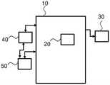
图1示出了根据本发明示例性实施例的用于在医学成像设备中嵌入光纤形状感测的图像处理系统的示意性设置;1 shows a schematic setup of an image processing system for embedding optical fiber shape sensing in a medical imaging device according to an exemplary embodiment of the present invention;
图2示出了根据本发明示例性实施例的在医学成像设备中嵌入光纤形状感测的方法;2 illustrates a method of embedding optical fiber shape sensing in a medical imaging device according to an exemplary embodiment of the present invention;
图3示出了根据本发明示例性实施例的叠加在术前CT上的示出了它们在脉管系统内的位置的光学形状感测设备的示例;3 shows an example of an optical shape sensing device showing their position within the vasculature superimposed on a preoperative CT according to an exemplary embodiment of the present invention;
图4示出了根据本发明示例性实施例的示出支架的更好图像质量的StentBoost的图像的示例;并且FIG. 4 shows an example of an image of StentBoost showing better image quality of a stent according to an exemplary embodiment of the present invention; and
图5示出了根据本发明示例性实施例的基于FORS设备和治疗设备位置定义用于增强的局部区域的示例;Figure 5 shows an example of defining a local area for enhancement based on FORS device and treatment device location according to an exemplary embodiment of the present invention;
图6示出了根据本发明示例性实施例的与治疗设备相组合使用FORS设备周围的局部区域来限制用于图像处理的X射线图像的区域的示例。6 shows an example of using a local area around a FORS device in combination with a treatment device to limit the area of an X-ray image for image processing according to an exemplary embodiment of the present invention.
具体实施方式Detailed ways
图1示出了根据本发明示例性实施例的例如用于在医学成像设备中嵌入光纤形状感测的图像处理系统的示意性设置。图像处理系统10包括处理器单元20。图像处理系统10被配置为例如连接到显示单元30。图像处理系统10被配置为连接到成像系统40。Figure 1 shows a schematic setup of an image processing system, eg, for embedding optical fiber shape sensing in a medical imaging device, according to an exemplary embodiment of the present invention. The
图像处理系统10被配置为连接到与成像系统40配准的光学形状感测系统50。
处理器单元20被布置为接收与成像系统40相关联的成像数据以及与和成像系统40配准的光学形状感测系统50相关联的光学形状感测数据,使得光学形状感测数据能够被定位在成像系统中;The
所述处理器单元20被配置为基于所述成像数据和/或所述光学形状感测数据在所述成像数据中定义感兴趣区域,并且还被配置为使用所述光学形状感测数据作为所述感兴趣区域内的标记物,使得所述处理器单元基于接收到的光学形状感测数据对所述感兴趣区域上的成像数据应用图像增强。The
图2示出了根据本发明示例性实施例的在医学成像设备中嵌入光纤形状感测的方法。所述方法包括:2 illustrates a method of embedding optical fiber shape sensing in a medical imaging device according to an exemplary embodiment of the present invention. The method includes:
作为第一步,执行以下操作:借助于处理器单元来接收S1与成像系统相关联的成像数据以及与和所述成像系统配准的光学形状感测系统相关联的光学形状感测数据,使得所述光学形状感测数据可以被定位在所述成像系统中;As a first step, the following operations are performed: receiving S1 imaging data associated with the imaging system and optical shape sensing data associated with the optical shape sensing system registered with the imaging system by means of the processor unit, such that the optical shape sensing data can be located in the imaging system;
作为第二步,执行以下操作:借助于处理器单元来基于所述成像数据和/或所述光学形状感测数据在所述成像数据中定义S2感兴趣区域,并且还使用所述光学形状感测数据作为所述感兴趣区域内的标记物,使得处理器单元基于接收到的光学形状感测数据对感兴趣区域的成像数据应用图像增强。As a second step, an S2 region of interest is defined in said imaging data based on said imaging data and/or said optical shape sensing data by means of a processor unit, and also using said optical shape sensing The measured data is used as a marker within the region of interest, causing the processor unit to apply image enhancement to the imaging data of the region of interest based on the received optical shape sensing data.
图3示出了根据本发明示例性实施例的叠加在术前CT上的示出了它们在脉管系统内的位置的光学形状感测设备的示例。Figure 3 shows an example of an optical shape sensing device showing their position within the vasculature superimposed on a preoperative CT according to an exemplary embodiment of the present invention.
根据本发明的示例性实施例,光纤真实形状,FORS,在外科手术期间使用沿多芯光纤的光用于设备定位和导航。According to an exemplary embodiment of the present invention, the fiber true shape, FORS, uses light along a multi-core fiber for device positioning and navigation during surgery.
根据本发明的示例性实施例,所涉及的原理利用特征瑞利背散射或受控光栅图案在光纤中使用分布式应变测量。According to an exemplary embodiment of the present invention, the principles involved use distributed strain measurements in optical fibers using characteristic Rayleigh backscatter or controlled grating patterns.
根据本发明的示例性实施例,沿着光纤的形状开始于沿着传感器的特定点,己知为起始点或者z=0,并且随后的开关位置和取向是相对于该点的。According to an exemplary embodiment of the invention, the shape along the fiber starts at a specific point along the sensor, known as the starting point or z=0, and the subsequent switch positions and orientations are relative to this point.
根据本发明的示例性实施例,所述光学形状感测光纤可以被集成到医学设备中,以便在微创手术期间提供对设备的实时引导。According to an exemplary embodiment of the present invention, the optical shape sensing fiber can be integrated into a medical device to provide real-time guidance of the device during minimally invasive procedures.
根据本发明的示例性实施例,所述集成纤维提供整个设备的位置和方向。According to an exemplary embodiment of the present invention, the integrated fibers provide the position and orientation of the entire device.
图3示出了叠加在术前CT图像上的用于导航至左肾动脉的形状感应导丝和形状感测导管。Figure 3 shows a shape-sensing guidewire and shape-sensing catheter for navigating to the left renal artery superimposed on a preoperative CT image.
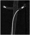
图4示出了根据本发明示例性实施例的示出支架的更好图像质量的StentBoost的图像的示例;并且FIG. 4 shows an example of an image of StentBoost showing better image quality of a stent according to an exemplary embodiment of the present invention; and
根据本发明的示例性实施例,增强由StentBoost提供,其本身是增强支架相对于血管壁的可视化的工具。According to an exemplary embodiment of the present invention, the enhancement is provided by StentBoost, which itself is a tool to enhance the visualization of the stent relative to the vessel wall.
根据本发明的示例性实施例,通过显示支架支柱的更精细细节来增强图像中的支架,同时淡出背景噪声和解剖结构。这使得支架的定位更加精确,并且能够立即纠正部署不足的情况。According to an exemplary embodiment of the present invention, a stent in an image is enhanced by revealing finer details of the stent struts while fading out background noise and anatomy. This allows for more precise positioning of the stent and immediate correction of under-deployment.
根据本发明的示例性实施例,StentBoost在提高支架图像质量的产品方面被使用。它在每幅图像帧中定位支架的标记物带,补偿任何运动,然后在图像帧之间进行平均以提高图像的对比度。在美国专利US728962B2:Medical Viewing System and Method forDetecting and Enhancing Structures in Noisy Images中描述了StentBoost。According to an exemplary embodiment of the present invention, StentBoost is used in products that improve the image quality of a stent. It locates the stent's marker band in each image frame, compensates for any motion, and then averages between image frames to improve the contrast of the image. StentBoost is described in US Patent US728962B2: Medical Viewing System and Method for Detecting and Enhancing Structures in Noisy Images.
根据本发明的示例性实施例,增强或StentBoost拍摄一系列X射线图像并定位已知标记物(例如球囊/支架标记物)以用于一系列图像的共同配准。当图像中存在多个标记物时,此技术会失败。在这种情况下,FORS实现的导丝可用于限制标记物的搜索范围,因为这些标记物将位于导丝的路径上。搜索范围可以根据以下因素围绕FORS丝确定:According to an exemplary embodiment of the present invention, Enhancement or StentBoost takes a series of X-ray images and locates known markers (eg balloon/stent markers) for co-registration of the series of images. This technique fails when multiple markers are present in the image. In this case, a FORS-implemented guidewire can be used to limit the search for markers as these markers will be located in the guidewire's path. The search range can be determined around the FORS wire based on the following factors:
·设备类型,例如针对支架/球囊的窄搜索,针对植入物的宽搜索;Device type, e.g. narrow search for stents/balloons, broad search for implants;
·估计的FORS误差,例如作为沿设备的曲率、扭曲、长度的函数;Estimated FORS error, e.g. as a function of curvature, twist, length along the device;
·用户定义的搜索区域User-defined search area
·成像系统设置,例如像素分辨率、成像协议类型;Imaging system settings, such as pixel resolution, imaging protocol type;
该技术仍然使用例如X射线或超声图像中的标记物进行运动补偿,并降低了对FORS设备的精度要求。FORS设备和X射线系统必须共同配准,使得坐标系对齐。The technique still uses markers in, for example, X-ray or ultrasound images for motion compensation and reduces the precision requirements on FORS equipment. The FORS device and the X-ray system must be co-registered so that the coordinate systems are aligned.
图5示出了根据本发明示例性实施例的基于FORS设备和治疗设备位置定义用于增强的局部区域的示例。FIG. 5 shows an example of defining a local area for enhancement based on FORS device and treatment device position according to an exemplary embodiment of the present invention.
图5示出了FORS GW的示例,可用于定位X射线图像中的支架标记物。FORS设备是一种与UniCath集线器结合使用以定义支架位置的导丝。支架区域用于定义支架标记物的搜索范围。Figure 5 shows an example of a FORS GW that can be used to locate stent markers in X-ray images. The FORS device is a guidewire used in conjunction with the UniCath hub to define the position of the stent. The scaffold region is used to define the search range for scaffold markers.
在使用图像中的标记物执行运动补偿的情况下,考虑平面外运动仍然具有挑战性。根据本发明的示例性实施例,FORS 3D位置可以用于过滤掉平面外的帧并且不将它们包括在平均化过程中,或者用于校正平面外运动将会影响到的缩放。Considering out-of-plane motion remains challenging when motion compensation is performed using markers in the image. According to an exemplary embodiment of the present invention, FORS 3D position may be used to filter out out-of-plane frames and not include them in the averaging process, or to correct for scaling that out-of-plane motion would affect.
根据本发明的示例性实施例,两个标记物之间的FORS形状可用于评估支架何时发生形状变化(与考虑平移的当前技术相反)。这些帧可以被丢弃,或者可以使用FORS来改变设备的形状以匹配其他帧。According to an exemplary embodiment of the present invention, the FORS shape between the two markers can be used to assess when the stent undergoes a shape change (as opposed to current techniques that consider translation). These frames can be dropped, or FORS can be used to reshape the device to match other frames.
根据本发明的示例性实施例,在图像中存在捕获设备(例如,生物可降解支架)的有限的标记物。在这种情况下,一个或多个节点的FORS位置和形状可以直接用作运动补偿的定位器。According to an exemplary embodiment of the invention, there are limited markers of the capture device (eg, a biodegradable stent) in the image. In this case, the FORS position and shape of one or more nodes can be directly used as motion-compensated localizers.
根据本发明的示例性实施例,FORS装置和成像系统被共同配准,使得坐标系被对齐。According to an exemplary embodiment of the present invention, the FORS device and the imaging system are co-registered such that the coordinate systems are aligned.
根据本发明的示例性实施例,FORS准确度将是该策略的性能的限制因素。有一些额外的方法可用于提高准确性,特别是为了提高StentBoost期间的性能。According to an exemplary embodiment of the present invention, FORS accuracy will be the limiting factor for the performance of this strategy. There are some additional methods that can be used to improve accuracy, especially to improve performance during StentBoost.
根据本发明的示例性实施例,在配准之后FORS准确度很高,因此该方法可以包括自动配准步骤(必要时包括多幅图像投影)以便在StentBoost算法之前校正FORS误差。According to an exemplary embodiment of the present invention, the FORS accuracy is high after registration, so the method may include an automatic registration step (including multiple image projections if necessary) to correct FORS errors before the StentBoost algorithm.
根据本发明的示例性实施例,与绝对精度相比,FORS相对精度也较高。因此,相对于绝对FORS位置,相对FORS运动可用于校正设备运动。According to an exemplary embodiment of the present invention, the relative accuracy of FORS is also high compared to the absolute accuracy. Thus, relative FORS motion can be used to correct for device motion relative to absolute FORS position.
根据本发明的示例性实施例,增强或StentBoost拍摄一系列X射线图像并定位已知标记物(例如球囊/支架标记物)以用于一系列图像的共同配准。该技术可以推广到自动识别图像中的合适定位器,以用作针对运动补偿的标记物,前提是用于稳定的区域仅限于图像的相关部分。在这种情况下,支持FORS的设备可用于沿着导丝的路径为定位器建立搜索区域。然后,所述技术在(例如X射线或超声)图像中使用那些自动生成的定位器进行运动补偿,并降低了对FORS设备的精度要求。According to an exemplary embodiment of the present invention, Enhancement or StentBoost takes a series of X-ray images and locates known markers (eg balloon/stent markers) for co-registration of the series of images. This technique can be generalized to automatically identify suitable localizers in images for use as markers for motion compensation, provided that the regions used for stabilization are limited to relevant parts of the image. In this case, a FORS-enabled device can be used to establish a search area for the localizer along the path of the guidewire. The technique then uses those automatically generated localizers in images (eg X-ray or ultrasound) for motion compensation and reduces the precision requirements on the FORS device.
根据本发明的示例性实施例,FORS装置和成像系统必须被共同配准,使得坐标系被对齐。According to an exemplary embodiment of the present invention, the FORS device and the imaging system must be co-registered so that the coordinate systems are aligned.
定位器既可以是解剖学的,也可以是基于设备的,例如:Locators can be either anatomical or device-based, such as:
·导丝的弯曲的线·Bent wire of guide wire
·植入物上的标记物· Markers on implants
·夹子或阀装置· Clamp or valve device
·DSA中的血管轮廓(在FORS导管尖端)或血管分叉处Vessel contours (at the FORS catheter tip) or vessel bifurcations in DSA
该系统还可以有预定义的定位器的库,以便在设备附近进行搜索,例如不透射线的标记物带、开窗、二尖瓣等。替代地,所述系统可以具有一组预定义的典型特征(例如,边缘、线、点,然后它会在设备附近自动找到和识别)。The system can also have a library of predefined locators to search near the device, such as radiopaque marker strips, fenestrations, mitral valves, etc. Alternatively, the system may have a predefined set of typical features (eg, edges, lines, points, which it then automatically finds and recognizes in the vicinity of the device).
图6示出了根据本发明示例性实施例的与治疗设备相组合使用FORS设备周围的局部区域来限制用于图像处理的X射线图像的区域的示例。6 shows an example of using a local area around a FORS device in combination with a treatment device to limit the area of an X-ray image for image processing according to an exemplary embodiment of the present invention.
图6示出了FORS GW的示例,所述示例可用于识别搜索范围,然后识别植入物导线附近的定位器,以用于稳定图像。Figure 6 shows an example of a FORS GW that can be used to identify a search range and then a localizer near the implant lead for image stabilization.
本发明可以应用到许多应用,如血管的(导丝、导管,支架护套,部署系统等),腔内的(内窥镜或支气管镜),整形外科的(K线和螺丝刀)以及非医学应用。The present invention can be applied to many applications such as vascular (guide wires, catheters, stent sheaths, deployment systems, etc.), intraluminal (endoscope or bronchoscope), orthopaedic (K-wires and screwdrivers) and non-medical application.
在另一个示例性实施例中,本发明可以应用于形状感测光纤的瑞利(增强和规则)以及光纤布拉格实现。它也适用于此类设备的手动和机器人操作。In another exemplary embodiment, the present invention can be applied to Rayleigh (boost and regular) and fiber Bragg implementations of shape sensing fibers. It is also suitable for manual and robotic operation of such equipment.
在另一个示例性实施例中,本发明可以应用于与FORS结合使用的任何成像系统,包括X射线、超声、MRI、CT、OCT、IVUS、内窥镜等。In another exemplary embodiment, the present invention may be applied to any imaging system used in conjunction with FORS, including X-ray, ultrasound, MRI, CT, OCT, IVUS, endoscopy, and the like.
在另一示范性实施例中,提供了一种计算机程序或计算机程序单元,其特征在于,其被配置为在合适的系统上执行根据前述实施例中的一个的方法的方法步骤。In a further exemplary embodiment, a computer program or computer program element is provided, characterized in that it is configured to perform the method steps of the method according to one of the preceding embodiments on a suitable system.
计算机程序单元因此可以被存储在计算单元上,其也可以是实施例的一部分。该计算单元可以被配置为执行上述方法的步骤或引起上述方法的步骤的执行。The computer program element may thus be stored on the computing unit, which may also be part of an embodiment. The computing unit may be configured to perform or cause the steps of the above-described method to be performed.
此外,其可以被配置为操作上述装置和/或系统的部件。计算单元可以被配置为自动操作和/或执行用户的命令。计算机程序可被加载到数据处理器的工作存储器中。数据处理器因此可以被配备为执行根据前述实施例中的一项的方法。Furthermore, it may be configured to operate a component of the above-described apparatus and/or system. The computing unit may be configured to operate automatically and/or execute commands of the user. The computer program can be loaded into the working memory of the data processor. The data processor may thus be equipped to carry out the method according to one of the preceding embodiments.
本发明的该示范性实施例覆盖正好从开始就使用本发明的计算机程序以及借助于更新而将现有程序转变为使用本发明的程序的计算机程序两者。This exemplary embodiment of the invention covers both a computer program that uses the invention right from the start and a computer program that, by means of an update, transforms an existing program into a program that uses the invention.
另外,计算机程序单元可以能够提供所有必要的步骤来完成如以上所描述的方法的示范性实施例的流程。In addition, a computer program element may be able to provide all necessary steps to carry out the flow of an exemplary embodiment of the method as described above.
根据本发明的另一个示例性实施例,提出了一种计算机可读介质,例如CD-ROM、USB记忆棒等,其中,所述计算机可读介质具有存储在其上的计算机程序单元,所述计算机程序单元由前一部分所描述。According to another exemplary embodiment of the present invention, a computer-readable medium, such as a CD-ROM, a USB memory stick, etc., is proposed, wherein the computer-readable medium has a computer program element stored thereon, the The computer program elements are described in the previous section.
计算机程序可以存储和/或分布在适合的介质上,例如与其他硬件一起被提供或作为其他硬件的部分被提供的光学存储介质或固态介质,但是计算机程序也可以以其他形式分布,例如经由因特网或其他的有线或无线的电信系统分布。The computer program may be stored and/or distributed on suitable media, such as optical storage media or solid state media provided with or as part of other hardware, but the computer program may also be distributed in other forms, such as via the Internet or other wired or wireless telecommunication system distribution.
然而,计算机程序也可以通过如万维网的网络来提供并且可以被从这样的网络下载到数据处理器的工作存储器中。根据本发明的另外的示范性实施例,提供了一种用于使得计算机程序单元可供下载的介质,所述计算机程序单元被布置为执行本发明的先前描述的实施例中的一个。However, the computer program can also be provided over a network such as the World Wide Web and can be downloaded from such a network into the working memory of a data processor. According to a further exemplary embodiment of the present invention, there is provided a medium for making a computer program element available for download, the computer program element being arranged to perform one of the previously described embodiments of the present invention.
必须指出,本发明的实施例参考不同主题进行描述。尤其地,一些实施例是参考方法型权利要求来描述的,而其他实施例是参考设备型权利要求来描述的。然而,本领域技术人员以上和以下描述可以得出,除非另行指出,除了属于同一类型的主题的特任的任何组合之外,涉及不同主题的特征之间的任何组合也被认为由本申请公开。然而,所有特征能够被组合,提供超过所述特征的简单加和的协同效应。It must be pointed out that embodiments of the invention are described with reference to different subject matters. In particular, some embodiments have been described with reference to method type claims whereas other embodiments have been described with reference to apparatus type claims. However, those skilled in the art can derive from the above and the following description that, unless otherwise indicated, any combination between features relating to different subject matter is considered to be disclosed by this application, in addition to any specific combination of subject matter belonging to the same type. However, all features can be combined, providing synergistic effects that exceed the simple summation of the features.
尽管已经在附图和前面的描述中详细图示和描述了本发明,但是这样的图示和描述应当被认为是说明性或示范性的,而非限制性的。本发明不限于所公开的实施例。本领域技术人员通过研究附图、公开内容以及从属权利要求,在实践请求保护的本发明时能够理解并且实现对所公开的实施例的其他变型。While the invention has been illustrated and described in detail in the drawings and foregoing description, such illustration and description are to be considered illustrative or exemplary and not restrictive. The invention is not limited to the disclosed embodiments. Other modifications to the disclosed embodiments can be understood and effected by those skilled in the art in practicing the claimed invention, from a study of the drawings, the disclosure, and the dependent claims.
在权利要求书中,词语“包括”不排除其他元件或步骤,并且词语“一”或“一个”不排除多个。单个处理器或其他单元可以实现在权利要求中记载的若干项目的功能。尽管在互相不同的从属权利要求中列举了特定措施,但是这并不指示不能有利地使用这些措施的组合。权利要求书中的任何附图标记不应被解释为对范围的限制。In the claims, the word "comprising" does not exclude other elements or steps, and the word "a" or "an" does not exclude a plurality. A single processor or other unit may fulfill the functions of several items recited in the claims. The mere fact that certain measures are recited in mutually different dependent claims does not indicate that a combination of these measures cannot be used to advantage. Any reference signs in the claims shall not be construed as limiting the scope.
Claims (16)
Translated fromChineseApplications Claiming Priority (3)
| Application Number | Priority Date | Filing Date | Title |
|---|---|---|---|
| US202062960964P | 2020-01-14 | 2020-01-14 | |
| US62/960,964 | 2020-01-14 | ||
| PCT/EP2021/050399WO2021144228A1 (en) | 2020-01-14 | 2021-01-11 | Image enhancement based on fiber optic shape sensing |
Publications (1)
| Publication Number | Publication Date |
|---|---|
| CN115088010Atrue CN115088010A (en) | 2022-09-20 |
Family
ID=74183166
Family Applications (1)
| Application Number | Title | Priority Date | Filing Date |
|---|---|---|---|
| CN202180014342.7APendingCN115088010A (en) | 2020-01-14 | 2021-01-11 | Image enhancement based on fiber optic shape sensing |
Country Status (5)
| Country | Link |
|---|---|
| US (1) | US20230005135A1 (en) |
| EP (1) | EP4091129A1 (en) |
| JP (1) | JP2023510852A (en) |
| CN (1) | CN115088010A (en) |
| WO (1) | WO2021144228A1 (en) |
Families Citing this family (1)
| Publication number | Priority date | Publication date | Assignee | Title |
|---|---|---|---|---|
| EP4447816A1 (en)* | 2021-12-17 | 2024-10-23 | Koninklijke Philips N.V. | Systems, devices, and methods for coregistration of intravascular data to enhanced stent deployment x-ray images |
Citations (4)
| Publication number | Priority date | Publication date | Assignee | Title |
|---|---|---|---|---|
| US20090169080A1 (en)* | 2005-08-09 | 2009-07-02 | Koninklijke Philips Electronics, N.V. | System and method for spatially enhancing structures in noisy images with blind de-convolution |
| WO2013144912A1 (en)* | 2012-03-29 | 2013-10-03 | Koninklijke Philips Electronics N.V. | Artifact removal using shape sensing |
| CN104282036A (en)* | 2013-07-09 | 2015-01-14 | 韦伯斯特生物官能(以色列)有限公司 | Model based reconstruction of the heart from sparse samples |
| CN104379062A (en)* | 2012-06-22 | 2015-02-25 | 皇家飞利浦有限公司 | Temporal anatomical target tagging in angiograms |
Family Cites Families (13)
| Publication number | Priority date | Publication date | Assignee | Title |
|---|---|---|---|---|
| US728962A (en) | 1902-05-24 | 1903-05-26 | William E Mcnutt | Dough-raiser. |
| US7415169B2 (en)* | 2001-11-30 | 2008-08-19 | Koninklijke Philips Electronics N.V. | Medical viewing system and method for enhancing structures in noisy images |
| WO2012101563A2 (en)* | 2011-01-27 | 2012-08-02 | Koninklijke Philips Electronics N.V. | Integration of fiber optic shape sensing within an nterventional environment |
| RU2622371C2 (en)* | 2011-02-17 | 2017-06-14 | Конинклейке Филипс Электроникс Н.В. | Electrical activity map provision system |
| JP6195822B2 (en)* | 2011-03-31 | 2017-09-13 | コーニンクレッカ フィリップス エヌ ヴェKoninklijke Philips N.V. | Shape detection to support medical procedures |
| BR112015019171A2 (en)* | 2013-02-14 | 2017-07-18 | Koninklijke Philips Nv | intervention system, intervention method, and computer program |
| WO2015092581A1 (en)* | 2013-12-17 | 2015-06-25 | Koninklijke Philips N.V. | Shape sensed robotic ultrasound for minimally invasive interventions |
| WO2015110928A1 (en)* | 2014-01-24 | 2015-07-30 | Koninklijke Philips N.V. | Virtual image with optical shape sensing device perspective |
| EP3247301B1 (en)* | 2015-01-22 | 2020-10-28 | Koninklijke Philips N.V. | Endograft visualization with optical shape sensing |
| JP7167030B2 (en)* | 2017-01-03 | 2022-11-08 | コーニンクレッカ フィリップス エヌ ヴェ | MEDICAL NAVIGATION SYSTEM USING SHAPE SENSING DEVICE AND METHOD OF OPERATION |
| CN110087576B (en)* | 2017-01-09 | 2023-03-17 | 直观外科手术操作公司 | System and method for registering an elongated device to a three-dimensional image in an image-guided procedure |
| DE102017201162B4 (en)* | 2017-01-25 | 2020-09-17 | Siemens Healthcare Gmbh | Method for operating an X-ray device with an improved display of a medical component |
| EP3542747A1 (en)* | 2018-03-22 | 2019-09-25 | Koninklijke Philips N.V. | Visualization system for visualizing an alignment accuracy |
- 2021
- 2021-01-11EPEP21700294.8Apatent/EP4091129A1/enactivePending
- 2021-01-11WOPCT/EP2021/050399patent/WO2021144228A1/ennot_activeCeased
- 2021-01-11CNCN202180014342.7Apatent/CN115088010A/enactivePending
- 2021-01-11JPJP2022542737Apatent/JP2023510852A/enactivePending
- 2021-01-11USUS17/792,526patent/US20230005135A1/enactivePending
Patent Citations (4)
| Publication number | Priority date | Publication date | Assignee | Title |
|---|---|---|---|---|
| US20090169080A1 (en)* | 2005-08-09 | 2009-07-02 | Koninklijke Philips Electronics, N.V. | System and method for spatially enhancing structures in noisy images with blind de-convolution |
| WO2013144912A1 (en)* | 2012-03-29 | 2013-10-03 | Koninklijke Philips Electronics N.V. | Artifact removal using shape sensing |
| CN104379062A (en)* | 2012-06-22 | 2015-02-25 | 皇家飞利浦有限公司 | Temporal anatomical target tagging in angiograms |
| CN104282036A (en)* | 2013-07-09 | 2015-01-14 | 韦伯斯特生物官能(以色列)有限公司 | Model based reconstruction of the heart from sparse samples |
Also Published As
| Publication number | Publication date |
|---|---|
| EP4091129A1 (en) | 2022-11-23 |
| JP2023510852A (en) | 2023-03-15 |
| WO2021144228A1 (en) | 2021-07-22 |
| US20230005135A1 (en) | 2023-01-05 |
Similar Documents
| Publication | Publication Date | Title |
|---|---|---|
| JP6581598B2 (en) | Device for determining a specific position of a catheter | |
| JP5711729B2 (en) | Assistance in setting device size during intervention | |
| JP6434171B2 (en) | Image alignment | |
| JP6388632B2 (en) | Operating method of processor unit | |
| US12029504B2 (en) | Electromagnetic navigation device for guiding and tracking an interventional tool | |
| JP5432463B2 (en) | System for tracking tool movement in percutaneous replacement of heart valves | |
| EP2680755B1 (en) | Visualization for navigation guidance | |
| JP2008534109A (en) | Apparatus and method for positioning a device within a tubular organ | |
| JP6559532B2 (en) | Real-time simulation of fluoroscopic images | |
| CN106456080B (en) | Apparatus for modifying imaging of a TEE probe in X-ray data | |
| WO2012117321A1 (en) | Visualization for navigation guidance | |
| JP2018192287A (en) | Method of operating processor device | |
| US20120022366A1 (en) | Registration of aorta to patient via two 2d images for placement of a stent | |
| CN111403017A (en) | Medical assistance device, system, and method for determining a deformation of an object | |
| US11887236B2 (en) | Animated position display of an OSS interventional device | |
| US20090310842A1 (en) | Model-based determination of the contraction status of a periodically contracting object | |
| CN115088010A (en) | Image enhancement based on fiber optic shape sensing | |
| CN102821697A (en) | Automated identification of anatomy part | |
| US20250090115A1 (en) | Determining a length of a stent | |
| US20230172571A1 (en) | Providing a result data set | |
| US11478301B2 (en) | Modeling anatomical structures using an anatomical measurement wire | |
| EP4542489A1 (en) | Providing an image representation of a vascular region | |
| EP3628238A1 (en) | Torsion correction for intravascular images | |
| RU2574374C2 (en) | Determination of object's particular orientation | |
| Baker et al. | Systems and Methods for Image Guided Surgery |
Legal Events
| Date | Code | Title | Description |
|---|---|---|---|
| PB01 | Publication | ||
| PB01 | Publication | ||
| SE01 | Entry into force of request for substantive examination | ||
| SE01 | Entry into force of request for substantive examination |