CN115054737B - A kind of endothelialization-promoting nickel-titanium alloy vascular stent surface coating and preparation method thereof - Google Patents
A kind of endothelialization-promoting nickel-titanium alloy vascular stent surface coating and preparation method thereofDownload PDFInfo
- Publication number
- CN115054737B CN115054737BCN202210732765.9ACN202210732765ACN115054737BCN 115054737 BCN115054737 BCN 115054737BCN 202210732765 ACN202210732765 ACN 202210732765ACN 115054737 BCN115054737 BCN 115054737B
- Authority
- CN
- China
- Prior art keywords
- titanium alloy
- endothelialization
- surface coating
- nickel
- micro
- Prior art date
- Legal status (The legal status is an assumption and is not a legal conclusion. Google has not performed a legal analysis and makes no representation as to the accuracy of the status listed.)
- Active
Links
- 229910001000nickel titaniumInorganic materials0.000titleclaimsabstractdescription77
- 230000002792vascularEffects0.000titleclaimsabstractdescription58
- 239000011248coating agentSubstances0.000titleclaimsabstractdescription48
- 238000000576coating methodMethods0.000titleclaimsabstractdescription48
- 238000002360preparation methodMethods0.000titleclaimsabstractdescription23
- 229920001184polypeptidePolymers0.000claimsabstractdescription62
- 108090000765processed proteins & peptidesProteins0.000claimsabstractdescription62
- 102000004196processed proteins & peptidesHuman genes0.000claimsabstractdescription62
- BLRPTPMANUNPDV-UHFFFAOYSA-NSilaneChemical compound[SiH4]BLRPTPMANUNPDV-UHFFFAOYSA-N0.000claimsabstractdescription27
- 229910000077silaneInorganic materials0.000claimsabstractdescription27
- 230000001737promoting effectEffects0.000claimsabstractdescription19
- 239000000463materialSubstances0.000claimsdescription77
- 239000000243solutionSubstances0.000claimsdescription67
- 238000011282treatmentMethods0.000claimsdescription38
- HEMHJVSKTPXQMS-UHFFFAOYSA-MSodium hydroxideChemical compound[OH-].[Na+]HEMHJVSKTPXQMS-UHFFFAOYSA-M0.000claimsdescription30
- 239000007822coupling agentSubstances0.000claimsdescription29
- TXDNPSYEJHXKMK-UHFFFAOYSA-NsulfanylsilaneChemical compoundS[SiH3]TXDNPSYEJHXKMK-UHFFFAOYSA-N0.000claimsdescription29
- 238000000034methodMethods0.000claimsdescription26
- PEDCQBHIVMGVHV-UHFFFAOYSA-NGlycerineChemical compoundOCC(O)COPEDCQBHIVMGVHV-UHFFFAOYSA-N0.000claimsdescription18
- OKKJLVBELUTLKV-UHFFFAOYSA-NMethanolChemical compoundOCOKKJLVBELUTLKV-UHFFFAOYSA-N0.000claimsdescription18
- GRYLNZFGIOXLOG-UHFFFAOYSA-NNitric acidChemical compoundO[N+]([O-])=OGRYLNZFGIOXLOG-UHFFFAOYSA-N0.000claimsdescription17
- 238000006243chemical reactionMethods0.000claimsdescription17
- 229910017604nitric acidInorganic materials0.000claimsdescription17
- QTBSBXVTEAMEQO-UHFFFAOYSA-NAcetic acidChemical compoundCC(O)=OQTBSBXVTEAMEQO-UHFFFAOYSA-N0.000claimsdescription15
- 238000001035dryingMethods0.000claimsdescription12
- 125000003396thiol groupChemical group[H]S*0.000claimsdescription12
- XLYOFNOQVPJJNP-UHFFFAOYSA-NwaterChemical compoundOXLYOFNOQVPJJNP-UHFFFAOYSA-N0.000claimsdescription12
- DZGWFCGJZKJUFP-UHFFFAOYSA-NTyramineNatural productsNCCC1=CC=C(O)C=C1DZGWFCGJZKJUFP-UHFFFAOYSA-N0.000claimsdescription11
- PRQROPMIIGLWRP-BZSNNMDCSA-Nchemotactic peptideChemical compoundCSCC[C@H](NC=O)C(=O)N[C@@H](CC(C)C)C(=O)N[C@H](C(O)=O)CC1=CC=CC=C1PRQROPMIIGLWRP-BZSNNMDCSA-N0.000claimsdescription11
- 239000008367deionised waterSubstances0.000claimsdescription11
- 229910021641deionized waterInorganic materials0.000claimsdescription11
- 210000002889endothelial cellAnatomy0.000claimsdescription11
- 229960003732tyramineDrugs0.000claimsdescription11
- 239000002253acidSubstances0.000claimsdescription10
- 230000033444hydroxylationEffects0.000claimsdescription10
- 238000005805hydroxylation reactionMethods0.000claimsdescription10
- 238000004140cleaningMethods0.000claimsdescription8
- DDFHBQSCUXNBSA-UHFFFAOYSA-N5-(5-carboxythiophen-2-yl)thiophene-2-carboxylic acidChemical compoundS1C(C(=O)O)=CC=C1C1=CC=C(C(O)=O)S1DDFHBQSCUXNBSA-UHFFFAOYSA-N0.000claimsdescription7
- IYMAXBFPHPZYIK-BQBZGAKWSA-NArg-Gly-AspChemical compoundNC(N)=NCCC[C@H](N)C(=O)NCC(=O)N[C@@H](CC(O)=O)C(O)=OIYMAXBFPHPZYIK-BQBZGAKWSA-N0.000claimsdescription7
- 239000010405anode materialSubstances0.000claimsdescription7
- ZOMNIUBKTOKEHS-UHFFFAOYSA-Ldimercury dichlorideChemical classCl[Hg][Hg]ClZOMNIUBKTOKEHS-UHFFFAOYSA-L0.000claimsdescription7
- 239000008151electrolyte solutionSubstances0.000claimsdescription7
- 239000004475ArginineSubstances0.000claimsdescription6
- XUJNEKJLAYXESH-UHFFFAOYSA-NcysteineNatural productsSCC(N)C(O)=OXUJNEKJLAYXESH-UHFFFAOYSA-N0.000claimsdescription6
- 235000018417cysteineNutrition0.000claimsdescription6
- 230000003511endothelial effectEffects0.000claimsdescription6
- 210000000130stem cellAnatomy0.000claimsdescription6
- DCQBZYNUSLHVJC-UHFFFAOYSA-N3-triethoxysilylpropane-1-thiolChemical groupCCO[Si](OCC)(OCC)CCCSDCQBZYNUSLHVJC-UHFFFAOYSA-N0.000claimsdescription5
- MWOGMBZGFFZBMK-LJZWMIMPSA-N(2s)-2-[[(2s)-2-[[2-[[(2s,3s)-2-[[(2s)-2-amino-3-(4-hydroxyphenyl)propanoyl]amino]-3-methylpentanoyl]amino]acetyl]amino]-3-hydroxypropanoyl]amino]-5-(diaminomethylideneamino)pentanoic acidChemical compoundNC(N)=NCCC[C@@H](C(O)=O)NC(=O)[C@H](CO)NC(=O)CNC(=O)[C@H]([C@@H](C)CC)NC(=O)[C@@H](N)CC1=CC=C(O)C=C1MWOGMBZGFFZBMK-LJZWMIMPSA-N0.000claimsdescription4
- UUEWCQRISZBELL-UHFFFAOYSA-N3-trimethoxysilylpropane-1-thiolChemical compoundCO[Si](OC)(OC)CCCSUUEWCQRISZBELL-UHFFFAOYSA-N0.000claimsdescription4
- 230000003746surface roughnessEffects0.000claimsdescription4
- 108010052768tyrosyl-isoleucyl-glycyl-seryl-arginineProteins0.000claimsdescription4
- 239000004472LysineSubstances0.000claimsdescription3
- DIUWVMBPZWARSF-UMFOICNISA-NNCC(O)=O.NCC(O)=O.OC[C@H](N)C(O)=O.SC[C@H](N)C(O)=O.OC(=O)[C@@H]1CCCN1.OC(=O)[C@@H](N)CC(O)=O.OC(=O)[C@@H](N)CCCNC(N)=NChemical compoundNCC(O)=O.NCC(O)=O.OC[C@H](N)C(O)=O.SC[C@H](N)C(O)=O.OC(=O)[C@@H]1CCCN1.OC(=O)[C@@H](N)CC(O)=O.OC(=O)[C@@H](N)CCCNC(N)=NDIUWVMBPZWARSF-UMFOICNISA-N0.000claimsdescription3
- 239000006087Silane Coupling AgentSubstances0.000claimsdescription3
- 239000004474valineSubstances0.000claimsdescription3
- 239000000956alloySubstances0.000claimsdescription2
- 230000000694effectsEffects0.000claimsdescription2
- 150000003573thiolsChemical group0.000claimsdescription2
- 108010001336Horseradish PeroxidaseProteins0.000claims3
- 210000004204blood vesselAnatomy0.000abstractdescription7
- 208000007536ThrombosisDiseases0.000abstractdescription6
- 230000003111delayed effectEffects0.000abstractdescription6
- 238000002513implantationMethods0.000abstractdescription5
- 208000037803restenosisDiseases0.000abstractdescription5
- 230000008827biological functionEffects0.000abstractdescription4
- 238000001727in vivoMethods0.000abstractdescription4
- 238000010276constructionMethods0.000description10
- 230000004048modificationEffects0.000description10
- 238000012986modificationMethods0.000description10
- LFQSCWFLJHTTHZ-UHFFFAOYSA-NEthanolChemical compoundCCOLFQSCWFLJHTTHZ-UHFFFAOYSA-N0.000description9
- 235000011330Armoracia rusticanaNutrition0.000description8
- 240000003291Armoracia rusticanaSpecies0.000description8
- 102000016938CatalaseHuman genes0.000description8
- 108010053835CatalaseProteins0.000description8
- 238000001228spectrumMethods0.000description7
- 230000012010growthEffects0.000description6
- 238000003756stirringMethods0.000description6
- 239000003513alkaliSubstances0.000description5
- 230000035755proliferationEffects0.000description5
- 230000008569processEffects0.000description4
- 239000000126substanceSubstances0.000description4
- 208000026106cerebrovascular diseaseDiseases0.000description3
- 238000005516engineering processMethods0.000description3
- 239000000203mixtureSubstances0.000description3
- 238000002791soakingMethods0.000description3
- 238000012360testing methodMethods0.000description3
- 238000012876topographyMethods0.000description3
- 208000024172Cardiovascular diseaseDiseases0.000description2
- 102000008946FibrinogenHuman genes0.000description2
- 108010049003FibrinogenProteins0.000description2
- PXHVJJICTQNCMI-UHFFFAOYSA-NNickelChemical compound[Ni]PXHVJJICTQNCMI-UHFFFAOYSA-N0.000description2
- 238000000026X-ray photoelectron spectrumMethods0.000description2
- HZEWFHLRYVTOIW-UHFFFAOYSA-N[Ti].[Ni]Chemical group[Ti].[Ni]HZEWFHLRYVTOIW-UHFFFAOYSA-N0.000description2
- 230000001028anti-proliverative effectEffects0.000description2
- 230000009286beneficial effectEffects0.000description2
- 210000004369bloodAnatomy0.000description2
- 239000008280bloodSubstances0.000description2
- 239000012503blood componentSubstances0.000description2
- 230000002526effect on cardiovascular systemEffects0.000description2
- 229940012952fibrinogenDrugs0.000description2
- 230000006870functionEffects0.000description2
- 239000007943implantSubstances0.000description2
- 230000007774longtermEffects0.000description2
- 238000012545processingMethods0.000description2
- 239000000758substrateSubstances0.000description2
- 208000031481Pathologic ConstrictionDiseases0.000description1
- 208000018262Peripheral vascular diseaseDiseases0.000description1
- -1REDVChemical compound0.000description1
- RTAQQCXQSZGOHL-UHFFFAOYSA-NTitaniumChemical compound[Ti]RTAQQCXQSZGOHL-UHFFFAOYSA-N0.000description1
- 238000010521absorption reactionMethods0.000description1
- 230000001133accelerationEffects0.000description1
- 239000013543active substanceSubstances0.000description1
- 230000032683agingEffects0.000description1
- 238000005275alloyingMethods0.000description1
- 230000002429anti-coagulating effectEffects0.000description1
- 239000003146anticoagulant agentSubstances0.000description1
- 229940127219anticoagulant drugDrugs0.000description1
- 230000010100anticoagulationEffects0.000description1
- ODKSFYDXXFIFQN-UHFFFAOYSA-NarginineNatural productsOC(=O)C(N)CCCNC(N)=NODKSFYDXXFIFQN-UHFFFAOYSA-N0.000description1
- 210000000601blood cellAnatomy0.000description1
- 239000013590bulk materialSubstances0.000description1
- 230000015556catabolic processEffects0.000description1
- 230000010261cell growthEffects0.000description1
- 238000012512characterization methodMethods0.000description1
- 238000006731degradation reactionMethods0.000description1
- 238000003745diagnosisMethods0.000description1
- 201000010099diseaseDiseases0.000description1
- 208000037265diseases, disorders, signs and symptomsDiseases0.000description1
- 238000005868electrolysis reactionMethods0.000description1
- 238000002149energy-dispersive X-ray emission spectroscopyMethods0.000description1
- 238000011156evaluationMethods0.000description1
- 238000002474experimental methodMethods0.000description1
- 125000000524functional groupChemical group0.000description1
- 238000010438heat treatmentMethods0.000description1
- 125000002887hydroxy groupChemical group[H]O*0.000description1
- 238000007654immersionMethods0.000description1
- 230000001939inductive effectEffects0.000description1
- 229960000310isoleucineDrugs0.000description1
- 238000003698laser cuttingMethods0.000description1
- 239000012528membraneSubstances0.000description1
- 208000031225myocardial ischemiaDiseases0.000description1
- 229910052759nickelInorganic materials0.000description1
- 230000003647oxidationEffects0.000description1
- 238000007254oxidation reactionMethods0.000description1
- 238000005498polishingMethods0.000description1
- 125000002924primary amino groupChemical group[H]N([H])*0.000description1
- 235000018102proteinsNutrition0.000description1
- 102000004169proteins and genesHuman genes0.000description1
- 108090000623proteins and genesProteins0.000description1
- 230000035484reaction timeEffects0.000description1
- 230000008929regenerationEffects0.000description1
- 238000011069regeneration methodMethods0.000description1
- 238000001338self-assemblyMethods0.000description1
- 125000003808silyl groupChemical group[H][Si]([H])([H])[*]0.000description1
- 238000001179sorption measurementMethods0.000description1
- 208000037804stenosisDiseases0.000description1
- 230000036262stenosisEffects0.000description1
- 230000035882stressEffects0.000description1
- 238000002560therapeutic procedureMethods0.000description1
- 239000010936titaniumSubstances0.000description1
- 229910052719titaniumInorganic materials0.000description1
- 230000007704transitionEffects0.000description1
- PQDJYEQOELDLCP-UHFFFAOYSA-NtrimethylsilaneChemical compoundC[SiH](C)CPQDJYEQOELDLCP-UHFFFAOYSA-N0.000description1
- 210000003606umbilical veinAnatomy0.000description1
Images


Classifications
- A—HUMAN NECESSITIES
- A61—MEDICAL OR VETERINARY SCIENCE; HYGIENE
- A61L—METHODS OR APPARATUS FOR STERILISING MATERIALS OR OBJECTS IN GENERAL; DISINFECTION, STERILISATION OR DEODORISATION OF AIR; CHEMICAL ASPECTS OF BANDAGES, DRESSINGS, ABSORBENT PADS OR SURGICAL ARTICLES; MATERIALS FOR BANDAGES, DRESSINGS, ABSORBENT PADS OR SURGICAL ARTICLES
- A61L31/00—Materials for other surgical articles, e.g. stents, stent-grafts, shunts, surgical drapes, guide wires, materials for adhesion prevention, occluding devices, surgical gloves, tissue fixation devices
- A61L31/02—Inorganic materials
- A61L31/022—Metals or alloys
- A—HUMAN NECESSITIES
- A61—MEDICAL OR VETERINARY SCIENCE; HYGIENE
- A61L—METHODS OR APPARATUS FOR STERILISING MATERIALS OR OBJECTS IN GENERAL; DISINFECTION, STERILISATION OR DEODORISATION OF AIR; CHEMICAL ASPECTS OF BANDAGES, DRESSINGS, ABSORBENT PADS OR SURGICAL ARTICLES; MATERIALS FOR BANDAGES, DRESSINGS, ABSORBENT PADS OR SURGICAL ARTICLES
- A61L31/00—Materials for other surgical articles, e.g. stents, stent-grafts, shunts, surgical drapes, guide wires, materials for adhesion prevention, occluding devices, surgical gloves, tissue fixation devices
- A61L31/08—Materials for coatings
- A61L31/10—Macromolecular materials
- A—HUMAN NECESSITIES
- A61—MEDICAL OR VETERINARY SCIENCE; HYGIENE
- A61L—METHODS OR APPARATUS FOR STERILISING MATERIALS OR OBJECTS IN GENERAL; DISINFECTION, STERILISATION OR DEODORISATION OF AIR; CHEMICAL ASPECTS OF BANDAGES, DRESSINGS, ABSORBENT PADS OR SURGICAL ARTICLES; MATERIALS FOR BANDAGES, DRESSINGS, ABSORBENT PADS OR SURGICAL ARTICLES
- A61L31/00—Materials for other surgical articles, e.g. stents, stent-grafts, shunts, surgical drapes, guide wires, materials for adhesion prevention, occluding devices, surgical gloves, tissue fixation devices
- A61L31/14—Materials characterised by their function or physical properties, e.g. injectable or lubricating compositions, shape-memory materials, surface modified materials
- A61L31/16—Biologically active materials, e.g. therapeutic substances
- C—CHEMISTRY; METALLURGY
- C23—COATING METALLIC MATERIAL; COATING MATERIAL WITH METALLIC MATERIAL; CHEMICAL SURFACE TREATMENT; DIFFUSION TREATMENT OF METALLIC MATERIAL; COATING BY VACUUM EVAPORATION, BY SPUTTERING, BY ION IMPLANTATION OR BY CHEMICAL VAPOUR DEPOSITION, IN GENERAL; INHIBITING CORROSION OF METALLIC MATERIAL OR INCRUSTATION IN GENERAL
- C23C—COATING METALLIC MATERIAL; COATING MATERIAL WITH METALLIC MATERIAL; SURFACE TREATMENT OF METALLIC MATERIAL BY DIFFUSION INTO THE SURFACE, BY CHEMICAL CONVERSION OR SUBSTITUTION; COATING BY VACUUM EVAPORATION, BY SPUTTERING, BY ION IMPLANTATION OR BY CHEMICAL VAPOUR DEPOSITION, IN GENERAL
- C23C22/00—Chemical surface treatment of metallic material by reaction of the surface with a reactive liquid, leaving reaction products of surface material in the coating, e.g. conversion coatings, passivation of metals
- C23C22/05—Chemical surface treatment of metallic material by reaction of the surface with a reactive liquid, leaving reaction products of surface material in the coating, e.g. conversion coatings, passivation of metals using aqueous solutions
- C23C22/06—Chemical surface treatment of metallic material by reaction of the surface with a reactive liquid, leaving reaction products of surface material in the coating, e.g. conversion coatings, passivation of metals using aqueous solutions using aqueous acidic solutions with pH less than 6
- C23C22/48—Chemical surface treatment of metallic material by reaction of the surface with a reactive liquid, leaving reaction products of surface material in the coating, e.g. conversion coatings, passivation of metals using aqueous solutions using aqueous acidic solutions with pH less than 6 not containing phosphates, hexavalent chromium compounds, fluorides or complex fluorides, molybdates, tungstates, vanadates or oxalates
- C—CHEMISTRY; METALLURGY
- C23—COATING METALLIC MATERIAL; COATING MATERIAL WITH METALLIC MATERIAL; CHEMICAL SURFACE TREATMENT; DIFFUSION TREATMENT OF METALLIC MATERIAL; COATING BY VACUUM EVAPORATION, BY SPUTTERING, BY ION IMPLANTATION OR BY CHEMICAL VAPOUR DEPOSITION, IN GENERAL; INHIBITING CORROSION OF METALLIC MATERIAL OR INCRUSTATION IN GENERAL
- C23C—COATING METALLIC MATERIAL; COATING MATERIAL WITH METALLIC MATERIAL; SURFACE TREATMENT OF METALLIC MATERIAL BY DIFFUSION INTO THE SURFACE, BY CHEMICAL CONVERSION OR SUBSTITUTION; COATING BY VACUUM EVAPORATION, BY SPUTTERING, BY ION IMPLANTATION OR BY CHEMICAL VAPOUR DEPOSITION, IN GENERAL
- C23C22/00—Chemical surface treatment of metallic material by reaction of the surface with a reactive liquid, leaving reaction products of surface material in the coating, e.g. conversion coatings, passivation of metals
- C23C22/05—Chemical surface treatment of metallic material by reaction of the surface with a reactive liquid, leaving reaction products of surface material in the coating, e.g. conversion coatings, passivation of metals using aqueous solutions
- C23C22/60—Chemical surface treatment of metallic material by reaction of the surface with a reactive liquid, leaving reaction products of surface material in the coating, e.g. conversion coatings, passivation of metals using aqueous solutions using alkaline aqueous solutions with pH greater than 8
- C—CHEMISTRY; METALLURGY
- C23—COATING METALLIC MATERIAL; COATING MATERIAL WITH METALLIC MATERIAL; CHEMICAL SURFACE TREATMENT; DIFFUSION TREATMENT OF METALLIC MATERIAL; COATING BY VACUUM EVAPORATION, BY SPUTTERING, BY ION IMPLANTATION OR BY CHEMICAL VAPOUR DEPOSITION, IN GENERAL; INHIBITING CORROSION OF METALLIC MATERIAL OR INCRUSTATION IN GENERAL
- C23G—CLEANING OR DE-GREASING OF METALLIC MATERIAL BY CHEMICAL METHODS OTHER THAN ELECTROLYSIS
- C23G1/00—Cleaning or pickling metallic material with solutions or molten salts
- C23G1/02—Cleaning or pickling metallic material with solutions or molten salts with acid solutions
- C23G1/10—Other heavy metals
- C—CHEMISTRY; METALLURGY
- C25—ELECTROLYTIC OR ELECTROPHORETIC PROCESSES; APPARATUS THEREFOR
- C25F—PROCESSES FOR THE ELECTROLYTIC REMOVAL OF MATERIALS FROM OBJECTS; APPARATUS THEREFOR
- C25F1/00—Electrolytic cleaning, degreasing, pickling or descaling
- C25F1/02—Pickling; Descaling
- C25F1/04—Pickling; Descaling in solution
- C25F1/08—Refractory metals
- A—HUMAN NECESSITIES
- A61—MEDICAL OR VETERINARY SCIENCE; HYGIENE
- A61L—METHODS OR APPARATUS FOR STERILISING MATERIALS OR OBJECTS IN GENERAL; DISINFECTION, STERILISATION OR DEODORISATION OF AIR; CHEMICAL ASPECTS OF BANDAGES, DRESSINGS, ABSORBENT PADS OR SURGICAL ARTICLES; MATERIALS FOR BANDAGES, DRESSINGS, ABSORBENT PADS OR SURGICAL ARTICLES
- A61L2300/00—Biologically active materials used in bandages, wound dressings, absorbent pads or medical devices
- A61L2300/20—Biologically active materials used in bandages, wound dressings, absorbent pads or medical devices containing or releasing organic materials
- A61L2300/252—Polypeptides, proteins, e.g. glycoproteins, lipoproteins, cytokines
- A—HUMAN NECESSITIES
- A61—MEDICAL OR VETERINARY SCIENCE; HYGIENE
- A61L—METHODS OR APPARATUS FOR STERILISING MATERIALS OR OBJECTS IN GENERAL; DISINFECTION, STERILISATION OR DEODORISATION OF AIR; CHEMICAL ASPECTS OF BANDAGES, DRESSINGS, ABSORBENT PADS OR SURGICAL ARTICLES; MATERIALS FOR BANDAGES, DRESSINGS, ABSORBENT PADS OR SURGICAL ARTICLES
- A61L2300/00—Biologically active materials used in bandages, wound dressings, absorbent pads or medical devices
- A61L2300/40—Biologically active materials used in bandages, wound dressings, absorbent pads or medical devices characterised by a specific therapeutic activity or mode of action
- A61L2300/416—Anti-neoplastic or anti-proliferative or anti-restenosis or anti-angiogenic agents, e.g. paclitaxel, sirolimus
- A—HUMAN NECESSITIES
- A61—MEDICAL OR VETERINARY SCIENCE; HYGIENE
- A61L—METHODS OR APPARATUS FOR STERILISING MATERIALS OR OBJECTS IN GENERAL; DISINFECTION, STERILISATION OR DEODORISATION OF AIR; CHEMICAL ASPECTS OF BANDAGES, DRESSINGS, ABSORBENT PADS OR SURGICAL ARTICLES; MATERIALS FOR BANDAGES, DRESSINGS, ABSORBENT PADS OR SURGICAL ARTICLES
- A61L2300/00—Biologically active materials used in bandages, wound dressings, absorbent pads or medical devices
- A61L2300/40—Biologically active materials used in bandages, wound dressings, absorbent pads or medical devices characterised by a specific therapeutic activity or mode of action
- A61L2300/42—Anti-thrombotic agents, anticoagulants, anti-platelet agents
- A—HUMAN NECESSITIES
- A61—MEDICAL OR VETERINARY SCIENCE; HYGIENE
- A61L—METHODS OR APPARATUS FOR STERILISING MATERIALS OR OBJECTS IN GENERAL; DISINFECTION, STERILISATION OR DEODORISATION OF AIR; CHEMICAL ASPECTS OF BANDAGES, DRESSINGS, ABSORBENT PADS OR SURGICAL ARTICLES; MATERIALS FOR BANDAGES, DRESSINGS, ABSORBENT PADS OR SURGICAL ARTICLES
- A61L2300/00—Biologically active materials used in bandages, wound dressings, absorbent pads or medical devices
- A61L2300/60—Biologically active materials used in bandages, wound dressings, absorbent pads or medical devices characterised by a special physical form
- A61L2300/606—Coatings
- A—HUMAN NECESSITIES
- A61—MEDICAL OR VETERINARY SCIENCE; HYGIENE
- A61L—METHODS OR APPARATUS FOR STERILISING MATERIALS OR OBJECTS IN GENERAL; DISINFECTION, STERILISATION OR DEODORISATION OF AIR; CHEMICAL ASPECTS OF BANDAGES, DRESSINGS, ABSORBENT PADS OR SURGICAL ARTICLES; MATERIALS FOR BANDAGES, DRESSINGS, ABSORBENT PADS OR SURGICAL ARTICLES
- A61L2400/00—Materials characterised by their function or physical properties
- A61L2400/18—Modification of implant surfaces in order to improve biocompatibility, cell growth, fixation of biomolecules, e.g. plasma treatment
- A—HUMAN NECESSITIES
- A61—MEDICAL OR VETERINARY SCIENCE; HYGIENE
- A61L—METHODS OR APPARATUS FOR STERILISING MATERIALS OR OBJECTS IN GENERAL; DISINFECTION, STERILISATION OR DEODORISATION OF AIR; CHEMICAL ASPECTS OF BANDAGES, DRESSINGS, ABSORBENT PADS OR SURGICAL ARTICLES; MATERIALS FOR BANDAGES, DRESSINGS, ABSORBENT PADS OR SURGICAL ARTICLES
- A61L2420/00—Materials or methods for coatings medical devices
- A61L2420/02—Methods for coating medical devices
- A—HUMAN NECESSITIES
- A61—MEDICAL OR VETERINARY SCIENCE; HYGIENE
- A61L—METHODS OR APPARATUS FOR STERILISING MATERIALS OR OBJECTS IN GENERAL; DISINFECTION, STERILISATION OR DEODORISATION OF AIR; CHEMICAL ASPECTS OF BANDAGES, DRESSINGS, ABSORBENT PADS OR SURGICAL ARTICLES; MATERIALS FOR BANDAGES, DRESSINGS, ABSORBENT PADS OR SURGICAL ARTICLES
- A61L2420/00—Materials or methods for coatings medical devices
- A61L2420/08—Coatings comprising two or more layers
- C—CHEMISTRY; METALLURGY
- C23—COATING METALLIC MATERIAL; COATING MATERIAL WITH METALLIC MATERIAL; CHEMICAL SURFACE TREATMENT; DIFFUSION TREATMENT OF METALLIC MATERIAL; COATING BY VACUUM EVAPORATION, BY SPUTTERING, BY ION IMPLANTATION OR BY CHEMICAL VAPOUR DEPOSITION, IN GENERAL; INHIBITING CORROSION OF METALLIC MATERIAL OR INCRUSTATION IN GENERAL
- C23C—COATING METALLIC MATERIAL; COATING MATERIAL WITH METALLIC MATERIAL; SURFACE TREATMENT OF METALLIC MATERIAL BY DIFFUSION INTO THE SURFACE, BY CHEMICAL CONVERSION OR SUBSTITUTION; COATING BY VACUUM EVAPORATION, BY SPUTTERING, BY ION IMPLANTATION OR BY CHEMICAL VAPOUR DEPOSITION, IN GENERAL
- C23C2222/00—Aspects relating to chemical surface treatment of metallic material by reaction of the surface with a reactive medium
- C23C2222/20—Use of solutions containing silanes
- Y—GENERAL TAGGING OF NEW TECHNOLOGICAL DEVELOPMENTS; GENERAL TAGGING OF CROSS-SECTIONAL TECHNOLOGIES SPANNING OVER SEVERAL SECTIONS OF THE IPC; TECHNICAL SUBJECTS COVERED BY FORMER USPC CROSS-REFERENCE ART COLLECTIONS [XRACs] AND DIGESTS
- Y02—TECHNOLOGIES OR APPLICATIONS FOR MITIGATION OR ADAPTATION AGAINST CLIMATE CHANGE
- Y02P—CLIMATE CHANGE MITIGATION TECHNOLOGIES IN THE PRODUCTION OR PROCESSING OF GOODS
- Y02P20/00—Technologies relating to chemical industry
- Y02P20/50—Improvements relating to the production of bulk chemicals
- Y02P20/55—Design of synthesis routes, e.g. reducing the use of auxiliary or protecting groups
Landscapes
- Health & Medical Sciences (AREA)
- Chemical & Material Sciences (AREA)
- Life Sciences & Earth Sciences (AREA)
- Heart & Thoracic Surgery (AREA)
- Surgery (AREA)
- Vascular Medicine (AREA)
- Epidemiology (AREA)
- Animal Behavior & Ethology (AREA)
- General Health & Medical Sciences (AREA)
- Public Health (AREA)
- Veterinary Medicine (AREA)
- Engineering & Computer Science (AREA)
- Chemical Kinetics & Catalysis (AREA)
- Materials Engineering (AREA)
- Metallurgy (AREA)
- Organic Chemistry (AREA)
- General Chemical & Material Sciences (AREA)
- Mechanical Engineering (AREA)
- Inorganic Chemistry (AREA)
- Electrochemistry (AREA)
- Biomedical Technology (AREA)
- Medicinal Chemistry (AREA)
- Molecular Biology (AREA)
- Materials For Medical Uses (AREA)
- Media Introduction/Drainage Providing Device (AREA)
Abstract
Description
Translated fromChinese技术领域technical field
本发明属于医疗技术领域,具体涉及一种促内皮化的镍钛合金血管支架表面涂层及其制备方法。The invention belongs to the field of medical technology, and in particular relates to a nickel-titanium alloy vascular stent surface coating for promoting endothelialization and a preparation method thereof.
背景技术Background technique
目前,全球范围内死亡率居前十五位的主要疾病中,缺血性心脏病和脑血管病位列榜首。在我国,随着人口老龄化及城镇化进程加速,心脑血管疾病危险因素流行趋势明显,导致患病人数逐年增加。近年来,生物医用镍钛合金血管支架已广泛应用于心脑血管疾病、外周血管疾病的植介入诊疗和电生理介入治疗,国内及全球市场应用前景广阔。血管植入支架植入人体后除部分发生降解吸收外,绝大多数会伴随其终生,因此,该类材料不仅要具备与天然组织相适应的力学特性及优异的耐磨耐蚀性能,更需具有良好地诱导内皮细胞快速生长性能,以抑制血小板、纤维蛋白原等血液成分的粘附,进而实现材料表面内皮化,从而降低植入后血管再狭窄和迟发性血栓的发生几率。因此,提出以快速促内皮化为目的血管支架表面改性方法尤为重要。Currently, ischemic heart disease and cerebrovascular disease top the list among the top 15 major diseases with the highest mortality rate worldwide. In my country, with the aging of the population and the acceleration of urbanization, the prevalence of risk factors for cardiovascular and cerebrovascular diseases is obvious, resulting in an increase in the number of patients year by year. In recent years, biomedical nickel-titanium alloy vascular stents have been widely used in implant interventional diagnosis and electrophysiological interventional therapy of cardiovascular and cerebrovascular diseases, peripheral vascular diseases, and have broad application prospects in domestic and global markets. After the vascular implant stent is implanted in the human body, except for some degradation and absorption, most of it will accompany it for life. It has good performance in inducing the rapid growth of endothelial cells to inhibit the adhesion of blood components such as platelets and fibrinogen, and then realize the endothelialization of the surface of the material, thereby reducing the incidence of vascular restenosis and delayed thrombosis after implantation. Therefore, it is particularly important to propose a method for surface modification of vascular stents for the purpose of rapidly promoting endothelialization.
但是,现有技术中,仅仅采用单一的物理或化学表面改性技术,其最终的促内皮化效果有限。故如何构建一种能够快速促内皮化的镍钛合金血管支架表面涂层成为本领域技术人员急需解决的问题。However, in the prior art, only a single physical or chemical surface modification technology is used, and its ultimate effect of promoting endothelialization is limited. Therefore, how to construct a nickel-titanium alloy vascular stent surface coating that can rapidly promote endothelialization has become an urgent problem to be solved by those skilled in the art.
发明内容Contents of the invention
因此,本发明要解决的技术问题在于提供一种能够快速促内皮化的镍钛合金血管支架表面涂层及其制备方法,能够赋予镍钛合金血管支架在体内实现快速内皮化的生物学功能,降低其植入后发生血管内再狭窄和迟发性血栓等并发症的几率。Therefore, the technical problem to be solved by the present invention is to provide a nickel-titanium alloy stent surface coating capable of rapidly promoting endothelialization and its preparation method, which can endow the nickel-titanium alloy stent with the biological function of rapid endothelialization in vivo, Reduce the chance of complications such as intravascular restenosis and delayed thrombosis after implantation.
为了解决上述问题,本发明提供一种促内皮化的镍钛合金血管支架表面涂层,所述促内皮化的镍钛合金血管支架表面涂层包括依次设置的微纳起伏结构、硅烷层和活性多肽层;微纳起伏结构包括凹陷区和凸出区,凹陷区包括纳米级凹陷区和微米级凹陷区,凸出区包括纳米级凸出区和微米级凸出区。In order to solve the above problems, the present invention provides a nickel-titanium alloy vascular stent surface coating for promoting endothelialization, which includes a micro-nano relief structure, a silane layer and an active Polypeptide layer; the micro-nano relief structure includes a concave area and a convex area, the concave area includes a nano-scale concave area and a micro-scale concave area, and the convex area includes a nano-scale convex area and a micron-scale convex area.
进一步地,硅烷层由巯基硅烷偶联剂反应制备;Further, the silane layer is prepared by reacting a mercaptosilane coupling agent;
优选地,巯基硅烷偶联剂为γ-巯丙基三乙氧基硅烷或γ-巯丙基三甲氧基硅烷。Preferably, the mercaptosilane coupling agent is γ-mercaptopropyltriethoxysilane or γ-mercaptopropyltrimethoxysilane.
进一步地,活性多肽层的多肽序列包括精氨酸—甘氨酸—天冬氨酸(RGD)或精氨酸—谷氨酸—天冬氨酸—缬氨酸(REDV)或酪氨酸—异亮氨酸—甘氨酸—丝氨酸—精氨酸(YIGSR)的多肽序列;Further, the polypeptide sequence of the active polypeptide layer includes arginine-glycine-aspartic acid (RGD) or arginine-glutamic acid-aspartic acid-valine (REDV) or tyrosine-isoleucine The polypeptide sequence of amino acid-glycine-serine-arginine (YIGSR);
进一步地,活性多肽层的多肽序列的端部接入含巯基官能团的半胱氨酸(C);Further, the end of the polypeptide sequence of the active polypeptide layer is connected to a cysteine (C) containing a sulfhydryl functional group;
优选地,含巯基官能团的半胱氨酸后的多肽序列为甘氨酸—精氨酸—甘氨酸—天冬氨酸—丝氨酸—脯氨酸—半胱氨酸(GRGDSPC);或,半胱氨酸—精氨酸—谷氨酸—天冬氨酸—缬氨酸—赖氨酸(CREDVK);或,半胱氨酸—酪氨酸—异亮氨酸—甘氨酸—丝氨酸—精氨酸(CYIGSR);Preferably, the polypeptide sequence after cysteine containing a thiol functional group is glycine-arginine-glycine-aspartic acid-serine-proline-cysteine (GRGDSPC); or, cysteine- Arginine-glutamate-aspartate-valine-lysine (CREDVK); or, cysteine-tyrosine-isoleucine-glycine-serine-arginine (CYIGSR) ;
和/或,活性多肽层具有特异性选择性粘附内皮细胞/内皮祖细胞的作用。And/or, the active polypeptide layer has the function of specifically and selectively adhering endothelial cells/endothelial progenitor cells.
根据本发明的再一方面,提供了一种如上所述促内皮化的镍钛合金血管支架表面涂层的制备方法,包括如下步骤:According to another aspect of the present invention, there is provided a kind of preparation method of the nickel-titanium alloy stent surface coating that promotes endothelialization as mentioned above, comprising the steps:
步骤(1):对支架本体材料表面进行微纳起伏化处理,以使得支架本体材料表面形成微纳起伏结构;Step (1): performing micro-nano relief treatment on the surface of the stent body material, so that the surface of the stent body material forms a micro-nano relief structure;
步骤(2):在微纳起伏结构上制备硅烷层;Step (2): preparing a silane layer on the micro-nano relief structure;
步骤(3):在硅烷层上制备活性多肽层。Step (3): preparing an active polypeptide layer on the silane layer.
进一步地,在步骤(1)之前,促内皮化的镍钛合金血管支架表面涂层的制备方法还包括如下步骤:对支架本体材料的表面依次进行去污、清洗,并将支架本体材料置于酸液中处理,以初步去除所述支架本体材料表面的氧化膜;进一步地,酸液含1-2M硝酸和0.5-1.5M氟化铵;和/或,处理时间为5-15min;Further, before step (1), the preparation method of the surface coating of the nickel-titanium alloy stent for promoting endothelialization also includes the following steps: sequentially decontaminating and cleaning the surface of the stent body material, and placing the stent body material on Treatment in acid solution to preliminarily remove the oxide film on the surface of the stent body material; further, the acid solution contains 1-2M nitric acid and 0.5-1.5M ammonium fluoride; and/or, the treatment time is 5-15min;
和/或,在步骤(1)之后,在步骤(2)之前,促内皮化的镍钛合金血管支架表面涂层的制备方法还包括如下步骤:对微纳起伏结构进行羟基化处理;进一步地,对微纳起伏结构进行羟基化处理包括如下步骤:将支架本体材料浸泡在氢氧化钠溶液中,反应后进行烘干处理,使得支架本体材料微纳起伏结构表面羟基化;进一步地,浸泡反应的温度为70-85℃;进一步地,氢氧化钠溶液浓度为1-10M。And/or, after step (1), before step (2), the preparation method of the nickel-titanium alloy stent surface coating for promoting endothelialization also includes the following steps: carrying out hydroxylation treatment on the micro-nano relief structure; further Carrying out the hydroxylation treatment on the micro-nano undulating structure includes the following steps: soaking the stent body material in a sodium hydroxide solution, and performing drying treatment after the reaction, so that the surface of the stent body material micro-nano undulating structure is hydroxylated; further, the immersion reaction The temperature is 70-85°C; further, the concentration of sodium hydroxide solution is 1-10M.
进一步地,步骤(1)中,对支架本体材料的表面进行微纳起伏化处理,具体包括如下操作:采用三电极电化学体系,将支架本体材料作为阳极,惰性电极作为阴极,饱和甘汞电极作为参比电极,以硝酸溶液作为电解质溶液,首先在阳极表面施加-1V电压,持续预设时间,以进一步充分去除阳极材料表面氧化膜;随后对阳极进行两次脱合金化处理,以在支架本体材料表面形成微纳起伏结构;Further, in step (1), micro-nano relief treatment is performed on the surface of the stent body material, specifically including the following operations: using a three-electrode electrochemical system, using the stent body material as the anode, the inert electrode as the cathode, and the saturated calomel electrode As a reference electrode, nitric acid solution was used as the electrolyte solution, and a voltage of -1V was first applied on the surface of the anode for a preset time to further fully remove the oxide film on the surface of the anode material; The surface of the bulk material forms a micro-nano undulating structure;
进一步地,预设时间为1-5min;和/或,两次脱合金化处理包括如下步骤:第一次脱合金化处理施加电压为3-5V,处理时间为1-60min,第二次脱合金化处理施加电压为1-3V,处理时间为10-120min;Further, the preset time is 1-5min; and/or, the two dealloying treatments include the following steps: the applied voltage of the first dealloying treatment is 3-5V, the treatment time is 1-60min, and the second dealloying treatment The applied voltage of alloying treatment is 1-3V, and the treatment time is 10-120min;
进一步地,硝酸溶液浓度为0.1-2M;Further, the concentration of the nitric acid solution is 0.1-2M;
和/或,步骤(2)微纳起伏结构的表面粗糙度为0.1-10μm。And/or, the surface roughness of the micro-nano relief structure in step (2) is 0.1-10 μm.
进一步地,步骤(2)中,对微纳起伏结构上制备硅烷层包括如下步骤:将经过步骤(1)中处理后的支架本体材料表面浸入巯基硅烷偶联剂溶液中,持续预设时间,随后进行干燥处理,以形成硅烷层;Further, in step (2), preparing the silane layer on the micro-nano relief structure includes the following steps: immersing the surface of the stent body material treated in step (1) in the mercaptosilane coupling agent solution for a preset time, followed by drying to form a silane layer;
进一步地,巯基硅烷偶联剂溶液包括体积分数为35-45%去离子水、45-55%甲醇和5-20%的巯基硅烷偶联剂;Further, the mercaptosilane coupling agent solution includes a volume fraction of 35-45% deionized water, 45-55% methanol and 5-20% mercaptosilane coupling agent;
进一步地,巯基硅烷偶联剂溶液用乙酸溶液调节pH至4.5-5.5之间,并滴加50-80μl丙三醇以提高溶液稳定性;Further, the pH of the mercaptosilane coupling agent solution is adjusted to 4.5-5.5 with acetic acid solution, and 50-80 μl of glycerol is added dropwise to improve the stability of the solution;
进一步地,预设时间为12-48h。Further, the preset time is 12-48h.
进一步地,步骤(3)中,在硅烷层上进行活性多肽层制备包括如下步骤:Further, in step (3), the preparation of the active polypeptide layer on the silane layer includes the following steps:
将经过步骤(2)处理的支架本体材料浸入含活性多肽、辣根过氧化氢酶、酪胺盐酸盐的PBS溶液中进行充分反应;Immersing the stent body material treated in step (2) in a PBS solution containing active polypeptide, horseradish catalase, and tyramine hydrochloride to fully react;
优选地,活性多肽的浓度为0.3-2mg/mL;和/或,辣根过氧化氢酶的浓度为5-30U/mL;和/或,酪胺盐酸盐的浓度为10-50mM;Preferably, the concentration of the active polypeptide is 0.3-2mg/mL; and/or, the concentration of horseradish catalase is 5-30U/mL; and/or, the concentration of tyramine hydrochloride is 10-50mM;
优选地,反应时间为12-48h。Preferably, the reaction time is 12-48h.
本发明提供的促内皮化的镍钛合金血管支架表面涂层及其制备方法,本发明能够赋予镍钛合金血管支架在体内快速实现内皮化的生物学功能,降低其植入后发生血管内再狭窄和迟发性血栓等并发症的几率。The surface coating of the nickel-titanium alloy vascular stent for promoting endothelialization and the preparation method thereof provided by the present invention can endow the nickel-titanium alloy vascular stent with the biological function of rapidly realizing endothelialization in vivo, and reduce the occurrence of intravascular regeneration after implantation. Chances of complications such as stenosis and delayed thrombosis.
附图说明Description of drawings
图1为本发明实施例的促内皮化的镍钛合金血管支架表面涂层制备方法的工艺流程图;Fig. 1 is the process flow chart of the method for preparing the surface coating of the nickel-titanium alloy vascular stent that promotes endothelialization of the embodiment of the present invention;
图2为本发明实施例1所用激光切割镍钛合金血管支架的尺寸和成分:(a)支架照片,(b)组成支架的单菱形单元和双菱形单元结构,(c、d)支架结构局部放大图和(e)支架化学成分的能谱(EDS)图;Figure 2 is the size and composition of the laser-cut nickel-titanium alloy vascular stent used in Example 1 of the present invention: (a) photo of the stent, (b) single rhombus unit and double rhombus unit structure forming the stent, (c, d) partial structure of the stent Enlarged view and (e) energy spectrum (EDS) map of the chemical composition of the scaffold;
图3为本发明实施例2的促内皮化的镍钛合金血管支架表面涂层表面的微纳起伏结构的微观形貌,其中图(a)为该基底的扫描电子显微镜形貌图,图(b)为该基底的激光共聚焦形貌图;Fig. 3 is the microscopic topography of the micro-nano undulating structure on the nickel-titanium alloy vascular stent surface coating surface that promotes endothelialization of the embodiment of the present invention 2, and wherein figure (a) is the scanning electron microscope topography figure of this substrate, figure ( b) is the laser confocal topography of the substrate;
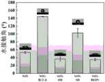
图4为本发明实施例3的促内皮化的镍钛合金血管支架表面涂层的水接触角测试结果;Fig. 4 is the water contact angle test result of the nickel-titanium alloy vascular stent surface coating that promotes endothelialization of embodiment 3 of the present invention;
图5为本发明实施例3的促内皮化的镍钛合金血管支架表面涂层表面的X射线光电子能谱(XPS)测试结果,其中图(a)为血管支架用镍钛合金改性表面全谱图,图(b)为血管支架用镍钛合金表面O1s轨道精细谱图,图(c)为血管支架用镍钛合金表面羟基化后O1s轨道精细谱图,图(d)为血管支架用镍钛合金硅烷层改性后S 2p轨道精细谱图,图(e)为血管支架用镍钛合金活性多肽层构建后C1s轨道精细谱图,图(f)为血管支架用镍钛合金活性多肽层构建后C1s轨道精细谱图;Fig. 5 is the X-ray photoelectron spectrum (XPS) test result of the nickel-titanium alloy vascular stent surface coating surface that promotes the endothelialization of the embodiment of the present invention 3, wherein figure (a) is that the vascular stent is fully modified with nickel-titanium alloy surface Spectrum, picture (b) is the fine spectrum of O1s orbital on the surface of nickel-titanium alloy for blood vessel stent, picture (c) is the fine spectrum of O1s orbital after hydroxylation on the surface of nickel-titanium alloy for blood vessel stent, picture (d) is for blood vessel stent Fine spectrum of
图6为人脐静脉内皮细胞(HUVECs)在本发明实施例3的支架用镍钛合金活性多肽层改性表面的生长增殖情况。Fig. 6 shows the growth and proliferation of human umbilical vein endothelial cells (HUVECs) on the surface modified with a nickel-titanium alloy active polypeptide layer for the scaffold in Example 3 of the present invention.
具体实施方式Detailed ways
结合参见图1-6所示,一种促内皮化的镍钛合金血管支架表面涂层;微纳起伏结构包括凹陷区和凸出区,凹陷区包括纳米级凹陷区和微米级凹陷区,凸出区包括纳米级凸出区和微米级凸出区。本发明综合利用微纳起伏结构及选择性粘附内皮细胞活性多肽化学接枝的突出优势,构建一种能够快速促内皮化的血管支架表面涂层技术。As shown in Figure 1-6, a nickel-titanium alloy stent surface coating for promoting endothelialization; the micro-nano undulating structure includes a concave area and a convex area, and the concave area includes a nano-scale concave area and a micron-scale concave area, and the convex area The out region includes a nanoscale protruding region and a microscale protruding region. The present invention comprehensively utilizes the outstanding advantages of the micro-nano undulating structure and the selective adhesion of endothelial cell active polypeptide chemical grafting to construct a surface coating technology for blood vessel stents that can rapidly promote endothelialization.
一方面,本发明所提供的促内皮化的镍钛合金血管支架表面涂层由微纳起伏结构和活性多肽层所构成,其中,微纳起伏结构有利于血液环境中血细胞、蛋白及活性物质的吸附,生物相容性良好,而活性多肽层能够特异性地促进内皮细胞有序生长,从而构建特异性的促内皮细胞生长表面,两者协同作用可有效促进材料表面快速实现内皮化,以起到良好的长期抗凝血,抗增生作用,避免血管再狭窄及迟发性血栓等问题出现;On the one hand, the nickel-titanium alloy stent surface coating for promoting endothelialization provided by the present invention is composed of a micro-nano undulating structure and an active polypeptide layer, wherein the micro-nano undulating structure is beneficial to the blood cells, proteins and active substances in the blood environment. Adsorption, good biocompatibility, and the active polypeptide layer can specifically promote the orderly growth of endothelial cells, thereby constructing a specific surface for promoting endothelial cell growth. Good long-term anticoagulant and antiproliferative effects, avoiding vascular restenosis and delayed thrombosis;
另一方面,本发明实施例提供的上述一种促内皮化的镍钛合金血管支架表面涂层及其制备方法,除了具有上述有益效果之外,还具有工艺简单易行,成本低廉等特点,可广泛适用于医用镍钛合金血管支架材料功能化活性表面制备。On the other hand, the above-mentioned endothelialization-promoting nickel-titanium alloy stent surface coating and its preparation method provided by the embodiments of the present invention, in addition to the above-mentioned beneficial effects, also have the characteristics of simple process and low cost. It can be widely used in the preparation of functionalized active surface of medical nickel-titanium alloy vascular stent material.
本发明还公开了一些实施例,硅烷层由巯基硅烷偶联剂反应制备,以在支架本体材料表面接枝巯基官能团;The invention also discloses some embodiments, the silane layer is prepared by reacting a mercaptosilane coupling agent to graft mercapto functional groups on the surface of the stent body material;
本发明还公开了一些实施例,巯基硅烷偶联剂为γ-巯丙基三乙氧基硅烷或γ-巯丙基三甲氧基硅烷,可充分利用巯基硅烷偶联剂中所含硅烷基(即三甲基硅烷)与羟基的反应,将巯基引入至支架本体材料表面。The present invention also discloses some embodiments, the mercaptosilane coupling agent is gamma-mercaptopropyltriethoxysilane or gamma-mercaptopropyltrimethoxysilane, which can make full use of the silyl groups contained in the mercaptosilane coupling agent ( That is, the reaction between trimethylsilane) and hydroxyl groups introduces mercapto groups to the surface of the stent body material.
本发明还公开了一些实施例,活性多肽层的多肽序列包括精氨酸—甘氨酸—天冬氨酸(RGD)或精氨酸—谷氨酸—天冬氨酸—缬氨酸(REDV)或酪氨酸—异亮氨酸—甘氨酸—丝氨酸—精氨酸(YIGSR)的多肽序列,上述活性多肽对血管环境中的内皮细胞/血液环境中的内皮祖细胞具有特异性选择性粘附作用,能够促进内皮细胞/内皮祖细胞的粘附、增殖;The present invention also discloses some embodiments, the polypeptide sequence of the active polypeptide layer includes arginine-glycine-aspartic acid (RGD) or arginine-glutamic acid-aspartic acid-valine (REDV) or The polypeptide sequence of tyrosine-isoleucine-glycine-serine-arginine (YIGSR), the above-mentioned active polypeptide has specific and selective adhesion to endothelial cells in the vascular environment/endothelial progenitor cells in the blood environment, Can promote the adhesion and proliferation of endothelial cells/endothelial progenitor cells;
本发明还公开了一些实施例,活性多肽层的多肽序列的端部接入含巯基官能团的半胱氨酸(C);The present invention also discloses some embodiments, the end of the polypeptide sequence of the active polypeptide layer is connected with cysteine (C) containing a sulfhydryl functional group;
本发明还公开了一些实施例,含巯基官能团的半胱氨酸后的多肽序列为甘氨酸—精氨酸—甘氨酸—天冬氨酸—丝氨酸—脯氨酸—半胱氨酸(GRGDSPC);或,半胱氨酸—精氨酸—谷氨酸—天冬氨酸—缬氨酸—赖氨酸(CREDVK);或,半胱氨酸—酪氨酸—异亮氨酸—甘氨酸—丝氨酸—精氨酸(CYIGSR),这些多肽序列一方面含有上述所述活性多肽层的多肽序列(如:RGD、REDV、YIGSR),仍保持对内皮细胞/内皮祖细胞的特异性选择性粘附作用,另一方面,其含有的半胱氨酸(C)中含有巯基官能团,因此可与上述支架本体材料表面已接枝的巯基官能团脱氢形成二硫键,从而产生多肽自组装反应,完成活性多肽层的构建;The present invention also discloses some embodiments, the polypeptide sequence after cysteine containing sulfhydryl functional group is glycine-arginine-glycine-aspartic acid-serine-proline-cysteine (GRGDSPC); or , cysteine-arginine-glutamic acid-aspartic acid-valine-lysine (CREDVK); or, cysteine-tyrosine-isoleucine-glycine-serine- Arginine (CYIGSR), on the one hand, these polypeptide sequences contain the polypeptide sequences of the above-mentioned active polypeptide layer (such as: RGD, REDV, YIGSR), and still maintain the specific and selective adhesion to endothelial cells/endothelial progenitor cells, On the other hand, the cysteine (C) contained in it contains a sulfhydryl functional group, so it can dehydrogenate with the grafted sulfhydryl functional group on the surface of the scaffold body material to form a disulfide bond, thereby generating a polypeptide self-assembly reaction and completing the active polypeptide Layer construction;
本发明还公开了一些实施例,活性多肽层具有特异性选择性粘附内皮细胞/内皮祖细胞的作用。The invention also discloses some embodiments, the active polypeptide layer has the function of specifically and selectively adhering endothelial cells/endothelial progenitor cells.
根据本发明的实施例,提供了一种促内皮化的镍钛合金血管支架,镍钛合金血管支架包括支架本体材料;支架本体材料上设置有表面涂层,表面涂层为上述的血管支架的表面涂层。According to an embodiment of the present invention, a nickel-titanium alloy vascular stent for promoting endothelialization is provided. The nickel-titanium alloy vascular stent includes a stent body material; surface coating.
根据本发明的实施例,提供了一种如上所述血管支架的制备方法,包括如下步骤:According to an embodiment of the present invention, a kind of preparation method of vascular stent as above is provided, comprising the steps:
步骤(1):对支架本体材料表面进行微纳起伏化处理,以使得支架本体材料表面形成微纳起伏结构;Step (1): performing micro-nano relief treatment on the surface of the stent body material, so that the surface of the stent body material forms a micro-nano relief structure;
步骤(2):在微纳起伏结构上制备硅烷层;Step (2): preparing a silane layer on the micro-nano relief structure;
步骤(3):在硅烷层上制备活性多肽层。Step (3): preparing an active polypeptide layer on the silane layer.
本发明提供的方法工艺简单,能够赋予血管支架材料快速内皮化,最终实现长期抗凝血、抗增生等多重生物学功能,有效抑制血管再狭窄和迟发性血栓等并发症发生。The method provided by the invention has a simple process, can endothelialize the vascular stent material quickly, finally realize multiple biological functions such as long-term anticoagulation and anti-proliferation, and effectively inhibit the occurrence of complications such as vascular restenosis and delayed thrombosis.
本发明还公开了一些实施例,在步骤(1)之前,促内皮化的镍钛合金血管支架表面涂层的制备方法还包括如下步骤:对支架本体材料表面依次进行去污、清洗,并将支架本体材料置于酸液中进行处理;进一步地,酸液含1-2M硝酸和0.5-1.5M氟化铵;和/或,处理时间为5-15min,以初步去除支架本体材料表面的氧化膜;The present invention also discloses some embodiments. Before step (1), the preparation method of the nickel-titanium alloy vascular stent surface coating for promoting endothelialization also includes the following steps: sequentially decontaminating and cleaning the surface of the stent body material, and applying The stent body material is placed in an acid solution for treatment; further, the acid solution contains 1-2M nitric acid and 0.5-1.5M ammonium fluoride; and/or, the treatment time is 5-15min to initially remove the oxidation on the surface of the stent body material membrane;
本发明还公开了一些实施例,在步骤(1)之后,在步骤(2)之前,促内皮化的镍钛合金血管支架表面涂层的制备方法还包括如下步骤:对微纳起伏结构进行羟基化处理;进一步地,对微纳起伏结构进行羟基化处理包括如下步骤:将支架本体材料浸泡在氢氧化钠溶液中,反应后进行烘干处理,使得支架本体材料微纳起伏结构表面羟基化;进一步地,浸泡反应的温度为70-85℃;进一步地,氢氧化钠溶液浓度为1-10M。The present invention also discloses some embodiments. After step (1) and before step (2), the preparation method of the nickel-titanium alloy vascular stent surface coating for promoting endothelialization further includes the following steps: performing hydroxylation on the micro-nano undulating structure Further, the hydroxylation treatment of the micro-nano relief structure includes the following steps: soaking the stent body material in a sodium hydroxide solution, and performing drying treatment after the reaction, so that the surface of the stent body material micro-nano relief structure is hydroxylated; Further, the temperature of soaking reaction is 70-85°C; further, the concentration of sodium hydroxide solution is 1-10M.
本发明还公开了一些实施例,步骤(1)中,对支架本体材料的表面进行微纳起伏化处理,具体包括如下操作:采用三电极电化学体系,将支架本体材料作为阳极,惰性电极作为阴极,饱和甘汞电极作为参比电极,以硝酸溶液作为电解质溶液,首先在阳极表面施加-1V电压,持续预设时间;随后对阳极进行两次脱合金化处理,以在支架本体材料表面形成微纳起伏结构;The invention also discloses some embodiments. In step (1), the surface of the stent body material is subjected to micro-nano relief treatment, which specifically includes the following operations: using a three-electrode electrochemical system, using the stent body material as the anode, and the inert electrode as the anode. Cathode, saturated calomel electrode as reference electrode, nitric acid solution as electrolyte solution, firstly apply -1V voltage on the surface of the anode for a preset time; then dealloy the anode twice to form on the surface of the stent body material Micro-nano relief structure;
本发明还公开了一些实施例,预设时间为1-5min,以进一步充分去除阳极材料表面氧化膜;和/或,所述两次脱合金化处理包括如下步骤:第一次脱合金化处理施加电压为3-5V,处理时间为1-60min,第二次脱合金化处理施加电压为1-3V,处理时间为10-120min,其具体效果为第一次脱合金化处理是通过采用相对高电压(3-5V)产生的大电流,使整体支架本体材料表面发生脱合金反应,以快速获得均匀的微纳起伏结构层,第二次脱合金化处理是通过采用相对低电压(1-3V)使微纳起伏结构持续发生均匀脱合金,以增加微纳起伏结构层深度。The present invention also discloses some embodiments, the preset time is 1-5min, so as to further fully remove the oxide film on the surface of the anode material; and/or, the two dealloying treatments include the following steps: the first dealloying treatment The applied voltage is 3-5V, the treatment time is 1-60min, the applied voltage of the second dealloying treatment is 1-3V, and the treatment time is 10-120min. The high current generated by the high voltage (3-5V) causes a dealloying reaction to occur on the surface of the overall stent body material to quickly obtain a uniform micro-nano undulating structure layer. The second dealloying treatment is performed by using a relatively low voltage (1- 3V) Continuously and uniformly dealloying the micro-nano relief structure to increase the depth of the micro-nano relief structure layer.
本发明还公开了一些实施例,硝酸溶液浓度为0.1-2M;The invention also discloses some embodiments, the concentration of the nitric acid solution is 0.1-2M;
本发明还公开了一些实施例,步骤(2)微纳起伏结构的表面粗糙度为0.1-10μm。The invention also discloses some embodiments, the surface roughness of the micro-nano relief structure in step (2) is 0.1-10 μm.
本发明还公开了一些实施例,步骤(2)中,对微纳起伏结构上制备硅烷层包括如下步骤:将经过步骤(1)中处理后的支架本体材料表面浸入巯基硅烷偶联剂溶液中,持续预设时间,随后进行干燥处理,以形成硅烷层;The present invention also discloses some embodiments. In step (2), preparing the silane layer on the micro-nano undulating structure includes the following steps: immersing the surface of the stent body material treated in step (1) in the mercaptosilane coupling agent solution , for a predetermined time, followed by drying to form a silane layer;
本发明还公开了一些实施例,巯基硅烷偶联剂溶液用乙酸溶液调节pH至4.5-5.5之间,并滴加50-80μl丙三醇以提高溶液稳定性,以保证微纳起伏结构表面巯基官能团的有效接枝;The invention also discloses some embodiments. The pH of the mercaptosilane coupling agent solution is adjusted to 4.5-5.5 with acetic acid solution, and 50-80 μl of glycerol is added dropwise to improve the stability of the solution, so as to ensure that the mercapto groups on the surface of the micro-nano undulating structure Effective grafting of functional groups;
本发明还公开了一些实施例,巯基硅烷偶联剂溶液包括体积分数为35-45%去离子水、45-55%甲醇和5-20%的巯基硅烷偶联剂;The invention also discloses some embodiments, the mercaptosilane coupling agent solution includes 35-45% deionized water, 45-55% methanol and 5-20% mercaptosilane coupling agent by volume fraction;
本发明还公开了一些实施例,预设时间为12-48h。The present invention also discloses some embodiments, and the preset time is 12-48h.
本发明还公开了一些实施例,步骤(3)中,在硅烷层上进行活性多肽层制备包括如下步骤:The present invention also discloses some embodiments. In step (3), the preparation of the active polypeptide layer on the silane layer includes the following steps:
将经过步骤(2)处理的支架本体材料浸入含活性多肽、辣根过氧化氢酶、酪胺盐酸盐的PBS溶液中进行充分反应;Immersing the stent body material treated in step (2) in a PBS solution containing active polypeptide, horseradish catalase, and tyramine hydrochloride to fully react;
本发明还公开了一些实施例,活性多肽的浓度为0.3-2mg/mL;和/或,辣根过氧化氢酶的浓度为5-30U/mL;和/或,酪胺盐酸盐的浓度为10-50mM;The present invention also discloses some embodiments, the concentration of active polypeptide is 0.3-2mg/mL; and/or, the concentration of horseradish catalase is 5-30U/mL; and/or, the concentration of tyramine hydrochloride 10-50mM;
本发明还公开了一些实施例,将经过步骤(3)处理的支架本体材料浸入含活性多肽、辣根过氧化氢酶、酪胺盐酸盐的PBS溶液中反应的时间为12-48h。The invention also discloses some embodiments. The time for immersing the scaffold body material treated in step (3) in the PBS solution containing active polypeptide, horseradish catalase and tyramine hydrochloride is 12-48h.
下面通过具体实验实施例进一步对本发明说明如下:Below by concrete experiment embodiment, the present invention is further described as follows:
实施例1Example 1
以镍钛合金血管支架进行促内皮化涂层构建,其中,镍钛合金支架由镍钛合金薄壁管(OD=2.8mm、WT=100μm)经激光切割、清洗、机械扩张、热处理定型、电解抛光、清洗等步骤加工而成,使用扫描电子显微镜观察支架的微观形貌,并进行支架材料化学成分的EDS表征。The endothelialization-promoting coating is constructed with nickel-titanium alloy stents. The nickel-titanium alloy stent is made of nickel-titanium alloy thin-walled tubes (OD=2.8mm, WT=100μm) after laser cutting, cleaning, mechanical expansion, heat treatment and electrolysis. Polishing, cleaning and other steps of processing, using scanning electron microscope to observe the microscopic morphology of the scaffold, and carry out the EDS characterization of the chemical composition of the scaffold material.
加工后的支架长度约为25.5mm(附图2(a)),由单菱形单元和双菱形单元组成(附图2(b)),两结构单元之间以圆角过渡(附图2(c,d))连接以减少应力集中,并防止支架过于尖锐导致血管损伤,支架网丝宽度为0.051mm±0.007mm,壁厚为0.056mm±0.006mm。EDS分析表明,支架材料为含重量百分比为45.4%钛元素和54.6%镍元素的镍钛合金,加工后依次经去离子水、无水乙醇冲洗,烘干备用。The length of the processed stent is about 25.5mm (attachment 2(a)), consisting of a single rhombus unit and a double rhombus unit (attachment 2(b)), and the transition between the two structural units is rounded (attachment 2( c, d)) Connect to reduce stress concentration and prevent the stent from being too sharp to cause blood vessel damage. The width of the stent mesh is 0.051mm±0.007mm, and the wall thickness is 0.056mm±0.006mm. EDS analysis showed that the scaffold material was a nickel-titanium alloy containing 45.4% titanium and 54.6% nickel by weight. After processing, it was rinsed with deionized water and absolute ethanol in turn, and dried for later use.
实施例2Example 2
本实施例构建促内皮化的镍钛合金血管支架表面涂层的微纳起伏结构,主要实施如下:In this embodiment, the micro-nano undulating structure of the surface coating of the nickel-titanium alloy stent that promotes endothelialization is constructed, and the main implementation is as follows:
(1)预处理:将血管支架用镍钛合金去污、清洗,于含1.5M硝酸,1.5M氟化铵的酸液中浸泡10min,以初步去除材料表面氧化膜;(1) Pretreatment: decontaminate and clean the vascular stent with nickel-titanium alloy, and soak it in an acid solution containing 1.5M nitric acid and 1.5M ammonium fluoride for 10 minutes to initially remove the oxide film on the surface of the material;
(2)微纳起伏结构构建:采用三电极电化学体系,将步骤(1)处理材料作为阳极,惰性电极作为阴极,饱和甘汞电极作为参比电极,以1M硝酸溶液作为电解质溶液,首先在电极表面施加-1V电压,持续时间为1min,以充分去除阳极材料表面氧化膜,再在电极表面施加3V电压,持续时间为5min,最后在电极表面施加2V电压,持续时间为30min。之后依次使用去离子水、乙醇清洗支架材料,烘干备用,如附图3所示,微纳起伏结构表面粗糙度均在微纳米级尺度。(2) Micro-nano undulating structure construction: a three-electrode electrochemical system is adopted, the material treated in step (1) is used as the anode, the inert electrode is used as the cathode, the saturated calomel electrode is used as the reference electrode, and 1M nitric acid solution is used as the electrolyte solution. Apply a voltage of -1V to the surface of the electrode for 1min to fully remove the oxide film on the surface of the anode material, then apply a voltage of 3V to the surface of the electrode for 5min, and finally apply a voltage of 2V to the surface of the electrode for 30min. Afterwards, the support material was washed with deionized water and ethanol in sequence, and dried for later use. As shown in Figure 3, the surface roughness of the micro-nano undulating structure was in the micro-nano scale.
实施例3Example 3
(1)预处理:将血管支架用镍钛合金材料去污、清洗,于含1M硝酸,1M氟化铵的酸液中浸泡15min,以除去材料表面氧化膜;(1) Pretreatment: decontaminate and clean the vascular stent with nickel-titanium alloy material, and soak it in an acid solution containing 1M nitric acid and 1M ammonium fluoride for 15 minutes to remove the oxide film on the surface of the material;
(2)微纳起伏结构构建:采用三电极电化学体系,将步骤(1)处理材料作为阳极,惰性电极作为阴极,饱和甘汞电极作为参比电极,以2M硝酸溶液作为电解质溶液,首先在电极表面施加-1V电压,持续时间为5min,以充分去除阳极材料表面氧化膜,再在电极表面施加5V电压,持续时间为3min,最后在电极表面施加2V电压,持续时间为20min。之后依次使用去离子水、乙醇清洗支架材料,烘干备用;(2) Construction of micro-nano undulating structure: a three-electrode electrochemical system is adopted, the material treated in step (1) is used as the anode, the inert electrode is used as the cathode, the saturated calomel electrode is used as the reference electrode, and 2M nitric acid solution is used as the electrolyte solution. Apply a voltage of -1V to the surface of the electrode for 5 minutes to fully remove the oxide film on the surface of the anode material, then apply a voltage of 5V to the surface of the electrode for 3 minutes, and finally apply a voltage of 2V to the surface of the electrode for 20 minutes. Afterwards, use deionized water and ethanol to clean the scaffold material in turn, and dry it for later use;
(3)热碱溶液改性:将步骤(2)处理材料浸泡在2M氢氧化钠溶液中,80℃反应24h,随后进行烘干处理,如附图5(b)、(c)所示,两者对比可知,热碱处理后材料表面在能量约为531eV处形成-OH基团特征峰,-OH基团特征峰对O1s峰面积占比由初始20.76at%上升到34.24at%,表明该热碱改性过程实现了镍钛合金血管支架表面羟基化;(3) Modification with hot alkali solution: soak the material treated in step (2) in 2M sodium hydroxide solution, react at 80°C for 24 hours, and then perform drying treatment, as shown in Figure 5(b) and (c), The comparison between the two shows that after the thermal alkali treatment, the characteristic peak of the -OH group is formed on the surface of the material at an energy of about 531eV, and the area ratio of the characteristic peak of the -OH group to the O1s peak increases from the initial 20.76at% to 34.24at%. The surface hydroxylation of nickel-titanium alloy vascular stents was realized by the hot-alkali modification process;
(4)硅烷层制备:将步骤(3)处理材料浸入巯基硅烷偶联剂溶液中反应12h,随后于110℃下干燥1h,使材料表面硅烷化。其中,巯基硅烷偶联剂溶液由体积分数为40%去离子水、50%甲醇和10%的γ-巯丙基三乙氧基硅烷所组成,用乙酸调节巯基硅烷偶联剂溶液pH至4.5-5.5之间,并滴加75μ的丙三醇以提高巯基硅烷偶联剂溶液的稳定性。如附图5(d)所示,改性后材料表面在能量约为163eV处出现明显峰位,其为巯基特征峰,证明硅烷偶联剂成功偶联至镍钛合金血管支架表面上;(4) Preparation of silane layer: immerse the material treated in step (3) into the mercaptosilane coupling agent solution for 12 hours to react, and then dry at 110° C. for 1 hour to silanize the surface of the material. Wherein, the mercaptosilane coupling agent solution is composed of 40% deionized water, 50% methanol and 10% γ-mercaptopropyltriethoxysilane by volume fraction, and the pH of the mercaptosilane coupling agent solution is adjusted to 4.5 with acetic acid Between -5.5, and 75 μ of glycerol was added dropwise to improve the stability of the mercaptosilane coupling agent solution. As shown in Figure 5(d), the surface of the modified material has an obvious peak at an energy of about 163eV, which is a characteristic peak of mercapto, which proves that the silane coupling agent is successfully coupled to the surface of the nickel-titanium alloy stent;
(5)活性多肽层构建:将步骤(4)处理材料浸入含1mg/mL活性多肽CREDVK、10U/mL辣根过氧化氢酶、35mM酪胺盐酸盐的PBS溶液中,反应12h后取出,制备过程中进行搅拌,搅拌速度为100rpm,随后进行清洗、烘干。对附图5(e)、(f)进行分析比较,材料表面出现NH-C=O特征峰,表明活性多肽REDV成功接枝在镍钛支架材料表面。(5) Construction of the active polypeptide layer: immerse the material treated in step (4) into a PBS solution containing 1 mg/mL active polypeptide CREDVK, 10 U/mL horseradish catalase, and 35 mM tyramine hydrochloride, and take it out after 12 hours of reaction. Stirring is carried out during the preparation process, and the stirring speed is 100 rpm, followed by cleaning and drying. Analyzing and comparing accompanying drawings 5(e) and (f), the characteristic peaks of NH-C=O appeared on the surface of the material, indicating that the active polypeptide REDV was successfully grafted on the surface of the nickel-titanium scaffold material.
现基于实施例3,将镍钛合金抛光样品表面设置成对照组,将支架用镍钛合金活性多肽层改性表面样品表面设置为实验组,对血管支架用镍钛合金活性多肽层改性表面进行HUVECs生长增殖实验评价。Now based on Example 3, the surface of the nickel-titanium alloy polished sample is set as a control group, the surface of the sample surface modified by the nickel-titanium alloy active polypeptide layer for the stent is set as the experimental group, and the surface modified by the nickel-titanium alloy active polypeptide layer for the vascular stent HUVECs growth and proliferation test evaluation was carried out.
37℃下,将实验组和对照组样品置于含10000个HUVECs/孔完全培养基的48孔板中,分别培养1天、3天,固定表面HUVECs后,采用扫描电镜对样品表面进行观察和分析。附图6显示了支架用镍钛合金活性多肽层改性表面HUVECs生长增殖结果,表明活性多肽层可显著促进HUVECs的粘附、生长及增殖,同时抑制血小板、纤维蛋白原等血液成分的粘附,有助于镍钛支架材料植入体内后快速内皮化。At 37°C, the samples of the experimental group and the control group were placed in 48-well plates containing 10,000 HUVECs/well complete medium, and cultured for 1 day and 3 days respectively. After fixing the HUVECs on the surface, the surface of the samples was observed and analyzed by scanning electron microscope analyze. Accompanying drawing 6 shows the growth and proliferation results of HUVECs on the surface modified with nickel-titanium alloy active polypeptide layer, indicating that the active polypeptide layer can significantly promote the adhesion, growth and proliferation of HUVECs, and at the same time inhibit the adhesion of blood components such as platelets and fibrinogen , contribute to rapid endothelialization of nickel-titanium stent material after implantation in vivo.
实施例4Example 4
(1)预处理:将血管支架用镍钛合金去污、清洗,于含1M硝酸,0.8M氟化铵的酸液中浸泡15min,以除去材料表面氧化膜;(1) Pretreatment: decontaminate and clean the vascular stent with nickel-titanium alloy, and soak it in an acid solution containing 1M nitric acid and 0.8M ammonium fluoride for 15 minutes to remove the oxide film on the surface of the material;
(2)微纳起伏结构构建:采用三电极电化学体系,将步骤(1)处理材料作为阳极,惰性电极作为阴极,饱和甘汞电极作为参比电极,以1.5M硝酸溶液作为电解质溶液,首先在电极表面施加-1V电压,持续时间为2min,以充分去除阳极材料表面氧化膜,再在电极表面施加5V电压,持续时间为3min,最后在电极表面施加2.5V电压,持续时间为30min。之后依次使用去离子水、乙醇清洗支架材料,烘干备用;(2) Micro-nano undulating structure construction: using a three-electrode electrochemical system, the material treated in step (1) is used as an anode, an inert electrode is used as a cathode, a saturated calomel electrode is used as a reference electrode, and 1.5M nitric acid solution is used as an electrolyte solution. Apply a voltage of -1V on the surface of the electrode for 2 minutes to fully remove the oxide film on the surface of the anode material, then apply a voltage of 5V on the surface of the electrode for 3 minutes, and finally apply a voltage of 2.5V on the surface of the electrode for 30 minutes. Afterwards, use deionized water and ethanol to clean the scaffold material in turn, and dry it for later use;
(3)热碱溶液改性:将步骤(2)处理材料浸泡在10M氢氧化钠溶液中。80℃反应12h,随后进行烘干处理,实现镍钛合金血管支架表面羟基化;(3) Modification with hot alkali solution: soak the material treated in step (2) in 10M sodium hydroxide solution. Reaction at 80°C for 12 hours, followed by drying to achieve surface hydroxylation of the nickel-titanium alloy stent;
(4)硅烷层制备:将步骤(3)处理材料浸入巯基硅烷偶联剂溶液中反应24h,随后于80℃下干燥3h,使材料表面硅烷化。其中,巯基硅烷偶联剂溶液由体积分数为35%去离子水、45%甲醇和20%的γ-巯丙基三乙氧基硅烷所组成,用乙酸调节巯基硅烷偶联剂溶液pH至4.5-5.5之间,并滴加60μl的丙三醇以提高巯基硅烷偶联剂溶液的稳定性;(4) Preparation of silane layer: immerse the material treated in step (3) into the mercaptosilane coupling agent solution for 24 hours, and then dry at 80° C. for 3 hours to silanize the surface of the material. Wherein, the mercaptosilane coupling agent solution is composed of 35% deionized water, 45% methanol and 20% γ-mercaptopropyltriethoxysilane by volume fraction, and the pH of the mercaptosilane coupling agent solution is adjusted to 4.5 with acetic acid -5.5, and drop 60 μl of glycerol to improve the stability of the mercaptosilane coupling agent solution;
(5)活性多肽层构建:将步骤(4)处理材料浸入含1.5mg/mL活性多肽CYIGSR、15U/mL辣根过氧化氢酶、30mM酪胺盐酸盐的PBS溶液中,反应18h后取出,制备过程中进行搅拌,搅拌速度为150rpm,随后进行清洗、烘干。(5) Active polypeptide layer construction: immerse the material treated in step (4) into a PBS solution containing 1.5mg/mL active polypeptide CYIGSR, 15U/mL horseradish catalase, and 30mM tyramine hydrochloride, and take it out after 18 hours of reaction , stirring during the preparation process, the stirring speed is 150rpm, followed by cleaning and drying.
实施例5Example 5
(1)预处理:将血管支架用镍钛合金去污、清洗,于含2M硝酸,0.5M氟化铵的酸液中浸泡5min,以除去材料表面氧化膜;(1) Pretreatment: decontaminate and clean the vascular stent with nickel-titanium alloy, and soak it in an acid solution containing 2M nitric acid and 0.5M ammonium fluoride for 5 minutes to remove the oxide film on the surface of the material;
(2)微纳起伏结构构建:采用三电极电化学体系,将步骤(1)处理材料作为阳极,惰性电极作为阴极,饱和甘汞电极作为参比电极,以0.5M硝酸溶液作为电解质溶液,首先在电极表面施加-1V电压,持续时间为1min,以充分去除阳极材料表面氧化膜,再在电极表面施加3V电压,持续时间为5min,最后在电极表面施加5V电压,持续时间为20min。之后依次使用去离子水、乙醇清洗支架材料,烘干备用;(2) Construction of micro-nano undulating structure: a three-electrode electrochemical system is adopted, the material treated in step (1) is used as the anode, the inert electrode is used as the cathode, the saturated calomel electrode is used as the reference electrode, and 0.5M nitric acid solution is used as the electrolyte solution. Apply a voltage of -1V on the surface of the electrode for 1min to fully remove the oxide film on the surface of the anode material, then apply a voltage of 3V on the surface of the electrode for 5min, and finally apply a voltage of 5V on the surface of the electrode for 20min. Afterwards, use deionized water and ethanol to clean the scaffold material in turn, and dry it for later use;
(3)热碱溶液改性:将步骤(2)处理材料浸泡在5M氢氧化钠溶液中。80℃反应18h,随后进行烘干处理,实现镍钛合金血管支架表面羟基化;(3) Modification with hot alkali solution: soak the material treated in step (2) in 5M sodium hydroxide solution. Reaction at 80°C for 18 hours, followed by drying to achieve surface hydroxylation of the nickel-titanium alloy stent;
(4)硅烷层制备:将步骤(3)处理材料浸入巯基硅烷偶联剂溶液中反应48h,随后于100℃下干燥2h,使材料表面硅烷化。其中,巯基硅烷偶联剂溶液由体积分数为40%去离子水、45%甲醇和15%的γ-巯丙基三甲氧基硅烷所组成,用乙酸调节巯基硅烷偶联剂溶液pH至4.5-5.5之间,并滴加80μl的丙三醇以提高巯基硅烷偶联剂溶液的稳定性;(4) Preparation of silane layer: immerse the material treated in step (3) into the mercaptosilane coupling agent solution for reaction for 48 hours, and then dry at 100° C. for 2 hours to silanize the surface of the material. Wherein, the mercaptosilane coupling agent solution is composed of 40% deionized water, 45% methanol and 15% γ-mercaptopropyltrimethoxysilane by volume fraction, and the pH of the mercaptosilane coupling agent solution is adjusted to 4.5- 5.5, and drop 80 μl of glycerol to improve the stability of the mercaptosilane coupling agent solution;
(5)活性多肽层构建:将步骤(4)处理材料浸入含0.5mg/mL活性多肽GRGDSPC、20U/mL辣根过氧化氢酶、25mM酪胺盐酸盐的PBS溶液中,反应12h后取出,制备过程中进行搅拌,搅拌速度为100rpm,随后进行清洗、烘干。(5) Active polypeptide layer construction: immerse the material treated in step (4) into a PBS solution containing 0.5 mg/mL active polypeptide GRGDSPC, 20 U/mL horseradish catalase, and 25 mM tyramine hydrochloride, and take it out after 12 hours of reaction , stirring during the preparation process, the stirring speed is 100rpm, followed by cleaning and drying.
本领域的技术人员容易理解的是,在不冲突的前提下,上述各有利方式可以自由地组合、叠加。Those skilled in the art can easily understand that, on the premise of no conflict, the above-mentioned advantageous modes can be freely combined and superimposed.
以上仅为本发明的较佳实施例而已,并不用以限制本发明,凡在本发明的精神和原则之内所作的任何修改、等同替换和改进等,均应包含在本发明的保护范围之内。以上仅是本发明的优选实施方式,应当指出,对于本技术领域的普通技术人员来说,在不脱离本发明技术原理的前提下,还可以做出若干改进和变型,这些改进和变型也应视为本发明的保护范围。The above are only preferred embodiments of the present invention, and are not intended to limit the present invention. Any modifications, equivalent replacements and improvements made within the spirit and principles of the present invention should be included in the protection scope of the present invention. Inside. The above are only preferred embodiments of the present invention, it should be pointed out that for those of ordinary skill in the art, without departing from the technical principles of the present invention, some improvements and modifications can also be made, and these improvements and modifications should also be It is regarded as the protection scope of the present invention.
Claims (19)
Priority Applications (1)
| Application Number | Priority Date | Filing Date | Title |
|---|---|---|---|
| CN202210732765.9ACN115054737B (en) | 2022-06-27 | 2022-06-27 | A kind of endothelialization-promoting nickel-titanium alloy vascular stent surface coating and preparation method thereof |
Applications Claiming Priority (1)
| Application Number | Priority Date | Filing Date | Title |
|---|---|---|---|
| CN202210732765.9ACN115054737B (en) | 2022-06-27 | 2022-06-27 | A kind of endothelialization-promoting nickel-titanium alloy vascular stent surface coating and preparation method thereof |
Publications (2)
| Publication Number | Publication Date |
|---|---|
| CN115054737A CN115054737A (en) | 2022-09-16 |
| CN115054737Btrue CN115054737B (en) | 2023-06-06 |
Family
ID=83202939
Family Applications (1)
| Application Number | Title | Priority Date | Filing Date |
|---|---|---|---|
| CN202210732765.9AActiveCN115054737B (en) | 2022-06-27 | 2022-06-27 | A kind of endothelialization-promoting nickel-titanium alloy vascular stent surface coating and preparation method thereof |
Country Status (1)
| Country | Link |
|---|---|
| CN (1) | CN115054737B (en) |
Family Cites Families (6)
| Publication number | Priority date | Publication date | Assignee | Title |
|---|---|---|---|---|
| WO2011057107A2 (en)* | 2009-11-06 | 2011-05-12 | The University Of Notre Dame Du Lac | Microchamber electrochemical cell having a nanoslot |
| KR20160123075A (en)* | 2015-04-15 | 2016-10-25 | 오스템임플란트 주식회사 | Implant having a surface of micro-nano composite structure and method of surface-treating implant |
| CN109988814B (en)* | 2019-03-20 | 2022-08-16 | 华南理工大学 | Bioactive polypeptide grafting density high-throughput screening method for gold/titanium surface modification |
| CN112535560B (en)* | 2020-11-30 | 2022-01-14 | 中国科学院金属研究所 | Super-soft smooth nickel-titanium alloy intracranial intravascular stent with micro-nano structure |
| CN113425914B (en)* | 2021-06-17 | 2022-09-16 | 河北工业大学 | Medical material for promoting cell growth and inhibiting bacterial adhesion and processing method thereof |
| CN114504723A (en)* | 2021-12-22 | 2022-05-17 | 融冲(深圳)生物医疗科技有限责任公司 | Polymer balloon and preparation method thereof |
- 2022
- 2022-06-27CNCN202210732765.9Apatent/CN115054737B/enactiveActive
Non-Patent Citations (1)
| Title |
|---|
| Recent progress on magnetic iron oxide nanoparticles: synthesis, surface functional strategies and biomedical applications;Wei Wu 等;National Institute for Materials Science;第16卷(第2期);1-43* |
Also Published As
| Publication number | Publication date |
|---|---|
| CN115054737A (en) | 2022-09-16 |
Similar Documents
| Publication | Publication Date | Title |
|---|---|---|
| JP3485264B2 (en) | Biocompatible medical metal material to which a physiologically active substance is bound and method for producing the same | |
| Syed et al. | Evaluation of decellularization protocols for production of tubular small intestine submucosa scaffolds for use in oesophageal tissue engineering | |
| US10729821B1 (en) | Method for preparing chitosan/heparinized graphene oxide composite multilayer film on surface of medical magnesium alloy | |
| CN101310778B (en) | Method for fixing antibody on medical appliance | |
| CN107096068A (en) | A kind of preparation method of dentistry implant and its bioactivity antimicrobial surface | |
| CN101130114A (en) | Biocompatible surface coating for implantable medical devices and coating method thereof | |
| CN101361988B (en) | Preparation method of blood vessel support or cardiac valve surface coating with good biocompatibility | |
| CN101357241B (en) | CD34 antibody or CD133 antibody surface orientation fixing method of titanium and titanium alloy cardiovascular implantation device | |
| CN107185055B (en) | A kind of surface modification method of medical magnesium alloy | |
| CN102793947B (en) | Degradable magnesium and surface modification method of alloy thereof | |
| US12303616B2 (en) | Anti-thrombotic and endothelialization-enhanced bioprosthetic valve material and preparation method thereof | |
| CN111467574A (en) | Biological valve material based on EDC/NHS activation and recombinant human collagen modification and preparation method thereof | |
| CN106267342A (en) | A kind of dentistry implant and preparation method thereof | |
| CN110292665A (en) | A kind of composite coating and preparation method thereof of the corrosion-resistant anti-oxidant anti-inflammatory of Mg alloy surface | |
| CN116236614A (en) | TiO for catalyzing and releasing CO 2 Nanotube material, preparation method and application thereof | |
| CN114904062B (en) | A vascular stent with selective biofunctionalization and its preparation method | |
| CN115054737B (en) | A kind of endothelialization-promoting nickel-titanium alloy vascular stent surface coating and preparation method thereof | |
| CN112915260B (en) | A kind of non-glutaraldehyde cross-linked biological material and preparation method thereof | |
| CN113368315B (en) | A kind of medical nickel-titanium alloy material with temperature-sensitive hydrogel coating and its preparation method and application | |
| CN111603615B (en) | Controllable degradable high-strength magnesium-based composite stent composite coating and preparation method thereof | |
| CN109646722A (en) | A kind of intravascular stent surface treatment method | |
| CN116712607A (en) | Application of a covalently grafted albumin coating | |
| CN107119269A (en) | The acid coated method of the winestone with corrosion protection effect is built in magnesium based metal | |
| CN102085125B (en) | Method for fixing polypeptide aptamers on surface of cardiovascular implanting device | |
| CN115261939B (en) | A method for preparing composite coating on titanium surface |
Legal Events
| Date | Code | Title | Description |
|---|---|---|---|
| PB01 | Publication | ||
| PB01 | Publication | ||
| SE01 | Entry into force of request for substantive examination | ||
| SE01 | Entry into force of request for substantive examination | ||
| GR01 | Patent grant | ||
| GR01 | Patent grant |