CN114848846A - Drug delivery system and preparation method and application thereof - Google Patents
Drug delivery system and preparation method and application thereofDownload PDFInfo
- Publication number
- CN114848846A CN114848846ACN202210585525.0ACN202210585525ACN114848846ACN 114848846 ACN114848846 ACN 114848846ACN 202210585525 ACN202210585525 ACN 202210585525ACN 114848846 ACN114848846 ACN 114848846A
- Authority
- CN
- China
- Prior art keywords
- quantum dots
- graphene quantum
- magnetic nanoparticles
- drug delivery
- silica
- Prior art date
- Legal status (The legal status is an assumption and is not a legal conclusion. Google has not performed a legal analysis and makes no representation as to the accuracy of the status listed.)
- Granted
Links
- 238000012377drug deliveryMethods0.000titleclaimsabstractdescription48
- 238000002360preparation methodMethods0.000titleclaimsabstractdescription27
- VYPSYNLAJGMNEJ-UHFFFAOYSA-NSilicium dioxideChemical compoundO=[Si]=OVYPSYNLAJGMNEJ-UHFFFAOYSA-N0.000claimsabstractdescription75
- 239000002122magnetic nanoparticleSubstances0.000claimsabstractdescription51
- VNWKTOKETHGBQD-UHFFFAOYSA-NmethaneChemical compoundCVNWKTOKETHGBQD-UHFFFAOYSA-N0.000claimsabstractdescription51
- 238000002866fluorescence resonance energy transferMethods0.000claimsabstractdescription27
- 206010028980NeoplasmDiseases0.000claimsabstractdescription22
- 239000000377silicon dioxideSubstances0.000claimsabstractdescription17
- 229940079593drugDrugs0.000claimsabstractdescription15
- 239000003814drugSubstances0.000claimsabstractdescription15
- 238000003384imaging methodMethods0.000claimsabstractdescription14
- 238000011068loading methodMethods0.000claimsabstractdescription14
- 235000012239silicon dioxideNutrition0.000claimsabstractdescription10
- 238000012544monitoring processMethods0.000claimsabstractdescription9
- 239000002246antineoplastic agentSubstances0.000claimsabstractdescription8
- 229940041181antineoplastic drugDrugs0.000claimsabstractdescription7
- 239000003937drug carrierSubstances0.000claimsabstractdescription7
- 230000002902bimodal effectEffects0.000claimsabstractdescription6
- 239000012792core layerSubstances0.000claimsabstractdescription5
- 239000010410layerSubstances0.000claimsabstractdescription5
- OVBPIULPVIDEAO-LBPRGKRZSA-Nfolic acidChemical compoundC=1N=C2NC(N)=NC(=O)C2=NC=1CNC1=CC=C(C(=O)N[C@@H](CCC(O)=O)C(O)=O)C=C1OVBPIULPVIDEAO-LBPRGKRZSA-N0.000claimsdescription174
- AOJJSUZBOXZQNB-TZSSRYMLSA-NDoxorubicinChemical compoundO([C@H]1C[C@@](O)(CC=2C(O)=C3C(=O)C=4C=CC=C(C=4C(=O)C3=C(O)C=21)OC)C(=O)CO)[C@H]1C[C@H](N)[C@H](O)[C@H](C)O1AOJJSUZBOXZQNB-TZSSRYMLSA-N0.000claimsdescription96
- 239000011724folic acidSubstances0.000claimsdescription89
- 229960000304folic acidDrugs0.000claimsdescription88
- OVBPIULPVIDEAO-UHFFFAOYSA-NN-Pteroyl-L-glutaminsaeureNatural productsC=1N=C2NC(N)=NC(=O)C2=NC=1CNC1=CC=C(C(=O)NC(CCC(O)=O)C(O)=O)C=C1OVBPIULPVIDEAO-UHFFFAOYSA-N0.000claimsdescription87
- 235000019152folic acidNutrition0.000claimsdescription87
- OKTJSMMVPCPJKN-UHFFFAOYSA-NCarbonChemical class[C]OKTJSMMVPCPJKN-UHFFFAOYSA-N0.000claimsdescription24
- 239000010941cobaltSubstances0.000claimsdescription22
- 229910017052cobaltInorganic materials0.000claimsdescription22
- GUTLYIVDDKVIGB-UHFFFAOYSA-Ncobalt atomChemical compound[Co]GUTLYIVDDKVIGB-UHFFFAOYSA-N0.000claimsdescription21
- 229960004679doxorubicinDrugs0.000claimsdescription21
- 229910021389grapheneInorganic materials0.000claimsdescription21
- HEMHJVSKTPXQMS-UHFFFAOYSA-MSodium hydroxideChemical compound[OH-].[Na+]HEMHJVSKTPXQMS-UHFFFAOYSA-M0.000claimsdescription18
- 229910000859α-FeInorganic materials0.000claimsdescription17
- KRKNYBCHXYNGOX-UHFFFAOYSA-Ncitric acidChemical compoundOC(=O)CC(O)(C(O)=O)CC(O)=OKRKNYBCHXYNGOX-UHFFFAOYSA-N0.000claimsdescription16
- 239000000243solutionSubstances0.000claimsdescription15
- XEEYBQQBJWHFJM-UHFFFAOYSA-NIronChemical compound[Fe]XEEYBQQBJWHFJM-UHFFFAOYSA-N0.000claimsdescription14
- 230000008685targetingEffects0.000claimsdescription14
- 238000002156mixingMethods0.000claimsdescription13
- LMDZBCPBFSXMTL-UHFFFAOYSA-N1-ethyl-3-(3-dimethylaminopropyl)carbodiimideChemical compoundCCN=C=NCCCN(C)CLMDZBCPBFSXMTL-UHFFFAOYSA-N0.000claimsdescription11
- DWANEFRJKWXRSG-UHFFFAOYSA-N1,2-tetradecanediolChemical compoundCCCCCCCCCCCCC(O)CODWANEFRJKWXRSG-UHFFFAOYSA-N0.000claimsdescription10
- LFQSCWFLJHTTHZ-UHFFFAOYSA-NEthanolChemical compoundCCOLFQSCWFLJHTTHZ-UHFFFAOYSA-N0.000claimsdescription9
- WYTZZXDRDKSJID-UHFFFAOYSA-N(3-aminopropyl)triethoxysilaneChemical compoundCCO[Si](OCC)(OCC)CCCNWYTZZXDRDKSJID-UHFFFAOYSA-N0.000claimsdescription8
- 238000005576amination reactionMethods0.000claimsdescription8
- 238000006243chemical reactionMethods0.000claimsdescription8
- 238000006482condensation reactionMethods0.000claimsdescription8
- 229910052742ironInorganic materials0.000claimsdescription7
- 239000005642Oleic acidSubstances0.000claimsdescription6
- 239000008367deionised waterSubstances0.000claimsdescription6
- 229910021641deionized waterInorganic materials0.000claimsdescription6
- ZQPPMHVWECSIRJ-KTKRTIGZSA-Noleic acidChemical compoundCCCCCCCC\C=C/CCCCCCCC(O)=OZQPPMHVWECSIRJ-KTKRTIGZSA-N0.000claimsdescription6
- LIVNPJMFVYWSIS-UHFFFAOYSA-Nsilicon monoxideInorganic materials[Si-]#[O+]LIVNPJMFVYWSIS-UHFFFAOYSA-N0.000claimsdescription6
- 239000002904solventSubstances0.000claimsdescription6
- XLYOFNOQVPJJNP-UHFFFAOYSA-NwaterChemical compoundOXLYOFNOQVPJJNP-UHFFFAOYSA-N0.000claimsdescription6
- WRIDQFICGBMAFQ-UHFFFAOYSA-N(E)-8-Octadecenoic acidNatural productsCCCCCCCCCC=CCCCCCCC(O)=OWRIDQFICGBMAFQ-UHFFFAOYSA-N0.000claimsdescription5
- QGLWBTPVKHMVHM-KTKRTIGZSA-N(z)-octadec-9-en-1-amineChemical compoundCCCCCCCC\C=C/CCCCCCCCNQGLWBTPVKHMVHM-KTKRTIGZSA-N0.000claimsdescription5
- LQJBNNIYVWPHFW-UHFFFAOYSA-N20:1omega9c fatty acidNatural productsCCCCCCCCCCC=CCCCCCCCC(O)=OLQJBNNIYVWPHFW-UHFFFAOYSA-N0.000claimsdescription5
- QSBYPNXLFMSGKH-UHFFFAOYSA-N9-HeptadecensaeureNatural productsCCCCCCCC=CCCCCCCCC(O)=OQSBYPNXLFMSGKH-UHFFFAOYSA-N0.000claimsdescription5
- NQTADLQHYWFPDB-UHFFFAOYSA-NN-HydroxysuccinimideChemical compoundON1C(=O)CCC1=ONQTADLQHYWFPDB-UHFFFAOYSA-N0.000claimsdescription5
- -1N-hydroxysuccinimide amineChemical class0.000claimsdescription5
- ZQPPMHVWECSIRJ-UHFFFAOYSA-NOleic acidNatural productsCCCCCCCCC=CCCCCCCCC(O)=OZQPPMHVWECSIRJ-UHFFFAOYSA-N0.000claimsdescription5
- 150000001408amidesChemical class0.000claimsdescription5
- 239000003153chemical reaction reagentSubstances0.000claimsdescription5
- BKFAZDGHFACXKY-UHFFFAOYSA-Ncobalt(II) bis(acetylacetonate)Chemical compound[Co+2].CC(=O)[CH-]C(C)=O.CC(=O)[CH-]C(C)=OBKFAZDGHFACXKY-UHFFFAOYSA-N0.000claimsdescription5
- QXJSBBXBKPUZAA-UHFFFAOYSA-Nisooleic acidNatural productsCCCCCCCC=CCCCCCCCCC(O)=OQXJSBBXBKPUZAA-UHFFFAOYSA-N0.000claimsdescription5
- VHUUQVKOLVNVRT-UHFFFAOYSA-NAmmonium hydroxideChemical compound[NH4+].[OH-]VHUUQVKOLVNVRT-UHFFFAOYSA-N0.000claimsdescription4
- 229910018557Si OInorganic materials0.000claimsdescription4
- 230000004913activationEffects0.000claimsdescription4
- 239000007825activation reagentSubstances0.000claimsdescription4
- 239000003513alkaliSubstances0.000claimsdescription4
- 235000011114ammonium hydroxideNutrition0.000claimsdescription4
- 238000010556emulsion polymerization methodMethods0.000claimsdescription4
- 238000010438heat treatmentMethods0.000claimsdescription4
- UQSXHKLRYXJYBZ-UHFFFAOYSA-Niron oxideInorganic materials[Fe]=OUQSXHKLRYXJYBZ-UHFFFAOYSA-N0.000claimsdescription4
- 239000011259mixed solutionSubstances0.000claimsdescription4
- NDLPOXTZKUMGOV-UHFFFAOYSA-Noxo(oxoferriooxy)iron hydrateChemical compoundO.O=[Fe]O[Fe]=ONDLPOXTZKUMGOV-UHFFFAOYSA-N0.000claimsdescription4
- 238000009833condensationMethods0.000claimsdescription3
- 230000005494condensationEffects0.000claimsdescription3
- USIUVYZYUHIAEV-UHFFFAOYSA-Ndiphenyl etherChemical compoundC=1C=CC=CC=1OC1=CC=CC=C1USIUVYZYUHIAEV-UHFFFAOYSA-N0.000claimsdescription3
- 239000000839emulsionSubstances0.000claimsdescription2
- 230000035484reaction timeEffects0.000claimsdescription2
- LZKLAOYSENRNKR-LNTINUHCSA-Niron;(z)-4-oxoniumylidenepent-2-en-2-olateChemical compound[Fe].C\C(O)=C\C(C)=O.C\C(O)=C\C(C)=O.C\C(O)=C\C(C)=OLZKLAOYSENRNKR-LNTINUHCSA-N0.000claims1
- 201000011510cancerDiseases0.000abstractdescription9
- 238000003745diagnosisMethods0.000abstractdescription8
- 239000000090biomarkerSubstances0.000abstractdescription3
- 238000013270controlled releaseMethods0.000abstractdescription3
- 229910004298SiO 2Inorganic materials0.000description88
- 210000004027cellAnatomy0.000description19
- 238000002595magnetic resonance imagingMethods0.000description17
- 229910052681coesiteInorganic materials0.000description12
- 229910052906cristobaliteInorganic materials0.000description12
- 239000002953phosphate buffered salineSubstances0.000description12
- 229910052682stishoviteInorganic materials0.000description12
- 229910052905tridymiteInorganic materials0.000description12
- 239000002105nanoparticleSubstances0.000description11
- 238000000034methodMethods0.000description10
- IAZDPXIOMUYVGZ-UHFFFAOYSA-NDimethylsulphoxideChemical compoundCS(C)=OIAZDPXIOMUYVGZ-UHFFFAOYSA-N0.000description8
- 238000000799fluorescence microscopyMethods0.000description7
- 239000002096quantum dotSubstances0.000description7
- 238000012360testing methodMethods0.000description7
- BOTDANWDWHJENH-UHFFFAOYSA-NTetraethyl orthosilicateChemical compoundCCO[Si](OCC)(OCC)OCCBOTDANWDWHJENH-UHFFFAOYSA-N0.000description6
- 230000005415magnetizationEffects0.000description6
- 239000002609mediumSubstances0.000description6
- 239000004005microsphereSubstances0.000description6
- 239000000203mixtureSubstances0.000description6
- 239000000872bufferSubstances0.000description5
- 230000000694effectsEffects0.000description5
- 231100000135cytotoxicityToxicity0.000description4
- 230000003013cytotoxicityEffects0.000description4
- 239000012091fetal bovine serumSubstances0.000description4
- AQBLLJNPHDIAPN-LNTINUHCSA-Kiron(3+);(z)-4-oxopent-2-en-2-olateChemical compound[Fe+3].C\C([O-])=C\C(C)=O.C\C([O-])=C\C(C)=O.C\C([O-])=C\C(C)=OAQBLLJNPHDIAPN-LNTINUHCSA-K0.000description4
- 238000001000micrographMethods0.000description4
- 239000002245particleSubstances0.000description4
- 108091003079Bovine Serum AlbuminProteins0.000description3
- HPZOOQSXPMEJBV-ODCFVKFUSA-NTirilazad mesylateChemical groupCS(O)(=O)=O.O=C([C@@H]1[C@@]2(C)CC=C3[C@@]4(C)C=CC(=O)C=C4CC[C@H]3[C@@H]2C[C@H]1C)CN(CC1)CCN1C(N=1)=CC(N2CCCC2)=NC=1N1CCCC1HPZOOQSXPMEJBV-ODCFVKFUSA-N0.000description3
- 238000002835absorbanceMethods0.000description3
- 239000000370acceptorSubstances0.000description3
- 239000007864aqueous solutionSubstances0.000description3
- 230000015572biosynthetic processEffects0.000description3
- 125000003178carboxy groupChemical group[H]OC(*)=O0.000description3
- 239000011248coating agentSubstances0.000description3
- 238000000576coating methodMethods0.000description3
- 238000002474experimental methodMethods0.000description3
- 239000006249magnetic particleSubstances0.000description3
- 239000002114nanocompositeSubstances0.000description3
- 239000002086nanomaterialSubstances0.000description3
- 230000008569processEffects0.000description3
- 229920006395saturated elastomerPolymers0.000description3
- 239000000725suspensionSubstances0.000description3
- 238000003786synthesis reactionMethods0.000description3
- 230000001225therapeutic effectEffects0.000description3
- CSCPPACGZOOCGX-UHFFFAOYSA-NAcetoneChemical compoundCC(C)=OCSCPPACGZOOCGX-UHFFFAOYSA-N0.000description2
- HEDRZPFGACZZDS-UHFFFAOYSA-NChloroformChemical compoundClC(Cl)ClHEDRZPFGACZZDS-UHFFFAOYSA-N0.000description2
- 229910003321CoFeInorganic materials0.000description2
- 239000002616MRI contrast agentSubstances0.000description2
- 230000002378acidificating effectEffects0.000description2
- 239000011543agarose gelSubstances0.000description2
- 125000003277amino groupChemical group0.000description2
- 230000008901benefitEffects0.000description2
- 230000001186cumulative effectEffects0.000description2
- 231100000263cytotoxicity testToxicity0.000description2
- DTPCFIHYWYONMD-UHFFFAOYSA-Ndecaethylene glycolPolymersOCCOCCOCCOCCOCCOCCOCCOCCOCCOCCODTPCFIHYWYONMD-UHFFFAOYSA-N0.000description2
- 230000007423decreaseEffects0.000description2
- 238000001514detection methodMethods0.000description2
- 238000010586diagramMethods0.000description2
- 102000006815folate receptorHuman genes0.000description2
- 108020005243folate receptorProteins0.000description2
- 230000006872improvementEffects0.000description2
- 238000000338in vitroMethods0.000description2
- 238000011534incubationMethods0.000description2
- 230000004048modificationEffects0.000description2
- 238000012986modificationMethods0.000description2
- ZPIRTVJRHUMMOI-UHFFFAOYSA-NoctoxybenzeneChemical compoundCCCCCCCCOC1=CC=CC=C1ZPIRTVJRHUMMOI-UHFFFAOYSA-N0.000description2
- 229920000767polyanilinePolymers0.000description2
- 102000005962receptorsHuman genes0.000description2
- 108020003175receptorsProteins0.000description2
- 238000000926separation methodMethods0.000description2
- 238000003756stirringMethods0.000description2
- 239000000126substanceSubstances0.000description2
- 230000002195synergetic effectEffects0.000description2
- 238000010998test methodMethods0.000description2
- 238000000870ultraviolet spectroscopyMethods0.000description2
- 238000005406washingMethods0.000description2
- OHVLMTFVQDZYHP-UHFFFAOYSA-N1-(2,4,6,7-tetrahydrotriazolo[4,5-c]pyridin-5-yl)-2-[4-[2-[[3-(trifluoromethoxy)phenyl]methylamino]pyrimidin-5-yl]piperazin-1-yl]ethanoneChemical compoundN1N=NC=2CN(CCC=21)C(CN1CCN(CC1)C=1C=NC(=NC=1)NCC1=CC(=CC=C1)OC(F)(F)F)=OOHVLMTFVQDZYHP-UHFFFAOYSA-N0.000description1
- OMIHGPLIXGGMJB-UHFFFAOYSA-N7-oxabicyclo[4.1.0]hepta-1,3,5-trieneChemical compoundC1=CC=C2OC2=C1OMIHGPLIXGGMJB-UHFFFAOYSA-N0.000description1
- 239000006144Dulbecco’s modified Eagle's mediumSubstances0.000description1
- 239000007987MES bufferSubstances0.000description1
- 231100000002MTT assayToxicity0.000description1
- 238000000134MTT assayMethods0.000description1
- 229920004890Triton X-100Polymers0.000description1
- 239000013504Triton X-100Substances0.000description1
- 229910021529ammoniaInorganic materials0.000description1
- 238000004458analytical methodMethods0.000description1
- 230000009286beneficial effectEffects0.000description1
- 230000005540biological transmissionEffects0.000description1
- 229940045348brown mixtureDrugs0.000description1
- 229910052799carbonInorganic materials0.000description1
- 239000000969carrierSubstances0.000description1
- 230000003833cell viabilityEffects0.000description1
- 230000001413cellular effectEffects0.000description1
- 238000012412chemical couplingMethods0.000description1
- XLJKHNWPARRRJB-UHFFFAOYSA-Ncobalt(2+)Chemical compound[Co+2]XLJKHNWPARRRJB-UHFFFAOYSA-N0.000description1
- LBFUKZWYPLNNJC-UHFFFAOYSA-Ncobalt(ii,iii) oxideChemical compound[Co]=O.O=[Co]O[Co]=OLBFUKZWYPLNNJC-UHFFFAOYSA-N0.000description1
- 239000002872contrast mediaSubstances0.000description1
- 239000000599controlled substanceSubstances0.000description1
- 238000001816coolingMethods0.000description1
- 239000013078crystalSubstances0.000description1
- 230000003247decreasing effectEffects0.000description1
- 230000007812deficiencyEffects0.000description1
- LOKCTEFSRHRXRJ-UHFFFAOYSA-Idipotassium trisodium dihydrogen phosphate hydrogen phosphate dichlorideChemical compoundP(=O)(O)(O)[O-].[K+].P(=O)(O)([O-])[O-].[Na+].[Na+].[Cl-].[K+].[Cl-].[Na+]LOKCTEFSRHRXRJ-UHFFFAOYSA-I0.000description1
- 229940014144folateDrugs0.000description1
- 239000012737fresh mediumSubstances0.000description1
- 231100000171higher toxicityToxicity0.000description1
- 125000002887hydroxy groupChemical group[H]O*0.000description1
- 238000000099in vitro assayMethods0.000description1
- 238000005462in vivo assayMethods0.000description1
- 230000003993interactionEffects0.000description1
- 230000005865ionizing radiationEffects0.000description1
- SZVJSHCCFOBDDC-UHFFFAOYSA-Niron(II,III) oxideInorganic materialsO=[Fe]O[Fe]O[Fe]=OSZVJSHCCFOBDDC-UHFFFAOYSA-N0.000description1
- 230000004807localizationEffects0.000description1
- 231100000053low toxicityToxicity0.000description1
- 229920002521macromoleculePolymers0.000description1
- 238000003760magnetic stirringMethods0.000description1
- 239000003550markerSubstances0.000description1
- 239000000463materialSubstances0.000description1
- 238000005259measurementMethods0.000description1
- VMGAPWLDMVPYIA-HIDZBRGKSA-Nn'-amino-n-iminomethanimidamideChemical compoundN\N=C\N=NVMGAPWLDMVPYIA-HIDZBRGKSA-N0.000description1
- 239000002077nanosphereSubstances0.000description1
- 238000013086organic photovoltaicMethods0.000description1
- 230000002093peripheral effectEffects0.000description1
- 238000005424photoluminescenceMethods0.000description1
- 230000005588protonationEffects0.000description1
- 238000011002quantificationMethods0.000description1
- 239000002994raw materialSubstances0.000description1
- 230000009467reductionEffects0.000description1
- BOLDJAUMGUJJKM-LSDHHAIUSA-Nrenifolin DNatural productsCC(=C)[C@@H]1Cc2c(O)c(O)ccc2[C@H]1CC(=O)c3ccc(O)cc3OBOLDJAUMGUJJKM-LSDHHAIUSA-N0.000description1
- 238000002165resonance energy transferMethods0.000description1
- 230000011664signalingEffects0.000description1
- 210000004872soft tissueAnatomy0.000description1
- 238000000527sonicationMethods0.000description1
- 229960005322streptomycinDrugs0.000description1
- 239000006228supernatantSubstances0.000description1
- 238000002626targeted therapyMethods0.000description1
- 238000002560therapeutic procedureMethods0.000description1
- 238000012876topographyMethods0.000description1
- 210000004881tumor cellAnatomy0.000description1
- 230000002476tumorcidal effectEffects0.000description1
- 230000035899viabilityEffects0.000description1
Images


Classifications
- A—HUMAN NECESSITIES
- A61—MEDICAL OR VETERINARY SCIENCE; HYGIENE
- A61K—PREPARATIONS FOR MEDICAL, DENTAL OR TOILETRY PURPOSES
- A61K31/00—Medicinal preparations containing organic active ingredients
- A61K31/70—Carbohydrates; Sugars; Derivatives thereof
- A61K31/7028—Compounds having saccharide radicals attached to non-saccharide compounds by glycosidic linkages
- A61K31/7034—Compounds having saccharide radicals attached to non-saccharide compounds by glycosidic linkages attached to a carbocyclic compound, e.g. phloridzin
- A61K31/704—Compounds having saccharide radicals attached to non-saccharide compounds by glycosidic linkages attached to a carbocyclic compound, e.g. phloridzin attached to a condensed carbocyclic ring system, e.g. sennosides, thiocolchicosides, escin, daunorubicin
- A—HUMAN NECESSITIES
- A61—MEDICAL OR VETERINARY SCIENCE; HYGIENE
- A61K—PREPARATIONS FOR MEDICAL, DENTAL OR TOILETRY PURPOSES
- A61K41/00—Medicinal preparations obtained by treating materials with wave energy or particle radiation ; Therapies using these preparations
- A—HUMAN NECESSITIES
- A61—MEDICAL OR VETERINARY SCIENCE; HYGIENE
- A61K—PREPARATIONS FOR MEDICAL, DENTAL OR TOILETRY PURPOSES
- A61K47/00—Medicinal preparations characterised by the non-active ingredients used, e.g. carriers or inert additives; Targeting or modifying agents chemically bound to the active ingredient
- A61K47/50—Medicinal preparations characterised by the non-active ingredients used, e.g. carriers or inert additives; Targeting or modifying agents chemically bound to the active ingredient the non-active ingredient being chemically bound to the active ingredient, e.g. polymer-drug conjugates
- A61K47/51—Medicinal preparations characterised by the non-active ingredients used, e.g. carriers or inert additives; Targeting or modifying agents chemically bound to the active ingredient the non-active ingredient being chemically bound to the active ingredient, e.g. polymer-drug conjugates the non-active ingredient being a modifying agent
- A61K47/52—Medicinal preparations characterised by the non-active ingredients used, e.g. carriers or inert additives; Targeting or modifying agents chemically bound to the active ingredient the non-active ingredient being chemically bound to the active ingredient, e.g. polymer-drug conjugates the non-active ingredient being a modifying agent the modifying agent being an inorganic compound, e.g. an inorganic ion that is complexed with the active ingredient
- A—HUMAN NECESSITIES
- A61—MEDICAL OR VETERINARY SCIENCE; HYGIENE
- A61K—PREPARATIONS FOR MEDICAL, DENTAL OR TOILETRY PURPOSES
- A61K47/00—Medicinal preparations characterised by the non-active ingredients used, e.g. carriers or inert additives; Targeting or modifying agents chemically bound to the active ingredient
- A61K47/50—Medicinal preparations characterised by the non-active ingredients used, e.g. carriers or inert additives; Targeting or modifying agents chemically bound to the active ingredient the non-active ingredient being chemically bound to the active ingredient, e.g. polymer-drug conjugates
- A61K47/51—Medicinal preparations characterised by the non-active ingredients used, e.g. carriers or inert additives; Targeting or modifying agents chemically bound to the active ingredient the non-active ingredient being chemically bound to the active ingredient, e.g. polymer-drug conjugates the non-active ingredient being a modifying agent
- A61K47/54—Medicinal preparations characterised by the non-active ingredients used, e.g. carriers or inert additives; Targeting or modifying agents chemically bound to the active ingredient the non-active ingredient being chemically bound to the active ingredient, e.g. polymer-drug conjugates the non-active ingredient being a modifying agent the modifying agent being an organic compound
- A61K47/545—Heterocyclic compounds
- A—HUMAN NECESSITIES
- A61—MEDICAL OR VETERINARY SCIENCE; HYGIENE
- A61K—PREPARATIONS FOR MEDICAL, DENTAL OR TOILETRY PURPOSES
- A61K47/00—Medicinal preparations characterised by the non-active ingredients used, e.g. carriers or inert additives; Targeting or modifying agents chemically bound to the active ingredient
- A61K47/50—Medicinal preparations characterised by the non-active ingredients used, e.g. carriers or inert additives; Targeting or modifying agents chemically bound to the active ingredient the non-active ingredient being chemically bound to the active ingredient, e.g. polymer-drug conjugates
- A61K47/69—Medicinal preparations characterised by the non-active ingredients used, e.g. carriers or inert additives; Targeting or modifying agents chemically bound to the active ingredient the non-active ingredient being chemically bound to the active ingredient, e.g. polymer-drug conjugates the conjugate being characterised by physical or galenical forms, e.g. emulsion, particle, inclusion complex, stent or kit
- A61K47/6921—Medicinal preparations characterised by the non-active ingredients used, e.g. carriers or inert additives; Targeting or modifying agents chemically bound to the active ingredient the non-active ingredient being chemically bound to the active ingredient, e.g. polymer-drug conjugates the conjugate being characterised by physical or galenical forms, e.g. emulsion, particle, inclusion complex, stent or kit the form being a particulate, a powder, an adsorbate, a bead or a sphere
- A61K47/6927—Medicinal preparations characterised by the non-active ingredients used, e.g. carriers or inert additives; Targeting or modifying agents chemically bound to the active ingredient the non-active ingredient being chemically bound to the active ingredient, e.g. polymer-drug conjugates the conjugate being characterised by physical or galenical forms, e.g. emulsion, particle, inclusion complex, stent or kit the form being a particulate, a powder, an adsorbate, a bead or a sphere the form being a solid microparticle having no hollow or gas-filled cores
- A61K47/6929—Medicinal preparations characterised by the non-active ingredients used, e.g. carriers or inert additives; Targeting or modifying agents chemically bound to the active ingredient the non-active ingredient being chemically bound to the active ingredient, e.g. polymer-drug conjugates the conjugate being characterised by physical or galenical forms, e.g. emulsion, particle, inclusion complex, stent or kit the form being a particulate, a powder, an adsorbate, a bead or a sphere the form being a solid microparticle having no hollow or gas-filled cores the form being a nanoparticle, e.g. an immuno-nanoparticle
- A—HUMAN NECESSITIES
- A61—MEDICAL OR VETERINARY SCIENCE; HYGIENE
- A61K—PREPARATIONS FOR MEDICAL, DENTAL OR TOILETRY PURPOSES
- A61K49/00—Preparations for testing in vivo
- A61K49/001—Preparation for luminescence or biological staining
- A61K49/0013—Luminescence
- A61K49/0017—Fluorescence in vivo
- A61K49/005—Fluorescence in vivo characterised by the carrier molecule carrying the fluorescent agent
- A61K49/0052—Small organic molecules
- A—HUMAN NECESSITIES
- A61—MEDICAL OR VETERINARY SCIENCE; HYGIENE
- A61K—PREPARATIONS FOR MEDICAL, DENTAL OR TOILETRY PURPOSES
- A61K49/00—Preparations for testing in vivo
- A61K49/001—Preparation for luminescence or biological staining
- A61K49/0063—Preparation for luminescence or biological staining characterised by a special physical or galenical form, e.g. emulsions, microspheres
- A61K49/0065—Preparation for luminescence or biological staining characterised by a special physical or galenical form, e.g. emulsions, microspheres the luminescent/fluorescent agent having itself a special physical form, e.g. gold nanoparticle
- A61K49/0067—Preparation for luminescence or biological staining characterised by a special physical or galenical form, e.g. emulsions, microspheres the luminescent/fluorescent agent having itself a special physical form, e.g. gold nanoparticle quantum dots, fluorescent nanocrystals
- A—HUMAN NECESSITIES
- A61—MEDICAL OR VETERINARY SCIENCE; HYGIENE
- A61K—PREPARATIONS FOR MEDICAL, DENTAL OR TOILETRY PURPOSES
- A61K49/00—Preparations for testing in vivo
- A61K49/001—Preparation for luminescence or biological staining
- A61K49/0063—Preparation for luminescence or biological staining characterised by a special physical or galenical form, e.g. emulsions, microspheres
- A61K49/0069—Preparation for luminescence or biological staining characterised by a special physical or galenical form, e.g. emulsions, microspheres the agent being in a particular physical galenical form
- A61K49/0089—Particulate, powder, adsorbate, bead, sphere
- A61K49/0091—Microparticle, microcapsule, microbubble, microsphere, microbead, i.e. having a size or diameter higher or equal to 1 micrometer
- A61K49/0093—Nanoparticle, nanocapsule, nanobubble, nanosphere, nanobead, i.e. having a size or diameter smaller than 1 micrometer, e.g. polymeric nanoparticle
- A—HUMAN NECESSITIES
- A61—MEDICAL OR VETERINARY SCIENCE; HYGIENE
- A61K—PREPARATIONS FOR MEDICAL, DENTAL OR TOILETRY PURPOSES
- A61K49/00—Preparations for testing in vivo
- A61K49/06—Nuclear magnetic resonance [NMR] contrast preparations; Magnetic resonance imaging [MRI] contrast preparations
- A61K49/08—Nuclear magnetic resonance [NMR] contrast preparations; Magnetic resonance imaging [MRI] contrast preparations characterised by the carrier
- A61K49/10—Organic compounds
- A—HUMAN NECESSITIES
- A61—MEDICAL OR VETERINARY SCIENCE; HYGIENE
- A61K—PREPARATIONS FOR MEDICAL, DENTAL OR TOILETRY PURPOSES
- A61K49/00—Preparations for testing in vivo
- A61K49/06—Nuclear magnetic resonance [NMR] contrast preparations; Magnetic resonance imaging [MRI] contrast preparations
- A61K49/18—Nuclear magnetic resonance [NMR] contrast preparations; Magnetic resonance imaging [MRI] contrast preparations characterised by a special physical form, e.g. emulsions, microcapsules, liposomes
- A61K49/1818—Nuclear magnetic resonance [NMR] contrast preparations; Magnetic resonance imaging [MRI] contrast preparations characterised by a special physical form, e.g. emulsions, microcapsules, liposomes particles, e.g. uncoated or non-functionalised microparticles or nanoparticles
- A61K49/1821—Nuclear magnetic resonance [NMR] contrast preparations; Magnetic resonance imaging [MRI] contrast preparations characterised by a special physical form, e.g. emulsions, microcapsules, liposomes particles, e.g. uncoated or non-functionalised microparticles or nanoparticles coated or functionalised microparticles or nanoparticles
- A61K49/1824—Nuclear magnetic resonance [NMR] contrast preparations; Magnetic resonance imaging [MRI] contrast preparations characterised by a special physical form, e.g. emulsions, microcapsules, liposomes particles, e.g. uncoated or non-functionalised microparticles or nanoparticles coated or functionalised microparticles or nanoparticles coated or functionalised nanoparticles
- A—HUMAN NECESSITIES
- A61—MEDICAL OR VETERINARY SCIENCE; HYGIENE
- A61K—PREPARATIONS FOR MEDICAL, DENTAL OR TOILETRY PURPOSES
- A61K9/00—Medicinal preparations characterised by special physical form
- A61K9/0002—Galenical forms characterised by the drug release technique; Application systems commanded by energy
- A61K9/0009—Galenical forms characterised by the drug release technique; Application systems commanded by energy involving or responsive to electricity, magnetism or acoustic waves; Galenical aspects of sonophoresis, iontophoresis, electroporation or electroosmosis
- A—HUMAN NECESSITIES
- A61—MEDICAL OR VETERINARY SCIENCE; HYGIENE
- A61P—SPECIFIC THERAPEUTIC ACTIVITY OF CHEMICAL COMPOUNDS OR MEDICINAL PREPARATIONS
- A61P35/00—Antineoplastic agents
- G—PHYSICS
- G01—MEASURING; TESTING
- G01N—INVESTIGATING OR ANALYSING MATERIALS BY DETERMINING THEIR CHEMICAL OR PHYSICAL PROPERTIES
- G01N21/00—Investigating or analysing materials by the use of optical means, i.e. using sub-millimetre waves, infrared, visible or ultraviolet light
- G01N21/62—Systems in which the material investigated is excited whereby it emits light or causes a change in wavelength of the incident light
- G01N21/63—Systems in which the material investigated is excited whereby it emits light or causes a change in wavelength of the incident light optically excited
- G01N21/64—Fluorescence; Phosphorescence
- G01N21/645—Specially adapted constructive features of fluorimeters
- G01N21/6456—Spatial resolved fluorescence measurements; Imaging
Landscapes
- Health & Medical Sciences (AREA)
- Chemical & Material Sciences (AREA)
- Life Sciences & Earth Sciences (AREA)
- General Health & Medical Sciences (AREA)
- Animal Behavior & Ethology (AREA)
- Public Health (AREA)
- Veterinary Medicine (AREA)
- Epidemiology (AREA)
- Engineering & Computer Science (AREA)
- Medicinal Chemistry (AREA)
- Pharmacology & Pharmacy (AREA)
- Nanotechnology (AREA)
- Nuclear Medicine, Radiotherapy & Molecular Imaging (AREA)
- Bioinformatics & Cheminformatics (AREA)
- Biomedical Technology (AREA)
- Radiology & Medical Imaging (AREA)
- Immunology (AREA)
- Inorganic Chemistry (AREA)
- General Physics & Mathematics (AREA)
- Pathology (AREA)
- Physics & Mathematics (AREA)
- Biochemistry (AREA)
- Analytical Chemistry (AREA)
- Materials Engineering (AREA)
- Chemical Kinetics & Catalysis (AREA)
- General Chemical & Material Sciences (AREA)
- Organic Chemistry (AREA)
- Molecular Biology (AREA)
- Medicines Containing Antibodies Or Antigens For Use As Internal Diagnostic Agents (AREA)
Abstract
Translated fromChinese
Description
Translated fromChinese技术领域technical field
本发明属于生物医学领域,具体涉及一种药物递送系统及其制备方法与应用。The invention belongs to the field of biomedicine, and in particular relates to a drug delivery system and a preparation method and application thereof.
背景技术Background technique
近些年来,纳米技术的进步推动了其在生物医学领域不断增长的需求,这归功于多功能纳米平台对纳米级癌症诊断和治疗的贡献。多功能纳米平台是一种集医学靶向、生物成像、诊断和治疗为一体的系统,对未来癌症诊断和治疗具有广阔的前景。目前,多功能纳米平台仍然需要减少伪影并显着提高诊断准确性,减少有害的非特异性副作用,旨在克服与常规抗癌药物相关的限制,例如快速清除、在水溶液中不溶和缺乏选择性等。Advances in nanotechnology in recent years have driven its growing demand in the biomedical field, thanks to the contribution of multifunctional nanoplatforms to nanoscale cancer diagnosis and therapy. The multifunctional nanoplatform is a system that integrates medical targeting, bioimaging, diagnosis and treatment, and holds great promise for future cancer diagnosis and treatment. Currently, multifunctional nanoplatforms are still required to reduce artifacts and significantly improve diagnostic accuracy, reduce harmful non-specific side effects, and aim to overcome limitations associated with conventional anticancer drugs, such as rapid clearance, insolubility in aqueous solutions, and lack of selectivity Wait.
目前,荧光成像技术作为监测策略中最灵敏的技术之一,可以提供灵敏、快速的全身成像,甚至可以检测微观肿瘤。磁共振成像技术(Magnetic Resonance Imaging,MRI)由于针对不同软组织之间的对比度好、无电离辐射、空气分辨率高的优点,被广泛应用于癌症的诊断和治疗,但其需要的扫描时间长,阻止了体内和体外检测。由于每种成像方式都有其优点和缺点,因此通过协同多模式成像来提高治疗效果的成像方式组合引起了极大的兴趣,这是单一方式无法实现的。Currently, fluorescence imaging, as one of the most sensitive techniques in surveillance strategies, can provide sensitive and rapid whole-body imaging and even detect microscopic tumors. Magnetic Resonance Imaging (MRI) is widely used in the diagnosis and treatment of cancer due to its advantages of good contrast between different soft tissues, no ionizing radiation, and high air resolution, but it requires a long scanning time. In vivo and in vitro assays are blocked. Since each imaging modality has its own advantages and disadvantages, there is great interest in the combination of imaging modalities to improve treatment outcomes through synergistic multimodal imaging, which cannot be achieved by a single modality.
CN103571493A公开了一种磁性荧光纳米微球,通过在磁性纳米粒子表面包裹上聚苯胺大分子,通过聚苯胺分子末端的功能氨基成功的连接了荧光纳米材料量子点,得到了具有磁性荧光双功能的纳米微球。所述磁性荧光纳米微球既保留了量子点优异的荧光特性,又具有磁性粒子的超顺磁性,通过简单的外加磁场即可进行采集和分离,可应用于免疫检测、靶向治疗和细胞分离等领域。CN103571493A discloses a magnetic fluorescent nano-microsphere. By wrapping a polyaniline macromolecule on the surface of the magnetic nano-particle, and successfully connecting the fluorescent nano-material quantum dots through the functional amino group at the end of the polyaniline molecule, a fluorescent nanomaterial quantum dot is obtained. Nanospheres. The magnetic fluorescent nano-microspheres not only retain the excellent fluorescence properties of quantum dots, but also have the superparamagnetic properties of magnetic particles. They can be collected and separated by simply applying a magnetic field, and can be applied to immune detection, targeted therapy and cell separation. and other fields.
CN103571494A公开了一种磁性荧光纳米复合微球,具有以下组成:磁性纳米粒子和荧光量子点;所述磁性纳米粒子与荧光量子点通过甘露寡糖分子末端的功能性羟基与荧光量子点的羧基基团通过化学偶联连接。磁性荧光纳米微球既具有磁性粒子的超顺磁性,又保留了量子点优异的荧光特性,通过简单的外加磁场即可进行采集和分离,可应用于化学分析检测、免疫检测、荧光追踪和磁性分离等领域。CN103571494A discloses a magnetic fluorescent nanocomposite microsphere, which has the following composition: magnetic nanoparticles and fluorescent quantum dots; the magnetic nanoparticles and fluorescent quantum dots pass through functional hydroxyl groups at the ends of mannooligosaccharide molecules and carboxyl groups of fluorescent quantum dots. The groups are linked by chemical coupling. Magnetic fluorescent nano-microspheres not only have the superparamagnetic properties of magnetic particles, but also retain the excellent fluorescence properties of quantum dots. They can be collected and separated by simply applying a magnetic field, and can be used in chemical analysis detection, immunodetection, fluorescence tracking and magnetic fields. separation, etc.
基于以上研究,可以看出采用磁性和荧光双功能的纳米复合微球,能够使得微球既具有荧光特性又具有磁性,能够实现荧光和MRI协同成像。但目前,针对磁性颗粒和荧光粒子的选择还有待进一步优化。Based on the above studies, it can be seen that the use of magnetic and fluorescent dual-functional nanocomposite microspheres can make the microspheres have both fluorescent and magnetic properties, and can realize the synergistic imaging of fluorescence and MRI. But at present, the selection of magnetic particles and fluorescent particles needs to be further optimized.
发明内容SUMMARY OF THE INVENTION
针对现有技术的不足,本发明的目的在于提供一种药物递送系统及其制备方法与应用,所述药物递送系统可以实现药物输送和荧光/MRI双模态细胞成像的监测。In view of the deficiencies of the prior art, the purpose of the present invention is to provide a drug delivery system, a preparation method and an application thereof, which can realize the monitoring of drug delivery and fluorescence/MRI dual-modality cell imaging.
为达到此发明目的,本发明采用以下技术方案:In order to achieve this object of the invention, the present invention adopts the following technical solutions:
第一方面,本发明提供一种药物递送系统,所述药物递送系统以磁性纳米颗粒为核层,以石墨烯量子点修饰的二氧化硅为壳层,所述石墨烯量子点负载阿霉素。In a first aspect, the present invention provides a drug delivery system, wherein the drug delivery system uses magnetic nanoparticles as a core layer, and uses silica modified with graphene quantum dots as a shell layer, and the graphene quantum dots are loaded with doxorubicin .
石墨烯量子点(Graphene Quantum Dots,GQDs)是一类新型的零维荧光碳基纳米材料,可作为标记生物标志物和抗癌药物载体,具有离域电子的大表面积、理想的光致发光、稳定的荧光、在多种溶剂中的溶解性、优异的生物相容性和低毒性的特点。另外,它们较小的横向尺寸和丰富的外围羧基,能够与靶向分子结合,为癌症诊断的肿瘤靶向成像的潜在应用提供支持。Graphene Quantum Dots (GQDs) are a new class of zero-dimensional fluorescent carbon-based nanomaterials, which can be used as marker biomarkers and anticancer drug carriers, with large surface area of delocalized electrons, ideal photoluminescence, Stable fluorescence, solubility in a variety of solvents, excellent biocompatibility and low toxicity. In addition, their small lateral size and abundant peripheral carboxyl groups enable binding to targeting molecules, supporting the potential application of tumor-targeted imaging for cancer diagnosis.
磁性纳米颗粒在酸性条件下容易聚集和溶解,而二氧化硅涂层可以有效保护磁性纳米颗粒的内部磁铁矿核,保持磁性纳米颗粒的稳定性。Magnetic nanoparticles are easy to aggregate and dissolve under acidic conditions, and the silica coating can effectively protect the inner magnetite core of magnetic nanoparticles and maintain the stability of magnetic nanoparticles.
在本发明中,所述磁性纳米颗粒包括钴铁氧体、四氧化三铁或三氧化二铁中的任意一种或至少两种的组合,优选为钴铁氧体。In the present invention, the magnetic nanoparticles include any one or a combination of at least two of cobalt ferrite, ferric oxide or ferric oxide, preferably cobalt ferrite.
钴铁氧体(CoFe2O4)是一种尖晶石型铁氧体,具有高饱和磁化强度和矫顽力,且比表面积大,化学稳定性好,易于合成,是作为癌症治疗过程中的药物载体的极好候选物。Cobalt ferrite (CoFe2 O4 ) is a spinel-type ferrite with high saturation magnetization and coercivity, large specific surface area, good chemical stability, and easy synthesis. excellent candidates for drug carriers.
优选地,所述磁性纳米颗粒与二氧化硅之间通过Si-O键进行连接。Preferably, the magnetic nanoparticles and silica are connected through Si-O bonds.
优选地,所述石墨烯量子点与二氧化硅之间通过酰胺键进行连接。Preferably, the graphene quantum dots and the silicon dioxide are connected through an amide bond.
所述石墨量子点与阿霉素之间通过π-π键进行相互作用。The graphite quantum dots interact with doxorubicin through π-π bonds.
优选地,所述药物递送系统的载药量为60-70%(例如可以是60%、61%、62%、63%、64%、65%、66%、67%、68%、69%、70%等)。Preferably, the drug delivery system has a drug load of 60-70% (for example, it can be 60%, 61%, 62%, 63%, 64%, 65%, 66%, 67%, 68%, 69% , 70%, etc.).
在本发明中,所述石墨烯量子点还负载有肿瘤靶向分子。In the present invention, the graphene quantum dots are also loaded with tumor targeting molecules.
优选地,所述肿瘤靶向分子包括叶酸。Preferably, the tumor targeting molecule comprises folic acid.
所述靶向分子均能够实现药物递送过程中的靶向作用,其中叶酸(Folic acid,FA)作为一个活跃的肿瘤定位信号对过表达叶酸受体的肿瘤细胞具有高亲和力。All of the targeting molecules can achieve targeting in the process of drug delivery, wherein folic acid (FA), as an active tumor localization signal, has high affinity for tumor cells overexpressing folate receptors.
优选地,所述肿瘤靶向分子与石墨烯量子点之间通过酰胺键连接。Preferably, the tumor targeting molecule and the graphene quantum dots are connected by an amide bond.
优选地,所述肿瘤靶向分子的负载量占所述药物递送系统总质量的60-70%(例如可以是60%、61%、62%、63%、64%、65%、66%、67%、68%、69%、70%等)。Preferably, the load of the tumor targeting molecule accounts for 60-70% of the total mass of the drug delivery system (for example, it can be 60%, 61%, 62%, 63%, 64%, 65%, 66%, 67%, 68%, 69%, 70%, etc.).
在本发明中,所述药物递送系统以钴铁氧体为核层,以石墨烯量子点修饰的二氧化硅为壳层,所述石墨烯量子点负载有阿霉素和叶酸;In the present invention, the drug delivery system uses cobalt ferrite as the core layer, and uses the silicon dioxide modified by graphene quantum dots as the shell layer, and the graphene quantum dots are loaded with doxorubicin and folic acid;
所述钴铁氧体与二氧化硅之间通过Si-O键进行连接,所述石墨烯量子点与二氧化硅之间通过酰胺键进行连接;通过π-π堆积将所述阿霉素负载到石墨烯量子点表面;所述石墨烯量子点与叶酸之间通过酰胺键进行连接。The cobalt ferrite and silicon dioxide are connected through Si-O bonds, and the graphene quantum dots and silicon dioxide are connected through amide bonds; the doxorubicin is loaded through π-π stacking to the surface of the graphene quantum dots; the graphene quantum dots and folic acid are connected through an amide bond.
第二方面,本发明提供一种FRET模型,所述FRET模型包括第一方面所述的药物递送系统。In a second aspect, the present invention provides a FRET model, wherein the FRET model includes the drug delivery system described in the first aspect.
优选地,所述石墨烯量子点作为所述FRET模型中的药物载体和/或供体。Preferably, the graphene quantum dots are used as drug carriers and/or donors in the FRET model.
优选地,所述阿霉素作为所述FRET模型的中的抗癌药物和/或受体。Preferably, the doxorubicin is used as an anticancer drug and/or receptor in the FRET model.
DOX/GQD-CFO@SiO2/FA纳米平台用于受控药物输送系统,允许通过FRET信号实时监测药物释放过程。GQDs在FRET模型中同时作为药物载体和供体,而DOX在FRET模型中作为抗癌药物和受体。FRET“开”/FRET“关”的变化可以通过将DOX加载到GQD上/从GQD释放来调整,从而允许通过FRET信号监测药物输送。The DOX/GQD-CFO@SiO2 /FA nanoplatform was used in a controlled drug delivery system, allowing real-time monitoring of the drug release process via FRET signals. GQDs act as both drug carriers and donors in the FRET model, while DOX acts as an anticancer drug and acceptor in the FRET model. Changes in FRET "on"/FRET "off" can be tuned by loading/releasing DOX onto/from GQDs, allowing monitoring of drug delivery by FRET signaling.
在本发明中,通过FRET的相应变化来监测药物输送。通过对GQD-CFO@SiO2/FA进行MRI测试;T2加权MRI图像的亮度降低表明GQD-CFO@SiO2/FA作为负MRI造影剂的潜力。荧光/MRI双峰细胞成像可以促进药物递送的运输。此外,细胞毒性试验证实了GQD-CFO@SiO2/FA纳米平台的理想生物相容性,DOX/GQD-CFO@SiO2/FA对HeLa细胞表现出显著的细胞毒性,这有助于增强FA修饰的DOX/GQD-CFO@SiO2/FA的靶向作用。这些结果表明本发明提供的药物递送系统的治疗效率显着提高。因此,本发明提供的药物递送系统可监测药物输送和荧光/MRI双峰细胞成像,用于同时诊断和治疗癌症。In the present invention, drug delivery is monitored by corresponding changes in FRET. By performing MRI tests on GQD-CFO@SiO2 /FA; the reduced brightness of T2-weighted MRI images indicates the potential of GQD-CFO@SiO2 /FA as a negative MRI contrast agent. Fluorescence/MRI bimodal cellular imaging can facilitate transport for drug delivery. Furthermore, the cytotoxicity test confirmed the ideal biocompatibility of the GQD-CFO@SiO2 /FA nanoplatform, and DOX/GQD-CFO@SiO2 /FA exhibited significant cytotoxicity against HeLa cells, which contributed to the enhancement of FA Targeting effect of modified DOX/GQD-CFO@SiO2 /FA. These results indicate that the therapeutic efficiency of the drug delivery system provided by the present invention is significantly improved. Therefore, the drug delivery system provided by the present invention can monitor drug delivery and fluorescence/MRI bimodal cell imaging for simultaneous diagnosis and treatment of cancer.
第三方面,本发明提供一种根据第一方面所述的药物递送系统的制备方法,所述制备方法包括以下步骤:In a third aspect, the present invention provides a preparation method of the drug delivery system according to the first aspect, the preparation method comprising the following steps:
(1)将磁性纳米颗粒通过乳液聚合法修饰二氧化硅,得到磁性纳米颗粒@二氧化硅;(1) The magnetic nanoparticles are modified by the emulsion polymerization method to obtain magnetic nanoparticles@silica;
(2)将磁性纳米颗粒@二氧化硅进行氨基化反应,然后与石墨烯量子点进行反应,得到石墨烯量子点-磁性纳米颗粒@二氧化硅;(2) subjecting magnetic nanoparticles@silica to an amination reaction, and then reacting with graphene quantum dots to obtain graphene quantum dots-magnetic nanoparticles@silica;
(3)将石墨烯量子点-磁性纳米颗粒@二氧化硅与阿霉素混合,得到阿霉素/石墨烯量子点-磁性纳米颗粒@二氧化硅。(3) Mixing graphene quantum dots-magnetic nanoparticles@silica with doxorubicin to obtain doxorubicin/graphene quantum dots-magnetic nanoparticles@silica.
在本发明中,步骤(1)中,所述磁性纳米颗粒为钴铁氧体,所述钴铁氧体的制备方法包括以下步骤:将钴源、铁源、油酸、1,2-十四烷二醇、油胺和溶剂混合,得到所述钴铁氧体。In the present invention, in step (1), the magnetic nanoparticles are cobalt ferrite, and the preparation method of the cobalt ferrite includes the following steps: combining cobalt source, iron source, oleic acid, 1,2-ten Tetraalkanediol, oleylamine and solvent are mixed to obtain the cobalt ferrite.
优选地,所述钴源包括Co(acac)2,所述铁源包括Fe(acac)3。Preferably, the cobalt source includes Co(acac)2 and the iron source includes Fe(acac)3 .
优选地,所述钴源和铁源的摩尔比为1:(1.5-2.5);Preferably, the molar ratio of the cobalt source and the iron source is 1:(1.5-2.5);
其中,“1.5-2.5”可以是1.5、1.7、1.9、2.1、2.3、2.5等。Wherein, "1.5-2.5" can be 1.5, 1.7, 1.9, 2.1, 2.3, 2.5 and so on.
优选地,所述溶剂包括苯醚。Preferably, the solvent includes phenyl ether.
优选地,所述油酸、1,2-十四烷二醇和油胺的摩尔比为(4-8):(8-12):(4-8);Preferably, the molar ratio of oleic acid, 1,2-tetradecanediol and oleylamine is (4-8):(8-12):(4-8);
其中,“4-8”可以是4、5、6、7、8等;Among them, "4-8" can be 4, 5, 6, 7, 8, etc.;
“8-12”可以是8、9、10、11、12等。"8-12" can be 8, 9, 10, 11, 12, etc.
优选地,所述混合的温度为180-300℃(例如可以是180℃、200℃、220℃、240℃、260℃、280℃、300℃等),时间为1-5h(例如可以是1h、2h、3h、4h、5h等)。Preferably, the mixing temperature is 180-300°C (for example, it can be 180°C, 200°C, 220°C, 240°C, 260°C, 280°C, 300°C, etc.), and the time is 1-5h (for example, it can be 1h , 2h, 3h, 4h, 5h, etc.).
优选地,步骤(1)中,所述乳液聚合法的制备方法包括以下步骤:将磁性纳米颗粒分散到含有去离子水和无水乙醇的混合溶液中,超声,然后加入氨水;再与四乙氧基硅烷混合反应,得到所述磁性纳米颗粒@二氧化硅。Preferably, in step (1), the preparation method of the emulsion polymerization method includes the following steps: dispersing the magnetic nanoparticles into a mixed solution containing deionized water and absolute ethanol, ultrasonicating, and then adding ammonia water; Oxysilane mixed reaction to obtain the magnetic nanoparticles@silica.
优选地,所述去离子水、无水乙醇和氨水的体积比为(8-12):(30-50):(0.5-1.5);Preferably, the volume ratio of the deionized water, absolute ethanol and ammonia water is (8-12):(30-50):(0.5-1.5);
其中,“8-12”可以是8、9、10、11、12等;Among them, "8-12" can be 8, 9, 10, 11, 12, etc.;
“30-50”可以是30、35、40、45、50等;"30-50" can be 30, 35, 40, 45, 50, etc.;
“0.5-1.5”可以是0.5、0.7、0.9、1.1、1.3、1.5等。"0.5-1.5" can be 0.5, 0.7, 0.9, 1.1, 1.3, 1.5, etc.
优选地,所述超声的时间为0.5-1.5h(例如可以是0.5h、0.7h、0.9h、1.1h、1.3h、1.5h等)。Preferably, the ultrasonic time is 0.5-1.5h (for example, it can be 0.5h, 0.7h, 0.9h, 1.1h, 1.3h, 1.5h, etc.).
优选地,所述混合反应的时间为0.5-1.5h(例如可以是0.5h、0.7h、0.9h、1.1h、1.3h、1.5h等)。Preferably, the mixing reaction time is 0.5-1.5h (for example, it can be 0.5h, 0.7h, 0.9h, 1.1h, 1.3h, 1.5h, etc.).
在本发明中,步骤(2)中,所述氨基化反应所采用的氨基化试剂为3-氨丙基三乙氧基硅烷。In the present invention, in step (2), the amination reagent used in the amination reaction is 3-aminopropyltriethoxysilane.
优选地,步骤(2)中,所述石墨烯量子点的制备方法包括以下步骤:将柠檬酸进行加热,然后与碱混合,得到所述石墨烯量子点。Preferably, in step (2), the preparation method of the graphene quantum dots includes the following steps: heating citric acid, and then mixing with alkali to obtain the graphene quantum dots.
优选地,所述加热的温度为180-200℃(例如可以是180℃、185℃、190℃、195℃、200℃等),时间为10-40min(例如可以是10min、20min、30min、40min等)。Preferably, the heating temperature is 180-200°C (for example, it can be 180°C, 185°C, 190°C, 195°C, 200°C, etc.), and the time is 10-40min (for example, it can be 10min, 20min, 30min, 40min) Wait).
优选地,所述碱包括氢氧化钠水溶液,所述氢氧化钠水溶液的摩尔浓度为0.05-0.2M(例如可以是0.05M、0.07M、0.09M、0.11M、0.13M、0.15M、0.18M、0.2M等)。Preferably, the alkali includes an aqueous sodium hydroxide solution, and the molar concentration of the aqueous sodium hydroxide solution is 0.05-0.2M (for example, it can be 0.05M, 0.07M, 0.09M, 0.11M, 0.13M, 0.15M, 0.18M , 0.2M, etc.).
优选地,步骤(2)中,所述石墨烯量子点进行反应前需要进行活化,所述活化采用的活化试剂包括1-乙基-(3-二甲基氨基丙基)碳酰二亚胺和N-羟基琥珀酰亚胺。Preferably, in step (2), the graphene quantum dots need to be activated before the reaction, and the activation reagent used for the activation includes 1-ethyl-(3-dimethylaminopropyl)carbodiimide and N-hydroxysuccinimide.
优选地,步骤(3)中,所述将石墨烯量子点-磁性纳米颗粒@二氧化硅与阿霉素混合之前,还包括以下步骤:将石墨烯量子点-磁性纳米颗粒@二氧化硅与叶酸进行缩合反应。Preferably, in step (3), before the mixing of graphene quantum dots-magnetic nanoparticles@silica with doxorubicin, it further comprises the following steps: mixing graphene quantum dots-magnetic nanoparticles@silica with Folic acid undergoes a condensation reaction.
优选地,所述缩合反应所采用的缩合试剂包括1-乙基-(3-二甲基氨基丙基)碳酰二亚胺和N-羟基琥珀酰亚胺。Preferably, the condensation reagents used in the condensation reaction include 1-ethyl-(3-dimethylaminopropyl)carbodiimide and N-hydroxysuccinimide.
作为本发明优选地技术方案,所述药物递送系统的制备方法包括以下步骤:As a preferred technical solution of the present invention, the preparation method of the drug delivery system comprises the following steps:
(a)将磁性纳米颗粒通过反乳液相修饰二氧化硅,得到磁性纳米颗粒@二氧化硅;(a) The magnetic nanoparticles are modified by the inverse emulsion phase to obtain magnetic nanoparticles@silica;
(b)将磁性纳米颗粒@二氧化硅进行氨基化反应,然后与石墨烯量子点进行酰胺缩合反应,得到石墨烯量子点-磁性纳米颗粒@二氧化硅;(b) subjecting the magnetic nanoparticles@silica to an amination reaction, followed by an amide condensation reaction with graphene quantum dots to obtain graphene quantum dots-magnetic nanoparticles@silica;
其中,所述石墨烯量子点进行反应前需要进行活化,所述活化采用的活化试剂包括1-乙基-(3-二甲基氨基丙基)碳酰二亚胺和N-羟基琥珀酰亚胺;Wherein, the graphene quantum dots need to be activated before the reaction, and the activation reagents used in the activation include 1-ethyl-(3-dimethylaminopropyl)carbodiimide and N-hydroxysuccinimide amine;
(c)将石墨烯量子点-磁性纳米颗粒@二氧化硅与叶酸进行酰胺缩合反应,得到石墨烯量子点-磁性纳米颗粒@二氧化硅/叶酸;(c) amide condensation reaction of graphene quantum dots-magnetic nanoparticles@silica and folic acid to obtain graphene quantum dots-magnetic nanoparticles@silica/folic acid;
(d)将石墨烯量子点-磁性纳米颗粒@二氧化硅/叶酸与阿霉素进行π-π堆积,得到阿霉素/石墨烯量子点-磁性纳米颗粒@二氧化硅/叶酸。(d) π-π stacking graphene quantum dots-magnetic nanoparticles@silica/folic acid with doxorubicin to obtain doxorubicin/graphene quantum dots-magnetic nanoparticles@silica/folic acid.
第四方面,本发明提供一种根据第一方面所述的药物递送系统或第二方面所述的FRET模型在监测药物输送和/或荧光/MRI双峰细胞成像中的应用。In a fourth aspect, the present invention provides an application of the drug delivery system according to the first aspect or the FRET model according to the second aspect in monitoring drug delivery and/or fluorescence/MRI bimodal cell imaging.
术语解释Terminology Explanation
CoFe2O4/CFO------------钴铁氧体CoFe2 O4 /CFO------------Cobalt Ferrite
NPs------------纳米粒子NPs------------ Nanoparticles
SiO2------------二氧化硅SiO2 ------------Silica
GQDs------------石墨烯量子点GQDs------------Graphene quantum dots
FA------------叶酸(FA)FA------------Folic Acid (FA)
DOX------------阿霉素DOX------------ Doxorubicin
FRET------------
FR------------叶酸受体FR------------folate receptor
MRI------------核磁共振成像MRI------------MRI
Iron(III)acetylacetonate------------乙酰丙酮铁(III)、Fe(acac)3Iron(III)acetylacetonate------------Iron(III) acetylacetonate, Fe(acac)3
Cobalt(II)acetylacetonate------------乙酰丙酮钴(II)、Co(acac)2Cobalt(II)acetylacetonate------------acetylacetonate cobalt(II), Co(acac)2
Aqueous ammonia-----------氨水(25%)、Aqueous ammonia-------------ammonia (25%),
3-aminopropyl triethoxysilane(APTES,99%)------------3-氨基丙基三乙氧基硅烷(APTES,99%)、3-aminopropyl triethoxysilane(APTES,99%)------------3-aminopropyl triethoxysilane(APTES,99%),
Polyoxyethylene(10)octylphenyl ether(Triton X-100)-----------聚氧乙烯(10)辛基苯基醚Polyoxyethylene(10)octylphenyl ether(Triton X-100)------------Polyoxyethylene(10)octylphenyl ether
tetraethyl orthosilicate(TEOS,98%)------------原硅酸四乙酯(TEOS,98%),tetraethyl orthosilicate (TEOS, 98%)------------tetraethyl orthosilicate (TEOS, 98%),
Citric acid(99.5%)------------柠檬酸(99.5%)购自Sigma。Citric acid (99.5%)------------Citric acid (99.5%) was purchased from Sigma.
Oleic acid------------油酸(90%)、Oleic acid------------Oleic acid (90%),
Oleylamine------------油胺(>70%)、Oleylamine------------Oleylamine (>70%),
Phenyl ether(99%)------------苯醚(99%)、Phenyl ether (99%)------------ Phenyl ether (99%),
1,2-tetradecanediol(97%)------------1,2-十四烷二醇(97%)。1,2-tetradecanediol (97%)------------1,2-tetradecanediol (97%).
1-(3-dimethylaminopropyl)-N-ethylcarbodiimide hydrochloride(EDC,99%),------------1-(3-二甲氨基丙基)-N-乙基碳二亚胺盐酸盐(EDC,99%)、1-(3-dimethylaminopropyl)-N-ethylcarbodiimide hydrochloride(EDC,99%),------------1-(3-dimethylaminopropyl)-N-ethylcarbodiimide Amine hydrochloride (EDC, 99%),
N-hydroxysuccinimide(NHS,98%)------------N-羟基琥珀酰亚胺(NHS,98%)、N-hydroxysuccinimide (NHS, 98%)------------N-hydroxysuccinimide (NHS, 98%),
Methyl thiazolyl tetrazolium(MTT)------------甲基噻唑基四唑(MTT)、Methyl thiazolyl tetrazolium(MTT)------------Methyl thiazolyl tetrazolium(MTT),
Dimethyl sulfoxide(DMSO)------------二甲亚砜(DMSO)和Dimethyl sulfoxide (DMSO)------------ Dimethyl sulfoxide (DMSO) and
Phosphate buffered saline(PBS,7.4)------------磷酸盐缓冲盐水(PBS,7.4)Phosphate buffered saline (PBS, 7.4)------------ Phosphate buffered saline (PBS, 7.4)
Dulbecco's modified Eagle's medium-----------Eagle培养基(DMEM)、Dulbecco's modified Eagle's medium------------Eagle medium (DMEM),
Penicillin-streptomycin------------青霉素-链霉素、Penicillin-streptomycin------------Penicillin-streptomycin,
Fetal bovine serum(FBS)------------胎牛血清(FBS)和Fetal bovine serum (FBS)------------Fetal bovine serum (FBS) and
Trypsin-EDTA------------胰蛋白酶-EDTA。Trypsin-EDTA------------Trypsin-EDTA.
相对于现有技术,本发明具有以下有益效果:Compared with the prior art, the present invention has the following beneficial effects:
(1)本发明提供的药物递送系统可用于通过检测FRET信号来监测药物输送。GQDs在FRET模型中同时作为药物载体和供体,而DOX在FRET模型中作为抗癌药物和受体;通过FRET模型来进行有效的药物加载和控释。(1) The drug delivery system provided by the present invention can be used to monitor drug delivery by detecting FRET signals. GQDs act as both drug carriers and donors in the FRET model, while DOX acts as an anticancer drug and acceptor in the FRET model; efficient drug loading and controlled release are achieved through the FRET model.
(2)本发明提供的药物递送系统不仅可以作为生物标志物追踪靶向肿瘤,还可以实现对药物的监测交付,从而提高诊断准确性。(2) The drug delivery system provided by the present invention can not only be used as a biomarker to track and target tumors, but also can realize the monitoring and delivery of drugs, thereby improving the diagnostic accuracy.
(3)本发明提供的药物递送系统对HeLa细胞表现出显着的细胞毒性,可以显着增强治疗效果。(3) The drug delivery system provided by the present invention exhibits significant cytotoxicity to HeLa cells, which can significantly enhance the therapeutic effect.
(4)本发明提供的药物递送系统不仅可以用于监测药物输送和荧光/MRI双峰细胞成像,用于同时进行癌症诊断和治疗。(4) The drug delivery system provided by the present invention can not only be used for monitoring drug delivery and fluorescence/MRI bimodal cell imaging, but also for simultaneous cancer diagnosis and treatment.
附图说明Description of drawings
图1为CFO@SiO2 NPs和GQD的形貌图;Figure 1 shows the topography of CFO@SiO2 NPs and GQDs;
其中,图a为CFO@SiO2 NPs的电镜图,图b为GQD的电镜图。Among them, Figure a is the electron microscope image of CFO@SiO2 NPs, and Figure b is the electron microscope image of GQD.
图2为DOX/GQD-CFO@SiO2/FA的合成示意图。Figure 2 is a schematic diagram of the synthesis of DOX/GQD-CFO@SiO2 /FA.
图3为DOX的标准曲线图。Figure 3 is a graph of the standard curve of DOX.
图4为GQD-CFO@SiO2/FA负载能力图。Figure 4 is a graph of the loading capacity of GQD-CFO@SiO2 /FA.
图5为DOX/GQD-CFO@SiO2/FA体外DOX的释放图。Figure 5 shows the release profile of DOX in vitro from DOX/GQD-CFO@SiO2 /FA.
图6为CFO、CFO@SiO2和GQD-CFO@SiO2/FA在300K下的滞后回线;Figure 6 shows the hysteresis loops of CFO, CFO@SiO2 and GQD-CFO@SiO2 /FA at 300K;
其中,1为CFO,2为GQD-CFO@SiO2,3为GQD-CFO@SiO2/FA。Among them, 1 is CFO, 2 is GQD-CFO@SiO2 , and 3 is GQD-CFO@SiO2 /FA.
图7为GQD-CFO@SiO2/FA在不同CFO浓度下的T2加权MRI。Figure 7 shows the T2-weighted MRI of GQD-CFO@SiO2 /FA at different CFO concentrations.
图8为GQD-CFO@SiO2/FA在不同CFO浓度下的弛豫率。Figure 8 shows the relaxation rates of GQD-CFO@SiO2 /FA at different CFO concentrations.
具体实施方式Detailed ways
下面通过具体实施方式来进一步说明本发明的技术方案。本领域技术人员应该明了,所述实施例仅仅是帮助理解本发明,不应视为对本发明的具体限制。The technical solutions of the present invention are further described below through specific embodiments. It should be understood by those skilled in the art that the embodiments are only for helping the understanding of the present invention, and should not be regarded as a specific limitation of the present invention.
实施例1Example 1
本实施例提供一种CFO,所述CFO的制备方法包括以下步骤:The present embodiment provides a CFO, and the preparation method of the CFO comprises the following steps:
将Co(acac)2(1mmol)和Fe(acac)3(2mmol)添加到含有6mmol油酸、10mmol1,2-十四烷二醇、6mmol油胺和20mL苯醚的溶液中,在N2下进行磁力搅拌。随后将得到的混合物加热至200℃保持1.5h,并在280℃回流1h。除去热源并冷却后,将黑色混合物用丙酮和氯仿溶剂洗涤,得到CFO。Co(acac)2 (1 mmol) and Fe(acac)3 (2 mmol) were added to a solution containing 6 mmol of oleic acid, 10 mmol of 1,2-tetradecanediol, 6 mmol of oleylamine and20 mL of phenylene ether under N Magnetic stirring was performed. The resulting mixture was then heated to 200 °C for 1.5 h and refluxed at 280 °C for 1 h. After removing the heat source and cooling, the black mixture was washed with acetone and chloroform solvent to give CFO.
实施例2Example 2
本实施例提供一种CFO@SiO2 NPs,所述CFO@SiO2 NPs的制备方法包括以下步骤:This embodiment provides a CFO@SiO2 NPs, and the preparation method of the CFO@SiO2 NPs includes the following steps:
将4mL实施例1制备得到的CFO重新分散到含有10mL去离子水和40mL无水乙醇的混合溶液中。超声搅拌1h后,加入1mL NH3·H2O。然后加入2mL TEOS(原硅酸四乙酯),继续反应10h,得到CFO@SiO2 NPs。4 mL of the CFO prepared in Example 1 was redispersed into a mixed solution containing 10 mL of deionized water and 40 mL of absolute ethanol. After sonication for 1 h, 1 mL of NH3 ·H2 O was added. Then 2 mL of TEOS (tetraethyl orthosilicate) was added, and the reaction was continued for 10 h to obtain CFO@SiO2 NPs.
实施例3Example 3
本实施例提供一种NH2-CFO@SiO2 NPs,所述NH2-CFO@SiO2 NPs的制备方法包括以下步骤:This embodiment provides NH2 -CFO@SiO2 NPs, and the preparation method of the NH2 -CFO@SiO2 NPs includes the following steps:
将实施例2制备得到的CFO@SiO2 NPs(棕色混合物)重新分散在6mL无水乙醇中,取4mL悬浮液分散在含有10mL去离子水和40mL无水乙醇的混合溶液中。然后,将混合物滴加含有溶解在乙醇(10mL)中的3-氨丙基三乙氧基硅烷(APTES)(0.75mL)的溶液,然后轻轻搅拌8h。经过多次洗涤,得到NH2-CFO@SiO2 NPs,备用。The CFO@SiO2 NPs (brown mixture) prepared in Example 2 were redispersed in 6 mL of absolute ethanol, and 4 mL of the suspension was dispersed in a mixed solution containing 10 mL of deionized water and 40 mL of absolute ethanol. Then, a solution containing 3-aminopropyltriethoxysilane (APTES) (0.75 mL) dissolved in ethanol (10 mL) was added dropwise to the mixture, followed by gentle stirring for 8 h. After several washings, NH2 -CFO@SiO2 NPs were obtained, which were used for later use.
实施例4Example 4
本实施例提供一种GQD-CFO@SiO2纳米粒子,所述GQD-CFO@SiO2纳米粒子的制备方法如下包括以下步骤:This embodiment provides a GQD-CFO@SiO2 nanoparticle, and the preparation method of the GQD-CFO@SiO2 nanoparticle includes the following steps:
(1)GQD的制备:将0.5g柠檬酸加热至190℃,保持25min,将所得的黄色溶液加入至50mL 0.1M NaOH溶液中,NaOH与柠檬酸中和后得到GQDs的水溶液。(1) Preparation of GQDs: 0.5 g of citric acid was heated to 190 °C for 25 min, and the obtained yellow solution was added to 50 mL of 0.1 M NaOH solution, and the aqueous solution of GQDs was obtained after NaOH and citric acid were neutralized.
(2)GQD-CFO@SiO2/FA纳米粒子的制备:将EDC(50mg)和NHS(10mg)添加到10mL的GQDs溶液中(步骤(1)制备得到),搅拌30min,得到悬浮液。随后将实施例3制备的NH2-CFO@SiO2 NPs添加到上述悬浮液中,与GQD共轭以获得GQD-CFO@SiO2。其中GQDs的羧基和CFO@SiO2的胺基团之间形成经典的酰胺键,用胺改性的CFO@SiO2 NPs对GQDs进行改性(2) Preparation of GQD-CFO@SiO2 /FA nanoparticles: EDC (50 mg) and NHS (10 mg) were added to 10 mL of GQDs solution (prepared in step (1)) and stirred for 30 min to obtain a suspension. The NH2 -CFO@SiO2 NPs prepared in Example 3 were then added to the above suspension and conjugated with GQDs to obtain GQD-CFO@SiO2 . In which a classical amide bond is formed between the carboxyl groups of GQDs and the amine groups of CFO@SiO2 , the GQDs were modified with amine-modified CFO@SiO2 NPs
实施例5Example 5
本实施例提供一种GQD-CFO@SiO2/FA纳米粒子,所述GQD-CFO@SiO2/FA纳米粒子的制备方法包括以下步骤:This embodiment provides a GQD-CFO@SiO2 /FA nanoparticle, and the preparation method of the GQD-CFO@SiO2 /FA nanoparticle includes the following steps:
将FA通过EDC/NHS反应共价结合到GQD-CFO@SiO2纳米粒子上:将含有50mg用EDC和NHS预处理的FA的10mL MES缓冲液添加到实施例4制备得到的GQD-CFO@SiO2纳米粒子溶液中,轻轻摇动24h,得到GQD-CFO@SiO2/FA纳米粒子。最后,将得到的GQD-CFO@SiO2/FA纳米粒子纯化并重新分散在PBS溶液中。Covalent attachment of FA to GQD- CFO@SiO nanoparticles via EDC/NHS reaction: 10 mL of MES buffer containing 50 mg of FA pretreated with EDC and NHS was added to the GQD-CFO@SiO prepared in Example 42 nanoparticle solution, shake gently for 24h to obtain GQD-CFO@SiO2 /FA nanoparticles. Finally, the obtained GQD-CFO@SiO2 /FA nanoparticles were purified and redispersed in PBS solution.
如图1所示,采用飞利浦CM-100透射电子显微镜对CFO@SiO2 NPs和GQD进行表面形貌的表征;As shown in Figure 1, the surface morphologies of CFO@SiO2 NPs and GQDs were characterized by Philips CM-100 transmission electron microscope;
其中,图(a)为CFO@SiO2 NPs的电镜图,从图中可以看出平均尺寸约为25nm,具有近5nm的壳;图(b)为GQD粒子的电镜图,从图中可以看出GQD粒子是球形的,平均尺寸为3.5nm,标准偏差为0.6nm。Among them, Figure (a) is the electron microscope image of CFO@SiO2 NPs, it can be seen from the figure that the average size is about 25nm, with a shell of nearly 5nm; Figure (b) is the electron microscope image of GQD particles, which can be seen from the figure The GQD particles were spherical with an average size of 3.5 nm and a standard deviation of 0.6 nm.
实施例6Example 6
本实施例提供一种DOX/GQD-CFO@SiO2/FA,所述DOX/GQD-CFO@SiO2/FA的制备方法包括以下步骤:This embodiment provides a DOX/GQD-CFO@SiO2 /FA, and the preparation method of the DOX/GQD-CFO@SiO2 /FA includes the following steps:
将DOX(4.5mg/mL,PBS)与实施例5制备得到的GQD-CFO@SiO2/FA NPs的PBS缓冲液(2mg/mL)混合,进行药物加载。将混合物超声处理30分钟,然后在黑暗中室温搅拌24小时,得到DOX/GQD-CFO@SiO2/FA。DOX (4.5 mg/mL, PBS) was mixed with GQD-CFO@SiO2 /FA NPs prepared in Example 5 in PBS buffer (2 mg/mL) for drug loading. The mixture was sonicated for 30 minutes and then stirred at room temperature for 24 hours in the dark to yield DOX/GQD-CFO@SiO2 /FA.
如图2所示,DOX/GQD-CFO@SiO2/FA的合成示意图,从图中可以看出,Co(acac)2和Fe(acac)3首先合成CFO,然后和TEOS反应,生成CFO@SiO2 NPs;经APTES氨基化生成NH2-CFO@SiO2 NPs;再与GQD经过酰胺缩合反应生成GQD-CFO@SiO2;再与叶酸通过酰胺缩合反应生成GQD-CFO@SiO2/FA;然后经过π-π堆积得到DOX/GQD-CFO@SiO2/FA。GQDs作为FRET模型中的药物载体和供体,而DOX作为抗癌剂药物和受体。DOX/GQD-CFO@SiO2/FA进入癌细胞后,可与癌细胞上的叶酸受体结合,释放药物。As shown in Figure 2, the schematic diagram of the synthesis of DOX/GQD-CFO@SiO2 /FA, it can be seen from the figure that Co(acac) 2 and Fe(acac)3 first synthesize CFO, and then react with TEOS to generate CFO@ SiO2 NPs; aminated by APTES to generate NH2 -CFO@SiO2 NPs; then reacted with GQD through amide condensation to generate GQD-CFO@SiO2 ; then reacted with folic acid to generate GQD-CFO@SiO2 /FA; Then DOX/GQD-CFO@SiO2 /FA was obtained by π-π stacking. GQDs act as drug carriers and donors in the FRET model, while DOX act as anticancer agent drugs and acceptors. After DOX/GQD-CFO@SiO2 /FA enters cancer cells, it can bind to folate receptors on cancer cells to release drugs.
测试例1Test Example 1
GQD-CFO@SiO2/FA纳米平台的载药和释药实验Drug loading and drug release experiments on GQD-CFO@SiO2 /FA nanoplatform
测试方法:将DOX(0-0.9mg/mL,PBS)添加到GQD-CFO@SiO2/FA NPs的PBS缓冲液中的(2mg/mL)中来进行药物加载。将混合物超声处理30分钟,然后在黑暗中室温搅拌24小时。Test method: DOX (0-0.9 mg/mL, PBS) was added to GQD-CFO@SiO2 /FA NPs (2 mg/mL) in PBS buffer for drug loading. The mixture was sonicated for 30 minutes and then stirred at room temperature in the dark for 24 hours.
GQD-CFO@SiO2/FA NPs上的DOX负载通过紫外/可见光谱法通过480nm处的吸光度确定。负载在GQD-CFO@SiO2/FA NPs上的DOX量通过已报道文献(Dr.Liu在2008年发表在Adv.Mater.的改进方法进行量化。文献:Liu,Z.;Liu,Q.;Huang,Y.;Ma,Y.;Yin,S.;Zhang,X.;Sun,W.;Chen,Y.J.A.M.,Organic photovoltaic devices based on a novelacceptor material:graphene.Adv.Mater.,,2008,20(20),3924-3930.)的改进方法进行量化。DOX loading on GQD-CFO@SiO2 /FA NPs was determined by UV/Vis spectroscopy by absorbance at 480 nm. The amount of DOX loaded on GQD-CFO@SiO2 /FA NPs was quantified by an improved method reported in the literature (Dr. Liu published in Adv. Mater. in 2008. Literature: Liu, Z.; Liu, Q.; Huang, Y.; Ma, Y.; Yin, S.; Zhang, X.; Sun, W.; Chen, YJAM, Organic photovoltaic devices based on a novelacceptor material: graphene.Adv.Mater.,, 2008,20( 20), 3924-3930.) for quantification.
为了进一步探索基于FRET模型的药物递送,通过将DOX与GQD-CFO@SiO2/FA在PBS缓冲液中混合来进行载药量测量。DOX的UV-VIS标准曲线通过UV/Vis光谱法通过480nm处的吸光度确定。To further explore the FRET model-based drug delivery, drug loading measurements were performed by mixing DOX with GQD-CFO@SiO2 /FA in PBS buffer. The UV-VIS standard curve for DOX was determined by absorbance at 480 nm by UV/Vis spectroscopy.
如图3所示DOX的标准曲线图,Y=18.2077X-0.0079,R2=0.9997,线性关系良好。The standard curve of DOX is shown in Figure 3, Y=18.2077X-0.0079, R2 =0.9997, and the linear relationship is good.
如图4所示GQD-CFO@SiO2/FA负载能力图,GQD-CFO@SiO2/FA的负载能力随着初始DOX浓度的增加而增加,GQD-CFO@SiO2/FA的最大负载量为65.4wt%。As shown in Fig. 4, the loading capacity of GQD-CFO@SiO2 /FA is shown, the loading capacity of GQD-CFO@SiO2 /FA increases with the increase of initial DOX concentration, and the maximum loading capacity of GQD-CFO@SiO2 /FA is 65.4 wt%.
为了获得DOX的释放曲线,将DOX/GQD-CFO@SiO2/FA在37℃下连续搅拌分散在不同pH值的PBS溶液中。在预定的时间间隔内,将DOX/GQD-CFO@SiO2/FA样品与缓冲液分离,并根据DOX的标准曲线来确定上清液中释放的DOX浓度。DOX/GQD-CFO@SiO2/FA在48小时内释放的DOX累积量按下式计算:To obtain the release profiles of DOX, DOX/GQD-CFO@SiO2 /FA were dispersed in PBS solutions of different pH values at 37 °C with continuous stirring. At predetermined time intervals, the DOX/GQD-CFO@SiO2 /FA samples were separated from the buffer, and the DOX concentration released in the supernatant was determined according to the standard curve of DOX. The cumulative amount of DOX released by DOX/GQD-CFO@SiO2 /FA within 48 hours was calculated as follows:
累积DOX释放[%]=(Mt/M0)×100%;其中Mt是在时间t从DOX/GQD-CFO@SiO2/FA释放的DOX总量,M0是加载到GQD-CFO@SiO2/FA中的DOX的初始量。Cumulative DOX release [%] = (Mt/M0)×100%; where Mt is the total amount of DOX released from DOX/GQD-CFO@SiO2 /FA at time t, and M0 is the loading to GQD-CFO@SiO2 / Initial amount of DOX in FA.
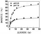
如图5所示,DOX/GQD-CFO@SiO2/FA体外DOX的释放图。在pH值7.4时,6小时内释放的DOX少于18%,并表现出极大的稳定性。相比之下,高达42.6%的DOX在pH值为5.5的情况下在6小时内迅速释放。48小时后,近71%的DOX在pH=5.5的酸性缓冲液中释放,而在pH=7.4时释放的DOX不到30%。pH控制释放依赖于DOX,在低pH介质中的质子化作用,这可以使DOX更具亲水性和水溶性,从而导致DOX从纳米平台在水溶液中的释放增加。实验结果与其他描述类似pH响应性药物释放行为的文献非常吻合。Figure 5 shows the release profile of DOX/GQD-CFO@SiO2 /FA in vitro. At pH 7.4, less than 18% DOX was released in 6 hours and showed great stability. In contrast, up to 42.6% of DOX was rapidly released within 6 hours at pH 5.5. After 48 hours, nearly 71% of the DOX was released in the acidic buffer at pH=5.5, while less than 30% of the DOX was released at pH=7.4. The pH-controlled release relies on the protonation of DOX in low pH media, which can make DOX more hydrophilic and water-soluble, leading to increased DOX release from nanoplatforms in aqueous solutions. The experimental results are in good agreement with other literature describing similar pH-responsive drug release behavior.
测试例2
GQD-CFO@SiO2/FA纳米平台的MRI实验MRI experiment of GQD-CFO@SiO2/FA nanoplatform
测试方法:为了研究获得的纳米粒子的磁性并评估GQD-CFO@SiO2/FA作为诊断MR造影剂的性能,对GQD-CFO@SiO2/FANPs进行了MRI测试。将制备的具有不同CFO浓度的GQD-CFO@SiO2/FA NPs分散并悬浮在200μL 1.0%琼脂糖凝胶中。然后将所有样品快速转移到6孔板中进行T2加权MRI成像。此外,HeLa细胞与GQD-CFO@SiO2/FA NPs一起孵育过夜,然后通过胰蛋白酶消化分离。HeLa细胞用PBS洗涤后,用200μL 1.0%琼脂糖凝胶分散悬浮,快速转移至试管中进行MRI实验。Test method: To study the magnetic properties of the obtained nanoparticles and evaluate the performance of GQD-CFO@SiO2 /FA as a diagnostic MR contrast agent, MRI tests of GQD-CFO@SiO2 /FANPs were performed. The prepared GQD-CFO@SiO2 /FA NPs with different CFO concentrations were dispersed and suspended in 200 μL of 1.0% agarose gel. All samples were then quickly transferred into 6-well plates for T2-weighted MRI imaging. Furthermore, HeLa cells were incubated with GQD-CFO@SiO2 /FA NPs overnight and then detached by trypsinization. HeLa cells were washed with PBS, dispersed and suspended with 200 μL of 1.0% agarose gel, and quickly transferred to a test tube for MRI experiments.
如图6所示,CFO、CFO@SiO2和GQD-CFO@SiO2/FA在300K下的滞后回线;结果表明CFO的饱核磁化强度(Ms)为69.4emu/g,而CFO@SiO2和CFO@SiO2-NH2的饱核磁化强度(Ms)分别降至22.1和15.3emu/g。由于纳米复合材料中CFO分数的减少和壳的非磁性贡献,二氧化硅涂层导致饱核磁化强度明显降低。尽管二氧化硅涂层和GQDs改性降低了CFO的饱核磁化强度,GQD-CFO@SiO2/FA表现出近乎超顺磁性的行为,这与CFO NPs相似。The hysteresis loops of CFO, CFO@SiO2 and GQD-CFO@SiO2 /FA at 300 K are shown in Fig. 6; the results show that the saturated nuclear magnetization (Ms) of CFO is 69.4 emu/g, while that of CFO@SiO2 and CFO@SiO2 -NH2 the saturated nuclear magnetization (Ms) decreased to 22.1 and 15.3 emu/g, respectively. The silica coating leads to a significant decrease in the saturation core magnetization due to the reduction of the CFO fraction in the nanocomposite and the non-magnetic contribution of the shell. Although the silica coating and GQDs modification reduced the saturated nuclear magnetization of CFO, GQD-CFO@SiO2 /FA exhibited near superparamagnetic behavior, which was similar to CFO NPs.
如图7所示,GQD-CFO@SiO2/FA在不同CFO浓度下的T2加权MRI。T2加权图像的亮度直接对应于T2的值。由于CFO的磁矩与周围介质中的质子之间的偶极相互作用,信号强度随着CFO浓度的增加而降低。由于GQD-CFO@SiO2/FA可以缩短T2、横向弛豫时间,因此导致MR图像更暗。As shown in Fig. 7, T2-weighted MRI of GQD-CFO@SiO2 /FA at different CFO concentrations. The brightness of a T2-weighted image corresponds directly to the value of T2. The signal intensity decreases with increasing CFO concentration due to the dipole interaction between the magnetic moment of the CFO and the protons in the surrounding medium. Since GQD-CFO@SiO2 /FA can shorten T2 and transverse relaxation time, it leads to darker MR images.
弛豫率通过单指数曲线提取,拟合信号强度与不同浓度下的回波时间,使用以下等式:The relaxation rate was extracted by a mono-exponential curve, fitting the signal intensity to the echo time at different concentrations, using the following equation:
Mxy(t)=Mxy(0)e-t/T2Mxy(t)=Mxy(0)e-t/T2
其中Mxy(0)和Mxy(t)分别为初始和时间等于t横向磁化。where Mxy(0) and Mxy(t) are the initial and time equal to t transverse magnetization, respectively.
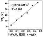
如图8所示,GQD-CFO@SiO2/FA在不同CFO浓度下的弛豫率,T2弛豫时间与粒子浓度成线性比例。GQD-CFO@SiO2/FA的R2弛豫系数值为75.6mM-1S-1。MRI结果表明GQD-CFO@SiO2/FA表现出理想的对比效果,表明GQD-CFO@SiO2/FA适合用作负MRI造影剂。As shown in Fig. 8, the relaxation rate, T2 relaxation time of GQD-CFO@SiO2 /FA at different CFO concentrations is linearly proportional to the particle concentration. The R2 relaxation coefficient value of GQD-CFO@SiO2 /FA is 75.6mM-1 S-1 . The MRI results show that GQD-CFO@SiO2 /FA exhibits ideal contrast effect, indicating that GQD-CFO@SiO2 /FA is suitable as a negative MRI contrast agent.
测试例3
GQD-CFO@SiO2/FA纳米平台的细胞毒性试验Cytotoxicity test of GQD-CFO@SiO2 /FA nanoplatform
MTT法测定了CFO@SiO2、GQD、GQD-CFO@SiO2和GQD-CFO@SiO2/FA对HeLa细胞的细胞毒性。简而言之,将HeLa细胞接种到96孔板中并过夜生长。然后将每个孔中的培养基更换为含有游离DOX、DOX/GQD-CFO@SiO2或DOX/GQD-CFO@SiO2/FA处理的培养基(150μL)。孵育24h后,将每孔培养基更换为含MTT试剂的新鲜培养基,浓度为0.5mg/mL,孵育4h。小心洗涤并除去培养基后,将100μL DMSO加入孔中以溶解甲臜晶体。记录酶标仪每个孔在490nm处的吸光度,以确定细胞活力。进行MTT测试以评估制造的纳米平台的杀肿瘤潜力。The cytotoxicity of CFO@SiO2 , GQD, GQD-CFO@SiO2 and GQD-CFO@SiO2 /FA on HeLa cells was determined by MTT assay. Briefly, HeLa cells were seeded into 96-well plates and grown overnight. The medium in each well was then replaced with free DOX, DOX/GQD-CFO@SiO2 , or DOX/GQD-CFO@SiO2 /FA-treated medium (150 μL). After 24 hours of incubation, the medium in each well was replaced with fresh medium containing MTT reagent at a concentration of 0.5 mg/mL, and incubated for 4 hours. After careful washing and removal of the medium, 100 μL of DMSO was added to the wells to dissolve the formazan crystals. Record the absorbance at 490 nm of each well of the microplate reader to determine cell viability. MTT tests were performed to evaluate the tumoricidal potential of the fabricated nanoplatforms.
HeLa细胞分别与CFO@SiO2、GQD-CFO@SiO2、GQD-CFO@SiO2/FA、GQD、游离DOX、DOX/GQD-CFO@SiO2和DOX/GQD-CFO@SiO2/FA一起孵育。HeLa cells together with CFO@SiO2 , GQD-CFO@SiO2 , GQD-CFO@SiO2 /FA, GQD, free DOX, DOX/GQD-CFO@SiO2 and DOX/GQD-CFO@SiO2 /FA, respectively Incubation.
结果表明CFO@SiO2、GQD-CFO@SiO2、GQD-CFO@SiO2/FA、GQDs对HeLa细胞的活力没有显着影响,这表明GQD-CFO@SiO2/FA具有理想的生物相容性。The results showed that CFO@SiO2 , GQD-CFO@SiO2 , GQD-CFO@SiO2 /FA, GQDs had no significant effect on the viability of HeLa cells, which indicated that GQD-CFO@SiO2 /FA had ideal biocompatibility sex.
与分别与DOX/GQD-CFO@SiO2和游离DOX孵育的HeLa细胞相比,负载DOX的DOX/GQD-CFO@SiO2/FA对Hela细胞表现出更高的毒性。显然,DOX/GQD-CFO@SiO2/FA表明显著增强对HeLa细胞的细胞毒性(84.7%),远高于非FA纳米平台DOX/GQD-CFO@SiO2(67.2%);在10μg/mL DOX浓度下,由于靶向效果的提高DOX/GQD-CFO@SiO2/FA纳米平台通过靶向分子修饰带来的HeLa细胞。这些结果表明基于FRET的-DOX/GQD-CFO@SiO2/FA显着提高了治疗效果。Compared with HeLa cells incubated with DOX/GQD-CFO@SiO2 and free DOX, respectively, DOX-loaded DOX/GQD-CFO@SiO2 /FA exhibited higher toxicity to HeLa cells. Apparently, DOX/GQD-CFO@SiO2 /FA showed significantly enhanced cytotoxicity to HeLa cells (84.7%), much higher than that of non-FA nanoplatform DOX/GQD-CFO@SiO2 (67.2%); at 10 μg/mL At DOX concentration, due to the improvement of targeting effect, the DOX/GQD-CFO@SiO2 /FA nanoplatform brought HeLa cells through targeted molecular modification. These results indicate that FRET-based -DOX/GQD-CFO@SiO2 /FA significantly enhances the therapeutic effect.
申请人声明,本发明通过上述实施例来说明本发明的工艺方法,但本发明并不局限于上述工艺步骤,即不意味着本发明必须依赖上述工艺步骤才能实施。所属技术领域的技术人员应该明了,对本发明的任何改进,对本发明所选用原料的等效替换及辅助成分的添加、具体方式的选择等,均落在本发明的保护范围和公开范围之内。The applicant declares that the present invention illustrates the process method of the present invention through the above-mentioned embodiments, but the present invention is not limited to the above-mentioned process steps, that is, it does not mean that the present invention must rely on the above-mentioned process steps to be implemented. Those skilled in the art should understand that any improvement of the present invention, the equivalent replacement of the selected raw materials of the present invention, the addition of auxiliary components, the selection of specific methods, etc., all fall within the protection scope and disclosure scope of the present invention.
Claims (10)
Translated fromChinesePriority Applications (1)
| Application Number | Priority Date | Filing Date | Title |
|---|---|---|---|
| CN202210585525.0ACN114848846B (en) | 2022-05-26 | 2022-05-26 | A drug delivery system and its preparation method and application |
Applications Claiming Priority (1)
| Application Number | Priority Date | Filing Date | Title |
|---|---|---|---|
| CN202210585525.0ACN114848846B (en) | 2022-05-26 | 2022-05-26 | A drug delivery system and its preparation method and application |
Publications (2)
| Publication Number | Publication Date |
|---|---|
| CN114848846Atrue CN114848846A (en) | 2022-08-05 |
| CN114848846B CN114848846B (en) | 2025-01-28 |
Family
ID=82640374
Family Applications (1)
| Application Number | Title | Priority Date | Filing Date |
|---|---|---|---|
| CN202210585525.0AActiveCN114848846B (en) | 2022-05-26 | 2022-05-26 | A drug delivery system and its preparation method and application |
Country Status (1)
| Country | Link |
|---|---|
| CN (1) | CN114848846B (en) |
Citations (18)
| Publication number | Priority date | Publication date | Assignee | Title |
|---|---|---|---|---|
| CN101942066A (en)* | 2010-09-03 | 2011-01-12 | 南京邮电大学 | Water-soluble polymer magnetic nanoparticle with fluorescence and preparation method thereof |
| CN102967706A (en)* | 2012-11-21 | 2013-03-13 | 济南大学 | Preparation method and application of flow injection chemiluminiscence immuno sensor for detecting tumor marker |
| CN103432590A (en)* | 2013-08-14 | 2013-12-11 | 华东理工大学 | Graphene quantum dot nuclear targeting medicine carrying system as well as preparation method and application thereof |
| CN103784407A (en)* | 2014-02-26 | 2014-05-14 | 哈尔滨医科大学 | Folic acid-mediated (polyethylene glycol) PEG-graphene oxide doxorubicine-loaded nanoparticle and preparation method thereof |
| CN104263358A (en)* | 2014-09-26 | 2015-01-07 | 重庆文理学院 | A magnetic fluorescent bifunctional graphene oxide nanocomposite material and its preparation method |
| CN104436210A (en)* | 2014-11-14 | 2015-03-25 | 上海交通大学 | Malignant-tumour-resistant graphene oxide nano-drug delivery system and preparation method thereof |
| CN104843715A (en)* | 2015-03-31 | 2015-08-19 | 中国科学院化学研究所 | Preparation method of silicon dioxide asymmetrically-modified magnetic colloidal particles |
| CN105457601A (en)* | 2015-12-01 | 2016-04-06 | 济南大学 | Preparation and application of folic acid modified magnetic graphene oxide adsorbent |
| CN105566535A (en)* | 2015-11-20 | 2016-05-11 | 陕西高华知本化工科技有限公司 | Method for preparing magnetic micro-spheres |
| US20170027168A1 (en)* | 2015-07-27 | 2017-02-02 | Stephan HEATH | Methods, products, and systems relating to making, providing, and using nanocrystalline (nc) products comprising nanocrystalline cellulose (ncc), nanocrystalline (nc) polymers and/or nanocrystalline (nc) plastics or other nanocrystals of cellulose composites or structures, in combination with other materials |
| CN106512023A (en)* | 2016-12-02 | 2017-03-22 | 武汉理工大学 | Preparation method of difunctional mesoporous silicon ball composite targeted drug delivery system |
| WO2017050979A1 (en)* | 2015-09-25 | 2017-03-30 | Consejo Superior De Investigaciones Científicas (Csic) | Nanoparticles for diagnosis and drug delivery |
| CN106727274A (en)* | 2017-03-07 | 2017-05-31 | 常州大学 | A kind of preparation method of the polypyrrole/mesoporous silicon oxide/graphene quantum dot nano composite material of core shell structure |
| CN106974882A (en)* | 2017-03-07 | 2017-07-25 | 常州大学 | A kind of polypyrrole/mesoporous silicon oxide of core shell structure/application of the graphene quantum dot nano composite material in medicine controlled releasing |
| CN107375241A (en)* | 2017-08-01 | 2017-11-24 | 大连理工大学 | A kind of preparation method of the film modified nano graphene oxide pharmaceutical carrier of phosphatide for cancer target transmission |
| CN109316465A (en)* | 2018-11-02 | 2019-02-12 | 孙世国 | A kind of biodegradable multiple target point targeting intelligent drug delivery system of inorganic nano, preparation method and application |
| CN110302397A (en)* | 2019-08-09 | 2019-10-08 | 西北工业大学 | pH-responsive graphene oxide nanosheet-coated mesoporous silica drug double-loaded composite nanoparticles and preparation method |
| CN112791064A (en)* | 2020-12-08 | 2021-05-14 | 清华大学 | A magnetic field-controlled helical nanorobot for targeted drug delivery |
- 2022
- 2022-05-26CNCN202210585525.0Apatent/CN114848846B/enactiveActive
Patent Citations (19)
| Publication number | Priority date | Publication date | Assignee | Title |
|---|---|---|---|---|
| CN101942066A (en)* | 2010-09-03 | 2011-01-12 | 南京邮电大学 | Water-soluble polymer magnetic nanoparticle with fluorescence and preparation method thereof |
| CN102967706A (en)* | 2012-11-21 | 2013-03-13 | 济南大学 | Preparation method and application of flow injection chemiluminiscence immuno sensor for detecting tumor marker |
| CN103432590A (en)* | 2013-08-14 | 2013-12-11 | 华东理工大学 | Graphene quantum dot nuclear targeting medicine carrying system as well as preparation method and application thereof |
| CN103784407A (en)* | 2014-02-26 | 2014-05-14 | 哈尔滨医科大学 | Folic acid-mediated (polyethylene glycol) PEG-graphene oxide doxorubicine-loaded nanoparticle and preparation method thereof |
| CN105860957A (en)* | 2014-09-26 | 2016-08-17 | 重庆文理学院 | Preparation method of oxidized graphene composite for drug carrier |
| CN104263358A (en)* | 2014-09-26 | 2015-01-07 | 重庆文理学院 | A magnetic fluorescent bifunctional graphene oxide nanocomposite material and its preparation method |
| CN104436210A (en)* | 2014-11-14 | 2015-03-25 | 上海交通大学 | Malignant-tumour-resistant graphene oxide nano-drug delivery system and preparation method thereof |
| CN104843715A (en)* | 2015-03-31 | 2015-08-19 | 中国科学院化学研究所 | Preparation method of silicon dioxide asymmetrically-modified magnetic colloidal particles |
| US20170027168A1 (en)* | 2015-07-27 | 2017-02-02 | Stephan HEATH | Methods, products, and systems relating to making, providing, and using nanocrystalline (nc) products comprising nanocrystalline cellulose (ncc), nanocrystalline (nc) polymers and/or nanocrystalline (nc) plastics or other nanocrystals of cellulose composites or structures, in combination with other materials |
| WO2017050979A1 (en)* | 2015-09-25 | 2017-03-30 | Consejo Superior De Investigaciones Científicas (Csic) | Nanoparticles for diagnosis and drug delivery |
| CN105566535A (en)* | 2015-11-20 | 2016-05-11 | 陕西高华知本化工科技有限公司 | Method for preparing magnetic micro-spheres |
| CN105457601A (en)* | 2015-12-01 | 2016-04-06 | 济南大学 | Preparation and application of folic acid modified magnetic graphene oxide adsorbent |
| CN106512023A (en)* | 2016-12-02 | 2017-03-22 | 武汉理工大学 | Preparation method of difunctional mesoporous silicon ball composite targeted drug delivery system |
| CN106727274A (en)* | 2017-03-07 | 2017-05-31 | 常州大学 | A kind of preparation method of the polypyrrole/mesoporous silicon oxide/graphene quantum dot nano composite material of core shell structure |
| CN106974882A (en)* | 2017-03-07 | 2017-07-25 | 常州大学 | A kind of polypyrrole/mesoporous silicon oxide of core shell structure/application of the graphene quantum dot nano composite material in medicine controlled releasing |
| CN107375241A (en)* | 2017-08-01 | 2017-11-24 | 大连理工大学 | A kind of preparation method of the film modified nano graphene oxide pharmaceutical carrier of phosphatide for cancer target transmission |
| CN109316465A (en)* | 2018-11-02 | 2019-02-12 | 孙世国 | A kind of biodegradable multiple target point targeting intelligent drug delivery system of inorganic nano, preparation method and application |
| CN110302397A (en)* | 2019-08-09 | 2019-10-08 | 西北工业大学 | pH-responsive graphene oxide nanosheet-coated mesoporous silica drug double-loaded composite nanoparticles and preparation method |
| CN112791064A (en)* | 2020-12-08 | 2021-05-14 | 清华大学 | A magnetic field-controlled helical nanorobot for targeted drug delivery |
Non-Patent Citations (3)
| Title |
|---|
| XIAOQIAN SU ET AL: "A graphene quantum dot@Fe3O4@SiO2 based nanoprobe for drug delivery sensing and dual-modal fluorescence and MRI imaging in cancer cells", 《BIOSENSORS AND BIOELECTRONICS》, vol. 92, 28 October 2016 (2016-10-28), pages 489 - 495* |
| 曾淑文等: "α-Fe2O3光电催化分解水研究进展", 《 表面技术》, vol. 46, no. 4, 19 May 2017 (2017-05-19), pages 67 - 70* |
| 李江等: "阿霉素在纳米递送系统中联合用药的研究进展",阿霉素在纳米递送系统中联合用药的研究进展, vol. 42, no. 9, 15 September 2023 (2023-09-15)* |
Also Published As
| Publication number | Publication date |
|---|---|
| CN114848846B (en) | 2025-01-28 |
Similar Documents
| Publication | Publication Date | Title |
|---|---|---|
| Dave et al. | Monodisperse magnetic nanoparticles for biodetection, imaging, and drug delivery: a versatile and evolving technology | |
| Ge et al. | Fluorescence modified chitosan-coated magnetic nanoparticles for high-efficient cellular imaging | |
| Lee et al. | Paramagnetic inorganic nanoparticles as T1 MRI contrast agents | |
| KR100851933B1 (en) | Magnetic resonance imaging agent containing manganese oxide nanoparticles | |
| Larsen et al. | Controlled aggregation of superparamagnetic iron oxide nanoparticles for the developmentof molecular magnetic resonance imaging probes | |
| CN101775280B (en) | Water-soluble nuclear-shell magneto-optic dual-function nanoparticle and preparation method thereof | |
| Xiong et al. | Preparation, characterization of 2-deoxy-D-glucose functionalized dimercaptosuccinic acid-coated maghemite nanoparticles for targeting tumor cells | |
| Park et al. | Surface design of Eu-doped iron oxide nanoparticles for tuning the magnetic relaxivity | |
| Hemalatha et al. | Fabrication and characterization of dual acting oleyl chitosan functionalised iron oxide/gold hybrid nanoparticles for MRI and CT imaging | |
| Forge et al. | An original route to stabilize and functionalize magnetite nanoparticles for theranosis applications | |
| CN103143041B (en) | Preparation method of targeted MRI (magnetic resonance imaging) contrast medium based on folic acid modified iron oxide nanoparticles | |
| WO2011150212A2 (en) | Magnetic nanoparticles | |
| EP2808036A1 (en) | Superparamagnetic nanoparticles as a contrast agent for magnetic resonance imaging (mri) of magnetic susceptibility (t2*) | |
| CN103933584B (en) | A kind of preparation method of the superparamagnetic iron oxide nano-particle of modified with folic acid | |
| Xu et al. | Superparamagnetic magnetite nanocrystal clusters: a sensitive tool for MR cellularimaging | |
| Wang et al. | Transferrin-conjugated superparamagnetic iron oxide nanoparticles as in vivo magnetic resonance imaging contrast agents | |
| CN106466488A (en) | There is ultra-fine magnetic core-shell nano material and its preparation and the application of tumor cell targeting | |
| CN111607094A (en) | A kind of polyaspartic acid-dopamine/polyethylene glycol polymer and its use in preparing contrast agent | |
| Teng et al. | A multifunctional nanoplatform based on graphene quantum dots‐cobalt ferrite for monitoring of drug delivery and fluorescence/magnetic resonance bimodal cellular imaging | |
| Xu et al. | Folic acid-conjugated GdPO4: Tb3+@ SiO2 Nanoprobe for folate receptor-targeted optical and magnetic resonance bi-modal imaging | |
| Kostevšek et al. | “Single-” and “multi-core” FePt nanoparticles: from controlled synthesis via zwitterionic and silica bio-functionalization to MRI applications | |
| CN101693011A (en) | Preparation method of magnetic medicine-carrying nano-particle | |
| Smitha et al. | Fluorescent superparamagnetic iron oxide core–shell nanoprobes for multimodal cellular imaging | |
| Grudzinski et al. | Comprehensive magnetic resonance characteristics of carbon-encapsulated iron nanoparticles: a new frontier for the core-shell–type contrast agents | |
| Gong et al. | Synthesis of streptavidin-conjugated magnetic nanoparticles for DNA detection |
Legal Events
| Date | Code | Title | Description |
|---|---|---|---|
| PB01 | Publication | ||
| PB01 | Publication | ||
| SE01 | Entry into force of request for substantive examination | ||
| SE01 | Entry into force of request for substantive examination | ||
| GR01 | Patent grant | ||
| GR01 | Patent grant |