CN114821176B - Viral encephalitis classification system for MR (magnetic resonance) images of children brain - Google Patents
Viral encephalitis classification system for MR (magnetic resonance) images of children brainDownload PDFInfo
- Publication number
- CN114821176B CN114821176BCN202210463034.9ACN202210463034ACN114821176BCN 114821176 BCN114821176 BCN 114821176BCN 202210463034 ACN202210463034 ACN 202210463034ACN 114821176 BCN114821176 BCN 114821176B
- Authority
- CN
- China
- Prior art keywords
- sub
- module
- children
- image
- classification
- Prior art date
- Legal status (The legal status is an assumption and is not a legal conclusion. Google has not performed a legal analysis and makes no representation as to the accuracy of the status listed.)
- Active
Links
Images


Classifications
- G—PHYSICS
- G06—COMPUTING OR CALCULATING; COUNTING
- G06F—ELECTRIC DIGITAL DATA PROCESSING
- G06F18/00—Pattern recognition
- G06F18/20—Analysing
- G06F18/24—Classification techniques
- G—PHYSICS
- G06—COMPUTING OR CALCULATING; COUNTING
- G06N—COMPUTING ARRANGEMENTS BASED ON SPECIFIC COMPUTATIONAL MODELS
- G06N3/00—Computing arrangements based on biological models
- G06N3/02—Neural networks
- G06N3/04—Architecture, e.g. interconnection topology
- G06N3/045—Combinations of networks
- G—PHYSICS
- G06—COMPUTING OR CALCULATING; COUNTING
- G06N—COMPUTING ARRANGEMENTS BASED ON SPECIFIC COMPUTATIONAL MODELS
- G06N3/00—Computing arrangements based on biological models
- G06N3/02—Neural networks
- G06N3/04—Architecture, e.g. interconnection topology
- G06N3/048—Activation functions
- G—PHYSICS
- G06—COMPUTING OR CALCULATING; COUNTING
- G06N—COMPUTING ARRANGEMENTS BASED ON SPECIFIC COMPUTATIONAL MODELS
- G06N3/00—Computing arrangements based on biological models
- G06N3/02—Neural networks
- G06N3/08—Learning methods
Landscapes
- Engineering & Computer Science (AREA)
- Theoretical Computer Science (AREA)
- Physics & Mathematics (AREA)
- Data Mining & Analysis (AREA)
- Evolutionary Computation (AREA)
- Life Sciences & Earth Sciences (AREA)
- Artificial Intelligence (AREA)
- General Physics & Mathematics (AREA)
- General Engineering & Computer Science (AREA)
- General Health & Medical Sciences (AREA)
- Software Systems (AREA)
- Molecular Biology (AREA)
- Computing Systems (AREA)
- Biophysics (AREA)
- Biomedical Technology (AREA)
- Mathematical Physics (AREA)
- Computational Linguistics (AREA)
- Health & Medical Sciences (AREA)
- Bioinformatics & Cheminformatics (AREA)
- Bioinformatics & Computational Biology (AREA)
- Computer Vision & Pattern Recognition (AREA)
- Evolutionary Biology (AREA)
- Image Analysis (AREA)
Abstract
Translated fromChinese
Description
Translated fromChinese技术领域technical field
本发明属于医学人工智能领域,尤其是涉及一种儿童脑部MR图像病毒性脑炎分类系统。The invention belongs to the field of medical artificial intelligence, in particular to a classification system for children's brain MR images of viral encephalitis.
背景技术Background technique
儿童脑炎是儿科一种较为常见的疾病。一般情况下,可通过临床症状、实验室检查,以及影像学和脑电图检测进行综合判断。若经确诊,患者需要在专业医生的指导下,进行针对性治疗。Childhood encephalitis is a relatively common disease in pediatrics. In general, a comprehensive judgment can be made through clinical symptoms, laboratory tests, and imaging and EEG tests. If diagnosed, patients need to be targeted for treatment under the guidance of professional doctors.
目前医生诊断主要通过临床症状、实验室检查(脑脊液检查)、影像学和脑电图检测等方法进行检查,然而临床症状不太准确;影像学和脑电图检测则只有重症时才能肉眼观察到病变区域;脑脊液检查较准确,但耗时较长,且需要抽取脑脊液,对儿童造成创伤和痛苦。At present, doctors diagnose mainly through clinical symptoms, laboratory tests (cerebrospinal fluid examination), imaging and EEG detection, etc. However, clinical symptoms are not very accurate; imaging and EEG detection can only be observed with the naked eye in severe cases Lesion area; cerebrospinal fluid examination is more accurate, but it takes a long time and needs to extract cerebrospinal fluid, which causes trauma and pain to children.
随着人工智能和深度学习的发展,在医学领域,很多研究者尝试使用智能算法对影像学数据脑电图数据进行自动识别。With the development of artificial intelligence and deep learning, in the medical field, many researchers try to use intelligent algorithms to automatically identify EEG data from imaging data.
如公开号为CN112561863A的中国专利文献公开了一种基于深度学习的粒细胞图片细粒度分类识别系统;包含定位模块和分类模块,其中定位模块利用Hourglass网络模型对输入的粒细胞图片进行特征提取,将粒细胞图片中的全部细胞分别进行定位,再将定位后的细胞裁剪出来,留下单个完整的细胞,并将全部裁剪出来的细胞进行尺寸归一化处理;分类模块采用构建的深度学习分类模型对定位模块定位出的粒细胞进行分类;辅助临床医生准确高效完成粒细胞分类识别计数任务,减小主观性带来的误差,减轻医生的工作量,辅助医生做出疾病判断;该系统能够有效解决非均衡数据下的细胞分类以及粒细胞间的细粒度分类,提升网络分类识别效果。For example, the Chinese patent document with the publication number CN112561863A discloses a fine-grained classification and recognition system for granulocyte pictures based on deep learning; it includes a positioning module and a classification module, wherein the positioning module uses the Hourglass network model to extract features from the input granulocyte pictures, Position all the cells in the granulocyte picture separately, then cut out the positioned cells, leaving a single complete cell, and normalize all the cut out cells in size; the classification module adopts the constructed deep learning classification The model classifies the granulocytes located by the positioning module; assists clinicians to accurately and efficiently complete the task of granulocyte classification, identification and counting, reduces the error caused by subjectivity, reduces the workload of doctors, and assists doctors in making disease judgments; the system can Effectively solve the cell classification under unbalanced data and the fine-grained classification between granulocytes, and improve the network classification recognition effect.
公开号为CN112132808A的中国专利文献公开了一种基于常态模型学习的乳腺X线图像病变检测方法和装置。所述方法包括从乳腺X线图像中分割出乳腺区域;提取图像块,进行亮度归一化处理;选取一部分正常区域图像块作为训练集,输入到双重深度卷积神经网络模型进行训练,得到常态模型;从训练集中选取若干正常区域图像块作为模板,输入到常态模型,得到模板图像的特征向量;将测试集输入到常态模型,得到测试图像的特征向量;将模板图像和测试图像的特征向量输入到最近邻分类器执行一类分类,得到测试结果。The Chinese patent document with publication number CN112132808A discloses a mammography image lesion detection method and device based on normal model learning. The method comprises the steps of segmenting a mammary gland area from a mammogram image; extracting image blocks, and performing brightness normalization processing; selecting a part of normal area image blocks as a training set, and inputting them into a double-depth convolutional neural network model for training to obtain normal model; select several normal area image blocks from the training set as templates, input them into the normal model, and obtain the feature vector of the template image; input the test set into the normal model, and obtain the feature vector of the test image; combine the template image and the feature vector of the test image Input to the nearest neighbor classifier to perform a class of classification, and get the test result.
然而,对于儿童病毒性脑炎而言,影像学数据的显示的特征不明显,使用常规的深度学习方法,很难对儿童是否患有病毒性脑炎进行准确的诊断。However, for children with viral encephalitis, the characteristics of imaging data are not obvious, and it is difficult to accurately diagnose whether children have viral encephalitis using conventional deep learning methods.
发明内容Contents of the invention
本发明提供了一种儿童脑部MR图像病毒性脑炎分类系统,可以在腰穿脑脊液检查、临床检查之外,只基于MR图像进行病毒性脑炎的诊断,具有较高的准确率。The invention provides a classification system for viral encephalitis in children's brain MR images, which can diagnose viral encephalitis based only on MR images in addition to lumbar puncture cerebrospinal fluid examination and clinical examination, and has a high accuracy rate.
一种儿童脑部MR图像病毒性脑炎分类系统,包括计算机存储器、计算机处理器以及存储在所述计算机存储器中并可在所述计算机处理器上执行的计算机程序,所述计算机存储器中存有训练好的分类模型,所述的分类模型采用改进的SE ResNet网络模型,共包含四个卷积部分,每个卷积部分由若干个子模块组构成,每个子模块组包含Inception子模块和SE Res子模块,最终通过全连接层获得最后的分类结果;A kind of viral encephalitis classification system of children's brain MR image, comprises computer memory, computer processor and is stored in described computer memory and can execute the computer program on described computer processor, stored in the described computer memory The trained classification model, the classification model adopts the improved SE ResNet network model, including four convolution parts, each convolution part is composed of several sub-module groups, and each sub-module group contains Inception sub-module and SE ResNet The sub-module finally obtains the final classification result through the fully connected layer;
其中,Inception子模块通过不同尺度的卷积来提升对不同大小的特征的学习能力;SE Res子模块包含SE和Res两部分,SE部分通过压缩-扩张通道数来提升模型对于有效特征的学习能力,Res部分通过跳跃连接对SE部分的输入特征矩阵X和输出特征矩阵
所述计算机处理器执行所述计算机程序时实现以下步骤:When the computer processor executes the computer program, the following steps are implemented:
将待分类的儿童脑部MR影像输入训练好的分类模型中,得到病毒性脑炎分类结果。Input the MR images of children's brains to be classified into the trained classification model to obtain the classification results of viral encephalitis.
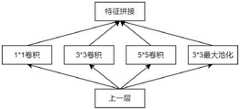
进一步地,所述Inception子模块的结构如下:在获得上一层的数据输入X后,进入到多核卷积层Lincep,此层设计三个不同尺寸的卷积核以及一个池化核,即Cincep=[C1,C2,C3,P1];其中,C1,C2,C3的卷积核大小分别是1*1、3*3、5*5,P1的核大小为3*3;通过这些卷积核得到四个不同的特征
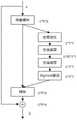
SE Res子模块中,SE部分的结构如下:获取上一层的的输入数据X(c*w*h),其中c,w,h分别代表特征矩阵的通道数、宽、高;首先用1*1大小的全局池化层进行池化,获得池化后的特征矩阵F1=GobelPool(X),其大小为c*w*h;然后用1/16*c的通道数进行全连接卷积,获得F2=FC(F1),其大小为c/16*w*h;然后用c通道数的卷积层进行卷积,获得F3=FC(F2),其大小为c*1*1;然后用sigmoid激活函数进行归一化操作,将权重归一化到0-1之间,获得F4=Sigmoid(F3),其大小为c*1*1;最后用此结果对输入数据进行加权,获得
Res部分的结构如下:特征矩阵X(c*w*h)通过SE部分后得到


所述分类模型的训练过程如下:The training process of the classification model is as follows:
(1)收集患有病毒性脑炎和正常儿童患者的T1W序列MR影像数据,对影像数据进行预处理;(1) Collect T1W sequence MR image data of children with viral encephalitis and normal children, and preprocess the image data;
(2)将预处理后的影像数据划分为训练集、验证集和测试集;(2) Divide the preprocessed image data into training set, verification set and test set;
(3)将训练集送入到构建的分类模型中进行训练,利用验证集对分类模型的性能进行评估,根据评估的效果对模型的超参数进行调整,通过反复训练、验证,最终得到性能达标的分类模型。(3) Send the training set into the constructed classification model for training, use the verification set to evaluate the performance of the classification model, adjust the hyperparameters of the model according to the evaluation effect, and finally obtain the performance standard through repeated training and verification classification model.
步骤(1)中,所述的预处理包括对影像进行缩放,选取最大切片数作为标准,未达到此切片数的数据通过复制首尾切片进行补充,使各案例的输入数据保持一致;同时对影像进行尺度的归一化,并采用高斯滤波器滤除噪声。In step (1), the preprocessing includes zooming the image, selecting the maximum number of slices as a standard, and supplementing the data that does not reach the number of slices by copying the first and last slices, so that the input data of each case is consistent; Normalize the scale and use a Gaussian filter to filter out noise.
步骤(2)中,将预处理后的影像数据按7:1:2划分为训练集、验证集和测试集。In step (2), the preprocessed image data is divided into training set, verification set and test set according to 7:1:2.
步骤(3)中,采用监督训练方法对分类模型进行训练。In step (3), the classification model is trained using a supervised training method.
与现有技术相比,本发明具有以下有益效果:Compared with the prior art, the present invention has the following beneficial effects:
1、本发明创新性地提出了利用儿童脑部MR图像对儿童是否患有病毒性脑炎进行判断,不再需要腰穿脑脊液检查,因而也无需进行手术,减少了儿童患者的痛苦,也极大地提升了诊断的效率。1. The present invention innovatively proposes to use children's brain MR images to judge whether children have viral encephalitis, no need for lumbar puncture and cerebrospinal fluid examination, and therefore no need for surgery, which reduces the pain of children and greatly reduces the pain of children. Greatly improved the efficiency of diagnosis.
2、本发明中的分类模型采用改进的SE ResNet网络模型,在Inception网络模型的基础上加入了SE Res模块,Inception网络模型通过不同尺度的卷积来提升对不同大小的特征的学习能力,SE Res模块首先通过压缩-扩张通道数来提升模型对于有效特征的学习能力,然后通过跳跃连接提升模型对于不同维度特征的学习能力;大大提升了诊断的效率和准确率。2. The classification model in the present invention adopts the improved SE ResNet network model, and the SE Res module is added on the basis of the Inception network model. The Inception network model improves the learning ability of features of different sizes through convolution of different scales. SE The Res module first improves the learning ability of the model for effective features by compressing and expanding the number of channels, and then improves the learning ability of the model for features of different dimensions through skip connections; greatly improving the efficiency and accuracy of diagnosis.
附图说明Description of drawings
图1为本发明一种儿童脑部MR图像病毒性脑炎分类系统的实施流程图;Fig. 1 is the implementation flowchart of a kind of children's brain MR image viral encephalitis classification system of the present invention;
图2为本发明中分类模型的整体结构图;Fig. 2 is the overall structural diagram of classification model among the present invention;
图3为分类模型中每个卷积部分的结构图;Figure 3 is a structural diagram of each convolutional part in the classification model;
图4为本发明分类模型中Inception子模块的网络结构示意图;Fig. 4 is a schematic diagram of the network structure of the Inception submodule in the classification model of the present invention;
图5为本发明分类模型中SE Res子模块的网络结构图。Fig. 5 is a network structure diagram of the SE Res sub-module in the classification model of the present invention.
具体实施方式Detailed ways
下面结合附图和实施例对本发明做进一步详细描述,需要指出的是,以下所述实施例旨在便于对本发明的理解,而对其不起任何限定作用。The present invention will be further described in detail below with reference to the accompanying drawings and embodiments. It should be noted that the following embodiments are intended to facilitate the understanding of the present invention, but do not limit it in any way.
一种儿童脑部MR图像病毒性脑炎分类系统,包括计算机存储器、计算机处理器以及存储在计算机存储器中并可在计算机处理器上执行的计算机程序,计算机存储器中存有训练好的分类模型。A classification system for viral encephalitis in children's brain MR images, comprising a computer memory, a computer processor and a computer program stored in the computer memory and executable on the computer processor, and a trained classification model is stored in the computer memory.
如图1所示,整个系统的实施流程如下:As shown in Figure 1, the implementation process of the entire system is as follows:
1、图像预处理1. Image preprocessing
收集患有病毒性脑炎和正常儿童T1W期MR影像数据,对影像进行缩放,因为每个案例扫描的MR切片数不一致,本方法选取最大切片数作为标准,未达到此切片数的数据通过复制首尾切片进行补充,使各案例的输入数据保持一致,也包括对影像进行尺度的归一化,采用高斯滤波器滤除噪声。Collect the T1W MR image data of children with viral encephalitis and normal children, and scale the images. Because the number of MR slices scanned in each case is inconsistent, this method selects the maximum number of slices as the standard, and the data that does not reach this number of slices are copied The first and last slices are supplemented to make the input data of each case consistent, which also includes normalizing the scale of the image, and using a Gaussian filter to filter out noise.
2、数据分组2. Data grouping
将70%的数据集作为训练集,10%的数据集作为验证集,20%的数据集作为测试集。70% of the data set is used as a training set, 10% of the data set is used as a validation set, and 20% of the data set is used as a test set.
3、模型构建3. Model construction
构建分类模型,分类模型采用改进的SE ResNet网络模型,此网络是在Inception网络模型的基础上加入了SE Res模块。Inception网络模型通过不同尺度的卷积来提升对不同大小的特征的学习能力,SE Res模块首先通过压缩-扩张通道数来提升模型对于有效特征的学习能力,然后通过跳跃连接提升模型对于不同维度特征的学习能力。Construct the classification model, the classification model adopts the improved SE ResNet network model, this network is added the SE Res module on the basis of the Inception network model. The Inception network model improves the learning ability of features of different sizes through convolution of different scales. The SE Res module first improves the learning ability of the model for effective features by compressing and expanding the number of channels, and then improves the model for features of different dimensions through skip connections. learning ability.
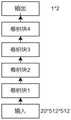
如图2和图3所示,模型共包含四个卷积块,卷积块1-卷积块4,每个卷积块由若干个子模块组构成,每个子模块组包含Inception子模块和SE Res子模块。As shown in Figure 2 and Figure 3, the model contains a total of four convolution blocks, convolution block 1-convolution block 4, each convolution block consists of several sub-module groups, each sub-module group contains Inception sub-modules and SE Res submodule.
Inception子模块的结构如图4所示,首先在通过Inception子模块获取不同尺度的特征。在获得上一层的数据输入X后,进入到多核卷积层Lincep,此层设计三个不同尺寸的卷积核以及一个池化核,即Cincep=[C1,C2,C3,P1],其中,C1,C2,C3的卷积核大小分别是1*1、3*3、5*5,P1的核大小为3*3。通过这些卷积核可以得到四个不同的特征
因为Inception子模块最终获取的特征数较多,为了提升计算速度和模型精度,本方法采用SE Res子模块对特征进行加权筛选。Because the Inception sub-module finally obtains a large number of features, in order to improve the calculation speed and model accuracy, this method uses the SE Res sub-module to perform weighted screening of features.
SE Res子模块的结构如图5所示,SE Res子模块包含SE和Res两部分。SE部分中,获取上一层的输入数据X(c*w*h),其中c,w,h分别代表特征矩阵的通道数、宽、高。首先用1*1大小的全局池化层进行池化,获得池化后的特征矩阵F1=GobelPool(X),其大小为c*w*h,然后用1/16*c的通道数进行全连接卷积,获得F2=FC(F1),其大小为c/16*w*h,然后用c通道数的卷积层进行卷积,获得F3=FC(F2),其大小为c*1*1,然后用sigmoid激活函数进行归一化操作,将权重归一化到0-1之间,获得F4=Sigmoid(F3),其大小为c*1*1,最后用此结果对输入数据进行加权,获得


最终模型通过全连接卷积层,得到输入案例是否为病毒性脑炎的概率。The final model obtains the probability of whether the input case is viral encephalitis through a fully connected convolutional layer.
4、模型训练和分类测试4. Model training and classification testing
分割模型训练时,将训练集送入到分类模型中;验证集对模型的超参数进行调整,使用优化器更新参数,对网络进行优化,对学习率进行自动调参,得到训练完成的分类网络;测试集用来估计学习过程完成之后的模型的泛化能力。When splitting model training, the training set is sent to the classification model; the verification set adjusts the hyperparameters of the model, uses the optimizer to update the parameters, optimizes the network, and automatically adjusts the learning rate to obtain the trained classification network ; The test set is used to estimate the generalization ability of the model after the learning process is completed.
5、评估阶段5. Evaluation stage
在测试集上,对模型的分类效果进行评估:对分类任务的评估,需要计算每一类的精确率(Precision)和召回率(Recall)。每一类的精确率用正确分类到本类的案例(TruePositive,TP)除以所有分类到本类的案例数(TP+FP),当不属于本类的案例被模型分到本类时,计数为假阳性(False Positive,FP)。每一类的召回率正确分类到本类的案例(TruePositive,TP)除以本类的真实案例数(TP+TN),当属于本类的案例被模型分到其它类时,计数为假阴性(True Negative,TN)。最终智能脑炎诊断分类模型在测试集上的分类性能AUC进行评估,AU曲线是以假正率(FP_rate)和假负率(TP_rate)为轴的ROC(ReceiverOperating Characteristic)曲线下面的面积。On the test set, evaluate the classification effect of the model: for the evaluation of the classification task, it is necessary to calculate the precision rate (Precision) and recall rate (Recall) of each category. The accuracy rate of each category is divided by the cases correctly classified into this category (TruePositive, TP) by the number of cases classified into this category (TP+FP). When the cases that do not belong to this category are classified into this category by the model, The count is False Positive (FP). The recall rate of each category is correctly classified into the case of this category (TruePositive, TP) divided by the number of real cases of this category (TP+TN). When the cases belonging to this category are classified into other categories by the model, the count is false negative (True Negative, TN). The classification performance AUC of the final intelligent encephalitis diagnostic classification model on the test set is evaluated. The AU curve is the area under the ROC (Receiver Operating Characteristic) curve with the false positive rate (FP_rate) and false negative rate (TP_rate) as the axes.
以上所述的实施例对本发明的技术方案和有益效果进行了详细说明,应理解的是以上所述仅为本发明的具体实施例,并不用于限制本发明,凡在本发明的原则范围内所做的任何修改、补充和等同替换,均应包含在本发明的保护范围之内。The embodiments described above have described the technical solutions and beneficial effects of the present invention in detail. It should be understood that the above descriptions are only specific embodiments of the present invention, and are not intended to limit the present invention. All within the scope of the principles of the present invention Any modifications, supplements and equivalent replacements should be included within the protection scope of the present invention.
Claims (8)
Translated fromChinese


Priority Applications (1)
| Application Number | Priority Date | Filing Date | Title |
|---|---|---|---|
| CN202210463034.9ACN114821176B (en) | 2022-04-28 | 2022-04-28 | Viral encephalitis classification system for MR (magnetic resonance) images of children brain |
Applications Claiming Priority (1)
| Application Number | Priority Date | Filing Date | Title |
|---|---|---|---|
| CN202210463034.9ACN114821176B (en) | 2022-04-28 | 2022-04-28 | Viral encephalitis classification system for MR (magnetic resonance) images of children brain |
Publications (2)
| Publication Number | Publication Date |
|---|---|
| CN114821176A CN114821176A (en) | 2022-07-29 |
| CN114821176Btrue CN114821176B (en) | 2022-11-01 |
Family
ID=82509353
Family Applications (1)
| Application Number | Title | Priority Date | Filing Date |
|---|---|---|---|
| CN202210463034.9AActiveCN114821176B (en) | 2022-04-28 | 2022-04-28 | Viral encephalitis classification system for MR (magnetic resonance) images of children brain |
Country Status (1)
| Country | Link |
|---|---|
| CN (1) | CN114821176B (en) |
Families Citing this family (1)
| Publication number | Priority date | Publication date | Assignee | Title |
|---|---|---|---|---|
| CN116363438B (en)* | 2023-04-14 | 2023-09-15 | 浙江大学 | A multimodality-based classification system for childhood encephalitis |
Citations (10)
| Publication number | Priority date | Publication date | Assignee | Title |
|---|---|---|---|---|
| EP0918458B1 (en)* | 1996-05-08 | 2006-12-27 | Meryl Squires | Antimicrobial treatment for herpes simplex virus and other infectious diseases |
| CN108427920A (en)* | 2018-02-26 | 2018-08-21 | 杭州电子科技大学 | A kind of land and sea border defense object detection method based on deep learning |
| CN109071610A (en)* | 2016-05-03 | 2018-12-21 | 无微华斯生物科技有限公司 | Method and system for diagnosing and treating virus infection |
| CN110739070A (en)* | 2019-09-26 | 2020-01-31 | 南京工业大学 | A brain disease diagnosis method based on 3D convolutional neural network |
| CN111104961A (en)* | 2019-10-31 | 2020-05-05 | 太原理工大学 | Method for classifying breast cancer based on improved MobileNet network |
| CN111223553A (en)* | 2020-01-03 | 2020-06-02 | 大连理工大学 | A two-stage deep transfer learning TCM tongue diagnosis model |
| CN111709446A (en)* | 2020-05-14 | 2020-09-25 | 天津大学 | X-ray chest X-ray classification device based on an improved densely connected network |
| CN111868080A (en)* | 2018-02-27 | 2020-10-30 | 磨石肿瘤生物技术公司 | Neoantigen identification using pan-allelic models |
| CN112906748A (en)* | 2021-01-25 | 2021-06-04 | 浙江大学 | 12-lead ECG arrhythmia detection classification model construction method based on residual error network |
| WO2021216467A1 (en)* | 2020-04-20 | 2021-10-28 | The Regents Of The University Of California | A nano-enabled vaccination approach for coronavirus disease (covid-19) and other viral diseases |
Family Cites Families (3)
| Publication number | Priority date | Publication date | Assignee | Title |
|---|---|---|---|---|
| CN108171232B (en)* | 2017-11-15 | 2021-12-28 | 中山大学 | Deep learning algorithm-based bacterial and viral pneumonia classification method for children |
| US11064902B2 (en)* | 2018-06-29 | 2021-07-20 | Mayo Foundation For Medical Education And Research | Systems, methods, and media for automatically diagnosing intraductal papillary mucinous neosplasms using multi-modal magnetic resonance imaging data |
| CN112116005B (en)* | 2020-09-18 | 2024-01-23 | 推想医疗科技股份有限公司 | Training method and device for image classification model, storage medium and electronic equipment |
- 2022
- 2022-04-28CNCN202210463034.9Apatent/CN114821176B/enactiveActive
Patent Citations (10)
| Publication number | Priority date | Publication date | Assignee | Title |
|---|---|---|---|---|
| EP0918458B1 (en)* | 1996-05-08 | 2006-12-27 | Meryl Squires | Antimicrobial treatment for herpes simplex virus and other infectious diseases |
| CN109071610A (en)* | 2016-05-03 | 2018-12-21 | 无微华斯生物科技有限公司 | Method and system for diagnosing and treating virus infection |
| CN108427920A (en)* | 2018-02-26 | 2018-08-21 | 杭州电子科技大学 | A kind of land and sea border defense object detection method based on deep learning |
| CN111868080A (en)* | 2018-02-27 | 2020-10-30 | 磨石肿瘤生物技术公司 | Neoantigen identification using pan-allelic models |
| CN110739070A (en)* | 2019-09-26 | 2020-01-31 | 南京工业大学 | A brain disease diagnosis method based on 3D convolutional neural network |
| CN111104961A (en)* | 2019-10-31 | 2020-05-05 | 太原理工大学 | Method for classifying breast cancer based on improved MobileNet network |
| CN111223553A (en)* | 2020-01-03 | 2020-06-02 | 大连理工大学 | A two-stage deep transfer learning TCM tongue diagnosis model |
| WO2021216467A1 (en)* | 2020-04-20 | 2021-10-28 | The Regents Of The University Of California | A nano-enabled vaccination approach for coronavirus disease (covid-19) and other viral diseases |
| CN111709446A (en)* | 2020-05-14 | 2020-09-25 | 天津大学 | X-ray chest X-ray classification device based on an improved densely connected network |
| CN112906748A (en)* | 2021-01-25 | 2021-06-04 | 浙江大学 | 12-lead ECG arrhythmia detection classification model construction method based on residual error network |
Non-Patent Citations (4)
| Title |
|---|
| 《CT和MRI对小儿病毒性脑炎的诊断价值(80例)》;吴蒙蒙 等;《医疗装备》;20161231;第29卷(第23期);第85-86页* |
| 《Differentiation of Glioma Mimicking Encephalitis and Encephalitis Using Multiparametric MR-Based Deep Learning》;Wenli Wu等;《Frontiers in Oncology》;20210331;第11卷;第1-9页* |
| 《基于人工智能技术的儿童慢病管理平台的研制与应用》;李哲明 等;《中国医疗设备》;20201220;第35卷(第S2期);第172-174页* |
| 《基于改进Inception-ResNet-v2的城市交通路面状态识别算法》;王佳 等;《科学技术与工程》;20220228;第22卷(第06期);第2524-2530页* |
Also Published As
| Publication number | Publication date |
|---|---|
| CN114821176A (en) | 2022-07-29 |
Similar Documents
| Publication | Publication Date | Title |
|---|---|---|
| CN114926477B (en) | A deep learning-based multimodal MRI image segmentation method for brain tumors | |
| CN106780475A (en) | A kind of image processing method and device based on histopathologic slide's image organizational region | |
| CN113076878B (en) | Constitution Identification Method Based on Convolutional Network Structure of Attention Mechanism | |
| CN112784856A (en) | Channel attention feature extraction method and identification method of chest X-ray image | |
| CN111738302A (en) | A system for classifying and diagnosing Alzheimer's disease based on multimodal data | |
| CN116363438B (en) | A multimodality-based classification system for childhood encephalitis | |
| CN113658151B (en) | Breast lesion magnetic resonance image classification method, equipment and readable storage medium | |
| Firke et al. | Convolutional neural network for diabetic retinopathy detection | |
| CN114926396B (en) | Mental disorder magnetic resonance image preliminary screening model construction method | |
| CN117094980A (en) | Ultrasonic breast nodule image interpretation method based on deep learning | |
| CN114418999A (en) | Retinopathy detection system based on lesion attention pyramid convolution neural network | |
| CN117352164A (en) | Multimodal tumor detection and diagnosis platform based on artificial intelligence and its processing method | |
| Chak et al. | Neural network and svm based kidney stone based medical image classification | |
| CN114821176B (en) | Viral encephalitis classification system for MR (magnetic resonance) images of children brain | |
| Fu et al. | MSEF-Net: A multi-scale EfficientNet fusion for diabetic retinopathy grading | |
| Archana et al. | Deep convolutional neural networks for multiclass cervical cell classification | |
| Gupta et al. | Brain tumor classification using mr images and transfer learning | |
| CN115205599B (en) | A multi-age classification system for children's chest radiograph pneumonia based on a domain generalization model | |
| CN114913169B (en) | A screening system for neonatal necrotizing enterocolitis | |
| CN114723937B (en) | A perivascular space classification method and system based on magnetic resonance imaging | |
| Arbab et al. | Automatic Detection and Classification of Acute Lymphoblastic Leukemia Using Convolution Neural Network | |
| CN116681951A (en) | Deep learning-based X-ray lung image classification system for pneumonia | |
| Yuningsih et al. | Anemia classification based on abnormal red blood cell morphology using convolutional neural network | |
| CN118379571A (en) | A multi-label tongue image recognition method and system with adjustable screening threshold | |
| Narne et al. | Evaluating Deep Learning Models for Accurate Pneumonia Diagnosis from Chest X-Rays |
Legal Events
| Date | Code | Title | Description |
|---|---|---|---|
| PB01 | Publication | ||
| PB01 | Publication | ||
| SE01 | Entry into force of request for substantive examination | ||
| SE01 | Entry into force of request for substantive examination | ||
| GR01 | Patent grant | ||
| GR01 | Patent grant |