CN114795414A - Visual interatrial septum puncture device and use method - Google Patents
Visual interatrial septum puncture device and use methodDownload PDFInfo
- Publication number
- CN114795414A CN114795414ACN202210431547.1ACN202210431547ACN114795414ACN 114795414 ACN114795414 ACN 114795414ACN 202210431547 ACN202210431547 ACN 202210431547ACN 114795414 ACN114795414 ACN 114795414A
- Authority
- CN
- China
- Prior art keywords
- module
- electrocoagulation
- bracket
- ultrasound
- host
- Prior art date
- Legal status (The legal status is an assumption and is not a legal conclusion. Google has not performed a legal analysis and makes no representation as to the accuracy of the status listed.)
- Pending
Links
Images


Classifications
- A—HUMAN NECESSITIES
- A61—MEDICAL OR VETERINARY SCIENCE; HYGIENE
- A61B—DIAGNOSIS; SURGERY; IDENTIFICATION
- A61B17/00—Surgical instruments, devices or methods
- A61B17/34—Trocars; Puncturing needles
- A61B17/3403—Needle locating or guiding means
- A—HUMAN NECESSITIES
- A61—MEDICAL OR VETERINARY SCIENCE; HYGIENE
- A61B—DIAGNOSIS; SURGERY; IDENTIFICATION
- A61B17/00—Surgical instruments, devices or methods
- A61B17/34—Trocars; Puncturing needles
- A61B17/3417—Details of tips or shafts, e.g. grooves, expandable, bendable; Multiple coaxial sliding cannulas, e.g. for dilating
- A61B17/3421—Cannulas
- A—HUMAN NECESSITIES
- A61—MEDICAL OR VETERINARY SCIENCE; HYGIENE
- A61B—DIAGNOSIS; SURGERY; IDENTIFICATION
- A61B17/00—Surgical instruments, devices or methods
- A61B17/34—Trocars; Puncturing needles
- A61B17/3476—Powered trocars, e.g. electrosurgical cutting, lasers, powered knives
- A—HUMAN NECESSITIES
- A61—MEDICAL OR VETERINARY SCIENCE; HYGIENE
- A61B—DIAGNOSIS; SURGERY; IDENTIFICATION
- A61B18/00—Surgical instruments, devices or methods for transferring non-mechanical forms of energy to or from the body
- A61B18/04—Surgical instruments, devices or methods for transferring non-mechanical forms of energy to or from the body by heating
- A61B18/12—Surgical instruments, devices or methods for transferring non-mechanical forms of energy to or from the body by heating by passing a current through the tissue to be heated, e.g. high-frequency current
- A61B18/14—Probes or electrodes therefor
- A61B18/1492—Probes or electrodes therefor having a flexible, catheter-like structure, e.g. for heart ablation
- A—HUMAN NECESSITIES
- A61—MEDICAL OR VETERINARY SCIENCE; HYGIENE
- A61B—DIAGNOSIS; SURGERY; IDENTIFICATION
- A61B8/00—Diagnosis using ultrasonic, sonic or infrasonic waves
- A61B8/08—Clinical applications
- A61B8/0833—Clinical applications involving detecting or locating foreign bodies or organic structures
- A—HUMAN NECESSITIES
- A61—MEDICAL OR VETERINARY SCIENCE; HYGIENE
- A61B—DIAGNOSIS; SURGERY; IDENTIFICATION
- A61B8/00—Diagnosis using ultrasonic, sonic or infrasonic waves
- A61B8/12—Diagnosis using ultrasonic, sonic or infrasonic waves in body cavities or body tracts, e.g. by using catheters
- A—HUMAN NECESSITIES
- A61—MEDICAL OR VETERINARY SCIENCE; HYGIENE
- A61B—DIAGNOSIS; SURGERY; IDENTIFICATION
- A61B8/00—Diagnosis using ultrasonic, sonic or infrasonic waves
- A61B8/44—Constructional features of the ultrasonic, sonic or infrasonic diagnostic device
- A61B8/4444—Constructional features of the ultrasonic, sonic or infrasonic diagnostic device related to the probe
- A61B8/445—Details of catheter construction
- A—HUMAN NECESSITIES
- A61—MEDICAL OR VETERINARY SCIENCE; HYGIENE
- A61B—DIAGNOSIS; SURGERY; IDENTIFICATION
- A61B17/00—Surgical instruments, devices or methods
- A61B17/34—Trocars; Puncturing needles
- A61B17/3403—Needle locating or guiding means
- A61B2017/3413—Needle locating or guiding means guided by ultrasound
- A—HUMAN NECESSITIES
- A61—MEDICAL OR VETERINARY SCIENCE; HYGIENE
- A61B—DIAGNOSIS; SURGERY; IDENTIFICATION
- A61B17/00—Surgical instruments, devices or methods
- A61B17/34—Trocars; Puncturing needles
- A61B17/3417—Details of tips or shafts, e.g. grooves, expandable, bendable; Multiple coaxial sliding cannulas, e.g. for dilating
- A61B2017/3454—Details of tips
- A—HUMAN NECESSITIES
- A61—MEDICAL OR VETERINARY SCIENCE; HYGIENE
- A61B—DIAGNOSIS; SURGERY; IDENTIFICATION
- A61B18/00—Surgical instruments, devices or methods for transferring non-mechanical forms of energy to or from the body
- A61B2018/00315—Surgical instruments, devices or methods for transferring non-mechanical forms of energy to or from the body for treatment of particular body parts
- A61B2018/00345—Vascular system
- A61B2018/00351—Heart
- A—HUMAN NECESSITIES
- A61—MEDICAL OR VETERINARY SCIENCE; HYGIENE
- A61B—DIAGNOSIS; SURGERY; IDENTIFICATION
- A61B18/00—Surgical instruments, devices or methods for transferring non-mechanical forms of energy to or from the body
- A61B2018/00571—Surgical instruments, devices or methods for transferring non-mechanical forms of energy to or from the body for achieving a particular surgical effect
- A61B2018/00607—Coagulation and cutting with the same instrument
- A—HUMAN NECESSITIES
- A61—MEDICAL OR VETERINARY SCIENCE; HYGIENE
- A61B—DIAGNOSIS; SURGERY; IDENTIFICATION
- A61B18/00—Surgical instruments, devices or methods for transferring non-mechanical forms of energy to or from the body
- A61B2018/0091—Handpieces of the surgical instrument or device
- A61B2018/00916—Handpieces of the surgical instrument or device with means for switching or controlling the main function of the instrument or device
- A61B2018/00958—Handpieces of the surgical instrument or device with means for switching or controlling the main function of the instrument or device for switching between different working modes of the main function
Landscapes
- Health & Medical Sciences (AREA)
- Life Sciences & Earth Sciences (AREA)
- Surgery (AREA)
- Engineering & Computer Science (AREA)
- Animal Behavior & Ethology (AREA)
- Veterinary Medicine (AREA)
- Biomedical Technology (AREA)
- Heart & Thoracic Surgery (AREA)
- Medical Informatics (AREA)
- Molecular Biology (AREA)
- Nuclear Medicine, Radiotherapy & Molecular Imaging (AREA)
- General Health & Medical Sciences (AREA)
- Public Health (AREA)
- Pathology (AREA)
- Physics & Mathematics (AREA)
- Biophysics (AREA)
- Radiology & Medical Imaging (AREA)
- Plasma & Fusion (AREA)
- Otolaryngology (AREA)
- Cardiology (AREA)
- Surgical Instruments (AREA)
Abstract
Description
Translated fromChinese技术领域technical field
本发明属于医疗器械技术领域,具体涉及一种可视化的房间隔穿刺装置及使用方法。The invention belongs to the technical field of medical devices, and in particular relates to a visualized atrial septal puncture device and a using method.
背景技术Background technique
房间隔穿刺术是一种治疗心脏疾病的手术。随着心血管介入诊疗技术领域的发展,需要在左心房进行各种介入诊疗操作的技术越来越多,比如,心房颤动导管消融、左房房速与房扑导管消融、二尖瓣球囊扩张、左心耳封堵等等。理论上,进入左心房的途径有两个:经右心房穿刺房间隔和经左心室逆行至左心房,但后者需要穿刺股动脉后逆行经过主动脉瓣和二尖瓣,导管操作的可行性小,因此,经股静脉顺行到右心房再经房间隔至左心房,是上述房颤导管消融等介入诊疗操作的唯一途径。Atrial septocentesis is a procedure to treat heart disease. With the development of the field of cardiovascular interventional diagnosis and treatment, there are more and more techniques for various interventional diagnosis and treatment operations in the left atrium, such as catheter ablation of atrial fibrillation, catheter ablation of left atrial tachycardia and atrial flutter, mitral valve balloon Dilation, left atrial appendage closure, etc. In theory, there are two ways to enter the left atrium: trans-atrial septal puncture through the right atrium and retrograde through the left ventricle to the left atrium, but the latter requires puncture of the femoral artery and retrograde through the aortic valve and mitral valve, the feasibility of catheter operation Therefore, anterograde through the femoral vein to the right atrium and then through the atrial septum to the left atrium is the only way for interventional diagnosis and treatment operations such as catheter ablation of atrial fibrillation.
现目前,房间隔穿刺技术由于房间隔穿刺位置很局限、左心房空间小、房顶与左心耳薄、升主动脉后压比邻等特点,难度较大,容易引起心肌穿孔、心包压塞、大动脉出血、体循环血栓或空气栓塞等严重并发症。传统的房间隔穿刺主要借助X线放射影像装置指导,但存在以下不足:At present, the atrial septal puncture technique is difficult due to the limited location of the atrial septal puncture, the small space of the left atrium, the thin roof and the left atrial appendage, and the adjacent posterior pressure of the ascending aorta. Serious complications such as bleeding, systemic thrombosis, or air embolism. The traditional atrial septal puncture is mainly guided by X-ray radiography, but it has the following shortcomings:
1、该X线放射影像指导方法技术难度较大,对术者的经验要求较高,学习曲线相对较长;1. The X-ray imaging guidance method is technically difficult, requires higher experience for the operator, and has a relatively long learning curve;
2、放射性影像对患者、术者均存在放射危害,对身体健康产生不利影响;2. Radioactive images present radiation hazards to both patients and operators, and have adverse effects on physical health;
3、X线影像下各种解剖结构存在较多的重叠,同时受房间隔解剖变异的不确定性增加,可视化效果较差,存在较大的安全隐患。3. There is a lot of overlap of various anatomical structures under X-ray images, and at the same time, the uncertainty of the anatomical variation of the atrial septum increases, the visualization effect is poor, and there is a great potential safety hazard.
因此,需要一种避免X射线的可视化房间隔穿刺装置,避免X射线伤害人体。Therefore, there is a need for a visualized atrial septal puncture device that avoids X-rays and avoids X-rays harming the human body.
发明内容SUMMARY OF THE INVENTION
为了解决上述技术问题,本发明设计了一种可视化的房间隔穿刺装置及使用方法,通过超声造影模块,实现可视化的房间隔穿刺,摒弃了传统的X线放射影像指导,保证患者、术者不受X线放射伤害。In order to solve the above technical problems, the present invention designs a visualized atrial septal puncture device and a method of use, which realizes a visualized atrial septal puncture through an ultrasound contrast module, abandons the traditional X-ray imaging guidance, and ensures that patients and operators do not Injured by X-ray radiation.
为了达到解决上述技术问题的技术效果,本发明是通过以下技术方案实现的:一种可视化的房间隔穿刺装置,其特征在于,包括:支架、主机、电凝模块、超声造影模块、存储模块;In order to achieve the technical effect of solving the above-mentioned technical problems, the present invention is realized by the following technical solutions: a visualized atrial septal puncture device, characterized in that it includes: a stent, a host, an electrocoagulation module, an ultrasound contrast module, and a storage module;
所述支架整体为“C”型,所述主机固定设置在支架底部,所述电凝模块、超声造影模块固定设置在支架的顶部,所述存储模块固定设置在主机的上端面;The bracket as a whole is of "C" shape, the host is fixedly arranged on the bottom of the bracket, the electrocoagulation module and the ultrasound contrast module are fixedly arranged on the top of the bracket, and the storage module is fixedly arranged on the upper end face of the host;
所述电凝模块和超声造影模块集成于支架上;The electrocoagulation module and the ultrasound contrast module are integrated on the bracket;
进一步的,所述电凝模块包括电凝执行器、电刀、负极板、电凝控制器,所述电刀、负极板可拆卸的连接在电凝执行器上,所述电凝控制器电连接电凝执行器和主机;Further, the electrocoagulation module includes an electrocoagulation actuator, an electrosurgery knife, a negative electrode plate, and an electrocoagulation controller. Connect the electrocoagulation actuator and the host;
进一步的,所述电凝控制器固定设置在支架的顶部上端面,所述电凝执行器固定设置在支架顶部的下端面;Further, the electrocoagulation controller is fixedly arranged on the upper end surface of the top of the bracket, and the electrocoagulation actuator is fixedly arranged on the lower end surface of the top of the bracket;
进一步的,所述超声造影模块包括超声导管、超声执行器、超声控制器,所述超声导管可拆卸的连接在超声执行器上,所述超声控制器电连接超声执行器和主机;Further, the ultrasound contrast module includes an ultrasound catheter, an ultrasound actuator, and an ultrasound controller, the ultrasound catheter is detachably connected to the ultrasound actuator, and the ultrasound controller is electrically connected to the ultrasound actuator and the host;
进一步的,所述超声控制器固定设置在支架的顶部上端面,超声执行器固定设置在支架顶部的下端面;Further, the ultrasonic controller is fixedly arranged on the upper end surface of the top of the bracket, and the ultrasonic actuator is fixedly arranged on the lower end surface of the top of the bracket;
进一步的,所述支架下底面固定设有可锁死的万向轮,侧面固定设有无菌储物格;所述主机内固定设有中央处理模块;Further, a lockable universal wheel is fixed on the bottom surface of the bracket, and a sterile storage compartment is fixed on the side; a central processing module is fixed in the main body;
进一步的,所述存储模块包括储物抽屉以及固定设置在储物抽屉上,且上端可旋转外展的储物台;Further, the storage module includes a storage drawer and a storage table fixedly arranged on the storage drawer, and the upper end can be rotated and spread out;
本发明的又一目的在于提供一种可视化的房间隔穿刺装置的使用方法及电凝穿刺方法,包括以下步骤:Another object of the present invention is to provide a visual method of using a septal puncture device and an electrocoagulation puncture method, comprising the following steps:
S1、将超声导管的连接连接至超声执行器;S1. Connect the connection of the ultrasonic catheter to the ultrasonic actuator;
S2、将11F的鞘管经股静脉途径进入人体;S2. Enter the 11F sheath into the human body through the femoral vein;
S4、将超声导管沿上述鞘管循静脉途径置入人体的右心房,于超声控制器上显影;S4, insert the ultrasound catheter into the right atrium of the human body along the above-mentioned sheath via the vein, and develop it on the ultrasound controller;
S5、将电刀和负极板连接至电凝执行器;S5. Connect the electrosurgical knife and the negative plate to the electrocoagulation actuator;
S6、超声指导下送房间隔穿刺鞘组合,将J型导丝头端(J型软头)送入上腔静脉;S6. The combination of atrial septal puncture sheath is sent under the guidance of ultrasound, and the J-shaped guide wire tip (J-shaped soft tip) is sent into the superior vena cava;
S7、沿导丝将房间隔穿刺鞘组合送入上腔静脉;S7. The atrial septal puncture sheath is combined into the superior vena cava along the guide wire;
S8、撤出导丝,经房间隔穿刺鞘组合尾端的三连三通注射生理盐水后产生的水花进一步确认房间隔穿刺鞘组合在上腔静脉内的位置;S8. The guide wire is withdrawn, and the spray generated after injecting the normal saline through the three-way three-way at the end of the atrial septum puncture sheath combination further confirms the position of the atrial septal puncture sheath combination in the superior vena cava;
S9、保持超声导管上腔静脉扇面不动,逐渐往下回撤房间隔穿刺鞘组合;S9. Keep the fan of the superior vena cava of the ultrasound catheter still, and gradually withdraw the atrial septal puncture sheath combination;
S10、房间隔穿刺鞘组合下移过程中通过超声导管手柄按钮调整超声导管扇面实时跟踪穿刺鞘组合的位置,当房间隔穿刺鞘组合顶在房间隔上并出现帐篷征时停止移动房间隔穿刺鞘组合及超声导管。S10. During the downward movement of the atrial septal puncture sheath combination, adjust the ultrasonic catheter fan surface to track the position of the puncture sheath combination in real time through the handle button of the ultrasonic catheter, and stop moving the atrial septal puncture sheath when the atrial septal puncture sheath combination is pressed against the atrial septum and a tent sign appears. Combination and Ultrasound Catheters.
S11、沿房间隔穿刺鞘组合尾端的内鞘孔径送入J型导丝尾端硬头(直头),使直头顶在房间隔穿刺鞘组合的房间隔接触面,导丝直头可在超声直视下直接突破房间隔进入左房,若用力突破存在明显阻力则使用电凝;S11. Send the hard tip (straight end) at the end of the J-shaped guide wire along the inner sheath aperture at the end of the atrial septal puncture sheath combination, so that the straight head is on the contact surface of the interatrial septum of the atrial septal puncture sheath combination, and the straight end of the guide wire can be used in ultrasound Directly break through the atrial septum and enter the left atrium under direct vision, and use electrocoagulation if there is obvious resistance to breaking through forcefully;
S12、将电刀头端的金属头与房间隔穿刺鞘组合尾端的J型导丝部分充分接触,长按电刀手柄的电凝按钮,热量经导丝传送至房间隔穿刺鞘组合的头端,硬头导丝在热量传递下可直接自动突破房间隔进入左房。S12. Fully contact the metal head of the electrosurgical head end with the J-shaped guide wire part at the tail end of the atrial septal puncture sheath combination, long press the coagulation button of the electrosurgical handle, and the heat is transmitted to the head end of the atrial septal puncture sheath combination through the guide wire. The rigid-tipped guide wire can directly and automatically break through the atrial septum and enter the left atrium under heat transfer.
S13、沿J型导丝硬头整体前送房间隔穿刺鞘组合约5mm,将J型导丝撤出S13. Send the combination of atrial septal puncture sheath about 5mm forward along the hard tip of the J-shaped guide wire as a whole, and withdraw the J-shaped guide wire
S14、将J型导丝的头端(软头)经房间隔穿刺鞘组合的内鞘尾端孔径送入左心房;通过超声导管手柄按钮调整超声导管扇面直至出现左侧肺静脉扇面;调节J型导丝方向,使J型导丝头端位于左侧肺静脉;固定J型导丝,沿导丝将房间隔穿刺鞘组合外鞘整体送入左房。S14. Send the head end (soft tip) of the J-shaped guide wire into the left atrium through the inner sheath tail aperture of the atrial septal puncture sheath; adjust the ultrasonic catheter fan through the handle button of the ultrasonic catheter until the left pulmonary vein fan appears; adjust the J-type The direction of the guide wire is so that the head end of the J-shaped guide wire is located in the left pulmonary vein; the J-shaped guide wire is fixed, and the atrial septal puncture sheath combined with the outer sheath is sent into the left atrium as a whole along the guide wire.
本发明的有益效果是:The beneficial effects of the present invention are:
1、该装置通过设计超声造影模块,摒弃传统的X光线透视形式,以超声造影导向房间隔穿刺,避免了X光线对患者和操作者的辐射伤害;同时设计电凝模块补足房间隔穿刺中导丝难以穿刺进入的问题,使得整个房间隔穿刺更顺利;1. By designing a contrast-enhanced ultrasound module, the device abandons the traditional form of X-ray fluoroscopy, and uses contrast-enhanced ultrasound to guide the atrial septal puncture, avoiding the radiation damage of X-rays to the patient and the operator; at the same time, the electrocoagulation module is designed to complement the middle-guided atrial septal puncture. The problem that the wire is difficult to puncture into, makes the whole atrial septal puncture more smoothly;
2、该装置将电凝模块和超声造影模块融合为一个整体,避免了在传统手术过程中多台设备共同运作占据手术空间的问题,为手术操作提供了便利,同时提升了手术中仪器使用的效率。2. The device integrates the electrocoagulation module and the ultrasound contrast module into a whole, which avoids the problem that multiple devices work together to occupy the surgical space during traditional surgery, provides convenience for surgical operations, and improves the use of instruments during surgery. efficiency.
3、该装置使用J型导丝的硬头取代传统的房间隔穿刺针进行房间隔穿刺,既保证了患者安全又节约了经济成本。3. The device uses the hard tip of the J-shaped guide wire to replace the traditional atrial septal puncture needle for atrial septal puncture, which not only ensures the safety of the patient but also saves the economic cost.
附图说明Description of drawings
为了更清楚地说明本发明实施例的技术方案,下面将对实施例描述所需要使用的附图作简单地介绍,显而易见地,下面描述中的附图仅仅是本发明的一些实施例,对于本领域普通技术人员来讲,在不付出创造性劳动的前提下,还可以根据这些附图获得其他的附图。In order to illustrate the technical solutions of the embodiments of the present invention more clearly, the following briefly introduces the accompanying drawings used in the description of the embodiments. Obviously, the drawings in the following description are only some embodiments of the present invention. For those of ordinary skill in the art, other drawings can also be obtained from these drawings without any creative effort.
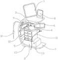
图1是一种可视化的房间隔穿刺装置的整体结构示意图;1 is a schematic diagram of the overall structure of a visualized atrial septal puncture device;
图2是一种可视化的房间隔穿刺装置的主机内部结构示意图;FIG. 2 is a schematic diagram of the internal structure of the main unit of a visualized atrial septal puncture device;
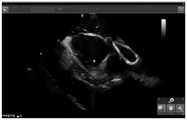
图3是示例超声扇面下的上腔静脉、右心房结构;Figure 3 is an example of the superior vena cava, right atrium structure below the ultrasound fan;
图4是示例J型导丝头端(软头)位于上腔静脉内呈现明显高密度影;Figure 4 is an example of the J-shaped guide wire tip (soft tip) located in the superior vena cava showing obvious high-density shadow;
图5是示例房间隔穿刺鞘组合沿导丝上行过程中淹没部分导丝;Figure 5 is an example of submerged part of the guide wire during the ascent of the transseptal sheath combination along the guide wire;
图6是示例房间隔穿刺鞘组合的水花指示房间隔穿刺鞘组合头端的位置;Fig. 6 is the position of the head end of the atrial transseptal sheath combination indicated by the splash of an example transseptal sheath combination;
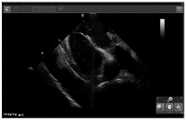
图7是示例房间隔穿刺鞘组合在下拉过程中的右房压迹;Figure 7 is a right atrial impression during pull-down of an example transseptal sheath combination;
图8是示例房间隔穿刺鞘组合顶在房间隔上使房间隔呈现帐篷征;FIG. 8 is an example of the interatrial septum puncture sheath combination being placed on the interatrial septum so that the interatrial septum presents a tent sign;
图9是示例J型导丝硬头房间隔接触面在电凝作用下呈现闪花;Figure 9 is an example of the J-shaped guide wire rigid tip atrial septal contact surface showing sparkle under the action of electrocoagulation;
图10是示例J型导丝硬头已突破房间隔进入左房内;Figure 10 is an example of a J-shaped guide wire that has broken through the atrial septum and entered the left atrium;
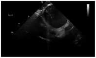
图11是示例J型导丝软头端位于左上肺静脉内;Figure 11 is an example of the soft tip of the J-shaped guide wire located in the left upper pulmonary vein;
附图中,各标号所代表的部件列表如下:In the accompanying drawings, the list of components represented by each number is as follows:
1-支架,2-主机,21-中央处理模块,3-电凝执行器,4-超声执行器,5-储物抽屉,6-储物台,7-超声控制器,8-电凝控制器,9-负极板,10-电刀,11-超声导管,12-无菌储物格,13-万向轮。1-stand, 2-host, 21-central processing module, 3-coagulation actuator, 4-ultrasonic actuator, 5-storage drawer, 6-storage table, 7-ultrasonic controller, 8-coagulation control device, 9-negative plate, 10-electric knife, 11-ultrasound catheter, 12-sterile storage compartment, 13-universal wheel.
具体实施方式Detailed ways
下面将结合本发明实施例中的附图,对本发明实施例中的技术方案进行清楚、完整地描述,显然,所描述的实施例仅仅是本发明一部分实施例,而不是全部的实施例。基于本发明中的实施例,本领域普通技术人员在没有作出创造性劳动前提下所获得的所有其它实施例,都属于本发明保护的范围。The technical solutions in the embodiments of the present invention will be clearly and completely described below with reference to the accompanying drawings in the embodiments of the present invention. Obviously, the described embodiments are only a part of the embodiments of the present invention, but not all of the embodiments. Based on the embodiments of the present invention, all other embodiments obtained by those of ordinary skill in the art without creative efforts shall fall within the protection scope of the present invention.
实施例1Example 1
参阅图1至图2所示,一种可视化的房间隔穿刺装置,其特征在于,包括:支架1、主机2、电凝模块、超声造影模块、存储模块;Referring to FIGS. 1 to 2, a visualized atrial septal puncture device is characterized in that it includes: a
所述支架1整体为“C”型,所述主机2固定设置在支架1底部,所述电凝模块、超声造影模块固定设置在支架1的顶部,所述存储模块固定设置在主机2的上端面;The
所述电凝模块和超声造影模块集成于支架1上,即该装置将传统的电凝器和超声造影有机的结合在一起,避免了在术中多个设备同时使用,占据手术空间的问题;The electrocoagulation module and the contrast-enhanced ultrasound module are integrated on the
所述电凝模块包括电凝执行器3、电刀10、负极板9、电凝控制器8,所述电刀10、负极板9可拆卸的连接在电凝执行器3上,所述电凝控制器8电连接电凝执行器3和主机2;The electrocoagulation module includes an
所述电凝控制器8固定设置在支架1的顶部上端面,所述电凝执行器3固定设置在支架1顶部的下端面;The electrocoagulation controller 8 is fixedly arranged on the upper end surface of the top of the
所述超声造影模块包括超声导管11、超声执行器4、超声控制器7,所述超声导管11可拆卸的连接在超声执行器4上,所述超声控制器7电连接超声执行器4和主机2;The ultrasound contrast module includes an
所述超声控制器7固定设置在支架1的顶部上端面,超声执行器7固定设置在支架1顶部的下端面;The
所述支架1下底面固定设有可锁死的万向轮13,侧面固定设有无菌储物格12,方便存储手术中需要用到的一些无菌用品,如鞘管之类;所述主机2内固定设有中央处理模块21;The bottom surface of the
所述存储模块包括储物抽屉5以及固定设置在储物抽屉5上,且上端可旋转外展的储物台6,无论是储物抽屉5还是储物台6的设计均是增加该装置中储物空间,方便手术中一些手术用具的摆放,使得整个手术过程井然有序。The storage module includes a
实施例2Example 2
本实施例为使用一种可视化的房间隔穿刺装置的房间隔穿刺流程及电凝方法:The present embodiment is a septal puncture procedure and electrocoagulation method using a visualized atrial septal puncture device:
1.装置的连接1. Device connection
①心腔内超声导管的连接:心腔内超声导管的尾端分别与超声机主机及Carto三维标测系统连接,超声导管的管身通过11F的鞘管经股静脉途径进入人体,循静脉途径进入人体的右心房。①Connection of the intracardiac ultrasound catheter: The tail end of the intracardiac ultrasound catheter is connected to the ultrasound machine host and the Carto three-dimensional mapping system respectively. into the right atrium of the human body.
②电刀装置的连接:将负极板的导线连接主机接口,负极板贴于人体腿部皮肤。电刀尾部与主机接口连接,电刀金属头端备用。②Connection of the electrosurgical device: Connect the wire of the negative plate to the host interface, and the negative plate is attached to the skin of the human leg. The tail of the electrosurgery is connected to the host interface, and the metal head of the electrosurgery is spare.
2.打出上腔静脉扇面2. Punch out the superior vena cava fan
通过超声导管的手柄操作打出上腔静脉扇面,具体的操作步骤为:首先旋转手柄上的锁住按钮;然后顺时针旋转手柄,当扇面中出现左侧肺静脉扇面时旋转手柄上的P弯直至出现一半的主动脉扇面时停止旋转P弯;最后旋转手柄上的R弯直至上腔静脉扇面出现。(见图3)The superior vena cava fan is created through the handle operation of the ultrasonic catheter. The specific operation steps are: firstly rotate the lock button on the handle; then rotate the handle clockwise, when the left pulmonary vein fan appears in the fan, rotate the P curve on the handle until it appears Stop rotating the P-curve halfway through the aortic fan; finally rotate the R-curve on the handle until the superior vena cava fan appears. (see Figure 3)
3.超声指导下送房间隔穿刺鞘组合3. Ultrasound-guided delivery of atrial septal puncture sheath combination
①穿刺股静脉后将J型导丝头端(J型软头)送入上腔静脉,超声扇面中可见J型导丝头端,呈高密度影。(见图4)①After puncturing the femoral vein, the tip of the J-shaped guide wire (J-shaped soft tip) was sent into the superior vena cava. The tip of the J-shaped guide wire was seen in the ultrasound fan, showing a high-density shadow. (see Figure 4)
②沿导丝将房间隔穿刺鞘组合送入上腔静脉,并逐渐淹没高亮度的导丝影。(见图5)②The interatrial puncture sheath was combined into the superior vena cava along the guide wire, and the high-brightness guide wire shadow was gradually submerged. (see Figure 5)
③撤出导丝,经房间隔穿刺鞘组合尾端的三连三通注射生理盐水,可见水花出现在上腔静脉内,从而指示房间隔穿刺鞘组合头端在上腔静脉内的位置。(图6)③ Withdraw the guide wire, inject normal saline through the three-way three-way at the end of the atrial septal sheath combination, and it can be seen that the spray appears in the superior vena cava, thus indicating the position of the head of the atrial septal sheath combination in the superior vena cava. (Image 6)
④保持超声导管上腔静脉扇面不动,逐渐往下回撤房间隔穿刺鞘组合,在回撤过程中超声导管手柄逐渐松R弯,实时跟踪房间隔穿刺鞘组合的位置。(图7)④Keep the fan of the superior vena cava of the ultrasound catheter still, and gradually withdraw the atrial septal puncture sheath combination. During the withdrawal process, the ultrasonic catheter handle gradually loosens the R-bend, and the position of the atrial septal puncture sheath combination is tracked in real time. (Figure 7)
⑤房间隔穿刺鞘组合滑落至房间隔位置,出现帐篷征。(图8)⑤ The combination of atrial septal puncture sheath slipped to the atrial septum, and a tent sign appeared. (Figure 8)
4.直视下电凝穿刺房间隔4. Electrocoagulation and puncture of the interatrial septum under direct vision
①沿房间隔穿刺鞘组合尾端的内鞘孔径送入J型导丝尾端硬头(直头),使直头顶在房间隔穿刺鞘组合的房间隔接触面从而房间隔帐篷征更为明显,并用力突破,导丝直头可在直视下直接突破房间隔进入左房。若用力突破存在明显阻力则使用以下电凝步骤。① Send the hard tip (straight end) of the J-shaped guide wire along the inner sheath aperture at the end of the atrial septal puncture sheath combination, so that the straight head tops the atrial septal contact surface of the atrial septal puncture sheath combination, so that the atrial septal tent sign is more obvious. And break through forcefully, the straight end of the guide wire can directly break through the atrial septum and enter the left atrium under direct vision. If there is significant resistance to breaking through forcefully, use the following coagulation procedure.
②将电刀头端的金属头与房间隔穿刺鞘组合尾端的J型导丝部分充分接触,长按电刀手柄的电凝按钮,热量经导丝传送至房间隔穿刺鞘组合的头端,同时J型导丝的硬头在帐篷征下出现闪花并突破房间隔进入左房。(图9,图10)② Fully contact the metal head of the electrosurgical head with the J-shaped guide wire at the tail end of the atrial septal puncture sheath combination, long press the coagulation button of the electrosurgical handle, and the heat is transmitted to the head end of the interatrial puncture sheath combination through the guide wire. The stiff tip of the J-type guide wire flashes under the tent sign and breaks through the interatrial septum into the left atrium. (Figure 9, Figure 10)
③固定超声导管扇面位置,沿J型导丝硬头前送房间隔穿刺鞘组合约5mm,将J型导丝撤出。③Fix the position of the fan surface of the ultrasonic catheter, send the interseptal sheath combination about 5mm forward along the hard tip of the J-shaped guide wire, and withdraw the J-shaped guide wire.
5.房间隔穿刺鞘组合外鞘进入左房5. Atrial septal puncture sheath combined with outer sheath into the left atrium
①将J型导丝的头端(软头)经房间隔穿刺鞘组合的内鞘尾端孔径送入左心房。① Send the tip (soft tip) of the J-shaped guide wire into the left atrium through the caudal aperture of the inner sheath combined with the atrial septal puncture sheath.
②通过超声导管手柄按钮调整超声导管扇面直至出现左侧肺静脉扇面。②Adjust the ultrasound catheter fan through the ultrasound catheter handle button until the left pulmonary vein fan appears.
③调节J型导丝方向,使J型导丝头端位于左侧肺静脉。③ Adjust the direction of the J-shaped guide wire so that the tip of the J-shaped guide wire is located in the left pulmonary vein.
④固定J型导丝,沿导丝将房间隔穿刺鞘组合外鞘整体送入左房。④Fix the J-shaped guide wire, and send the atrial septal puncture sheath combined with the outer sheath into the left atrium as a whole along the guide wire.
(图11)(Figure 11)
综上所述,1、该装置通过设计超声造影模块,摒弃传统的X光线透视形式,以超声造影导向房间隔穿刺,避免了X光线对患者和操作者的辐射伤害;同时设计电凝模块补足房间隔穿刺中导丝难以穿刺进入的问题,使得整个房间隔穿刺更顺利;To sum up, 1. By designing a contrast-enhanced ultrasound module, the device abandons the traditional form of X-ray fluoroscopy, and uses contrast-enhanced ultrasound to guide atrial septal puncture to avoid radiation damage to patients and operators by X-rays; at the same time, the design of an electrocoagulation module complements the The problem that the guide wire is difficult to puncture in the atrial septal puncture makes the whole atrial septal puncture more smoothly;
2、该装置将电凝模块和超声造影模块融合为一个整体,避免了在传统手术过程中多台设备共同运作占据手术空间的问题,为手术操作提供了便利,同时提升了手术中仪器使用的效率。2. The device integrates the electrocoagulation module and the ultrasound contrast module into a whole, which avoids the problem that multiple devices work together to occupy the surgical space during traditional surgery, provides convenience for surgical operations, and improves the use of instruments during surgery. efficiency.
3、该装置使用J型导丝的硬头取代传统的房间隔穿刺针进行房间隔穿刺,既保证了患者安全又节约了经济成本。3. The device uses the hard tip of the J-shaped guide wire to replace the traditional atrial septal puncture needle for atrial septal puncture, which not only ensures the safety of the patient but also saves the economic cost.
在本说明书的描述中,参考术语“一个实施例”、“示例”、“具体示例”等的描述意指结合该实施例或示例描述的具体特征、结构、材料或者特点包含于本发明的至少一个实施例或示例中。在本说明书中,对上述术语的示意性表述不一定指的是相同的实施例或示例。而且,描述的具体特征、结构、材料或者特点可以在任何的一个或多个实施例或示例中以合适的方式结合。In the description of this specification, description with reference to the terms "one embodiment," "example," "specific example," etc. means that a particular feature, structure, material, or characteristic described in connection with the embodiment or example is included in at least one aspect of the present invention. in one embodiment or example. In this specification, schematic representations of the above terms do not necessarily refer to the same embodiment or example. Furthermore, the particular features, structures, materials or characteristics described may be combined in any suitable manner in any one or more embodiments or examples.
以上公开的本发明优选实施例只是用于帮助阐述本发明。优选实施例并没有详尽叙述所有的细节,也不限制该发明仅为所述的具体实施方式。显然,根据本说明书的内容,可作很多的修改和变化。本说明书选取并具体描述这些实施例,是为了更好地解释本发明的原理和实际应用,从而使所属技术领域技术人员能很好地理解和利用本发明。本发明仅受权利要求书及其全部范围和等效物的限制。The above-disclosed preferred embodiments of the present invention are provided only to help illustrate the present invention. The preferred embodiments do not exhaust all the details, nor do they limit the invention to only the described embodiments. Obviously, many modifications and variations are possible in light of the contents of this specification. These embodiments are selected and described in this specification in order to better explain the principles and practical applications of the present invention, so that those skilled in the art can well understand and utilize the present invention. The present invention is to be limited only by the claims and their full scope and equivalents.
Claims (7)
Priority Applications (1)
| Application Number | Priority Date | Filing Date | Title |
|---|---|---|---|
| CN202210431547.1ACN114795414A (en) | 2022-04-22 | 2022-04-22 | Visual interatrial septum puncture device and use method |
Applications Claiming Priority (1)
| Application Number | Priority Date | Filing Date | Title |
|---|---|---|---|
| CN202210431547.1ACN114795414A (en) | 2022-04-22 | 2022-04-22 | Visual interatrial septum puncture device and use method |
Publications (1)
| Publication Number | Publication Date |
|---|---|
| CN114795414Atrue CN114795414A (en) | 2022-07-29 |
Family
ID=82507266
Family Applications (1)
| Application Number | Title | Priority Date | Filing Date |
|---|---|---|---|
| CN202210431547.1APendingCN114795414A (en) | 2022-04-22 | 2022-04-22 | Visual interatrial septum puncture device and use method |
Country Status (1)
| Country | Link |
|---|---|
| CN (1) | CN114795414A (en) |
Citations (10)
| Publication number | Priority date | Publication date | Assignee | Title |
|---|---|---|---|---|
| CN205126349U (en)* | 2015-10-19 | 2016-04-06 | 重庆腾跃医疗器械有限公司 | Clear treatment device that creates of water and electricity disconnect -type |
| CN206836942U (en)* | 2017-01-17 | 2018-01-05 | 山西医科大学 | A kind of medical sacral nerve Needle localization guidance system |
| CN107789011A (en)* | 2016-09-07 | 2018-03-13 | 美国西门子医疗解决公司 | The intracardiac echocardiogram conduit of acoustics ablation auxiliary |
| CN109925028A (en)* | 2018-12-29 | 2019-06-25 | 复旦大学附属华山医院北院 | A kind of ICE ultrasound through superior vena cava atrial septal puncture device |
| CN209695386U (en)* | 2018-09-10 | 2019-11-29 | 浙江伽奈维医疗科技有限公司 | Varicose treatment work station |
| CN111067599A (en)* | 2019-12-23 | 2020-04-28 | 云南省第二人民医院 | Percutaneous interatrial puncture suit |
| CN211409192U (en)* | 2019-08-22 | 2020-09-04 | 湖南埃普特医疗器械有限公司 | Dilator and interatrial puncture system |
| US20210259732A1 (en)* | 2020-02-25 | 2021-08-26 | Baylis Medical Company Inc. | Methods and Devices for Creation of Communication Between Aorta and Left Atrium |
| CN113473928A (en)* | 2019-02-04 | 2021-10-01 | 国际私人银行有限责任公司 | Needle system and method |
| CN113692257A (en)* | 2019-03-20 | 2021-11-23 | 东区医疗有限公司 | Directional balloon transseptal insertion device for medical procedures |
- 2022
- 2022-04-22CNCN202210431547.1Apatent/CN114795414A/enactivePending
Patent Citations (10)
| Publication number | Priority date | Publication date | Assignee | Title |
|---|---|---|---|---|
| CN205126349U (en)* | 2015-10-19 | 2016-04-06 | 重庆腾跃医疗器械有限公司 | Clear treatment device that creates of water and electricity disconnect -type |
| CN107789011A (en)* | 2016-09-07 | 2018-03-13 | 美国西门子医疗解决公司 | The intracardiac echocardiogram conduit of acoustics ablation auxiliary |
| CN206836942U (en)* | 2017-01-17 | 2018-01-05 | 山西医科大学 | A kind of medical sacral nerve Needle localization guidance system |
| CN209695386U (en)* | 2018-09-10 | 2019-11-29 | 浙江伽奈维医疗科技有限公司 | Varicose treatment work station |
| CN109925028A (en)* | 2018-12-29 | 2019-06-25 | 复旦大学附属华山医院北院 | A kind of ICE ultrasound through superior vena cava atrial septal puncture device |
| CN113473928A (en)* | 2019-02-04 | 2021-10-01 | 国际私人银行有限责任公司 | Needle system and method |
| CN113692257A (en)* | 2019-03-20 | 2021-11-23 | 东区医疗有限公司 | Directional balloon transseptal insertion device for medical procedures |
| CN211409192U (en)* | 2019-08-22 | 2020-09-04 | 湖南埃普特医疗器械有限公司 | Dilator and interatrial puncture system |
| CN111067599A (en)* | 2019-12-23 | 2020-04-28 | 云南省第二人民医院 | Percutaneous interatrial puncture suit |
| US20210259732A1 (en)* | 2020-02-25 | 2021-08-26 | Baylis Medical Company Inc. | Methods and Devices for Creation of Communication Between Aorta and Left Atrium |
Similar Documents
| Publication | Publication Date | Title |
|---|---|---|
| CN111491584B (en) | Augmented reality solution for optimizing directional approach and therapy delivery of interventional cardiology tools | |
| US12318632B2 (en) | Heart arrhythmia non-invasive treatment device and method | |
| CN103118595B (en) | Medical diagnostic imaging apparatus | |
| JP2008511413A (en) | Method and system for treating atrial fibrillation and other cardiac arrhythmias | |
| Mascia et al. | A new era in zero X-ray ablation | |
| Schwagten et al. | Initial experience with catheter ablation using remote magnetic navigation in adults with complex congenital heart disease and in small children | |
| US20080228079A1 (en) | Clinical utilization of contrast agents to define specific areas within the myocardial wall to provide guidance and localization for ablation, cyroablation, or other techniques in patients with post myocardial infarction | |
| US20230070457A1 (en) | Medical image processing apparatus, x-ray diagnostic apparatus, and storage medium | |
| JP5823096B2 (en) | X-ray diagnostic apparatus and image processing method | |
| Hua et al. | Advances of implantation techniques for conduction system pacing | |
| CN210785905U (en) | Coronary artery intervention guide wire fixer | |
| CN114795414A (en) | Visual interatrial septum puncture device and use method | |
| Knight et al. | Robotic positioning of standard electrophysiology catheters: a novel approach to catheter robotics | |
| US20050159798A1 (en) | Method and apparatus for cardiac ablation | |
| CN217138241U (en) | Chemical ablation balloon catheter and chemical ablation combined product | |
| Jia et al. | Percutaneous occluder device closure through femoral vein guidance by transthoracic echocardiography in adult atrial septal defect patients | |
| WO2018148525A1 (en) | Determining ablation location using probabilistic decision-making | |
| CN203852439U (en) | Large sheet for interventional therapy operations | |
| CN108451641A (en) | A kind of operating robot for vertebroplasty | |
| CN114869431A (en) | Interatrial septum puncture system | |
| Robledo-Nolasco et al. | Radiofrequency catheter ablation of cardiac arrhythmias using only three-dimentional mapping systems | |
| Drant et al. | Guidance of radiofrequency catheter ablation by transesophageal echocardiography in children with palliated single ventricle | |
| Chokr et al. | Catheter ablation of focal atrial tachycardia with early activation close to the his-bundle from the non coronary aortic cusp | |
| Robledo-Nolasco et al. | Ablación con catéter de Radiofrecuencia de taquiarritmias USANDO sólo sistemas de mapeo tridimensional | |
| CN221577964U (en) | Multifunctional puncturing device for gynecological intervention treatment |
Legal Events
| Date | Code | Title | Description |
|---|---|---|---|
| PB01 | Publication | ||
| PB01 | Publication | ||
| SE01 | Entry into force of request for substantive examination | ||
| SE01 | Entry into force of request for substantive examination |