CN114766427B - A severe preeclampsia animal model based on BCAM target and its construction method and application - Google Patents
A severe preeclampsia animal model based on BCAM target and its construction method and applicationDownload PDFInfo
- Publication number
- CN114766427B CN114766427BCN202210449712.6ACN202210449712ACN114766427BCN 114766427 BCN114766427 BCN 114766427BCN 202210449712 ACN202210449712 ACN 202210449712ACN 114766427 BCN114766427 BCN 114766427B
- Authority
- CN
- China
- Prior art keywords
- severe preeclampsia
- preeclampsia
- pregnant
- bcam
- animal model
- Prior art date
- Legal status (The legal status is an assumption and is not a legal conclusion. Google has not performed a legal analysis and makes no representation as to the accuracy of the status listed.)
- Active
Links
- 201000005608severe pre-eclampsiaDiseases0.000titleclaimsabstractdescription40
- 238000010171animal modelMethods0.000titleclaimsabstractdescription27
- 101000935638Homo sapiens Basal cell adhesion moleculeProteins0.000titleclaimsabstractdescription22
- 101000766294Homo sapiens Branched-chain-amino-acid aminotransferase, mitochondrialProteins0.000titleclaimsabstractdescription22
- 102100026413Branched-chain-amino-acid aminotransferase, mitochondrialHuman genes0.000titleclaimsabstract4
- 238000010276constructionMethods0.000titleabstractdescription5
- 241000699670Mus sp.Species0.000claimsabstractdescription36
- 238000011282treatmentMethods0.000claimsabstractdescription7
- 230000002265preventionEffects0.000claimsabstractdescription5
- 210000002700urineAnatomy0.000claimsdescription24
- 230000035935pregnancyEffects0.000claimsdescription17
- 210000002826placentaAnatomy0.000claimsdescription15
- 102100035194Placenta growth factorHuman genes0.000claimsdescription12
- 241000700159RattusSpecies0.000claimsdescription12
- 230000036772blood pressureEffects0.000claimsdescription12
- 238000000034methodMethods0.000claimsdescription12
- 210000004185liverAnatomy0.000claimsdescription9
- 238000010186stainingMethods0.000claimsdescription9
- 238000001514detection methodMethods0.000claimsdescription8
- 210000003734kidneyAnatomy0.000claimsdescription8
- 108010082093Placenta Growth FactorProteins0.000claimsdescription7
- 230000001575pathological effectEffects0.000claimsdescription7
- 238000002331protein detectionMethods0.000claimsdescription7
- 238000002360preparation methodMethods0.000claimsdescription5
- 102000004169proteins and genesHuman genes0.000claimsdescription5
- 108090000623proteins and genesProteins0.000claimsdescription5
- 238000002347injectionMethods0.000claimsdescription4
- 239000007924injectionSubstances0.000claimsdescription4
- 230000000877morphologic effectEffects0.000claimsdescription4
- 210000000056organAnatomy0.000claimsdescription4
- 231100000915pathological changeToxicity0.000claimsdescription4
- 230000036285pathological changeEffects0.000claimsdescription4
- 239000003153chemical reaction reagentSubstances0.000claimsdescription3
- 239000003814drugSubstances0.000claimsdescription3
- 229940079593drugDrugs0.000claimsdescription3
- 210000003462veinAnatomy0.000claimsdescription3
- 238000012544monitoring processMethods0.000claimsdescription2
- 238000003759clinical diagnosisMethods0.000abstractdescription3
- 230000008506pathogenesisEffects0.000abstractdescription3
- 238000011269treatment regimenMethods0.000abstractdescription3
- 208000032023Signs and SymptomsDiseases0.000abstractdescription2
- 102100028239Basal cell adhesion moleculeHuman genes0.000description18
- 201000011461pre-eclampsiaDiseases0.000description12
- 241000699666Mus <mouse, genus>Species0.000description10
- FAPWRFPIFSIZLT-UHFFFAOYSA-MSodium chlorideChemical compound[Na+].[Cl-]FAPWRFPIFSIZLT-UHFFFAOYSA-M0.000description7
- 210000002993trophoblastAnatomy0.000description7
- LFQSCWFLJHTTHZ-UHFFFAOYSA-NEthanolChemical compoundCCOLFQSCWFLJHTTHZ-UHFFFAOYSA-N0.000description6
- 101150062285PGF geneProteins0.000description5
- 239000002253acidSubstances0.000description5
- 238000011161developmentMethods0.000description5
- 230000003169placental effectEffects0.000description5
- WZUVPPKBWHMQCE-UHFFFAOYSA-NHaematoxylinChemical compoundC12=CC(O)=C(O)C=C2CC2(O)C1C1=CC=C(O)C(O)=C1OC2WZUVPPKBWHMQCE-UHFFFAOYSA-N0.000description4
- 102000003855L-lactate dehydrogenaseHuman genes0.000description4
- 108700023483L-lactate dehydrogenasesProteins0.000description4
- 230000001605fetal effectEffects0.000description4
- 230000008569processEffects0.000description4
- 235000018102proteinsNutrition0.000description4
- 239000000243solutionSubstances0.000description4
- 239000012192staining solutionSubstances0.000description4
- XLYOFNOQVPJJNP-UHFFFAOYSA-NwaterSubstancesOXLYOFNOQVPJJNP-UHFFFAOYSA-N0.000description4
- 239000012224working solutionSubstances0.000description4
- 210000004027cellAnatomy0.000description3
- 208000037265diseases, disorders, signs and symptomsDiseases0.000description3
- 238000002474experimental methodMethods0.000description3
- 201000007270liver cancerDiseases0.000description3
- 208000014018liver neoplasmDiseases0.000description3
- DHRLEVQXOMLTIM-UHFFFAOYSA-Nphosphoric acid;trioxomolybdenumChemical compoundO=[Mo](=O)=O.O=[Mo](=O)=O.O=[Mo](=O)=O.O=[Mo](=O)=O.O=[Mo](=O)=O.O=[Mo](=O)=O.O=[Mo](=O)=O.O=[Mo](=O)=O.O=[Mo](=O)=O.O=[Mo](=O)=O.O=[Mo](=O)=O.O=[Mo](=O)=O.OP(O)(O)=ODHRLEVQXOMLTIM-UHFFFAOYSA-N0.000description3
- 239000000047productSubstances0.000description3
- 239000008399tap waterSubstances0.000description3
- 235000020679tap waterNutrition0.000description3
- 238000012360testing methodMethods0.000description3
- XEEYBQQBJWHFJM-UHFFFAOYSA-NIronChemical compound[Fe]XEEYBQQBJWHFJM-UHFFFAOYSA-N0.000description2
- CTQNGGLPUBDAKN-UHFFFAOYSA-NO-XyleneChemical compoundCC1=CC=CC=C1CCTQNGGLPUBDAKN-UHFFFAOYSA-N0.000description2
- 102100024616Platelet endothelial cell adhesion moleculeHuman genes0.000description2
- 210000001367arteryAnatomy0.000description2
- 230000004791biological behaviorEffects0.000description2
- 230000008827biological functionEffects0.000description2
- 230000037396body weightEffects0.000description2
- 230000006378damageEffects0.000description2
- 230000018044dehydrationEffects0.000description2
- 238000006297dehydration reactionMethods0.000description2
- 230000035487diastolic blood pressureEffects0.000description2
- 201000010099diseaseDiseases0.000description2
- 239000012153distilled waterSubstances0.000description2
- 230000002503metabolic effectEffects0.000description2
- 230000007935neutral effectEffects0.000description2
- 238000007634remodelingMethods0.000description2
- 239000006228supernatantSubstances0.000description2
- 230000035488systolic blood pressureEffects0.000description2
- 241000701161unidentified adenovirusSpecies0.000description2
- 238000005406washingMethods0.000description2
- 238000005303weighingMethods0.000description2
- 239000008096xyleneSubstances0.000description2
- AXDJCCTWPBKUKL-UHFFFAOYSA-N4-[(4-aminophenyl)-(4-imino-3-methylcyclohexa-2,5-dien-1-ylidene)methyl]aniline;hydron;chlorideChemical compoundCl.C1=CC(=N)C(C)=CC1=C(C=1C=CC(N)=CC=1)C1=CC=C(N)C=C1AXDJCCTWPBKUKL-UHFFFAOYSA-N0.000description1
- 102100036475Alanine aminotransferase 1Human genes0.000description1
- 108010082126Alanine transaminaseProteins0.000description1
- VHUUQVKOLVNVRT-UHFFFAOYSA-NAmmonium hydroxideChemical compound[NH4+].[OH-]VHUUQVKOLVNVRT-UHFFFAOYSA-N0.000description1
- 108010003415Aspartate AminotransferasesProteins0.000description1
- 102000004625Aspartate AminotransferasesHuman genes0.000description1
- 108010067225Cell Adhesion MoleculesProteins0.000description1
- 102000016289Cell Adhesion MoleculesHuman genes0.000description1
- 102000008186CollagenHuman genes0.000description1
- 108010035532CollagenProteins0.000description1
- 238000008157ELISA kitMethods0.000description1
- 208000001362Fetal Growth RetardationDiseases0.000description1
- 102100031351Galectin-9Human genes0.000description1
- 101710121810Galectin-9Proteins0.000description1
- 241000282412HomoSpecies0.000description1
- 206010020772HypertensionDiseases0.000description1
- 108060003951ImmunoglobulinProteins0.000description1
- 241001465754MetazoaSpecies0.000description1
- 206010030113OedemaDiseases0.000description1
- 208000037273Pathologic ProcessesDiseases0.000description1
- 206010038536Renal tubular atrophyDiseases0.000description1
- 241000283984RodentiaSpecies0.000description1
- 235000011114ammonium hydroxideNutrition0.000description1
- 230000009286beneficial effectEffects0.000description1
- 230000033228biological regulationEffects0.000description1
- 239000000090biomarkerSubstances0.000description1
- 230000015572biosynthetic processEffects0.000description1
- 210000004369bloodAnatomy0.000description1
- 239000008280bloodSubstances0.000description1
- 238000009530blood pressure measurementMethods0.000description1
- 230000009134cell regulationEffects0.000description1
- 229920001436collagenPolymers0.000description1
- 230000003247decreasing effectEffects0.000description1
- 230000002950deficientEffects0.000description1
- 230000008021depositionEffects0.000description1
- 238000003745diagnosisMethods0.000description1
- 230000004069differentiationEffects0.000description1
- 208000035475disorderDiseases0.000description1
- 230000004064dysfunctionEffects0.000description1
- 208000002296eclampsiaDiseases0.000description1
- 230000032692embryo implantationEffects0.000description1
- 210000002889endothelial cellAnatomy0.000description1
- 210000003989endothelium vascularAnatomy0.000description1
- YQGOJNYOYNNSMM-UHFFFAOYSA-NeosinChemical compound[Na+].OC(=O)C1=CC=CC=C1C1=C2C=C(Br)C(=O)C(Br)=C2OC2=C(Br)C(O)=C(Br)C=C21YQGOJNYOYNNSMM-UHFFFAOYSA-N0.000description1
- 230000007705epithelial mesenchymal transitionEffects0.000description1
- 210000003743erythrocyteAnatomy0.000description1
- DZGCGKFAPXFTNM-UHFFFAOYSA-Nethanol;hydron;chlorideChemical compoundCl.CCODZGCGKFAPXFTNM-UHFFFAOYSA-N0.000description1
- 208000030941fetal growth restrictionDiseases0.000description1
- 210000003754fetusAnatomy0.000description1
- 210000003976gap junctionAnatomy0.000description1
- 238000007490hematoxylin and eosin (H&E) stainingMethods0.000description1
- 210000003494hepatocyteAnatomy0.000description1
- 238000003125immunofluorescent labelingMethods0.000description1
- 102000018358immunoglobulinHuman genes0.000description1
- 238000013115immunohistochemical detectionMethods0.000description1
- 230000008595infiltrationEffects0.000description1
- 238000001764infiltrationMethods0.000description1
- 230000009545invasionEffects0.000description1
- 229910052742ironInorganic materials0.000description1
- 238000004519manufacturing processMethods0.000description1
- 239000000463materialSubstances0.000description1
- 239000011159matrix materialSubstances0.000description1
- 230000004048modificationEffects0.000description1
- 238000012986modificationMethods0.000description1
- 238000010172mouse modelMethods0.000description1
- 230000017074necrotic cell deathEffects0.000description1
- 230000009054pathological processEffects0.000description1
- 210000005059placental tissueAnatomy0.000description1
- 230000036470plasma concentrationEffects0.000description1
- 210000003240portal veinAnatomy0.000description1
- 239000013641positive controlSubstances0.000description1
- 201000001474proteinuriaDiseases0.000description1
- 238000011552rat modelMethods0.000description1
- 238000011160researchMethods0.000description1
- 238000012216screeningMethods0.000description1
- 238000000926separation methodMethods0.000description1
- 238000012174single-cell RNA sequencingMethods0.000description1
- 230000008961swellingEffects0.000description1
- 208000024891symptomDiseases0.000description1
- 208000011580syndromic diseaseDiseases0.000description1
- 230000009885systemic effectEffects0.000description1
- 238000002560therapeutic procedureMethods0.000description1
- 210000001578tight junctionAnatomy0.000description1
- 210000001519tissueAnatomy0.000description1
- 210000004881tumor cellAnatomy0.000description1
- XOSXWYQMOYSSKB-LDKJGXKFSA-Lwater blueChemical compoundCC1=CC(/C(\C(C=C2)=CC=C2NC(C=C2)=CC=C2S([O-])(=O)=O)=C(\C=C2)/C=C/C\2=N\C(C=C2)=CC=C2S([O-])(=O)=O)=CC(S(O)(=O)=O)=C1N.[Na+].[Na+]XOSXWYQMOYSSKB-LDKJGXKFSA-L0.000description1
- 230000036266weeks of gestationEffects0.000description1
Images


Classifications
- A—HUMAN NECESSITIES
- A01—AGRICULTURE; FORESTRY; ANIMAL HUSBANDRY; HUNTING; TRAPPING; FISHING
- A01K—ANIMAL HUSBANDRY; AVICULTURE; APICULTURE; PISCICULTURE; FISHING; REARING OR BREEDING ANIMALS, NOT OTHERWISE PROVIDED FOR; NEW BREEDS OF ANIMALS
- A01K67/00—Rearing or breeding animals, not otherwise provided for; New or modified breeds of animals
- A01K67/02—Breeding vertebrates
Landscapes
- Life Sciences & Earth Sciences (AREA)
- Environmental Sciences (AREA)
- Animal Behavior & Ethology (AREA)
- Zoology (AREA)
- Animal Husbandry (AREA)
- Biodiversity & Conservation Biology (AREA)
- Investigating Or Analysing Biological Materials (AREA)
Abstract
Translated fromChinese
Description
Translated fromChinese技术领域technical field
本发明涉及生物医药领域,具体的说,是一种基于BCAM靶标的重度子痫前期动物模型及其构建方法和应用。The present invention relates to the field of biomedicine, specifically, a severe preeclampsia animal model based on BCAM target and its construction method and application.
背景技术Background technique
子痫前期是一种妊娠20周后以新发高血压(≥140/90 mmHg)为主要临床特征的多因素、多途径、多器官受累的妊娠期综合征,可根据严重程度分为子痫前期和重度子痫前期,滋养细胞浅浸润与螺旋动脉重塑障碍是其病理基础,但具体发病机制尚不明确。胚胎植入过程离不开细胞粘附分子对细胞—细胞、细胞—基质的紧密连接和间隙连接,这是成功妊娠的重要过程。粘附分子可促进细胞通过由外而内的信号对细胞外环境的变化做出反应,并且调控细胞命运,引起细胞生物学行为改变,是子痫前期发生发展的中心环节。Preeclampsia is a multifactorial, multipathway, and multiorgan-involved pregnancy syndrome characterized by new-onset hypertension (≥140/90 mmHg) after 20 weeks of gestation. It can be divided into eclampsia according to severity. In preeclampsia and severe preeclampsia, the shallow infiltration of trophoblasts and the disorder of spiral artery remodeling are the pathological basis, but the specific pathogenesis is still unclear. The process of embryo implantation is inseparable from the tight junctions and gap junctions of cell-cell and cell-matrix by cell adhesion molecules, which is an important process for successful pregnancy. Adhesion molecules can promote cells to respond to changes in the extracellular environment through outside-in signals, regulate cell fate, and cause changes in cell biological behavior, which is the central link in the development of preeclampsia.
由于人体实验的医学伦理学制约,动物模型成为研究子痫前期发生发展及探索防治措施的重要工具。啮齿类动物胎盘形成方式与滋养细胞侵袭程度与人类接近,母体有子宫螺旋动脉重塑的表现,造模成功后,可出现子痫前期的典型表型,既往采用的子痫前期大鼠模型为通过大鼠尾静脉注射腺病毒构建的sFlt-1,但此模型一般不会出现多器官脏器的损害,不能很好的反应严重的子痫前期。另外,为成功构建子痫前期的动物模型,公开号为CN111514277A的发明专利采用了Gal-9作为生物标记物来构建子痫前期的小鼠动物模型,以及公开号为CN112941102A的发明专利采用了RNA m6A作为靶点来构建子痫前期的小鼠动物模型。然而目前并没有能很好反应重度子痫前期这种疾病严重程度的动物模型。Due to the medical ethics constraints of human experiments, animal models have become an important tool to study the occurrence and development of preeclampsia and to explore prevention and treatment measures. The rodent placental formation method and the degree of trophoblast invasion are similar to those of humans. The mother has the performance of uterine spiral artery remodeling. After successful modeling, the typical phenotype of preeclampsia can appear. The preeclampsia rat model used in the past is The sFlt-1 constructed by injecting adenovirus into the tail vein of rats generally does not cause damage to multiple organs and cannot respond well to severe preeclampsia. In addition, in order to successfully construct an animal model of preeclampsia, the invention patent with publication number CN111514277A uses Gal-9 as a biomarker to construct a mouse animal model of preeclampsia, and the invention patent with publication number CN112941102A uses RNA m6A was used as a target to construct a mouse animal model of preeclampsia. However, there is currently no animal model that can well reflect the severity of severe preeclampsia.
BCAM属于免疫球蛋白超家族中的一类粘附分子,在与滋养细胞具有相似 “上皮-间质转化”进程的肿瘤细胞上,参与调控细胞生物学行为。例如:余增在“肝癌特异性抗体的筛选和鉴定即BCAM作为肝癌诊疗潜在靶点的初步研究”中揭示了BCAM作为肝癌相关靶点的临床研究价值,以及最新研究利用孕早期胎盘建立单细胞RNA测序,揭示BCAM标记着滋养细胞祖细胞建立、更新和分化(Shannon MJ et al. 2022;149(1):dev199840.Development)。然而,目前尚无研究报道BCAM在滋养细胞中的功能调控作用及其与重度子痫前期的关系。BCAM belongs to a class of adhesion molecules in the immunoglobulin superfamily, and participates in the regulation of cell biological behavior on tumor cells that have a similar "epithelial-mesenchymal transition" process to trophoblast cells. For example: Yu Zeng revealed the clinical research value of BCAM as a liver cancer-related target in "Screening and Identification of Liver Cancer-Specific Antibodies: A Preliminary Study on BCAM as a Potential Target for Liver Cancer Diagnosis and Therapy", and the latest study used the placenta in the first trimester to establish a single cell RNA sequencing reveals that BCAM marks trophoblast progenitor establishment, renewal and differentiation (Shannon MJ et al. 2022;149(1):dev199840.Development). However, no studies have reported the functional regulation of BCAM in trophoblasts and its relationship with severe preeclampsia.
发明内容Contents of the invention
本发明首次将BCAM作为靶标应用于重度子痫前期动物模型的构建中,为此,提供了一种基于BCAM靶标的重度子痫前期动物模型及其构建方法,通过孕鼠BCAM水平的降低,可以获得具有重度子痫前期的特征性症状和体征改变的动物模型,并能反应该疾病的严重程度,操作可控性好、稳定性佳。For the first time, the present invention uses BCAM as a target in the construction of a severe preeclampsia animal model. Therefore, a BCAM target-based severe preeclampsia animal model and its construction method are provided. By reducing the level of BCAM in pregnant mice, the Obtain an animal model with changes in the characteristic symptoms and signs of severe preeclampsia, which can reflect the severity of the disease, and has good controllability and stability.
由于BCAM缺乏的孕鼠模型可反映重度子痫前期这一涉及全身多器官组织疾病的严重程度,对探讨重度子痫前期临床诊疗和处置策略的选择具有重要价值,为此,本发明还提供了基于BCAM靶标的重度子痫前期动物模型在重度子痫前期中的应用,如用于制备防治重度子痫前期的药物,或制备诊断/预测重度子痫前期的产品,等。Since the BCAM-deficient pregnant mouse model can reflect the severity of severe preeclampsia, a disease involving multiple organs and tissues throughout the body, it is of great value in exploring the selection of clinical diagnosis, treatment and treatment strategies for severe preeclampsia. For this reason, the present invention also provides The application of severe preeclampsia animal models based on BCAM targets in severe preeclampsia, such as for the preparation of drugs for the prevention and treatment of severe preeclampsia, or the preparation of products for diagnosing/predicting severe preeclampsia, etc.
本发明通过下述技术方案实现:一种基于BCAM靶标的重度子痫前期动物模型的构建方法,通过对孕鼠注射Ad-shBCAM,并监测其有关重度子痫前期的病理指标,以构建得到重度子痫前期的动物模型。The present invention is achieved through the following technical solutions: a method for constructing a severe preeclampsia animal model based on BCAM targets, by injecting Ad-shBCAM into pregnant mice and monitoring its pathological indicators related to severe preeclampsia to construct severe preeclampsia Animal models of preeclampsia.
所述孕鼠采用10周龄SPF级的SD大鼠。The pregnant mice were 10-week-old SD rats of SPF grade.
所述孕鼠的注射时间为孕第9.5天。The injection time of the pregnant mice is the 9.5th day of pregnancy.
所述孕鼠采用尾静脉注射Ad-shBCAM,注射剂量为2×10⁹ PFU。The pregnant mice were injected with Ad-shBCAM through the tail vein, and the injection dose was 2×10⁹ PFU.
所述病理指标包括血压、24-h尿蛋白、生化指标、主要脏器病理变化及子代宫内发育变化。The pathological indicators include blood pressure, 24-h urine protein, biochemical indicators, pathological changes of major organs, and intrauterine development changes of offspring.
还包括对病理指标的检测:Also includes detection of pathological indicators:
在孕第6天、9天、12天、15天和17天分别对孕鼠进行称重,测量无创血压,并收集在孕第9天、12天、15天和17天尿液,行24-h尿蛋白检测;On the 6th, 9th, 12th, 15th and 17th day of pregnancy, the pregnant mice were weighed, and the non-invasive blood pressure was measured, and the urine was collected on the 9th, 12th, 15th and 17th day of pregnancy. -h urine protein detection;
在孕第19天测量孕鼠有创血压,并收集尿液行24-h尿蛋白检测,收集血浆行生化指标、PlGF和sFlt-1检测,收集胎盘、肝脏和肾脏作形态学染色观察;Measure the invasive blood pressure of pregnant rats on the 19th day of pregnancy, collect urine for 24-h urine protein detection, collect plasma for biochemical indicators, PlGF and sFlt-1 detection, and collect placenta, liver and kidney for morphological staining observation;
对仔鼠计数、称重和测量身长。The pups were counted, weighed and measured for length.
BCAM作为靶标在重度子痫前期动物模型中的应用,采用上述方法构建重度子痫前期动物模型。The application of BCAM as a target in animal models of severe preeclampsia, using the above method to construct animal models of severe preeclampsia.
采用上述方法构建的重度子痫前期动物模型。A severe preeclampsia animal model constructed by the above method.
采用上述方法构建的重度子痫前期动物模型在用于制备防治重度子痫前期的药物、或制备诊断/预测重度子痫前期的产品中的应用。The application of the severe preeclampsia animal model constructed by the above method in the preparation of drugs for the prevention and treatment of severe preeclampsia, or the production of products for diagnosing/predicting severe preeclampsia.
所述产品为试剂或试剂盒。The product is a reagent or a test kit.
本发明与现有技术相比,具有以下优点及有益效果:Compared with the prior art, the present invention has the following advantages and beneficial effects:
(1)本发明所建立的重度子痫前期动物模型,具有血压和24-h蛋白尿升高的子痫前期典型症状,这与人类子痫前期的病理过程相符合。此外,还检测到了胎盘、肝肾的结构功能损伤,并观察到胎鼠宫内发育受限,这些都能很好的反应重度子痫前期的全身多系统功能障碍及严重程度,更具代表性。(1) The severe preeclampsia animal model established by the present invention has typical preeclampsia symptoms of elevated blood pressure and 24-h proteinuria, which is consistent with the pathological process of human preeclampsia. In addition, the structural and functional damage of the placenta, liver and kidney was detected, and intrauterine growth restriction of fetal mice was observed. These can well reflect the systemic multi-system dysfunction and severity of severe preeclampsia, and are more representative .
(2)本发明提供了一种标准化、安全高效、稳定可靠的重度子痫前期动物模型,为探讨重度子痫前期的发病机制、临床诊疗和处置策略等提供了动物模型。(2) The present invention provides a standardized, safe, efficient, stable and reliable animal model of severe preeclampsia, which provides an animal model for exploring the pathogenesis, clinical diagnosis and treatment, and treatment strategies of severe preeclampsia.
附图说明Description of drawings
图1为SD孕鼠体重测定值变化。Figure 1 shows the changes in body weight of SD pregnant mice.
图2为SD孕鼠血压测定值变化。Figure 2 shows the changes in blood pressure measurements of SD pregnant mice.
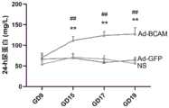
图3为SD孕鼠24-h尿蛋白测定值变化。Figure 3 shows the changes in the measured value of urine protein in SD pregnant mice at 24-h.
图4为SD孕鼠的血浆sFlt-1和PLGF水平变化。Figure 4 shows the changes of plasma sFlt-1 and PLGF levels in pregnant SD mice.
图5为SD孕鼠在孕19天的胎盘重量变化。Figure 5 shows the changes in placental weight of SD pregnant mice on day 19 of pregnancy.
图6为SD孕鼠胎盘与肝脏病理变化。Figure 6 shows the pathological changes of the placenta and liver of SD pregnant mice.
图7为SD孕鼠肾脏病理变化。Figure 7 shows the pathological changes of the kidneys of pregnant SD mice.
图8为仔鼠宫内发育情况。Figure 8 shows the intrauterine development of offspring.
图9为SD孕鼠胎盘BCAM表达变化。Figure 9 shows the changes in the expression of BCAM in the placenta of pregnant SD mice.
具体实施方式Detailed ways
下面结合实施例对本发明作进一步地详细说明,但本发明的实施方式不限于此。The present invention will be further described in detail below in conjunction with examples, but the embodiments of the present invention are not limited thereto.
主要试剂及材料:Main reagents and materials:
SPF级SD大鼠(北京华阜康生物科技有限公司)、重组腺病毒载体(上海汉恒生物科技有限公司)、BCA试剂盒(碧云天生物技术有限公司)。SPF grade SD rats (Beijing Huafukang Biotechnology Co., Ltd.), recombinant adenovirus vector (Shanghai Hanheng Biotechnology Co., Ltd.), BCA kit (Biyuntian Biotechnology Co., Ltd.).
主要仪器:Main instruments:
BP-98A型智能无创血压-鼠仪(北京软隆生物科技有限公司)、BL-420S生物机能实验系统(BL系列生物机能实验系统,成都泰盟科技有限公司)、大鼠代谢笼(四川大学基础医学院动物实验中心)、全自动生化分析仪BS-200(深圳迈瑞生物医疗公司)、扫描电子显微镜(日本Olympus公司)。BP-98A intelligent non-invasive blood pressure-mouse instrument (Beijing Ruanlong Biotechnology Co., Ltd.), BL-420S biological function experiment system (BL series biological function experiment system, Chengdu Taimeng Technology Co., Ltd.), rat metabolic cage (Sichuan University Basic Medical College Animal Experiment Center), automatic biochemical analyzer BS-200 (Shenzhen Mindray Biomedical Company), scanning electron microscope (Japan Olympus Company).
实施例1:Example 1:
选取周龄10周的SD大鼠,按雌雄合笼比例1∶2,确定妊娠后将雌鼠随机分为3组,每组6只雌鼠,在孕第9.5天经尾静脉分别注射2×10⁹ PFU的Ad-shBCAM(实验组)、Ad-GFP(载体对照组)及生理盐水(空白对照组)。Select 10-week-old SD rats, according to the ratio of male and female in a cage of 1:2, after confirming the pregnancy, the female mice are randomly divided into 3 groups, with 6 female mice in each group, and inject 2× 10⁹ PFU of Ad-shBCAM (experimental group), Ad-GFP (vehicle control group) and normal saline (blank control group).
在孕6天、9天、12天、15天和17天对孕鼠进行称重,测量无创血压,并收集在孕第9天、12天、15天和17天尿液,行24-h尿蛋白检测。Pregnant mice were weighed on
在孕第19天测量孕鼠有创血压,并收集尿液行24-h尿蛋白检测,收集血浆行生化指标、PlGF和sFlt-1检测,胎盘称重,收集胎盘、肝脏和肾脏作形态学染色观察,仔鼠计数、称重和测量身长。Measure the invasive blood pressure of pregnant mice on the 19th day of pregnancy, collect urine for 24-h urine protein detection, collect plasma for biochemical indicators, PlGF and sFlt-1 detection, weigh the placenta, and collect placenta, liver and kidney for morphology Staining observation, counting, weighing and measuring the length of offspring.
检测结果如下:The test results are as follows:
(一)孕鼠体重(1) Weight of pregnant mice
结果参见图1, 与对照组相比,Ad-shBCAM组孕鼠的体重在GD15和GD17上均显著降低。其中,*:P< 0.05,Ad-shBCAM组与Ad-GFP组比较;#:P< 0.05,Ad-shBCAM组与生理盐水组比较。The results are shown in Figure 1. Compared with the control group, the weight of the pregnant mice in the Ad-shBCAM group was significantly reduced on both GD15 and GD17. Among them, *: P<0.05, compared between Ad-shBCAM group and Ad-GFP group; #: P<0.05, compared between Ad-shBCAM group and normal saline group.
(二)孕鼠体重和血压(2) Body weight and blood pressure of pregnant mice
结果参见图2,A为收缩压;B为舒张压;C为孕19天孕鼠的有创血压。与对照组相比,Ad-shBCAM组孕鼠的收缩压、舒张压和有创血压均显著升高。其中,*:P< 0.05,Ad-shBCAM组与Ad-GFP组比较;#:P< 0.05,Ad-shBCAM组与生理盐水组比较。The results are shown in Fig. 2, A is the systolic blood pressure; B is the diastolic blood pressure; C is the invasive blood pressure of the pregnant rat on the 19th day of pregnancy. Compared with the control group, the systolic blood pressure, diastolic blood pressure and invasive blood pressure of pregnant mice in the Ad-shBCAM group were significantly increased. Among them, *: P<0.05, compared between Ad-shBCAM group and Ad-GFP group; #: P<0.05, compared between Ad-shBCAM group and normal saline group.
(三)24-h尿蛋白检测(3) 24-h urine protein detection
24-h尿蛋白检测时,在需采集尿液的前一天早晨9:00将大鼠放入代谢笼中,大鼠自由活动及饮食,次日9:00收集尿杯中的尿液,记录尿量。取1 ml尿液于低温离心机离心(1600 rpm/10 min),分装上清并做好标记。参考说明书,利用BCA试剂盒(碧云天)对尿蛋白含量定量。For 24-h urine protein detection, the rats were put into the metabolic cage at 9:00 the morning before the urine collection, the rats were free to move and eat, and the urine in the urine cup was collected at 9:00 the next day, and recorded urine output. Take 1 ml of urine and centrifuge in a low-temperature centrifuge (1600 rpm/10 min), divide the supernatant and mark it. Referring to the instructions, use the BCA kit (Beiyuntian) to quantify the urine protein content.
结果参见图3,与对照组相比,Ad-shBCAM组孕鼠的24-h尿蛋白显著增加。其中,*:P< 0.05,Ad-shBCAM组与Ad-GFP组比较;#:P< 0.05,Ad-shBCAM组与生理盐水组比较。The results are shown in Figure 3. Compared with the control group, the 24-h urine protein of the pregnant mice in the Ad-shBCAM group was significantly increased. Among them, *: P<0.05, compared between Ad-shBCAM group and Ad-GFP group; #: P<0.05, compared between Ad-shBCAM group and normal saline group.
(四)血浆生化指标(4) Plasma biochemical indicators
血浆中ALT(谷丙转氨酶)、AST(谷草转氨酶)与LDH(乳酸脱氢酶)水平检测时,首先取出样本解冻,低温离心机中以速度1600 rpm/min离心5 min,取上清大于200 μl于新的离心管,然后使用全自动兽用生化仪检测ALT、AST和LDH水平。When detecting the levels of ALT (alanine aminotransferase), AST (aspartate aminotransferase) and LDH (lactate dehydrogenase) in plasma, first take out the sample and thaw it, centrifuge it in a low-temperature centrifuge at a speed of 1600 rpm/min for 5 minutes, and take the supernatant greater than 200 μl into a new centrifuge tube, and then use a fully automatic veterinary biochemical analyzer to detect the levels of ALT, AST and LDH.
结果如下表1所示。The results are shown in Table 1 below.
表1 SD孕鼠血浆ALT、AST与LDH水平Table 1 Plasma levels of ALT, AST and LDH in pregnant SD mice
上表1中,**:P< 0.01,Ad-sAxl组与Ad-Fc组比较;#:P< 0.05,##:P< 0.01,Ad-sAxl组与生理盐水组比较。In the above table 1, **: P<0.01, compared between Ad-sAxl group and Ad-Fc group; #: P<0.05, ##: P<0.01, compared between Ad-sAxl group and normal saline group.
(五)PlGF和sFlt-1检测(5) Detection of PlGF and sFlt-1
PlGF和sFlt-1检测时,PlGF和sFlt-1检测方法:参照说明书,采用ELISA试剂盒(CSB-E07350r, CSB-E07400r)分别检测血浆sFlt-1和PlGF水平。When detecting PlGF and sFlt-1, the detection method of PlGF and sFlt-1: refer to the instruction manual, and use ELISA kits (CSB-E07350r, CSB-E07400r) to detect the levels of plasma sFlt-1 and PlGF respectively.
结果参见图4,A为血浆sFlt-1水平;B为血浆PLGF水平;C为sFlt-1/PLGF比值比。与对照组相比,Ad-shBCAM组孕鼠的血浆sFlt-1水平与和sFlt-1/PLGF比值比均显著增加,而血浆PLGF水平显著降低。其中,*:P< 0.05,Ad-shBCAM组与Ad-GFP组比较;#:P< 0.05,Ad-shBCAM组与生理盐水组比较。See Figure 4 for the results, A is the plasma sFlt-1 level; B is the plasma PLGF level; C is the sFlt-1/PLGF ratio. Compared with the control group, the plasma sFlt-1 level and the sFlt-1/PLGF ratio of pregnant mice in the Ad-shBCAM group were significantly increased, while the plasma PLGF level was significantly decreased. Among them, *: P<0.05, compared between Ad-shBCAM group and Ad-GFP group; #: P<0.05, compared between Ad-shBCAM group and normal saline group.
(六)胎盘称重(6) Placenta weighing
结果参见图5,与对照组相比,Ad-shBCAM组孕鼠在孕19天的胎盘重量显著增加。其中,*:P< 0.05,Ad-shBCAM组与Ad-GFP组比较;#:P< 0.05,Ad-shBCAM组与生理盐水组比较。The results are shown in Figure 5. Compared with the control group, the placenta weight of pregnant mice in the Ad-shBCAM group increased significantly on the 19th day of pregnancy. Among them, *: P<0.05, compared between Ad-shBCAM group and Ad-GFP group; #: P<0.05, compared between Ad-shBCAM group and normal saline group.
(七)形态学染色观察(孕鼠胎盘和肝脏)(7) Morphological staining observation (pregnant mouse placenta and liver)
胎盘采用Masson三色染色法:切片常规脱蜡至水;用配制好的Weigert 铁苏木素染色液染色5 min-10 min;充分水洗,Masson蓝化液返蓝3-5 min,蒸馏水洗1 min;丽春红品红染色液染色5-10 min;在上述操作过程中按蒸馏水:弱酸溶液=2:1比例配置弱酸工作液,用弱酸工作液洗1 min;1%磷钼酸溶液洗1-2 min;用配置好的弱酸工作液洗1 min;不经水洗直接放入苯胺蓝染色液中染色1-2 min;用配置好的弱酸工作液洗1 min;95%乙醇快速脱水;无水乙醇脱水3次,每次5-10 s;二甲苯透明3次,每次1-2 min;中性树胶封固。The placenta was stained with Masson's trichrome method: the sections were routinely dewaxed to water; stained with the prepared Weigert iron hematoxylin staining solution for 5 min-10 min; fully washed with Masson blue solution for 3-5 min, and washed with distilled water for 1 min; Dye with ponceau fuchsin staining solution for 5-10 min; in the above operation process, prepare weak acid working solution according to the ratio of distilled water: weak acid solution = 2:1, wash with weak acid working solution for 1 min; wash with 1% phosphomolybdic acid solution for 1- 2 min; wash with prepared weak acid working solution for 1 min; directly put into aniline blue staining solution without washing for 1-2 min; wash with prepared weak acid working solution for 1 min; quickly dehydrate with 95% ethanol; anhydrous Dehydrate with
肝脏采用苏木精-伊红(HE)染色:脱水、切片、脱蜡至水,进行后续操作:HE染色:苏木精染色15 min→以自来水冲洗→1%盐酸乙醇中分色→直至切片颜色变浅蓝→自来水冲洗→0.6%氨水中恢复为蓝色→自来水冲洗→0.5%伊红染色液中复染2 min;脱水:分别将切片依次放入95%酒精I、II和无水乙醇Ⅰ、Ⅱ,静置5 min;透明:分别将切片依次放入二甲苯Ⅰ和Ⅱ中,静置5min;4)封固:将切片拿出充分晾干,以中性树胶封片。Hematoxylin-eosin (HE) staining of the liver: dehydration, sectioning, dewaxing to water, and subsequent operations: HE staining: hematoxylin staining for 15 min → washing with tap water → separation in 1% hydrochloric acid ethanol → until sectioning The color becomes light blue → rinse with tap water → return to blue in 0.6% ammonia water → rinse with tap water → counterstain in 0.5% eosin staining solution for 2 min; dehydration: put the sections in 95% alcohol I, II and absolute ethanol in turn Ⅰ, Ⅱ, stand still for 5 min; transparent: put the slices in xylene I and II in turn, and let stand for 5 min; 4) Mounting: take out the slices and dry them fully, and seal them with neutral gum.
结果参见图6,A为马松三色(MST)染色SD孕鼠胎盘(200×);B为苏木精-伊红(HE)染色SD孕鼠肝脏(600×)。与对照组相比,Ad-shBCAM组胎盘绒毛滋养细胞中存在胶原沉积(箭头符号表示)。Ad-shBCAM实验组与Ad-sFlt-1阳性对照组的结果相似,肝脏小叶间门静脉坏死,周围肝细胞排列紊乱。See Figure 6 for the results. A is Masson trichrome (MST) staining of SD pregnant mouse placenta (200×); B is hematoxylin-eosin (HE) staining of SD pregnant mouse liver (600×). Compared with the control group, there was collagen deposition in the placental villous trophoblasts in the Ad-shBCAM group (arrow symbol). The results of Ad-shBCAM experimental group and Ad-sFlt-1 positive control group were similar, portal vein necrosis between liver lobules, surrounding hepatocytes arranged disorderly.
(八)形态学染色观察(孕鼠肾脏)(8) Morphological staining observation (pregnant mouse kidney)
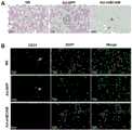
结果参见图7,图中A为PAS染色SD孕鼠肾脏(400×)。Ad-shBCAM组孕鼠肾小管萎缩,间质水肿,内皮细胞肿胀(剪头符号所示)。图中B为CD31免疫荧光染色肾脏血管内皮(400×)。Ad-shBCAM组孕鼠肾脏CD31荧光表达显著减弱(与白色剪头比较)。The results are shown in Figure 7, in which A is the kidney of a pregnant SD mouse stained with PAS (400×). Ad-shBCAM group pregnant mice renal tubular atrophy, interstitial edema, and endothelial cell swelling (shown by the cut-off symbol). Figure B is CD31 immunofluorescence staining of renal vascular endothelium (400×). The fluorescence expression of CD31 in the kidney of pregnant mice in the Ad-shBCAM group was significantly reduced (compared with the white cut head).
(九)仔鼠检测(9) Detection of offspring
测量胎鼠与胎盘的一般情况,结果参见图8,其中,B为胎仔数,C为胎鼠体重,D为胎鼠身长。Measure the general condition of the fetal mouse and the placenta, and the results are shown in Figure 8, wherein, B is the number of fetuses, C is the weight of the fetal mouse, and D is the length of the fetal mouse.
(十)孕鼠胎盘的BCAM蛋白表达(10) Expression of BCAM protein in the placenta of pregnant mice
结果参见图9,免疫组化检测孕鼠胎盘组织BCAM蛋白表达(400×)胎盘绒毛滋养细胞与红细胞中存在BCAM广泛表达(染色为棕黄色),而Ad-shBCAM组胎盘BCAM蛋白显著减少。The results are shown in Figure 9. Immunohistochemical detection of BCAM protein expression in placental tissue of pregnant mice (400×) BCAM was widely expressed in placental villous trophoblasts and erythrocytes (stained in brown), while the placental BCAM protein in the Ad-shBCAM group was significantly reduced.
以上所述,仅是本发明的较佳实施例,并非对本发明做任何形式上的限制,凡是依据本发明的技术实质对以上实施例所作的任何简单修改、等同变化,均落入本发明的保护范围之内。The above descriptions are only preferred embodiments of the present invention, and are not intended to limit the present invention in any form. Any simple modifications and equivalent changes made to the above embodiments according to the technical essence of the present invention all fall within the scope of the present invention. within the scope of protection.
Claims (8)
Translated fromChinesePriority Applications (1)
| Application Number | Priority Date | Filing Date | Title |
|---|---|---|---|
| CN202210449712.6ACN114766427B (en) | 2022-04-24 | 2022-04-24 | A severe preeclampsia animal model based on BCAM target and its construction method and application |
Applications Claiming Priority (1)
| Application Number | Priority Date | Filing Date | Title |
|---|---|---|---|
| CN202210449712.6ACN114766427B (en) | 2022-04-24 | 2022-04-24 | A severe preeclampsia animal model based on BCAM target and its construction method and application |
Publications (2)
| Publication Number | Publication Date |
|---|---|
| CN114766427A CN114766427A (en) | 2022-07-22 |
| CN114766427Btrue CN114766427B (en) | 2023-04-28 |
Family
ID=82432846
Family Applications (1)
| Application Number | Title | Priority Date | Filing Date |
|---|---|---|---|
| CN202210449712.6AActiveCN114766427B (en) | 2022-04-24 | 2022-04-24 | A severe preeclampsia animal model based on BCAM target and its construction method and application |
Country Status (1)
| Country | Link |
|---|---|
| CN (1) | CN114766427B (en) |
Family Cites Families (4)
| Publication number | Priority date | Publication date | Assignee | Title |
|---|---|---|---|---|
| WO2016201319A1 (en)* | 2015-06-10 | 2016-12-15 | The Broad Institute Inc. | Antibodies, compounds and screens for identifying and treating cachexia or pre-cachexia |
| US20170020930A1 (en)* | 2015-07-21 | 2017-01-26 | Therapeutic Solutions International, Inc. | Prevention of pregnancy complications by probiotic administration |
| CN106310238B (en)* | 2016-10-26 | 2020-02-14 | 四川大学华西第二医院 | Application of Ad-sAxl in establishing preeclampsia rat model and establishing method of preeclampsia rat model |
| CA3083949A1 (en)* | 2017-11-30 | 2020-06-06 | Novartis Ag | Bcma-targeting chimeric antigen receptor, and uses thereof |
- 2022
- 2022-04-24CNCN202210449712.6Apatent/CN114766427B/enactiveActive
Also Published As
| Publication number | Publication date |
|---|---|
| CN114766427A (en) | 2022-07-22 |
Similar Documents
| Publication | Publication Date | Title |
|---|---|---|
| CN115843748A (en) | Construction method and application of animal model of preeclampsia appearance | |
| Rapola et al. | Pathology of fetal congenital nephrosis: immunohistochemical and ultrastructural studies | |
| CN114766427B (en) | A severe preeclampsia animal model based on BCAM target and its construction method and application | |
| CN116370451A (en) | Application of pravastatin and pharmaceutically acceptable salts thereof in preparation of drugs for preventing or treating preeclampsia | |
| CN117925811A (en) | Application of circSLC8A1 in the preparation of chronic kidney disease detection kit | |
| CN114982708A (en) | Mouse contrast agent kidney disease model and construction method thereof | |
| CN115873943B (en) | Application of bone morphogenetic protein 2 in the diagnosis, prevention and treatment of preeclampsia | |
| CN115054595A (en) | Application of eupatilin in preparing medicine for preventing and treating diabetic nephropathy | |
| CN106310238B (en) | Application of Ad-sAxl in establishing preeclampsia rat model and establishing method of preeclampsia rat model | |
| CN102037930A (en) | Establishing method of PPHN (Persistent Pulmonary Hypertension of Newborn) animal model | |
| LU506900B1 (en) | A soluble recombinant protein for the treatment of preeclampsia | |
| CN117305371A (en) | Construction method and application of a severe preeclampsia rat model based on LncRNA DUXAP8 | |
| Pini et al. | Histamine and the Kidney: In Vivo Animal Models | |
| CN104698187B (en) | Application of membrane protein CD81 in early forecasting, parting, diagnosis and treatment of eclampsia | |
| CN117929754B (en) | Application of MUC1-C segment in the preparation of drugs for the prevention or treatment of pulmonary hypertension | |
| CN118384278B (en) | Application of USP53 gene silencing drugs in renal ischemia-reperfusion injury | |
| RU2782110C1 (en) | Method for predicting miscarriage in the early stages by determining vascular endothelial growth factor and hypoxia inducible factor 1α in the blood serum of pregnant women with cytomegalovirus infection | |
| CN114807353B (en) | Application of Nrf2 in the auxiliary diagnosis of pulmonary dysplasia in intrauterine growth restricted fetuses | |
| CN120381241A (en) | A method and system for evaluating the placenta in preeclampsia using photoacoustic imaging and ultrasound contrast imaging | |
| Akiyama et al. | Endothelial Cell–Targeted Deletion of PPARγ Blocks Rosiglitazone-Induced Plasma Volume Expansion and Vascular Remodeling in Adipose Tissue | |
| CN118901662A (en) | Improved MPO-AAV (MPO-AAV) related experimental vasculitis animal model construction method | |
| RU2256320C2 (en) | Method for detecting glycogen in extract out of organs and tissues in bees | |
| CN118236403A (en) | Use of mitochondrial grafting for treating renal fibrosis | |
| CN119925438A (en) | A method for constructing a severe preeclampsia rat model based on TET2 and its application | |
| CN116179674A (en) | Use of CSRP2BP gene as marker for diagnosing preeclampsia |
Legal Events
| Date | Code | Title | Description |
|---|---|---|---|
| PB01 | Publication | ||
| PB01 | Publication | ||
| SE01 | Entry into force of request for substantive examination | ||
| SE01 | Entry into force of request for substantive examination | ||
| GR01 | Patent grant | ||
| GR01 | Patent grant |