CN114730484A - 3D selective bone matching from 2D image data - Google Patents
3D selective bone matching from 2D image dataDownload PDFInfo
- Publication number
- CN114730484A CN114730484ACN202080079853.2ACN202080079853ACN114730484ACN 114730484 ACN114730484 ACN 114730484ACN 202080079853 ACN202080079853 ACN 202080079853ACN 114730484 ACN114730484 ACN 114730484A
- Authority
- CN
- China
- Prior art keywords
- bone
- patient
- surgical
- data
- model
- Prior art date
- Legal status (The legal status is an assumption and is not a legal conclusion. Google has not performed a legal analysis and makes no representation as to the accuracy of the status listed.)
- Pending
Links
Images


Classifications
- G—PHYSICS
- G06—COMPUTING OR CALCULATING; COUNTING
- G06T—IMAGE DATA PROCESSING OR GENERATION, IN GENERAL
- G06T19/00—Manipulating 3D models or images for computer graphics
- G06T19/20—Editing of 3D images, e.g. changing shapes or colours, aligning objects or positioning parts
- G—PHYSICS
- G06—COMPUTING OR CALCULATING; COUNTING
- G06T—IMAGE DATA PROCESSING OR GENERATION, IN GENERAL
- G06T7/00—Image analysis
- G06T7/30—Determination of transform parameters for the alignment of images, i.e. image registration
- G06T7/33—Determination of transform parameters for the alignment of images, i.e. image registration using feature-based methods
- G—PHYSICS
- G06—COMPUTING OR CALCULATING; COUNTING
- G06T—IMAGE DATA PROCESSING OR GENERATION, IN GENERAL
- G06T7/00—Image analysis
- G06T7/70—Determining position or orientation of objects or cameras
- G06T7/73—Determining position or orientation of objects or cameras using feature-based methods
- G06T7/75—Determining position or orientation of objects or cameras using feature-based methods involving models
- G—PHYSICS
- G06—COMPUTING OR CALCULATING; COUNTING
- G06T—IMAGE DATA PROCESSING OR GENERATION, IN GENERAL
- G06T2200/00—Indexing scheme for image data processing or generation, in general
- G06T2200/08—Indexing scheme for image data processing or generation, in general involving all processing steps from image acquisition to 3D model generation
- G—PHYSICS
- G06—COMPUTING OR CALCULATING; COUNTING
- G06T—IMAGE DATA PROCESSING OR GENERATION, IN GENERAL
- G06T2200/00—Indexing scheme for image data processing or generation, in general
- G06T2200/24—Indexing scheme for image data processing or generation, in general involving graphical user interfaces [GUIs]
- G—PHYSICS
- G06—COMPUTING OR CALCULATING; COUNTING
- G06T—IMAGE DATA PROCESSING OR GENERATION, IN GENERAL
- G06T2207/00—Indexing scheme for image analysis or image enhancement
- G06T2207/10—Image acquisition modality
- G06T2207/10116—X-ray image
- G—PHYSICS
- G06—COMPUTING OR CALCULATING; COUNTING
- G06T—IMAGE DATA PROCESSING OR GENERATION, IN GENERAL
- G06T2207/00—Indexing scheme for image analysis or image enhancement
- G06T2207/30—Subject of image; Context of image processing
- G06T2207/30004—Biomedical image processing
- G06T2207/30008—Bone
- G—PHYSICS
- G06—COMPUTING OR CALCULATING; COUNTING
- G06T—IMAGE DATA PROCESSING OR GENERATION, IN GENERAL
- G06T2210/00—Indexing scheme for image generation or computer graphics
- G06T2210/41—Medical
- G—PHYSICS
- G06—COMPUTING OR CALCULATING; COUNTING
- G06T—IMAGE DATA PROCESSING OR GENERATION, IN GENERAL
- G06T2219/00—Indexing scheme for manipulating 3D models or images for computer graphics
- G06T2219/20—Indexing scheme for editing of 3D models
- G06T2219/2021—Shape modification
Landscapes
- Engineering & Computer Science (AREA)
- Physics & Mathematics (AREA)
- General Physics & Mathematics (AREA)
- Theoretical Computer Science (AREA)
- Computer Vision & Pattern Recognition (AREA)
- Architecture (AREA)
- Computer Graphics (AREA)
- Computer Hardware Design (AREA)
- General Engineering & Computer Science (AREA)
- Software Systems (AREA)
- Surgical Instruments (AREA)
- Apparatus For Radiation Diagnosis (AREA)
Abstract
Translated fromChinese
Description
Translated fromChinese本申请要求2019年12月20日提交的美国临时申请序列号62/951,676的权益,该临时申请的内容以全文引用的方式并入本文中。This application claims the benefit of US Provisional Application Serial No. 62/951,676, filed December 20, 2019, the contents of which are incorporated herein by reference in their entirety.
技术领域technical field
本公开总体上涉及与计算机辅助手术系统有关的方法、系统和装置,所述计算机辅助手术系统包括一起工作以增强手术流程的各种硬件和软件部件。所公开的技术可以应用于例如肩、髋和膝关节置换术,以及其它外科手术,如关节镜手术、脊柱手术、颌面手术、肩袖手术、韧带修复和置换手术。更具体地,本公开涉及根据双平面二维(2D)图像创建三维(3D)解剖模型的方法。The present disclosure generally relates to methods, systems, and apparatuses related to computer-assisted surgical systems that include various hardware and software components that work together to enhance surgical procedures. The disclosed techniques can be applied to, for example, shoulder, hip and knee replacements, as well as other surgical procedures such as arthroscopic surgery, spinal surgery, maxillofacial surgery, rotator cuff surgery, ligament repair and replacement surgery. More particularly, the present disclosure relates to a method of creating a three-dimensional (3D) anatomical model from a biplanar two-dimensional (2D) image.
背景技术Background technique
随着提供医疗保健的成本持续上升,许多实体都在寻找降低成本的方法。在某些情况下,保险公司会实施更严格的报销标准,以摆脱更昂贵的治疗。例如,保险提供商可询问使用磁共振成像(MRI)设备是否必要,因为与其它成像系统,包括计算机断层扫描(CT)扫描器和X射线机相比,使用此类设备的成本较高。在其它情况下,由于获取和操作此类系统的成本问题,人口较少或新兴市场可能无法获得MRI技术。As the cost of providing healthcare continues to rise, many entities are looking for ways to reduce costs. In some cases, insurers implement stricter reimbursement standards to get rid of more expensive treatments. For example, insurance providers may ask whether the use of magnetic resonance imaging (MRI) equipment is necessary because of the high cost of using such equipment compared to other imaging systems, including computed tomography (CT) scanners and X-ray machines. In other cases, MRI technology may not be available in smaller populations or emerging markets due to the cost of acquiring and operating such systems.
目前,许多患者特定的全关节置换系统,包括Smith&Nephew的VISIONAIRE切割引导件,取决于根据通过MRI扫描产生的一系列图像解释患者的关节解剖结构的能力。特别地,患者特定的关节置换程序需要与例如膝盖中的包括软骨表面的区域匹配的形状拟合表面。目前需要提供包括软组织的扫描解剖特征的三维图像的MRI扫描,因为其它成像技术无法为此类表面的开发提供足够的细节。VISIONAIRE是田纳西州孟菲斯市的Smith&Nephew公司的注册商标。Currently, many patient-specific total joint replacement systems, including Smith & Nephew's VISIONAIRE cutting guide, depend on the ability to interpret a patient's joint anatomy based on a series of images generated through an MRI scan. In particular, patient-specific joint replacement procedures require shape-fitting surfaces that match, for example, areas in the knee that include cartilage surfaces. MRI scans that provide three-dimensional images including scanned anatomical features of soft tissue are currently required because other imaging techniques do not provide sufficient detail for the development of such surfaces. VISIONAIRE is a registered trademark of Smith & Nephew Corporation of Memphis, Tennessee.
此外,将MRI数据转换为患者特定的关节置换器械的过程可能需要大量的用户干预和在制造器械前的数据处理。用户通常花费大量的时间来确保使用MRI数据创建的骨模型尽可能与患者的骨匹配。简而言之,对MRI扫描的依赖可能在MRI系统不可用时阻止某些患者接受关节置换,或者在保险提供商拒绝承保并要求在全关节置换前进行其它处理时禁止或延迟审批流程。In addition, the process of converting MRI data into a patient-specific joint replacement device can require extensive user intervention and data processing prior to manufacturing the device. Users often spend considerable time making sure that the bone model created using the MRI data matches the patient's bone as closely as possible. In short, reliance on MRI scans can prevent certain patients from receiving joint replacements when MRI systems are unavailable, or prohibit or delay the approval process when insurance providers deny coverage and require additional processing before total joint replacement.
根据2D成像数据创建3D模型的先前尝试在很大程度上依赖于由处理器执行的复杂数学计算。例如,授予Dhruwdas的美国专利号10,217,217公开了一种用于使用常规2D x射线图像获得3D图像的方法。所述方法包括确定相机模型(源和x射线图像相对于彼此的位置)和2D x射线图像的数字放大率,从2D x射线图像提取骨的轮廓,以及识别轮廓的2D解剖值。所述方法还包括导入骨的3D模板模型,根据相机模型提取廓影顶点及其投影,以及相对于X射线图像对准3D模板模型。选择性地修改模板以匹配2D解剖值。针对每个廓影顶点投影确定轮廓上的最佳匹配点,然后根据相机模型反向投影,以找到最接近对应廓影顶点的目标位置。使用拉普拉斯网格变形算法使3D模板模型变形,使得廓影顶点达到对应的目标位置。然而,由于必须由处理器执行的复杂数学计算,Dhruwdas的方法具有高计算要求。Previous attempts to create 3D models from 2D imaging data have relied heavily on complex mathematical calculations performed by processors. For example, US Patent No. 10,217,217 to Dhruwdas discloses a method for obtaining 3D images using conventional 2D x-ray images. The method includes determining the camera model (position of the source and x-ray images relative to each other) and digital magnification of the 2D x-ray images, extracting the contours of the bone from the 2D x-ray images, and identifying 2D anatomical values of the contours. The method also includes importing a 3D template model of the bone, extracting silhouette vertices and their projections from the camera model, and aligning the 3D template model with respect to the X-ray image. Optionally modify templates to match 2D anatomical values. The best matching point on the silhouette is determined for each silhouette vertex projection, and then back-projected against the camera model to find the target position closest to the corresponding silhouette vertex. Use the Laplace mesh deformation algorithm to deform the 3D template model so that the silhouette vertices reach the corresponding target positions. However, Dhruwdas' method has high computational requirements due to the complex mathematical calculations that must be performed by the processor.
数学方法不具有与人类一样的3D直觉。因此,数学优化算法必须检查多个取向上的相似性,并且算法可能落入局部最小值,其中骨形状看起来与2D投影或轮廓线匹配,但旋转方向错误。当骨存在某种对称(像骨盆或肱骨或股骨的髁)时,就会发生这种情况。为了进行补偿,计算机必须执行许多计算上更昂贵的投影和比较,以便对这些局部最小值是鲁棒的。因此,主要数学过程是有效性降低的非常低效的方法。Mathematical methods do not have the same 3D intuition as humans. Therefore, mathematical optimization algorithms must check for similarity in multiple orientations, and the algorithm may fall into local minima, where the bone shape appears to match the 2D projection or contour, but the rotation is in the wrong direction. This happens when there is some kind of symmetry in the bone (like the condyles of the pelvis or humerus or femur). To compensate, the computer must perform many computationally more expensive projections and comparisons in order to be robust to these local minima. Therefore, the main mathematical process is a very inefficient method with reduced effectiveness.
附图说明Description of drawings
并入本说明书中且形成本说明书的一部分的附图说明本公开的实施例,且连同书面描述一起用于解释本发明的原理、特性和特征。在附图中:The accompanying drawings, which are incorporated in and form a part of this specification, illustrate embodiments of the present disclosure, and together with the written description serve to explain the principles, characteristics, and features of the invention. In the attached image:
图1示出了根据实施例的包括示例性计算机辅助手术系统(CASS)的手术室。FIG. 1 illustrates an operating room including an exemplary computer-assisted surgery system (CASS), according to an embodiment.
图2示出了根据一些实施例的电磁传感器装置的示例。Figure 2 shows an example of an electromagnetic sensor device according to some embodiments.
图3A示出了根据一些实施例的具有三个垂直线圈的电磁传感器装置的替代性示例。Figure 3A shows an alternative example of an electromagnetic sensor device with three vertical coils in accordance with some embodiments.
图3B示出了根据一些实施例的具有两个不平行固定线圈的电磁传感器装置的替代性示例。Figure 3B shows an alternative example of an electromagnetic sensor device with two non-parallel stationary coils in accordance with some embodiments.
图3C示出了根据一些实施例的具有两个不平行分离线圈的电磁传感器装置的替代性示例。Figure 3C shows an alternative example of an electromagnetic sensor device with two non-parallel separated coils in accordance with some embodiments.
图4示出了根据一些实施例的电磁传感器装置和患者骨的示例。Figure 4 shows an example of an electromagnetic sensor device and a patient's bone in accordance with some embodiments.
图5A示出了根据实施例的手术计算机提供给CASS的其它部件的说明性控制指令。5A shows illustrative control instructions provided by a surgical computer to other components of a CASS, according to an embodiment.
图5B示出了根据实施例的CASS的部件提供给手术计算机的说明性控制指令。5B shows illustrative control instructions provided to a surgical computer by components of a CASS, according to an embodiment.
图5C示出了根据实施例的手术计算机通过网络连接到手术数据服务器的说明性实施方式。5C shows an illustrative implementation of a surgical computer connected to a surgical data server through a network, according to an embodiment.
图6示出了根据实施例的手术患者护理系统和说明性数据源。6 shows a surgical patient care system and an illustrative data source according to an embodiment.
图7A示出了根据实施例的用于确定术前手术计划的示例性流程图。7A illustrates an exemplary flow diagram for determining a preoperative surgical plan, according to an embodiment.
图7B示出了根据实施例的用于确定护理期,包括术前、术中和术后动作的示例性流程图。7B illustrates an exemplary flowchart for determining a period of care, including preoperative, intraoperative, and postoperative actions, according to an embodiment.
图7C示出了根据实施例的说明性图形用户界面,包括描绘植入物放置的图像。7C shows an illustrative graphical user interface including an image depicting implant placement, according to an embodiment.
图8示出了根据实施例的产生关节的定制三维模型的示例性方法。8 illustrates an exemplary method of generating a customized three-dimensional model of a joint, according to an embodiment.
图9示出了根据实施例的生成定制三维骨模型的示例性方法。9 illustrates an exemplary method of generating a custom three-dimensional bone model, according to an embodiment.
图10A-10C示出了根据实施例的联合配准(co-registering)多个2D图像的过程。10A-10C illustrate a process of co-registering multiple 2D images according to an embodiment.
图11A-11B示出了根据实施例的相对于共同坐标系对准骨的过程。11A-11B illustrate a process for aligning bones relative to a common coordinate system, according to an embodiment.
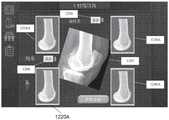
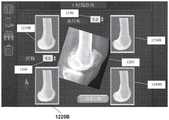
图12A-12C示出了根据实施例的从关于2D图像的库定向代表性骨的视图的过程。12A-12C illustrate a process for orienting a view of a representative bone from a library of 2D images, according to an embodiment.
图13A-13B示出了根据实施例的相对于至少一个2D图像缩放和重新定向3D骨模型的过程。13A-13B illustrate a process for scaling and reorienting a 3D bone model relative to at least one 2D image, according to an embodiment.
图14示出了根据实施例的修改3D骨模型的轮廓的过程。14 illustrates a process of modifying the contour of a 3D bone model, according to an embodiment.
图15A-15D示出了根据实施例的相对于髋臼股骨关节产生关节的定制三维模型的过程的各个阶段。15A-15D illustrate various stages of a process of generating a custom three-dimensional model of a joint relative to an acetabular femoral joint, according to an embodiment.
图16示出了根据实施例的从一组所识别的潜在代表性骨中选择代表性骨的过程。16 illustrates a process for selecting a representative bone from a set of identified potential representative bones, according to an embodiment.
图17示出了可以在其中实施示例性实施例的各方面的示例性数据处理系统的框图。17 illustrates a block diagram of an exemplary data processing system in which aspects of the exemplary embodiments may be implemented.
具体实施方式Detailed ways
本公开不限于所描述的特定系统、装置和方法,因为其可以变化。描述中使用的术语仅用于描述特定版本或实施例的目的,而不旨在限制范围。This disclosure is not limited to the particular systems, apparatus, and methods described, as they may vary. The terminology used in the description is for the purpose of describing a particular version or embodiment only and is not intended to limit the scope.
如本文件中所使用,除非上下文另外明确规定,否则单数形式“一个”、“一种”和“该/所述”包括复数引用。除非另有定义,否则本文所使用的所有科技术语具有与本领域普通技术人员通常所理解的相同含义。本公开中的任何内容均不应被解释为承认本公开中描述的实施例由于在前发明而无权把本公开的日期提前。如本文件中所使用,术语“包括”意指“包括但不限于”。As used in this document, the singular forms "a," "an," and "the/the" include plural references unless the context clearly dictates otherwise. Unless otherwise defined, all technical and scientific terms used herein have the same meaning as commonly understood by one of ordinary skill in the art. Nothing in this disclosure should be construed as an admission that the embodiments described in this disclosure are not entitled to advance the date of this disclosure by virtue of prior invention. As used in this document, the term "including" means "including but not limited to".
定义definition
出于本公开的目的,术语“植入物”用于指代制造成置换或增强生物结构的假体装置或结构。例如,在全髋关节置换手术中,使用假体髋臼杯(植入物)置换或增强患者的磨损或损坏的髋臼。虽然术语“植入物”通常被认为表示人造结构(与移植物形成对比),但是出于本说明书的目的,植入物可包括移植以置换或增强生物结构的生物组织或材料。For the purposes of this disclosure, the term "implant" is used to refer to a prosthetic device or structure manufactured to replace or augment a biological structure. For example, in total hip replacement surgery, a prosthetic acetabular cup (implant) is used to replace or augment a patient's worn or damaged acetabulum. While the term "implant" is generally considered to mean an artificial structure (as opposed to a graft), for the purposes of this specification, an implant may include biological tissue or material that is grafted to replace or enhance a biological structure.
出于本公开的目的,术语“实时”用于指代事件发生或可操作系统接收到输入时即时执行的计算或操作。然而,术语“实时”的使用并不旨在排除在输入和响应之间引起某些延迟的操作,只要延迟是机器的性能特性引起的意外结果即可。For the purposes of this disclosure, the term "real-time" is used to refer to computations or operations that are performed immediately when an event occurs or input is received by the operating system. However, the use of the term "real-time" is not intended to exclude operations that cause some delay between input and response, as long as the delay is an unintended consequence of the performance characteristics of the machine.
尽管本公开内容中的很多是通过特定职位或职务来指代外科医生或其他医学专业人员,但是本公开中的任何内容均不旨在限于特定职位或职务。外科医生或医疗专业人员可以包括任何医生、护士、医疗专业人员或技师。这些术语或职位中的任何一个都可以与本文公开的系统的用户互换使用,除非另有明确规定。例如,在一些实施例中,对外科医生的提及也可以适用于技师或护士。Although much of this disclosure refers to a surgeon or other medical professional by a particular position or function, nothing in this disclosure is intended to be limited to a particular position or function. A surgeon or medical professional may include any doctor, nurse, medical professional or technician. Any of these terms or positions may be used interchangeably with users of the systems disclosed herein, unless expressly stated otherwise. For example, in some embodiments a reference to a surgeon may also apply to a technician or nurse.
本文所公开的系统、方法和装置特别好地适合于利用手术导航系统(例如
CASS生态系统概述Overview of the CASS Ecosystem
图1提供了根据一些实施例的示例计算机辅助手术系统(CASS)100的图示。如以下各节中进一步详细所述,CASS使用计算机,机器人和成像技术来帮助外科医生执行骨科手术程序,例如全膝关节置换术(TKA)或全髋关节置换术(THA)。例如,手术导航系统可以帮助外科医生以高精度定位患者的解剖结构、引导手术器械以及植入医疗装置。诸如CASS 100的手术导航系统经常采用各种形式的计算技术来执行各种各样的标准和微创手术程序和技术。而且,这些系统允许外科医生更准确地计划、跟踪和导航相对于患者身体的器械和植入物的位置,以及进行术前和术中身体成像。FIG. 1 provides an illustration of an example computer-assisted surgery system (CASS) 100 in accordance with some embodiments. As described in further detail in the following sections, CASS uses computers, robotics, and imaging technologies to help surgeons perform orthopaedic surgical procedures, such as total knee arthroplasty (TKA) or total hip arthroplasty (THA). For example, surgical navigation systems can help surgeons locate patient anatomy, guide surgical instruments, and implant medical devices with high precision. Surgical navigation systems such as the CASS 100 often employ various forms of computing technology to perform a wide variety of standard and minimally invasive surgical procedures and techniques. Moreover, these systems allow surgeons to more accurately plan, track, and navigate the position of instruments and implants relative to a patient's body, as well as perform pre- and intra-operative body imaging.
执行器平台105在手术期间相对于患者定位手术工具。执行器平台105的确切部件将根据所采用的实施例而变化。例如,对于膝手术,执行器平台105可以包括在其使用期间保持手术工具或器械的末端执行器105B。末端执行器105B可以是外科医生使用的手持式装置或器械(例如
执行器平台105可以包括用于在手术期间定位患者的肢体的肢体定位器105C。肢体定位器105C的一个示例是SMITH AND NEPHEW SPIDER2系统。肢体定位器105C可以由外科医生手动操作,或者替代地,基于从手术计算机150(以下描述)接收到的指令来改变肢体位置。尽管在图1中示出了一个肢体定位器105C,但在一些实施例中可以有多个装置。作为示例,在手术台T的每一侧可以有一个肢体定位器105C,或者在手术台T的一侧可以有两个装置。肢体定位器105C可以直接安装到手术台T,在手术台T的旁边位于地板平台(未示出)上,安装在杆上,或安装在手术室的墙壁或天花板上。在一些实施例中,肢体定位器105C可以以非常规方式使用,例如牵开器或特定的骨保持器。作为示例,肢体定位器105C可以包括脚踝靴、软组织夹、骨夹或软组织牵开器匙,例如钩形、弯曲或成角的刀片。在一些实施例中,肢体定位器105C可以包括缝线保持器以辅助闭合伤口。The
执行器平台105可以包括工具,如螺丝刀,指示轴线或平面的光或激光,水准仪,销驱动器,销拔出器,平面检查器,指示器,指状件,或它们的某种组合。The
切除设备110(图1中未示出)使用例如机械、超声或激光技术执行骨或组织切除。切除设备110的示例包括钻孔装置、去毛刺装置、振荡锯切装置、振动冲击装置、扩孔器、超声骨切割装置、射频消融装置、往复运动装置(例如锉刀或拉刀),以及激光消融系统。在一些实施例中,切除设备110在手术期间由外科医生保持和操作。在其他实施例中,执行器平台105可以用于在使用期间保持切除设备110。Resection device 110 (not shown in FIG. 1 ) performs bone or tissue resection using, for example, mechanical, ultrasound, or laser techniques. Examples of
执行器平台105还可以包括切割引导件或夹具105D,其用于引导在手术期间用于切除组织的锯或钻。这样的切割引导件105D可以一体地形成为执行器平台105或机器人臂105A的一部分,或者切割引导件可以是可以配合地和/或可移除地附接到执行器平台105或机器人臂105A的独立结构。可以由CASS 100控制执行器平台105或机器人臂105A,以根据术前或术中制定的手术计划将切割引导件或夹具105D定位在患者的解剖结构附近,使得切割引导件或夹具将根据手术计划产生精确的骨切割。The
跟踪系统115使用一个或多个传感器来收集定位患者的解剖结构和手术器械的实时位置数据。例如,对于TKA程序,跟踪系统可以在程序期间提供末端执行器105B的位置和取向。除了位置数据之外,来自跟踪系统115的数据还可以用于推断解剖结构/器械的速度/加速度,其可以用于工具控制。在一些实施例中,跟踪系统115可以使用附接到末端执行器105B的跟踪器阵列来确定末端执行器105B的位置和取向。可以基于跟踪系统115的位置和取向以及跟踪系统115和末端执行器105B之间的三维空间中的已知关系来推断末端执行器105B的位置。在本发明的各种实施例中可以使用各种类型的跟踪系统,包括但不限于红外(IR)跟踪系统、电磁(EM)跟踪系统、基于视频或图像的跟踪系统以及超声配准和跟踪系统。使用由跟踪系统115提供的数据,手术计算机150可以检测对象并防止碰撞。例如,手术计算机150可以防止机器人臂105A和/或末端执行器105B与软组织碰撞。The
任何合适的跟踪系统都可以用于跟踪手术室中的手术对象和患者解剖结构。例如,可以在阵列中使用红外和可见光相机的组合。各种照明源(例如红外LED光源)可以照亮场景,从而可以进行三维成像。在一些实施例中,这可以包括立体,三视,四视等成像。除了在一些实施例中固定到推车的相机阵列之外,还可以在整个手术室中放置附加相机。例如,操作者/外科医生穿戴的手持式工具或头戴件可以包括将图像传回中央处理器以将那些图像与相机阵列获取的图像相关的成像功能。这可以为使用多个视角进行建模的环境提供更鲁棒的图像。此外,一些成像装置可以在场景上具有合适的分辨率或具有合适的视角以拾取存储在快速响应(QR)代码或条形码中的信息。这有助于识别未用系统手动配准的特定对象。在一些实施例中,相机可以安装在机器人臂105A上。Any suitable tracking system can be used to track surgical subjects and patient anatomy in the operating room. For example, a combination of infrared and visible light cameras can be used in the array. Various illumination sources, such as infrared LED light sources, can illuminate the scene, enabling three-dimensional imaging. In some embodiments, this may include stereoscopic, three-view, four-view, etc. imaging. In addition to the camera array fixed to the cart in some embodiments, additional cameras may be placed throughout the operating room. For example, a hand-held tool or headgear worn by the operator/surgeon may include imaging functionality that transmits images back to the central processor to correlate those images with those acquired by the camera array. This can provide more robust images for environments modeled with multiple viewpoints. In addition, some imaging devices may have the appropriate resolution on the scene or have the appropriate viewing angle to pick up information stored in Quick Response (QR) codes or barcodes. This helps identify specific objects that were not manually registered with the system. In some embodiments, the camera may be mounted on the
如本文所论述的,尽管大多数跟踪和/或导航技术利用基于图像的跟踪系统(例如,IR跟踪系统、基于视频或图像的跟踪系统等等)。然而,基于电磁(EM)的跟踪系统由于各种原因变得越来越常见。例如,标准光学跟踪器的植入需要组织切除(例如,向下到皮质)以及后续钻孔和驱动皮质销。另外,由于光学跟踪器需要与跟踪系统的直达视线,因此此类跟踪器的放置可能需要远离手术部位以确保它们不限制外科医生或医疗专业人员的移动。As discussed herein, although most tracking and/or navigation techniques utilize image-based tracking systems (eg, IR tracking systems, video or image-based tracking systems, etc.). However, electromagnetic (EM) based tracking systems are becoming more common for various reasons. For example, implantation of standard optical trackers requires tissue resection (eg, down to the cortex) and subsequent drilling and driving of cortical pins. Additionally, since optical trackers require a direct line-of-sight with the tracking system, placement of such trackers may need to be placed away from the surgical site to ensure that they do not restrict the movement of the surgeon or medical professional.
通常,基于EM的跟踪装置包括一个或多个线圈和参考场发生器。一个或多个线圈可(例如,经由有线或无线电源)通电。一旦通电,线圈就产生可(例如,由参考场发生器或附加装置)以允许确定一个或多个线圈的位置和取向的方式检测和测量的电磁场。如所属领域的普通技术人员应理解的,例如图2中所示的单个线圈限于检测五(5)个总自由度(DOF)。例如,传感器200能够跟踪/确定X、Y或Z方向上的移动,以及围绕Y轴202或Z轴201的旋转。然而,由于线圈的电磁特性,不可能正确地跟踪围绕X轴线的旋转运动。Typically, EM-based tracking devices include one or more coils and a reference field generator. One or more coils may be energized (eg, via a wired or wireless power source). Once energized, the coils generate electromagnetic fields that can be detected and measured (eg, by a reference field generator or additional device) in a manner that allows the position and orientation of one or more coils to be determined. As will be understood by one of ordinary skill in the art, a single coil such as that shown in FIG. 2 is limited to detecting five (5) total degrees of freedom (DOF). For example, the sensor 200 can track/determine movement in the X, Y or Z directions, and rotation about the
因此,在大多数电磁跟踪应用中,诸如图3A中所示的三线圈系统用于实现在可使刚性主体在三维空间中移动的所有六个自由度中的跟踪(即,前/后310、上/下320、左/右330、滚动340、俯仰350和偏航360)。然而,包括两个额外线圈及其定位的90°偏移角可能要求跟踪装置大得多。替代地,如本领域的技术人员所知,少于三个完整的线圈可用于跟踪所有6DOF。在一些基于EM的跟踪装置中,两个线圈可以彼此固定,例如图3B中所示的。由于两个线圈301B、302B彼此刚性地固定,不完全平行,并且具有相对于彼此已知的位置,因此可以使用此布置确定第六自由度303B。Thus, in most electromagnetic tracking applications, a three-coil system such as that shown in Figure 3A is used to achieve tracking in all six degrees of freedom that can move a rigid body in three-dimensional space (ie, front/back 310, up /down 320, left/right 330,
尽管使用两个固定线圈(例如,301B、302B)允许在6DOF中使用基于EM的跟踪,但由于附加线圈,传感器装置的直径比单个线圈大得多。因此,在手术环境中使用基于EM的跟踪系统的实际应用可能需要组织切除并对患者骨的一部分进行钻孔以允许插入EM跟踪器。替代地,在一些实施例中,可以仅使用销(例如,不需要钻孔或切除大量骨)将单个线圈或5DOFEM跟踪装置植入/插入患者骨中。Although the use of two stationary coils (eg, 301B, 302B) allows the use of EM-based tracking in 6DOF, the diameter of the sensor device is much larger than a single coil due to the additional coils. Therefore, practical application of using an EM-based tracking system in a surgical setting may require tissue resection and drilling of a portion of a patient's bone to allow insertion of an EM tracker. Alternatively, in some embodiments, a single coil or 5DOFEM tracking device may be implanted/inserted into a patient's bone using only pins (eg, without drilling or resection of a large amount of bone).
因此,如本文所述,需要一种解决方案,该解决方案可以将EM跟踪系统的使用限于小到足以使用小直径针或销(即,不需要在骨中制造新切口或大直径开口)插入/嵌入的装置。因此,在一些实施例中,未附接到第一传感器且因此具有小直径的第二5DOF传感器可用于跟踪所有6DOF。现在参考图3C,在一些实施例中,两个5DOF EM传感器(例如,301C、302C)可以在不同位置处以不同角取向(例如,角303C是非零的)插入到患者中(例如,患者骨中)。Therefore, as described herein, there is a need for a solution that can limit the use of EM tracking systems to small enough to be inserted using small diameter needles or pins (ie, without the need to make new incisions or large diameter openings in the bone) /Embedded device. Thus, in some embodiments, a second 5DOF sensor that is not attached to the first sensor and therefore has a small diameter can be used to track all 6DOF. Referring now to FIG. 3C, in some embodiments, two 5DOF EM sensors (eg, 301C, 302C) may be inserted into a patient (eg, in a patient's bone) at different locations with different angular orientations (eg,
现在参考图4,示出了使用在大多数OR中典型的标准中空针405将第一5DOF EM传感器401和第二5DOF EM传感器402插入到患者骨403中的示例实施例。在另一实施例中,第一传感器401和第二传感器402可具有“?”的角度偏移404。在一些实施例中,偏移角“?”404可能需要大于预定值(例如,0.50°、0.75°等的最小角)。在一些实施例中,此最小值可以在手术计划期间由CASS确定并提供给外科医生或医学专业人员。在一些实施例中,最小值可以基于一个或多个因素,例如,跟踪系统的定向准确度、第一EM传感器与第二EM传感器之间的距离。场发生器的位置、场检测器的位置、EM传感器的类型、EM传感器的质量、患者解剖结构等等。Referring now to FIG. 4, an example embodiment is shown in which a first
因此,如本文所论述的,在一些实施例中,销/针(例如,套管安装针等)可用于插入一个或多个EM传感器。通常,销/针将是一次性部件,而传感器本身可以是可重复使用的。然而,应理解,这仅是一个可能系统,并且可使用销/针和/或EM传感器是独立的一次性使用的或可重复使用的各种其它系统。在另一实施例中,EM传感器可(例如,使用鲁尔锁配件等)固定到安装针/销,这可以允许快速组装和拆卸。在另外的实施例中,EM传感器可以利用允许传感器最小地侵入放置的替代套筒和/或锚定系统。Thus, as discussed herein, in some embodiments, pins/needles (eg, trocar mount needles, etc.) may be used to insert one or more EM sensors. Typically, the pins/pins will be disposable parts, while the sensor itself can be reusable. However, it should be understood that this is only one possible system and that various other systems may be used where the pins/needles and/or EM sensors are self-contained single-use or reusable. In another embodiment, the EM sensor can be secured (eg, using a luer lock fitting, etc.) to the mounting pin/pin, which can allow for quick assembly and disassembly. In further embodiments, the EM sensor may utilize alternative sleeves and/or anchoring systems that allow for minimally invasive placement of the sensor.
在另一实施例中,上述系统可以允许多传感器导航系统,其可以检测和校正困扰电磁跟踪系统的场畸变。应理解,场畸变可以由参考场内的任何铁磁性材料的移动引起。因此,如本领域的普通技术人员所知,典型OR具有大量可造成干扰的装置(例如,手术台、LCD显示器、照明设备、成像系统、手术仪器等)。此外,众所周知,场畸变难以检测。使用多个EM传感器使得系统能够准确地检测场畸变,和/或警示用户当前位置测量值可能不准确。因为传感器(例如,经由销/针)牢固地固定到骨解剖结构,所以传感器位置(X、Y、Z)的相对测量可用于检测场畸变。作为非限制性示例,在一些实施例中,在EM传感器固定到骨之后,两个传感器之间的相对距离是已知的并且应当保持恒定。因此,此距离的任何改变都可以指示存在场畸变。In another embodiment, the system described above may allow for a multi-sensor navigation system that can detect and correct for field distortions that plague electromagnetic tracking systems. It will be appreciated that field distortion can be caused by movement of any ferromagnetic material within the reference field. Thus, as is known to those of ordinary skill in the art, a typical OR has a large number of devices (eg, operating tables, LCD displays, lighting, imaging systems, surgical instruments, etc.) that can cause interference. Furthermore, field distortion is notoriously difficult to detect. Using multiple EM sensors enables the system to accurately detect field distortion, and/or alert the user that current position measurements may be inaccurate. Because the sensor is firmly fixed to the bone anatomy (eg, via pins/pins), relative measurements of sensor position (X, Y, Z) can be used to detect field distortion. As a non-limiting example, in some embodiments, after the EM sensor is fixed to the bone, the relative distance between the two sensors is known and should remain constant. Therefore, any change in this distance can indicate the presence of field distortion.
在一些实施例中,外科医生可以在术前或术中用系统手动配准特定对象。例如,通过与用户界面交互,外科医生可以识别工具或骨结构的起始位置。通过跟踪与该工具或骨结构相关的基准标记,或者通过使用其他常规图像跟踪方式,处理器可以在工具或骨在三维模型中移动通过环境时对其进行跟踪。在其它示例中,2D至3D方法可以用作预对准或规划步骤,其提供指南和计划或定位可用于查看机器人系统的骨建模部分的原始数据。In some embodiments, the surgeon may manually register specific objects with the system either preoperatively or intraoperatively. For example, by interacting with the user interface, the surgeon can identify the starting location of a tool or bone structure. By tracking fiducial markers associated with the tool or bone structure, or by using other conventional image tracking means, the processor can track the tool or bone as it moves through the environment in the three-dimensional model. In other examples, a 2D to 3D approach can be used as a pre-alignment or planning step that provides guidelines and plans or positioning raw data that can be used to view the bone modeling portion of the robotic system.
在一些实施例中,诸如在手术室中识别个人、重要工具或骨的基准标记的某些标记物可以包括可以由与跟踪系统关联的相机或相机阵列拾取的被动或主动标识。例如,红外LED可以闪烁图案,所述图案将唯一的标识传达给该图案的来源,从而提供动态识别标记。类似地,一维或二维光学代码(条形码、QR代码等)可以固定到手术室的对象以提供基于图像分析可以发生的被动识别。如果这些代码非对称地放置在对象上,则它们也可以用于通过将标识的位置与图像中的对象的范围进行比较来确定对象的取向。例如,可以将QR码放置在工具托盘的角部中,从而允许跟踪该托盘的方向和标识。其他跟踪方式将在全文中进行说明。例如,在一些实施例中,外科医生和其他人员可以穿戴增强现实头戴件以提供附加的相机角度和跟踪能力。In some embodiments, certain markers, such as fiducial markers that identify individuals, vital tools, or bones in the operating room, may include passive or active markers that may be picked up by a camera or camera array associated with the tracking system. For example, an infrared LED can flash a pattern that conveys a unique identification to the source of the pattern, thereby providing a dynamic identification mark. Similarly, one-dimensional or two-dimensional optical codes (barcodes, QR codes, etc.) can be affixed to objects in the operating room to provide passive identification that can occur based on image analysis. If these codes are placed asymmetrically on the object, they can also be used to determine the orientation of the object by comparing the identified location to the extent of the object in the image. For example, QR codes can be placed in the corners of a tool tray, allowing the orientation and identification of that tray to be tracked. Other tracking methods will be explained in the full text. For example, in some embodiments, surgeons and other personnel may wear augmented reality headsets to provide additional camera angles and tracking capabilities.
除了光学跟踪外,还可以通过配准对象的物理性质并将其与可以跟踪的对象(例如固定到工具或骨的基准标记)关联来跟踪对象的某些特征。例如,外科医生可以执行手动配准过程,由此被跟踪工具和被跟踪骨可以相对于彼此被操纵。通过将工具的尖端撞击骨的表面,可以为该骨标绘三维表面,所述三维表面与相对于该基准标记的参考系的位置和取向关联。通过光学跟踪与该骨关联的基准标记的位置和取向(姿态),可以通过外推法在环境中跟踪该表面的模型。In addition to optical tracking, certain features of objects can be tracked by registering their physical properties and associating them with objects that can be tracked, such as fiducial markers affixed to tools or bone. For example, the surgeon may perform a manual registration process whereby the tracked tool and the tracked bone may be manipulated relative to each other. By striking the tip of the tool against the surface of the bone, a three-dimensional surface can be plotted for the bone, the three-dimensional surface being associated with the position and orientation of the reference frame relative to the fiducial marker. By optically tracking the position and orientation (pose) of the fiducial markers associated with the bone, a model of the surface can be tracked in the environment by extrapolation.
将CASS 100配准到患者的相关解剖结构的配准过程还可以涉及使用解剖学标志,例如骨或软骨上的标志。例如,CASS 100可以包括相关骨或关节的3D模型,并且外科医生可以使用连接到CASS的探针在术中收集有关患者实际骨上的骨标志的位置的数据。骨标志可以包括例如内踝和外踝,股骨近端和胫骨远端的端部,以及髋关节的中心。CASS 100可以将外科医生用探针收集的骨标志的位置数据与3D模型中相同标志的位置数据进行比较和配准。替代地,CASS 100可以通过使用由外科医生使用CASS探针或其他手段收集的骨标志和骨表面的位置数据来构建没有术前图像数据的骨或关节的3D模型。配准过程还可以包括确定关节的各个轴线。例如,对于TKA,外科医生可以使用CASS 100来确定股骨和胫骨的解剖和机械轴。外科医生和CASS 100可以通过在螺旋方向上移动患者的腿部(即,环转)来识别髋关节的中心,以便CASS可以确定髋关节中心的位置。The registration process of registering the CASS 100 to the relevant anatomy of the patient may also involve the use of anatomical landmarks, such as landmarks on bone or cartilage. For example, CASS 100 may include a 3D model of the relevant bone or joint, and the surgeon may use a probe attached to the CASS to intraoperatively collect data on the location of bone landmarks on the patient's actual bone. Bone landmarks may include, for example, the medial and lateral malleolus, the ends of the proximal femur and distal tibia, and the center of the hip joint. The CASS 100 can compare and register the position data of the bone landmarks collected by the surgeon with the probe with the position data of the same landmarks in the 3D model. Alternatively, CASS 100 may construct a 3D model of the bone or joint without preoperative image data by using bone landmarks and position data of the bone surface collected by the surgeon using a CASS probe or other means. The registration process may also include determining various axes of the joint. For example, for TKA, the surgeon may use the CASS 100 to determine the anatomical and mechanical axes of the femur and tibia. The surgeon and CASS 100 can identify the center of the hip joint by moving the patient's leg in a helical direction (ie, circling) so that the CASS can determine the location of the center of the hip joint.
组织导航系统120(图1中未示出)为外科医生提供手术区域周围的患者的骨、软骨、肌肉、神经和/或血管组织的术中实时可视化。可以用于组织导航的系统的示例包括荧光成像系统和超声系统。Tissue navigation system 120 (not shown in FIG. 1 ) provides the surgeon with intraoperative real-time visualization of the patient's bone, cartilage, muscle, nerve, and/or vascular tissue surrounding the surgical field. Examples of systems that can be used for tissue navigation include fluorescence imaging systems and ultrasound systems.
显示器125提供图形用户界面(GUI),其显示由组织导航系统120收集的图像以及与手术有关的其他信息。例如,在一个实施例中,显示器125覆盖术前或术中收集的从各种模态(例如,CT、MRI、X射线、荧光、超声等)收集的图像信息以为外科医生提供患者的解剖结构的各种视图以及实时状况。显示器125可以包括例如一个或多个计算机监视器。作为显示器125的替代或补充,手术人员中的一个或多个人员可以穿戴增强现实(AR)头戴式装置(HMD)。例如,在图1中,外科医生111穿戴AR HMD 155,其可以例如将术前图像数据覆盖在患者或提供手术计划建议。在以下各节中详细描述了AR HMD 155在手术程序中的各种示例性使用。
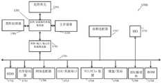
手术计算机150向CASS 100的各种部件提供控制指令,从那些部件收集数据,并为手术期间所需的各种数据提供一般处理。在一些实施例中,手术计算机150是通用计算机。在其他实施例中,手术计算机150可以是使用多个中央处理单元(CPU)或图形处理单元(GPU)来执行处理的并行计算平台。在一些实施例中,手术计算机150通过一个或多个计算机网络(例如,互联网)连接到远程服务器。远程服务器可以用于例如数据的存储或计算密集型处理任务的执行。The
可以使用本领域中公知的各种技术来将手术计算机150连接到CASS 100的其他部件。而且,计算机可以使用多种技术来连接到手术计算机150。例如,末端执行器105B可以通过有线(即,串行)连接而连接到手术计算机150。跟踪系统115、组织导航系统120和显示器125可以类似地使用有线连接来连接到手术计算机150。替代地,跟踪系统115、组织导航系统120和显示器125可以使用无线技术连接到手术计算机150,所述无线技术例如但不限于Wi-Fi、蓝牙、近场通信(NFC)或ZigBee。
动力冲击和髋臼扩孔器装置Dynamic Impact and Acetabular Reamer Devices
以上关于图1描述的CASS设计的灵活性的一部分在于可以根据需要向CASS 100添加额外的或替代的装置以支持特定的手术程序。例如,在髋手术的情况下,CASS 100可以包括动力冲击装置。冲击装置设计成重复施加外科医生可以用来执行诸如植入物对准的活动的冲击力。例如,在全髋关节置换术(THA)中,外科医生通常会使用冲击装置将假体髋臼杯插入到植入宿主的髋臼中。尽管冲击装置本质上可以是手动的(例如,由外科医生用槌敲击冲击器来操作),但是动力冲击装置通常在手术环境中更容易且更快速地使用。动力冲击装置可以例如使用附接到该装置的电池来供电。各种附接件可以连接至动力冲击装置以允许在手术期间根据需要以各种方式来引导冲击力。同样,在髋手术的情况下,CASS 100可以包括动力的、机器人控制的末端执行器,以使髋臼扩孔以容纳髋臼杯植入物。Part of the flexibility of the CASS design described above with respect to FIG. 1 is that additional or alternative devices can be added to CASS 100 as needed to support specific surgical procedures. For example, in the case of hip surgery, CASS 100 may include a powered impactor. Impact devices are designed to repeatedly apply impact forces that a surgeon can use to perform activities such as implant alignment. For example, in total hip arthroplasty (THA), surgeons typically use an impact device to insert a prosthetic acetabular cup into the implanted host's acetabulum. While impact devices may be manual in nature (eg, operated by a surgeon hitting an impactor with a mallet), powered impact devices are generally easier and faster to use in a surgical setting. A powered impact device may be powered, for example, using a battery attached to the device. Various attachments can be connected to the powered impact device to allow the impact force to be directed in various ways as desired during surgery. Also, in the case of hip surgery, CASS 100 may include a powered, robotically controlled end effector to ream the acetabulum to accommodate an acetabular cup implant.
在机器人辅助THA中,可以使用CT或其他图像数据、解剖标志的识别、附着到患者骨的跟踪器阵列以及一个或多个相机将患者的解剖结构配准到CASS 100。可以使用夹具和/或骨钉将跟踪器阵列安装在髂嵴上,并且可以在外部通过皮肤或在内部(后外侧或前外侧)通过为了执行THA而制造的切口来安装这样的跟踪器阵列。对于THA,CASS 100可以利用插入股骨近端的一个或多个股骨皮质螺钉作为检查点以帮助配准过程。CASS 100还可以使用插入骨盆中的一个或多个检查点螺钉作为附加检查点以帮助配准过程。股骨跟踪器阵列可以固定或安装在股骨皮质螺钉中。CASS 100可以采用以下步骤,其中使用外科医生精确地放置在显示器125上为外科医生所识别的股骨近端和骨盆的关键区域上的探针进行验证。跟踪器可以位于机器人臂105A或末端执行器105B上以将臂和/或末端执行器配准到CASS 100。验证步骤还可以利用股骨近端检查点和远端检查点。CASS 100可以利用颜色提示或其他提示来通知外科医生有关骨和机器人臂105A或末端执行器105B的配准过程已在一定程度的精度上(例如,在1mm内)被验证。In robotic-assisted THA, the patient's anatomy may be registered to the CASS 100 using CT or other image data, identification of anatomical landmarks, an array of trackers attached to the patient's bone, and one or more cameras. Tracker arrays can be mounted on the iliac crest using clamps and/or bone screws, and such tracker arrays can be mounted externally through the skin or internally (posterior lateral or anterolateral) through incisions made to perform THA. For THA, CASS 100 may utilize one or more cortical femoral screws inserted into the proximal femur as a checkpoint to aid the registration process. The CASS 100 can also use one or more checkpoint screws inserted into the pelvis as additional checkpoints to aid the registration process. Femoral tracker arrays can be fixed or installed in cortical femoral screws. The CASS 100 may employ steps for verification using probes that the surgeon places precisely on the
对于THA,CASS 100可以包括使用股骨阵列的拉刀跟踪选择,以允许外科医生在术中获取拉刀的位置和取向并计算患者的髋长度和偏移值。根据提供的有关患者髋关节的信息以及在完成拉刀跟踪后计划的植入物位置和取向的信息,外科医生可以对手术计划进行修改或调整。For THA, the CASS 100 may include a broach tracking option using a femoral array to allow the surgeon to acquire the position and orientation of the broach intraoperatively and calculate the patient's hip length and offset values. Based on the information provided about the patient's hip joint and the planned implant position and orientation after completing the broach tracking, the surgeon can make modifications or adjustments to the surgical plan.
对于机器人辅助THA,CASS 100可以包括连接或附接到机器人臂105A或末端执行器105B的一个或多个动力铰刀,其根据手术计划准备骨盆骨以接收髋臼植入物。机器人臂105A和/或末端执行器105B可以通知外科医生和/或控制扩孔器的动力,以确保根据手术计划切除(扩孔)髋臼。例如,如果外科医生根据手术计划试图在要切除的骨的边界之外切除骨,则CASS 100可以切断扩孔器的电源或指示外科医生切断扩孔器的电源。2D至3D建模方法可以提供关于骨体积预测的更大置信度,例如对于探针不可接近的骨的区域。CASS 100可以为外科医生提供选择以关闭或脱离扩孔器的机器人控制。与使用不同颜色的手术计划相比,显示器125可以示出正被切除(扩孔)的骨的进度。外科医生可以查看正被切除(扩孔)的骨的显示以根据手术计划引导扩孔器完成扩孔。CASS 100可以向外科医生提供视觉或听觉提示以警告外科医生正在进行不符合手术计划的切除。For robotically assisted THA, CASS 100 may include one or more powered reamers connected or attached to
在扩孔之后,CASS 100可以采用附接到或连接到机器人臂105A或末端执行器105B的手动或动力冲击器以将试验植入物和最终植入物冲击到髋臼中。机器人臂105A和/或末端执行器105B可以用于引导冲击器以根据手术计划将试验植入物和最终植入物冲击到髋臼中。CASS 100可以使试验植入物和最终植入物相对于骨的位置和取向被显示以告知外科医生如何将试验植入物和最终植入物的取向和位置与手术计划进行比较,显示器125可以在外科医生操纵腿部和髋部时显示植入物的位置和取向。如果外科医生对初始植入物位置和取向不满意,则通过准备新的手术计划,CASS 100可以为外科医生提供重新计划以及重做铰孔和植入物冲击的选择。After reaming, the CASS 100 may employ a manual or powered impactor attached or connected to the
术前,CASS 100可以基于髋关节的三维模型和患者特定的其他信息(例如腿骨的机械和解剖轴,上髁轴,股骨颈轴,股骨和髋的尺寸(例如,长度),髋关节的中线轴,髋关节的ASIS轴,以及诸如小转子标志,远侧标志和髋关节旋转中心的解剖学标志的位置)来制定拟议的手术计划。CASS制定的手术计划可以根据髋关节的三维模型和患者特定的其他信息提供建议的最佳植入物尺寸以及植入物的位置和取向。CASS制定的手术计划可以包括有关偏移值、倾斜度和前倾值、旋转中心、杯尺寸、中度值、上下配合度、股骨柄尺寸和长度的建议细节。Preoperatively, CASS 100 can be based on a 3D model of the hip joint and other patient-specific information (eg, mechanical and anatomical axis of the leg bone, epicondyle axis, femoral neck axis, femur and hip dimensions (eg, length), hip The midline axis, the ASIS axis of the hip, and the locations of anatomical landmarks such as the lesser trochanter landmark, the distal landmark, and the hip center of rotation) to develop the proposed surgical plan. The surgical plan developed by CASS can provide recommended optimal implant size and implant location and orientation based on a 3D model of the hip joint and other patient-specific information. The surgical plan developed by CASS can include recommended details for offset values, inclination and anteversion values, center of rotation, cup size, median value, superior and inferior fit, stem size and length.
对于THA,可以在术前和术中查看CASS制定的手术计划,而外科医生可以在术前或术中修改CASS制定的手术计划。CASS制定的手术计划可以显示计划的髋关节切除,并根据计划的切除将计划的植入物叠加到髋关节上。CASS 100可以为外科医生提供不同手术流程的选择,所述选择将根据外科医生的喜好显示给外科医生。例如,外科医生可以基于被检查和获取的解剖标志的数量和类型和/或在配准过程中使用的跟踪器阵列的位置和数量,从不同的工作流程进行选择。For THA, the surgical plan developed by CASS can be viewed preoperatively and intraoperatively, and the surgeon can modify the surgical plan developed by CASS either preoperatively or intraoperatively. The surgical plan developed by CASS can show the planned hip resection and superimpose the planned implant on the hip according to the planned resection. The CASS 100 may provide the surgeon with a selection of different surgical procedures that will be displayed to the surgeon according to the surgeon's preference. For example, the surgeon may select from different workflows based on the number and type of anatomical landmarks examined and acquired and/or the location and number of tracker arrays used in the registration process.
根据一些实施例,与CASS 100一起使用的动力冲击装置可以以各种不同的设置来操作。在一些实施例中,外科医生通过手动开关或动力冲击装置上的其他物理机构来调整设置。在其他实施例中,可以使用数字接口,所述数字接口允许例如经由动力冲击装置上的触摸屏进行设置输入。这样的数字接口可以允许可用设置基于例如连接到电力附接装置的附接件的类型而变化。在一些实施例中,可以通过与CASS 100内的机器人或其他计算机系统通信来改变设置,而不是调整动力冲击装置本身上的设置。可以使用例如动力冲击装置上的蓝牙或Wi-Fi联网模块来建立这样的连接。在另一实施例中,冲击装置和端部件可以包含允许冲击装置知道什么端部件(杯冲击器,拉刀手柄等)被附接的特征而外科医生不需要采取任何动作,并相应地调整设置。这可以例如通过QR码、条形码、RFID标签或其他方法来实现。According to some embodiments, the powered impact device used with CASS 100 may operate in a variety of different settings. In some embodiments, the surgeon adjusts the settings through a manual switch or other physical mechanism on the powered impact device. In other embodiments, a digital interface may be used that allows settings input, eg, via a touch screen on the powered impact device. Such a digital interface may allow the available settings to vary based on, for example, the type of attachment connected to the power attachment. In some embodiments, settings may be changed by communicating with a robot or other computer system within CASS 100, rather than adjusting settings on the powered impact device itself. Such a connection can be established using, for example, a Bluetooth or Wi-Fi networking module on the powered impact device. In another embodiment, the impact device and end piece may contain features that allow the impact device to know what end piece (cup impactor, broach handle, etc.) is attached without the surgeon needing to take any action and adjust the settings accordingly . This can be achieved, for example, by QR codes, barcodes, RFID tags or other methods.
可以使用的设置的示例包括杯冲击设置(例如,单向,指定频率范围,指定力和/或能量范围);拉刀冲击设置(例如,双向/在指定频率范围内振荡,指定力和/或能量范围);股骨头冲击设置(例如,以指定力或能量进行单向/单次打击);以及干冲击设置(例如,在指定频率下以指定力或能量进行的单向)。另外,在一些实施例中,动力冲击装置包括与髋臼内衬冲击有关的设置(例如,以指定力或能量进行的单向/单次打击)。对于每种类型的内衬(例如,聚合、陶瓷、黑晶(oxinium)或其他材料),可能会有多种设置。此外,动力冲击装置可以基于术前测试/成像/知识和/或外科医生的术中评估来提供针对不同骨质量的设置。在一些实施例中,动力冲击装置可以具有双重功能。例如,动力冲击装置不仅可以提供往复运动以提供冲击力,而且可以为拉刀或锉刀提供往复运动。Examples of settings that can be used include cup impact settings (eg, unidirectional, specified frequency range, specified force and/or energy range); broach impact settings (eg, bidirectional/oscillating within specified frequency range, specified force and/or energy range); femoral head impact settings (eg, unidirectional/single blow at a specified force or energy); and dry shock settings (eg, unidirectional with a specified force or energy at a specified frequency). Additionally, in some embodiments, the powered impact device includes settings related to acetabular liner impact (eg, one-way/single blow with a specified force or energy). For each type of liner (eg, polymeric, ceramic, oxinium, or other material), there may be multiple settings. Additionally, the dynamic impact device may provide settings for different bone qualities based on preoperative testing/imaging/knowledge and/or the surgeon's intraoperative assessment. In some embodiments, the powered impact device may have a dual function. For example, a powered impact device can provide reciprocation not only to provide impact force, but also to provide reciprocation for a broach or file.
在一些实施例中,动力冲击装置包括反馈传感器,该反馈传感器在器械使用期间收集数据,并将数据发送到计算装置,例如装置内的控制器或手术计算机150。然后,此计算装置可以例如通过不透射线的纹身记录数据以供稍后分析,所述纹身提供术前、术中和/或术后配准能力。可以收集的数据的示例包括但不限于声波、每种器械的预定共振频率、来自患者骨的反作用力或回弹能量、装置相对于配准的骨解剖结构的成像(例如,荧光、CT、超声、MRI等)的位置,和/或骨上的外部应变计。In some embodiments, the powered impact device includes a feedback sensor that collects data during instrument use and sends the data to a computing device, such as a controller or
一旦收集到数据,计算装置就可以实时或接近实时地执行一种或多种算法以帮助外科医生执行手术程序。例如,在一些实施例中,计算装置使用所收集的数据来导出诸如正确的最终拉刀尺寸(股骨)的信息;当干完全就位(股骨侧)时;或当杯对于THA就位(深度和/或取向)时。一旦知道该信息,其可以显示以供外科医生审查,或者其可以用于激活触觉或其他反馈机构以指导手术程序。Once the data is collected, the computing device can execute one or more algorithms in real-time or near real-time to assist the surgeon in performing the surgical procedure. For example, in some embodiments, the computing device uses the collected data to derive information such as the correct final broach size (femur); when the stem is fully seated (femoral side); or when the cup is seated for the THA (depth and/or orientation). Once this information is known, it can be displayed for review by the surgeon, or it can be used to activate haptic or other feedback mechanisms to guide the surgical procedure.
此外,从前述算法导出的数据可以用于驱动装置的操作。例如,在用动力冲击装置插入假体髋臼杯期间,装置可以自动伸出冲击头(例如,末端执行器),将植入物移动到适当的位置,或者一旦植入物完全就位就关闭装置的电源。在一个实施例中,导出的信息可以用于自动调整骨质量的设置,其中动力冲击装置应使用较少的动力来减轻股骨/髋臼/骨盆骨折或周围组织的损伤。Furthermore, data derived from the aforementioned algorithms can be used to drive the operation of the device. For example, during insertion of a prosthetic acetabular cup with a powered impact device, the device may automatically extend the impact head (eg, an end effector), move the implant into place, or close once the implant is fully seated power supply to the device. In one embodiment, the derived information can be used to automatically adjust bone quality settings where the powered impact device should use less power to mitigate femur/acetabular/pelvic fractures or damage to surrounding tissue.
机器人臂robotic arm
在一些实施例中,CASS 100包括机器人臂10SA,所述机器人臂用作稳定和保持手术程序期间使用的各种器械的接口。例如,在髋手术的情况下,这些器械可以包括但不限于牵开器、矢状或往复锯、扩孔器手柄、杯冲击器、拉刀手柄和干插入器。机器人臂105A可以具有多个自由度(类似Spider装置),并且具有锁定到位的能力(例如,通过按下按钮、语音激活、外科医生从机器人臂移开手或其他方法)。In some embodiments, CASS 100 includes a robotic arm 10SA that serves as an interface to stabilize and hold various instruments used during a surgical procedure. For example, in the case of hip surgery, these instruments may include, but are not limited to, retractors, sagittal or reciprocating saws, reamer handles, cup impactors, broach handles, and dry inserters. The
在一些实施例中,机器人臂105A的移动可以通过使用内置在机器人臂系统中的控制面板来实现。例如,显示屏可以包括一个或多个输入源,例如指导机器人臂105A移动的物理按钮或具有一个或多个图标的用户界面。外科医生或其他保健专业人员可以在执行手术程序期间与一个或多个输入源接合以定位机器人臂105A。In some embodiments, movement of the
附接或集成到机器人臂105A中的工具或末端执行器105B可以包括但不限于去毛刺装置、手术刀、切割装置、牵开器、关节张紧装置、任何类型的维度测量装置等。在一个特定示例中,机器人臂105A可以定位成获得相对准确的骨尺寸或形状测量值。在另外的示例中,机器人臂105A可以具有钳口或另一装置,该钳口或另一装置打开到已知植入物尺寸的宽度,使得外科医生可以关于植入物的正确尺寸设定或放置做出快速决策。在使用末端执行器105B的实施例中,末端执行器可以定位在机器人臂105A的端部处,使得在机器人臂系统内执行任何马达控制操作。在使用工具的实施例中,工具可以固定在机器人臂105A的远端处,但是马达控制操作可以位于工具本身内。A tool or
机器人臂105A可在内部被机动化以稳定机器人臂,从而防止其跌落并撞击患者、手术台、手术人员等,并允许外科医生移动机器人臂而不必完全支撑其重量。在外科医生移动机器人臂105A的同时,机器人臂可提供一些阻力以防止机器人臂移动太快或一次激活太多自由度。机器人臂105A的位置和锁定状态可以例如通过控制器或手术计算机150来跟踪。The
在一些实施例中,机器人臂105A可以用手(例如,由外科医生)或用内部马达移动到其理想位置和取向以进行正在执行的任务。在一些实施例中,机器人臂105A可以能够以“自由”模式操作,允许外科医生将臂定位在期望的位置而不受限制。在自由模式下,如上所述,仍可以跟踪机器人臂105A的位置和取向。在一个实施例中,在由手术计算机150跟踪的手术计划的指定部分期间,当来自用户(例如,外科医生)的输入时,可以选择性地释放一定的自由度。其中机器人臂105A通过液压或马达在内部提供动力或通过类似的手段提供对外部手动运动的阻力的设计可以被描述为动力机器人臂,而在没有动力反馈的情况下被手动操纵但是可以被手动或自动锁定在适当位置的臂可以被描述为被动机器人臂。In some embodiments, the
机器人臂105A或末端执行器105B可以包括触发器或其他装置以控制锯或钻的动力。外科医生对触发器或其他装置的接合可以使机器人臂105A或末端执行器105B从机动对准模式过渡到锯或钻被接合并通电的模式。另外,CASS 100可以包括脚踏板(未示出)、语音激活控制系统或AR系统,所述脚踏板、语音激活控制系统或AR系统在被激活时使系统执行某些功能。在一个示例中,用户查看膝盖,将其与模板对准,然后通知AR系统当前视图表示已对准的骨。该参考视图通知初始机器人臂105A位置,该位置接着可由操作员进一步微调。更具体地,所述系统使用该输入来定位机器人臂105A以三角测量大致起始姿态。在无源臂的情况下,当关节中的任一个达到其期望位置时,磁性离合器可以锁定。在该示例中,操作员仅移动臂,直到所有关节锁定到位。用户随后可(在有或没有AR协助的情况下)自由进行细微调整。The
在另一示例中,外科医生可以激活脚踏板以指示CASS 100将机器人臂105A或末端执行器105B置于自动模式,所述自动模式将机器人臂或末端执行器相对于患者的解剖结构置于适当的位置,以便执行必要的切除。CASS 100还可以将机器人臂105A或末端执行器105B置于协作模式,所述协作模式允许外科医生手动操纵机器人臂或末端执行器并将其定位在特定位置。协作模式可以配置成允许外科医生在限制其他方向上的运动的同时向内侧或外侧移动机器人臂105A或末端执行器105B。如所讨论的,机器人臂105A或末端执行器105B可以包括切割装置(锯、钻和磨刀)或将引导切割装置的切割引导件或夹具105D。在其他实施例中,机器人臂105A或机器人控制的末端执行器105B的运动可以完全由CASS100控制,而无需任何外科医生或其他医疗专业人员的帮助或输入,或仅需很少的帮助或输入。在另外的其他实施例中,外科医生或其他医疗专业人员可以使用与机器人臂或机器人控制的末端执行器装置分离的控制机构,例如使用操纵杆或交互式监视器或显示控制装置,来远程控制机器人臂105A或机器人控制的末端执行器105B的运动。In another example, the surgeon may activate a foot pedal to instruct the CASS 100 to place the
以下示例描述了在髋手术的情况下使用机器人装置;然而,应当理解,机器人臂在涉及膝、肩等的手术程序中可能还有其他应用。在于2019年8月28日提交的、标题为“机器人辅助韧带移植物放置和张紧(Robotic Assisted Ligament Graft Placement andTensioning)”的WIPO公开号WO 2020/047051中描述了在形成前十字韧带(ACL)移植隧道的情况下使用机器人臂的一个示例,该文献的全部内容通过引用并入本文中。The following examples describe the use of a robotic device in the context of hip surgery; however, it should be understood that there may be other applications of robotic arms in surgical procedures involving knees, shoulders, and the like. WIPO Publication No. WO 2020/047051, entitled "Robotic Assisted Ligament Graft Placement and Tensioning", filed on August 28, 2019, describes the formation of the anterior cruciate ligament (ACL) An example of the use of a robotic arm in the context of a graft tunnel, the entire contents of which are incorporated herein by reference.
机器人臂105A可以用于保持牵开器。例如在一个实施例中,外科医生可以将机器人臂105A移动到期望位置。此时,机器人臂105A可以锁定到位。在一些实施例中,机器人臂105A被提供有关于患者位置的数据,使得如果患者移动,则机器人臂可以相应地调节牵开器位置。在一些实施例中,可以使用多个机器人臂,由此允许保持多个牵开器或同时执行一个以上的动作(例如,牵开器保持和铰孔)。The
机器人臂105A还可以用于在制造股骨颈切口时帮助稳定外科医生的手。在该应用中,对机器人臂105A的控制可以施加某些限制以防止发生软组织损伤。例如,在一个实施例中,手术计算机150在机器人臂105A操作时跟踪其位置。如果跟踪的位置接近预测到组织损伤的区域,则可以向机器人臂105A发送命令以使其停止。替代地,在由手术计算机150自动控制机器人臂105A的情况下,手术计算机可以确保不向机器人臂提供导致其进入可能发生软组织损伤的区域的任何指令。手术计算机150可以对外科医生施加某些限制以防止外科医生扩孔到髋臼内侧壁中太深或以不正确的角度或取向扩孔。The
在一些实施例中,机器人臂105A可以用于在杯冲击期间将杯冲击器保持在期望的角度或取向。当已达到最终位置时,机器人臂105A可以防止任何进一步的就位以防止损坏骨盆。In some embodiments, the
外科医生可以使用机器人臂105A将拉刀手柄定位在期望位置,并允许外科医生以期望取向将拉刀冲击到股骨管中。在一些实施例中,一旦手术计算机150接收到拉刀已完全就位的反馈,机器人臂105A可以限制手柄以防止拉刀进一步前进。The surgeon can use the
机器人臂105A还可以用于表面重修应用。例如,机器人臂105A可以在使用传统器械的同时稳定外科医生,并提供某些约束或限制以允许适当地放置植入部件(例如,导丝放置、倒角切割器、套筒切割器、平面切割器等)。在仅使用骨钻的情况下,机器人臂105A可以使外科医生的手持件稳定并且可以对手持件施加限制以防止外科医生违反手术计划而去除非期望的骨。The
机器人臂105A可以是被动臂。作为示例,机器人臂105A可以是可从Brainlab AG获得的CIRQ机器人臂。CIRQ是德国慕尼黑Olof-Palme-Str.981829,Brainlab AG的注册商标。在一个特定实施例中,机器人臂105A是智能握持臂,如在授予Krinninger等人的美国专利申请第15/525,585号,授予Nowatschin等人的美国专利申请第15/561,042号,授予Nowatschin等人的美国专利第15/561,048号以及授予Nowatschin等人的美国专利第10,342,636号中所公开,上述每个专利的全部内容通过引用并入本文。The
手术程序数据的生成和收集Surgical procedure data generation and collection
医疗专业人员为治疗临床状况而提供的各种服务统称为“护理期”。对于特定的外科手术,护理期可以包括三个阶段:术前、术中和术后。在每个阶段期间,都会收集或生成可用于分析护理期的数据,以便了解程序的各个特征并识别可用于例如在训练模型中以最少的人工干预即可做出决策的模式。在护理期收集的数据可以作为完整数据集存储在手术计算机150或手术数据服务器180处。因此,对于每个护理期,存在一个数据集,所述数据集包括关于患者术前集体收集的所有数据,由CASS 100术中收集或存储的所有数据,以及由患者或由监控患者的医疗专业人员提供的任何术后数据。The various services that medical professionals provide to treat clinical conditions are collectively referred to as a "period of care." For a given surgical procedure, the care period can consist of three phases: preoperative, intraoperative, and postoperative. During each phase, data is collected or generated that can be used to analyze the period of care in order to understand the various characteristics of the procedure and identify patterns that can be used to make decisions with minimal human intervention, for example, in training models. The data collected during the nursing period may be stored at the
如进一步详细解释,在护理期中收集的数据可以用于增强手术程序的执行或提供对手术程序和患者结果的整体理解。例如,在一些实施例中,在护理期中收集的数据可以用于生成手术计划。在一个实施例中,在手术期间收集数据时,在术中完善高级的术前计划。以该方式,当通过CASS 100的部件收集新数据时,可以将手术计划视为实时或近实时动态变化。在其他实施例中,可以使用术前图像或其他输入数据来术前制定在手术期间简单执行的鲁棒计划。在该情况下,由CASS 100在手术期间收集的数据可用于提出建议,以确保外科医生处于术前手术计划之内。例如,如果外科医生不确定如何实现某些规定的切割或植入物对准,则可以向手术计算机150查询以得到建议。在另外的其他实施例中,可以组合术前和术中计划方案,使得可以在手术程序期间根据需要或期望动态修改完善的术前计划。在一些实施例中,患者解剖结构的基于生物力学的模型贡献模拟数据以在制定术前、术中和术后/康复程序中由CASS 100考虑以优化患者的植入物性能结果。As explained in further detail, the data collected during the nursing period can be used to enhance the performance of the surgical procedure or to provide a holistic understanding of the surgical procedure and patient outcomes. For example, in some embodiments, data collected during a nursing session can be used to generate a surgical plan. In one embodiment, advanced preoperative planning is refined intraoperatively when data is collected during surgery. In this manner, the surgical plan can be viewed as dynamically changing in real-time or near real-time as new data is collected through the components of CASS 100. In other embodiments, preoperative images or other input data may be used to preoperatively develop robust plans that are simply performed during surgery. In this case, the data collected by CASS 100 during surgery can be used to make recommendations to ensure that the surgeon is within the preoperative surgical plan. For example, if the surgeon is unsure how to achieve certain prescribed cuts or implant alignments, the
除了改变手术程序本身之外,在护理期中收集的数据还可以用作其他手术辅助程序的输入。例如,在一些实施例中,可以使用护理期数据来设计植入物。在2011年8月15日提交的、标题为“用于骨科手术的参数优化的系统和方法(Systems and Methods forOptimizing Parameters for Orthopaedic Procedures)”的美国专利申请第13/814,531号;2012年7月20日提交的、标题为“用于优化植入物与解剖结构的配合的系统和方法(Systems and Methods for Optimizing Fit of an Implant to Anatomy)”的美国专利申请第14/232,958号;以及2008年9月19日提交的、标题为“用于提高性能的手术调节植入物(Operatively Tuning Implants for Increased Performance)”的美国专利申请第12/234,444号中描述了用于设计、尺寸确定和配合植入物的示例数据驱动技术,上述专利的每一个的全部内容在此通过引用并入本专利申请。In addition to altering the surgical procedure itself, the data collected during the nursing period can also be used as input for other surgical adjunct procedures. For example, in some embodiments, care-period data can be used to design implants. US Patent Application No. 13/814,531, entitled "Systems and Methods for Optimizing Parameters for Orthopaedic Procedures," filed Aug. 15, 2011; Jul. 20, 2012 U.S. Patent Application No. 14/232,958, entitled "Systems and Methods for Optimizing Fit of an Implant to Anatomy," filed on 2008; and September 2008 U.S. Patent Application No. 12/234,444, filed on March 19, and entitled "Operatively Tuning Implants for Increased Performance," describes a design, sizing, and fit implant Examples of data-driven techniques for things, the entire contents of each of the aforementioned patents are hereby incorporated by reference into this patent application.
此外,数据可用于教育、培训或研究目的。例如,使用下面在图5C中描述的基于网络的方案,其他医生或学生可以在允许他们选择性地查看从CASS 100各个部件收集的数据的界面中远程地查看手术。在手术程序之后,可以使用类似的界面来“回放”手术以进行培训或其他教育目的,或找出手术中任何问题或并发症的根源。Additionally, the data may be used for educational, training or research purposes. For example, using the web-based approach described below in Figure 5C, other physicians or students can remotely view procedures in an interface that allows them to selectively view data collected from various components of CASS 100. After a surgical procedure, a similar interface can be used to "play back" the procedure for training or other educational purposes, or to find the source of any problems or complications in the procedure.
术前阶段期获取的数据通常包括手术之前收集或生成的所有信息。因此,例如,可以从患者录入表或电子病历(EMR)获取有关患者的信息。可以收集的患者信息的示例包括但不限于患者人口统计学、诊断、病史、病历记录、生命体征、病史信息、过敏和实验室检查结果。术前数据还可以包括与感兴趣的解剖区域有关的图像。这些图像可以例如使用磁共振成像(MRI)、计算机断层摄影(CT)、X射线、超声或本领域已知的任何其他方式来获取。术前数据还可以包括从患者获取的生活质量数据。例如,在一个实施例中,术前患者使用移动应用程序(“app”)来回答关于他们当前生活质量的问卷。在一些实施例中,CASS 100所使用的术前数据包括关于患者的人口统计学、人体测量学、文化或其他特定特征,其可以与活动水平和特定患者活动相符合以为患者定制手术计划。例如,某些文化或人口统计学的人可能更愿意使用每天蹲便的厕所。Data acquired during the preoperative phase typically includes all information collected or generated prior to surgery. Thus, for example, information about a patient can be obtained from a patient entry form or an electronic medical record (EMR). Examples of patient information that may be collected include, but are not limited to, patient demographics, diagnosis, medical history, medical records, vital signs, medical history information, allergies, and laboratory test results. Preoperative data may also include images related to anatomical regions of interest. These images may be acquired, for example, using magnetic resonance imaging (MRI), computed tomography (CT), X-ray, ultrasound, or any other means known in the art. Preoperative data may also include quality of life data obtained from the patient. For example, in one embodiment, preoperative patients use a mobile application ("app") to answer a questionnaire regarding their current quality of life. In some embodiments, the preoperative data used by CASS 100 includes demographic, anthropometric, cultural, or other specific characteristics about the patient, which can be aligned with activity levels and specific patient activities to customize a surgical plan for the patient. For example, certain cultures or demographics may prefer to use a toilet that squats daily.
图5A和5B提供了可在护理期的术中阶段期间获取的数据的示例。这些示例基于以上参考图1描述的CASS 100的各种部件;然而,应当理解,可以基于手术期间使用的设备的类型及其使用来使用其他类型的数据。5A and 5B provide examples of data that may be acquired during the intraoperative phase of a nursing session. These examples are based on the various components of CASS 100 described above with reference to FIG. 1; however, it should be understood that other types of data may be used based on the type of equipment used and its use during the procedure.
图5A示出了根据一些实施例的手术计算机150提供给CASS 100的其他部件的一些控制指令的示例。注意,图5A的示例假定执行器平台105的部件均由手术计算机150直接控制。在其中部件由外科医生111手动控制的实施例中,可以在显示器125或AR HMD 155上提供指令以指示外科医生111如何移动部件。FIG. 5A shows an example of some control instructions provided by
包括在执行器平台105中的各种部件由手术计算机150控制,所述手术计算机提供位置指令,所述位置指令指示该部件在坐标系内移动的位置。在一些实施例中,手术计算机150向执行器平台105提供指令,所述指令定义当执行器平台105的部件偏离手术计划时如何反应。这些命令在图SA中作为“触觉”命令被参考。例如,末端执行器105B可以提供力以抵抗在计划切除的区域之外的运动。执行器平台105可以使用的其他命令包括振动和音频提示。The various components included in the
在一些实施例中,机器人臂105A的末端执行器105B与切割引导件105D可操作地联接。响应于手术场景的解剖模型,机器人臂105A可以将末端执行器105B和切割引导件105D移动到适当位置以匹配根据手术计划要进行的股骨或胫骨切割的位置。这可以减少错误的可能性,从而允许视觉系统和利用该视觉系统的处理器实施手术计划,以将切割引导件105D放置在相对于胫骨或股骨的精确位置和取向以将切割引导件的切槽与要根据手术计划执行的切割对准。然后,外科医生可以使用任何合适的工具,例如振动或旋转锯或钻,以完美的放置和取向来执行切割(或钻孔),原因是该工具在机械上受到切割引导件105D的特征限制。在一些实施例中,切割引导件105D可以包括一个或多个销孔,在使用切割引导件执行患者组织的切除之前,外科医生使用所述钉孔来将切割引导件钻进和拧紧或钉扎到适当的位置。这可以释放机器人臂105A或确保切割引导件105D完全固定而不相对于要切除的骨移动。例如,该程序可以用于在全膝关节置换术期间制造股骨的第一远侧切口。在一些实施例中,在关节置换术是髋关节置换术的情况下,切割引导件105D可以固定到股骨头或髋臼以用于相应的髋关节置换术切除。应当理解,利用精确切口的任何关节置换术都可以以该方式使用机器人臂105A和/或切割引导件105D。In some embodiments, the
切除设备110提供有多种命令来执行骨或组织操作。与执行器平台105一样,位置信息可以被提供给切除设备110以指定在执行切除时应将其定位在何处。提供给切除设备110的其他命令可以取决于切除设备的类型。例如,对于机械或超声切除工具,命令可以指定工具的速度和频率。对于射频消融(RFA)和其他激光消融工具,这些命令可以指定强度和脉冲持续时间。
CASS 100的一些部件不需要由手术计算机150直接控制;而是,手术计算机150仅需要激活部件,所述部件然后在本地执行软件以指定收集数据并将其提供给手术计算机150的方式。在图5A的示例中,有两个部件以该方式操作:跟踪系统115和组织导航系统120。Some components of CASS 100 need not be directly controlled by
手术计算机150向显示器125提供外科医生111在手术期间所需的任何可视化。对于监视器,手术计算机150可以使用本领域已知的技术来提供用于显示图像、GUI等的指令。显示器125可以包括手术计划的工作流程的各个部分。例如,在配准过程期间,显示器125可以显示术前构建的3D骨模型,并在外科医生使用探针收集患者身上的解剖学标志的位置时示出探针的位置。显示器125可以包括关于手术目标区域的信息。例如,结合TKA,显示器125可以示出股骨和胫骨的机械和解剖轴。显示器125可以基于手术计划示出膝关节的内翻和外翻角,并且CASS 100可以示出如果对手术计划进行预期的修正将如何影响这样的角。因此,显示器125是交互式接口,其可以动态地更新和显示对手术计划的改变将如何影响程序以及安装在骨上的植入物的最终位置和取向。
随着工作流程进行到骨切割或切除的准备,显示器125可以在执行任何切割之前示出计划的或推荐的骨切割。外科医生111可以操纵图像显示以提供目标区域的不同解剖视角,并且可以具有基于患者的术中评估来改变或修正计划的骨切割的选择。显示器125可以示出如果执行计划的骨切割将如何将选定植入物安装在骨上。如果外科医生111选择改变先前计划的骨切割,则显示器125可以示出经修正的骨切割将如何改变安装在骨上时的植入物的位置和取向。As the workflow progresses to preparation for a bone cut or resection, the
显示器125可以向外科医生111提供关于患者,计划的外科手术和植入物的各种数据和信息。可以显示各种患者特定的信息,包括有关患者健康的实时数据,例如心率、血压等。显示器125还可以包括关于手术目标区域(包括标志的位置)的解剖结构的信息、解剖结构的当前状态(例如,是否进行了任何切除,计划和执行的骨切割的深度和角度)以及随着手术计划的进展解剖结构的未来状态。显示器125还可以提供或示出关于手术目标区域的附加信息。对于TKA,显示器125可以提供关于股骨和胫骨之间的间隙(例如,间隙平衡)以及如果执行计划的手术计划将如何改变这样的间隙的信息。对于TKA,显示器125可以提供关于膝关节的附加相关信息,例如关于关节的张力(例如,韧带松弛)的数据以及关于关节的旋转和对准的信息。显示器125可以示出当膝关节屈曲时计划的植入物的定位和位置将如何影响患者。显示器125可以示出不同植入物的使用或不同尺寸的相同植入物的使用将如何影响手术计划,并预览这样的植入物将如何定位在骨上。CASS 100可以在TKA或THA中为每个计划的骨切除术提供这样的信息。在TKA中,CASS 100可以为一个或多个计划的骨切除术提供机器人控制。例如,CASS 100只能为初始股骨远端切割提供机器人控制,并且外科医生111可以使用常规手段(例如4合1切割引导件或夹具105D)手动执行其他切除(前、后和倒角切割)。
显示器125可以采用不同的颜色来通知外科医生手术计划的状态。例如,可以以第一颜色显示未切除的骨,可以以第二颜色显示切除的骨,并且可以以第三颜色显示计划的切除。植入物可以叠加在显示器125中的骨上,并且植入物颜色可以改变或对应于不同类型或尺寸的植入物。The
显示器125上示出的信息和选项可以根据所执行的手术程序的类型而变化。此外,外科医生111可以请求或选择与他或她的手术计划偏好匹配或一致的特定手术流程显示。例如,对于通常在TKA中在股骨切割之前执行胫骨切割的外科医生111,显示器125和关联的工作流程可以适于考虑该偏好。外科医生111还可以预先选择从标准手术工作流程显示中包括或删除某些步骤。例如,如果外科医生111使用切除测量来最终确定植入计划,但在最终确定植入计划时不分析韧带间隙平衡,则可以将手术流程显示组织为模块,并且外科医生可以根据外科医生的喜好或特定手术的情况选择要显示的模块和提供模块的顺序。例如,涉及韧带和间隙平衡的模块可以包括切除前和切除后的韧带/间隙平衡,并且外科医生111可以根据在进行骨切除术之前或之后(或之前和之后)是否执行这种韧带和间隙平衡来选择将哪些模块包括在其默认手术计划工作流程中。The information and options shown on
对于更专业的显示设备,例如AR HMD,手术计算机150可以使用设备支持的数据格式来提供图像、文本等。例如,如果显示器125是诸如Microsoft HoloLensTM或Magic LeapOneTM的全息设备,则手术计算机150可以使用HoloLens应用程序接口(API)来发送命令,所述命令指定显示在外科医生111的视野中的全息图的位置和内容。For more specialized display devices, such as AR HMDs, the
在一些实施例中,可以将一个或多个手术计划模型结合到CASS 100中,并在提供给外科医生111的手术计划的制定中使用。术语“手术计划模型”是指模拟各种情况下的解剖结构的生物力学性能以确定执行切割和其他手术活动的最佳方式的软件。例如,对于膝关节置换手术,手术计划模型可以测量功能活动的参数,例如深屈膝、步态等,并选择膝上的切割位置以优化植入物放置。手术计划模型的一个示例是来自SMITH AND NEPHEW公司的LIFEMODTM仿真软件。在一些实施例中,手术计算机150包括允许在手术期间完全执行手术计划模型的计算架构(例如,基于GPU的并行处理环境)。在其他实施例中,手术计算机150可以通过网络连接到允许这种执行的远程计算机,例如手术数据服务器180(参见图5C)。作为对手术计划模型的完全执行的替代,在一些实施例中,导出一组传递函数,其将由模型获取的数学运算简化为一个或多个预测方程。然后,不同于在手术期间执行完全模拟,而是使用预测方程。在于2019年8月19日提交的、标题为“患者特定手术方法和系统(Patient SpecificSurgical Method and System)”的WIPO公开号2020/037308中描述了关于传递函数的使用的更多细节,该文献的全部内容通过引用并入本文。In some embodiments, one or more surgical planning models may be incorporated into CASS 100 and used in the development of surgical plans provided to surgeon 111 . The term "surgical planning model" refers to software that simulates the biomechanical properties of anatomical structures under various conditions to determine the best way to perform cuts and other surgical activities. For example, for knee replacement surgery, the surgical planning model can measure parameters of functional activity, such as deep knee flexion, gait, etc., and select cutting locations on the knee to optimize implant placement. An example of a surgical planning model is the LIFEMOD™ simulation software from the company SMITH AND NEPHEW. In some embodiments,
图5B示出了可以从CASS 100的各个部件提供给手术计算机150的一些类型的数据的示例。在一些实施例中,部件可以在手术期间实时或接近实时地将数据流传输到手术计算机150。在其他实施例中,部件可以使数据排队并且以设定的间隔(例如,每秒)将其发送到手术计算机150。可以使用本领域中已知的任何格式来传送数据。因此,在一些实施例中,所有部件均以通用格式将数据传输至手术计算机150。在其他实施例中,每个部件可以使用不同的数据格式,并且手术计算机150配置有能够转换数据的一个或多个软件应用。5B shows an example of some types of data that may be provided to
通常,手术计算机150可以用作收集CASS数据的中心点。数据的确切内容将取决于来源。例如,执行器平台105的每个部件向手术计算机150提供测量位置。因此,通过将测量位置与手术计算机150最初指定的位置(参见图5B)进行比较,手术计算机可以识别手术期间发生的偏差。In general, the
切除设备110可以根据所用设备的类型将各种类型的数据发送到手术计算机150。可以发送的示例性数据类型包括测量的扭矩、音频签名和测量的位移值。类似地,跟踪技术115可以根据所采用的跟踪方法来提供不同类型的数据。示例性跟踪数据类型包括被跟踪项(例如,解剖结构、工具等)、超声图像以及表面或标志收集点或轴的位置值。当系统操作时,组织导航系统120向手术计算机150提供解剖位置、形状等。
尽管显示器125通常用于输出数据以呈现给用户,但它也可以向手术计算机150提供数据。例如,对于将监视器用作显示器125的一部分的实施例,外科医生111可以与GUI交互以提供输入,所述输入被发送到手术计算机150以进行进一步处理。对于AR应用,可以将HMD的测量位置和位移发送到手术计算机150,使得其可以根据需要更新所呈现的视图。Although
在护理期的术后阶段期间,可以收集各种类型的数据以量化由于手术而导致的患者状况的总体改善或恶化。数据可以采取例如患者通过问卷调查报告的自我报告信息的形式。例如,在进行膝关节置换手术的情况下,可以使用牛津(Oxford)膝评分问卷来测量功能状态,并且可以通过EQ5D-5L问卷来测量术后生活质量。在髋关节置换手术的情况下的其他示例可以包括牛津髋评分、哈里斯(Harris)髋评分和WOMAC(西安大略和麦克马斯特大学骨关节炎指数)。这样的问卷可以例如由医护专业人员直接在临床环境中进行管理,或者使用允许患者直接回答问题的移动应用进行管理。在一些实施例中,可以为患者配备收集与手术有关的数据的一个或多个可穿戴设备。例如,在进行膝手术之后,可以为患者配备膝支架,所述膝支架包括用于监测膝位置、柔韧性等的传感器。可以收集该信息并将其传输到患者的移动设备以供外科医生审查以评估手术的结果并解决任何问题。在一些实施例中,一个或多个相机可以在术后指定的活动期间获取并记录患者的身体部位的运动。可以将该运动获取与生物力学模型进行比较以更好地了解患者关节的功能,并且更好地预测康复进展并确定可能需要的任何修正。During the postoperative phase of the care period, various types of data can be collected to quantify the overall improvement or deterioration of a patient's condition as a result of surgery. The data may take the form, for example, of self-reported information reported by patients through questionnaires. For example, in the case of knee replacement surgery, functional status can be measured using the Oxford Knee Score Questionnaire, and postoperative quality of life can be measured by the EQ5D-5L questionnaire. Other examples in the context of hip replacement surgery may include the Oxford Hip Score, Harris Hip Score and WOMAC (Western Ontario and McMaster Universities Osteoarthritis Index). Such questionnaires may be administered, for example, by healthcare professionals directly in a clinical setting, or using mobile applications that allow patients to answer questions directly. In some embodiments, the patient may be equipped with one or more wearable devices that collect data related to the procedure. For example, following knee surgery, a patient may be equipped with a knee brace that includes sensors for monitoring knee position, flexibility, and the like. This information can be collected and transmitted to the patient's mobile device for review by the surgeon to assess the outcome of the procedure and resolve any concerns. In some embodiments, the one or more cameras may acquire and record the movement of the patient's body part during post-operatively designated activities. This movement acquisition can be compared to a biomechanical model to better understand the function of the patient's joints, and to better predict rehabilitation progress and identify any modifications that may be needed.
护理期的术后阶段可以在患者的整个生命周期中持续进行。例如,在一些实施例中,手术计算机150或包括CASS 100的其他部件可以在执行手术之后继续接收和收集与手术程序有关的数据。该数据可以包括例如图像、问题答案、“正常”患者数据(例如,血型、血压、状况、药物等)、生物统计数据(例如,步态等),以及有关特定问题(例如,膝或髋关节疼痛)的客观和主观数据。该数据可以由患者或患者的医师明确地提供给手术计算机150或其他CASS部件。替代地或附加地,手术计算机150或其他CASS部件可以监测患者的EMR并在其可用时检索相关信息。患者康复的该纵向视图允许手术计算机150或其他CASS部件提供对患者结果的更客观分析以测量和跟踪给定程序的成功与否。例如,可以通过对护理期中收集的各种数据项进行回归分析,将患者在手术程序后很长时间所经历的状况与手术联系起来。通过对具有相似程序和/或相似解剖结构的患者组进行分析,可以进一步增强该分析。The post-operative phase of the care period can continue throughout the life of the patient. For example, in some embodiments,
在一些实施例中,在中心位置收集数据以提供更容易的分析和使用。在一些情况下,可以从各种CASS部件手动收集数据。例如,可以将便携式存储设备(例如,USB棒)附接到手术计算机150,以便检索在手术期间收集的数据。然后可以将数据例如经由台式计算机传输到集中式存储装置。替代地,在一些实施例中,手术计算机150经由网络175直接连接到集中式存储装置,如图5C中所不。In some embodiments, data is collected at a central location to provide easier analysis and use. In some cases, data can be collected manually from various CASS components. For example, a portable storage device (eg, a USB stick) can be attached to the
图5C示出了“基于云”的实施方式,其中手术计算机150经由网络175连接到手术数据服务器180。该网络175可以是例如私有内联网或因特网。除了来自手术计算机150的数据之外,其他来源也可以将相关数据传输到手术数据服务器180。图5C的示例示出了3个附加数据源:患者160、医疗保健专业人员165和EMR数据库170。因此,患者160可以例如使用移动应用将术前和术后数据发送到手术数据服务器180。医疗保健专业人员165包括外科医生和他或她的职员以及与患者160一起工作的任何其他专业人员(例如,私人医生,康复专家等)。还应当注意的是,EMR数据库170可以用于术前和术后数据。例如,假设患者160已给予足够的许可,则手术数据服务器180可以收集患者术前的EMR。然后,手术数据服务器180可以继续监测EMR以进行手术后的任何更新。FIG. 5C shows a "cloud-based" embodiment in which the
在手术数据服务器180处,护理期数据库185用于存储在患者的护理期中收集的各种数据。护理期数据库185可以使用本领域中已知的任何技术来实现。例如,在一些实施例中,可以使用基于SQL的数据库,其中所有各种数据项都以允许它们容易地并入行和列的两个SQL集合中的方式结构化。然而,在其他实施例中,可以采用No-SQL数据库来允许非结构化数据,同时提供快速处理和响应查询的能力。如本领域中所理解的,术语“No-SQL”用于定义在其设计中不相关的一类数据库。各种类型的No-SQL数据库通常可以根据其基础数据模型进行分组。这些分组可以包括使用基于列的数据模型(例如,Cassandra)、基于文档的数据模型(例如,MongoDB)、基于键值的数据模型(例如,Redis)和/或基于图形的数据模型(例如Allego)的数据库。可以使用任何类型的No-SQL数据库来实现本文所述的各种实施例,并且在一些实施例中,不同类型的数据库可以支持护理期数据库185。At the
可以使用本领域中已知的任何数据格式和传输技术在各种数据源和手术数据服务器180之间传输数据。应当注意的是,图5C中所示的架构允许从数据源到手术数据服务器180的传输,以及由数据源从手术数据服务器180检索数据。例如,如下面详细解释的,在一些实施例中,手术计算机150可以使用来自过去手术、机器学习模型等的数据来帮助指导手术程序。Data may be transferred between the various data sources and the
在一些实施例中,手术计算机150或手术数据服务器180可以执行去标识过程以确保存储在护理期数据库185中的数据满足健康保险流通与责任法案(HIPAA)标准或法律规定的其他要求。HIPAA提供了去标识期间必须从数据删除的某些标识列表。前述的去标识过程可以在传输到护理期数据库185以进行存储的数据中扫描这些标识。例如,在一个实施例中,手术计算机150在刚开始将特定数据项或一组数据项传输到手术数据服务器180之前执行去识别过程。在一些实施例中,唯一标识被分配给来自特定护理期的数据以便在必要时重新识别数据。In some embodiments,
尽管图5A-5C讨论了在单个护理期的情况下的数据收集,但是应当理解,一般概念可以扩展到多个护理期的数据收集。例如,每次使用CASS 100进行手术时可以在整个护理期中收集手术数据,并将其存储在手术计算机150或手术数据服务器180处。如下面进一步详细解释,护理期数据的鲁棒数据库允许生成优化值、测量结果、距离或其他参数以及与手术程序有关的其他建议。在一些实施例中,以允许在手术程序期间快速检索相关信息的方式在数据库或其他存储介质中索引各种数据集。例如,在一个实施例中,可以使用以患者为中心的一组索引,以便可以容易地提取与特定患者或与特定患者相似的一组患者的数据。该概念可以类似地应用于外科医生、植入物特性、CASS部件型式等。Although FIGS. 5A-5C discuss data collection in the context of a single period of care, it should be understood that the general concept can be extended to data collection for multiple periods of care. For example, surgical data may be collected and stored at the
管理护理期数据的更多细节在2018年12月21日提交的、标题为“提供护理期的方法和系统(Methods and Systems for Providing an Episode ofCare)”的美国专利申请第62/783,858号中进行了描述,其全部内容通过引用并入本文。Further details on managing care period data are presented in US Patent Application No. 62/783,858, filed December 21, 2018, and entitled "Methods and Systems for Providing an Episode of Care" description, the entire contents of which are incorporated herein by reference.
开放与封闭数字生态系统Open and closed digital ecosystems
在一些实施例中,CASS 100被设计成用作独立或“封闭”数字生态系统。CASS 100的每个部件专门设计成在封闭生态系统中使用,并且数字生态系统外部的装置通常无法访问数据。例如,在一些实施例中,每个部件包括实现用于诸如通信、存储、安全等的活动的专有协议的软件或固件。对于想要控制CASS 100的所有部件以确保满足某些兼容性、安全性和可靠性标准的公司而言,封闭数字生态系统的概念可能是理想的。例如,CASS 100可以设计成使得除非获得公司的认证,否则不能将新部件与CASS一起使用。In some embodiments, CASS 100 is designed to function as a stand-alone or "closed" digital ecosystem. Each component of the CASS 100 is specifically designed to be used in a closed ecosystem, and devices outside the digital ecosystem typically have no access to the data. For example, in some embodiments, each component includes software or firmware implementing proprietary protocols for activities such as communication, storage, security, and the like. The concept of a closed digital ecosystem may be ideal for companies that want to control all parts of the CASS 100 to ensure certain compatibility, safety and reliability standards are met. For example, CASS 100 can be designed so that new components cannot be used with CASS unless certified by the company.
在其他实施例中,CASS 100被设计成用作“开放”数字生态系统。在这些实施例中,部件可以由各种不同的公司根据诸如通信、存储和安全性的活动的标准来生产。因此,通过使用这些标准,任何公司都可以自由地构建CASS平台的独立、合规部件。可以使用公共可用的应用编程接口(API)和开放的、可共享的数据格式在部件之间传输数据。In other embodiments, CASS 100 is designed to function as an "open" digital ecosystem. In these embodiments, components may be produced by a variety of different companies according to standards for activities such as communications, storage, and security. Therefore, by using these standards, any company is free to build independent, compliant components of the CASS platform. Data can be transferred between components using publicly available application programming interfaces (APIs) and open, sharable data formats.
为了说明可以用CASS 100执行的一种类型的推荐,下面公开了一种用于优化手术参数的技术。在本文中术语“优化”是指基于某些指定的标准选择最佳的参数。在极端情况下,优化可以指基于来自整个护理期的数据(包括任何术前数据,给定时间点的CASS数据状态,以及术后目标)选择最佳参数。而且,可以使用历史数据来执行优化,例如在涉及例如相同外科医生、具有与当前患者相似的身体特性的过去患者等的过去手术期间生成的数据。To illustrate one type of recommendation that may be performed with CASS 100, a technique for optimizing surgical parameters is disclosed below. The term "optimizing" as used herein refers to selecting the best parameters based on some specified criteria. In extreme cases, optimization can refer to selecting optimal parameters based on data from the entire duration of care (including any preoperative data, CASS data status at a given time point, and postoperative goals). Furthermore, the optimization may be performed using historical data, such as data generated during past surgeries involving, eg, the same surgeon, past patients with similar physical characteristics to the current patient, and the like.
优化的参数可以取决于要对其进行手术的患者解剖结构的部分。例如,对于膝手术,手术参数可以包括股骨和胫骨部件的定位信息,包括但不限于旋转对准(例如,内翻/外翻旋转、外旋、股骨部件的屈曲旋转、胫骨部件的后倾角),切除深度(例如内翻膝,外翻膝),以及植入物的类型、大小和位置。定位信息还可以包括用于组合植入物的手术参数,例如整体肢体对准,组合胫股过度伸展和组合胫股切除。CASS 100可以针对给定的TKA股骨植入物优化参数的其他示例包括以下:The optimized parameters may depend on the portion of the patient's anatomy on which the operation is to be performed. For example, for knee surgery, surgical parameters may include positioning information of the femoral and tibial components, including but not limited to rotational alignment (eg, varus/valgus rotation, external rotation, flexion rotation of the femoral component, retroversion of the tibial component) , depth of resection (eg, varus knee, valgus knee), and implant type, size, and location. The positioning information may also include surgical parameters for the combined implant, such as global limb alignment, combined tibiofemoral hyperextension, and combined tibiofemoral resection. Other examples of parameters that the CASS 100 can optimize for a given TKA femoral implant include the following:

CASS 100可以为给定的TKA胫骨植入物优化的参数的其他示例包括以下:Other examples of parameters that CASS 100 can optimize for a given TKA tibial implant include the following:

对于髋手术,手术参数可以包括股骨颈切除位置和角度、杯倾斜角、杯前倾角、杯深度、股骨柄设计、股骨柄尺寸、股骨柄在管内的适合度、股骨偏移、腿长以及植入物的股骨型式。For hip surgery, surgical parameters can include femoral neck resection location and angle, cup inclination, cup anteversion, cup depth, stem design, stem size, stem fit within the canal, femoral offset, leg length, and implant The femoral pattern of the entry.
肩参数可以包括但不限于肱骨切除深度/角度、肱骨干型式、肱骨偏移、关节盂型式和倾斜度,以及反向肩参数,例如肱骨切除深度/角度、肱骨干型式、关节盂倾斜度/型式、盂球方位、盂球偏移和偏移方向。Shoulder parameters may include, but are not limited to, humeral resection depth/angle, humeral shaft pattern, humeral offset, glenoid pattern and inclination, and reverse shoulder parameters such as humeral resection depth/angle, humeral shaft pattern, glenoid inclination/ Type, glenosphere orientation, glenosphere offset and offset direction.
存在用于优化手术参数的各种常规技术。然而,这些技术通常需要大量的计算,因此,通常需要在术前确定参数。结果,外科医生基于在手术期间可能出现的问题来修改优化参数的能力受到限制。而且,常规的优化技术通常以“黑匣子”方式操作,很少或没有关于推荐参数值的解释。因此,如果外科医生决定偏离建议的参数值,则外科医生通常会在没有完全理解该偏离对其余手术流程的影响或偏离对患者手术后生活质量的影响的情况下这样做。Various conventional techniques exist for optimizing surgical parameters. However, these techniques are often computationally intensive and, therefore, parameters often need to be determined preoperatively. As a result, the surgeon's ability to modify optimization parameters based on problems that may arise during surgery is limited. Moreover, conventional optimization techniques often operate in a "black box" fashion with little or no interpretation of recommended parameter values. Therefore, if a surgeon decides to deviate from recommended parameter values, the surgeon will often do so without fully understanding the impact of the deviation on the rest of the surgical procedure or the impact of the deviation on the patient's post-operative quality of life.
手术患者护理系统Surgical Patient Care System
使用手术患者护理系统620可以将优化的一般概念扩展到整个护理期,所述手术患者护理系统使用手术数据以及来自患者605和医疗保健专业人员630的其他数据来优化结果和患者满意度,如图6中所示。The general concept of optimization can be extended to the entire period of care using a surgical
常规地,术前诊断,术前手术计划,术中执行既定计划以及术后全关节置换术的管理都基于个人经验,已发表的文献和外科医生的培训知识库(最终,个体外科医生的部落知识及其对等“网络”和期刊出版物)以及他们利用指导和视觉提示对“平衡”进行准确的术中触觉辨别以及对平面切除进行准确的手动执行的本能。该现有的知识库和执行方式在为需要护理的患者提供的结果优化方面受到限制。例如,在以下方面存在限制:准确诊断患者以进行适当的,微创的既定护理;使动态的患者,医疗经济和外科医生的偏好与患者期望的结果保持一致;执行手术计划以使骨正确对准和保持平衡等;以及从具有难以调和到整体患者框架中的不同偏差的断开连接源接收数据。因此,更精确地模拟解剖反应并指导手术计划的数据驱动工具可以改善现有方法。Conventionally, preoperative diagnosis, preoperative surgical planning, intraoperative execution of established plans, and management of postoperative total joint replacement are based on personal experience, published literature, and the surgeon's training knowledge base (ultimately, a tribe of individual surgeons). knowledge and its equivalent “web” and journal publications) and their instinct to utilize guidance and visual cues for accurate intraoperative tactile discrimination of “balance” and accurate manual execution of plane resections. This existing knowledge base and implementation is limited in optimizing outcomes for patients in need of care. For example, there are limitations in: accurately diagnosing patients for appropriate, minimally invasive, established care; aligning dynamic patient, medical economics, and surgeon preferences with patient-desired outcomes; performing surgical planning to allow correct bone alignment alignment and balance, etc.; and receiving data from disconnected sources with different biases that are difficult to reconcile into the overall patient frame. Therefore, data-driven tools that more accurately model anatomical responses and guide surgical planning could improve existing approaches.
手术患者护理系统620设计成利用患者特定数据、外科医生数据、医疗机构数据和历史结果数据来制定算法,所述算法基于期望的临床结果为患者的整个护理期(术前、术中和术后)建议或推荐最佳整体治疗计划。例如,在一个实施例中,手术患者护理系统620跟踪对建议或推荐计划的遵守,并基于患者/护理提供者的表现来调整计划。一旦手术治疗计划完成,手术患者护理系统620就将收集的数据记录在历史数据库中。该数据库可供将来的患者访问和制定将来的治疗计划。除了利用统计和数学模型之外,还可以使用模拟工具(例如
在一些实施例中,手术患者护理系统620采用数据收集和管理方法来提供详细的手术病例计划,该计划具有使用CASS 100监视和/或执行的不同步骤。用户的执行在每个步骤完成时计算并且用于建议对病例计划的后续步骤的更改。病例计划的生成依赖于存储在本地或云存储数据库中的一系列输入数据。输入数据既可以与当前接受治疗的患者相关,也可以与来自接受过类似治疗的患者的历史数据相关。In some embodiments, the surgical
患者605向手术患者护理系统620提供诸如当前患者数据610和历史患者数据615的输入。本领域中通常已知的各种方法可以用于从患者605收集这样的输入。例如,在一些实施例中,患者605填写手术患者护理系统620解析的纸质或数字调查以提取患者数据。在其他实施例中,手术患者护理系统620可以从诸如电子病历(EMR)、健康历史文件以及付款人/提供商历史文件的现有信息源提取患者数据。在另外的其他实施例中,手术患者护理系统620可以提供允许外部数据源将数据推送到手术患者护理系统的应用程序接口(API)。例如,患者605可以具有移动电话、可穿戴装置或其他移动装置,其收集数据(例如,心率、疼痛或不适水平、运动或活动水平,或患者提交的对患者对任何数目的术前计划标准或条件依从性的反应)并将该数据提供给手术患者护理系统620。类似地,患者605可能在其移动或可穿戴装置上具有数字应用程序,该数字应用程序可以收集数据并将其传输到手术患者护理系统620。The
当前患者数据610可以包括但不限于:活动水平、既往状况、合并症、康复前表现、健康和健身水平、术前预期水平(与医院、手术和康复有关)、都市统计区域(MSA)驱动评分、遗传背景、以前的损伤(运动、创伤等)、以前的关节置换术、以前的创伤手术、以前的运动医学手术、对侧关节或肢体的治疗、步态或生物力学信息(背和踝组织)、疼痛或不适水平、护理基础设施信息(付款人承保类型、家庭医疗基础设施水平等)以及手术预期理想结果的指示。
历史患者数据615可以包括但不限于:活动水平、既往状况、合并症、康复前表现、健康和健身水平、术前预期水平(与医院、手术和康复有关)、MSA驱动评分、遗传背景、以前的损伤(运动、创伤等)、以前的关节置换术、以前的创伤手术、以前的运动医学手术、对侧关节或肢体的治疗、步态或生物力学信息(背和踝组织)、疼痛或不适水平、护理基础设施信息(付款人承保类型、家庭医疗基础设施水平等)、手术的预期理想结果、手术的实际结果(患者报告结果[PRO]、植入物的生存期、疼痛程度、活动水平等)、所使用的植入物的尺寸、所使用的植入物的位置/取向/对准,实现的软组织平衡等。
进行手术或治疗的医疗保健专业人员630可以向手术患者护理系统620提供各种类型的数据625。该医疗保健专业人员数据625可包括例如对已知或优选手术技术的描述(例如,十字形保持(CR)与后稳定(PS)、尺寸增大与尺寸减小、有止血带与无止血带、股骨柄样式、THA的优选方案等),医疗保健专业人员630的培训水平(例如,从业年限、受训的职位、培训的地方、模仿的技术),包括历史数据(结果、患者满意度)的先前成功水平,以及关于运动范围、恢复天数和装置的生存期的预期理想结果。可以例如通过提供给医疗保健专业人员630的纸质或数字调查,经由医疗保健专业人员对移动应用的输入,或通过从EMR提取相关数据来获取医疗保健专业人员数据625。另外,CASS 100可以提供诸如概况数据(例如,患者专用膝器械概况)的数据或描述手术期间CASS的使用的历史记录。A healthcare professional 630 performing surgery or treatment may provide various types of
与要进行手术或治疗所在的设施有关的信息可以包括在输入数据中。该数据可以包括但不限于以下:门诊手术中心(ASC)与医院,设施创伤水平,关节置换术全面医疗计划(CJR)或捆绑候选,MSA驱动评分,社区与都市,学术与非学术,术后网络访问(仅熟练护理设施[SNF],家庭健康等),医疗专业人员的可用性,植入物的可用性,以及手术设备的可用性。Information about the facility where the surgery or treatment is to be performed may be included in the input data. This data may include, but is not limited to, the following: Ambulatory Surgery Centers (ASCs) vs. Hospitals, Facility Trauma Levels, Comprehensive Medical Plans (CJRs) or Bundled Candidates for Joint Replacement, MSA-Driven Scores, Community vs. Metropolitan, Academic vs. Non-Academic, Postoperative Network access (skilled nursing facility [SNF] only, home health, etc.), availability of medical professionals, availability of implants, and availability of surgical equipment.
这些设施输入可以例如但不限于通过调查(纸/数字),手术计划工具(例如,应用,网站,电子病历[EMR]等),医院信息数据库(在因特网上)等。也可以获取与关联医疗保健经济有关的输入数据,包括但不限于患者的社会经济概况,患者将获得的预期报销水平,以及治疗是否是患者特定的。These facility inputs can be, for example, but not limited to, through surveys (paper/digital), surgical planning tools (eg, apps, websites, electronic medical records [EMR], etc.), hospital information databases (on the Internet), and the like. Input data related to the economics of linked healthcare may also be obtained, including, but not limited to, the patient's socioeconomic profile, the expected level of reimbursement that the patient will receive, and whether treatment is patient-specific.
可以(例如但不限于)通过调查(纸/数字),直接付款人信息,社会经济状况数据库(在互联网上提供邮政编码)等来获取这些医疗保健经济输入。最后,获取从该程序的模拟导出的数据。模拟输入包括植入物尺寸、位置和取向。可以使用定制或可商购的解剖建模软件程序(例如
在手术之前,可以获取患者数据610、615和医疗保健专业人员数据625并将其存储在基于云的数据库或在线数据库(例如,图5C中所示的手术数据服务器180)中。与该程序有关的信息通过无线数据传输或使用便携式媒体存储手动提供给计算系统。计算系统被配置成生成用于CASS 100的病例计划。以下将描述病例计划的生成。应当注意,该系统可以访问以前接受治疗的患者的历史数据,包括由计算机辅助的患者特定的膝器械(PSKI)选择系统或CASS 100本身自动生成的植入物尺寸、位置和取向。为此,外科销售代表或病例工程师使用在线门户将病例日志数据上传到历史数据库。在一些实施例中,到在线数据库的数据传输是无线的和自动化的。Prior to surgery,
来自在线数据库的历史数据集用作机器学习模型(例如循环神经网络(RNN)或其他形式的人工神经网络)的输入。如本领域通常所理解的,人工神经网络的功能类似于生物神经网络,并且由一系列节点和连接组成。训练机器学习模型以基于输入数据预测一个或多个值。对于以下各节,假定对机器学习模型进行训练以生成预测方程。可以优化这些预测方程以确定植入物的最佳尺寸、位置和取向,以实现最佳结果或满意度。Historical datasets from online databases are used as input to machine learning models such as recurrent neural networks (RNNs) or other forms of artificial neural networks. As generally understood in the art, artificial neural networks function similarly to biological neural networks and consist of a series of nodes and connections. Train a machine learning model to predict one or more values based on input data. For the following sections, it is assumed that a machine learning model is trained to generate prediction equations. These predictive equations can be optimized to determine the optimal size, location and orientation of the implant to achieve the best outcome or satisfaction.
一旦程序完成,就收集所有患者数据和可用结果数据,包括由CASS 100确定的植入物尺寸、位置和取向,并将其存储在历史数据库中。通过RNN进行的目标方程的任何后续计算都将以该方式包括来自先前患者的数据,从而可以对系统进行持续改进。Once the procedure is complete, all patient data and available outcome data, including implant size, location and orientation determined by CASS 100, are collected and stored in a historian. Any subsequent computation of the target equation by the RNN will include data from previous patients in this way, allowing continuous improvement of the system.
除了或作为确定植入物定位的替代,在一些实施例中,预测方程和关联的优化可以用于生成与PSKI系统一起使用的切除平面。当与PSKI系统一起使用时,预测方程的计算和优化在手术前完成。使用医学图像数据(X射线,CT,MRI)估计患者的解剖结构。预测方程的全局优化可以提供植入物部件的理想尺寸和位置。植入物部件和患者解剖结构的布尔交集定义为切除体积。可以产生PSKI以去除优化的切除包络。在该实施例中,外科医生不能在术中改变手术计划。In addition to or as an alternative to determining implant positioning, in some embodiments, prediction equations and associated optimizations may be used to generate resection planes for use with PSKI systems. When used with the PSKI system, the calculation and optimization of the prediction equation is done before surgery. Estimate the patient's anatomy using medical image data (X-ray, CT, MRI). Global optimization of the prediction equations can provide ideal sizes and locations of implant components. The Boolean intersection of the implant component and the patient's anatomy is defined as the resection volume. PSKI can be generated to remove the optimized resection envelope. In this embodiment, the surgeon cannot change the surgical plan intraoperatively.
外科医生可以选择在手术之前或期间的任何时候更改手术病例计划。如果外科医生选择偏离手术病例计划,则锁定更改后的部件的尺寸、位置和/或取向,并根据部件的新尺寸、位置和/或取向刷新全局优化(使用先前描述的技术)以找到其他部件的新理想位置,以及实现部件的新优化尺寸、位置和/或取向需要执行的相应切除。例如,如果外科医生确定术中需要更新或修改TKA中股骨植入物的尺寸、位置和/或取向,则股骨植入物的位置将相对于解剖结构锁定,并且将通过考虑外科医生对股骨植入物尺寸、位置和/或取向的改变计算(通过全局优化)胫骨的新最佳位置。此外,如果用于实施病例计划的手术系统是机器人辅助的(例如,使用
图7A示出了手术患者护理系统620如何可以适于执行病例计划匹配服务。在该示例中,获取与当前患者610有关的数据,并将其与患者数据和相关结果615的历史数据库的全部或部分进行比较。例如,外科医生可以选择将当前患者的计划与历史数据库的子集进行比较。可以对历史数据库中的数据进行过滤以包括例如仅具有良好结果的数据集,与概况与当前患者概况相同或相似的患者的历史手术对应的数据集,与特定外科医生对应的数据集,与手术计划的特定要素(例如,仅保留特定韧带的手术)对应的数据集,或由外科医生或医疗专业人员选择的任何其他标准。例如,如果当前患者数据与经历良好结果的先前患者的数据匹配或相关,则可以访问并适应或采用先前患者的病例计划以用于当前患者。预测方程可以与识别或确定与病例计划相关的动作的术中算法结合使用。基于来自历史数据库的相关信息和/或预先选择的信息,术中算法确定一系列推荐操作以供外科医生执行。算法的每次执行都会在病例计划中产生下一个动作。如果外科医生执行该动作,则评估结果。外科医生执行动作的结果用于完善和更新术中算法的输入,用于生成病例计划中的下一步骤。一旦已完全执行病例计划,与病例计划相关的所有数据(包括外科医生对执行建议动作的任何偏差)都将存储在历史数据的数据库中。在一些实施例中,系统以分段方式使用术前、术中或术后模块,而不是整个连续护理。换句话说,护理人员可以规定治疗模块的任何排列或组合,包括使用单个模块。这些概念在图7B中示出,并且可以应用于使用CASS100的任何类型的手术。Figure 7A illustrates how the surgical
手术过程显示surgical procedure display
如以上关于图1和图5A-5C所述,CASS 100的各个部件在手术期间产生详细的数据记录。CASS 100可以在手术的每个步骤期间跟踪和记录外科医生的各种动作和活动,并将实际活动与术前或术中手术计划进行比较。在一些实施例中,可以采用软件工具将该数据处理为可以有效地“回放”手术的格式。例如,在一个实施例中,可以使用一个或多个GUI,其示出手术期间在显示器125上呈现的所有信息。这可以用示出由不同工具收集的数据的图形和图像进行补充。例如,在组织切除期间提供膝的视觉图示的GUI可以提供与视觉图示相邻的切除设备的测量扭矩和位移,以更好地提供对发生的与计划切除区域的任何偏差的理解。审查手术计划的回放或在实际手术与手术计划的不同阶段之间进行切换的能力可以为外科医生和/或手术人员带来好处,从而使此类人员可以识别出手术的任何不足或挑战性阶段,使得可以在未来手术中对其进行修改。类似地,在学术环境中,上述GUI可以用作培训未来的外科医生和/或手术人员的教学工具。此外,由于数据集有效地记录了外科医生活动的许多要素,因此它也可以出于其他原因(例如,法律或合规性原因)用作特定手术程序正确或不正确执行的证据。As described above with respect to Figures 1 and 5A-5C, the various components of CASS 100 generate detailed data records during surgery. The CASS 100 can track and record the various movements and activities of the surgeon during each step of the procedure, and compare the actual activity with the preoperative or intraoperative surgical plan. In some embodiments, software tools can be employed to process this data into a format in which the procedure can be effectively "played back". For example, in one embodiment, one or more GUIs may be used that show all of the information presented on the
随着时间,随着越来越多的手术数据被收集,可能会获取丰富的数据库,其描述由不同的外科医生为不同患者针对各种类型的解剖结构(膝、肩、髋等)执行手术程序。而且,诸如植入物类型和尺寸、患者人口统计学等的信息可以进一步用于增强整体数据集。一旦已建立数据集,就可以将其用于训练机器学习模型(例如,RNN),以基于CASS 100的当前状态来预测手术将如何进行。Over time, as more and more surgical data is collected, it is possible to acquire a rich database describing procedures performed on various types of anatomy (knee, shoulder, hip, etc.) by different surgeons for different patients program. Also, information such as implant type and size, patient demographics, etc. can be further used to enhance the overall dataset. Once the dataset has been established, it can be used to train a machine learning model (eg, an RNN) to predict how surgery will proceed based on the current state of the CASS 100 .
机器学习模型的训练可以如下进行。在手术期间,可以在多个时间段内对CASS100的总体状态进行采样。然后可以训练机器学习模型以将第一时间段的当前状态转换为不同时间段的未来状态。通过分析CASS 100的整个状态而不是单独的数据项,可以获取CASS 100的不同部件之间相互作用的任何因果效应。在一些实施例中,可以使用多个机器学习模型而不是单个模型。在一些实施例中,不仅可以利用CASS 100的状态,而且还可以利用患者数据(例如,从EMR获取)和手术人员的身份来训练机器学习模型。这使模型可以以更大的特异性进行预测。而且,如果需要,它允许外科医生仅根据他们自己的手术经验选择性地做出预测。The training of the machine learning model can proceed as follows. During surgery, the overall state of the CASS 100 can be sampled over multiple time periods. A machine learning model can then be trained to convert the current state for a first time period to a future state for a different time period. By analyzing the entire state of CASS 100 rather than individual data items, any causal effects of interactions between different components of CASS 100 can be captured. In some embodiments, multiple machine learning models may be used instead of a single model. In some embodiments, not only the state of the CASS 100, but also patient data (eg, obtained from the EMR) and the identity of the surgical staff can be used to train a machine learning model. This allows the model to make predictions with greater specificity. And, if desired, it allows surgeons to selectively make predictions based solely on their own surgical experience.
在一些实施例中,可以将由前述机器学习模型做出的预测或推荐直接整合到手术流程中。例如,在一些实施例中,手术计算机150可以在后台执行机器学习模型,从而为即将来临的动作或手术状况做出预测或推荐。因此可以为每个时期预测或推荐多个状态。例如,手术计算机150可以以30秒的增量预测或推荐接下来的5分钟的状态。使用该信息,外科医生可以利用手术的“过程显示”视图以允许未来状态的可视化。例如,图7C示出了可以显示给外科医生的一系列图像,其示出了植入物放置界面。外科医生可以例如通过在CASS 100的显示器125中输入特定时间或指示系统使用触觉、口头或其他指令以特定的时间增量来前进或倒回显示来遍历这些图像。在一个实施例中,过程显示可以呈现在AR HMD中的外科医生视野的上部。在一些实施例中,过程显示可以被实时更新。例如,当外科医生在计划切除区域周围移动切除工具时,可以更新过程显示,使得外科医生可以看到他或她的动作如何影响手术的其他因素。In some embodiments, predictions or recommendations made by the aforementioned machine learning models can be integrated directly into the surgical procedure. For example, in some embodiments,
在一些实施例中,不是简单地使用CASS 100的当前状态作为机器学习模型的输入,而是模型的输入可以包括计划的未来状态。例如,外科医生可以指示他或她正计划进行膝关节的特定骨切除。该指示可以手动输入到手术计算机150中,或者外科医生可以口头提供指示。然后手术计算机150可以产生胶片,显示切口对手术的预期效果。这样的胶片可以在特定的时间增量上示出如果将执行预期的动作过程,则手术将如何受到影响,包括例如患者解剖结构的变化,植入物位置和取向的变化,以及有关外科手术和器械的变化。外科医生或医疗专业人员可以在手术的任何时候调用或请求该类型的胶片,以预览如果将执行预期动作则预期动作过程将如何影响手术计划。In some embodiments, rather than simply using the current state of CASS 100 as an input to a machine learning model, the input to the model may include planned future states. For example, a surgeon may indicate that he or she is planning a specific bone resection of the knee joint. The instructions may be manually entered into the
应当进一步指出的是,使用经充分训练的机器学习模型和机器人CASS,可以使手术的各个要素实现自动化,使得外科医生仅需要例如通过仅为手术的各个步骤提供批准而最少地参与。例如,随着时间,使用臂或其他手段的机器人控制可以逐渐整合到手术流程中,外科医生与机器人操作之间的手动交互逐渐越来越少。在该情况下,机器学习模型可以学习需要哪些机器人命令来实现CASS实施计划的某些状态。最终,机器学习模型可以用于产生胶片或类似的视图或显示,其可以预测并可以从初始状态预览整个手术。例如,可以定义包括患者信息、手术计划、植入物特性和外科医生偏好的初始状态。基于该信息,外科医生可以预览整个手术以确认CASS推荐的计划满足外科医生的期望和/或要求。而且,由于机器学习模型的输出是CASS 100本身的状态,因此可以导出命令来控制CASS的部件以实现每个预测状态。因此,在极端情况下,仅基于初始状态信息就可以使整个手术自动化。It should be further noted that using a well-trained machine learning model and robotic CASS, various elements of the procedure can be automated such that the surgeon requires only minimal involvement, eg, by only providing approval for each step of the procedure. For example, over time, robotic control using arms or other means can be integrated into the surgical process, with less and less manual interaction between the surgeon and the robotic operation. In this case, the machine learning model can learn which robot commands are required to achieve certain states of the CASS implementation plan. Ultimately, machine learning models can be used to generate film or similar views or displays that can predict and preview the entire procedure from an initial state. For example, an initial state including patient information, surgical plan, implant characteristics, and surgeon preferences can be defined. Based on this information, the surgeon can preview the entire procedure to confirm that the CASS-recommended plan meets the surgeon's expectations and/or requirements. Also, since the output of the machine learning model is the state of the CASS 100 itself, commands can be derived to control the components of the CASS to achieve each predicted state. Therefore, in extreme cases, the entire procedure can be automated based on only initial state information.
使用点探针在髋手术期间获得关键区域的高分辨率Obtain high resolution of critical areas during hip surgery using point probes
在标题为“用于计划和执行无图像植入物修正手术的系统和方法(Systems andMethods for Planning and Performing Image Free Implant Revision Surgery)”的美国专利申请第14/955,742号中描述了点探针的使用,其全部内容通过引用并入本文。简而言之,可以使用光学跟踪的点探针来标绘需要新植入物的目标骨的实际表面。在移除有缺陷或磨损的植入物之后,以及在移除任何患病或其他不需要的骨之后,执行标绘。通过用点探针的尖端刷或刮擦剩余的整个骨,可以在骨表面上收集多个点。这称为跟踪或“绘制”骨。所收集的点用于在计算机计划系统中创建骨表面的三维模型或表面图。然后将剩余骨的创建3D模型用作规划手术和必要植入物尺寸的基础。在于2019年4月17日提交的、标题为“三维选择性骨匹配(Three-Dimensional Selective Bone Matching)”的美国专利申请第16/387,151号和2020年2月13日提交的、标题为“三维选择性骨匹配(Three-DimensionalSelective Bone Matching)”的美国专利申请第16/789,930号中描述了使用X射线确定3D模型的替代技术,上述专利中的每一个的全部内容通过引用并入本文。The use of point probes is described in US Patent Application No. 14/955,742 entitled "Systems and Methods for Planning and Performing Image Free Implant Revision Surgery" is incorporated herein by reference in its entirety. Briefly, an optically tracked point probe can be used to map the actual surface of the target bone that requires a new implant. Mapping is performed after removal of defective or worn implants, and after removal of any diseased or otherwise unwanted bone. Multiple spots can be collected on the bone surface by brushing or scraping the remaining entire bone with the tip of the spot probe. This is called tracing or "painting" the bone. The collected points are used to create a three-dimensional model or surface map of the bone surface in a computer planning system. The created 3D model of the remaining bone is then used as a basis for planning the surgery and necessary implant dimensions. U.S. Patent Application Serial Nos. 16/387,151, filed April 17, 2019, and entitled "Three-Dimensional Selective Bone Matching," and filed on February 13, 2020, and entitled "Three-Dimensional Selective Bone Matching." Alternative techniques for determining 3D models using X-rays are described in US Patent Application Serial No. 16/789,930 to Three-Dimensional Selective Bone Matching, each of which is incorporated herein by reference in its entirety.
对于髋应用,点探针绘制可用于获取诸如髋臼缘和髋臼窝的关键区域的高分辨率数据。这可以使外科医生在开始扩孔之前获得详细的视图。例如,在一个实施例中,点探针可以用于识别髋臼的底部(窝)。如本领域中众所周知,在髋手术中,重要的是确保在扩孔期间不损害髋臼的底部以避免破坏内侧壁。如果内侧壁被无意中破坏,则手术将需要额外的骨移植步骤。考虑到这一点,在手术程序期间,来自点探针的信息可用于为髋臼扩孔器提供操作指导。例如,髋臼扩孔器可以被配置成在外科医生到达底部或以其他方式偏离手术计划时向外科医生提供触觉反馈。替代地,当到达底部时或当扩孔器在阈值距离内时,CASS100可以自动停止扩孔器。For hip applications, point probe rendering can be used to obtain high-resolution data on critical areas such as the acetabular rim and acetabular fossa. This allows the surgeon to get a detailed view before starting the reaming. For example, in one embodiment, a point probe may be used to identify the floor (socket) of the acetabulum. As is well known in the art, in hip surgery it is important to ensure that the floor of the acetabulum is not damaged during reaming to avoid damaging the medial wall. If the medial wall is inadvertently damaged, the surgery will require an additional bone grafting step. With this in mind, information from the point probe can be used to provide operational guidance for the acetabular reamer during the surgical procedure. For example, an acetabular reamer can be configured to provide tactile feedback to the surgeon when the surgeon reaches the bottom or otherwise deviates from the surgical plan. Alternatively, the CASS 100 may automatically stop the reamer when the bottom is reached or when the reamer is within a threshold distance.
作为额外的保障,可以估计髋臼和内侧壁之间区域的厚度。例如,一旦将髋臼缘和髋臼窝绘制并将其配准到术前3D模型,即可通过比较髋臼表面的位置与内侧壁的位置来容易地估计厚度。使用该知识,在预测在扩孔时任何手术活动通过髋臼壁突出的情况下,CASS100可以提供警报或其他响应。As an additional safeguard, the thickness of the area between the acetabulum and the medial wall can be estimated. For example, once the acetabular rim and acetabular fossa have been mapped and registered to the preoperative 3D model, thickness can be easily estimated by comparing the location of the acetabular surface with the location of the medial wall. Using this knowledge, CASS100 can provide an alert or other response in the event that any surgical activity is predicted to protrude through the acetabular wall during reaming.
点探针还可以用于收集将3D模型定向到患者时使用的常见参考点的高分辨率数据。例如,对于像ASIS和耻骨联合之类的骨盆平面标志,外科医生可以使用点探针来绘制骨以代表真实的骨盆平面。已知这些标志的更完整视图,配准软件将具有更多信息来定向3D模型。Point probes can also be used to collect high-resolution data on common reference points used when orienting 3D models to patients. For example, for pelvic plane landmarks like ASIS and the symphysis pubis, the surgeon can use a point probe to map the bone to represent the true pelvic plane. Knowing a more complete view of these landmarks, the registration software will have more information to orient the 3D model.
点探针还可用于收集描述可用于提高植入物放置的准确性的股骨近端参考点的高分辨率数据。例如,大转子(GT)的尖端与股骨头中心之间的关系通常用作在髋关节置换术期间对准股骨部件的参考点。对准高度取决于GT的正确位置;因此,在一些实施例中,点探针用于绘制GT以提供该区域的高分辨率视图。类似地,在一些实施例中,具有小转子(LT)的高分辨率视图可能是有用的。例如,在髋关节置换术期间,Dorr分类有助于选择将在手术期间最大化实现压入配合的能力的干,从而防止术后股骨部件发生微动并确保最佳的骨长入。如本领域中所理解的,Dorr分类测量LT处的管宽度与LT下方10cm处的管宽度之间的比率。分类的准确性高度取决于相关解剖结构的正确位置。因此,绘制LT以提供该区域的高分辨率视图可能是有利的。Point probes can also be used to collect high-resolution data describing reference points in the proximal femur that can be used to improve the accuracy of implant placement. For example, the relationship between the tip of the greater trochanter (GT) and the center of the femoral head is often used as a reference point for aligning femoral components during hip arthroplasty. The alignment height depends on the correct location of the GT; thus, in some embodiments, point probes are used to map the GT to provide a high-resolution view of the region. Similarly, in some embodiments it may be useful to have a high resolution view of the lesser trochanter (LT). For example, during hip arthroplasty, the Dorr classification facilitates selection of the stem that will maximize the ability to achieve a press fit during surgery, thereby preventing post-operative micromotion of the femoral component and ensuring optimal bone ingrowth. As understood in the art, the Dorr classification measures the ratio between the tube width at the LT and the tube width at 10 cm below the LT. The accuracy of the classification is highly dependent on the correct location of the relevant anatomical structures. Therefore, it may be advantageous to map the LT to provide a high-resolution view of the region.
在一些实施例中,点探针用于绘制股骨颈以提供高分辨率数据,允许外科医生更好地理解在哪里制造颈切口。然后导航系统可以在外科医生进行颈切割时引导他们。例如,如本领域中所理解的,通过将一条线放置在股骨柄的中心下方并且将第二条线放置在股骨颈的中心下方来测量股骨颈角度。因此,股骨颈(以及可能还有股骨柄)的高分辨率视图将提供股骨颈角度的更准确的计算。In some embodiments, point probes are used to map the femoral neck to provide high resolution data, allowing the surgeon to better understand where to make the neck incision. The navigation system can then guide the surgeon as they make the neck cut. For example, as understood in the art, the femoral neck angle is measured by placing one line under the center of the femoral stem and a second line under the center of the femoral neck. Therefore, a high resolution view of the femoral neck (and possibly the femoral stem) will provide a more accurate calculation of the femoral neck angle.
高分辨率股骨头颈数据也可以用于导航表面重修程序,其中软件/硬件帮助外科医生准备股骨近端并放置股骨部件。如本领域中通常所理解的,在髋表面重修期间,股骨头和颈未被移除;而是修剪头部并用光滑的金属覆盖物盖住。在该情况下,对于外科医生来说,对股骨和帽进行绘制将是有利的,使得可以理解它们各自几何形状的精确评估并将其用于指导股骨部件的修剪和放置。High-resolution femoral head and neck data can also be used to navigate resurfacing procedures, where software/hardware assists the surgeon in preparing the proximal femur and placing femoral components. As generally understood in the art, during hip resurfacing, the femoral head and neck are not removed; instead, the head is trimmed and covered with a smooth metal covering. In this case, it would be advantageous for the surgeon to map the femur and cap so that an accurate assessment of their respective geometries can be understood and used to guide trimming and placement of the femoral components.
使用点探针将术前数据配准到患者解剖结构Register preoperative data to patient anatomy using point probes
如上所述,在一些实施例中,在术前阶段期间基于感兴趣的解剖区域的2D或3D图像来开发3D模型。在这样的实施例中,在手术程序之前执行3D模型与手术部位之间的配准。配准的3D模型可用于在术中跟踪和测量患者的解剖结构和手术工具。As mentioned above, in some embodiments, the 3D model is developed during the preoperative phase based on 2D or 3D images of the anatomical region of interest. In such an embodiment, the registration between the 3D model and the surgical site is performed prior to the surgical procedure. The registered 3D model can be used to track and measure the patient's anatomy and surgical tools intraoperatively.
在手术程序期间,获取标志以便于将该术前3D模型配准到患者的解剖结构。对于膝手术,这些点可能包括股骨头中心、股骨远端轴点、内侧和外侧上髁、内侧和外侧踝、胫骨近端机械轴点以及胫骨A/P方向。对于髋手术,这些点可能包括髂前上棘(ASIS)、耻骨联合、沿髋臼缘和半球内的点、大转子(GT)和小转子(LT)。During the surgical procedure, landmarks are acquired to facilitate registration of the preoperative 3D model to the patient's anatomy. For knee surgery, these points may include the center of the femoral head, the distal femoral pivot point, the medial and lateral epicondyles, the medial and lateral malleolus, the proximal tibial mechanical pivot point, and the tibial A/P orientation. For hip surgery, these points may include the anterior superior iliac spine (ASIS), the symphysis pubis, points along the acetabular rim and within the hemisphere, the greater trochanter (GT), and the lesser trochanter (LT).
在翻修手术中,外科医生可能绘制包含解剖缺陷的某些区域以便更好地可视化和导航植入物插入。这些缺陷可以基于术前图像的分析来识别。例如,在一个实施例中,将每个术前图像与示出“健康”解剖结构(即,无缺陷)的图像库进行比较。患者图像和健康图像之间的任何重大偏差都可以标记为潜在缺陷。然后,在手术期间,可以通过CASS 100的显示器125上的视觉警报来警告外科医生可能存在的缺陷。然后外科医生可以绘制该区域以向手术计算机150提供有关潜在缺陷的更多详细信息。In revision surgery, the surgeon may map certain areas containing anatomical defects to better visualize and navigate implant insertion. These defects can be identified based on analysis of preoperative images. For example, in one embodiment, each preoperative image is compared to a library of images showing "healthy" anatomy (ie, defect-free). Any significant deviation between the patient image and the healthy image can be flagged as a latent defect. Then, during surgery, the surgeon may be alerted to possible deficiencies by visual alerts on the
在一些实施例中,外科医生可以使用非接触方法来进行骨解剖内切口的配准。例如,在一个实施例中,采用激光扫描进行配准。激光条投射在感兴趣的解剖区域上,并且该区域的高度变化被检测为线的变化。其他非接触式光学方法,例如白光干涉法或超声,也可以替代地用于表面高度测量或对解剖结构进行配准。例如,在配准点和被配准的骨之间存在软组织的地方(例如,髋手术中的ASIS,耻骨联合),超声技术可能是有益的,从而提供对解剖平面的更精确限定。In some embodiments, the surgeon may use a non-contact method to perform the registration of the incisions within the bone anatomy. For example, in one embodiment, laser scanning is employed for registration. A laser bar is projected on the anatomical region of interest, and height changes in this region are detected as line changes. Other non-contact optical methods, such as white light interferometry or ultrasound, can alternatively be used for surface height measurement or registration of anatomical structures. For example, where there is soft tissue between the registration point and the bone being registered (eg, ASIS in hip surgery, symphysis pubis), ultrasound techniques may be beneficial to provide a more precise definition of the anatomical plane.
如本文所论述,实施例可以允许从2D图像数据创建一个或多个3D模型。2D图像数据可以用比诸如MRI或CT图像的体积图像数据低的成本来获取。应理解,尽管本文中主要使用术语“3D图像数据”,但所述模型可包括CAD、IGES、STL、VRML、DXF、OBJ或类似文件/应用程序类型中的一种或多种。在一些实施例中,如本文所论述的,在解剖模型的细分期间,可能产生极其大的骨形状库(即,比通常用标准非可置换骨形状标准统计形状模型(SSM)技术的数据库可获得的更多)。因此,如本文所论述的,一些实施例可利用半自动化系统。在一些实施例中,半自动化系统可以从用户(例如,提供质量控制的医疗成像专家)接收额外输入,从而利用用户的医疗专业知识来简化系统的计算要求。在一些实施例中,半自动化系统可以不从用户接收任何额外输入。As discussed herein, embodiments may allow one or more 3D models to be created from 2D image data. 2D image data can be acquired at a lower cost than volumetric image data such as MRI or CT images. It should be understood that although the term "3D image data" is primarily used herein, the model may comprise one or more of CAD, IGES, STL, VRML, DXF, OBJ or similar file/application types. In some embodiments, as discussed herein, during the subdivision of the anatomical model, an extremely large library of bone shapes may be generated (ie, larger than those typically used with standard non-replaceable bone shape Standard Statistical Shape Model (SSM) techniques) more available). Thus, as discussed herein, some embodiments may utilize semi-automated systems. In some embodiments, the semi-automated system may receive additional input from a user (eg, a medical imaging specialist providing quality control), thereby utilizing the user's medical expertise to simplify the computational requirements of the system. In some embodiments, the semi-automated system may not receive any additional input from the user.
现在参考图8,示出了与根据2D图像数据创建患者解剖结构的至少一部分的3D模型有关的示例性实施例800。因此,在一些实施例中,系统可接收801捕获患者的骨解剖结构的至少一部分(例如,形成关节或感兴趣区的一个或多个骨或骨区段)的多个2D图像(例如,投影放射照相、平片X射线、锥形束X射线、荧光透视、断层扫描、心脏超声检查、超声波或任何已知或未来2D图像格式)。在一些实施例中,用户(例如,外科医生或医学专家)可以利用图形用户界面(GUI)从本地或远程存储装置上传一个或多个2D图像。GUI可以在各种平台(包括但不限于计算机、平板电脑、智能手机或其它移动装置)上实施,并且可以在各种装置(包括但不限于显示器、耳机、医学图像查看器以及照片存档和通信系统(PACS))上显示。在另外的实施例中,2D图像可以由系统从辅助装置直接或自主地下载,所述辅助装置包括但不限于计算机、移动装置、平板电脑、服务器、远程数据库等。在一些情况下,一个或多个2D图像可以由远程装置传输,并且由系统接收和存储以供将来使用。在其他情况下,系统可以访问远程装置并请求2D图像。此外,除了2D图像之外,还可以接收与其相关联的区分数据。例如,系统可以另外获取包括与2D图像的骨有关的一个或多个特性(例如尺寸、测量值、计算的特性、变形、特征或如本文所述的其它区分信息)的数据。与骨的特性有关的数据可通过来自用户的手动输入、从数据库或通过本领域普通技术人员已知的其它方法与2D图像一起接收。Referring now to FIG. 8, an
一旦被接收801,多个2D图像可以被联合配准502以产生修改的和/或合成2D图像(例如,下文讨论的图10A-10C)。在一些实施例中,联合配准过程802可包括对准两个或更多个2D图像以重新创建一个或多个感兴趣解剖特征。如本文所论述的,具体参考图10A-10C,每个所接收的2D图像801的视场的一部分可以与相邻(即,相关联的)2D图像的视场的一部分重叠。因此,在一些实施例中,可以(例如,自主地或通过人员审查)分析这些重叠区域,以使得共同特征能够彼此对准,并且因此可以将多个2D图像“拼接”在一起以形成合成2D图像。在图10A-10C中描绘的实施例中,将多个图像拼接在一起以形成合成的全长腿部X射线图像。在一些示例中,可以由不透射线的纹身或电子测量装置辅助拼接,所述不透射线的纹身或电子测量装置传送在获取X射线图像或其它2D图像时的位置/取向。Once received 801, the multiple 2D images may be co-registered 502 to generate modified and/or composite 2D images (eg, FIGS. 10A-10C discussed below). In some embodiments, the co-registration process 802 may include aligning two or more 2D images to recreate one or more anatomical features of interest. As discussed herein, with specific reference to FIGS. 10A-10C, a portion of the field of view of each received 2D image 801 may overlap with a portion of the field of view of an adjacent (ie, associated) 2D image. Thus, in some embodiments, these overlapping regions can be analyzed (eg, autonomously or through human review) to enable common features to align with each other, and thus multiple 2D images can be "stitched" together to form a composite 2D image. In the embodiment depicted in Figures 10A-10C, multiple images are stitched together to form a composite full-length leg X-ray image. In some examples, the stitching may be assisted by a radiopaque tattoo or electronic measurement device that communicates the position/orientation when acquiring the X-ray image or other 2D image.
在另一实施例中,所述系统可以将由系统获取的区分信息803与图像相关联,所述区分信息包括与2D图像的骨有关的一个或多个特性,例如尺寸、测量值、计算的特性、变形、特征,或如本文所述的其它区分信息。与骨的特性有关的数据可通过来自用户的手动输入、从数据库或通过本领域普通技术人员已知的其它方法与2D图像一起接收。In another embodiment, the system may associate with the image differentiated information 803 obtained by the system, the differentiated information including one or more properties related to the bone of the 2D image, eg dimensions, measurements, computed properties , variants, features, or other distinguishing information as described herein. Data related to the properties of the bone may be received with the 2D image by manual input from the user, from a database, or by other methods known to those of ordinary skill in the art.
所述系统可以对合成图像(或各个2D图像中的至少一个)进行标记,并将标志和任何其它已知区分信息与图像相关联。可以通过识别关于患者解剖结构(例如,骨、关节、韧带等)的一个或多个关键点来执行标记过程,以便进一步表征感兴趣区域。标志可以与合成图像和/或各个2D图像相关联,以在如本文进一步描述的过程中用作区分数据。应注意,标记过程是任选的,并且在一些示例中可以不执行。在这些示例中,骨的廓影在2D投影(例如,X射线)上的印记、叠加在X射线上的半透明骨或者模拟的X射线可用作配准标志。The system may tag the composite image (or at least one of the individual 2D images) and associate the tag and any other known distinguishing information with the image. The labeling process may be performed by identifying one or more key points about the patient's anatomy (eg, bones, joints, ligaments, etc.) to further characterize the region of interest. The markers may be associated with the composite image and/or the individual 2D images for use as differentiating data in processes as further described herein. It should be noted that the marking process is optional and may not be performed in some examples. In these examples, imprints of bone silhouettes on 2D projections (eg, X-rays), translucent bone superimposed on X-rays, or simulated X-rays can be used as registration landmarks.
在一些实施例中,关键点可以与一个或多个解剖特征有关,并且/或者与已知部分、解剖特征或标志相关联。例如,在一些实施例中,一个或多个关键点可以指骨解剖结构的部分、韧带附着的位置,和/或大小和方向极值(例如,自适应引导VISIONAIRE系统的点、骨标志、解剖标志、几何拐点等)。在另一实施例中,关键点与特征有关,并且/或者与解剖特征或标志的一部分相关联。在另外的实施例中,关键点可以与细分区段相关联。在另外的实施例中,可以通过使2D图像标志在3D空间的投影射线相对于3D候选骨模型相交来获得关键点。在其它另外的实施例中,将2D或3D实体分成分立区段,使得可以如本文所述个别地操纵表示骨的特定区段或区域。In some embodiments, keypoints may be associated with one or more anatomical features and/or associated with known parts, anatomical features or landmarks. For example, in some embodiments, one or more keypoints may refer to portions of bone anatomy, locations of ligament attachments, and/or magnitude and orientation extrema (eg, points, bone landmarks, anatomical landmarks for adaptive guidance of the VISIONAIRE system) , geometric inflection points, etc.). In another embodiment, the keypoint is associated with a feature and/or is associated with a portion of an anatomical feature or landmark. In further embodiments, keypoints may be associated with segmented segments. In further embodiments, keypoints may be obtained by intersecting projected rays of 2D image markers in 3D space relative to 3D candidate bone models. In other further embodiments, the 2D or 3D entity is divided into discrete segments such that specific segments or regions representing bone can be individually manipulated as described herein.
在另外的实施例中,一个或多个关键点可以与一个或多个特定解剖特征相关联,例如膝关节或髋关节中心、患者髁(例如,外侧和内侧)上的一个或多个后点、前切迹点、上髁(例如,外侧和内侧)、沿着股骨AP轴线的点、中间平面或交点等。在另外的实施例中,关键点可以识别一个或多个手术工具(例如,切割引导件、试验植入物等)相对于一个或多个解剖特征或标志的预期切除位置或预期位置。在一些实施例中,关键点可以位于解剖特征或标志上。在其它实施例中,一个或多个关键点可以位于距一个或多个特征或标志的预定偏移位置处。因此,在一些实施例中,关键点可以与特征、标志或已知位置(例如,在相对于可识别解剖位置的已知矢量处)相关联。因此,应理解,在一些实施例中,选择用于识别的每个关键点或每组关键点可基于患者解剖结构(例如,特定关节)或待执行的程序类型而变化。In additional embodiments, one or more keypoints may be associated with one or more specific anatomical features, such as the center of the knee or hip joint, one or more posterior points on the patient's condyles (eg, lateral and medial) , anterior notch points, epicondyles (eg, lateral and medial), points along the femoral AP axis, mid-plane or intersection, etc. In further embodiments, keypoints may identify expected resection locations or expected locations of one or more surgical tools (eg, cutting guides, trial implants, etc.) relative to one or more anatomical features or landmarks. In some embodiments, keypoints may be located on anatomical features or landmarks. In other embodiments, one or more keypoints may be located at predetermined offset positions from one or more features or landmarks. Thus, in some embodiments, keypoints may be associated with features, landmarks, or known locations (eg, at known vectors relative to identifiable anatomical locations). Thus, it should be understood that, in some embodiments, each keypoint or set of keypoints selected for identification may vary based on patient anatomy (eg, a particular joint) or the type of procedure to be performed.
一旦联合配准802完成,就可以执行标记过程803。如本文进一步讨论的,在一些实施例中,标记过程803可以(例如,经由GUI)手动执行。在其它实施例中,计算装置可以(例如,基于机器学习、人工智能、人工神经网络等)识别(即,自动标记)一个或多个关键点。在一些实施例中,可以对关键点进行手动调整。在一些实施例中,选择用于识别的一组关键点可以在多个患者的多个程序中一致地且准确地可识别。在一些实施例中,由于标记过程803的一致性和准确性,系统可以计算患者的骨的一个或多个特性。在其它实施例中,可以识别和/或计算骨的各种尺寸和/或变形。例如,关于膝关节,系统可以计算股骨、胫骨和/或整条腿的内翻、外翻和/或弯曲角变形。如所描述的,进一步设想了当计算一个或多个特性时,标记803步骤还可以涵盖以相同方式使由系统获取的任何另外区分数据与2D图像(包括与2D图像的骨有关的一个或多个特性)相关联。例如,关于骨的特性的数据可以通过来自用户的手动输入、从数据库或通过本领域普通技术人员已知的其它方法与2D图像一起接收。Once the co-registration 802 is complete, a labeling process 803 can be performed. As discussed further herein, in some embodiments, the marking process 803 may be performed manually (eg, via a GUI). In other embodiments, the computing device may identify (ie, automatically label) one or more keypoints (eg, based on machine learning, artificial intelligence, artificial neural networks, etc.). In some embodiments, keypoints may be manually adjusted. In some embodiments, a set of key points selected for identification may be consistently and accurately identifiable across multiple procedures across multiple patients. In some embodiments, due to the consistency and accuracy of the marking process 803, the system can calculate one or more properties of the patient's bone. In other embodiments, various dimensions and/or deformations of the bone may be identified and/or calculated. For example, with respect to the knee joint, the system may calculate varus, valgus, and/or flexion angle deformations of the femur, tibia, and/or the entire leg. As described, it is further envisaged that when calculating one or more properties, the labeling 803 step may also encompass in the same manner any additional distinguishing data acquired by the system from the 2D image (including one or more related to the bone of the 2D image). characteristics) are associated. For example, data about the properties of the bone may be received with the 2D image by manual input from a user, from a database, or by other methods known to those of ordinary skill in the art.
虽然如本文所述的关键点的识别以及骨的特性的计算可能是有益的,但在一些实施例中,可以省略此步骤。由于用户提供的用于对准的视觉辅助,因此生成3D骨模型不需要识别关键点。还有,关键点以及各种特性的计算可以有助于系统更准确地估计骨的特征,并且因此更准确且高效地提供可比较的代表性骨,如下文进一步描述的。在步骤804中,系统为与患者骨解剖结构的关节相关联的每个骨生成定制3D骨模型。While the identification of keypoints and the computation of bone properties as described herein may be beneficial, in some embodiments this step may be omitted. The generation of 3D bone models does not require identification of keypoints due to user-provided visual aids for alignment. Also, the computation of key points and various properties may help the system to more accurately estimate the characteristics of the bone, and thus provide a comparable representative bone more accurately and efficiently, as described further below. In step 804, the system generates a custom 3D bone model for each bone associated with a joint of the patient's bone anatomy.
参考图9,示出了用于在图8的步骤804中为患者的关节的骨生成定制3D骨模型的示例性方法900。在一些实施例中,执行方法900的系统可以使用如上文更详细地描述的所获得的和联合配准的2D图像使候选骨与共同或已知坐标系901相符。同样,如上文所解释的,2D图像可以是患者骨的骨解剖结构的不同视图,并且可以包括通过X射线或超声波生成的射线照片,但是也可以在其它示例中使用其它类型的2D图像。Referring to FIG. 9, an
在另一实施例中,可以识别902基本上与患者骨匹配的(例如,来自候选骨的历史3D模型库的)候选骨的3D模型,并将其从一个或多个视图呈现(例如,在显示装置上),任选地,与叠加在3D模型上的患者骨解剖结构的一个或多个2D图像一起呈现。可以基于骨解剖结构的骨特性、包括与患者相关联的人口统计信息的背景信息,和/或方法900的先前迭代中3D模型或2D图像中的一个或多个的重新定位来识别3D模型。可选地,可以使用计算机自动化(例如,图像处理或分析技术或人工智能(AI)模型的应用)从2D图像自动提取骨特性,但骨特性可以是输入的或手动提取的或以其它方式获得的。骨特性可以与轮廓、表面区域、特征和相关联的位置、变形或患者骨解剖结构的任何其它特征有关。In another embodiment, a 3D model of a candidate bone that substantially matches the patient's bone (eg, from a library of historical 3D models of candidate bones) can be identified 902 and presented from one or more views (eg, at on a display device), optionally, presented with one or more 2D images of the patient's bony anatomy superimposed on the 3D model. The 3D model may be identified based on bony properties of the bony anatomy, contextual information including demographic information associated with the patient, and/or repositioning of one or more of the 3D model or 2D images in previous iterations of
在一些示例中,系统可以输出交互显示,该交互显示便于选择有助于从库中识别3D模型的输出滤波器。在这些示例中,滤波器中的每一个与骨特性中的一个或背景信息的一部分相关联。另外,在方法900的第二和后续迭代中,3D模型可以基于3D模型或2D图像中的一个或多个的重新定位来识别902,以便改进3D模型或2D图像的对准。在这些示例中,可以基于在重新定位之后骨解剖结构在2D图像中的廓影的部分与第一历史3D模型的2D表示(其也可以是廓影)的比较来识别3D模型。所述比较有利地且高效地解析一个或多个模糊的骨膜投影。系统可以分析骨解剖结构的廓影或其它轮廓或外形以及关于重新定位以选择与骨解剖结构更紧密匹配的后续3D模型的信息。In some examples, the system may output an interactive display that facilitates selection of output filters that aid in identifying the 3D model from the library. In these examples, each of the filters is associated with one of the bone properties or a portion of the background information. Additionally, in second and subsequent iterations of
因此,在一些实施例中,与候选骨相关联的模板3D骨模型可以是默认用于初始比较的理想化骨或优选骨。在其它实施例中,模板3D骨模型可以是选自代表性骨的库的初始代表性骨,如本文所论述的。在一些情况下,基于任何和/或所有已知数据(例如,基于2D图像、从患者收集的人口统计数据、历史医学图像等的关键点、标志、轴线、解剖结构大小、取向、角度等)选择代表性骨以与候选骨紧密匹配。Thus, in some embodiments, the template 3D bone model associated with the candidate bone may be the idealized bone or the preferred bone by default for the initial comparison. In other embodiments, the template 3D bone model may be an initial representative bone selected from a library of representative bones, as discussed herein. In some cases, based on any and/or all known data (eg, keypoints, landmarks, axes, anatomical size, orientation, angles, etc. based on 2D images, demographic data collected from patients, historical medical images, etc.) A representative bone was selected to closely match the candidate bone.
接着,例如通过重新缩放、竖直或水平平移或重新定向患者的骨的2D图像以改善与3D模型的对准来重新定位903对应于候选骨的所识别的3D骨模型。替代地,3D骨模型可以相对于2D图像重新定位、缩放和/或重新定向以实现相同结果。有助于改进对准的比较可以总体地或以开放壳表面(open shell surface)基于2D图像。开放壳表面可以包括例如线框,模拟的反向投影表示(例如,以类似于CT图像),经解释的像素灰度值或轮廓线,或一个或多个得到的标志、轴线、线或轮廓,但也可使用其它类型的表面。另外,骨表面可以由患者的骨解剖结构的可见部分或患者的骨解剖结构的形状表示,而在2D图像中不表示软组织,并且其它类型的表示也可以用于其它示例中。Next, the identified 3D bone model corresponding to the candidate bone is repositioned 903, eg, by rescaling, vertically or horizontally translating, or reorienting the 2D image of the patient's bone to improve alignment with the 3D model. Alternatively, the 3D bone model can be repositioned, scaled and/or reoriented relative to the 2D image to achieve the same result. Comparisons that help to improve alignment can be based on 2D images either collectively or with open shell surfaces. The open shell surface may include, for example, a wireframe, a simulated backprojected representation (eg, in a manner similar to a CT image), an interpreted pixel gray value or contour lines, or one or more resulting landmarks, axes, lines, or contours , but other types of surfaces can also be used. Additionally, the bone surface may be represented by a visible portion of the patient's bony anatomy or the shape of the patient's bony anatomy, while the soft tissue is not represented in the 2D image, and other types of representations may be used in other examples.
在步骤904中,系统确定3D模型是否满足关于2D图像的阈值准确度和3D模型中反映的骨解剖结构的表示的质量。该确定可以基于2D图像中的骨的廓影的手动输入或自动分析或者任何其他自动比较以及建立或定义的阈值。如果不满足阈值准确度,则否(No)分支回到步骤902,并且在后续迭代中重复步骤902-904。在后续迭代中,系统使用关于重新定位的信息以改进识别并呈现相关联的3D模型的候选骨的选择。In
基于如本文所述的重新定位903,系统能够识别各种所识别的关键点1011相对于模板3D骨模型的已知关键点的位置。如图16中所示,系统可以呈现符合相似度阈值的一组潜在代表性骨(即,一个或多个代表性骨1610),用户和/或AI系统可以从该组潜在代表性骨选择代表性骨。在一些实施例中,这可能由于用户和/或AI系统确定潜在代表性骨与2D图像在可接受程度上不匹配而发生。该组潜在代表性骨可以以本文所述的相同方式与初始的一组代表性骨(即,库中的相邻骨)有关,并且可以以本文所述的相同方式(即,通过对相似度的量值进行排序)排列或呈现。在一些实施例中,用户和/或AI系统可以选择返回到前一组潜在代表性骨。此过程可以迭代地继续,直到识别出可接受的结果。Based on the repositioning 903 as described herein, the system is able to identify the location of the various identified keypoints 1011 relative to the known keypoints of the template 3D bone model. As shown in Figure 16, the system may present a set of potential representative bones (ie, one or more representative bones 1610) that meet the similarity threshold, from which the user and/or the AI system may select a representative Sexual bone. In some embodiments, this may occur due to the user and/or AI system determining that the potentially representative bone does not match the 2D image to an acceptable degree. The set of potential representative bones can be related to the initial set of representative bones (ie, adjacent bones in the library) in the same manner as described herein, and can be order of magnitudes) arrangement or presentation. In some embodiments, the user and/or the AI system may choose to return to the previous set of potentially representative bones. This process can continue iteratively until acceptable results are identified.
因此,重新定位可以基于对准信息和本文讨论的各种因素中的任一种产生更新的结果(即,一组新的所识别的潜在代表性骨)。用户和/或AI系统可以审查该组新的所识别的潜在代表性骨(例如,如图16中所示)并选择新的代表性骨或候选骨。此外,用户和/或AI系统可以选择返回到重新定位步骤903。替代地,在审查时,用户和/或AI系统可以选择继续初始代表性骨。在其它实施例中,AI系统可以提示用户执行所描述的动作中的任一个。然而,如果在步骤904中满足阈值准确度,则是(Yes)分支进行到步骤905。Thus, repositioning may yield updated results (ie, a new set of identified potential representative bones) based on the alignment information and any of the various factors discussed herein. The user and/or AI system can review the new set of identified potential representative bones (eg, as shown in Figure 16) and select new representative bones or candidate bones. Additionally, the user and/or the AI system may choose to return to the repositioning step 903 . Alternatively, upon review, the user and/or the AI system may choose to continue with the initial representative bone. In other embodiments, the AI system may prompt the user to perform any of the actions described. However, if the threshold accuracy is met in
然后,3D骨模型可以通过改变一个或多个轮廓或表面或接触区域来修改905,以与患者的骨解剖结构更紧密地匹配。在完成表面区域修改后,所得3D模型基本上对应于骨解剖结构。因此,该技术有利地以更有效且更高效的方式根据2D图像生成3D模型。例如,所生成的3D模型可用于促进数字模板化以确定植入物大小或建立用于手术规划或术后评估的临床标志。在其它示例中,所生成的3D模型可用于生成或修改手术计划,或限定与患者的骨解剖结构相关联的机器人手术的手术体积。在另外其它示例中,所生成的3D模型可用于产生患者特定的切口或针引导件、抓握旋量-空间触点或用于切口或针引导件的视觉参考,并且3D骨模型还可以其它方式用于其它示例中。The 3D bone model can then be modified 905 by changing one or more contours or surfaces or contact areas to more closely match the patient's bone anatomy. After completing the surface area modification, the resulting 3D model essentially corresponds to the bone anatomy. Therefore, this technique advantageously generates 3D models from 2D images in a more efficient and efficient manner. For example, the generated 3D models can be used to facilitate digital templating to determine implant sizing or to establish clinical landmarks for surgical planning or postoperative evaluation. In other examples, the generated 3D model may be used to generate or modify a surgical plan, or to define a surgical volume for robotic surgery associated with a patient's bony anatomy. In yet other examples, the generated 3D model can be used to generate patient-specific incisions or needle guides, grasping screw-spatial contacts, or visual references for incisions or needle guides, and the 3D bone model can also be method is used in other examples.
在一些实施例中,对于与患者的关节相关联的每个骨,系统可以重复过程900的每个步骤。例如,在膝关节的非限制性示例中,可以针对股骨和胫骨两者单独地执行过程900。另外或替代地,在髋关节的情况下,可以针对股骨和髋臼两者单独地执行过程900。对于其它关节,可以在单独的步骤中通过过程900对每个感兴趣骨进行建模。还设想了可以在单个过程中同时对多于一个骨进行建模。设想了,对于每个候选骨,可以使用一组不同的关键点来计算特性以及参考历史骨数据和与历史骨数据进行比较。在另一实施例中,可以加速该过程以用于额外骨。在第一骨的过程900期间,一旦从库中选择了代表性3D骨模型,也可以呈现来自相同历史记录(即,相同历史患者)的关节的对应额外骨的一个或多个3D骨模型,使得可以跳过或简化步骤902以用于后续骨。对于每个3D骨模型,仍可以单独地执行对尺度和取向的调整以及对轮廓或表面区域的修改。In some embodiments, the system may repeat each step of
返回参考图8,可以通过产生关节805的定制三维模型来完成过程800。在根据方法900生成定制3D骨模型中的每一个之后,可以将它们组合805以产生关节的定制三维模型。由于在整个此过程中已定义和调整每个关节的各种轴线,因此每个3D骨模型的对准可以是已知的,使得它们可以相对于彼此自动定向。在一些示例中,可以针对患者的关节识别对应于与不同患者相关联的多个候选骨的多个历史3D骨模型。Referring back to FIG. 8 ,
关节的三维模型随后可以由系统打包并传输到各个位置和系统。作为非限制性示例,关节的三维模型可以传输到患者记录数据库、临床研究数据库、手术规划系统、植入物规划或制造系统、引导件规划或制造系统、工具规划或制造系统和/或训练系统。此外,历史骨图像数据库可以包括关于从其中衍生代表性骨的历史骨和/或与其相关联的历史患者的额外信息。在一些实施例中,代表性骨可以与外科医生的注释或指示相关联。注释或指示可以包括疾病诊断,例如对指示疾病原因(例如,指示骨变形由创伤或骨关节炎引起)的历史骨周围的软组织和/或骨刺的评估。注释或指示可以另外或替代地包括与历史骨有关的治疗计划信息。例如,外科医生可以指示变形的程度,并且其中该变形未得到全面治疗(例如,由于软组织可能不能充分适应此类治疗,因此可能无法治疗极端变形),注释也可以指示治疗的程度。作为另一示例,注释或指示可以描述对用于最大化运动范围的股骨头植入物大小的选择。作为额外示例,注释或指示可包括使用2D或3D数字模板软件的计划的骨校正。在另一实施例中,代表性骨可以与植入物信息相关联。植入物信息可以是具体植入物的描述(例如,大小)或者患者或骨制备的描述(例如,切割平面、扩孔表面、针或板的位置)。植入物信息还可以指示额外工具、引导件或部件在手术或患者的手术史中的使用。例如,植入物信息可以指示哪些增强件、楔形件、柄、创伤板、螺钉等曾用于历史患者。本文所述的任何附加信息可以预测植入物、附加部件或最适合当前患者的行动方案。在另一实施例中,代表性骨可以与手术计划或手术结果数据、植入物设计数据等相关联。应注意,本文所述的附加信息的实施例旨在为非限制性示例。此附加信息可以与关节的三维模型一起中继到外部位置和系统。由于患者的关节与代表性骨的相似度,附加信息不仅可以具有信息性并有助于医疗专业人员的未来决策,而且可以甚至进一步帮助与手术程序相关的规划。在某些情况下,附加信息可以完全减轻规划或制造过程的一个或多个步骤。The 3D model of the joint can then be packaged by the system and transferred to various locations and systems. As non-limiting examples, the three-dimensional model of the joint may be transferred to a patient record database, clinical research database, surgical planning system, implant planning or manufacturing system, guide planning or manufacturing system, tool planning or manufacturing system, and/or training system . Additionally, the historical bone image database may include additional information about the historical bone from which the representative bone was derived and/or the historical patient associated therewith. In some embodiments, the representative bone may be associated with a surgeon's annotation or indication. An annotation or indication may include a diagnosis of disease, such as an assessment of soft tissue and/or bone spurs around a historical bone that indicates the cause of the disease (eg, indicates that the deformation of the bone is caused by trauma or osteoarthritis). The annotation or indication may additionally or alternatively include treatment planning information related to the historical bone. For example, the surgeon may indicate the degree of deformation, and where the deformation is not fully treated (eg, extreme deformation may not be treated because the soft tissue may not be adequately adapted to such treatment), the annotation may also indicate the degree of treatment. As another example, a note or indication may describe the selection of a femoral head implant size for maximizing range of motion. As an additional example, the annotations or indications may include planned bone corrections using 2D or 3D digital template software. In another embodiment, the representative bone can be associated with implant information. Implant information may be a description of the specific implant (eg, size) or a description of the patient or bone preparation (eg, cutting planes, reaming surfaces, needle or plate locations). Implant information may also indicate the use of additional tools, guides or components in the procedure or the patient's surgical history. For example, the implant information may indicate which augments, wedges, stems, trauma plates, screws, etc. were used on historical patients. Any additional information described herein can predict the implant, additional component, or course of action best suited to the current patient. In another embodiment, the representative bone may be associated with surgical planning or surgical outcome data, implant design data, and the like. It should be noted that the examples of additional information described herein are intended to be non-limiting examples. This additional information can be relayed to external locations and systems along with the 3D model of the joint. Due to the similarity of the patient's joint to the representative bone, the additional information can not only be informative and aid future decisions by the medical professional, but can even further aid planning related to the surgical procedure. In some cases, the additional information can completely alleviate one or more steps of the planning or manufacturing process.
设想了,在不停止或中断所述过程的情况下,可以忽略本文所述的过程800、900的一些步骤。例如,联合配准步骤802可以预先执行或完全忽略,例如,所述过程可以继续利用单个短片2D x射线图像1005代替合成2D图像1010。作为另一示例,可以在不停止所述过程的情况下忽略标记步骤803。然而,应注意,标记步骤803的包括向系统(即,关键点1011)提供进一步的信息,以用于识别潜在代表性骨。作为另一示例,修改步骤905可以在稍后单独执行,或完全忽略。在一些实施例中,3D骨模型可以不经过进一步修改而充分代表候选骨。在其它实施例中,可以在不执行步骤905的情况下完成所得的关节的3D模型。3D模型可以被打包并传递到另一系统,在此系统中可能发生此类修改。It is contemplated that some steps of the
还设想了本文所述的过程800、900中的任一步骤可以由计算装置执行。通过机器学习,计算装置可能能够在没有用户干预的情况下执行若干步骤。例如,计算装置可能能够通过自动识别共同特征并相应地对准2D图像来联合配准多个2D图像。在一些实施例中,计算装置可以自动识别关键点(即,自动标记)。例如,利用机器学习的计算装置可能能够更一致地且更准确地识别关键点。甚至更进一步的,计算装置能够识别的关键点的数目可能比可行地手动识别的数目更多(例如,由于时间约束、缺乏一致性、缺乏准确性等)。此额外标记过程可使得更准确地生成3D骨模型。在另外的实施例中,系统可以自动对准候选骨并定向代表性骨的视图。设想了本文所述的所有步骤的自动化。另外,在并入一个或多个自动化步骤的实施例中,用户还可以选择对由系统完成的步骤进行调整。用户可能希望修改自动识别的关键点的位置,调整多个2D图像的对准等。It is also contemplated that any of the steps of the
本文所述的过程并不旨在限于所描述的特定实施例,所述特定实施例旨在作为各种特征的图示。在不脱离本领域技术人员显然明白的精神和范围的情况下,可以对该过程进行许多修改和变型。例如,虽然本文所述的过程的各个步骤可包括重新定位、缩放、定向、旋转或以其它方式修改2D图像以更好地匹配额外视觉表示(例如,额外2D图像、3D模型、模板等),但设想了可替代地以对应方式修改额外视觉表示以更好地匹配2D图像。此外,尽管本文所述的过程的各个步骤可包括比较、标记、对准、定向、调整或以其它方式修改合成2D图像,但设想了可以针对一个或多个单独的2D图像或包括合成2D图像的一部分的2D图像进行修改。The processes described herein are not intended to be limited to the particular embodiments described, which are intended as illustrations of various features. Many modifications and variations can be made in this process without departing from the spirit and scope apparent to those skilled in the art. For example, while various steps of the processes described herein may include repositioning, scaling, orienting, rotating, or otherwise modifying the 2D image to better match additional visual representations (eg, additional 2D images, 3D models, templates, etc.), But it is envisaged that the additional visual representation may alternatively be modified in a corresponding manner to better match the 2D image. Furthermore, although the various steps of the processes described herein may include comparing, marking, aligning, orienting, adjusting, or otherwise modifying the composite 2D image, it is contemplated that one or more individual 2D images may be addressed or included in the composite 2D image. part of the 2D image to be modified.
现在参考图10A,多个所接收的2D图像(即1005A、1005B和1005C)的示意性示例示出为射线照片“x射线”图像。然而,如所论述的,设想了各种形式的2D图像。在另外的实施例中,2D图像可包括荧光透视图像、投影射线照片、2D计算机断层扫描图像、2D心电图图像、超声波图像等。多个2D图像中的每一个可以提供患者身体的区域的一个或多个截面视场,使得总之,多个2D图像捕获形成感兴趣解剖结构(例如,关节)的骨的全部。Referring now to FIG. 10A, an illustrative example of a plurality of received 2D images (ie, 1005A, 1005B, and 1005C) is shown as a radiographic "x-ray" image. However, as discussed, various forms of 2D images are contemplated. In further embodiments, the 2D images may include fluoroscopic images, projection radiographs, 2D computed tomography images, 2D electrocardiogram images, ultrasound images, and the like. Each of the plurality of 2D images may provide one or more cross-sectional fields of view of a region of the patient's body, such that collectively, the plurality of 2D images capture all of the bone that forms the anatomy of interest (eg, a joint).
作为非限制性示例,当膝关节是感兴趣解剖结构时,多个2D图像可包括捕获患者股骨的上部的第一图像1005A、捕获股骨的下部和胫骨的上部的第二图像1005B,以及捕获胫骨的下部的第三图像1005C。此外,虽然单个视图可能足够,但可以提供多个骨的额外视图。在一些实施例中,例如,可以从前-后(AP)视图和/或内-外(ML)视图提供股骨和/或胫骨的图像。因此,针对所示2D图像中的每一个,实施例可具有来自第二视图的对应2D图像。在其它实施例中,仅来自第一视图的2D图像中的一些可以具有来自第二视图的对应2D图像。As a non-limiting example, when the knee joint is the anatomy of interest, the plurality of 2D images may include a
在一些实施例中,如图10A-C中所示,2D图像的对准可能需要重新定位、旋转和/或缩放每个2D图像以与其他图像对准。如所描绘的,合成图像1010还可被裁剪以分离关节的骨。在一些实施例中,联合配准可以经由对准工具1001(例如,移动、旋转、缩放等)通过用户界面手动地执行。在另一实施例中,联合配准可以由识别一个或多个解剖特征的计算装置执行。在一些实施例中,用户可以对计算机生成的联合配准进行手动调整。In some embodiments, as shown in Figures 10A-C, alignment of the 2D images may require repositioning, rotating and/or scaling each 2D image to align with other images. As depicted, composite image 1010 may also be cropped to separate the bones of the joint. In some embodiments, co-registration may be performed manually through a user interface via alignment tool 1001 (eg, move, rotate, zoom, etc.). In another embodiment, co-registration may be performed by a computing device that identifies one or more anatomical features. In some embodiments, the user may make manual adjustments to the computer-generated co-registration.
在一些实施例中,并且如图10C中所示,合成图像1010可用于确定骨大小、骨对准、骨变形、机械轴线、关节线等。在一些实施例中,并且如图所示,可以(例如,手动或自主)识别一个或多个关键点1011。作为非限制性示例,在手动选择关键点的实施例中,用户可以选择测量工具/引导件1002以使得能够选择这些点,并且选择使一个或多个点与一个或多个其它点相关联(例如,以创建轴线(例如,机械轴线或解剖轴线)、最佳拟合曲线等)的能力。在其它实施例中,计算装置可以(例如,基于机器学习、人工智能、人工神经网络等)识别(即,自动分段和/或自动标记)一个或多个关键点1011。用户可以对自动识别的关键点进行手动调整。在一些实施例中,该组关键点是计算预先确定的骨的一组特性所需的预定组。例如,系统和/或用户可以识别股骨头的中心、一个或多个关节表面、一个或多个髁骨、髁间窝、轴的中心、沿着轴的轴线的一个或多个额外点等。系统可计算患者的骨的一个或多个特性,例如骨大小、骨长度、解剖轴线、机械轴线等。系统还可以识别骨的变形和/或计算变形程度。例如,可以计算股骨、胫骨和/或整条腿的内翻、外翻和/或弯曲角变形。另外,可以收集周围软组织的测量值,以与患者性别相关,或区分由于肥胖与其它类型的退行性骨关节炎引起的变形。在一些示例中,此类测量值可基于X射线灰度值与周围空气的灰度值的比较。In some embodiments, and as shown in Figure 1OC, the composite image 1010 may be used to determine bone size, bone alignment, bone deformation, mechanical axes, joint lines, and the like. In some embodiments, and as shown, one or more keypoints 1011 may be identified (eg, manually or autonomously). As a non-limiting example, in embodiments where keypoints are manually selected, the user may select the measurement tool/
现在参考图11A和11B,患者的骨可以符合共同的或已知的坐标系(即,图9的901处)。例如,如图11A中可见,可以将候选骨的2D图像1105(即,合成图像1010、其一部分,或多个2D图像1005A-1005C中的一个)与模板骨1115比较(例如,覆盖)并对准,以便将候选骨置于预定取向。相对于共同坐标系(例如,基于关键点的坐标系)具有已知位置、取向和缩放的模板骨1115提供候选骨可以对准的表示。在一些实施例中,模板骨1115可以是默认用于初始比较的理想化骨或优选骨。在其它实施例中,模板骨1115可以是选自代表性骨库的初始代表性骨,如本文进一步讨论的。在一些情况下,基于任何和/或所有已知数据(基于2D图像、从患者收集的人口统计数据、历史医学图像等的关键点、标志、轴线、解剖结构大小、取向、角度等)来选择代表性骨以与候选骨紧密匹配。2D图像1105的对准可包括重新定位、旋转和/或缩放2D图像1105以与模板骨1115基本上匹配和对准,如图11B中所示,使得2D图像1105的候选骨被置于预定取向。替代地,模板骨1115可以被重新定位、旋转和/或缩放以与2D图像1105匹配和对准,从而实现相同的结果。如本文所论述的,2D图像的对准可以由用户和/或软件(例如,基于图像分析、人工智能系统或其它基于神经网络的系统)执行。Referring now to Figures 11A and 11B, the patient's bone may conform to a common or known coordinate system (ie, at 901 of Figure 9). For example, as can be seen in FIG. 11A, a
基于2D图像1105相对于模板1115(即,图9的901处)的重新定位、旋转和/或缩放,系统可以识别各个所识别的关键点1011相对于模板1115的已知关键点的位置。因此,在一些实施例中,系统可以利用任何和/或所有可用患者数据(如关于模板骨1115简要描述的)识别一个或多个潜在代表性骨,以与患者骨进行比较。可以从代表性骨库(例如,历史骨图像数据库)中识别潜在代表性骨。作为非限制性示例,系统可以利用一个或多个关键点(及其相对于彼此和/或模板1115的对应关键点的位置)和/或患者骨的任何计算的特性(包括但不限于骨尺寸、骨变形、骨厚度、机械轴线和解剖轴线)来识别历史骨图像数据中的实质性匹配。在一些实施例中,系统还可以收集各种生物测定和人口统计数据,例如年龄、身高、体重、种族、活动水平、先前损伤和医疗数据等,其可以与历史骨图像数据交叉参考。应理解,代表性骨可仅大致等同于患者的解剖结构。Based on the repositioning, rotation and/or scaling of the
在一些实施例中,当用户和/或AI系统对准2D图像时,可以确定模板1115和2D图像1105在形状、大小、位置、旋转等方面具有足够的差异,使得需要进一步对准。例如,当用户将2D图像1105与模板1115对准时,进一步的不一致可能变得明显,从而提示用户进一步定位、旋转和/或缩放2D图像以更适当地匹配模板1115。在一些实施例中,可以迭代地执行关于图11A-11B描述的过程。In some embodiments, when the user and/or AI system aligns the 2D image, it may be determined that the
使用本文所论述的迭代更新对准过程可以基于骨尺寸、计算的特性、关键点1011相对于彼此的位置、模板1115的关键点以及本文所论述的各种因素中的任一个的任何改变而产生一个或多个更新的结果/选项(即,新的一组所识别的潜在代表性骨和/或经修改或更新的模板)。在一些实施例中,用户和/或AI系统可以选择返回到前一组潜在代表性骨。此过程可以迭代地继续,直到识别出可接受的结果。Using the iterative update alignment process discussed herein may result based on any changes in bone dimensions, calculated properties, positions of
在一些实施例中,新的一组潜在代表性骨可以全部或部分包括初始组的潜在代表性骨。此外,在一些实施例中,新的一组潜在代表性骨可以全部或部分是初始的一组潜在代表性骨(基于共同坐标系)的“邻居”,即,基于关键点和如本文所述的所有其他可用数据在库中识别为表现出与初始的一组潜在代表性骨具有实质相似性的骨。在一些实施例中,可以通过相似度的量值来量化库中的骨的相似性,使得潜在代表性骨可以彼此相似的排序顺序排列和/或呈现。In some embodiments, the new set of potential representative bones may include all or part of the initial set of potential representative bones. Additionally, in some embodiments, the new set of potential representative bones may be "neighbors" (based on a common coordinate system) of the original set of potential representative bones in whole or in part, ie, based on keypoints and as described herein All other available data for are identified in the library as bones that exhibit substantial similarity to an initial set of potentially representative bones. In some embodiments, the similarity of bones in a library can be quantified by a measure of similarity, such that potentially representative bones can be arranged and/or presented in a sort order that is similar to each other.
库可包括来自多个患者的历史骨图像数据。例如,库可包括跨多个医院和位置从多个医生接收的骨图像数据。可以各种方式处理历史骨图像数据以允许与候选骨的2D图像进行更准确的比较。例如,在一些实施例中,历史骨图像数据可以包括可以被直接呈现(例如1210A、1220A、1230A和1240A)以与患者骨的2D图像1105进行比较的2D图像。在替代实施例中,库可包括至少一个3D图像、3D图像数据、3D实体或表示骨的其它3D数据,而没有对应的2D图像数据。例如,在一些情况下,患者的解剖结构的历史3D表示(例如,MRI或CT)可以包括在库中,而没有对应的2D图像(例如,当患者仅经历3D医学成像时)。在此类情况下,实施例可利用转换模块,该转换模块可将3D图像数据转换成2D图像的表示或在视觉上类似于标准或现有2D图像样式(例如,x射线)的2D图像的重建。The library may include historical bone image data from multiple patients. For example, the library may include bone image data received from multiple physicians across multiple hospitals and locations. Historical bone image data can be processed in various ways to allow for more accurate comparisons with 2D images of candidate bones. For example, in some embodiments, historical bone image data may include 2D images that may be directly presented (eg, 1210A, 1220A, 1230A, and 1240A) for comparison with the
在另一实施例中,库中的代表性骨或候选骨可以符合共同坐标系或与其对准(即,具有预定取向、角度和/或视图),使得代表性骨具有已知的初始取向、角度和/或视图。在一些实施例中,代表性骨的初始取向/角度符合预定视图。预定视图可以是标准成像视图,例如临床场景中常用的视图或根据教科书说明的视图。通过关于解剖和/或临床环境(例如,x射线台或x射线盒)以临床医生通常可以对患者进行定向的相同方式定向视图,可以最初向用户呈现类似于临床场景且因此为具有医学成像经验的个体所熟悉的视图。因此,用户可能能够例如通过从通常在临床环境中出现的相同视角识别骨的特征或标志来更好地评估潜在代表性骨。在一些实施例中,预定视图可对应于患者的特定姿态,所述特定姿态在临床环境中是常见的,例如,仰卧位、站立位或坐位。在另外的实施例中,预定视图可以是不同地显示一个或多个解剖特征或标志以允许清晰可视化和比较的视图。In another embodiment, representative bones or candidate bones in the library may conform to or be aligned with a common coordinate system (ie, have a predetermined orientation, angle, and/or view) such that the representative bone has a known initial orientation, angle and/or view. In some embodiments, the initial orientation/angle of the representative bone conforms to a predetermined view. The predetermined views may be standard imaging views, such as views commonly used in clinical scenarios or according to textbook descriptions. By orienting the view with respect to the anatomy and/or clinical environment (eg, x-ray table or x-ray box) in the same way that a clinician would normally orient a patient, the user may initially be presented with a clinical scenario that resembles a clinical scene and is therefore experienced in medical imaging a view familiar to the individual. Thus, a user may be able to better assess a potentially representative bone, eg, by identifying features or landmarks of the bone from the same perspective that typically occurs in a clinical setting. In some embodiments, the predetermined view may correspond to a particular posture of the patient that is common in a clinical setting, eg, supine, standing, or sitting. In further embodiments, the predetermined views may be views that display one or more anatomical features or landmarks differently to allow for clear visualization and comparison.
现在参考图12A-12C,示出了对图9的步骤902进行说明的实施例,其中审查代表性骨的多个视图以选择与2D图像1105的患者骨的最佳匹配。在一些实施例中,代表性骨可以由用户和/或AI系统基于对该组潜在代表性骨的审查来选择。例如,如图16中所示,系统呈现符合相似度阈值的一组潜在代表性骨(即,一个或多个代表性骨1610),用户和/或AI系统可以从该组潜在代表性骨选择代表性骨。在一些实施例中,除了一个或多个代表性骨1610之外,系统还呈现患者的候选骨的2D图像1605(例如2D图像1105)以便于比较,使得用户可以通过比较从中选择代表性骨。在其它实施例中,可以由系统基于与2D图像1105的相似度的量化量值从一组潜在代表性骨选择代表性骨(例如,展现相似度的最大量值的代表性骨)。如图12A中可见,代表性骨可以从多个视图(例如,1210A、1220A、1230A和1240A)呈现,以便最小化或消除在从单个2D视图观察代表性骨时可能呈现的模糊性。具体就使用AI而言,一些示例中的AI系统用于预测视图偏离理想化前-后或内-外视图或另一标准视图的量。通过使用正确定向的骨,将相对高效地缩放和定位骨。Referring now to FIGS. 12A-12C , an embodiment illustrating step 902 of FIG. 9 is shown in which multiple views of a representative bone are reviewed to select the best match to the patient's bone of the
如图12A-12C中展示的,可以关于代表性骨的一个或多个所呈现的视图(例如,1210A-C、1220A-C、1230A-C和1240A-C)分析(即,患者骨的)2D图像1105以识别适当匹配的视图。这样,用户和/或自动化软件可以直接比较骨的对应解剖特征、关键点或标志。例如,可以在患者的代表性骨和2D图像上比较髁的大小、形状和位置。虽然视图1210A-C、1220A-C、1230A-C和1240A-C与2D图像1105的候选骨对准到共同坐标系,但图像可能需要就其旋转角1250和/或尾角1260进一步定向或细化。例如,比较从不同视点显示的解剖特征、关键点或标志以便消除2D视图中的模糊性可能是有用的。因此,在一些实施例中,用户和/或自动化软件可能能够调整视图的旋转角1250和/或尾角1260(例如,单独地或一致地调整1210A-C、1220A-C、1230A-C和1240A-C),以便识别和确定视图与2D图像的候选骨之间的更好的可能匹配。As shown in Figures 12A-12C, one or more of the presented views (eg, 1210A-C, 1220A-C, 1230A-C, and 1240A-C) of the representative bone can be analyzed (ie, of the patient's bone)
图12D中示出了2D视图的非限制性图示。因此,如图所示,第一列(即,1210A、1220A、1230A和1240A)表示没有调整(即,如图12A中所示)的四个视图中的每一个。第二列(即,1210B、1220B、1230B和1240B)表示对旋转角进行五度(5°)调整的四个视图中的每一个。如图12B中所示,视图1210、1220已在第一方向上从其原始取向(如图12A中所见的1210A和1220A)旋转地调整5°,而视图1230、1240已在相反方向上从其原始取向旋转地调整5°。最后,第三列(即,1210C、1220C、1230C和1240C)表示对旋转角进行五度(5°)调整且对尾角进行五度(5°)调整的四个潜在视图中的每一个。如图12C中所示,视图1210、1230已在第一方向上从其原始取向(如图12A中所见的1210A和1230A)向尾部调整5°,而视图1220、1240已在相反方向上从其原始取向向尾部调整5°。因此,呈现了代表性骨的四个独特视图(如图12C中所示)。A non-limiting illustration of a 2D view is shown in Figure 12D. Thus, as shown, the first column (ie, 1210A, 1220A, 1230A, and 1240A) represents each of the four views without adjustment (ie, as shown in Figure 12A). The second column (ie, 1210B, 1220B, 1230B, and 1240B) represents each of the four views with a five-degree (5°) adjustment to the rotation angle. As shown in Figure 12B, the views 1210, 1220 have been rotationally adjusted by 5° in the first direction from their original orientation (1210A and 1220A as seen in Figure 12A), while the views 1230, 1240 have been adjusted in the opposite direction from Its original orientation is rotationally adjusted by 5°. Finally, the third column (ie, 1210C, 1220C, 1230C, and 1240C) represents each of the four potential views with a five-degree (5°) adjustment to the rotation angle and a five-degree (5°) adjustment to the tail angle. As shown in Figure 12C, views 1210, 1230 have been adjusted 5° aft from their original orientation (1210A and 1230A as seen in Figure 12A) in a first direction, while views 1220, 1240 have been adjusted in the opposite direction from Its original orientation is adjusted 5° to the tail. Thus, four unique views of a representative bone are presented (as shown in Figure 12C).
因此,如图12D中所示,可通过调整视图来实现2D视图中的至少一个与2D图像1105的更紧密匹配。虽然大小和形状相似的骨的呈现可以提供用于选择适当匹配的代表性骨和/或视图的充分比较,但是在2D比较中仍可能存在模糊性。例如,如1230A中所示,描绘了小阴影区1230A-1。在一些实施例中,阴影区可能太小而无法辨别其代表的是软组织还是髁的边缘。因此,在一些实施例中,可以旋转视图以更好地确定所描绘的解剖结构。如本文所论述的,确定应改变取向/角可以由用户和/或软件(例如,基于图像分析、人工智能系统或其它基于神经网络的系统)做出。因此,如图所示,1230B是2D表示,其中1230A中所示的视图已旋转五度(5°)。如1230B中所示,阴影区1230B-1比阴影区1230A-1大得多。这是因为在此特定示例中,两个髁(即,内侧髁和外侧髁)在2D图像中彼此重叠。因此,当旋转骨时,髁的对准被移位以允许更好的观察。Thus, as shown in Figure 12D, a closer match of at least one of the 2D views to the
在另一实施例中,如参考图12C所论述的,可以进一步修改2D视图(例如,可以调整尾角)以进一步增加或增强解剖结构的2D视图。因此,如图所示,可以通过增加五度旋转并将尾角调整五度1230C来修改原始视图1230A。因此,基于这种修改,可以清楚地看到,可以通过对潜在代表性骨的微小调整来增强或更好地观察1230C-1小的难以辨别的阴影1230A-1。如还示出的,可能存在这些视觉增强的额外示例(例如,1240A-1、1240B-1和1240C-1)。In another embodiment, as discussed with reference to Figure 12C, the 2D view may be further modified (eg, the tail angle may be adjusted) to further increase or enhance the 2D view of the anatomy. Thus, as shown, the
应注意,图12A-12D的图示旨在为非限制性示例。因此,虽然图12A-12D描绘了被调整5°的旋转角和尾角,旋转角和/或尾角可以被调整高达10°、高达15°或大于15°。此外,本文设想了所有可能的增量。旋转角和/或尾角可以较小增量(例如,一度或一度的一小部分)或较大增量(例如,10°或更大)调整。在一些实施例中,可以选择增量,其实施角度的可展示且临床相关的变化,同时仍提供将视图与患者骨的2D图像精确匹配所需的粒度水平。另外,尽管本文以度为单位论述了旋转角和尾角的调整,但可以其它增量单位调整角。一个或多个非限制性示例可包括按角的百分比、弧长、任意或最佳拟合、单位等进行调整。It should be noted that the illustrations of Figures 12A-12D are intended to be non-limiting examples. Thus, while FIGS. 12A-12D depict the swivel and tail angles adjusted by 5°, the swivel and/or tail angles may be adjusted by up to 10°, up to 15°, or more than 15°. Furthermore, this paper envisages all possible increments. The rotation angle and/or the tail angle may be adjusted in small increments (eg, a degree or fraction of a degree) or larger increments (eg, 10° or more). In some embodiments, increments may be selected that implement a demonstrable and clinically relevant change in angle while still providing the level of granularity required to accurately match the view to the 2D image of the patient's bone. Additionally, although the adjustment of the rotation and tail angles is discussed herein in units of degrees, the angles may be adjusted in other incremental units. One or more non-limiting examples may include adjusting by percentage of angle, arc length, arbitrary or best fit, units, and the like.
如本文所论述的,1250A是原始取向上的潜在代表性骨。应理解,尽管初始视图被描绘为是相同的,但潜在代表性骨的初始视图可变化。在一些实施例中,可以适应任何原始取向/角度,只要它们是系统已知的或由系统限定的。多个不同的初始视图可以允许与2D图像1105更快且更清晰地比较。As discussed herein, 1250A is a potentially representative bone in its original orientation. It should be understood that although the initial view is depicted as being the same, the initial view of the underlying representative bone may vary. In some embodiments, any original orientation/angle may be accommodated as long as they are known or defined by the system. Multiple different initial views may allow for faster and clearer comparisons with the
在一些实施例中,当用户和/或AI系统将视图定向到2D图像1105时,可以确定视图和2D图像1105在旋转角和/或尾角上具有足够差异,使得需要进一步对准。例如,当用户对视图1210/1220/1250/1240进行定向以匹配2D图像1105时,进一步的不一致可能变得明显,从而提示用户对视图进行进一步定向。在一些实施例中,用户和/或AI系统可以选择返回到该组潜在代表性骨(例如,如图16中所示),并且选择新的代表性骨。In some embodiments, when the user and/or AI system orients the view to the
可以迭代地执行参考图12A-12D描述的过程。在另一实施例中,当用户对视图1210/1220/1250/1240进行定向以匹配2D图像1105时,系统可以利用视图的取向作为指示2D图像1105中存在的取向(即,旋转角和尾角)。取向信息可以基于取向信息和本文论述的各种因素中的任一个产生更新的结果(即,新的一组所识别的潜在代表性骨)。用户和/或AI系统可以审查新的一组所识别的潜在代表性骨(例如,如图16中所示)并选择新的代表性骨。因此,用户和/或AI系统可以返回到具有新的代表性骨的定向步骤。此外,用户和/或AI系统可以选择返回到对准步骤901。替代地,在审查时,用户和/或AI系统可以选择继续初始代表性骨。在其它实施例中,AI系统可以提示用户执行所描述的动作中的任一个。所述过程可以迭代地继续,直到可以识别出符合或超过相似度阈值的代表性骨的2D视图(即,理想视图)。在一些实施例中,如果不能找到符合阈值的代表性骨和/或对应视图,则用户或AI系统可以减少或修改与阈值相关联的各种因素,直到找到代表性骨的合适视图。The process described with reference to Figures 12A-12D may be performed iteratively. In another embodiment, when the user orients the view 1210/1220/1250/1240 to match the
图13A和13B提供了关于调整3D骨模型(即,对应于所选代表性骨的3D骨模型)的步骤903的非限制性示例。如图13A中所示,基于所选理想视图,3D骨模型1325覆盖在2D图像1305(例如,2D图像1005A-1005C)中的一个或多个上。在一些实施例中,可以单独地选择2D图像中的一个(例如,AP股骨、AP胫骨、侧胫骨、侧股骨等)以进行直接比较,如图13B中所示,其中可以进一步重新定位、重新缩放和/或重新定向2D图像以较大程度地匹配3D骨模型。替代地,可以进一步重新定位、重新缩放和/或重新定向3D骨模型以较大程度地匹配2D图像,从而实现相同的相对位置、取向和缩放。可以对一个或多个额外2D图像重复此过程,使得3D模型被进一步调整为最佳拟合。在另一实施例中,所选2D图像1305可以沿着锁定平面(例如,在A至P、P至A、L至M,或M至L中)沿着单个轴线移动,以便确认和/或评估各点处的3D骨。13A and 13B provide a non-limiting example of step 903 with respect to adjusting the 3D bone model (ie, the 3D bone model corresponding to the selected representative bone). As shown in Figure 13A, based on the selected ideal view, a
现在参考图14,示出了与修改3D骨模型905的步骤相关联的所示实施例。如图14中所示,代表性3D骨模型1425与2D图像1405(例如,2D图像1005A-1005C)之间可持续存在一些不一致性。因此,在一些实施例中,可以通过调整3D骨模型上的一个或多个点1430来改变3D骨模型的一个或多个轮廓。这些改变可以依赖于骨图像数据(未示出)的库。因此,在一些实施例中,如果特定点对应于(例如,来自库的)已知3D骨模型的轮廓,则3D骨模型的轮廓可以仅可调整到该特定点。Referring now to FIG. 14, the illustrated embodiment associated with the steps of modifying the 3D bone model 905 is shown. As shown in Figure 14, some inconsistency may persist between the representative
例如,当选择3D骨模型上的点1430进行调整时,系统可以访问和/或创建基于接近度的点云,该点云确定在库中的多个3D骨模型中的对应点的任何和所有潜在位置。当在给定方向(例如,上、下、左、右等)上调整所选点1430时,它可以“卡合”到点云内的相邻位置。因此,由于所选点1430的新位置对应于存储在库中的至少一个3D图像上的现有点,因此利用来自其的数据调整3D骨模型的轮廓以考虑所选点1430的新位置。在用户手动修改3D骨模型的一个或多个轮廓的实施例中,系统可以提供视觉辅助。例如,当调整所选点1430时,可以实时地向用户显示从所选点1430的即时位置产生的新轮廓。可以通过将所得的切口或额外骨质量叠加在3D骨模型上来图示新轮廓(例如,显示为模仿骨的外观、3D模型或模拟x射线的不透明或半透明特征)。虽然图14中展示了分立的一组点1430,但这仅为说明性的。设想了可以根据对应点云选择和重新定位沿着3D骨模型的外围的任何点。一旦完成所有轮廓修改,结果是表示患者的候选骨的定制3D模型。For example, when a
本文所述的表示3D骨模型的方式旨在为示例性和非限制性的。3D骨模型可以替代地以多种方式中的任一种表示,以用于修改轮廓或区域的目的。例如,3D骨模型可以表示为来自库的对应x射线数据的接合碎片或区段,使得各个碎片或区段可以被重新定位以匹配2D图像1405。The manner of representing the 3D bone model described herein is intended to be exemplary and non-limiting. The 3D bone model may alternatively be represented in any of a number of ways for purposes of modifying contours or regions. For example, the 3D bone model can be represented as joined fragments or segments of corresponding x-ray data from the library, such that the individual fragments or segments can be repositioned to match the
在另一实施例中,3D骨模型可以基于统计形状(即,统计形状模型)。除了整体缩放或以其它方式调整3D骨模型之外,可以缩放或调整单个统计形状以便修改3D骨模型的分立部分或区域以更好地匹配2D图像1405。例如,在一些实施例中,2D图像的特定髁的尺寸可能与3D骨模型的尺寸不匹配,而骨的其它特征以高准确度匹配。在这种情况下,对应于髁的3D骨模型的一个或多个单个统计形状可以整体缩放以更好地匹配2D图像的髁。在另一实施例中,变形可以被所选3D骨模型错误呈现或根本不呈现(例如,库可以包含极少表示罕见变形的数据)。在这种情况下,对应于包括变形的区域的3D骨模型的一个或多个单个统计形状可以被缩放和/或调整以更好地呈现变形并且更好地匹配2D图像。In another embodiment, the 3D bone model may be based on a statistical shape (ie, a statistical shape model). In addition to scaling or otherwise adjusting the 3D bone model as a whole, individual statistical shapes may be scaled or adjusted in order to modify discrete portions or regions of the 3D bone model to better match the
应注意,所得的候选骨的定制3D模型相对于候选骨的一些特征可能比其它特征准确程度更大。在一些实施例中,在定制3D模型以已知目的产生的情况下,骨的特定特征或区域对定制3D模型的效用可能更重要,而其它特征或区域不太重要或完全不相关。例如,当产生定制3D模型以进一步用于设计和制造定制切割引导件时,切割引导件将抵靠骨安放并接触骨的骨的特定表面和区域对于确保与骨的适当配合和定向至关重要。然而,其它区域在使用中可能不接触切割引导件,并且除了确保这些区域不干扰切割引导件的配合和安放之外,可能与设计切割引导件不相关。换句话说,虽然MRI 3D骨表示所有表面,但仅骨的一部分可用于特定应用。例如,急症护理和修复应用可能无权接近关节空间,但周围的骨可用于预测关节空间或待校正的骨形状的其它方面。It should be noted that the resulting customized 3D model of the candidate bone may be more accurate with respect to some features of the candidate bone than others. In some embodiments, where the custom 3D model is produced for a known purpose, certain features or regions of the bone may be more important to the utility of the custom 3D model, while other features or regions are less important or irrelevant at all. For example, when creating custom 3D models for further use in designing and manufacturing custom cutting guides, the specific surfaces and areas of the bone where the cutting guide will rest against and contact the bone are critical to ensuring proper fit and orientation with the bone . However, other areas may not contact the cutting guide in use and may not be relevant to designing the cutting guide other than to ensure that these areas do not interfere with the fit and seating of the cutting guide. In other words, while MRI 3D bone represents all surfaces, only a portion of the bone can be used for a specific application. For example, acute care and prosthetic applications may not have access to joint space, but surrounding bone may be used to predict joint space or other aspects of bone shape to be corrected.
因此,在一些实施例中,用户和/或AI系统可以集中于实现2D图像与定制3D模型之间关于感兴趣特征、表面和区域的高度相似性。其他特征、表面和区域可具有较低的相似度阈值,其可以以各种方式(例如,定制3D模型的表面中的间隙或裂口、具有指示误差裕度的通用或近似表面形状)在定制3D模型中实施,使得利用定制3D模型的系统可以考虑这些不确定性。例如,可以产生限制与任何不确定表面或区域的接触的切割引导件,以便防止切割引导件抵靠骨的不当或不明确的放置。Thus, in some embodiments, the user and/or AI system may focus on achieving a high degree of similarity between the 2D image and the customized 3D model with respect to features, surfaces, and regions of interest. Other features, surfaces, and regions can have lower similarity thresholds, which can be used in custom 3D in various ways (eg, gaps or rips in the surface of the custom 3D model, generic or approximate surface shapes with an indicative margin of error). Models are implemented so that systems utilizing custom 3D models can account for these uncertainties. For example, a cutting guide can be created that limits contact with any indeterminate surface or area in order to prevent inappropriate or ambiguous placement of the cutting guide against the bone.
此外,尽管本文概述的过程已相对于膝关节示出,但设想了可以执行所述程序以产生各种其它类型的关节的定制三维模型,所述其它类型的关节包括但不限于髋、肩、脊柱和诸如手腕和踝部的四肢。例如,图15A-15D展示了在髋臼股骨关节(即,髋关节)上执行的过程800和900的各个步骤。可能需要髋关节的定制三维模型来规划全髋关节成形术或全髋关节修复成形术。图15A和15B在髋关节的2D图像上展示了联合配准802和标记803步骤。图15C和15D展示了使候选骨与共同坐标系相符的步骤901。Additionally, although the procedures outlined herein have been shown with respect to the knee joint, it is contemplated that the procedures may be performed to produce custom three-dimensional models of various other types of joints, including but not limited to hip, shoulder, The spine and extremities such as wrists and ankles. For example, Figures 15A-15D illustrate various steps of
此外,如本文所论述的,虽然单个视图可能足够,但可提供提供多个骨的额外视图的额外2D图像。图15A和15C展示了髋关节的AP视图的使用,而15B和15D描绘了髋关节的ML视图的使用。每个视图的图像可以联合配准、标记,并且用于产生定制3D骨模型。如本领域的普通技术人员将显而易见的,所提供的视图可基于各种因素,包括但不限于关节类型、预期的程序类型、可用性和辐射暴露而变化。例如,在髋关节的情况下,可以利用每个骨的多个视图。在一些实施例中,可利用骨盆的AP视图和骨盆的侧视图。在一些实施例中,AP视图可以是低骨盆视图。此外,倾斜视图也可以代替视图中的一个。另外,可利用股骨的AP视图和蛙式(Lauenstein)视图(即,蛙腿)。此外,来自相同视图(例如,来自关节的一侧的AP视图和来自关节的相对侧的AP视图)的多个图像可以一起使用。在一些情况下,各视图可以限于一个髋臼股骨关节,与骨盆的两个髋臼股骨关节相对。在一些情况下,可以在单个2D图像中捕获特定视图(例如,骨盆的AP视图和股骨的AP视图)处的一个或多个骨。所描述的实施例旨在为非限制性示例,并且设想了所述系统可与任何2D视图或2D视图的组合一起使用。由于患者成像的变化,可以提供各种类型的图像和各种视图,并且因此所述系统被设计成利用任何此类2D图像。Furthermore, as discussed herein, while a single view may be sufficient, additional 2D images may be provided that provide additional views of multiple bones. Figures 15A and 15C demonstrate the use of the AP view of the hip joint, while 15B and 15D depict the use of the ML view of the hip joint. The images for each view can be co-registered, labeled, and used to generate a custom 3D bone model. As will be apparent to one of ordinary skill in the art, the views provided may vary based on various factors including, but not limited to, joint type, intended procedure type, availability, and radiation exposure. For example, in the case of a hip joint, multiple views of each bone can be utilized. In some embodiments, an AP view of the pelvis and a side view of the pelvis may be utilized. In some embodiments, the AP view may be a low pelvis view. Also, an oblique view can replace one of the views. Additionally, AP views and Lauenstein views of the femur (ie, frog legs) are available. Furthermore, multiple images from the same view (eg, AP view from one side of the joint and AP view from the opposite side of the joint) can be used together. In some cases, each view may be limited to one acetabular-femoral joint, as opposed to the two acetabular-femoral joints of the pelvis. In some cases, one or more bones at a particular view (eg, AP view of the pelvis and AP view of the femur) may be captured in a single 2D image. The described embodiments are intended to be non-limiting examples and it is contemplated that the system may be used with any 2D view or combination of 2D views. Due to variations in patient imaging, various types of images and various views can be provided, and thus the system is designed to utilize any such 2D images.
虽然本文的各过程被描述和示出为利用多个2D图像,但在一些实施例中,可以仅提供患者的单个视图的单个2D图像。例如,所述系统可识别2D图像的关键点和特征,通过历史骨图像数据的库交叉参考所述关键点和特征以识别基本相似的历史骨图像数据。在一些实施例中,库包含对应于所识别的历史骨图像数据的3D模型和/或额外2D视图,其可用于合成患者的单个2D图像的备用视图。单个2D图像可以与合成的备用视图一起使用以产生关节的定制3D模型,如本文另外描述的。Although the procedures herein are described and illustrated as utilizing multiple 2D images, in some embodiments, only a single 2D image of a single view of the patient may be provided. For example, the system may identify keypoints and features of 2D images that are cross-referenced through a library of historical bone image data to identify substantially similar historical bone image data. In some embodiments, the library contains 3D models and/or additional 2D views corresponding to the identified historical bone image data, which can be used to synthesize alternate views of a single 2D image of the patient. A single 2D image can be used with the synthesized alternate views to generate a custom 3D model of the joint, as further described herein.
如本文所论述的,选择用于识别的一组关键点将基于关节类型而变化。如图15A中所示,例如,髋关节的关键点可包括股骨头的中心(例如,近似为球体)、骨盆泪滴、坐骨点和/或转子。髋关节的关键点的其它非限制性示例是髂嵴、髂前上棘(ASIS)、髌骨点、坐骨最低点、大转子、小转子、髋臼、鞍点、髋臼顶、闭孔、骨联合、骶骨、骶尾关节和股骨干。此外,除了关键点之外,所述系统还可以利用本文所述的任何两个关键点之间的线。在一些实施例中,可利用骨盆的相对侧上的对应特征之间的线,例如股骨头之间的线、坐骨间线、转子间线或泪滴线。对于其它的关节类型,不同的关键点和线对于进行标记可能是有意义的,如本领域的普通技术人员已知的。另外,可以针对不同的关节类型执行不同的计算。例如,在如图15B中所见的髋关节的情况下,计算可包括骨盆倾斜、变形和股骨移位,以及其它测量。As discussed herein, the set of keypoints selected for identification will vary based on joint type. As shown in Figure 15A, for example, the key points of the hip joint may include the center of the femoral head (eg, approximately a sphere), the pelvic teardrop, the ischial point, and/or the trochanter. Other non-limiting examples of key points of the hip joint are iliac crest, anterior superior iliac spine (ASIS), patella point, ischial nadir, greater trochanter, lesser trochanter, acetabulum, saddle point, acetabular roof, obturator foramen, syndesmosis , sacrum, sacrococcygeal joint and femoral shaft. Furthermore, in addition to keypoints, the system may utilize a line between any two keypoints described herein. In some embodiments, lines between corresponding features on opposite sides of the pelvis may be utilized, such as the line between the femoral heads, the interischial line, the intertrochanteric line, or the teardrop line. For other joint types, different key points and lines may be meaningful for marking, as known to those of ordinary skill in the art. Additionally, different calculations can be performed for different joint types. For example, in the case of a hip joint as seen in Figure 15B, calculations may include pelvic tilt, deformation, and femoral displacement, among other measurements.
图17示出了可以在其中实施示例性实施例的各方面的示例性数据处理系统1700的框图。数据处理系统1700是计算机的示例,例如服务器或客户端,实现本发明的示例性实施例的过程的计算机可用代码或指令位于其中。在一些实施例中,数据处理系统1700可以是服务器计算装置。例如,数据处理系统1700可以在可操作地连接到如上所述的手术系统100的服务器或另一类似计算装置中实施。数据处理系统1700可以被配置成例如传输和接收与患者有关的信息和/或与手术系统100相关的手术计划。17 illustrates a block diagram of an exemplary
在所描绘的示例中,数据处理系统1700可以采用集线器架构,所述集线器架构包括北桥和存储器控制器集线器(NB/MCH)1701以及南桥和输入/输出(I/O)控制器集线器(SB/ICH)1702。处理单元1703、主存储器1704和图形处理器1705可以连接到NB/MCH 1701。图形处理器1705可以通过例如加速图形端口(AGP)连接到NB/MCH 1701。In the depicted example,
在所描绘的示例中,网络适配器1706连接到SB/ICH 1702。音频适配器1707、键盘和鼠标适配器1708、调制解调器1709、只读存储器(ROM)1710、硬盘驱动器(HDD)1711、光学驱动器(例如,CD或DVD)1712、通用串行总线(USB)端口和其他通信端口1713,以及PCI/PCIe装置1714可通过总线系统1716连接到SB/ICH 1702。PCI/PCIe装置1714可以包括以太网适配器、附加卡和笔记本电脑的PC卡。ROM 1710可以是例如闪存基本输入/输出系统(BIOS)。HDD 1711和光学驱动器1712可以使用集成驱动器电子器件(IDE)或串行高级技术附件(SATA)接口。超级I/O(SIO)装置1715可以连接到SB/ICH 1702。In the depicted example,
操作系统可以在处理单元1703上运行。操作系统可以协调并提供对数据处理系统1700内的各种部件的控制。作为客户端,操作系统可以是可商购的操作系统。诸如JavaTM编程系统的面向对象的编程系统可以与操作系统一起运行,并从面向对象的程序或在数据处理系统1700上执行的应用程序向操作系统提供调用。作为服务器,数据处理系统1700可以是运行高级交互式Executive操作系统或Linux操作系统的

用于操作系统、面向对象的编程系统以及应用程序或程序的指令位于诸如HDD1711的存储装置上,并且被加载到主存储器1704中以供处理单元1703执行。本文所述的实施例的过程可以由处理单元1703使用计算机可用程序代码执行,所述计算机可用程序代码可以位于诸如主存储器1704、ROM 1710的存储器中,或位于一个或多个外围设备中。Instructions for the operating system, object-oriented programming system, and applications or programs are located on storage devices such as
总线系统1716可以由一个或多个总线构成。可以使用任何类型的通信构造或架构来实施总线系统1716,所述通信构造或架构可以提供在附接到该构造或架构的不同部件或装置之间的数据传输。诸如调制解调器1709或网络适配器1706的通信单元可以包括可用于传输和接收数据的一个或多个装置。The
本领域的普通技术人员将理解,图17中所描绘的硬件可以根据实施方式而变化。除了示出的硬件之外或代替示出的硬件,可以使用其他内部硬件或外围设备,如闪存、等效的非易失性存储器或光盘驱动器。此外,数据处理系统1700可以采用许多不同数据处理系统中的任一种的形式,包括但不限于客户端计算装置、服务器计算装置、平板电脑、笔记本电脑、电话或其它通信装置、个人数字助理等等。本质上,数据处理系统1700可以是不受架构限制的任何已知或以后开发的数据处理系统。Those of ordinary skill in the art will understand that the hardware depicted in Figure 17 may vary from implementation to implementation. In addition to or in place of the hardware shown, other internal hardware or peripheral devices such as flash memory, equivalent non-volatile memory, or optical disk drives may be used. Furthermore,
虽然已公开了结合本教导的原理的各种示例性实施例,但是本教导不限于所公开的实施例。相反,本申请旨在涵盖本教导的任何变型、使用或更改,并使用其一般原理。此外,本申请旨在涵盖属于这些教导所属领域中的已知或习惯实践内的对本公开的偏离。Although various exemplary embodiments incorporating the principles of the present teachings have been disclosed, the present teachings are not limited to the disclosed embodiments. On the contrary, this application is intended to cover any adaptations, uses, or adaptations of the present teachings, using its general principles. Furthermore, this application is intended to cover departures from the present disclosure that come within known or customary practice in the art to which these teachings pertain.
在以上详细描述中,参考形成其一部分的附图。在附图中,除非上下文另外规定,类似符号通常标识类似的部件。本公开中描述的说明性实施例并不意味着是限制性的。可以使用其它实施例,并且可以在不脱离本文所呈现的主题的精神或范围的情况下进行其它改变。容易理解的是,本公开的各种特征(如本文大体上描述并在附图中图示的)可以被布置、取代、组合、分离和设计成各种各样的不同构型,这些构型全部在本文中明确设想。In the foregoing detailed description, reference is made to the accompanying drawings which form a part hereof. In the drawings, similar symbols typically identify similar components, unless context dictates otherwise. The illustrative embodiments described in this disclosure are not meant to be limiting. Other embodiments may be utilized, and other changes may be made, without departing from the spirit or scope of the subject matter presented herein. It will be readily understood that the various features of the present disclosure (as generally described herein and illustrated in the accompanying drawings) may be arranged, substituted, combined, separated and designed in a wide variety of different configurations, which All are explicitly envisaged in this article.
本公开不限于本申请中所描述的特定实施例方面,其旨在作为各种特征的说明。在不脱离本领域技术人员显然明白的精神和范围的情况下,可以进行许多修改和变化。根据前述描述,本公开的范围内的功能等效方法和设备(除本文中所列举的那些之外)对于本领域技术人员将显而易见。应当理解,本公开不限于特定的方法、试剂、化合物、组合物或生物系统,其当然可以变化。还应理解,本文中所使用的术语仅用于描述特定实施例的目的,而不意图是限制性的。This disclosure is not to be limited in terms of the specific embodiments described in this application, which are intended as illustrations of various features. Numerous modifications and variations can be made without departing from the spirit and scope apparent to those skilled in the art. Functionally equivalent methods and apparatus (other than those listed herein) within the scope of the present disclosure will be apparent to those skilled in the art from the foregoing description. It is to be understood that this disclosure is not limited to particular methods, reagents, compounds, compositions or biological systems, which can, of course, vary. It is also to be understood that the terminology used herein is for the purpose of describing particular embodiments only and is not intended to be limiting.
关于本文中基本上任何复数和/或单数术语的使用,本领域技术人员可以根据上下文和/或应用酌情从复数转换成单数和/或从单数转换为复数。为了清楚起见,各种单数/复数排列可在本文中明确阐述。With respect to the use of substantially any plural and/or singular term herein, one of ordinary skill in the art can convert from plural to singular and/or from singular to plural as appropriate depending on the context and/or application. Various singular/plural permutations may be expressly set forth herein for the sake of clarity.
本领域的技术人员应理解,一般来说,本文中所使用的术语通常意图为“开放性”术语(例如,术语“包括”应解释为“包括但不限于”,术语“具有”应解释为“至少具有”,术语“包含”应解释为“包含但不限于”等等)。虽然各种组合物、方法和装置按照“包括”各种部件或步骤(解释为意为“包括但不限于”)描述,但组合物、方法和装置还可“基本上由各种部件和步骤组成”或“由各种部件和步骤组成”,并且此类术语应解释为定义基本上封闭的构件组。It will be understood by those of skill in the art that, in general, terms used herein are generally intended to be "open-ended" terms (eg, the term "including" should be interpreted as "including but not limited to," the term "having" should be interpreted as "Having at least", the term "comprising" should be interpreted as "including but not limited to" etc.). Although various compositions, methods and apparatuses are described as "comprising" the various components or steps (interpreted to mean "including but not limited to"), the compositions, methods and apparatus can also be "consisted essentially of the various components and steps" consisting of" or "consisting of various parts and steps" and such terms should be construed as defining a substantially closed group of components.
另外,即使明确叙述特定数目,本领域的技术人员将认识到,此类叙述应解释为意指至少所叙述的数字(例如,无其它修饰词只叙述“两个叙述物”,意味着至少两个叙述物或两个或更多个叙述物)。此外,在使用类似于“A、B和C等中的至少一个”的用语的那些情况下,一般来说,这种构造意在本领域技术人员将理解该用语的意义(例如,“具有A、B和C中的至少一个的系统”将包括但不限于只具有A、只具有B、只具有C、一起具有A和B、一起具有A和C、一起具有B和C和/或一起具有A、B和C的系统,等等)。在使用类似于“A、B或C等中的至少一个”的用语的那些情况下,一般来说,这种构造意在本领域技术人员将理解该用语的意义(例如,“具有A、B或C中的至少一个的系统”将包括但不限于只具有A、只具有B、只具有C、一起具有A和B、一起具有A和C、一起具有B和C和/或一起具有A、B和C的系统,等等)。本领域技术人员还将理解,不管在说明书、样品实施例或者附图中,呈现两个或更多个替代术语的几乎任何转折词和/或短语都应理解为考虑了包括术语之一、术语中任一个或两个术语的可能性。例如,短语“A或B”将理解为包括“A”或“B”或“A和B”的可能性。Additionally, even if a specific number is explicitly recited, those skilled in the art will recognize that such recitation should be construed to mean at least the recited number (eg, the recitation of "two recitations" without other modifiers means at least two narrative or two or more narratives). Furthermore, in those instances where terms similar to "at least one of A, B, and C, etc." are used, in general, this construction is intended to mean that those skilled in the art will understand the meaning of the term (eg, "has A "A system of at least one of , B, and C" will include, but is not limited to, having A only, B only, C only, A and B together, A and C together, B and C together, and/or having systems of A, B, and C, etc.). In those cases where terms like "at least one of A, B, or C, etc.," are used, this construction is generally intended to mean that those skilled in the art will understand the meaning of the terms (eg, "has A, B, etc." "A system of at least one of or C" will include, but is not limited to, having A only, B only, C only, A and B together, A and C together, B and C together, and/or A together, systems of B and C, etc.). Those skilled in the art will also understand that virtually any transition word and/or phrase presenting two or more alternative terms, whether in the specification, sample examples, or drawings, should be understood to be considered to include one of the terms, the term possibility of either or both terms. For example, the phrase "A or B" will be understood to include the possibilities of "A" or "B" or "A and B".
另外,在根据马库什组描述本公开的特征的情况下,本领域的技术人员将认识到,本公开还根据马库什组的任何个别成员或成员的子组描述。In addition, where the disclosure is described in terms of Markush groups, those skilled in the art will recognize that the disclosure is also described in terms of any individual member or subgroup of members of the Markush group.
本领域技术人员将理解,出于任何和所有目的,例如就提供书面描述而言,本文公开的所有范围还涵盖任何可能的子范围和所有可能的子范围及其子范围的组合。任何列出的范围可被容易地认为是充分描述并且实现分解为至少相等的二分之一、三分之一、四分之一、五分之一、十分之一等等的相同范围。作为非限制性示例,本文中论述的每个范围可以容易地分解为下三分之一、中三分之一和上三分之一等等。本领域技术人员还将理解,诸如“达”、“至少”等的所有语言包括叙述的数字,并且指可以随后如上所述分解成子范围的范围。最后,所属领域的技术人员将理解,范围包括每个个别成员。因此,例如,具有1-3个细胞的基团是指具有1、2或3个细胞的基团。类似地,具有1-5个细胞的基团是指具有1、2、3、4或5个细胞的基团,诸如此类。It will be understood by those of skill in the art that all ranges disclosed herein also encompass any and all possible subranges and combinations of subranges thereof for any and all purposes, such as for providing a written description. Any listed range can be readily considered to be fully descriptive and implement the same range broken down into at least equal halves, thirds, quarters, fifths, tenths, etc. As a non-limiting example, each range discussed herein can be easily broken down into lower thirds, middle thirds, upper thirds, and so on. Those of skill in the art will also understand that all language such as "up to," "at least," etc. includes recited numbers and refers to ranges that can be subsequently broken down into sub-ranges as described above. Finally, as will be understood by those skilled in the art, a range includes each individual member. Thus, for example, a group having 1-3 cells refers to a group having 1, 2 or 3 cells. Similarly, a group with 1-5 cells refers to a group with 1, 2, 3, 4 or 5 cells, and so on.
如本文所用,术语“约”是指数值量的变化,该变化例如可以通过在现实世界中的测量或处理程序,通过这些程序中的无意错误,通过组合物或试剂的制造、来源或纯度方面的差异等而发生。通常,本文所用的术语“约”是指大于或小于由所述值的1/10(例如,±10%)表示的值或值的范围。术语“约”还指的是本领域技术人员可以理解为等同的变体,只要这样的变体不包含现有技术实践的已知值即可。术语“约”之后的每个值或值的范围也意图涵盖所述绝对值或值的范围的实施例。无论是否被术语“约”修饰,在本公开中所引用的定量值包括与所引用的值的等同物,例如,可能发生的这样的值的数值变化,但是本领域技术人员将认识到等同物。As used herein, the term "about" refers to a change in an amount of an index value, which may vary, for example, by measurement or processing procedures in the real world, by inadvertent error in such procedures, by the manufacture, origin, or purity of a composition or reagent differences, etc. Generally, the term "about" as used herein refers to a value or a range of values that is greater or less than 1/10 (eg, ±10%) of the stated value. The term "about" also refers to variations that those skilled in the art would interpret as equivalents, so long as such variations do not contain known values of prior art practice. Each value or range of values following the term "about" is also intended to encompass embodiments of the absolute value or range of values. Whether or not modified by the term "about," recited quantitative values in this disclosure include equivalents to the recited values, eg, numerical variations of such values may occur, but those skilled in the art will recognize equivalents .
以上公开的各种特征和功能以及其替代方案可以组合成许多其它不同的系统或应用。本领域的技术人员随后可以进行各种目前不可预见或非预期的替代方案、修改、变化或改进,其中每一个也旨在由所公开的实施例涵盖。The various features and functions disclosed above, as well as alternatives thereof, may be combined into many other different systems or applications. Various presently unforeseeable or unanticipated alternatives, modifications, changes or improvements may then be made by those skilled in the art, each of which is also intended to be encompassed by the disclosed embodiments.
Claims (15)
Translated fromChineseApplications Claiming Priority (3)
| Application Number | Priority Date | Filing Date | Title |
|---|---|---|---|
| US201962951676P | 2019-12-20 | 2019-12-20 | |
| US62/951,676 | 2019-12-20 | ||
| PCT/US2020/066357WO2021127625A1 (en) | 2019-12-20 | 2020-12-21 | Three-dimensional selective bone matching from two-dimensional image data |
Publications (1)
| Publication Number | Publication Date |
|---|---|
| CN114730484Atrue CN114730484A (en) | 2022-07-08 |
Family
ID=74175970
Family Applications (1)
| Application Number | Title | Priority Date | Filing Date |
|---|---|---|---|
| CN202080079853.2APendingCN114730484A (en) | 2019-12-20 | 2020-12-21 | 3D selective bone matching from 2D image data |
Country Status (6)
| Country | Link |
|---|---|
| US (2) | US12272017B2 (en) |
| EP (1) | EP4078539B1 (en) |
| CN (1) | CN114730484A (en) |
| AU (1) | AU2020407662A1 (en) |
| CA (1) | CA3162370A1 (en) |
| WO (1) | WO2021127625A1 (en) |
Cited By (2)
| Publication number | Priority date | Publication date | Assignee | Title |
|---|---|---|---|---|
| CN116019548A (en)* | 2022-12-27 | 2023-04-28 | 杭州键嘉医疗科技股份有限公司 | A method, device, equipment, and storage medium for knee replacement surgery planning based on X-ray images |
| CN119741266A (en)* | 2024-12-04 | 2025-04-01 | 西安电子科技大学广州研究院 | A method for detecting and analyzing lower limb deformity |
Families Citing this family (7)
| Publication number | Priority date | Publication date | Assignee | Title |
|---|---|---|---|---|
| CA3201287A1 (en) | 2020-11-10 | 2022-05-19 | Bright Machines, Inc. | Method and system for improved auto-calibration of a robotic cell |
| IL310120A (en)* | 2021-07-16 | 2024-03-01 | Bright Machines Inc | Method and device for the localization of a vision-based tool |
| US11955228B2 (en)* | 2021-12-10 | 2024-04-09 | GE Precision Healthcare LLC | Methods and system for simulated radiology studies based on prior imaging data |
| WO2023148427A1 (en)* | 2022-02-03 | 2023-08-10 | Aikoa Technologies Oy | Method for training computing arrangement to provide prognosis of progression of tissue condition |
| CN117171407A (en)* | 2023-11-03 | 2023-12-05 | 山东中联佳裕软件股份有限公司 | Intelligent platform management system based on big data analysis |
| US20250177700A1 (en)* | 2023-12-04 | 2025-06-05 | Freenav, Inc. | Device, system, and method for dual purpose tracking |
| CN119399284B (en)* | 2025-01-03 | 2025-04-11 | 安徽省儿童医院(安徽省新华医院、安徽省儿科医学研究所、复旦大学附属儿科医院安徽医院) | Pelvic key point positioning method, system and storage medium |
Citations (12)
| Publication number | Priority date | Publication date | Assignee | Title |
|---|---|---|---|---|
| EP1348394A1 (en)* | 2002-03-27 | 2003-10-01 | BrainLAB AG | Planning or navigation assistance by generic obtained patient data with two-dimensional adaptation |
| US20040068187A1 (en)* | 2000-04-07 | 2004-04-08 | Krause Norman M. | Computer-aided orthopedic surgery |
| US20070177783A1 (en)* | 2006-01-27 | 2007-08-02 | Canon Kabushiki Kaisha | Information processing apparatus and method |
| US7796790B2 (en)* | 2003-10-17 | 2010-09-14 | Koninklijke Philips Electronics N.V. | Manual tools for model based image segmentation |
| US20160157751A1 (en)* | 2014-12-09 | 2016-06-09 | Mohamed R. Mahfouz | Bone reconstruction and orthopedic implants |
| US20160171128A1 (en)* | 2010-07-19 | 2016-06-16 | Align Technology, Inc. | Methods and systems for creating and interacting with three dimensional virtual models |
| CN105792781A (en)* | 2013-10-15 | 2016-07-20 | 穆罕默德·拉什万·马赫福兹 | Bone reconstruction and orthopedic implants |
| EP3080619A1 (en)* | 2013-12-09 | 2016-10-19 | Mohamed R. Mahfouz | Bone reconstruction and orthopedic implants |
| US20170258526A1 (en)* | 2016-03-12 | 2017-09-14 | Philipp K. Lang | Devices and methods for surgery |
| CN109496143A (en)* | 2016-05-27 | 2019-03-19 | 马科外科公司 | It is preoperative to plan and be registrated in the related art to surgery systems |
| US20190122330A1 (en)* | 2015-12-18 | 2019-04-25 | Orthogrid Systems Inc. | Method of providing surgical guidance |
| US20190380792A1 (en)* | 2018-06-19 | 2019-12-19 | Tornier, Inc. | Virtual guidance for orthopedic surgical procedures |
Family Cites Families (9)
| Publication number | Priority date | Publication date | Assignee | Title |
|---|---|---|---|---|
| US10575875B2 (en)* | 2007-12-18 | 2020-03-03 | Howmedica Osteonics Corporation | Systems and methods for surgical planning of arthroplasty procedures |
| WO2015023787A1 (en)* | 2013-08-13 | 2015-02-19 | Coffey Dane | Computer visualization of anatomical items |
| WO2016116946A2 (en) | 2015-01-20 | 2016-07-28 | Indian Institute Of Technology, Bombay | A system and method for obtaining 3-dimensional images using conventional 2-dimensional x-ray images |
| EP3130305B1 (en) | 2015-08-12 | 2021-01-13 | medineering GmbH | Medical holding arm |
| CN110603002A (en)* | 2017-03-10 | 2019-12-20 | 拜欧米特制造有限责任公司 | Augmented reality supported knee surgery |
| EP3836832A4 (en) | 2018-08-17 | 2022-09-28 | Smith & Nephew, Inc. | PATIENT-SPECIFIC SURGICAL PROCEDURE AND SYSTEM |
| WO2020047051A1 (en) | 2018-08-28 | 2020-03-05 | Smith & Nephew, Inc. | Robotic assisted ligament graft placement and tensioning |
| US11612438B2 (en)* | 2018-09-05 | 2023-03-28 | Point Robotics Medtech Inc. | Navigation system and method for medical operation by a robotic system using a tool |
| US20210393330A1 (en)* | 2020-06-17 | 2021-12-23 | Smith & Nephew, Inc. | Knee imaging co-registration devices and methods |
- 2020
- 2020-12-21CNCN202080079853.2Apatent/CN114730484A/enactivePending
- 2020-12-21WOPCT/US2020/066357patent/WO2021127625A1/ennot_activeCeased
- 2020-12-21CACA3162370Apatent/CA3162370A1/enactivePending
- 2020-12-21EPEP20839206.8Apatent/EP4078539B1/enactiveActive
- 2020-12-21AUAU2020407662Apatent/AU2020407662A1/enactivePending
- 2020-12-21USUS17/779,479patent/US12272017B2/enactiveActive
- 2025
- 2025-02-28USUS19/066,456patent/US20250200921A1/enactivePending
Patent Citations (12)
| Publication number | Priority date | Publication date | Assignee | Title |
|---|---|---|---|---|
| US20040068187A1 (en)* | 2000-04-07 | 2004-04-08 | Krause Norman M. | Computer-aided orthopedic surgery |
| EP1348394A1 (en)* | 2002-03-27 | 2003-10-01 | BrainLAB AG | Planning or navigation assistance by generic obtained patient data with two-dimensional adaptation |
| US7796790B2 (en)* | 2003-10-17 | 2010-09-14 | Koninklijke Philips Electronics N.V. | Manual tools for model based image segmentation |
| US20070177783A1 (en)* | 2006-01-27 | 2007-08-02 | Canon Kabushiki Kaisha | Information processing apparatus and method |
| US20160171128A1 (en)* | 2010-07-19 | 2016-06-16 | Align Technology, Inc. | Methods and systems for creating and interacting with three dimensional virtual models |
| CN105792781A (en)* | 2013-10-15 | 2016-07-20 | 穆罕默德·拉什万·马赫福兹 | Bone reconstruction and orthopedic implants |
| EP3080619A1 (en)* | 2013-12-09 | 2016-10-19 | Mohamed R. Mahfouz | Bone reconstruction and orthopedic implants |
| US20160157751A1 (en)* | 2014-12-09 | 2016-06-09 | Mohamed R. Mahfouz | Bone reconstruction and orthopedic implants |
| US20190122330A1 (en)* | 2015-12-18 | 2019-04-25 | Orthogrid Systems Inc. | Method of providing surgical guidance |
| US20170258526A1 (en)* | 2016-03-12 | 2017-09-14 | Philipp K. Lang | Devices and methods for surgery |
| CN109496143A (en)* | 2016-05-27 | 2019-03-19 | 马科外科公司 | It is preoperative to plan and be registrated in the related art to surgery systems |
| US20190380792A1 (en)* | 2018-06-19 | 2019-12-19 | Tornier, Inc. | Virtual guidance for orthopedic surgical procedures |
Non-Patent Citations (2)
| Title |
|---|
| HANS LAMECKER等: "A 3D statistical shape model of the pelvic bone for segmentation", 《PRCO SPIE》, 31 December 2004 (2004-12-31)* |
| 杨彬;何伟;魏秋实;余嘉;庞智晖;周广全;陈镇秋;刘允;: "高仿真股骨头坏死的数字化表达", 中华关节外科杂志(电子版), no. 04, 1 August 2012 (2012-08-01)* |
Cited By (2)
| Publication number | Priority date | Publication date | Assignee | Title |
|---|---|---|---|---|
| CN116019548A (en)* | 2022-12-27 | 2023-04-28 | 杭州键嘉医疗科技股份有限公司 | A method, device, equipment, and storage medium for knee replacement surgery planning based on X-ray images |
| CN119741266A (en)* | 2024-12-04 | 2025-04-01 | 西安电子科技大学广州研究院 | A method for detecting and analyzing lower limb deformity |
Also Published As
| Publication number | Publication date |
|---|---|
| WO2021127625A1 (en) | 2021-06-24 |
| CA3162370A1 (en) | 2021-06-24 |
| US12272017B2 (en) | 2025-04-08 |
| AU2020407662A1 (en) | 2022-06-09 |
| US20230019873A1 (en) | 2023-01-19 |
| EP4078539A1 (en) | 2022-10-26 |
| EP4078539B1 (en) | 2025-06-11 |
| US20250200921A1 (en) | 2025-06-19 |
Similar Documents
| Publication | Publication Date | Title |
|---|---|---|
| US12053242B2 (en) | Three-dimensional selective bone matching | |
| US11832893B2 (en) | Methods of accessing joints for arthroscopic procedures | |
| US12272017B2 (en) | Three-dimensional selective bone matching from two-dimensional image data | |
| US20230329794A1 (en) | Systems and methods for hip modeling and simulation | |
| CN114901195A (en) | Modified and CASS-assisted osteotomy | |
| JP2023545257A (en) | Computer-implemented method for planning patella replacement surgery | |
| CN114901217A (en) | Primary trial measurement device for use during total knee replacement revision surgery | |
| US20230056596A1 (en) | System and method for implant surface matching for joint revision surgery | |
| WO2021003275A1 (en) | Surgical assistant device | |
| CN115136253A (en) | Method for arthroscopic video analysis and apparatus therefor | |
| CN113873962A (en) | Computer controlled rotary tool for operation | |
| US12303340B2 (en) | Calibration of 2D images for digital templating using monomarker | |
| CN115087408A (en) | Method and apparatus for optical tracking and surface acquisition in a surgical environment | |
| JP7730822B2 (en) | Joint Extension System | |
| CN114466625A (en) | Registration of intramedullary canal during total knee replacement revision | |
| CN114040717A (en) | Method and system for ligament balance | |
| CN115210821A (en) | User interface for digital marking in arthroscope | |
| US12089831B2 (en) | Actuated retractor with tension feedback | |
| CN113573647A (en) | Method of measuring forces using a tracking system | |
| US20230087709A1 (en) | Fiducial tracking knee brace device and methods thereof | |
| US11931107B1 (en) | Intraoperative three-dimensional bone model generation | |
| WO2021231349A1 (en) | Dual scale calibration monomarker for digital templating in 2d imaging | |
| CN114901131A (en) | Tensioner Tool and Protective Sleeve with Pressure Sensor Grid for use with it | |
| CN114929124A (en) | Method and system for multi-stage robotically-assisted bone preparation for non-osseous cement implants |
Legal Events
| Date | Code | Title | Description |
|---|---|---|---|
| PB01 | Publication | ||
| PB01 | Publication | ||
| SE01 | Entry into force of request for substantive examination | ||
| SE01 | Entry into force of request for substantive examination |