CN114592065A - A group of combined markers for predicting the prognosis of liver cancer and their applications - Google Patents
A group of combined markers for predicting the prognosis of liver cancer and their applicationsDownload PDFInfo
- Publication number
- CN114592065A CN114592065ACN202210421628.3ACN202210421628ACN114592065ACN 114592065 ACN114592065 ACN 114592065ACN 202210421628 ACN202210421628 ACN 202210421628ACN 114592065 ACN114592065 ACN 114592065A
- Authority
- CN
- China
- Prior art keywords
- liver cancer
- prognosis
- expression
- gene
- patients
- Prior art date
- Legal status (The legal status is an assumption and is not a legal conclusion. Google has not performed a legal analysis and makes no representation as to the accuracy of the status listed.)
- Granted
Links
- 201000007270liver cancerDiseases0.000titleclaimsabstractdescription111
- 208000014018liver neoplasmDiseases0.000titleclaimsabstractdescription111
- 238000004393prognosisMethods0.000titleclaimsabstractdescription63
- 230000014509gene expressionEffects0.000claimsabstractdescription62
- 239000000090biomarkerSubstances0.000claimsabstractdescription26
- 101000697353Homo sapiens FACT complex subunit SSRP1Proteins0.000claimsabstractdescription20
- 101000762405Homo sapiens BorealinProteins0.000claimsabstractdescription19
- 101000980907Homo sapiens Cell division cycle-associated protein 3Proteins0.000claimsabstractdescription19
- 102100028166FACT complex subunit SSRP1Human genes0.000claimsabstractdescription17
- 102100024486BorealinHuman genes0.000claimsabstractdescription16
- 102100024479Cell division cycle-associated protein 3Human genes0.000claimsabstractdescription14
- 102100020736Chromosome-associated kinesin KIF4AHuman genes0.000claimsabstractdescription14
- 101001139157Homo sapiens Chromosome-associated kinesin KIF4AProteins0.000claimsabstractdescription14
- 101000988651Homo sapiens Humanin-like 1Proteins0.000claimsabstractdescription14
- 101001050286Homo sapiens Jupiter microtubule associated homolog 1Proteins0.000claimsabstractdescription14
- 102100023133Jupiter microtubule associated homolog 1Human genes0.000claimsabstractdescription14
- 238000002626targeted therapyMethods0.000claimsabstract2
- 108090000623proteins and genesProteins0.000claimsdescription68
- 238000003559RNA-seq methodMethods0.000claimsdescription10
- 238000004458analytical methodMethods0.000claimsdescription10
- 238000007427paired t-testMethods0.000claimsdescription10
- 238000011870unpaired t-testMethods0.000claimsdescription9
- 238000000611regression analysisMethods0.000claimsdescription8
- 238000010201enrichment analysisMethods0.000claimsdescription6
- 238000011529RT qPCRMethods0.000claimsdescription5
- 238000012216screeningMethods0.000claimsdescription4
- 108091028043Nucleic acid sequenceProteins0.000claimsdescription2
- 230000002159abnormal effectEffects0.000claimsdescription2
- 239000003550markerSubstances0.000claimsdescription2
- 238000002360preparation methodMethods0.000claimsdescription2
- 238000007619statistical methodMethods0.000claimsdescription2
- 230000004083survival effectEffects0.000abstractdescription17
- 206010073071hepatocellular carcinomaDiseases0.000abstractdescription3
- 238000010837poor prognosisMethods0.000abstractdescription2
- 239000000092prognostic biomarkerSubstances0.000abstractdescription2
- 230000005773cancer-related deathEffects0.000abstract1
- 231100000844hepatocellular carcinomaToxicity0.000abstract1
- 238000012502risk assessmentMethods0.000abstract1
- 210000001519tissueAnatomy0.000description45
- 210000005228liver tissueAnatomy0.000description15
- 108091032973(ribonucleotides)n+mProteins0.000description11
- 206010028980NeoplasmDiseases0.000description11
- 238000000034methodMethods0.000description8
- 238000012163sequencing techniqueMethods0.000description7
- 201000011510cancerDiseases0.000description6
- 238000010839reverse transcriptionMethods0.000description4
- 102000006382RibonucleasesHuman genes0.000description3
- 108010083644RibonucleasesProteins0.000description3
- 108091023040Transcription factorProteins0.000description3
- 102000040945Transcription factorHuman genes0.000description3
- 230000034994deathEffects0.000description3
- 231100000517deathToxicity0.000description3
- 108020004999messenger RNAProteins0.000description3
- 238000003757reverse transcription PCRMethods0.000description3
- 239000000523sampleSubstances0.000description3
- 230000035945sensitivityEffects0.000description3
- 108010093502E2F Transcription FactorsProteins0.000description2
- 108091092584GDNAProteins0.000description2
- 239000013614RNA sampleSubstances0.000description2
- 238000010276constructionMethods0.000description2
- 238000002156mixingMethods0.000description2
- 230000001575pathological effectEffects0.000description2
- 230000003234polygenic effectEffects0.000description2
- 239000011541reaction mixtureSubstances0.000description2
- 238000003753real-time PCRMethods0.000description2
- 210000004881tumor cellAnatomy0.000description2
- 238000010200validation analysisMethods0.000description2
- XLYOFNOQVPJJNP-UHFFFAOYSA-NwaterSubstancesOXLYOFNOQVPJJNP-UHFFFAOYSA-N0.000description2
- 208000005623CarcinogenesisDiseases0.000description1
- 108020004414DNAProteins0.000description1
- 230000004568DNA-bindingEffects0.000description1
- 108050006730E2F FamilyProteins0.000description1
- 102000019274E2F FamilyHuman genes0.000description1
- 102100031181Glyceraldehyde-3-phosphate dehydrogenaseHuman genes0.000description1
- 238000012408PCR amplificationMethods0.000description1
- 238000000137annealingMethods0.000description1
- 230000009286beneficial effectEffects0.000description1
- 230000027455bindingEffects0.000description1
- 230000033228biological regulationEffects0.000description1
- 230000036952cancer formationEffects0.000description1
- 231100000504carcinogenesisToxicity0.000description1
- 230000022131cell cycleEffects0.000description1
- 230000030833cell deathEffects0.000description1
- 230000024245cell differentiationEffects0.000description1
- 239000003153chemical reaction reagentSubstances0.000description1
- 238000007405data analysisMethods0.000description1
- 230000007812deficiencyEffects0.000description1
- 238000004925denaturationMethods0.000description1
- 230000036425denaturationEffects0.000description1
- 238000013461designMethods0.000description1
- 238000011161developmentMethods0.000description1
- 230000018109developmental processEffects0.000description1
- 201000010099diseaseDiseases0.000description1
- 208000037265diseases, disorders, signs and symptomsDiseases0.000description1
- 238000000605extractionMethods0.000description1
- 108020004445glyceraldehyde-3-phosphate dehydrogenaseProteins0.000description1
- 230000012010growthEffects0.000description1
- 230000009545invasionEffects0.000description1
- 239000000203mixtureSubstances0.000description1
- 238000010606normalizationMethods0.000description1
- 238000012257pre-denaturationMethods0.000description1
- 238000011160researchMethods0.000description1
- 230000028617response to DNA damage stimulusEffects0.000description1
- 238000001356surgical procedureMethods0.000description1
- 208000024891symptomDiseases0.000description1
- 238000002560therapeutic procedureMethods0.000description1
- 238000013518transcriptionMethods0.000description1
- 230000035897transcriptionEffects0.000description1
- 238000011144upstream manufacturingMethods0.000description1
- 238000012795verificationMethods0.000description1
Images


Classifications
- C—CHEMISTRY; METALLURGY
- C12—BIOCHEMISTRY; BEER; SPIRITS; WINE; VINEGAR; MICROBIOLOGY; ENZYMOLOGY; MUTATION OR GENETIC ENGINEERING
- C12Q—MEASURING OR TESTING PROCESSES INVOLVING ENZYMES, NUCLEIC ACIDS OR MICROORGANISMS; COMPOSITIONS OR TEST PAPERS THEREFOR; PROCESSES OF PREPARING SUCH COMPOSITIONS; CONDITION-RESPONSIVE CONTROL IN MICROBIOLOGICAL OR ENZYMOLOGICAL PROCESSES
- C12Q1/00—Measuring or testing processes involving enzymes, nucleic acids or microorganisms; Compositions therefor; Processes of preparing such compositions
- C12Q1/68—Measuring or testing processes involving enzymes, nucleic acids or microorganisms; Compositions therefor; Processes of preparing such compositions involving nucleic acids
- C12Q1/6876—Nucleic acid products used in the analysis of nucleic acids, e.g. primers or probes
- C12Q1/6883—Nucleic acid products used in the analysis of nucleic acids, e.g. primers or probes for diseases caused by alterations of genetic material
- C12Q1/6886—Nucleic acid products used in the analysis of nucleic acids, e.g. primers or probes for diseases caused by alterations of genetic material for cancer
- C—CHEMISTRY; METALLURGY
- C12—BIOCHEMISTRY; BEER; SPIRITS; WINE; VINEGAR; MICROBIOLOGY; ENZYMOLOGY; MUTATION OR GENETIC ENGINEERING
- C12Q—MEASURING OR TESTING PROCESSES INVOLVING ENZYMES, NUCLEIC ACIDS OR MICROORGANISMS; COMPOSITIONS OR TEST PAPERS THEREFOR; PROCESSES OF PREPARING SUCH COMPOSITIONS; CONDITION-RESPONSIVE CONTROL IN MICROBIOLOGICAL OR ENZYMOLOGICAL PROCESSES
- C12Q1/00—Measuring or testing processes involving enzymes, nucleic acids or microorganisms; Compositions therefor; Processes of preparing such compositions
- C12Q1/68—Measuring or testing processes involving enzymes, nucleic acids or microorganisms; Compositions therefor; Processes of preparing such compositions involving nucleic acids
- C12Q1/6844—Nucleic acid amplification reactions
- C12Q1/6851—Quantitative amplification
- G—PHYSICS
- G16—INFORMATION AND COMMUNICATION TECHNOLOGY [ICT] SPECIALLY ADAPTED FOR SPECIFIC APPLICATION FIELDS
- G16B—BIOINFORMATICS, i.e. INFORMATION AND COMMUNICATION TECHNOLOGY [ICT] SPECIALLY ADAPTED FOR GENETIC OR PROTEIN-RELATED DATA PROCESSING IN COMPUTATIONAL MOLECULAR BIOLOGY
- G16B25/00—ICT specially adapted for hybridisation; ICT specially adapted for gene or protein expression
- G—PHYSICS
- G16—INFORMATION AND COMMUNICATION TECHNOLOGY [ICT] SPECIALLY ADAPTED FOR SPECIFIC APPLICATION FIELDS
- G16B—BIOINFORMATICS, i.e. INFORMATION AND COMMUNICATION TECHNOLOGY [ICT] SPECIALLY ADAPTED FOR GENETIC OR PROTEIN-RELATED DATA PROCESSING IN COMPUTATIONAL MOLECULAR BIOLOGY
- G16B50/00—ICT programming tools or database systems specially adapted for bioinformatics
- G16B50/30—Data warehousing; Computing architectures
- G—PHYSICS
- G16—INFORMATION AND COMMUNICATION TECHNOLOGY [ICT] SPECIALLY ADAPTED FOR SPECIFIC APPLICATION FIELDS
- G16H—HEALTHCARE INFORMATICS, i.e. INFORMATION AND COMMUNICATION TECHNOLOGY [ICT] SPECIALLY ADAPTED FOR THE HANDLING OR PROCESSING OF MEDICAL OR HEALTHCARE DATA
- G16H50/00—ICT specially adapted for medical diagnosis, medical simulation or medical data mining; ICT specially adapted for detecting, monitoring or modelling epidemics or pandemics
- G16H50/30—ICT specially adapted for medical diagnosis, medical simulation or medical data mining; ICT specially adapted for detecting, monitoring or modelling epidemics or pandemics for calculating health indices; for individual health risk assessment
- C—CHEMISTRY; METALLURGY
- C12—BIOCHEMISTRY; BEER; SPIRITS; WINE; VINEGAR; MICROBIOLOGY; ENZYMOLOGY; MUTATION OR GENETIC ENGINEERING
- C12Q—MEASURING OR TESTING PROCESSES INVOLVING ENZYMES, NUCLEIC ACIDS OR MICROORGANISMS; COMPOSITIONS OR TEST PAPERS THEREFOR; PROCESSES OF PREPARING SUCH COMPOSITIONS; CONDITION-RESPONSIVE CONTROL IN MICROBIOLOGICAL OR ENZYMOLOGICAL PROCESSES
- C12Q2600/00—Oligonucleotides characterized by their use
- C12Q2600/118—Prognosis of disease development
- C—CHEMISTRY; METALLURGY
- C12—BIOCHEMISTRY; BEER; SPIRITS; WINE; VINEGAR; MICROBIOLOGY; ENZYMOLOGY; MUTATION OR GENETIC ENGINEERING
- C12Q—MEASURING OR TESTING PROCESSES INVOLVING ENZYMES, NUCLEIC ACIDS OR MICROORGANISMS; COMPOSITIONS OR TEST PAPERS THEREFOR; PROCESSES OF PREPARING SUCH COMPOSITIONS; CONDITION-RESPONSIVE CONTROL IN MICROBIOLOGICAL OR ENZYMOLOGICAL PROCESSES
- C12Q2600/00—Oligonucleotides characterized by their use
- C12Q2600/158—Expression markers
Landscapes
- Health & Medical Sciences (AREA)
- Life Sciences & Earth Sciences (AREA)
- Engineering & Computer Science (AREA)
- Chemical & Material Sciences (AREA)
- Physics & Mathematics (AREA)
- Organic Chemistry (AREA)
- General Health & Medical Sciences (AREA)
- Proteomics, Peptides & Aminoacids (AREA)
- Zoology (AREA)
- Biotechnology (AREA)
- Biophysics (AREA)
- Wood Science & Technology (AREA)
- Genetics & Genomics (AREA)
- Medical Informatics (AREA)
- Bioinformatics & Cheminformatics (AREA)
- Immunology (AREA)
- Theoretical Computer Science (AREA)
- Molecular Biology (AREA)
- Analytical Chemistry (AREA)
- Pathology (AREA)
- General Engineering & Computer Science (AREA)
- Databases & Information Systems (AREA)
- Evolutionary Biology (AREA)
- Biochemistry (AREA)
- Spectroscopy & Molecular Physics (AREA)
- Microbiology (AREA)
- Public Health (AREA)
- Bioinformatics & Computational Biology (AREA)
- Chemical Kinetics & Catalysis (AREA)
- Data Mining & Analysis (AREA)
- Biomedical Technology (AREA)
- Epidemiology (AREA)
- Primary Health Care (AREA)
- Hospice & Palliative Care (AREA)
- Oncology (AREA)
- Bioethics (AREA)
- Measuring Or Testing Involving Enzymes Or Micro-Organisms (AREA)
Abstract
Translated fromChinese
Description
Translated fromChinese发明领域Field of Invention
本发明属于生物医药领域,具体涉及一组预测肝癌预后的联合标志物及其应用,具体涉及一组新的转录因子家族E2F相关的基因集,该基因集可以作为肝癌的预后标志物。The invention belongs to the field of biomedicine, in particular to a group of combined markers for predicting the prognosis of liver cancer and their application, and in particular to a group of novel transcription factor family E2F-related gene sets, which can be used as prognosis markers for liver cancer.
背景技术Background technique
肝癌是临床上最常见的恶性肿瘤之一,死亡人数在所有癌症中位居第三。早期难以发现,70%以上的肝癌患者在晚期确诊,因此,肝癌患者的预后极差。此外,传统的可识别的临床和病理症状在预测肝癌预后方面存在很大缺陷,为了延长肝癌患者的总体存活率,需要寻找更好发预测预后的新方法。Liver cancer is one of the most common malignant tumors in clinical practice, and the death toll ranks third among all cancers. It is difficult to detect in the early stage, and more than 70% of liver cancer patients are diagnosed in the advanced stage. Therefore, the prognosis of liver cancer patients is extremely poor. In addition, the traditional identifiable clinical and pathological symptoms have great deficiencies in predicting the prognosis of HCC. In order to prolong the overall survival rate of HCC patients, it is necessary to find new methods to better predict the prognosis.
肝癌作为一种异质性疾病,并非由单个基因或其产物所决定,越来越多的文献报道,来自患者肿瘤组织的多基因预后特征比单基因更能准确地预测癌症患者的预后,特别是mRNA的多基因预后特征比非编码预后基因具有更好的预后准确性,可以提供更有效的个体化治疗。然而,目前在肝癌中尚缺乏mRNA联合生物标志物对肝癌预后的研究。因此,寻找有效的联合生物标志物对于评估肝癌的预后具有重要的意义。As a heterogeneous disease, liver cancer is not determined by a single gene or its product. More and more literatures report that polygenic prognostic features from patient tumor tissue can more accurately predict the prognosis of cancer patients than single gene, especially Polygenic prognostic signatures that are mRNAs have better prognostic accuracy than noncoding prognostic genes and can provide more effective individualized therapy. However, there is still a lack of research on mRNA combined biomarkers for the prognosis of liver cancer. Therefore, finding effective combined biomarkers is of great significance for evaluating the prognosis of liver cancer.
E2F是编码一系列转录因子、具有多功能的转录因子家族。目前已报道E2F家族可通过结合共识DNA结合序列参与调控肿瘤细胞周期、DNA损伤反应、细胞分化和细胞死亡,从而影响肿瘤细胞的生长和侵袭。大量的证据表明,在多种癌症类型中,E2F通过控制其下游的靶标因子参与肿瘤的发生发展。本发明公开的联合生物标志物可用于肝癌患者的预后判断,对我国肝癌的治疗与预后判断现状具有显著意义。E2F is a multifunctional transcription factor family encoding a series of transcription factors. It has been reported that the E2F family can participate in the regulation of tumor cell cycle, DNA damage response, cell differentiation and cell death by binding to consensus DNA binding sequences, thereby affecting the growth and invasion of tumor cells. A large body of evidence indicates that E2F is involved in tumorigenesis and development by controlling its downstream target factors in various cancer types. The combined biomarkers disclosed in the invention can be used for the prognosis judgment of liver cancer patients, and have significant significance for the current situation of the treatment and prognosis judgment of liver cancer in my country.
发明内容SUMMARY OF THE INVENTION
鉴于在预测肝癌预后的现有技术中缺乏足够的生物标志物,本发明提供一组用于预测肝癌预后的联合生物标记物及其确立方法和应用。为实现该目的,将进行如下说明:In view of the lack of sufficient biomarkers in the prior art for predicting the prognosis of liver cancer, the present invention provides a set of combined biomarkers for predicting the prognosis of liver cancer and methods for their establishment and applications. To achieve this, the following descriptions will be made:
第一方面,本发明提供了一组用于预测肝癌预后的联合生物标记物,所述联合生物标记物包括CDCA3,CDCA8,HN1,KIF4A以及SSRP1;所述联合标志物由Risk score表征:Risk score=0.3915*expression of gene HN1-0.3864*expression ofgene KIF4A-0.2886*expression ofgene CDCA3+0.4415*expression ofgene CDCA8+0.8842*expression ofgene SSRP1。In the first aspect, the present invention provides a set of combined biomarkers for predicting the prognosis of liver cancer, the combined biomarkers include CDCA3, CDCA8, HN1, KIF4A and SSRP1; the combined markers are characterized by Risk score: Risk score =0.3915*expression of gene HN1-0.3864*expression ofgene KIF4A-0.2886*expression ofgene CDCA3+0.4415*expression ofgene CDCA8+0.8842*expression ofgene SSRP1.
第二方面,本发明提供了将上述联合生物标记物用于预测肝癌预后的方法,所述方法包括如下步骤:In a second aspect, the present invention provides a method for predicting the prognosis of liver cancer using the above-mentioned combined biomarkers, the method comprising the following steps:
(1)从TCGA数据库,检索肝癌患者的癌组织及癌旁组织的RNA-Seq测序数据,并下载患者的临床病理资料;(1) From the TCGA database, retrieve the RNA-Seq sequencing data of cancer tissues and adjacent tissues of liver cancer patients, and download the clinicopathological data of the patients;
(2)利用GSEA功能富集分析,筛选肝癌组织和癌旁正常组织中存在差异的基因集:采用GSEA功能富集分析,以|NES|>1并且NOM p-val<0.05为标准,选取具有显著统计学差异的基因集,用于确定肝癌治疗中有价值的标志性联合生物标志物;NES代表归一化后的富集分析评分,NOM p-val代表校正后的p value,表征富集结果的可信度;其中,转录因子E2F基因集|NES|=2.071552,NOM p-val=0.001961,是在肝癌组织和癌旁正常组织中差异最大的基因集,并对其进行了进一步的分析;(2) Use GSEA functional enrichment analysis to screen gene sets with differences between liver cancer tissues and adjacent normal tissues: GSEA functional enrichment analysis was used to select genes with |NES|>1 and NOM p-val<0.05 as the standard. Statistically significant gene set for identifying valuable landmark combined biomarkers in liver cancer treatment; NES stands for normalized enrichment analysis score, NOM p-val stands for corrected p-value, characterizing enrichment The reliability of the results; among them, the transcription factor E2F gene set |NES|=2.071552, NOM p-val=0.001961, is the gene set with the largest difference between liver cancer tissue and adjacent normal tissue, and further analysis was carried out. ;
(3)单因素COX筛选差异基因集中影响预后的基因:利用单因素Cox回归分析,筛选出差异基因集中,影响肝癌患者预后的基因,以P<0.05为标准;(3) Univariate COX screening of genes that affect prognosis in differential gene sets: using univariate Cox regression analysis to screen out genes in differential gene sets that affect the prognosis of patients with liver cancer, with P<0.05 as the standard;
(4)多因素COX构建肝癌预后的风险模型:从单因素Cox分析结果中筛选P<0.001的预后基因,采用多因素Cox回归分析模拟并建立肝癌的预后模型,最终筛选出CDCA3、CDCA8、SSRP1、HN1、KIF4A构建预测肝癌患者预后的风险模型;对所选基因的表达水平进行加权,与多因素Cox回归分析得到的回归系数进行线性积分,风险评分=0.3915*expressionofgene HN1-0.3864*expression of gene KIF4A-0.2886*expression of gene CDCA3+0.4415*expression of gene CDCA8+0.8842*expression ofgene SSRP1,风险评分公式可用于计算每个肝癌患者的风险值,根据风险值的大小可以预测肝癌患者的预后;(4) Multivariate COX to construct a risk model for the prognosis of liver cancer: Screen the prognostic genes with P<0.001 from the results of univariate Cox analysis, use multivariate Cox regression analysis to simulate and establish a prognosis model for liver cancer, and finally screen out CDCA3, CDCA8, SSRP1 , HN1 and KIF4A to construct a risk model for predicting the prognosis of liver cancer patients; the expression levels of the selected genes were weighted, and the regression coefficients obtained by multivariate Cox regression analysis were linearly integrated, risk score=0.3915*expressionofgene HN1-0.3864*expression of gene KIF4A-0.2886*expression of gene CDCA3+0.4415*expression of gene CDCA8+0.8842*expression ofgene SSRP1, the risk score formula can be used to calculate the risk value of each liver cancer patient, and the prognosis of liver cancer patients can be predicted according to the size of the risk value;
(5)预后模型中基因的异常表达:利用TCGA数据库和GEO数据库中肝癌组织、正常肝组织的RNA-Seq数据,采用配对以及非配对T检验,对比CDCA3、CDCA8、SSRP1、HN1和KIF4A在肝癌组织及正常肝组织中的表达量的差异;(5) Abnormal expression of genes in prognostic models: Using RNA-Seq data of liver cancer tissue and normal liver tissue in TCGA database and GEO database, paired and unpaired T tests were used to compare the expression of CDCA3, CDCA8, SSRP1, HN1 and KIF4A in liver cancer. The difference of expression between tissue and normal liver tissue;
(6)风险模型准确性的验证:使用ROC曲线和Kaplan–Meier(K-M)曲线评估模型的准确性;ROC曲线下面积反映该预后模型的准确性和特异性,K-M曲线反映高风险组和低风险组患者预后的差异,以P<0.05为标准确定是否具有统计学意义;(6) Validation of the accuracy of the risk model: use the ROC curve and the Kaplan–Meier (K-M) curve to evaluate the accuracy of the model; the area under the ROC curve reflects the accuracy and specificity of the prognostic model, and the K-M curve reflects the high-risk group and the low-risk group. The difference in prognosis of patients in the risk group was determined by P<0.05 as the standard to determine whether it was statistically significant;
(7)收集肝癌组织和癌旁正常肝组织,通过实时荧光定量PCR检测预后模型中基因CDCA3、CDCA8、SSRP1、HN1和KIF4A在肝癌组织和癌旁正常肝组织中的表达差异;(7) Collect liver cancer tissue and adjacent normal liver tissue, and detect the differences in the expression of genes CDCA3, CDCA8, SSRP1, HN1 and KIF4A in liver cancer tissue and adjacent normal liver tissue in the prognostic model by real-time fluorescence quantitative PCR;
(8)统计分析:数据显示为平均值±SD/SEM,P值小于0.05认为是统计学有差异;(8) Statistical analysis: The data are shown as the mean ± SD/SEM, and the P value less than 0.05 is considered to be statistically different;
其中,所述步骤(1)中,检索数据并处理RNA序列数据,具体为:从TCGA下载了422例肝癌组织和88例癌旁正常肝组织的RNA-Seq数据和临床数据,网址如下:https://portal.gdc.cancer.gov/。Among them, in the step (1), the data is retrieved and the RNA sequence data is processed, specifically: the RNA-Seq data and clinical data of 422 cases of liver cancer tissues and 88 cases of adjacent normal liver tissues are downloaded from TCGA, and the website is as follows: https ://portal.gdc.cancer.gov/.
第三方面,本发明提供一组用于预测肝癌预后的标记物,该标记物含有上述的一组用于预测肝癌预后的生物标记物,该标记物在制备辅助判断肝癌预后试剂盒中的应用。In a third aspect, the present invention provides a set of markers for predicting the prognosis of liver cancer, the marker contains the above-mentioned set of biomarkers for predicting the prognosis of liver cancer, and the application of the markers in the preparation of a kit for judging the prognosis of liver cancer .
最后,本发明还提供一种辅助判断肝癌预后的试剂盒,该试剂盒含有上述一组用于预测肝癌预后的联合生物标记物。Finally, the present invention also provides a kit for assisting in judging the prognosis of liver cancer, the kit contains the above-mentioned group of combined biomarkers for predicting the prognosis of liver cancer.
有益效果beneficial effect
本发明提供一种联合生物标志物及其作为肝癌预后预测的方法,区别于单基因生物标志物,有更准确、有效的优点,将大大提高肝癌预后判断的准确性。总体生存分析显示,联合生物标志物中CDCA3,CDCA8,HN1,KIF4A以及SSRP1的基因表达水平高,患者的总生存时间缩短,ROC曲线下面积为0.755,表明上述联合生物标志物具有较高的灵敏度和准确性,因此,由这5个基因组成的联合生物标志物可作为优异的肝癌预后生物标志物。The invention provides a combined biomarker and a method for predicting the prognosis of liver cancer, which is different from the single-gene biomarker and has the advantages of being more accurate and effective, and will greatly improve the accuracy of predicting the prognosis of liver cancer. Overall survival analysis showed that the gene expression levels of CDCA3, CDCA8, HN1, KIF4A and SSRP1 in the combined biomarkers were high, the overall survival time of patients was shortened, and the area under the ROC curve was 0.755, indicating that the combined biomarkers have high sensitivity and accuracy, therefore, a combined biomarker consisting of these 5 genes could serve as an excellent prognostic biomarker for liver cancer.
附图1为实施例3中肝癌预后模型的构建。其中,图1A是患者由低到高的风险评分;图1B横坐标是患者评分由低到高,纵坐标是患者的生存时间,星号*和加号+分别代表患者的生存状态为死亡和存活;图1C的横坐标是按照患者的风险评分依次升高,该图代表随着患者风险评分的升高模型中5个基因的表达情况;图1D是模型公式中5个基因表达量的系数值。Figure 1 shows the construction of the liver cancer prognosis model in Example 3. Among them, Figure 1A is the patient's risk score from low to high; Figure 1B abscissa is the patient's score from low to high, the ordinate is the patient's survival time, asterisk * and plus sign + represent the patient's survival status as death and Survival; the abscissa of Figure 1C is the increase in order according to the patient's risk score, the figure represents the expression of the 5 genes in the model with the increase of the patient's risk score; Figure 1D is the coefficient of expression of the 5 genes in the model formula value.
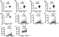
附图2为实施例4中非配对以及配对t检验检测这5个基因在TCGA数据库,肝癌组织和癌旁正常肝组织中的表达量差异。其中,图2A-2E是模型中5个基因在肝癌组织和癌旁正常肝组织表达量的非配对t检验的结果,图2F-2J是模型中5个基因在肝癌组织和癌旁正常肝组织表达量的配对t检验的结果;非配对t检验和配对t检验结果均表明模型中5个基因在肝癌组织中存在异常高表达,且具有统计学意义。Figure 2 shows the differences in the expression levels of these five genes detected by unpaired and paired t tests in the TCGA database, liver cancer tissue and adjacent normal liver tissue in Example 4. Among them, Figures 2A-2E are the results of the unpaired t-test of the expression levels of five genes in the model in liver cancer tissue and adjacent normal liver tissue, and Figures 2F-2J are the expression levels of five genes in the model in liver cancer tissue and adjacent normal liver tissue. The results of the paired t test of the expression level; the results of the unpaired t test and the paired t test showed that the five genes in the model were abnormally high expressed in the liver cancer tissue, and it was statistically significant.
附图3为实施例4中非配对以及配对t检验检测这5个基因在GEO数据库,肝癌组织和癌旁正常肝组织中的表达量差异。其中,图3A-3E是模型中5个基因在肝癌组织和癌旁正常肝组织表达量的非配对t检验的结果,图3F-3J是模型中5个基因在肝癌组织和癌旁正常肝组织表达量的配对t检验的结果;非配对t检验和配对t检验结果均表明模型中5个基因在肝癌组织中存在异常高表达,除SSRP1的配对t检验无统计学意义外,其它结果均有统计学意义;分析原因是可能由于样本量少导致的。FIG. 3 shows the differences in the expression levels of these five genes detected by unpaired and paired t tests in the GEO database, liver cancer tissue and adjacent normal liver tissue in Example 4. Among them, Figures 3A-3E are the results of the unpaired t-test of the expression levels of five genes in the model in liver cancer tissue and adjacent normal liver tissue, and Figures 3F-3J are the expression levels of five genes in the model in liver cancer tissue and adjacent normal liver tissue. The results of the paired t test of expression; the results of the unpaired t test and the paired t test both showed that the five genes in the model had abnormally high expression in the liver cancer tissue, except for the paired t test of SSRP1, which had no statistical significance Statistical significance; reason for analysis may be due to small sample size.
附图4为实施例5中ROC曲线和Kaplan-Meier生存曲线验证预后模型的准确性和特异性。其中,4A是高风险评分患者和低风险评分患者的Kaplan-Meier生存曲线,高风险评分患者的预后明显差于低风险评分患者;模型的ROC曲线下面积为0.755,证明该模型在预测患者预后方面具有很好的敏感性和特异性。FIG. 4 shows the ROC curve and Kaplan-Meier survival curve in Example 5 to verify the accuracy and specificity of the prognostic model. Among them, 4A is the Kaplan-Meier survival curve of patients with high risk score and patients with low risk score. The prognosis of patients with high risk score is significantly worse than that of patients with low risk score; the area under the ROC curve of the model is 0.755, which proves that the model can predict the prognosis of patients. Aspects have good sensitivity and specificity.
附图5为实施例6中,肝癌预后模型中的基因在临床样本中表达量的验证。通过提取临床肝癌组织和癌旁正常组织中的总RNA,分别进行逆转录和qRT-PCR检测模型中5个基因在临床肝癌组织和癌旁正常肝组织中的表达量,实验结果和生物信息学分析的结果一致,模型中的5个基因在肝癌组织中均存在显著高表达。FIG. 5 shows the verification of the expression levels of the genes in the liver cancer prognosis model in the clinical samples in Example 6. FIG. By extracting total RNA from clinical liver cancer tissue and adjacent normal tissue, reverse transcription and qRT-PCR were performed to detect the expression levels of five genes in the model in clinical liver cancer tissue and adjacent normal liver tissue, experimental results and bioinformatics The results of the analysis were consistent, and the five genes in the model were significantly highly expressed in liver cancer tissues.
具体实施方式Detailed ways
为了使本发明的目的、技术方案及优点更加清楚明白,以下结合实施例,对本发明进行进一步详细说明。应当理解,此处所描述的具体实施例仅用以解释本发明,并不用于限定本发明。In order to make the objectives, technical solutions and advantages of the present invention clearer, the present invention will be further described in detail below with reference to the embodiments. It should be understood that the specific embodiments described herein are only used to explain the present invention, but not to limit the present invention.
本发明适用的数据包括转录组数据和临床数据,适用标本包括冻存在-80℃的组织标本。The data applicable to the present invention include transcriptome data and clinical data, and applicable specimens include tissue specimens frozen at -80°C.
实施例1TCGA数据库RNA-Seq测序数据和患者临床病理资料的下载Example 1 Downloading of RNA-Seq sequencing data and patient clinicopathological data from TCGA database
从TCGA数据库下载肝癌患者的肝癌组织、癌旁正常组织的RNA-Seq测序数据,并下载肝癌患者的临床病理资料,下载网址为:https://portal.gdc.cancer.gov/。TCGA数据库中,422例肝癌患者具有临床病理资料;373例肝癌患者有肝癌组织的RNA-Seq测序数据。既有临床病理资料又有肝癌组织测序数据的肝癌患者总计369例,其中,50例肝癌患者具有肝癌组织和癌旁正常肝组织的配对的RNA-Seq测序数据。由于mRNA的表达谱数据已经通过TCGA标准化,因此不对这些数据进行进一步的标准化,肝癌患者病理参数如表1所示:Download the RNA-Seq sequencing data of liver cancer tissues and adjacent normal tissues of liver cancer patients from the TCGA database, and download the clinicopathological data of liver cancer patients. The download website is: https://portal.gdc.cancer.gov/. In the TCGA database, 422 liver cancer patients had clinicopathological data; 373 liver cancer patients had RNA-Seq sequencing data of liver cancer tissue. A total of 369 liver cancer patients had both clinicopathological data and liver cancer tissue sequencing data. Among them, 50 liver cancer patients had paired RNA-Seq sequencing data of liver cancer tissue and adjacent normal liver tissue. Since the mRNA expression profile data have been normalized by TCGA, no further normalization was performed on these data, and the pathological parameters of liver cancer patients are shown in Table 1:
表1.肝癌患者的临床病理参数Table 1. Clinicopathological parameters of liver cancer patients

实施例2肝癌患者中差异表达基因集的筛选Example 2 Screening of differentially expressed gene sets in liver cancer patients
采用GSEA 4.1.0版,利用肝癌组织和癌旁正常肝组织的RNA-Seq测序数据,分析肝癌组织和癌旁正常肝组织中差异表达的基因集。以|NES|>1.5,NOM P-val<0.05为标准筛选在肝癌组织中异常表达的基因集,用于确定肝癌治疗中对预后有预测价值的基因集;|NES|代表归一化后的富集分析评分,NOM p-val代表校正后的p value,表征富集结果的可信度;其中,转录因子E2F基因集中含有197个基因,其|NES|=2.071552,NOM p-val=0.001961,是在肝癌组织和癌旁正常组织中差异最大的基因集(表2)。Using GSEA version 4.1.0, RNA-Seq sequencing data of liver cancer tissue and adjacent normal liver tissue were used to analyze the differentially expressed gene sets in liver cancer tissue and adjacent normal liver tissue. Using |NES|>1.5, NOM P-val<0.05 as the standard to screen gene sets that are abnormally expressed in liver cancer tissues, to determine the gene sets with predictive value in liver cancer treatment; |NES| represents the normalized Enrichment analysis score, NOM p-val represents the corrected p value, indicating the reliability of the enrichment results; among them, the transcription factor E2F gene set contains 197 genes, of which |NES|=2.071552, NOM p-val=0.001961 , was the most different gene set in liver cancer tissue and adjacent normal tissue (Table 2).
表2.肝癌患者中异常表达的基因集Table 2. Aberrantly expressed gene sets in liver cancer patients

实施例3肝癌预后模型的构建Example 3 Construction of a liver cancer prognosis model
利用单因素Cox回归分析,从GSEA筛选出的差异基因集中筛选影响肝癌患者预后,且P<0.001的基因,共筛选出20个基因;采用多因素Cox回归分析建立预后相关模型,最终筛选出HN1、KIF4A、CDCA3、CDCA8及SSRP1构建预测肝癌患者预后的风险模型,所选预后基因的多变量COX生存分析的详细结果如表3所示。风险评分=0.3915*expression of gene HN1-0.3864*expression of gene KIF4A-0.2886*expression of gene CDCA3+0.4415*expression of gene CDCA8+0.8842*expression of gene SSRP1。在构建的风险模型中,B(COX)是相应基因表达量的系数,P value是基因多因素COX生存分析影响预后的P值,HR是基因的风险系数(表3)。根据风险评分中位数将肝癌患者分为低危组和高危组(图1A),本发明发现高危组总生存时间较低危组短,死亡人数较高(图1B)。Univariate Cox regression analysis was used to screen the differential genes screened by GSEA to screen the genes affecting the prognosis of patients with liver cancer, and P<0.001, and a total of 20 genes were screened; multivariate Cox regression analysis was used to establish a prognosis correlation model, and finally HN1 was screened out. , KIF4A, CDCA3, CDCA8 and SSRP1 to construct a risk model for predicting the prognosis of liver cancer patients. The detailed results of the multivariate COX survival analysis of the selected prognostic genes are shown in Table 3. Risk score = 0.3915*expression of gene HN1-0.3864*expression of gene KIF4A-0.2886*expression of gene CDCA3+0.4415*expression of gene CDCA8+0.8842*expression of gene SSRP1. In the constructed risk model, B(COX) is the coefficient of the corresponding gene expression, P value is the P value of the gene multivariate COX survival analysis affecting the prognosis, and HR is the risk coefficient of the gene (Table 3). Liver cancer patients were divided into low-risk and high-risk groups according to the median risk score (Fig. 1A). The present invention found that the high-risk group had a shorter overall survival time and a higher number of deaths (Fig. 1B).
表3.肝癌预后模型中基因的多因素COX分析结果Table 3. Results of multivariate COX analysis of genes in the liver cancer prognostic model


实施例4肝癌预后模型中基因在肝癌组织和癌旁正常组织中的表达差异Example 4 Expression differences of genes in liver cancer tissue and adjacent normal tissue in a liver cancer prognosis model
采用TCGA以及GEO数据库,使用非配对以及配对t检验在肝癌组织以及正常组织中分析预后模型中5个基因表达量的差异,结果显示筛选的5个基因在肝癌组织中的表达均高于相邻正常组织(图2、图3)。Using the TCGA and GEO databases, unpaired and paired t tests were used to analyze the differences in the expression of five genes in the prognostic model in liver cancer tissue and normal tissue. normal tissue (Figure 2, Figure 3).
实施例5ROC曲线和Kaplan-Meier生存曲线验证预后模型的准确性和特异性Example 5 ROC curve and Kaplan-Meier survival curve to verify the accuracy and specificity of the prognostic model
将肝癌患者按风险评分中位数分为高低风险组,构建ROC曲线,ROC曲线下面积为0.755,表明风险评分在预测肝癌患者预后方面具有较高的特异性和敏感性(图4B)。该结果证明预后模型在预测肝癌患者预后方面具有较好的准确性和特异性。The patients with liver cancer were divided into high and low risk groups according to the median risk score, and the ROC curve was constructed. The area under the ROC curve was 0.755, indicating that the risk score had high specificity and sensitivity in predicting the prognosis of patients with liver cancer (Figure 4B). The results demonstrate that the prognostic model has good accuracy and specificity in predicting the prognosis of liver cancer patients.
利用Kaplan-Meier生存曲线,分析高低风险组患者的预后,低危组患者的生存时间明显优于高危组(图4A)。对肝癌患者的预后进行分层分析,以确定风险评分的有效性。该结果表明风险值高的患者的预后较差,预后模型可以很好的预测肝癌患者的预后。Using the Kaplan-Meier survival curve to analyze the prognosis of patients in the high- and low-risk groups, the survival time of patients in the low-risk group was significantly better than that in the high-risk group (Figure 4A). A stratified analysis of the prognosis of patients with liver cancer to determine the validity of the risk score. The results indicate that patients with high risk values have poor prognosis, and the prognostic model can predict the prognosis of patients with liver cancer well.
实施例6肝癌预后模型中的基因在临床样本中表达量的验证(图5)Example 6 Validation of the expression levels of genes in the liver cancer prognosis model in clinical samples (Fig. 5)
(1)肝癌肿瘤组织及配对正常组织样本的获得及总RNA提取(1) Acquisition of liver cancer tumor tissue and paired normal tissue samples and extraction of total RNA
获得经手术分离的肝癌肿瘤组织样本21个,癌旁正常组织样本21个,用聚合美生物科技公司的TRIgent试剂提取总的RNA,对提取的总RNA进行定量。21 liver cancer tumor tissue samples and 21 adjacent normal tissue samples were obtained after surgery. Total RNA was extracted with TRIgent reagent from Polymei Biotechnology Company, and the extracted total RNA was quantified.
(2)实时荧光定量RT-PCR检测CDCA3、CDCA8、HN1、KIF4A以及SSRP1在肝癌肿瘤样本中的表达。(2) Real-time fluorescence quantitative RT-PCR to detect the expressions of CDCA3, CDCA8, HN1, KIF4A and SSRP1 in liver cancer tumor samples.
利用实时荧光定量RT-PCR检测步骤(1)中获得的21个肝癌肿瘤样本及21个癌旁正常组织样本中5个基因的表达情况,具体步骤如下:Real-time fluorescence quantitative RT-PCR was used to detect the expression of 5 genes in 21 liver cancer tumor samples and 21 adjacent normal tissue samples obtained in step (1). The specific steps are as follows:
1)RNA反转录:使用艾科瑞生物工程有限公司的反转录试剂盒(Evo M-MLV MixKit with gDNA Clean for qPCR,货号:AG11706-S)进行RNA样本的反转录反应,具体按照试剂盒说明书的方法进行,步骤如下:①去除基因组DNA,取1微克提取的总RNA样本,加入2μl 5×gDNA Clean Reaction Mix,3μl RNase free water,混匀后置42℃,反应2分钟;②反转录反应,在①的反应液中加入4μl 5×Evo M-MLV RT Reaction Mix,6μl RNase freewater,总体积为20μl;混匀后短暂离心,置于BIO-RAD T100 Thermal Cycler仪中进行反转录反应,反应参数为37℃,15分钟;85℃,5秒;随后置于4℃保存。1) RNA reverse transcription: Use the reverse transcription kit (Evo M-MLV MixKit with gDNA Clean for qPCR, Cat. No.: AG11706-S) of Ekerui Bioengineering Co., Ltd. to perform reverse transcription reaction of RNA samples. The method described in the kit instructions is carried out, and the steps are as follows: ①Remove genomic DNA, take 1 μg of extracted total RNA sample, add
2)实时荧光定量PCR:模型中基因的引物设计来源于生工生物工程有限公式,使用艾科瑞生物工程有限公司的
表4.肝癌预后模型中基因的引物序列如下:Table 4. The primer sequences of the genes in the liver cancer prognosis model are as follows:

3)数据分析:对同一样本分别检测其中目标RNA及内参RNA的表达;以内参RNA的表达量为基准,对目标RNA的表达进行归一化处理;随后采用通常使用的delta delta Ct法对目标RNA的表达量进行定量,本发明的内参为GAPDH。3) Data analysis: Detect the expression of target RNA and internal reference RNA in the same sample respectively; normalize the expression of target RNA based on the expression of internal reference RNA; then use the commonly used delta delta Ct method to analyze the target RNA. The expression level of RNA was quantified, and the internal reference of the present invention was GAPDH.
Claims (6)
Translated fromChinesePriority Applications (1)
| Application Number | Priority Date | Filing Date | Title |
|---|---|---|---|
| CN202210421628.3ACN114592065B (en) | 2022-04-21 | 2022-04-21 | Combined marker for predicting prognosis of liver cancer and application thereof |
Applications Claiming Priority (1)
| Application Number | Priority Date | Filing Date | Title |
|---|---|---|---|
| CN202210421628.3ACN114592065B (en) | 2022-04-21 | 2022-04-21 | Combined marker for predicting prognosis of liver cancer and application thereof |
Publications (2)
| Publication Number | Publication Date |
|---|---|
| CN114592065Atrue CN114592065A (en) | 2022-06-07 |
| CN114592065B CN114592065B (en) | 2023-12-12 |
Family
ID=81812073
Family Applications (1)
| Application Number | Title | Priority Date | Filing Date |
|---|---|---|---|
| CN202210421628.3AActiveCN114592065B (en) | 2022-04-21 | 2022-04-21 | Combined marker for predicting prognosis of liver cancer and application thereof |
Country Status (1)
| Country | Link |
|---|---|
| CN (1) | CN114592065B (en) |
Cited By (2)
| Publication number | Priority date | Publication date | Assignee | Title |
|---|---|---|---|---|
| CN116606932A (en)* | 2023-03-22 | 2023-08-18 | 唐山市人民医院 | Construction method of gastric cancer prognosis prediction risk model |
| CN116741271A (en)* | 2023-06-09 | 2023-09-12 | 唐山市人民医院 | Construction method and application of esophageal squamous carcinoma prognosis prediction risk model |
Citations (6)
| Publication number | Priority date | Publication date | Assignee | Title |
|---|---|---|---|---|
| CN1852974A (en)* | 2003-06-09 | 2006-10-25 | 密歇根大学董事会 | Compositions and methods for treating and diagnosing cancer |
| CN104769131A (en)* | 2012-09-21 | 2015-07-08 | 英特盖根公司 | Methods for Prognosing Overall Survival and Relapse-Free Survival in Hepatocellular Carcinoma |
| US20170298443A1 (en)* | 2014-09-25 | 2017-10-19 | Moffitt Genetics Corporation | Prognostic tumor biomarkers |
| CN113436673A (en)* | 2021-06-29 | 2021-09-24 | 北京泱深生物信息技术有限公司 | Molecular marker for liver cancer prognosis prediction and application thereof |
| CN113785076A (en)* | 2019-05-03 | 2021-12-10 | 株式会社递希真 | Methods and compositions for predicting cancer prognosis |
| CN113981085A (en)* | 2021-11-08 | 2022-01-28 | 右江民族医学院附属医院 | mRNA genome, mRNA gene pair group, prediction model and application for liver cancer prognosis |
- 2022
- 2022-04-21CNCN202210421628.3Apatent/CN114592065B/enactiveActive
Patent Citations (6)
| Publication number | Priority date | Publication date | Assignee | Title |
|---|---|---|---|---|
| CN1852974A (en)* | 2003-06-09 | 2006-10-25 | 密歇根大学董事会 | Compositions and methods for treating and diagnosing cancer |
| CN104769131A (en)* | 2012-09-21 | 2015-07-08 | 英特盖根公司 | Methods for Prognosing Overall Survival and Relapse-Free Survival in Hepatocellular Carcinoma |
| US20170298443A1 (en)* | 2014-09-25 | 2017-10-19 | Moffitt Genetics Corporation | Prognostic tumor biomarkers |
| CN113785076A (en)* | 2019-05-03 | 2021-12-10 | 株式会社递希真 | Methods and compositions for predicting cancer prognosis |
| CN113436673A (en)* | 2021-06-29 | 2021-09-24 | 北京泱深生物信息技术有限公司 | Molecular marker for liver cancer prognosis prediction and application thereof |
| CN113981085A (en)* | 2021-11-08 | 2022-01-28 | 右江民族医学院附属医院 | mRNA genome, mRNA gene pair group, prediction model and application for liver cancer prognosis |
Non-Patent Citations (5)
| Title |
|---|
| BOWEN WU等: "The diagnostic and prognostic value of cell division cycle associated gene family in Hepatocellular Carcinoma", vol. 11, no. 19* |
| GUANSHUI LUO等: "SSRP1 Is a Prognostic Biomarker Correlated with CD8+T CellInfiltration in Hepatocellular Carcinoma (HCC)"* |
| ZHICHENG LIU等: "HN1 as a diagnostic and prognostic biomarker for liver cancer", vol. 40, no. 7* |
| 张路遥;黄辉星;曹立环;余龙;: "CDCA8和INCENP mRNA在肝细胞癌组织中的表达及其临床意义", vol. 24, no. 02, pages 158 - 168* |
| 蓝祝晶等: "KIF4A 在肝细胞癌中的表达及预后价值分析", vol. 31, no. 1* |
Cited By (2)
| Publication number | Priority date | Publication date | Assignee | Title |
|---|---|---|---|---|
| CN116606932A (en)* | 2023-03-22 | 2023-08-18 | 唐山市人民医院 | Construction method of gastric cancer prognosis prediction risk model |
| CN116741271A (en)* | 2023-06-09 | 2023-09-12 | 唐山市人民医院 | Construction method and application of esophageal squamous carcinoma prognosis prediction risk model |
Also Published As
| Publication number | Publication date |
|---|---|
| CN114592065B (en) | 2023-12-12 |
Similar Documents
| Publication | Publication Date | Title |
|---|---|---|
| CN113785076B (en) | Methods of predicting prognosis of cancer and compositions thereof | |
| CN113286883A (en) | Methods for detecting disease using RNA analysis | |
| CN108277283B (en) | Application of lncRNA combination in preparation of product for predicting renal clear cell carcinoma prognosis and molecular targeted drug treatment sensitivity | |
| CN104140967B (en) | The long-chain non-coding RNA CLMAT1 relevant to colorectal cancer hepatic metastases and application thereof | |
| JP2014530619A (en) | Plasma microRNA for detection of early colorectal cancer | |
| Cai et al. | Serum miR-21 expression in human esophageal squamous cell carcinomas | |
| JP2015535176A (en) | A novel method for predicting overall and relapse-free survival in hepatocellular carcinoma | |
| CN114592065B (en) | Combined marker for predicting prognosis of liver cancer and application thereof | |
| CN107523647A (en) | Detect the LncRNA combinations of early stage cancer of the esophagus prognosis situation and the kit containing the combination | |
| CN107674916B (en) | Application of circular RNA in colorectal cancer biomarker | |
| CN105176983A (en) | Kit for detecting esophageal squamous carcinoma associated serum miRNAs genes | |
| US20210214807A1 (en) | Method and kit for the diagnosis of colorectal cancer | |
| CN108949969B (en) | Application of long non-coding RNA in colorectal cancer | |
| CN106967719A (en) | A kind of long-chain non-coding RNA as prostate cancer molecular marker application | |
| CN111763740B (en) | A system for predicting the efficacy and prognosis of neoadjuvant chemoradiotherapy in patients with esophageal squamous cell carcinoma based on lncRNA molecular model | |
| CN113736879A (en) | System for small cell lung cancer patient prognosis and application thereof | |
| CN106148511B (en) | Predictive marker and kit for recurrence risk of liver cancer patient after resection | |
| CN106755330B (en) | Cancer-related gene expression difference detection kit and application thereof | |
| CN115961042A (en) | Application of IGFBP1 gene or CHAF1A gene as gastric adenocarcinoma prognostic molecular marker | |
| CN113528670A (en) | A biomarker and detection kit for predicting the risk of late postoperative recurrence in patients with liver cancer | |
| Li et al. | Unveiling the clinical significance of RNA pseudouridine in colorectal cancer | |
| WO2021102228A1 (en) | Small unannotated, non-coding rnas for the detection of liver cancer | |
| CN113604576B (en) | Lung adenocarcinoma detection kit, storage medium and electronic device | |
| KR20200084435A (en) | Bio-marker for non-invasive differential diagnosis of acute rejection in kidney transplanted patients and uses thereof | |
| CN120138161B (en) | Application of reagent for detecting esophageal squamous carcinoma marker in preparation of esophageal squamous carcinoma auxiliary diagnosis or diagnosis product |
Legal Events
| Date | Code | Title | Description |
|---|---|---|---|
| PB01 | Publication | ||
| PB01 | Publication | ||
| SE01 | Entry into force of request for substantive examination | ||
| SE01 | Entry into force of request for substantive examination | ||
| GR01 | Patent grant | ||
| GR01 | Patent grant | ||
| TR01 | Transfer of patent right | Effective date of registration:20241120 Address after:230000, 4th Floor, Building 4, Mingzhu Industrial Park, No. 106 Chuangxin Avenue, High tech Zone, Hefei City, Anhui Province Patentee after:Anhui Huapei Biotechnology Co.,Ltd. Country or region after:China Address before:266000 5 Donghai Middle Road, Qingdao, Shandong Patentee before:QINGDAO MUNICIPAL Hospital Country or region before:China | |
| TR01 | Transfer of patent right |