CN114563869B - Surface mount type mobile phone microscope detection system and microscopic result obtaining method thereof - Google Patents
Surface mount type mobile phone microscope detection system and microscopic result obtaining method thereofDownload PDFInfo
- Publication number
- CN114563869B CN114563869BCN202210048147.2ACN202210048147ACN114563869BCN 114563869 BCN114563869 BCN 114563869BCN 202210048147 ACN202210048147 ACN 202210048147ACN 114563869 BCN114563869 BCN 114563869B
- Authority
- CN
- China
- Prior art keywords
- image
- microscopic image
- smart phone
- picture
- microscopic
- Prior art date
- Legal status (The legal status is an assumption and is not a legal conclusion. Google has not performed a legal analysis and makes no representation as to the accuracy of the status listed.)
- Expired - Fee Related
Links
Images


Classifications
- G—PHYSICS
- G02—OPTICS
- G02B—OPTICAL ELEMENTS, SYSTEMS OR APPARATUS
- G02B21/00—Microscopes
- G02B21/36—Microscopes arranged for photographic purposes or projection purposes or digital imaging or video purposes including associated control and data processing arrangements
- G02B21/362—Mechanical details, e.g. mountings for the camera or image sensor, housings
- G—PHYSICS
- G01—MEASURING; TESTING
- G01M—TESTING STATIC OR DYNAMIC BALANCE OF MACHINES OR STRUCTURES; TESTING OF STRUCTURES OR APPARATUS, NOT OTHERWISE PROVIDED FOR
- G01M11/00—Testing of optical apparatus; Testing structures by optical methods not otherwise provided for
- G01M11/02—Testing optical properties
- G—PHYSICS
- G02—OPTICS
- G02B—OPTICAL ELEMENTS, SYSTEMS OR APPARATUS
- G02B21/00—Microscopes
- G02B21/36—Microscopes arranged for photographic purposes or projection purposes or digital imaging or video purposes including associated control and data processing arrangements
- G02B21/365—Control or image processing arrangements for digital or video microscopes
- G02B21/367—Control or image processing arrangements for digital or video microscopes providing an output produced by processing a plurality of individual source images, e.g. image tiling, montage, composite images, depth sectioning, image comparison
- G—PHYSICS
- G02—OPTICS
- G02B—OPTICAL ELEMENTS, SYSTEMS OR APPARATUS
- G02B21/00—Microscopes
- G02B21/36—Microscopes arranged for photographic purposes or projection purposes or digital imaging or video purposes including associated control and data processing arrangements
- G02B21/368—Microscopes arranged for photographic purposes or projection purposes or digital imaging or video purposes including associated control and data processing arrangements details of associated display arrangements, e.g. mounting of LCD monitor
- H—ELECTRICITY
- H04—ELECTRIC COMMUNICATION TECHNIQUE
- H04M—TELEPHONIC COMMUNICATION
- H04M1/00—Substation equipment, e.g. for use by subscribers
- H04M1/02—Constructional features of telephone sets
- H04M1/0202—Portable telephone sets, e.g. cordless phones, mobile phones or bar type handsets
- H04M1/026—Details of the structure or mounting of specific components
- H—ELECTRICITY
- H04—ELECTRIC COMMUNICATION TECHNIQUE
- H04N—PICTORIAL COMMUNICATION, e.g. TELEVISION
- H04N7/00—Television systems
- H04N7/18—Closed-circuit television [CCTV] systems, i.e. systems in which the video signal is not broadcast
- Y—GENERAL TAGGING OF NEW TECHNOLOGICAL DEVELOPMENTS; GENERAL TAGGING OF CROSS-SECTIONAL TECHNOLOGIES SPANNING OVER SEVERAL SECTIONS OF THE IPC; TECHNICAL SUBJECTS COVERED BY FORMER USPC CROSS-REFERENCE ART COLLECTIONS [XRACs] AND DIGESTS
- Y02—TECHNOLOGIES OR APPLICATIONS FOR MITIGATION OR ADAPTATION AGAINST CLIMATE CHANGE
- Y02D—CLIMATE CHANGE MITIGATION TECHNOLOGIES IN INFORMATION AND COMMUNICATION TECHNOLOGIES [ICT], I.E. INFORMATION AND COMMUNICATION TECHNOLOGIES AIMING AT THE REDUCTION OF THEIR OWN ENERGY USE
- Y02D30/00—Reducing energy consumption in communication networks
- Y02D30/70—Reducing energy consumption in communication networks in wireless communication networks
Landscapes
- Physics & Mathematics (AREA)
- Engineering & Computer Science (AREA)
- Multimedia (AREA)
- Chemical & Material Sciences (AREA)
- Analytical Chemistry (AREA)
- General Physics & Mathematics (AREA)
- Optics & Photonics (AREA)
- Signal Processing (AREA)
- Computer Vision & Pattern Recognition (AREA)
- Microscoopes, Condenser (AREA)
- Investigating, Analyzing Materials By Fluorescence Or Luminescence (AREA)
Abstract
Description
Translated fromChinese技术领域technical field
本发明涉及显微装置技术领域,尤其涉及一种贴片式手机显微镜检测系统及其显微结果获取方法。The invention relates to the technical field of microscopic devices, in particular to a patch-type mobile phone microscope detection system and a method for obtaining microscopic results.
背景技术Background technique
贴片式手机显微镜,是一种基于智能手机的显微镜模块,其内设有镜片组,可以提高手机拍摄物体照片是的方法倍数,从而可使手机能拍摄拍摄物体的微观结构。目前,基于手机的贴片式手机显微镜,在用于实验观察时存在装置不够简约、使用起来比较麻烦的问题,且贴片式手机显微镜因成像质量有限,其直接拍摄出的图片存在成像不清晰、肉眼难以观察实验结果的问题;例如:现有的贴片式手机显微镜在观察蓝氏贾第虫、雨生红球藻细胞的图片,要对待测样品做荧光特异性处,其拍摄出的图片会容易出现对比度差、难以观察目标单元(蓝氏贾第虫和雨生红球藻细胞)的具体结构的、难以统计目标单元的个数的问题,影响实验过程。The patch-type mobile phone microscope is a microscope module based on a smart phone, which is equipped with a lens group, which can increase the multiples of the method of taking pictures of objects on the mobile phone, so that the mobile phone can take pictures of the microstructure of the object. At present, the SMD mobile phone microscope based on the mobile phone has the problem that the device is not simple enough and it is troublesome to use when it is used for experimental observation, and the image quality of the SMD mobile phone microscope is limited, and the pictures directly taken by it are unclear. , The problem that it is difficult to observe the experimental results with the naked eye; for example: when the existing patch-type mobile phone microscope is observing the pictures of Giardia lamblia and Haematococcus pluvialis cells, it is necessary to perform fluorescence specificity on the samples to be tested. The picture will be prone to problems such as poor contrast, difficulty in observing the specific structure of the target unit (Giardia lamblia and Haematococcus pluvialis cells), and difficulty in counting the number of target units, which will affect the experimental process.
发明内容Contents of the invention
有鉴于此,为提高贴片式手机显微镜的图像获取能力以及获取图片的质量,使其获取的样品图片能满足试验要求,并简化实验观察过程,本发明提供一种贴片式手机显微镜检测系统,包括支架、智能手机、贴片式显微镜模块以及服务器;In view of this, in order to improve the image acquisition ability of the SMD mobile phone microscope and the quality of the obtained pictures, so that the sample pictures obtained by it can meet the test requirements, and simplify the experimental observation process, the present invention provides a SMD mobile phone microscope detection system , including brackets, smartphones, SMD microscope modules and servers;
所述支架包括基座和两支撑板,两支撑板下端连接于所述基座两侧,两支撑板之间还设有载物板,The bracket includes a base and two support plates, the lower ends of the two support plates are connected to both sides of the base, and a loading plate is also provided between the two support plates.
所述贴片式显微镜模块贴紧于所述智能手机的镜头上,The patch microscope module is attached to the lens of the smart phone,
所述智能手机支撑于两所述支撑板之间,且位于所述载物板上方,The smart phone is supported between the two support plates and is located above the loading plate,
所述载物板上设有一透光孔,所述载物板用于支撑样品,A light-transmitting hole is provided on the loading plate, and the loading plate is used to support samples,
所述基座上设有一光源组件,所述光源组件用于照亮样品,A light source assembly is provided on the base, and the light source assembly is used to illuminate the sample,
所述智能手机用于通过贴片式显微镜模块拍摄样品的显微图像,并将其发送至服务器,所述服务器对显微图像处理得到显微图像和统计数据,并将其回传至智能手机显示。The smart phone is used to take the microscopic image of the sample through the patch microscope module, and send it to the server, and the server processes the microscopic image to obtain the microscopic image and statistical data, and sends it back to the smart phone show.
进一步地,包括所述光源组件包括LED灯和电源,所述电源位于所述基座内。Further, the light source assembly includes an LED lamp and a power supply, and the power supply is located in the base.
进一步地,两所述支撑板顶部分别设有一卡槽,所述智能手机两侧方别可滑动地卡于两所述卡槽内。Further, the tops of the two support plates are respectively provided with a card slot, and the two sides of the smart phone are slidably locked in the two card slots.
进一步地,所述智能手机和所述载物板之间还设有一安装板,所述安装板中部设有一滑槽,所述贴片式显微镜模块可滑动地限位于所述滑槽中。Further, a mounting plate is provided between the smart phone and the loading plate, and a chute is provided in the middle of the mounting plate, and the patch microscope module is slidably limited in the chute.
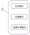
进一步地,所述服务器内设有依次相连的收发模块、存储模块以及图像处理模块,所述收发模块接收显微图像并将其传递至存储模块保存,所述图像处理模块读取显微图像,处理显微图像得到显微图像和统计数据,并将显微图像和统计数据回传至存储模块存储且通过收发模块发送回智能手机显示。Further, the server is provided with a transceiver module, a storage module and an image processing module connected in sequence, the transceiver module receives the microscopic image and transfers it to the storage module for storage, and the image processing module reads the microscopic image, Microscopic images are processed to obtain microscopic images and statistical data, and the microscopic images and statistical data are sent back to the storage module for storage and sent back to the smart phone for display through the transceiver module.
本发明还提供一种基于上述贴片式手机显微镜检测系统的显微图像获取方法,包括如下步骤:The present invention also provides a microscopic image acquisition method based on the above patch-type mobile phone microscope detection system, comprising the following steps:
S1:将样品安装在载物板上,且使样品位于透光孔上方;S1: Install the sample on the loading plate, and make the sample above the light-transmitting hole;
S2:将贴片式显微镜模块安装在智能手机上,滑动智能手机使智能手机镜头正对透光孔,并开启光源组件;S2: Install the SMD microscope module on the smartphone, slide the smartphone so that the lens of the smartphone faces the light hole, and turn on the light source assembly;
S3:开启智能手机的相机功能,智能手机通过贴片式显微镜模块拍摄样品显微图像;S3: Turn on the camera function of the smartphone, and the smartphone takes microscopic images of the sample through the patch microscope module;
S4:智能手机将拍摄到的限位图像发送至服务器;S4: the smart phone sends the captured image to the server;
S5:服务器将显微图像进行处理,处理结果,并将处理结果回传至只能智能手机,所述处理结果为显微图像和统计数据。S5: The server processes the microscopic image, processes the result, and returns the processing result to the smart phone, and the processing result is the microscopic image and statistical data.
进一步地,智能手机将样品图像发送至服务器之间,还通过其内的APP预先判定样品图像时候符合要求,若符合,则进行S5,否则返回S3;Furthermore, the smart phone sends the sample image to the server, and pre-determines that the sample image meets the requirements through the APP in it. If it meets the requirements, go to S5, otherwise return to S3;
进一步地,步骤S5中,服务器对样品图像进行处理获取,获取处理结果的方法如下:Further, in step S5, the server processes and obtains the sample image, and the method for obtaining the processing result is as follows:
S51:获取样品图像;S51: acquiring a sample image;
S52:将样品图像转化为灰度图,并通过中值滤波方式去除该灰度图椒盐噪声;S52: Convert the sample image into a grayscale image, and remove the salt and pepper noise of the grayscale image by means of median filtering;
S53:将步骤S52中的图像进行梯度化处理,选取梯度阈值将灰度图像二值化,得到多个目标区域,获取目标区域的位置及边缘轮廓,此时得到图片A;S53: Perform gradient processing on the image in step S52, select a gradient threshold to binarize the grayscale image, obtain multiple target areas, obtain the position and edge contour of the target area, and obtain picture A at this time;
S54:将步骤S52的图像进行多次腐蚀及膨胀操作,缩小灰度图中颜色深的部分的大小,此时得到图片B;S54: Perform multiple erosion and expansion operations on the image in step S52 to reduce the size of the dark part in the grayscale image, and obtain image B at this time;
S55:将图片A和图片B重合,且用图片A减去图片B,获得图片C;S55: Overlap picture A and picture B, and subtract picture B from picture A to obtain picture C;
S56:重新给定图片C和图片B不同的灰度值,将图片C于图片进行重合得到图片D;S56: Redefine the different gray values of picture C and picture B, and overlap picture C with picture D to obtain picture D;
S57:对图片D进行基于欧式距离变换的分水岭算法进行二值图像分隔,得到多个目标点,统计目标点数量;S57: Carrying out the binary image segmentation based on the Euclidean distance transform watershed algorithm on the image D, obtaining multiple target points, and counting the number of target points;
S58:对图片D中的目标区域进行RGB填充,即获得显微图像;S58: Perform RGB filling on the target area in the picture D, that is, obtain a microscopic image;
S59:将步骤S57中的统计数量以及S58获取的显微图像回传至智能手机。S59: Send back the statistics in step S57 and the microscopic image acquired in S58 to the smart phone.
本发明一种贴片式手机显微镜系统的有益效果为:该手机显微镜系统结构简单,操作便捷,其通过支架结构可提高样品图像拍摄的质量;且该系统的服务器可对样品图像进行处理,得到可清楚显示样目标单元数量以及微观结构的显微图像,从而简化实验观察过程,提高实验效率;采用本发明的显微结果获取方法,可通过对样品图像进行处理,提高显微图像的清晰度,并能自动统计目标单元数量,提高实验效率。The beneficial effects of a patch-type mobile phone microscope system of the present invention are: the mobile phone microscope system has a simple structure and is convenient to operate, and the quality of sample image shooting can be improved through the bracket structure; and the server of the system can process the sample image to obtain The number of sample target units and the microscopic image of the microstructure can be clearly displayed, thereby simplifying the experimental observation process and improving the experimental efficiency; using the microscopic result acquisition method of the present invention, the clarity of the microscopic image can be improved by processing the sample image , and can automatically count the number of target units to improve experimental efficiency.
附图说明Description of drawings
图1是本发明实施例一种贴片式手机显微镜检测系统的第一立体结构图。FIG. 1 is a first three-dimensional structure diagram of a patch-type mobile phone microscope detection system according to an embodiment of the present invention.
图2是本发明实施例一种贴片式手机显微镜检测系统的第二立体结构图。Fig. 2 is a second three-dimensional structure diagram of a patch-type mobile phone microscope detection system according to an embodiment of the present invention.
图3是本发明实施例一种贴片式手机显微镜检测系统的侧视图。Fig. 3 is a side view of a patch-type mobile phone microscope inspection system according to an embodiment of the present invention.
图4是本发明实施例一种贴片式手机显微镜检测系统的服务器的结构图。Fig. 4 is a structural diagram of a server of a patch-type mobile phone microscope detection system according to an embodiment of the present invention.
上述图中:1-基座,2-支撑板,21-卡槽,3-智能手机,4-贴片式显微镜模块,5-安装板,6-载物板,7-光源组件,71-LED灯,72-电池,8-服务器。In the above figure: 1-base, 2-support plate, 21-card slot, 3-smart phone, 4-SMD microscope module, 5-installation plate, 6-object carrier, 7-light source assembly, 71- LED light, 72-battery, 8-server.
具体实施方式Detailed ways
为使本发明的目的、技术方案和优点更加清楚,下面将结合附图对本发明实施方式作进一步地描述。In order to make the purpose, technical solution and advantages of the present invention clearer, the embodiments of the present invention will be further described below in conjunction with the accompanying drawings.
请参考图1至图4,一种贴片式手机显微镜检测系统,包括支架、智能手机3、贴片式显微镜模块4以及服务器8。Please refer to FIG. 1 to FIG. 4 , a SMD mobile phone microscope inspection system, including a bracket, a
所述支架1包括基座1和两支撑板2,两支撑板2竖直设置,两支撑板2下端连接于基座1两侧,两支撑板2顶部分别设有一卡槽21,两支撑板2之间还设有一安装板5和一载物板6,所述载物板6上设有一透光孔,载物板6上支撑有样品,本实施例中,载物台5是一个长条形不透光的涂黑的铝板,所述安装板5位于所述载物台5上方,所述安装板5中部设有一滑槽,优选地,所述安装板由透明材质制成。Described
所述基座1上设有一光源组件7,所述光源组件7包括LED灯71以及与LED灯71相连的电源72,所述电源72位于所述基座1内,本实施例中,所述电源72为纽扣电池,基座1上还设有控制LED灯71电亮和熄灭的电源开关,光源组件7用于使光线通过透光孔照亮样品。A
所述贴片式显微镜模块4通过粘贴的方式固定于所述智能手机3的镜头上,所述智能手机3两侧可滑动地限位于所述卡槽21中,所述贴片式显微镜模块4可滑动地限位于所述滑槽内,从而使智能手机3以及贴片式显微镜模块4可在支撑板2之间前后移动,便于智能手机镜头对准载物板6上的透光孔。优选地,所述贴片式显微镜模块4为TipScope贴片式手机显微镜,贴片式显微镜模块4可提高样品影像的放大倍数,从而使智能手机能拍摄样品的微观结构的图片,所述智能手机4用于将拍摄到的样品图片传递至服务器8进行处理,并接收并显示服务器8回传的处理结果。The patch-
所述服务器8内设有依次相连的收发模块、存储模块以及图像处理模块,所述收发模块接收显微图像并将其传递至存储模块保存,所述图像处理模块读取显微图像,处理显微图像得到显微图像和统计数据,并将显微图像和统计数据回传至存储模块存储且通过收发模块发送回智能手机4显示。The
本发明贴片式手机显微镜检测系统的工作过程如下:The working process of the patch type mobile phone microscope detection system of the present invention is as follows:
S1:将样品安装在载物板6上,且使样品位于透光孔上方;S1: install the sample on the
S2:将贴片式显微镜模块4安装在智能手机3上,滑动智能手机3使智能手机镜头正对透光孔,并开启光源组件7;S2: install the
S3:开启智能手机的相机功能,智能手机3通过贴片式显微镜模块4拍摄样品图像;S3: Turn on the camera function of the smart phone, and the
S4:智能手机3将拍摄到的样品图像发送至服务器8;S4: the
S5:服务器8将显微图像进行处理,得到处理结果,并将处理结果回传至智能手机3,所述处理结果为显微图像和统计数据。S5: The
上述步骤S4中,智能手机3将样品图像发送至服务器之前,还通过其内的APP预先判定样品图像时候符合服务器8处理要求,若符合,则进行S5,否则返回S3。In the above step S4, before the
上述步骤S5中,服务器8对样品图像进行处理,获取处理结果的方法具体包括如下步骤In the above step S5, the
S51:获取样品图像;样品图像即为智能手机通过贴片式手机显微镜模块4拍摄到的包含样品微观结构的图像;S51: Obtain a sample image; the sample image is an image including the microstructure of the sample captured by the smartphone through the SMD mobile
S52:将样品图像转化为灰度图,并通过中值滤波方式去除该灰度图椒盐噪声;S52: Convert the sample image into a grayscale image, and remove the salt and pepper noise of the grayscale image by means of median filtering;
S53:将步骤S52中的图像进行梯度化处理,选取梯度阈值将灰度图像二值化,得到多个目标区域,获取目标区域的位置及边缘轮廓,此时得到图片A;目标区域即为含有目标单元的区域,目标单元即为观察的对象(例如蓝氏贾第虫和雨生红球藻细胞);S53: Perform gradient processing on the image in step S52, select a gradient threshold to binarize the grayscale image, obtain multiple target areas, obtain the position and edge contour of the target area, and obtain picture A at this time; the target area is the one containing The area of the target unit, which is the object of observation (such as Giardia lamblia and Haematococcus pluvialis cells);
S54:将步骤S52的图像进行多次腐蚀及膨胀操作,缩小灰度图中颜色深的部分的大小,此时得到图片B;S54: Perform multiple erosion and expansion operations on the image in step S52 to reduce the size of the dark part in the grayscale image, and obtain image B at this time;
S55:将图片A和图片B重合,且用图片A减去图片B,获得图片C,图片C;图片C即为去除背景区域的目标区域的图像S55: Overlap picture A and picture B, and subtract picture B from picture A to obtain picture C, picture C; picture C is the image of the target area to remove the background area
S56:重新给定图片C和图片B不同的灰度值,将图片C于图片进行重合,使图片C覆盖在图片B上,并去除图片B上被覆盖的部分得到图片D;S56: redefine the different gray values of picture C and picture B, overlap picture C with the picture, make picture C cover picture B, and remove the covered part of picture B to obtain picture D;
S57:对图片D进行基于欧式距离变换的分水岭算法进行二值图像分隔,得到多个目标点,统计目标点数量;S57: Carrying out the binary image segmentation based on the Euclidean distance transform watershed algorithm on the image D, obtaining multiple target points, and counting the number of target points;
S58:对图片D中的目标区域进行RGB填充,即获得显微图像;S58: Perform RGB filling on the target area in the picture D, that is, obtain a microscopic image;
S59:将步骤S57中的统计数量以及S58获取的显微图像回传至智能手机。S59: Send back the statistics in step S57 and the microscopic image acquired in S58 to the smart phone.
在本文中,所涉及的前、后、上、下等方位词是以附图中零部件位于图中以及零部件相互之间的位置来定义的,只是为了表达技术方案的清楚及方便。应当理解,所述方位词的使用不应限制本申请请求保护的范围。In this article, the orientation words such as front, rear, upper, and lower involved are defined by the parts in the drawings and the positions between the parts in the drawings, just for the clarity and convenience of expressing the technical solution. It should be understood that the use of the location words should not limit the scope of protection claimed in this application.
在不冲突的情况下,本文中上述实施例及实施例中的特征可以相互结合。In the case of no conflict, the above-mentioned embodiments and features in the embodiments herein may be combined with each other.
以上所述仅为本发明的较佳实施例,并不用以限制本发明,凡在本发明的精神和原则之内,所作的任何修改、等同替换、改进等,均应包含在本发明的保护范围之内。The above descriptions are only preferred embodiments of the present invention, and are not intended to limit the present invention. Any modifications, equivalent replacements, improvements, etc. made within the spirit and principles of the present invention shall be included in the protection of the present invention. within range.
Claims (5)
Priority Applications (1)
| Application Number | Priority Date | Filing Date | Title |
|---|---|---|---|
| CN202210048147.2ACN114563869B (en) | 2022-01-17 | 2022-01-17 | Surface mount type mobile phone microscope detection system and microscopic result obtaining method thereof |
Applications Claiming Priority (1)
| Application Number | Priority Date | Filing Date | Title |
|---|---|---|---|
| CN202210048147.2ACN114563869B (en) | 2022-01-17 | 2022-01-17 | Surface mount type mobile phone microscope detection system and microscopic result obtaining method thereof |
Publications (2)
| Publication Number | Publication Date |
|---|---|
| CN114563869A CN114563869A (en) | 2022-05-31 |
| CN114563869Btrue CN114563869B (en) | 2023-04-18 |
Family
ID=81712508
Family Applications (1)
| Application Number | Title | Priority Date | Filing Date |
|---|---|---|---|
| CN202210048147.2AExpired - Fee RelatedCN114563869B (en) | 2022-01-17 | 2022-01-17 | Surface mount type mobile phone microscope detection system and microscopic result obtaining method thereof |
Country Status (1)
| Country | Link |
|---|---|
| CN (1) | CN114563869B (en) |
Families Citing this family (1)
| Publication number | Priority date | Publication date | Assignee | Title |
|---|---|---|---|---|
| CN118258812B (en)* | 2024-03-28 | 2025-03-18 | 中国地质大学(武汉) | A portable microscopic observation system and field slope rock and soil sample observation method |
Citations (2)
| Publication number | Priority date | Publication date | Assignee | Title |
|---|---|---|---|---|
| CN108846802A (en)* | 2018-01-25 | 2018-11-20 | 湖南省自兴人工智能研究院 | A kind of removal chromosomal G-banding mid-term gray level image Noise Method |
| CN212512901U (en)* | 2020-07-14 | 2021-02-09 | 张辉 | Smartphone-based portable concrete crack width measurement system |
Family Cites Families (17)
| Publication number | Priority date | Publication date | Assignee | Title |
|---|---|---|---|---|
| EP2227711A4 (en)* | 2008-01-02 | 2014-01-22 | Univ California | TELEMICROSCOPY APPARATUS WITH HIGH DIGITAL OPENING |
| WO2013191665A2 (en)* | 2012-06-21 | 2013-12-27 | Grimed Saglik Hizmetleri Ve Bilgisayar Urunleri Sanayi Ticaret Limited Sirketi | A mobile microscopy device being able to take images in different wavelengths (multispectral) |
| TWI533025B (en)* | 2014-07-07 | 2016-05-11 | 億觀生物科技股份有限公司 | Portable microscope |
| TWI572896B (en)* | 2014-11-07 | 2017-03-01 | 邦睿生技股份有限公司 | Testing equipment with magnifying function |
| CN104651462B (en)* | 2015-01-29 | 2017-02-22 | 华南农业大学 | Method for detecting magnaporthe grisea spore based on microscopic image analysis |
| CN105629451A (en)* | 2016-01-13 | 2016-06-01 | 大连理工大学 | High resolution portable microscope system and measurement method |
| KR101791003B1 (en)* | 2016-04-18 | 2017-11-21 | 신요식 | Apparatus for Taking Zoom-In Picture Using Mobile Terminal Equipped with Camera, and External-Optical Device therefor |
| CN106327442A (en)* | 2016-08-22 | 2017-01-11 | 上海奥通激光技术有限公司 | Multispectral micro-imaging field depth extension method and system |
| WO2019103909A1 (en)* | 2017-11-21 | 2019-05-31 | The Regents Of The University Of California | Portable microscopy device with enhanced image performance using deep learning and methods of using the same |
| CN209731356U (en)* | 2019-06-05 | 2019-12-03 | 武汉兰丁医学高科技有限公司 | A kind of device of the micro- scanning of mobile phone |
| KR102516836B1 (en)* | 2019-07-24 | 2023-04-03 | 주식회사 알틱스 | Sample analysis device compatible with mobile device and sample observing method using thereof |
| CN110596878B (en)* | 2019-10-14 | 2021-11-16 | 南京大学 | Double-lens microscope system with ultra-short focal length |
| CN110702672A (en)* | 2019-10-22 | 2020-01-17 | 中国科学院新疆理化技术研究所 | Explosive detection method based on portable mobile phone microscope |
| CN111583227B (en)* | 2020-05-08 | 2023-03-24 | 华侨大学 | Method, device, equipment and medium for automatically counting fluorescent cells |
| CN111709911B (en)* | 2020-05-18 | 2023-05-05 | 杭州电子科技大学 | A method for automatic counting of ovarian follicles based on neural network |
| CN111935404B (en)* | 2020-08-14 | 2021-10-15 | 腾讯科技(深圳)有限公司 | Macro imaging system, method and apparatus |
| CN112731641A (en)* | 2020-12-31 | 2021-04-30 | 安徽医科大学 | Multi-mode imaging mobile phone microscope device |
- 2022
- 2022-01-17CNCN202210048147.2Apatent/CN114563869B/ennot_activeExpired - Fee Related
Patent Citations (2)
| Publication number | Priority date | Publication date | Assignee | Title |
|---|---|---|---|---|
| CN108846802A (en)* | 2018-01-25 | 2018-11-20 | 湖南省自兴人工智能研究院 | A kind of removal chromosomal G-banding mid-term gray level image Noise Method |
| CN212512901U (en)* | 2020-07-14 | 2021-02-09 | 张辉 | Smartphone-based portable concrete crack width measurement system |
Also Published As
| Publication number | Publication date |
|---|---|
| CN114563869A (en) | 2022-05-31 |
Similar Documents
| Publication | Publication Date | Title |
|---|---|---|
| US10935484B2 (en) | Automated assessment of sperm samples | |
| Lee et al. | A smartphone-based chip-scale microscope using ambient illumination | |
| CN209764751U (en) | Surface defect detection system | |
| CN110579485B (en) | A device and method for rapid detection of surface defects on smartphone glass cover | |
| CN106062606B (en) | Image processing method and image processing apparatus | |
| US8736751B2 (en) | Digital presenter for displaying image captured by camera with illumination system | |
| CN110441323B (en) | Product surface polishing method and system | |
| US10088655B2 (en) | Preview station and method for taking preview images of microscope slides | |
| CN114563869B (en) | Surface mount type mobile phone microscope detection system and microscopic result obtaining method thereof | |
| CN103336404B (en) | A kind of lens test device and method | |
| US8237785B2 (en) | Automatic focusing apparatus for use in a microscope in which fluorescence emitted from a cell is captured so as to acquire a cell image, and automatic focusing method therefor | |
| CN203444234U (en) | Lens testing device | |
| JP2015143656A (en) | Inspection apparatus and inspection method | |
| CN112927190B (en) | Type-C multi-station multi-defect unified measurement system based on deep learning | |
| EP3620838A1 (en) | Systems and methods for calibrating a structured illumination imaging system and for capturing a structured illumination image | |
| CN211741094U (en) | Surface defect detection device for transparent mirror printing and gold stamping process | |
| CN211122566U (en) | Quick detection device of smart mobile phone glass apron surface defect | |
| CN104977154B (en) | Spatial light modulator defect classification method with sub-pixel structure | |
| CN109785290A (en) | Normalized steel plate defect detection method is shone based on local light | |
| CN220542797U (en) | Defect detection device | |
| CN112748110A (en) | Surface defect detection device for transparent mirror printing and gold stamping process | |
| CN109959498A (en) | Array type light-emitting element quality detection system and using method thereof | |
| CN213544365U (en) | Image forming apparatus with a plurality of image forming units | |
| WO2019176614A1 (en) | Image processing device, image processing method, and computer program | |
| CN116660154A (en) | A 2D slight scratch defect detection system and method for a transparent glass cover |
Legal Events
| Date | Code | Title | Description |
|---|---|---|---|
| PB01 | Publication | ||
| PB01 | Publication | ||
| SE01 | Entry into force of request for substantive examination | ||
| SE01 | Entry into force of request for substantive examination | ||
| GR01 | Patent grant | ||
| GR01 | Patent grant | ||
| CF01 | Termination of patent right due to non-payment of annual fee | ||
| CF01 | Termination of patent right due to non-payment of annual fee | Granted publication date:20230418 |