CN114563560A - Use of laminin 2 for diagnosis of hepatocellular carcinoma and pancreatic cancer - Google Patents
Use of laminin 2 for diagnosis of hepatocellular carcinoma and pancreatic cancerDownload PDFInfo
- Publication number
- CN114563560A CN114563560ACN202210131434.XACN202210131434ACN114563560ACN 114563560 ACN114563560 ACN 114563560ACN 202210131434 ACN202210131434 ACN 202210131434ACN 114563560 ACN114563560 ACN 114563560A
- Authority
- CN
- China
- Prior art keywords
- laminin
- monomer
- level
- afp
- pivka
- Prior art date
- Legal status (The legal status is an assumption and is not a legal conclusion. Google has not performed a legal analysis and makes no representation as to the accuracy of the status listed.)
- Pending
Links
Images


Classifications
- G—PHYSICS
- G01—MEASURING; TESTING
- G01N—INVESTIGATING OR ANALYSING MATERIALS BY DETERMINING THEIR CHEMICAL OR PHYSICAL PROPERTIES
- G01N33/00—Investigating or analysing materials by specific methods not covered by groups G01N1/00 - G01N31/00
- G01N33/48—Biological material, e.g. blood, urine; Haemocytometers
- G01N33/50—Chemical analysis of biological material, e.g. blood, urine; Testing involving biospecific ligand binding methods; Immunological testing
- G01N33/53—Immunoassay; Biospecific binding assay; Materials therefor
- G01N33/543—Immunoassay; Biospecific binding assay; Materials therefor with an insoluble carrier for immobilising immunochemicals
- G01N33/54313—Immunoassay; Biospecific binding assay; Materials therefor with an insoluble carrier for immobilising immunochemicals the carrier being characterised by its particulate form
- G01N33/54326—Magnetic particles
- G—PHYSICS
- G01—MEASURING; TESTING
- G01N—INVESTIGATING OR ANALYSING MATERIALS BY DETERMINING THEIR CHEMICAL OR PHYSICAL PROPERTIES
- G01N33/00—Investigating or analysing materials by specific methods not covered by groups G01N1/00 - G01N31/00
- G01N33/48—Biological material, e.g. blood, urine; Haemocytometers
- G01N33/50—Chemical analysis of biological material, e.g. blood, urine; Testing involving biospecific ligand binding methods; Immunological testing
- G01N33/53—Immunoassay; Biospecific binding assay; Materials therefor
- G01N33/574—Immunoassay; Biospecific binding assay; Materials therefor for cancer
- G01N33/57407—Specifically defined cancers
- G01N33/57438—Specifically defined cancers of liver, pancreas or kidney
- G—PHYSICS
- G01—MEASURING; TESTING
- G01N—INVESTIGATING OR ANALYSING MATERIALS BY DETERMINING THEIR CHEMICAL OR PHYSICAL PROPERTIES
- G01N33/00—Investigating or analysing materials by specific methods not covered by groups G01N1/00 - G01N31/00
- G01N33/48—Biological material, e.g. blood, urine; Haemocytometers
- G01N33/50—Chemical analysis of biological material, e.g. blood, urine; Testing involving biospecific ligand binding methods; Immunological testing
- G01N33/53—Immunoassay; Biospecific binding assay; Materials therefor
- G01N33/577—Immunoassay; Biospecific binding assay; Materials therefor involving monoclonal antibodies binding reaction mechanisms characterised by the use of monoclonal antibodies; monoclonal antibodies per se are classified with their corresponding antigens
- G—PHYSICS
- G01—MEASURING; TESTING
- G01N—INVESTIGATING OR ANALYSING MATERIALS BY DETERMINING THEIR CHEMICAL OR PHYSICAL PROPERTIES
- G01N2333/00—Assays involving biological materials from specific organisms or of a specific nature
- G01N2333/435—Assays involving biological materials from specific organisms or of a specific nature from animals; from humans
- G01N2333/78—Connective tissue peptides, e.g. collagen, elastin, laminin, fibronectin, vitronectin, cold insoluble globulin [CIG]
- G—PHYSICS
- G01—MEASURING; TESTING
- G01N—INVESTIGATING OR ANALYSING MATERIALS BY DETERMINING THEIR CHEMICAL OR PHYSICAL PROPERTIES
- G01N2800/00—Detection or diagnosis of diseases
- G01N2800/50—Determining the risk of developing a disease
- G—PHYSICS
- G01—MEASURING; TESTING
- G01N—INVESTIGATING OR ANALYSING MATERIALS BY DETERMINING THEIR CHEMICAL OR PHYSICAL PROPERTIES
- G01N2800/00—Detection or diagnosis of diseases
- G01N2800/56—Staging of a disease; Further complications associated with the disease
Landscapes
- Health & Medical Sciences (AREA)
- Life Sciences & Earth Sciences (AREA)
- Immunology (AREA)
- Engineering & Computer Science (AREA)
- Urology & Nephrology (AREA)
- Chemical & Material Sciences (AREA)
- Hematology (AREA)
- Molecular Biology (AREA)
- Biomedical Technology (AREA)
- Microbiology (AREA)
- Pathology (AREA)
- Biotechnology (AREA)
- Cell Biology (AREA)
- General Physics & Mathematics (AREA)
- General Health & Medical Sciences (AREA)
- Food Science & Technology (AREA)
- Medicinal Chemistry (AREA)
- Physics & Mathematics (AREA)
- Analytical Chemistry (AREA)
- Biochemistry (AREA)
- Gastroenterology & Hepatology (AREA)
- Hospice & Palliative Care (AREA)
- Oncology (AREA)
- Chemical Kinetics & Catalysis (AREA)
- Investigating Or Analysing Biological Materials (AREA)
- Measuring Or Testing Involving Enzymes Or Micro-Organisms (AREA)
Abstract
Description
Translated fromChinese本申请是申请日为2016年9月28日,申请号为201680069426.X,发明名称为“层粘连蛋白2用于诊断肝细胞癌和胰腺癌的用途”的发明专利申请的分案申请。This application is a divisional application of an invention patent application with an application date of September 28, 2016, an application number of 201680069426.X, and the invention titled "Use of Laminin 2 for Diagnosing Hepatocellular Carcinoma and Pancreatic Cancer".
本申请要求在2015年9月28日提交的美国临时专利申请号62/233,745和在2016年2月16日提交的美国临时专利申请号62/295,607的优先权,所述申请的全部内容通过引用全部并入本文。This application claims priority to US Provisional Patent Application No. 62/233,745, filed September 28, 2015, and US Provisional Patent Application No. 62/295,607, filed February 16, 2016, the entire contents of which are incorporated by reference All incorporated herein.
技术领域technical field
本公开涉及用于通过检测患者中的一种或多种生物标志物以及测定其量来确定患者中肝细胞癌或胰腺癌的预后、诊断或风险鉴定的方法和免疫测定平台。所述一种或多种生物标志物可以用于鉴定具有肝细胞癌或胰腺癌的患者,将患者鉴定为肝细胞癌治疗或胰腺癌治疗的候选,分类患者发展肝细胞癌或胰腺癌的风险,或分类患者的肝细胞癌或胰腺癌的分期或肝细胞癌或胰腺癌进展的风险,以及确定诊断、预后或治疗方案。The present disclosure relates to methods and immunoassay platforms for determining prognosis, diagnosis, or risk identification of hepatocellular or pancreatic cancer in a patient by detecting and determining the amount of one or more biomarkers in the patient. The one or more biomarkers can be used to identify patients with hepatocellular carcinoma or pancreatic cancer, identify patients as candidates for hepatocellular carcinoma therapy or pancreatic cancer therapy, classify patients at risk of developing hepatocellular carcinoma or pancreatic cancer , or classify a patient's stage of hepatocellular or pancreatic cancer or risk of progression of hepatocellular or pancreatic cancer, and determine diagnosis, prognosis, or treatment options.
背景技术Background technique
癌症是以多种形式发生的疾病,例如肝细胞癌和胰腺癌。肝细胞癌是肝癌的最常见的形式,并且经常继发于病毒性肝炎感染和/或肝硬化。由于许多症状与肝脏疾病相关的症状重叠,因此肝细胞癌的检测可能是困难的。在一些情况下,可以通过腹部成像和/或穿刺活检来检测肝细胞癌。Cancer is a disease that occurs in many forms, such as hepatocellular carcinoma and pancreatic cancer. Hepatocellular carcinoma is the most common form of liver cancer and is often secondary to viral hepatitis infection and/or cirrhosis. Detection of hepatocellular carcinoma can be difficult because many symptoms overlap with those associated with liver disease. In some cases, hepatocellular carcinoma can be detected by abdominal imaging and/or needle biopsy.
胰腺癌是由于癌症导致的死亡的主要原因,因为其难以在早期检测到并且迅速扩散。因此,胰腺癌的检测通常直到在癌症晚期出现症状才发生。Pancreatic cancer is the leading cause of death due to cancer because it is difficult to detect early and spreads rapidly. Therefore, detection of pancreatic cancer usually does not occur until symptoms appear in the advanced stages of the cancer.
因此,本领域需要鉴定提供肝细胞癌和胰腺癌的更早和/或更准确检测的方法。Accordingly, there is a need in the art to identify methods that provide earlier and/or more accurate detection of hepatocellular carcinoma and pancreatic cancer.
发明内容SUMMARY OF THE INVENTION
在一个方面,本发明涉及诊断有需要的受试者中的肝细胞癌(HCC)的方法,所述方法包括:(a) 从所述受试者获得生物样品;(b) 测定所述生物样品中的层粘连蛋白γ2单体的水平;(c) 将层粘连蛋白γ2单体的水平与层粘连蛋白γ2单体的参考水平进行比较;和(d) 当层粘连蛋白γ2单体的水平高于层粘连蛋白γ2单体的参考水平时,将所述受试者鉴定为具有HCC。In one aspect, the invention relates to a method of diagnosing hepatocellular carcinoma (HCC) in a subject in need thereof, the method comprising: (a) obtaining a biological sample from the subject; (b) assaying the biological the level of laminin γ2 monomer in the sample; (c) when the level of laminin γ2 monomer was compared to a reference level of laminin γ2 monomer; and (d) when the level of laminin γ2 monomer Above the reference level of laminin γ2 monomer, the subject is identified as having HCC.
在另一个方面,本发明涉及确定受试者是否具有肝细胞癌(HCC)或是否处于发展肝细胞癌(HCC)的风险中的方法,所述方法包括:(a) 从所述受试者获得生物样品;(b) 测量所述生物样品中的层粘连蛋白γ2单体的水平;(c) 将层粘连蛋白γ2单体的水平与层粘连蛋白γ2单体的参考水平进行比较;和(d) 当层粘连蛋白γ2单体的水平高于层粘连蛋白γ2单体的参考水平时,确定所述受试者具有HCC或处于发展HCC的风险中。In another aspect, the invention relates to a method of determining whether a subject has or is at risk of developing hepatocellular carcinoma (HCC), the method comprising: (a) from the subject obtaining a biological sample; (b) measuring the level of laminin γ2 monomer in the biological sample; (c) comparing the level of laminin γ2 monomer to a reference level of laminin γ2 monomer; and ( d) When the level of laminin γ2 monomer is higher than the reference level of laminin γ2 monomer, the subject is determined to have HCC or to be at risk of developing HCC.
在另一个方面,本发明涉及监测有需要的受试者中肝细胞癌(HCC)的进展的方法,所述方法包括:(a) 从所述受试者获得第一和第二生物样品;(b) 测量第一生物样品中的粘连蛋白γ2单体的第一水平和第二生物样品中的粘连蛋白γ2单体的第二水平;(c) 比较层粘连蛋白γ2单体的第一和第二水平;和(d) (i) 当层粘连蛋白γ2单体的第二水平大于层粘连蛋白γ2单体的第一水平时,确定HCC在所述受试者中已进展,或(ii) 当层粘连蛋白γ2单体的第二水平等于或低于层粘连蛋白γ2单体的第一水平时,确定HCC在所述受试者中未进展。In another aspect, the invention relates to a method of monitoring the progression of hepatocellular carcinoma (HCC) in a subject in need thereof, the method comprising: (a) obtaining first and second biological samples from the subject; (b) measuring the first level of laminin γ2 monomer in the first biological sample and the second level of laminin γ2 monomer in the second biological sample; (c) comparing the first and and (d) (i) determining that HCC has progressed in the subject when the second level of laminin γ2 monomer is greater than the first level of laminin γ2 monomer, or (ii) ) HCC is determined not to have progressed in the subject when the second level of laminin γ2 monomer is equal to or lower than the first level of laminin γ2 monomer.
在另一个方面,本发明涉及用于检测有需要的受试者中的HCC的试剂盒,所述试剂盒包含一种或多种用于检测层粘连蛋白γ2单体的试剂。In another aspect, the invention relates to a kit for detecting HCC in a subject in need thereof, the kit comprising one or more reagents for detecting laminin γ2 monomer.
在另一个方面,本发明涉及诊断有需要的受试者中的肝细胞癌(HCC)的方法,所述方法包括:(a) 从所述受试者获得生物样品;(b) 确定(i) 所述生物样品中的层粘连蛋白γ2单体的水平和蛋白诱导的维生素K拮抗剂-II(PIVKA-II)的水平;(ii) 所述生物样品中的层粘连蛋白γ2单体的水平和甲胎蛋白(AFP)的水平;或(iii) 所述生物样品中的层粘连蛋白γ2单体的水平、PIVKA-II的水平和AFP的水平;(c) 比较(i) 层粘连蛋白γ2单体的水平与层粘连蛋白γ2单体的参考水平和PIVKA-II的水平与PIVKA-II的参考水平;(ii) 层粘连蛋白γ2单体的水平与层粘连蛋白γ2单体的参考水平和AFP的水平与AFP的参考水平;或(iii) 层粘连蛋白γ2单体的水平与层粘连蛋白γ2单体的参考水平,PIVKA-II的水平与PIVKA-II的参考水平,和AFP的水平与AFP的参考水平;和(d) 当(i) 层粘连蛋白γ2单体的水平高于层粘连蛋白γ2单体的参考水平且PIVKA-II的水平高于PIVKA-II的参考水平;(ii) 层粘连蛋白γ2单体的水平高于层粘连蛋白γ2单体的参考水平且AFP的水平高于AFP的参考水平;或(iii) 层粘连蛋白γ2单体的水平高于层粘连蛋白γ2单体的参考水平,PIVKA-II的水平高于PIVKA-II的参考水平,且AFP的水平高于AFP的参考水平时,将所述受试者鉴定为具有HCC。In another aspect, the invention relates to a method of diagnosing hepatocellular carcinoma (HCC) in a subject in need thereof, the method comprising: (a) obtaining a biological sample from the subject; (b) determining (i) ) the level of laminin γ2 monomer and the level of protein-induced vitamin K antagonist-II (PIVKA-II) in the biological sample; (ii) the level of laminin γ2 monomer in the biological sample and alpha-fetoprotein (AFP); or (iii) the level of laminin γ2 monomer, the level of PIVKA-II, and the level of AFP in the biological sample; (c) compared to (i) laminin γ2 The level of the monomers was compared with the reference level of laminin γ2 monomer and the level of PIVKA-II and the reference level of PIVKA-II; (ii) the level of laminin γ2 monomer and the reference level of laminin γ2 monomer and or (iii) the level of
在另一个方面,本发明涉及确定受试者是否具有肝细胞癌(HCC)或是否处于发展肝细胞癌(HCC)的风险中的方法,所述方法包括:(a) 从所述受试者获得生物样品;(b) 测量(i) 所述生物样品中的层粘连蛋白γ2单体的水平和PIVKA-II的水平;(ii) 所述生物样品中的层粘连蛋白γ2单体的水平和AFP的水平;或(iii) 所述生物样品中的层粘连蛋白γ2单体的水平、PIVKA-II的水平和AFP的水平;(c) 比较(i) 层粘连蛋白γ2单体的水平与层粘连蛋白γ2单体的参考水平和PIVKA-II的水平与PIVKA-II的参考水平;(ii) 层粘连蛋白γ2单体的水平与层粘连蛋白γ2单体的参考水平和AFP的水平与AFP的参考水平;或(iii)层粘连蛋白γ2单体的水平与层粘连蛋白γ2单体的参考水平,PIVKA-II的水平与PIVKA-II的参考水平,和AFP的水平与AFP的参考水平;和(d) 当(i) 层粘连蛋白γ2单体的水平高于层粘连蛋白γ2单体的参考水平且PIVKA-II的水平高于PIVKA-II的参考水平;(ii) 层粘连蛋白γ2单体的水平高于层粘连蛋白γ2单体的参考水平且AFP的水平高于AFP的参考水平;或(iii) 层粘连蛋白γ2单体的水平高于层粘连蛋白γ2单体的参考水平,PIVKA-II的水平高于PIVKA-II的参考水平,且AFP的水平高于AFP的参考水平时,确定所述受试者具有HCC或处于发展HCC的风险中。In another aspect, the invention relates to a method of determining whether a subject has or is at risk of developing hepatocellular carcinoma (HCC), the method comprising: (a) from the subject obtaining a biological sample; (b) measuring (i) the level of laminin γ2 monomer and the level of PIVKA-II in the biological sample; (ii) the level of laminin γ2 monomer in the biological sample and the level of AFP; or (iii) the level of laminin γ2 monomer, the level of PIVKA-II and the level of AFP in the biological sample; (c) comparing (i) the level of laminin γ2 monomer with the level of laminin γ2 monomer The reference level of laminin γ2 monomer and the level of PIVKA-II and the reference level of PIVKA-II; (ii) the level of laminin γ2 monomer and the reference level of laminin γ2 monomer and the level of AFP and the reference level of AFP the reference level; or (iii) the level of laminin γ2 monomer to the reference level of laminin γ2 monomer, the level of PIVKA-II to the reference level of PIVKA-II, and the level of AFP to the reference level of AFP; and (d) When (i) the level of laminin γ2 monomer is higher than the reference level of laminin γ2 monomer and the level of PIVKA-II is higher than the reference level of PIVKA-II; (ii) the level of laminin γ2 monomer level above the reference level of laminin γ2 monomer and the level of AFP above the reference level of AFP; or (iii) the level of laminin γ2 monomer is higher than the reference level of laminin γ2 monomer, PIVKA- The subject is determined to have HCC or be at risk of developing HCC when the level of II is higher than the reference level of PIVKA-II, and the level of AFP is higher than the reference level of AFP.
在另一个方面,本发明涉及监测有需要的受试者中肝细胞癌(HCC)的进展的方法,所述方法包括:(a) 从所述受试者获得第一和第二生物样品;(b) 测量(i) 第一生物样品中的层粘连蛋白γ2单体的第一水平和PIVKA-II的第一水平和第二生物样品中的层粘连蛋白γ2单体的第二水平和PIVKA-II的第二水平;(ii) 第一生物样品中的层粘连蛋白γ2单体的第一水平和AFP的第一水平和第二生物样品中的层粘连蛋白γ2单体的第二水平和AFP的第二水平;或(iii) 第一生物样品中的层粘连蛋白γ2单体的第一水平、PIVKA-II的第一水平和AFP的第一水平,和第二生物样品中的层粘连蛋白γ2单体的第二水平、PIVKA-II的第二水平和AFP的第二水平;(c) 比较(i) 层粘连蛋白γ2单体的第一和第二水平以及PIVKA-II的第一和第二水平;(ii) 层粘连蛋白γ2单体的第一和第二水平以及AFP的第一和第二水平;或(iii) 层粘连蛋白γ2单体的第一和第二水平、PIVKA-II的第一和第二水平以及AFP的第一和第二水平;和(d) (1) 当(i) 层粘连蛋白γ2单体的第二水平高于层粘连蛋白γ2单体的第一水平且PIVKA-II的第二水平高于PIVKA-II的第一水平;(ii) 层粘连蛋白γ2单体的第二水平高于层粘连蛋白γ2单体的第一水平且AFP的第二水平高于AFP的第一水平;或(iii) 层粘连蛋白γ2单体的第二水平高于层粘连蛋白γ2单体的第一水平,PIVKA-II的第二水平高于PIVKA-II的第一水平,且AFP的第二水平高于AFP的第一水平时,确定HCC在所述受试者中已进展;或(2) 当(i) 层粘连蛋白γ2单体的第二水平等于或低于层粘连蛋白γ2单体的第一水平且PIVKA-II的第二水平等于或低于PIVKA-II的第一水平;(ii) 层粘连蛋白γ2单体的第二水平等于或低于层粘连蛋白γ2单体的第一水平且AFP的第二水平等于或低于AFP的第一水平;或(iii) 层粘连蛋白γ2单体的第二水平等于或低于层粘连蛋白γ2单体的第一水平,PIVKA-II的第二水平等于或低于PIVKA-II的第一水平,且AFP的第二水平等于或低于AFP的第一水平时,确定HCC在所述受试者中未进展。In another aspect, the invention relates to a method of monitoring the progression of hepatocellular carcinoma (HCC) in a subject in need thereof, the method comprising: (a) obtaining first and second biological samples from the subject; (b) Measurement of (i) a first level of laminin γ2 monomer and a first level of PIVKA-II in a first biological sample and a second level of laminin γ2 monomer and PIVKA in a second biological sample - a second level of II; (ii) a first level of laminin γ2 monomer and a first level of AFP in the first biological sample and a second level of laminin γ2 monomer in the second biological sample and a second level of AFP; or (iii) a first level of laminin γ2 monomer, a first level of PIVKA-II, and a first level of AFP in a first biological sample, and laminin in a second biological sample Second level of protein γ2 monomer, second level of PIVKA-II and second level of AFP; (c) comparison of (i) first and second level of laminin γ2 monomer and first level of PIVKA-II and second levels; (ii) first and second levels of laminin γ2 monomers and first and second levels of AFP; or (iii) first and second levels of laminin γ2 monomers, PIVKA - the first and second levels of II and the first and second levels of AFP; and (d) (1) when (i) the second level of laminin γ2 monomer is higher than the first and second level of laminin γ2 monomer one level and the second level of PIVKA-II is higher than the first level of PIVKA-II; (ii) the second level of laminin γ2 monomer is higher than the first level of laminin γ2 monomer and the second level of AFP The level is higher than the first level of AFP; or (iii) the second level of laminin γ2 monomer is higher than the first level of laminin γ2 monomer, and the second level of PIVKA-II is higher than the first level of PIVKA-II HCC is determined to have progressed in the subject when the second level of AFP is higher than the first level of AFP; or (2) when (i) the second level of laminin γ2 monomer is equal to or is lower than the first level of laminin γ2 monomer and the second level of PIVKA-II is equal to or lower than the first level of PIVKA-II; (ii) the second level of laminin γ2 monomer is equal to or lower than the laminin γ2 monomer A first level of laminin γ2 monomer and a second level of AFP equal to or lower than the first level of AFP; or (iii) a second level of laminin γ2 monomer equal to or lower than that of laminin γ2 monomer When the first level, the second level of PIVKA-II is equal to or lower than the first level of PIVKA-II, and the second level of AFP is equal to or lower than the first level of AFP, it is determined that HCC is not present in the subject. progress.
在另一个方面,本发明涉及用于检测有需要的受试者中的HCC的试剂盒,所述试剂盒包含(i) 一种或多种用于检测层粘连蛋白γ2单体的试剂和一种或多种用于检测PIVKA-II的试剂;(ii) 一种或多种用于检测层粘连蛋白γ2单体的试剂和一种或多种用于检测AFP的试剂;或(iii) 一种或多种用于检测层粘连蛋白γ2单体的试剂、一种或多种用于检测PIVKA-II的试剂和一种或多种用于检测AFP的试剂。In another aspect, the invention relates to a kit for detecting HCC in a subject in need thereof, the kit comprising (i) one or more reagents for detecting laminin γ2 monomer and a one or more reagents for the detection of PIVKA-II; (ii) one or more reagents for the detection of laminin γ2 monomers and one or more reagents for the detection of AFP; or (iii) a One or more reagents for detecting laminin γ2 monomer, one or more reagents for detecting PIVKA-II, and one or more reagents for detecting AFP.
在另一个方面,本发明涉及用于诊断有需要的受试者中的胰腺癌的方法,所述方法包括:(a) 从所述受试者获得生物样品;(b) 测定所述生物样品中的层粘连蛋白γ2单体的水平;(c) 测定所述生物样品中的至少一种额外生物标志物的水平,其中所述至少一种额外生物标志物选自:癌胚抗原(CEA)和碳水化合物抗原19-9 (CA19-9);(d) 比较层粘连蛋白γ2单体的水平与层粘连蛋白γ2单体的参考水平以及所述至少一种额外生物标志物的水平与所述至少一种额外生物标志物的参考水平;和(e) 当层粘连蛋白γ2单体和所述至少一种额外生物标志物的水平高于层粘连蛋白γ2单体和所述至少一种额外生物标志物的参考水平时,将所述受试者鉴定为具有胰腺癌。In another aspect, the invention relates to a method for diagnosing pancreatic cancer in a subject in need thereof, the method comprising: (a) obtaining a biological sample from the subject; (b) assaying the biological sample the level of laminin γ2 monomer in the biological sample; (c) determining the level of at least one additional biomarker in the biological sample, wherein the at least one additional biomarker is selected from: carcinoembryonic antigen (CEA) and carbohydrate antigen 19-9 (CA19-9); (d) comparing the level of
在另一个方面,本发明涉及确定受试者是否具有胰腺癌或是否处于发展胰腺癌的风险中的方法,所述方法包括:(a) 从所述受试者获得生物样品;(b) 测量所述生物样品中的层粘连蛋白γ2单体的水平;(c) 测量所述生物样品中的至少一种额外生物标志物的水平,其中所述至少一种额外生物标志物选自:癌胚抗原(CEA)和碳水化合物抗原19-9(CA19-9);(d) 比较层粘连蛋白γ2单体的水平与层粘连蛋白γ2单体的参考水平以及所述至少一种额外生物标志物的水平与所述至少一种额外生物标志物的参考水平;和(e) 当层粘连蛋白γ2单体和所述至少一种额外生物标志物的水平高于层粘连蛋白γ2单体和所述至少一种额外生物标志物的参考水平时,确定所述受试者具有胰腺癌或处于发展胰腺癌的风险中。In another aspect, the invention relates to a method of determining whether a subject has or is at risk of developing pancreatic cancer, the method comprising: (a) obtaining a biological sample from the subject; (b) measuring the level of
在另一个方面,本发明涉及监测有需要的受试者中胰腺癌的进展的方法,所述方法包括:(a) 从所述受试者获得第一和第二生物样品;(b) 测量第一生物样品中的粘连蛋白γ2单体的第一水平和第二生物样品中的粘连蛋白γ2单体的第二水平;(c) 测量第一生物样品中的至少一种额外生物标志物的第一水平和第二生物样品中的至少一种额外生物标志物的第二水平,其中所述至少一种额外生物标志物选自:癌胚抗原(CEA)和碳水化合物抗原19-9 (CA19-9);(d) 比较层粘连蛋白γ2单体的第一和第二水平;(e) 比较所述至少一种额外生物标志物的第一和第二水平;和(f) (i) 当层粘连蛋白γ2单体和所述至少一种额外生物标志物的第二水平高于层粘连蛋白γ2单体和所述至少一种额外生物标志物的第一水平时,确定胰腺癌在所述受试者中已进展,或(ii) 当层粘连蛋白γ2单体和所述至少一种额外生物标志物的第二水平等于或低于层粘连蛋白γ2单体和所述至少一种额外生物标志物的第一水平时,确定胰腺癌在所述受试者中未进展。In another aspect, the invention relates to a method of monitoring the progression of pancreatic cancer in a subject in need thereof, the method comprising: (a) obtaining first and second biological samples from the subject; (b) measuring a first level of cohesin γ2 monomers in the first biological sample and a second level of cohesin γ2 monomers in a second biological sample; (c) measuring the level of at least one additional biomarker in the first biological sample The first level and the second level of at least one additional biomarker in the second biological sample, wherein the at least one additional biomarker is selected from the group consisting of: carcinoembryonic antigen (CEA) and carbohydrate antigen 19-9 (CA19 -9); (d) comparing the first and second levels of laminin γ2 monomer; (e) comparing the first and second levels of the at least one additional biomarker; and (f) (i) When the second level of laminin γ2 monomer and the at least one additional biomarker is higher than the first level of laminin γ2 monomer and the at least one additional biomarker, pancreatic cancer is determined to be in the has progressed in said subject, or (ii) when the second level of
在另一个方面,本发明涉及用于检测有需要的受试者中的胰腺癌的试剂盒,所述试剂盒包含:(a) 一种或多种用于检测层粘连蛋白γ2单体的试剂;和(b) 一种或多种用于检测至少一种额外生物标志物的试剂,其中所述至少一种额外生物标志物选自:癌胚抗原(CEA)和碳水化合物抗原19-9 (CA19-9)。In another aspect, the invention relates to a kit for detecting pancreatic cancer in a subject in need thereof, the kit comprising: (a) one or more reagents for detecting
附图说明Description of drawings
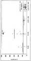
图1显示针对相对光单位(RLU)绘制层粘连蛋白γ2单体浓度(pg/mL)的图。具体地,各图描绘了实施例2中描述的免疫测定的代表性校准曲线。对于左图,层粘连蛋白γ2单体浓度为0 pg/mL至20,000 pg/mL。对于右图,层粘连蛋白γ2单体浓度为0 pg/mL至100pg/mL。每个校准曲线都源自三次重复。Figure 1 shows a graph of laminin γ2 monomer concentration (pg/mL) plotted against relative light units (RLU). Specifically, each figure depicts a representative calibration curve for the immunoassay described in Example 2. For the left panel, laminin γ2 monomer concentrations ranged from 0 pg/mL to 20,000 pg/mL. For the right panel, laminin γ2 monomer concentrations ranged from 0 pg/mL to 100 pg/mL. Each calibration curve was derived from three replicates.
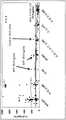
图2A显示针对层粘连蛋白γ2单体的血清浓度(pg/mL)绘制受试者组的图。对于每个受试者组,括号内的数字代表组中的受试者的数目,且实线代表该组的层粘连蛋白γ2单体的平均血清浓度(pg/mL)。沿x-轴长度延伸的实线代表健康供体受试者组的平均值加2倍标准偏差(平均值±2SD),其为81.3 pg/mL。Figure 2A shows a graph plotting groups of subjects against serum concentrations (pg/mL) of laminin γ2 monomer. For each subject group, the numbers in parentheses represent the number of subjects in the group, and the solid line represents the mean serum concentration (pg/mL) of laminin γ2 monomer for that group. The solid line extending along the length of the x-axis represents the mean plus 2 standard deviations (mean ± 2SD) for the group of healthy donor subjects, which was 81.3 pg/mL.
图2B显示针对层粘连蛋白γ2单体的血清浓度(pg/mL)绘制受试者组的图。对于每个受试者组,括号内的数字代表组中的受试者的数目,且实线代表该组的层粘连蛋白γ2单体的平均血清浓度(pg/mL)。沿x-轴长度延伸的实线代表健康供体受试者组的平均值加2倍标准偏差(平均值±2SD),其为79.5 pg/mL。箭头标示了指定AFP和CA19-9浓度的受试者。Figure 2B shows a graph plotting groups of subjects against serum concentrations (pg/mL) of laminin γ2 monomer. For each subject group, the numbers in parentheses represent the number of subjects in the group, and the solid line represents the mean serum concentration (pg/mL) of laminin γ2 monomer for that group. The solid line extending along the length of the x-axis represents the mean plus 2 standard deviations (mean ± 2SD) for the group of healthy donor subjects, which was 79.5 pg/mL. Arrows indicate subjects with indicated AFP and CA19-9 concentrations.
图2C显示针对层粘连蛋白γ2单体的血清浓度(pg/mL)绘制受试者组的图。对于每个受试者组,括号内的数字代表组中的受试者的数目,且实线代表该组的层粘连蛋白γ2单体的平均血清浓度(pg/mL)。沿x-轴长度延伸的实线代表源自ROC分析的截止值,其为75.9pg/mL。Figure 2C shows a graph plotting groups of subjects against serum concentrations (pg/mL) of laminin [gamma]2 monomer. For each subject group, the numbers in parentheses represent the number of subjects in the group, and the solid line represents the mean serum concentration (pg/mL) of laminin γ2 monomer for that group. The solid line extending along the length of the x-axis represents the cutoff value derived from the ROC analysis, which was 75.9 pg/mL.
图2D显示针对层粘连蛋白γ2单体的血清浓度(pg/mL)绘制受试者组的图。对于每个受试者组,括号内的数字代表组中的受试者的数目,且实线代表该组的层粘连蛋白γ2单体的平均血清浓度(pg/mL)。沿x-轴长度延伸的实线代表源自ROC分析的截止值,其为75.9pg/mL。箭头标示了指定AFP和CA19-9浓度的受试者。Figure 2D shows a graph plotting groups of subjects against serum concentrations (pg/mL) of laminin [gamma]2 monomer. For each subject group, the numbers in parentheses represent the number of subjects in the group, and the solid line represents the mean serum concentration (pg/mL) of laminin γ2 monomer for that group. The solid line extending along the length of the x-axis represents the cutoff value derived from the ROC analysis, which was 75.9 pg/mL. Arrows indicate subjects with indicated AFP and CA19-9 concentrations.
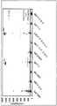
图3A显示针对层粘连蛋白γ2单体的血清浓度(pg/mL)绘制受试者组的图。名称“I”、“II”、“III”和“IV”分别代表具有I期HCC、II期HCC、III期HCC和IV期HCC的受试者组。“LC”代表肝硬化,且“CH”代表肝炎。对于每个受试者组,括号内的数字代表组中的受试者的数目,且实线代表该组的层粘连蛋白γ2单体的平均血清浓度(pg/mL)。沿x-轴长度延伸的实线代表健康供体受试者组的平均值加2倍标准偏差(平均值±2SD),其为79.5 pg/mL。箭头标示了指定AFP浓度的受试者。Figure 3A shows a graph plotting groups of subjects against serum concentrations (pg/mL) of laminin γ2 monomer. The designations "I", "II", "III" and "IV" represent groups of subjects with stage I HCC, stage II HCC, stage III HCC and stage IV HCC, respectively. "LC" stands for cirrhosis, and "CH" stands for hepatitis. For each subject group, the numbers in parentheses represent the number of subjects in the group, and the solid line represents the mean serum concentration (pg/mL) of laminin γ2 monomer for that group. The solid line extending along the length of the x-axis represents the mean plus 2 standard deviations (mean ± 2SD) for the group of healthy donor subjects, which was 79.5 pg/mL. Arrows indicate subjects with indicated AFP concentrations.
图3B显示针对层粘连蛋白γ2单体的血清浓度(pg/mL)绘制受试者组的图。名称“I”、“II”、“III”和“IV”分别代表具有I期HCC、II期HCC、III期HCC和IV期HCC的受试者组。“LC”代表肝硬化,且“CH”代表肝炎。对于每个受试者组,括号内的数字代表组中的受试者的数目,且实线代表该组的层粘连蛋白γ2单体的平均血清浓度(pg/mL)。沿x-轴长度延伸的实线代表源自ROC分析的截止值,其为75.9 pg/mL。箭头标示了指定AFP浓度的受试者。Figure 3B shows a graph of groups of subjects plotted against serum concentrations (pg/mL) of laminin γ2 monomer. The designations "I", "II", "III" and "IV" represent groups of subjects with stage I HCC, stage II HCC, stage III HCC and stage IV HCC, respectively. "LC" stands for cirrhosis, and "CH" stands for hepatitis. For each subject group, the numbers in parentheses represent the number of subjects in the group, and the solid line represents the mean serum concentration (pg/mL) of laminin γ2 monomer for that group. The solid line extending along the length of the x-axis represents the cutoff value derived from the ROC analysis, which was 75.9 pg/mL. Arrows indicate subjects with indicated AFP concentrations.
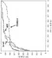
图4A显示标志物层粘连蛋白γ2单体(Ln-γ2)、PIVKA-II和AFP的受试者工作特征(ROC)图,其中将具有HCC的受试者与肝硬化(LC)、肝炎(CH)和健康的供体受试者进行比较。Figure 4A shows receiver operating characteristic (ROC) plots for the markers laminin γ2 monomer (Ln-γ2), PIVKA-II and AFP, in which subjects with HCC were compared with liver cirrhosis (LC), hepatitis ( CH) and healthy donor subjects.
图4B显示标志物层粘连蛋白γ2单体(Ln-γ2)、PIVKA-II和AFP的受试者工作特征(ROC)图,其中将具有HCC的受试者与健康的供体受试者进行比较。Figure 4B shows receiver operating characteristic (ROC) plots for the markers laminin γ2 monomer (Ln-γ2), PIVKA-II and AFP, in which subjects with HCC were compared with healthy donor subjects Compare.
图4C显示标志物层粘连蛋白γ2单体(Ln-γ2)、PIVKA-II和AFP的受试者工作特征(ROC)图,其中将具有HCC的受试者与肝炎(CH)进行比较。Figure 4C shows receiver operating characteristic (ROC) plots for the markers laminin γ2 monomer (Ln-γ2), PIVKA-II and AFP, comparing subjects with HCC to hepatitis (CH).
图5A显示具有HCC且与截止值(79.5 pg/mL,健康供体组的平均值 + 2SD)相比层粘连蛋白γ2单体(Ln-γ2)、AFP和/或PIVKA-II水平升高的受试者的Venn图。每个百分比值代表与每个截止值相比具有所示标志物的升高水平的HCC受试者的百分比。Figure 5A shows patients with HCC and elevated levels of laminin γ2 monomer (Ln-γ2), AFP and/or PIVKA-II compared to cutoff value (79.5 pg/mL, mean + 2SD for healthy donor group) Venn diagram of the subject. Each percentage value represents the percentage of HCC subjects with elevated levels of the indicated markers compared to each cutoff value.
图5B显示具有HCC且与截止值(75.9 pg/mL,源自ROC分析)相比层粘连蛋白γ2单体(Ln-γ2)、AFP和/或PIVKA-II水平升高的受试者的Venn图。每个百分比值代表与每个截止值相比具有所示标志物的升高水平的HCC受试者的百分比。Figure 5B shows Venn of subjects with HCC and elevated levels of laminin γ2 monomer (Ln-γ2), AFP and/or PIVKA-II compared to cutoff value (75.9 pg/mL, derived from ROC analysis) picture. Each percentage value represents the percentage of HCC subjects with elevated levels of the indicated markers compared to each cutoff value.
图5C显示具有HCC且与截止值(116.6 pg/mL,区分HCC与非恶性慢性肝脏疾病的最佳截止值)相比层粘连蛋白γ2单体(Ln-γ2)、AFP和/或PIVKA-II水平升高的受试者的Venn图。每个百分比值代表与每个截止值相比具有所示标志物的升高水平的HCC受试者的百分比。Figure 5C shows laminin γ2 monomer (Ln-γ2), AFP and/or PIVKA-II with HCC and compared to cutoff (116.6 pg/mL, the best cutoff to differentiate HCC from non-malignant chronic liver disease) Venn plot of subjects with elevated levels. Each percentage value represents the percentage of HCC subjects with elevated levels of the indicated markers compared to each cutoff value.
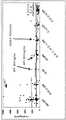
图6A显示针对层粘连蛋白γ2单体的血清浓度(pg/mL)绘制受试者组的图。名称“III”和“IV”分别代表具有III期胰腺癌和IV期胰腺癌的受试者组。对于每个受试者组,括号内的数字代表组中的受试者的数目,且实线代表该组的层粘连蛋白γ2单体的平均血清浓度(pg/mL)。沿x-轴长度延伸的实线代表健康供体受试者组的平均值加2倍标准偏差(平均值±2SD),其为79.5 pg/mL。Figure 6A shows a graph plotting groups of subjects against serum concentrations (pg/mL) of laminin γ2 monomer. The designations "III" and "IV" represent groups of subjects with stage III pancreatic cancer and stage IV pancreatic cancer, respectively. For each subject group, the numbers in parentheses represent the number of subjects in the group, and the solid line represents the mean serum concentration (pg/mL) of laminin γ2 monomer for that group. The solid line extending along the length of the x-axis represents the
图6B显示针对层粘连蛋白γ2单体的血清浓度(pg/mL)绘制受试者组的图。名称“III”和“IV”分别代表具有III期胰腺癌和IV期胰腺癌的受试者组。对于每个受试者组,括号内的数字代表组中的受试者的数目,且实线代表该组的层粘连蛋白γ2单体的平均血清浓度(pg/mL)。沿x-轴长度延伸的实线代表源自ROC分析的截止值,其为75.9 pg/mL。Figure 6B shows a graph plotting groups of subjects against serum concentrations (pg/mL) of laminin [gamma]2 monomer. The designations "III" and "IV" represent groups of subjects with stage III pancreatic cancer and stage IV pancreatic cancer, respectively. For each subject group, the numbers in parentheses represent the number of subjects in the group, and the solid line represents the mean serum concentration (pg/mL) of laminin γ2 monomer for that group. The solid line extending along the length of the x-axis represents the cutoff value derived from the ROC analysis, which was 75.9 pg/mL.
图7显示标志物层粘连蛋白γ2单体(Ln-γ2)、CEA和CA19-9的受试者工作特征(ROC)图,其中将具有胰腺癌的受试者与具有胰腺炎和健康的供体受试者进行比较。Figure 7 shows receiver operating characteristic (ROC) plots for the markers laminin γ2 monomer (Ln-γ2), CEA and CA19-9, in which subjects with pancreatic cancer were compared with those with pancreatitis and healthy donors. subjects for comparison.
图8显示具有胰腺癌且与截止值相比层粘连蛋白γ2单体(Ln-γ2)、CEA和/或CA19-9水平升高的受试者的Venn图。每个百分比值代表与每个截止值相比具有所示标志物的升高水平的胰腺癌受试者的百分比。Figure 8 shows Venn plots for subjects with pancreatic cancer and elevated levels of laminin γ2 monomer (Ln-γ2), CEA and/or CA19-9 compared to cutoff. Each percentage value represents the percentage of pancreatic cancer subjects with elevated levels of the indicated marker compared to each cutoff value.
图9显示来自正常受试者(对照)(n =52)、具有慢性肝脏疾病(CLD)的患者(n =24)和具有HCC的患者(n = 57)的血清样品中测定的血清Ln-γ2浓度的散点图。水平线代表中值浓度。缩写:Ln-γ2,层粘连蛋白γ2。Figure 9 shows serum Ln-determined in serum samples from normal subjects (controls) (n= 52), patients with chronic liver disease (CLD) (n= 24), and patients with HCC (n= 57) Scatter plot of γ2 concentration. The horizontal line represents the median concentration. Abbreviations: Ln-γ2, laminin γ2.
图10显示来自正常受试者(对照)(n =52)、具有慢性肝脏疾病的患者(n =24)和具有HCC的患者(n = 57)的血清样品中测定的血清AFP浓度的散点图。缩写:CLD,慢性肝脏疾病;HCC,肝细胞癌;AFP,甲胎蛋白;PIVKA-II,通过维生素K缺乏诱导的凝血酶原-II;SE,标准误差。Figure 10 shows a scatter plot of serum AFP concentrations determined in serum samples from normal subjects (controls) (n= 52), patients with chronic liver disease (n= 24), and patients with HCC (n= 57) picture. Abbreviations: CLD, chronic liver disease; HCC, hepatocellular carcinoma; AFP, alpha-fetoprotein; PIVKA-II, prothrombin-II induced by vitamin K deficiency; SE, standard error.
图11显示来自正常受试者(对照)(n =52)、具有慢性肝脏疾病的患者(n =24)和具有HCC的患者(n = 57)的血清样品中测定的血清PIVKA-II浓度的散点图。水平线代表中值浓度。缩写:CLD,慢性肝脏疾病;HCC,肝细胞癌;AFP,甲胎蛋白;PIVKA-II,通过维生素K缺乏诱导的凝血酶原-II;SE,标准误差。Figure 11 shows serum PIVKA-II concentrations determined in serum samples from normal subjects (controls) (n= 52), patients with chronic liver disease (n= 24), and patients with HCC (n= 57). Scatter plot. The horizontal line represents the median concentration. Abbreviations: CLD, chronic liver disease; HCC, hepatocellular carcinoma; AFP, alpha-fetoprotein; PIVKA-II, prothrombin-II induced by vitamin K deficiency; SE, standard error.
图12显示根据基于TNM分类的肿瘤分期的Ln-γ2值。水平线代表中值浓度。缩写:CLD,慢性肝脏疾病。Figure 12 shows Ln-γ2 values according to tumor stage based on TNM classification. The horizontal line represents the median concentration. Abbreviation: CLD, chronic liver disease.
图13显示根据基于TNM分类的肿瘤分期的AFP值。水平线代表中值浓度。缩写:CLD,慢性肝脏疾病。Figure 13 shows AFP values according to tumor staging based on TNM classification. The horizontal line represents the median concentration. Abbreviation: CLD, chronic liver disease.
图14显示根据基于TNM分类的肿瘤分期的PIVKA-II值。水平线代表中值浓度。缩写:CLD,慢性肝脏疾病。Figure 14 shows PIVKA-II values according to tumor stage based on TNM classification. The horizontal line represents the median concentration. Abbreviation: CLD, chronic liver disease.
具体实施方案specific implementation
本公开涉及用于检测肝细胞癌和/或胰腺癌的生物标志物。本公开涉及分析或定量层粘连蛋白γ2单体、PIVKA-II、AFP、CEA、CA19-9或其组合的水平,用于诊断、预测受试者中的肝细胞癌或胰腺癌、分类受试者中的肝细胞癌或胰腺癌的风险和监测受试者中的肝细胞癌或胰腺癌的进展。用于检测肝细胞癌的生物标志物可以是层粘连蛋白γ2单体、PIVKA-II、AFP或其组合。用于检测胰腺癌的生物标志物可以是层粘连蛋白γ2单体、CEA、CA19-9或其组合。The present disclosure relates to biomarkers for the detection of hepatocellular carcinoma and/or pancreatic cancer. The present disclosure relates to the analysis or quantification of levels of
本公开公开了这样的方法,其中生物标志物层粘连蛋白γ2单体可用于鉴定或诊断(其包括帮助鉴别或诊断)具有肝细胞癌和/或胰腺癌的患者,鉴定或帮助鉴定患者是否患有肝细胞癌和/或胰腺癌,分类患者的发展肝细胞癌和/或胰腺癌的风险,以及确定或帮助确定诊断、预后或治疗方案。与未患有肝细胞癌或胰腺癌或未处于患有肝细胞癌或胰腺癌的风险中的受试者相比,在患有肝细胞癌或胰腺癌或处于患有肝细胞癌或胰腺癌的风险中的受试者中,层粘连蛋白γ2单体的水平更高。层粘连蛋白γ2单体的水平可以随着肝细胞癌或胰腺癌的晚期阶段而增加。本文所述的方法可以与其他生物标志物(诸如PIVKA-II、AFP、CEA、CA19-9或其组合)结合使用,以鉴定和诊断(其包括辅助鉴定或诊断)患有肝细胞癌和/或胰腺癌的患者。The present disclosure discloses methods wherein the
本节段和本文中整个公开中使用的节段标题仅仅用于组织的目的,并且不旨在进行限制。The section headings used in this section and throughout the disclosure herein are for organizational purposes only and are not intended to be limiting.
1. 定义1. Definition
除非另有定义,本文所用的所有技术和科学术语具有与本领域普通技术人员通常理解的相同含义。在冲突的情况下,将以本文件,包括定义,为准。下面描述了优选的方法和材料,但与本文所述的方法和材料相同或等同的那些可以用于实施或测试本发明。本文中提及的所有出版物、专利申请、专利和其他参考文献都以其整体通过引用并入。本文公开的材料、方法和实例仅仅是说明性的,且并不意欲是限制性的。Unless otherwise defined, all technical and scientific terms used herein have the same meaning as commonly understood by one of ordinary skill in the art. In case of conflict, the present document, including definitions, will control. Preferred methods and materials are described below, but methods and materials similar or equivalent to those described herein can be used in the practice or testing of the present invention. All publications, patent applications, patents, and other references mentioned herein are incorporated by reference in their entirety. The materials, methods, and examples disclosed herein are illustrative only and are not intended to be limiting.
如本文所用的术语“包含”、“包括”、“具有(having)”、“具有(has)”、“可以”、“含有”及其变体意欲为开放式的过渡性短语、术语或词语,其不排除额外行为或结构的可能性。除非上下文另有清楚地规定,否则,单数形式("a", "and"和"the")包括复数所指物。本公开还考虑“包含”本文呈现的实施方案或要素、“由本文呈现的实施方案或要素组成”和“基本上由本文呈现的实施方案或要素组成”的其他实施方案,无论是否明确阐述。The terms "comprising," "including," "having," "has," "may," "containing," and variations thereof as used herein are intended to be open-ended transitional phrases, terms, or words , which does not exclude the possibility of additional behavior or structure. The singular forms ("a", "and" and "the") include plural referents unless the context clearly dictates otherwise. The present disclosure also contemplates other embodiments that "comprise", "consist of", and "consist essentially of" the embodiments or elements presented herein, whether expressly stated or not.
对于本文所述的数字范围,在所述范围之间具有相同精确度的每个中间数值均明确地考虑在内。例如,对于6-9的范围,除了6和9之外,数字7和8也考虑在内,且对于6.0-7.0的范围,数字6.0、6.1、6.2、6.3、6.4、6.5、6.6、6.7、6.8、6.9和7.0均明确地考虑在内。For numerical ranges recited herein, each intervening numerical value between the ranges is expressly contemplated with the same precision. For example, for the range 6-9, the
“曲线下面积”或“AUC”是指ROC曲线下面积。ROC曲线下AUC是精确度的量度。面积1表示完美测试,而面积0.5表示无意义测试。优选的AUC可以是至少约0.700、至少约0.750、至少约0.800、至少约0.850、至少约0.900、至少约0.910、至少约0.920、至少约0.930、至少约0.940、至少约0.950、至少约0.960、至少约0.970、至少约0.980、至少约0.990或至少约0.995。"Area under the curve" or "AUC" refers to the area under the ROC curve. AUC under the ROC curve is a measure of accuracy. An area of 1 represents a perfect test, while an area of 0.5 represents a pointless test. Preferred AUCs can be at least about 0.700, at least about 0.750, at least about 0.800, at least about 0.850, at least about 0.900, at least about 0.910, at least about 0.920, at least about 0.930, at least about 0.940, at least about 0.950, at least about 0.960, at least about About 0.970, at least about 0.980, at least about 0.990, or at least about 0.995.
如本文所用的“癌症”是指机体中异常细胞的不受控制和不受调节的生长。癌细胞也称为恶性细胞。癌症可能侵入机体的附近部分,并且还可以通过淋巴系统或血流传播至机体的更远端部分。癌症可以包括、但不限于肾上腺皮质癌、肛门癌、膀胱癌、脑肿瘤、乳腺癌、类癌肿瘤、胃肠道癌、未知原发性癌、宫颈癌、结肠癌、子宫内膜癌、食道癌、肝外胆管癌、Ewings肿瘤家族(PNET)、颅外生殖细胞肿瘤、眼内黑色素瘤眼癌、胆囊癌、胃癌(胃)、性腺外生殖细胞肿瘤、妊娠滋养细胞肿瘤、头颈癌、下咽癌、胰岛细胞癌、肾癌(肾细胞癌)、喉癌、急性淋巴细胞白血病、白血病、急性骨髓瘤、慢性淋巴细胞白血病、慢性骨髓性白血病、毛细胞白血病、唇和口腔癌、肝癌(包括肝细胞癌)、非小细胞肺癌、小细胞肺癌、AIDS相关淋巴瘤、中枢神经系统(原发性)淋巴瘤、皮肤T细胞淋巴瘤、霍奇金病淋巴瘤、非霍奇金病淋巴瘤、恶性间皮瘤、黑色素瘤、梅克尔细胞癌、伴有隐匿性原发性的转移性鳞状颈癌、多发性骨髓瘤和其他血浆细胞瘤、蕈样真菌病(Mycosis Fungoides)、骨髓增生异常综合征、骨髓增生性病症、鼻咽癌、成神经细胞瘤、口腔癌、口咽癌、骨肉瘤、卵巢上皮癌、卵巢生殖细胞肿瘤、胰腺癌、外分泌癌、胰腺癌、胰岛细胞癌、鼻旁窦和鼻腔癌、甲状旁腺癌、阴茎癌、垂体癌、浆细胞肿瘤、前列腺癌、横纹肌肉瘤、直肠癌、肾细胞癌(肾癌)、过渡细胞肾盂和输尿管癌、唾液腺癌、Sezary综合征、皮肤癌、小肠癌、软组织肉瘤、睾丸癌、恶性胸腺瘤、甲状腺癌、尿道癌、子宫癌、儿童不寻常癌症、阴道癌、外阴癌症和Wilms肿瘤。"Cancer" as used herein refers to the uncontrolled and unregulated growth of abnormal cells in the body. Cancer cells are also called malignant cells. Cancer can invade nearby parts of the body, and it can also spread to more distant parts of the body through the lymphatic system or the bloodstream. Cancers may include, but are not limited to, adrenal cortical cancer, anal cancer, bladder cancer, brain tumor, breast cancer, carcinoid tumors, gastrointestinal cancer, cancer of unknown primary, cervical cancer, colon cancer, endometrial cancer, esophagus Carcinoma, extrahepatic cholangiocarcinoma, Ewings tumor family (PNET), extracranial germ cell tumor, intraocular melanoma eye cancer, gallbladder cancer, gastric cancer (stomach), extragonadal germ cell tumor, gestational trophoblastic tumor, head and neck cancer, lower Pharyngeal cancer, pancreatic islet cell cancer, kidney cancer (renal cell carcinoma), laryngeal cancer, acute lymphocytic leukemia, leukemia, acute myeloma, chronic lymphocytic leukemia, chronic myeloid leukemia, hairy cell leukemia, lip and oral cavity cancer, liver cancer ( including hepatocellular carcinoma), non-small cell lung cancer, small cell lung cancer, AIDS-related lymphoma, central nervous system (primary) lymphoma, cutaneous T-cell lymphoma, Hodgkin's disease lymphoma, non-Hodgkin's disease lymphoma tumor, malignant mesothelioma, melanoma, Merkel cell carcinoma, metastatic squamous neck carcinoma with occult primary, multiple myeloma and other plasmacytomas, Mycosis Fungoides, Myelodysplastic Syndrome, Myeloproliferative Disorders, Nasopharyngeal Cancer, Neuroblastoma, Oral Cancer, Oropharyngeal Cancer, Osteosarcoma, Epithelial Ovarian Cancer, Ovarian Germ Cell Tumor, Pancreatic Cancer, Exocrine Cancer, Pancreatic Cancer, Islet Cells Cancer, paranasal sinus and nasal cavity cancer, parathyroid cancer, penile cancer, pituitary cancer, plasma cell tumor, prostate cancer, rhabdomyosarcoma, rectal cancer, renal cell cancer (kidney cancer), transitional cell renal pelvis and ureter cancer, salivary gland cancer , Sezary syndrome, skin cancer, small bowel cancer, soft tissue sarcoma, testicular cancer, malignant thymoma, thyroid cancer, urethral cancer, uterine cancer, unusual cancers in children, vaginal cancer, vulvar cancer, and Wilms tumor.
“组分”、“多种组分”或“至少一种组分”一般是指捕获抗体、检测或缀合校准物(calibrator)、对照、灵敏度实验对象组(sensitivity panel)、容器、缓冲液、稀释剂、盐、酶、酶的辅因子、检测试剂、预处理试剂/溶液、底物(例如作为溶液)、终止溶液等,根据本文所述方法和其他本领域已知方法,其可以包括于用于测试样品(诸如患者尿、血清或血浆样品)测定的试剂盒中。一些组分可以在溶液中或者被冻干以重构用于测定中。"Component", "components" or "at least one component" generally refers to capture antibodies, detection or conjugation calibrators, controls, sensitivity panels, containers, buffers , diluents, salts, enzymes, cofactors for enzymes, detection reagents, pretreatment reagents/solutions, substrates (eg, as solutions), stop solutions, etc., which may include, according to the methods described herein and other methods known in the art in kits for assays of test samples such as patient urine, serum or plasma samples. Some components can be in solution or lyophilized for reconstitution for use in assays.
如本文所用的“对照受试者”是指健康受试者,即没有肝细胞癌、胰腺癌或除了肝细胞癌或胰腺癌以外的癌症的临床体征或症状的受试者。在临床上评估对照受试者的肝细胞癌、胰腺癌或除了肝细胞癌或胰腺癌以外的癌症的其他未检测体征或症状,所述评估可包括常规体检和/或实验室测试。A "control subject" as used herein refers to a healthy subject, ie a subject without clinical signs or symptoms of hepatocellular carcinoma, pancreatic cancer, or cancer other than hepatocellular carcinoma or pancreatic cancer. Control subjects are clinically assessed for hepatocellular carcinoma, pancreatic cancer, or other undetected signs or symptoms of cancer other than hepatocellular carcinoma or pancreatic cancer, which assessment may include routine physical examination and/or laboratory tests.
如本文所用的“对照组”是指对照受试者或健康受试者的组,即不具有肝细胞癌、胰腺癌或除了肝细胞癌或胰腺癌以外的癌症的临床体征或症状的受试者组。A "control group" as used herein refers to a group of control subjects or healthy subjects, ie, subjects that do not have clinical signs or symptoms of hepatocellular carcinoma, pancreatic cancer, or cancer other than hepatocellular carcinoma or pancreatic cancer group.
如本文所用的“标记”和“可检测的标记”是指结合至抗体或分析物以使得抗体和分析物之间的反应可检测的部分,并且如此标记的抗体或分析物被称为“可检测标记的”。标记可以产生可通过视觉或仪器装置检测的信号。各种标记包括产生信号的物质,诸如色原、荧光化合物、化学发光化合物、放射性化合物等。代表性标记实例包括产生光的部分,例如吖啶鎓(acridinium)化合物,以及产生荧光的部分,例如荧光素。本文描述了其他标记。在这方面,所述部分自身可以不是可检测的,但是与另一个部分反应后,可变得可检测。术语“可检测标记的”的使用旨在涵盖此类标记。"Label" and "detectable label" as used herein refer to a moiety that binds to an antibody or analyte such that the reaction between the antibody and analyte is detectable, and the antibody or analyte so labeled is referred to as "detectable" detection markers". Markers can generate signals that can be detected by visual or instrumental means. Various labels include signal-generating substances such as chromogens, fluorescent compounds, chemiluminescent compounds, radioactive compounds, and the like. Representative examples of labels include light-generating moieties, such as acridinium compounds, and fluorescence-generating moieties, such as fluorescein. Additional tags are described in this article. In this regard, the moiety itself may not be detectable, but may become detectable upon reaction with another moiety. The use of the term "detectably labeled" is intended to encompass such labels.
可以使用本领域已知的任何合适的可检测标记。例如,可检测标记可以是放射性标记(诸如3H、14C、32P、33P、35S、90Y、99Tc、111In、125I、131I、177Lu、166Ho和153Sm)、酶促标记(诸如辣根过氧化物酶、碱性磷酸酶、葡萄糖6-磷酸脱氢酶等)、化学发光标记(诸如吖啶鎓酯(acridinium esters)、硫酯、或磺胺;鲁米诺、异鲁米诺、菲啶鎓酯(phenanthridinium esters)等)、荧光标记(诸如荧光素(例如5-荧光素、6-羧基荧光素、3’6-羧基荧光素、5(6)-羧基荧光素、6-六氯-荧光素、6-四氯荧光素、异硫氰酸荧光素等))、罗丹明、藻胆蛋白、R-藻红蛋白、量子点(例如硫化锌加帽的硒化镉)、温度测量标记、或免疫-聚合酶链式反应标记。标记、标记程序和标记的检测的介绍见于Polak和Van Noorden,Introduction to Immunocytochemistry,第二版, Springer Verlag, N.Y. (1997), 以及Haugland,Handbook of Fluorescent Probes and Research Chemicals(1996), 其是由Molecular Probes, Inc., Eugene, Oregon出版的组合手册和目录。荧光标记可用于FPIA (参见,例如美国专利号5,593,896, 5,573,904, 5,496,925, 5,359,093, 和5,352,803,其在此以其整体通过引用并入)。吖啶鎓化合物可以在均质化学发光测定中用作可检测标记(参见,例如Adamczyk等人, Bioorg. Med. Chem. Lett.16:1324-1328 (2006);Adamczyk等人, Bioorg. Med. Chem. Lett. 4:2313-2317 (2004);Adamczyk等人,Biorg. Med. Chem. Lett. 14:3917-3921 (2004);和Adamczyk等人, Org. Lett. 5:3779-3782 (2003))。Any suitable detectable label known in the art can be used. For example, detectable labels can be radioactive labels (such as 3H, 14C, 32P, 33P, 35S, 90Y, 99Tc, 111In, 125I, 131I, 177Lu, 166Ho, and 153Sm), enzymatic labels (such as horseradish peroxidase, alkaline phosphatase, glucose 6-phosphate dehydrogenase, etc.), chemiluminescent labels (such as acridinium esters, thioesters, or sulfonamides; luminol, isoluminol, phenanthridinium esters), etc.), fluorescent labels (such as fluorescein (e.g. 5-fluorescein, 6-carboxyfluorescein, 3'6-carboxyfluorescein, 5(6)-carboxyfluorescein, 6-hexachloro-fluorescein, 6 - Fluorescein tetrachloro, fluorescein isothiocyanate, etc.), rhodamine, phycobiliprotein, R-phycoerythrin, quantum dots (eg, zinc sulfide capped cadmium selenide), temperature measurement labels, or immuno- Polymerase chain reaction labeling. An introduction to labels, labeling procedures, and detection of labels is found in Polak and Van Noorden,Introduction to Immunocytochemistry, 2nd Edition, Springer Verlag, NY (1997), and Haugland,Handbook of Fluorescent Probes and Research Chemicals (1996), which was published by Molecular Portfolio Brochure and Catalog published by Probes, Inc., Eugene, Oregon. Fluorescent labels can be used for FPIA (see, eg, US Pat. Nos. 5,593,896, 5,573,904, 5,496,925, 5,359,093, and 5,352,803, which are hereby incorporated by reference in their entireties). Acridinium compounds can be used as detectable labels in homogeneous chemiluminescent assays (see, eg, Adamczyk et al, Bioorg. Med. Chem. Lett. 16:1324-1328 (2006); Adamczyk et al, Bioorg. Med. Chem. Lett. 4: 2313-2317 (2004); Adamczyk et al, Biorg. Med. Chem. Lett. 14: 3917-3921 (2004); and Adamczyk et al, Org. Lett. 5: 3779-3782 (2003 )).
在一个方面,吖啶鎓化合物是吖啶鎓-9-甲酰胺。制备吖啶鎓-9-甲酰胺的方法描述于Mattingly, J. Biolumin. Chemilumin. 6:107-114 (1991);Adamczyk等人, J.Org. Chem. 63:5636-5639 (1998);Adamczyk等人, Tetrahedron 55:10899-10914(1999);Adamczyk等人, Org. Lett. 1:779-781 (1999);Adamczyk等人, BioconjugateChem. 11:714-724 (2000);Mattingly等人, InLuminescence Biotechnology:Instruments and Applications;Dyke, K. V. Ed.;CRC Press:Boca Raton, pp. 77–105(2002);Adamczyk等人, Org. Lett. 5:3779-3782 (2003);以及美国专利号5,468,646、5,543,524和5,783,699 (其各自就其关于所述内容的教导以其整体通过引用并入本文)。In one aspect, the acridinium compound is acridinium-9-carboxamide. Methods for the preparation of acridinium-9-carboxamides are described in Mattingly, J. Biolumin. Chemilumin. 6: 107-114 (1991); Adamczyk et al, J. Org. Chem. 63: 5636-5639 (1998); Adamczyk et al, Tetrahedron 55: 10899-10914 (1999); Adamczyk et al, Org. Lett. 1: 779-781 (1999); Adamczyk et al, Bioconjugate Chem. 11: 714-724 (2000); Mattingly et al, InLuminescence Biotechnology:Instruments and Applications; Dyke, KV Ed.; CRC Press: Boca Raton, pp. 77-105 (2002); Adamczyk et al., Org. Lett. 5:3779-3782 (2003); and U.S. Patent No. 5,468,646 , 5,543,524 and 5,783,699 (each of which is hereby incorporated by reference in its entirety for its teachings on the subject matter).
吖啶鎓(acridinium)化合物的另一个实例是吖啶鎓-9-甲酸芳基酯。式II的吖啶鎓-9-甲酸芳基酯的一个实例是10-甲基-9-(苯氧基羰基)吖啶鎓氟磺酸酯(可获自CaymanChemical, Ann Arbor, MI)。制备吖啶鎓-9-甲酸芳基酯的方法描述于McCapra等人,Photochem. Photobiol.4:1111-21 (1965);Razavi等人, Luminescence 15:245-249(2000);Razavi等人, Luminescence 15:239-244 (2000);以及美国专利号5,241,070 (其各自就其关于所述内容的教导以其整体通过引用并入本文)。此类吖啶鎓-9-甲酸芳基酯在信号的强度和/或信号的快速性方面是过氧化氢的有效化学发光指示剂,过氧化氢在由至少一种氧化酶对分析物的氧化中产生。吖啶鎓-9-甲酸芳基酯的化学发光发射的过程迅速完成,即,在小于1秒内,而吖啶鎓-9-甲酰胺的化学发光发射延长超过2秒。然而,吖啶鎓-9-甲酸芳基酯在蛋白存在的情况下失去了其化学发光特性。因此,其使用需要信号生成和检测过程中不存在蛋白。用于分离或去除样品中的蛋白的方法对于本领域技术人员是众所周知的,并且包括,但不限于,超滤、提取、沉淀、透析、层析和/或消化(参见,例如,Wells,High Throughput Bioanalytical Sample Preparation. Methods and AutomationStrategies, Elsevier (2003))。从测试样品去除或分离的蛋白的量可以是约40%、约45%、约50%、约55%、约60%、约65%、约70%、约75%、约80%、约85%、约90%、或约95%。关于吖啶鎓-9-甲酸芳基酯及其用途的进一步细节描述于2007年4月9日提交的美国专利申请号11/697,835。吖啶鎓-9-甲酸芳基酯可以溶解在任何合适的溶剂,诸如脱气无水N,N-二甲基甲酰胺(DMF)或含水胆酸钠。Another example of an acridinium compound is aryl acridinium-9-carboxylate. An example of an acridinium-9-carboxylic acid aryl ester of formula II is 10-methyl-9-(phenoxycarbonyl)acridinium fluorosulfonate (available from Cayman Chemical, Ann Arbor, MI). Methods for the preparation of acridinium-9-carboxylate aryl esters are described in McCapra et al., Photochem. Photobiol. 4:1111-21 (1965); Razavi et al., Luminescence 15:245-249 (2000); Razavi et al., Luminescence 15:239-244 (2000); and US Patent No. 5,241,070 (each of which is incorporated herein by reference in its entirety for its teachings regarding the subject matter). Such acridinium-9-carboxylate aryl esters are effective chemiluminescent indicators of hydrogen peroxide in terms of the intensity of the signal and/or the rapidity of the signal, which is important in the oxidation of the analyte by at least one oxidase. produced in. The process of chemiluminescent emission of acridinium-9-carboxylate aryl esters was completed quickly, ie, in less than 1 second, while the chemiluminescent emission of acridinium-9-carboxamide was prolonged for more than 2 seconds. However, acridinium-9-carboxylate aryl esters lost their chemiluminescent properties in the presence of proteins. Therefore, its use requires the absence of proteins during signal generation and detection. Methods for isolating or removing proteins from a sample are well known to those skilled in the art and include, but are not limited to, ultrafiltration, extraction, precipitation, dialysis, chromatography and/or digestion (see, e.g., Wells,High Throughput Bioanalytical Sample Preparation. Methods and AutomationStrategies , Elsevier (2003)). The amount of protein removed or isolated from the test sample can be about 40%, about 45%, about 50%, about 55%, about 60%, about 65%, about 70%, about 75%, about 80%, about 85% %, about 90%, or about 95%. Further details regarding acridinium-9-carboxylic acid aryl esters and their uses are described in US Patent Application No. 11/697,835, filed April 9, 2007. The aryl acridinium-9-carboxylate can be dissolved in any suitable solvent, such as degassed anhydrousN,N -dimethylformamide (DMF) or aqueous sodium cholate.
如本文可互换使用的“用于将肝细胞癌分期的TNM系统”或“用于将HCC分期的TNM系统”是指用于将肝细胞癌(HCC)分期的系统。“T”描述以厘米(cm)测量的原发性肿瘤的数量和大小,以及癌症是否已经生长至附近的血管或器官中。“N”描述扩散至附近(区域)淋巴结的程度。“M”指示癌症是否已转移(传播)至身体的远端部分。在“T”、“N”和“M”之后可以出现数字或字母,以提供这些因素中的每一种的更多详细信息。具体地,数字0-4指示渐增的严重程度。字母“X”指示“无法评估”,因为信息不可用。"TNM system for staging hepatocellular carcinoma" or "TNM system for staging HCC" as used interchangeably herein refers to a system for staging hepatocellular carcinoma (HCC). "T" describes the number and size of the primary tumor, measured in centimeters (cm), and whether the cancer has grown into nearby blood vessels or organs. "N" describes the extent of spread to nearby (regional) lymph nodes. "M" indicates whether the cancer has metastasized (spread) to distant parts of the body. A number or letter may appear after the "T", "N" and "M" to provide more details on each of these factors. Specifically, numbers 0-4 indicate increasing severity. The letter "X" indicates "unable to evaluate" because the information is not available.
“T”组可以是“TX”、“T0”、“T1”、“T2”、“T3a”、“T3b”和“T4”。“TX”指示无法评估原发性肿瘤。“T0”指示没有原发性肿瘤的证据。“T1”指示没有生长进入血管的单个肿瘤(任何大小)。“T2”指示已经生长进入血管的单个肿瘤(任何大小),或多于一个肿瘤,但没有横向大于5cm (约2英寸)的肿瘤。“T3a”指示多于一个肿瘤,其中至少一个肿瘤横向大于5 cm。“T3b”指示已经生长进入肝大静脉(门静脉或肝静脉)的主要分支的至少一个肿瘤(任何大小)。“T4”指示已经生长进入附近器官(除了胆囊以外)的肿瘤(任何大小),或肿瘤生长至覆盖肝脏的薄层组织(称为内脏腹膜)中。The "T" group may be "TX", "T0", "T1", "T2", "T3a", "T3b" and "T4". "TX" indicates that the primary tumor could not be assessed. "T0" indicates no evidence of primary tumor. "T1" indicates a single tumor (of any size) that did not grow into blood vessels. "T2" indicates a single tumor (of any size) that has grown into a blood vessel, or more than one tumor, but no tumor larger than 5 cm (about 2 inches) laterally. "T3a" indicates more than one tumor, at least one of which is larger than 5 cm laterally. "T3b" indicates at least one tumor (of any size) that has grown into the major branch of the great hepatic vein (portal or hepatic vein). "T4" indicates a tumor (of any size) that has grown into nearby organs (other than the gallbladder), or a tumor that has grown into the thin layer of tissue that covers the liver (called the visceral peritoneum).
“N”组可以是“NX”、“N0”和“N1”。“NX”指示无法评估区域(附近)淋巴结。“N0”指示癌症尚未扩散至区域淋巴结。“N1”指示癌症已扩散至区域淋巴结。The "N" group can be "NX", "N0" and "N1". "NX" indicates that regional (nearby) lymph nodes cannot be assessed. "N0" indicates that the cancer has not spread to regional lymph nodes. "N1" indicates that the cancer has spread to regional lymph nodes.
“M”组可以是“M0”和“M1”。“M0”指示癌症尚未扩散至远端淋巴结或其他器官。“M1”指示癌症已扩散至远端淋巴结或器官。肝癌,包括肝细胞癌,可以扩散至腹部(腹膜)的内层、肺和骨骼。The "M" group can be "M0" and "M1". "M0" indicates that the cancer has not spread to distant lymph nodes or other organs. "M1" indicates that the cancer has spread to distant lymph nodes or organs. Liver cancer, including hepatocellular carcinoma, can spread to the lining of the abdomen (peritoneum), lungs, and bones.
如本文可互换使用的“I期肝细胞癌”和“I期HCC”是指T1、N0和M0的TNM分类。存在尚未生长进入任何血管的单个肿瘤(任何大小)。癌症尚未扩散至附近淋巴结或远端部位。"Stage I hepatocellular carcinoma" and "Stage I HCC" as used interchangeably herein refer to the TNM classifications of T1, NO and M0. There is a single tumor (of any size) that has not grown into any blood vessels. The cancer has not spread to nearby lymph nodes or distant sites.
如本文可互换使用的“II期肝细胞癌”和“II期HCC”是指T2、N0和M0的TNM分类。存在已生长进入血管的单个肿瘤(任何大小),或存在几个肿瘤,并且都是横向5 cm (2英寸)或更小。癌症尚未扩散至附近淋巴结或远端部位。"Stage II hepatocellular carcinoma" and "Stage II HCC" as used interchangeably herein refer to the TNM classifications of T2, NO and M0. There is a single tumor (of any size) that has grown into the blood vessels, or there are several tumors that are all 5 cm (2 inches) laterally or smaller. The cancer has not spread to nearby lymph nodes or distant sites.
如本文可互换使用的“IIIA期肝细胞癌”和“IIIA期HCC”是指T3a、N0和M0的TNM分类。存在多于一个肿瘤,并且至少一个肿瘤横向大于5 cm(2英寸)。癌症尚未扩散至附近淋巴结或远端部位。As used interchangeably herein, "stage IIIA hepatocellular carcinoma" and "stage IIIA HCC" refer to the TNM classifications of T3a, NO and M0. More than one tumor was present, and at least one tumor was larger than 5 cm (2 inches) laterally. The cancer has not spread to nearby lymph nodes or distant sites.
如本文可互换使用的“IIIB期肝细胞癌”和“IIIA期HCC”是指T3b、N0和M0的TNM分类。至少一个肿瘤正生长进入肝脏的主要静脉(门静脉或肝静脉)的分支。癌症尚未扩散至附近淋巴结或远端部位。"Stage IIIB hepatocellular carcinoma" and "Stage IIIA HCC" as used interchangeably herein refer to the TNM classifications of T3b, NO and M0. At least one tumor is growing into a branch of the liver's main vein (the portal or hepatic vein). The cancer has not spread to nearby lymph nodes or distant sites.
如本文可互换使用的“IIIC期肝细胞癌”和“IIIC期HCC”是指T4、N0和M0的TNM分类。肿瘤正生长进入附近器官(除了胆囊以外)或肿瘤已经生长进入肝脏的外层(outercovering)中。癌症尚未扩散至附近淋巴结或远端部位。As used interchangeably herein, "stage IIIC hepatocellular carcinoma" and "stage IIIC HCC" refer to the TNM classifications of T4, NO and M0. The tumor is growing into nearby organs (other than the gallbladder) or the tumor has grown into the outer covering of the liver. The cancer has not spread to nearby lymph nodes or distant sites.
如本文可互换使用的“IVA期肝细胞癌”和“IVA期HCC”是指任何T、N1和M0的TNM分类。肝脏中的肿瘤可以是任何大小或数量,并且肿瘤可能已经生长进入血管或附近器官中。癌症已扩散至附近淋巴结。癌症尚未扩散至远端部位。"Stage IVA hepatocellular carcinoma" and "Stage IVA HCC" as used interchangeably herein refer to any of the TNM classifications of T, N1 and M0. Tumors in the liver can be of any size or number, and tumors may have grown into blood vessels or nearby organs. Cancer has spread to nearby lymph nodes. The cancer has not spread to distant sites.
如本文可互换使用的“IVB期肝细胞癌”和“IVB期HCC”是指任何T、任何N和M1的TNM分类。癌症已扩散至身体的其他部位。肿瘤可以是任何大小或数量,并且附近的淋巴结可能被累及或可没有被累及。"Stage IVB hepatocellular carcinoma" and "Stage IVB HCC" as used interchangeably herein refer to any T, any N and M1 TNM classification. Cancer has spread to other parts of the body. Tumors can be of any size or number, and nearby lymph nodes may or may not be involved.
如本文所用的“肝癌”是指起源于肝脏的癌症。肝癌可以包括肝细胞癌(HCC)和纤维板层癌。在大多数情况下,肝癌的病因通常是肝脏的疤痕形成(即肝硬化)。"Liver cancer" as used herein refers to cancer originating in the liver. Liver cancer can include hepatocellular carcinoma (HCC) and fibrolamellar carcinoma. In most cases, the cause of liver cancer is usually scarring of the liver (ie, cirrhosis).
如本文所用的“肝硬化”是指特征在于用纤维化、瘢痕组织和再生结节(即由于受损组织被再生的过程而发生的结块,导致肝功能的丧失)代替肝组织的慢性肝脏疾病的结果。肝脏的功能单位的结构组织被破坏,使得通过肝脏的血流和肝脏的功能被破坏。肝硬化最常由酗酒、乙型肝炎和丙型肝炎以及脂肪肝引起,但具有其他许多可能的原因。一些病例是特发性的(即病因不明)。一旦已发生肝硬化,就可以发生肝脏疾病的严重并发症,包括门静脉高压症、肝功能衰竭和肝癌。一旦发生肝硬化,肝癌的风险就大大增加,并且肝硬化应该被认为是恶性前病况。肝硬化可以由酒精滥用、肝脏的自身免疫性疾病、乙型肝炎或丙型肝炎病毒感染、长期(慢性)的肝脏炎症和体内铁超载(血色素沉着病)引起。具有乙型肝炎或丙型肝炎的患者处于肝癌的风险中,即使他们没有发生肝硬化。"Cirrhosis" as used herein refers to chronic liver disease characterized by the replacement of liver tissue with fibrosis, scar tissue, and regenerative nodules (ie, clumping due to the process by which damaged tissue is regenerated, resulting in loss of liver function) disease outcome. The structural organization of the functional units of the liver is disrupted so that the blood flow through the liver and the function of the liver are disrupted. Cirrhosis is most commonly caused by alcohol abuse, hepatitis B and C, and fatty liver, but there are many other possible causes. Some cases are idiopathic (ie, the etiology is unknown). Once cirrhosis has developed, serious complications of liver disease can develop, including portal hypertension, liver failure, and liver cancer. Once cirrhosis develops, the risk of liver cancer greatly increases, and cirrhosis should be considered a premalignant condition. Cirrhosis can be caused by alcohol abuse, autoimmune disease of the liver, hepatitis B or C virus infection, long-term (chronic) inflammation of the liver, and iron overload in the body (hemochromatosis). Patients with hepatitis B or C are at risk for liver cancer, even if they do not develop cirrhosis.
如本文所用的“肝脏疾病”是指对肝脏的损伤或肝脏的疾病。肝功能障碍的症状包括身体体征和各种与消化问题、血糖问题、免疫病症、脂肪的吸收异常和代谢问题相关的症状。肝脏疾病包括肝纤维化、肝硬化和肝癌。所有慢性肝脏疾病都可以导致肝纤维化。慢性肝脏疾病可以由慢性病毒性乙型肝炎和酒精性肝脏疾病引起。"Liver disease" as used herein refers to damage to the liver or disease of the liver. Symptoms of liver dysfunction include physical signs and various symptoms related to digestive problems, blood sugar problems, immune disorders, abnormal fat absorption, and metabolic problems. Liver diseases include liver fibrosis, cirrhosis, and liver cancer. All chronic liver diseases can lead to liver fibrosis. Chronic liver disease can be caused by chronic viral hepatitis B and alcoholic liver disease.
如本文所用的“肝纤维化”是指细胞外基质蛋白(包括在大多数类型的慢性肝脏疾病中发生的胶原蛋白)的过度累积。肝纤维化是代表肝脏对损伤或疾病反应的瘢痕形成过程。肝纤维化可以由乙型肝炎和丙型肝炎、寄生虫的感染、过度使用酒精和暴露于有毒化学物质(包括药物)和胆管堵塞引起。晚期肝纤维化导致肝硬化、肝功能衰竭和门静脉高压,并且通常需要肝移植。"Liver fibrosis" as used herein refers to the excessive accumulation of extracellular matrix proteins, including collagen, which occurs in most types of chronic liver disease. Liver fibrosis is a scarring process that represents the liver's response to injury or disease. Liver fibrosis can be caused by hepatitis B and C, infection by parasites, excessive alcohol use and exposure to toxic chemicals (including drugs), and blocked bile ducts. Advanced liver fibrosis results in cirrhosis, liver failure, and portal hypertension, and often requires liver transplantation.
如本文所用的“瘤形成”是指组织的异常生长。瘤形成通常被称为肿瘤。但异常生长通常但不总是形成块。"Neoplasia" as used herein refers to abnormal growth of tissue. Neoplasia is often referred to as a tumor. But abnormal growths usually, but not always, form lumps.
如本文所用的术语“正常对照”或“健康对照”是指取自不具有肝细胞癌、胰腺癌或任何癌症或不处于发展癌症的风险中的受试者的样品或样本,或实际受试者。The term "normal control" or "healthy control" as used herein refers to a sample or sample taken from a subject who does not have hepatocellular carcinoma, pancreatic cancer or any cancer or is not at risk of developing cancer, or an actual subject By.
如本文所用的“正常”、“健康”和“对照”可互换地指取自不具有肝细胞癌、胰腺癌或任何癌症或不处于发展癌症的风险中的一个或多个受试者的样品。"Normal," "healthy," and "control" as used herein interchangeably refer to a blood sample taken from one or more subjects who do not have hepatocellular carcinoma, pancreatic cancer, or any cancer or who are not at risk of developing cancer. sample.
如本文所用的“良性胰腺疾病”和“胰腺疾病”可互换地指不是癌症或已变为癌症的胰腺疾病。良性胰腺疾病包括胰腺炎,各种类型的囊肿和肿瘤,胰腺上皮内瘤形成(PanIN)和导管内乳头状粘液性瘤形成(IPMN)病变和粘液性囊性瘤形成(MCN)。As used herein, "benign pancreatic disease" and "pancreatic disease" refer interchangeably to a pancreatic disease that is not cancer or has become cancer. Benign pancreatic diseases include pancreatitis, various types of cysts and tumors, pancreatic intraepithelial neoplasia (PanIN) and intraductal papillary myxomatous neoplasia (IPMN) lesions and myxocystic neoplasia (MCN).
如本文所用的“早期胰腺癌”是指限于胰腺、胰腺外或附近淋巴结、但尚未扩展至附近主要血管或神经或远端器官的胰腺癌。早期胰腺癌包括0期、I期和II期胰腺癌。参见Yachida等人(2010) Nature 467:1114-1119;还参见National Comprehensive CancerNetwork (NCCN) Guidelines Version 2.2012 Pancreatic Adenocarcinoma。As used herein, "early stage pancreatic cancer" refers to pancreatic cancer that is confined to the pancreas, extra-pancreatic, or nearby lymph nodes, but has not spread to nearby major blood vessels or nerves or distant organs. Early-stage pancreatic cancer includes
如本文所用的“晚期胰腺癌”是指已扩展至附近主要血管、神经或远端器官中的胰腺癌。晚期胰腺癌包括III期或IV期胰腺癌。As used herein, "advanced pancreatic cancer" refers to pancreatic cancer that has spread into nearby major blood vessels, nerves, or distant organs. Advanced pancreatic cancer includes stage III or IV pancreatic cancer.
如本文所用的“0期胰腺癌”是指限于胰腺中的单层细胞的胰腺癌。在成像测试中或肉眼看不到胰腺癌。肿瘤局限于胰腺导管细胞的顶层,并且没有侵入更深的组织或扩散至胰腺外。0期肿瘤有时被称为原位胰腺癌或胰腺上皮内瘤形成III (PanIn III)。"
如本文所用的“I期胰腺癌”是指局限于或限于胰腺且未扩散至邻近淋巴结的癌症。“IA期”是指局限于胰腺,且大小小于2 cm的肿瘤。“IB期”是指局限于胰腺,且大小大于2cm的肿瘤。"Stage I pancreatic cancer" as used herein refers to cancer that is confined or confined to the pancreas and has not spread to adjacent lymph nodes. "Stage IA" refers to tumors that are confined to the pancreas and less than 2 cm in size. "Stage IB" refers to tumors confined to the pancreas and larger than 2 cm in size.
如本文所用的“II期胰腺癌”是指已长出胰腺外,或已扩散至附近淋巴结的局部扩散癌症。“IIA期”是指长出胰腺外、但没有生长至大血管、附近淋巴结或远端部位的肿瘤。“IIB期”是指局限于胰腺或长出胰腺外、但没有扩散至附近大血管或主要神经的肿瘤。IIB期可以扩散至附近淋巴结,但尚未扩散至远端部位。"Stage II pancreatic cancer" as used herein refers to locally spread cancer that has grown outside the pancreas, or has spread to nearby lymph nodes. "Stage IIA" refers to tumors that have grown outside the pancreas, but have not grown into large blood vessels, nearby lymph nodes, or distant sites. "Stage IIB" refers to a tumor that is confined to the pancreas or that has grown outside the pancreas, but has not spread to nearby large blood vessels or major nerves. Stage IIB can spread to nearby lymph nodes but has not spread to distant sites.
如本文所用的“III期胰腺癌”是指已扩散至附近的主要血管或神经、但尚未转移的更广泛扩散的癌症。肿瘤长出胰腺外,至附近的大血管或主要神经中,并且可能扩散或未扩散至附近淋巴结。其尚未扩散至远端部位。"Stage III pancreatic cancer" as used herein refers to more widely spread cancer that has spread to nearby major blood vessels or nerves, but has not yet metastasized. The tumor has grown outside the pancreas into a nearby large blood vessel or major nerve, and may or may not have spread to nearby lymph nodes. It has not spread to distal sites.
如本文所用的“IV期胰腺癌”是指已扩散至远处器官或部位的证实的扩散癌症。IVA期胰腺癌局限于局部,但累及邻近器官或血管,由此妨碍手术切除。IVA期胰腺癌也被称为局部化或局部晚期的。IVB期胰腺癌已扩散至远端器官,最常见为肝脏。IVB期胰腺癌也称为转移性的。"Stage IV pancreatic cancer" as used herein refers to confirmed diffuse cancer that has spread to distant organs or sites. Stage IVA pancreatic cancer is localized but involves adjacent organs or blood vessels, thereby preventing surgical resection. Stage IVA pancreatic cancer is also known as localized or locally advanced. Stage IVB pancreatic cancer has spread to distant organs, most commonly the liver. Stage IVB pancreatic cancer is also called metastatic.
如本文所用的“预定截止(cutoff)”和“预定水平”是指测定截止值,其用于通过将测定结果与预定截止/水平比较,来评估诊断、预后或治疗效力结果,其中预定截止/水平已经与各种临床参数(例如疾病的存在、疾病阶段、疾病严重度、疾病进展、非进展或改进等)联系或相关。本公开提供了示例性预定水平。然而,众所周知,截止值可能依赖于免疫测定的性质(例如采用的抗体、反应条件、样品纯度等)而变化。进一步完全在本领域技术人员的一般能力内的是,基于公开提供的描述,将本文公开的内容针对其他免疫测定进行调整,以获得用于那些其他免疫测定的免疫测定特异性截止值。尽管测定之间预定截止/水平的精确值可不同,但本文所述的相关性应当是普遍适用的。As used herein, "predetermined cutoff" and "predetermined level" refer to an assay cutoff value for evaluating a diagnostic, prognostic or therapeutic efficacy outcome by comparing the assay result to a predetermined cutoff/level, wherein the predetermined cutoff/level Levels have been linked or correlated with various clinical parameters (eg, presence of disease, disease stage, disease severity, disease progression, non-progression or improvement, etc.). The present disclosure provides exemplary predetermined levels. However, it is well known that cutoff values may vary depending on the nature of the immunoassay (eg antibodies employed, reaction conditions, sample purity, etc.). It is further well within the general ability of those skilled in the art to adapt the disclosures herein for other immunoassays to obtain immunoassay specific cutoff values for those other immunoassays based on the description provided in the publication. Although the precise value of the predetermined cutoff/level may vary between assays, the correlations described herein should be generally applicable.
如在本文所述的诊断测定中所用的,“预处理试剂”,例如裂解、沉淀和/或增溶试剂,是将存在于测试样品中的任何细胞裂解和/或任何分析物增溶的试剂。如本文进一步所述,不是所有样品都需要预处理。此外,增溶分析物(即层粘连蛋白γ2单体、PIKVA-II、AFP、CEA和/或CA19-9,层粘连蛋白γ2单体、PIVKA-II、AFP、CEA和/或CA19-9的片段,层粘连蛋白γ2单体、PIVKA-II、AFP、CEA和/或CA19-9的变体,或其任何组合)引起该分析物从存在于样品中的任何内源结合蛋白释放。预处理试剂可以是均质的(不要求分离步骤)或异质的(要求分离步骤)。使用异质性预处理试剂时,在进行到测定的下一个步骤前,将任何沉淀的分析物结合蛋白从测试样品去除。预处理试剂任选地可以包含:(a) 一种或多种溶剂和盐,(b) 一种或多种溶剂、盐和去污剂,(c) 去污剂,(d) 去污剂和盐,或(e) 适合于细胞裂解和/或分析物的增溶的任何试剂或试剂组合。As used in the diagnostic assays described herein, a "pretreatment reagent," such as a lysis, precipitation, and/or solubilization reagent, is a reagent that lyses and/or solubilizes any analyte present in a test sample . As described further herein, not all samples require pretreatment. In addition, solubilizing analytes (ie, laminin γ2 monomers, PIKVA-II, AFP, CEA and/or CA19-9, laminin γ2 monomers, PIVKA-II, AFP, CEA and/or CA19-9) Fragments, variants of
在本文描述免疫测定和试剂盒上下文中,“质量控制试剂”包括但不限于校准物、对照、和灵敏度实验对象组。为了确立校准(标准)曲线以内推(interpolation)分析物(诸如抗体或分析物)的浓度,通常使用“校准物”或“标准品”(例如,一种或多种,诸如多种)。或者,可以使用接近预定阳性/阴性截止的单个校准物。可以共同使用多个校准物(即,多于一个校准物或不同量的校准物),以构成“灵敏度实验对象组”。In the context of immunoassays and kits described herein, "quality control reagents" include, but are not limited to, calibrators, controls, and sensitivity panels. To establish a calibration (standard) curve to interpolate the concentration of an analyte (such as an antibody or analyte), a "calibrator" or "standard" (eg, one or more, such as multiple) is typically used. Alternatively, a single calibrator close to a predetermined positive/negative cutoff can be used. Multiple calibrators (ie, more than one calibrator or different amounts of calibrators) can be used together to constitute a "sensitivity panel."
如本文所用的受试者(例如,患者)的“风险评估”、“风险分类”、“风险鉴定”或“风险分层”,是指对包括生物标志物在内的因素的评估,以预测包括疾病发作或疾病进展的未来事件的发生风险,从而可以在更为知情的基础上做出对受试者的治疗决定。"Risk assessment", "risk classification", "risk identification" or "risk stratification" of a subject (eg, patient) as used herein refers to the assessment of factors, including biomarkers, to predict Risk of future events including disease onset or disease progression so that treatment decisions for the subject can be made on a more informed basis.
如本文所用的“样品”、“生物样品”、“测试样品”、“样本”、“来自受试者的样品”、“获得自受试者的样品”和“患者样品”可以互换使用并且可以是血液、组织、尿、血清、血浆、羊水、脑脊髓液、胎盘细胞或组织、内皮细胞、白细胞或单核细胞的样品。所述样品可以如从患者获得直接使用,或者可以预处理,诸如通过过滤、蒸馏、提取、浓缩、离心、灭活干扰组分、添加试剂等,以便如本文讨论或另外以如本领域已知的一些方式修改样品的特征。As used herein "sample", "biological sample", "test sample", "sample", "sample from subject", "sample obtained from subject" and "patient sample" are used interchangeably and It can be a sample of blood, tissue, urine, serum, plasma, amniotic fluid, cerebrospinal fluid, placental cells or tissue, endothelial cells, leukocytes or monocytes. The sample may be used directly as obtained from the patient, or may be pretreated, such as by filtration, distillation, extraction, concentration, centrifugation, inactivation of interfering components, addition of reagents, etc., as discussed herein or otherwise as known in the art. Some ways to modify the characteristics of the sample.
任何细胞类型、组织或体液可用于获得样品。此类细胞类型、组织和流体可以包括组织诸如活检和尸检样品的切片,出于组织学目的取用的冰冻切片,血液(诸如全血),血浆,血清,痰,粪便,眼泪,粘液,唾液,支气管肺泡灌洗(BAL)流体,毛发,皮肤,红细胞,血小板,间质液,眼晶状体液,脑脊髓液,汗液,鼻液,滑液,月经液,羊水,精液等。细胞类型和组织也可包括淋巴液,腹水液,妇科液,尿液,腹膜液,脑脊髓液,通过阴道清洗收集的流体,或通过阴道冲洗收集的流体。组织或细胞类型可以通过从动物取出细胞样品来提供,但也可以通过使用先前分离的细胞(例如,由另一人在另一时间和/或出于另一目的分离)来完成。也可以使用存档组织,诸如具有治疗或结果历史的那些。蛋白或核苷酸分离和/或纯化可以不是必要的。Any cell type, tissue or body fluid can be used to obtain a sample. Such cell types, tissues and fluids may include sections of tissues such as biopsy and autopsy samples, frozen sections taken for histological purposes, blood (such as whole blood), plasma, serum, sputum, feces, tears, mucus, saliva , Bronchoalveolar lavage (BAL) fluid, hair, skin, red blood cells, platelets, interstitial fluid, eye lens fluid, cerebrospinal fluid, sweat, nasal fluid, synovial fluid, menstrual fluid, amniotic fluid, semen, etc. Cell types and tissues may also include lymph, ascites, gynecological fluid, urine, peritoneal fluid, cerebrospinal fluid, fluid collected by vaginal washing, or fluid collected by vaginal irrigation. A tissue or cell type can be provided by taking a sample of cells from an animal, but can also be accomplished by using previously isolated cells (eg, isolated by another person at another time and/or for another purpose). Archive tissues, such as those with treatment or outcome histories, can also be used. Protein or nucleotide isolation and/or purification may not be necessary.
在本公开内容的实践中,使用本领域众所周知的用于采集、处理和加工尿、血液、血清和血浆以及其他体液的方法。测试样品可以除目的分析物之外包含其他部分,诸如抗体、抗原、半抗原、激素、药物、酶、受体、蛋白、肽、多肽、寡核苷酸或多核苷酸。例如,样品可以是获得自受试者的全血样品。在如本文所述的免疫测定之前,将测试样品特别是全血进行处理(例如,用预处理试剂)可能是必需的或期望的。甚至在预处理不必要的情况下(例如,大多数尿样品,经预处理的存档样品等),样品的预处理是可以仅出于方便的目的进行的选项(例如,作为商业平台上的方案的部分)。样品可以如获得自受试者直接使用或在预处理之后使用以修改样品的特征。预处理可以包括提取、浓缩、灭活干扰组分和/或添加试剂。In the practice of the present disclosure, methods well known in the art for collecting, processing and processing urine, blood, serum and plasma, and other bodily fluids are used. The test sample may contain other moieties in addition to the analyte of interest, such as antibodies, antigens, haptens, hormones, drugs, enzymes, receptors, proteins, peptides, polypeptides, oligonucleotides or polynucleotides. For example, the sample can be a whole blood sample obtained from a subject. It may be necessary or desirable to process test samples, particularly whole blood, prior to immunoassays as described herein (eg, with pretreatment reagents). Even in cases where preprocessing is not necessary (eg, most urine samples, preprocessed archived samples, etc.), preprocessing of samples is an option that can be done for convenience only (eg, as a protocol on a commercial platform) part). The sample can be used directly as obtained from the subject or after pretreatment to modify the characteristics of the sample. Pretreatment may include extraction, concentration, inactivation of interfering components and/or addition of reagents.
如本文所用的术语“灵敏度”是指真阳性的数目除以真阳性的数目加上假阴性的数目,其中灵敏度(“sens”)可以在0 <sens <1的范围内。理想地,本文的方法实施方案具有等于零或接近等于零的假阴性的数目,使得当受试者确实具有肝细胞癌或胰腺癌时没有错误地将他们鉴定为没有肝细胞癌或胰腺癌。相反,通常进行预测算法正确分类阴性的能力的评价,这是对灵敏度的补充测量。The term "sensitivity" as used herein refers to the number of true positives divided by the number of true positives plus the number of false negatives, where sensitivity ("sens") may be in the
如本文所用的术语“特异性”是指真阴性的数目除以真阴性的数目加上假阳性的数目,其中特异性(“spec”)可以在0<spec<1的范围内。理想地,本文所述的方法具有等于零或接近等于零的真阳性的数目,使得当受试者事实上未具有肝细胞癌或胰腺癌时没有错误地将他们鉴定为具有腺瘤。因此,优选具有等于1或100%的灵敏度和特异性的方法。The term "specificity" as used herein refers to the number of true negatives divided by the number of true negatives plus the number of false positives, where specificity ("spec") may be in the
“校准组合物的系列”是指包含已知浓度的层粘连蛋白γ2单体、PIVKA-II、AFP、CEA和/或CA19-9的多种组合物,其中每种组合物与系列中的其他组合物的不同在于层粘连蛋白γ2单体、PIVKA-II、AFP、CEA和/或CA19-9的浓度。A "series of calibration compositions" refers to compositions comprising known concentrations of
如本文可互换使用的术语“受试者”、“患者”或“方法中的主题”可互换地使用,是指任何脊椎动物,包括但不限于,哺乳动物(例如,牛、猪、骆驼、美洲驼、马、山羊、兔、绵羊、仓鼠、豚鼠、猫、狗、大鼠和小鼠,非人的灵长类(例如,猴,诸如食蟹猴或猕猴、黑猩猩等)和人。在一些实施方案中,一个或多个受试者可以是人或非人。在一些实施方案中,受试者可以是处于或怀疑处于发生肝细胞癌和/或胰腺癌或除了肝细胞癌或胰腺癌以外的癌症的风险中或已经具有肝细胞癌和/或胰腺癌或除了肝细胞癌或胰腺癌以外的癌症的人受试者。As used interchangeably herein, the terms "subject", "patient" or "subject in the method" are used interchangeably to refer to any vertebrate including, but not limited to, mammals (eg, bovine, porcine, Camels, llamas, horses, goats, rabbits, sheep, hamsters, guinea pigs, cats, dogs, rats and mice, non-human primates (eg, monkeys such as cynomolgus or macaques, chimpanzees, etc.) and humans In some embodiments, one or more subjects may be human or non-human. In some embodiments, the subject may be at or suspected of developing hepatocellular carcinoma and/or pancreatic cancer or in addition to hepatocellular carcinoma A human subject who is at risk of or has had hepatocellular carcinoma and/or pancreatic cancer or a cancer other than hepatocellular carcinoma or pancreatic cancer.
如本文所用的术语“治疗(treat)”、“治疗(treated)”或“治疗(treating)”是指其中目的在于减缓(减轻)不期望的生理病况、病症或疾病或获得有益或期望的临床结果的疗法。为了本发明的目的,有益或期望的临床结果包括但不限于缓解症状;减轻病况、病症或疾病的程度;稳定(即不恶化)病况、病症或疾病的状态;延迟病况、病症或疾病的发作或减缓其进程;改善病况、病症或疾病状态;和缓解(无论部分或全部),无论是可检测的或不可检测的,或增强或改善的病况、病症或疾病。治疗还包括与如果不接受治疗的预期存活期相比延长存活期。The terms "treat", "treated" or "treating" as used herein refer to those wherein the purpose is to alleviate (lessen) an undesired physiological condition, disorder or disease or to obtain a beneficial or desired clinical Results of therapy. For the purposes of the present invention, beneficial or desired clinical outcomes include, but are not limited to, alleviation of symptoms; reduction in the extent of a condition, disorder or disease; stabilization (ie, not worsening) of the condition, disorder or disease state; delaying the onset of a condition, disorder or disease or slow its progression; ameliorate a condition, disorder or disease state; and alleviate (whether in part or in whole), whether detectable or undetectable, or enhance or ameliorate a condition, disorder or disease. Treatment also includes prolonging survival compared to expected survival if not receiving treatment.
如本文中关于核酸使用的“变体”是指(i) 参考核苷酸序列的部分或片段;(ii)参考核苷酸序列或其部分的互补序列;(iii) 与参考核酸或其互补序列基本上相同的核酸;或(iv)在严格条件下与参考核酸、其互补序列或与其基本上相同的序列杂交的核酸。"Variant" as used herein in reference to a nucleic acid refers to (i) a portion or fragment of a reference nucleotide sequence; (ii) the complement of a reference nucleotide sequence or a portion thereof; (iii) a reference nucleic acid or complement thereof A nucleic acid having substantially the same sequence; or (iv) a nucleic acid that hybridizes under stringent conditions to a reference nucleic acid, its complement, or a substantially identical sequence.
变体可以进一步定义为通过氨基酸的插入、缺失或保守取代而氨基酸序列不同、但保留至少一种生物学活性的肽或多肽。“生物活性”的代表性实例包括被特定抗体结合或促进免疫应答的能力。本文还使用变体来描述具有与参考蛋白基本上相同的氨基酸序列的蛋白,所述参考蛋白具有保留至少一种生物学活性的氨基酸序列。氨基酸的保守取代,即用具有特性(例如亲水性、程度和带电区域的分布)的不同氨基酸取代氨基酸,在本领域中被认为通常涉及微小变化。如本领域所理解,这些微小变化可以部分通过考虑氨基酸的亲水指数来鉴定。Kyte等人, J. Mol. Biol. 157:105-132 (1982)。氨基酸的亲水指数基于其疏水性和电荷的考虑。本领域已知具有类似亲水指数的氨基酸可以被取代并且仍然保留蛋白功能。在一个方面,具有±2的亲水指数的氨基酸被取代。氨基酸的亲水性也可用于揭示可导致蛋白保留生物学功能的取代。在肽背景中考虑氨基酸的亲水性允许计算该肽的最大局部平均亲水性,这是据报道与抗原性和免疫原性良好相关的有用的量度。美国专利号4,554,101,其全文通过引用并入本文。如本领域所理解,具有类似亲水性值的氨基酸的取代可以导致肽保留生物学活性,例如免疫原性。可以用具有彼此的±2内的亲水性值的氨基酸进行取代。氨基酸的疏水性指数和亲水性值两者均受该氨基酸的特定侧链影响。与该观察一致,与生物学功能相容的氨基酸取代被认为取决于氨基酸的相对相似性,特别是那些氨基酸的侧链,如疏水性、亲水性、电荷、大小和其他特性所揭示。“变体”也可以用于描述已经差异加工(例如通过蛋白水解、磷酸化或其他翻译后修饰)、但仍保留其抗原反应性的多肽或其片段。A variant can be further defined as a peptide or polypeptide that differs in amino acid sequence by insertion, deletion or conservative substitution of amino acids, but retains at least one biological activity. Representative examples of "biological activity" include the ability to be bound by a particular antibody or to promote an immune response. Variants are also used herein to describe proteins having substantially the same amino acid sequence as a reference protein having an amino acid sequence that retains at least one biological activity. Conservative substitutions of amino acids, ie replacing amino acids with different amino acids having properties such as hydrophilicity, degree and distribution of charged regions, are recognized in the art as generally involving minor changes. As understood in the art, these minor changes can be identified in part by considering the hydropathic index of amino acids. Kyte et al, J. Mol. Biol. 157:105-132 (1982). The hydropathic index of an amino acid is based on consideration of its hydrophobicity and charge. It is known in the art that amino acids with similar hydropathic indices can be substituted and still retain protein function. In one aspect, amino acids with a hydropathic index of ±2 are substituted. The hydrophilicity of amino acids can also be used to reveal substitutions that would result in a protein retaining biological function. Considering the hydrophilicity of amino acids in the context of a peptide allows calculation of the maximum local average hydrophilicity of the peptide, a useful measure that is reported to correlate well with antigenicity and immunogenicity. US Patent No. 4,554,101, which is incorporated herein by reference in its entirety. As understood in the art, substitution of amino acids with similar hydrophilicity values can result in peptides that retain biological activity, eg, immunogenicity. Substitutions can be made with amino acids having hydrophilicity values within ±2 of each other. Both the hydrophobicity index and the hydrophilicity value of an amino acid are affected by the particular side chain of that amino acid. Consistent with this observation, amino acid substitutions that are compatible with biological function are thought to depend on the relative similarity of amino acids, especially the side chains of those amino acids, as revealed by hydrophobicity, hydrophilicity, charge, size, and other properties. "Variants" can also be used to describe polypeptides or fragments thereof that have been differentially processed (eg, by proteolysis, phosphorylation, or other post-translational modifications), yet retain their antigenic reactivity.
变体可以是在完整基因序列或其片段的全长上基本上相同的核酸序列。核酸序列可以在基因序列或其片段的全长上具有80%、81%、82%、83%、84%、85%、86%、87%、88%、89%、90%、91%、92%、93%、94%、95%、96%、97%、98%、99%或100%同一性。变体可以是在氨基酸序列或其片段的全长上基本上相同的氨基酸序列。氨基酸序列可以在氨基酸序列或其片段的全长上具有80%、81%、82%、83%、84%、85%、86%、87%、88%、89%、90%、91%、92%、93%、94%、95%、96%、97%、98%、99%或100%同一性。Variants may be nucleic acid sequences that are substantially identical over the full length of the complete gene sequence or fragments thereof. The nucleic acid sequence may have 80%, 81%, 82%, 83%, 84%, 85%, 86%, 87%, 88%, 89%, 90%, 91%, 86%, 87%, 88%, 89%, 90%, 91%, 92%, 93%, 94%, 95%, 96%, 97%, 98%, 99% or 100% identity. Variants may be amino acid sequences that are substantially identical over the entire length of the amino acid sequence or fragments thereof. The amino acid sequence may have 80%, 81%, 82%, 83%, 84%, 85%, 86%, 87%, 88%, 89%, 90%, 91%, 86%, 87%, 88%, 89%, 90%, 91%, 92%, 93%, 94%, 95%, 96%, 97%, 98%, 99% or 100% identity.
2. 使用层粘连蛋白γ2单体诊断疾病的方法2. Methods of Diagnosing Diseases Using Laminin γ2 Monomers
本文提供了诊断有需要的受试者中的疾病、诸如肝细胞癌(HCC)或胰腺癌的方法。该方法包括从所述受试者获得样品,测定样品中的层粘连蛋白γ2单体的水平,和将样品中的层粘连蛋白γ2单体的水平与层粘连蛋白γ2单体的参考水平进行比较。获得自受试者的样品中的层粘连蛋白γ2单体的水平相对于参考水平的改变表明所述受试者患有疾病或处于患有疾病的风险中。在一些实施方案中,层粘连蛋白γ2单体的水平的改变可以是获得自受试者的样品中的层粘连蛋白γ2单体的水平相对于参考水平的增加。层粘连蛋白γ2单体的水平的这种增加可以表明受试者患有疾病或处于患有疾病的风险中。Provided herein are methods of diagnosing a disease, such as hepatocellular carcinoma (HCC) or pancreatic cancer, in a subject in need thereof. The method includes obtaining a sample from the subject, determining the level of laminin γ2 monomer in the sample, and comparing the level of laminin γ2 monomer in the sample to a reference level of laminin γ2 monomer . A change in the level of
诊断的方法还可以包括测定来自受试者的样品中的至少一种额外生物标志物(诸如PIVKA-II、AFP、CEA、CA19-9或其组合)的水平,和将样品中的至少一种额外生物标志物的水平与至少一种额外生物标志物的参考水平进行比较。获得自受试者的样品中的层粘连蛋白γ2单体和至少一种额外生物标志物的水平相对于参考水平的改变也可以表明所述受试者患有疾病或处于患有疾病的风险中。在一些实施方案中,The method of diagnosis can also include determining the level of at least one additional biomarker (such as PIVKA-II, AFP, CEA, CA19-9, or a combination thereof) in a sample from the subject, and adding the sample to the sample. The level of the additional biomarker is compared to a reference level of at least one additional biomarker. Changes in the levels of
层粘连蛋白γ2单体和至少一种额外生物标志物的水平的改变可以是获得自受试者的样品中的层粘连蛋白γ2单体和至少一种额外生物标志物的水平相对于参考水平的增加。层粘连蛋白γ2单体和至少一种额外生物标志物的水平的这种增加可以进一步表明受试者患有疾病或处于患有疾病的风险中。The change in the levels of the
a. 层粘连蛋白γ2单体a. Laminin γ2 monomer
诊断疾病、诸如肝细胞癌(HCC)或胰腺癌的方法包括测量来自受试者的样品中的层粘连蛋白γ2单体的水平。层粘连蛋白γ2单体(在本文中也称为“层粘连蛋白γ2”或“Ln-γ2”)是蛋白的层粘连蛋白家族的成员,其为细胞外基质糖蛋白和基膜的成分。每个异源三聚层粘连蛋白由三条不相同的链构成:层粘连蛋白α、层粘连蛋白β和层粘连蛋白γ。不同的α、β和γ链异构体可以组合以产生不同的异源三聚层粘连蛋白同种型。层粘连蛋白γ2是一种γ链异构体并且与层粘连蛋白α3和层粘连蛋白β3一起形成层粘连蛋白5。层粘连蛋白5主要位于罗勒层(basil lamina)和基膜中。在人类中,层粘连蛋白γ2单体由LAMC2基因编码,所述LAMC2基因位于人类染色体1的长(q)臂上的位置25和31之间(1q25-1q31)。人层粘连蛋白γ2单体可以是前体蛋白(其包括信号肽)或分泌蛋白(其中信号肽已通过切割而除去)。A method of diagnosing a disease, such as hepatocellular carcinoma (HCC) or pancreatic cancer, involves measuring the level of laminin γ2 monomer in a sample from a subject.
b. 额外生物标志物b. Additional Biomarkers
诊断疾病、诸如肝细胞癌(HCC)或胰腺癌的方法可以包括测量来自受试者的样品中的层粘连蛋白γ2单体的水平和至少一种额外生物标志物的水平。所述至少一种额外生物标志物可以是蛋白诱导的维生素K拮抗剂-II (PIVKA-II)、甲胎蛋白(AFP)、癌胚抗原(CEA)、car抗原19-9 (CA19-9)或其任何组合。A method of diagnosing a disease, such as hepatocellular carcinoma (HCC) or pancreatic cancer, can include measuring the level of laminin γ2 monomer and the level of at least one additional biomarker in a sample from the subject. The at least one additional biomarker can be protein-induced vitamin K antagonist-II (PIVKA-II), alpha-fetoprotein (AFP), carcinoembryonic antigen (CEA), car antigen 19-9 (CA19-9) or any combination thereof.
(1) 蛋白诱导的维生素K拮抗剂-II (PIVKA-II)(1) Protein-induced vitamin K antagonist-II (PIVKA-II)
诊断疾病、诸如肝细胞癌(HCC)的方法可以包括测量层粘连蛋白γ2单体的水平和蛋白诱导的维生素K拮抗剂-II (PIVKA-II)的水平。PIVKA-II (也称为脱-g-羧基凝血酶原(DCP))是凝血蛋白凝血酶原的异常形式。PIVKA-II对HCC的危险因素(诸如肝硬化)最不敏感,且因此可用于预测HCC。它将HCC与非恶性肝脏疾病区分开;然而,在具有由于肝内胆汁淤积导致的严重阻塞性黄疸的患者中,或在其中维生素K的作用在具有长期存在的维生素K缺乏的个体中受损的条件下以及已摄入华法林或广谱抗生素的患者中发现PIVKA-II浓度的假性增加。A method of diagnosing a disease, such as hepatocellular carcinoma (HCC), can include measuring the level of
(2) 甲胎蛋白(AFP)(2) Alpha-fetoprotein (AFP)
诊断疾病、诸如肝细胞癌(HCC)的方法可以包括测量层粘连蛋白γ2单体的水平和甲胎蛋白(AFP)的水平。AFP(也被称为甲胎蛋白、甲-1-胎蛋白、甲胎球蛋白和甲胎儿蛋白)是胎儿发育期间由卵黄囊和肝脏产生的主要血浆蛋白,并且被认为是血清白蛋白的胎儿形式。在人类中,由位于染色体4的长(q)臂上的AFP基因编码。AFP结合铜、镍、脂肪酸和胆红素,并且以单体、二聚体和三聚体形式存在。AFP是具有591个氨基酸和碳水化合物部分的糖蛋白。AFP可以使用Abbott ARCHITECT® AFP来检测和定量。A method of diagnosing a disease, such as hepatocellular carcinoma (HCC), can include measuring the level of laminin γ2 monomer and the level of alpha-fetoprotein (AFP). AFP (also known as alpha-fetoprotein, alpha-1-fetoprotein, alpha-fetoglobin, and alpha-fetoprotein) is the major plasma protein produced by the yolk sac and liver during fetal development, and is considered the fetal counterpart of serum albumin form. In humans, it is encoded by theAFP gene located on the long (q) arm of
(3) 癌胚抗原(CEA)(3) Carcinoembryonic Antigen (CEA)
诊断疾病、诸如胰腺癌的方法可以包括测量层粘连蛋白γ2单体的水平和癌胚抗原(CEA)的水平。CEA参与细胞粘附并且是糖基磷脂酰肌醇(GPI)-细胞表面锚定糖蛋白,其专门的唾液酸糖基化糖型充当功能性结肠癌L-选择蛋白和E-选择蛋白配体。CEA是肿瘤相关抗原,其特征在于大小约200kDa的糖蛋白。CEA是FDA批准的并且意欲用作在具有改变浓度的CEA的癌症患者的预后和管控中的辅助手段。A method of diagnosing a disease, such as pancreatic cancer, can include measuring the level of
此外,CEA是与结肠直肠癌(CRC)相关的标志物,并且已经临床批准用于监测胃肠(GI)癌症,包括CRC。其检测一些CRC的性能已经得到表征。已经在结肠直肠癌、胃癌、肺癌、前列腺癌、胰腺癌和卵巢癌(Abbott ARCHITECT CEA包装插页34-4067/R4)中显示临床相关性。CEA可以使用Abbott ARCHITECT® CEA来检测和定量。In addition, CEA is a marker associated with colorectal cancer (CRC) and has been clinically approved for monitoring gastrointestinal (GI) cancers, including CRC. Its performance in detecting some CRCs has been characterized. Clinical relevance has been shown in colorectal, gastric, lung, prostate, pancreatic and ovarian cancers (Abbott ARCHITECT CEA Package Insert 34-4067/R4). CEA can be detected and quantified using Abbott ARCHITECT® CEA.
(4) 碳水化合物抗原19-9 (CA19-9)(4) Carbohydrate Antigen 19-9 (CA19-9)
诊断疾病、诸如胰腺癌的方法可以包括测量层粘连蛋白γ2单体的水平和碳水化合物抗原19-9 (CA19-9或CA19-9)的水平。CA19-9是指源自人结肠直肠癌细胞系的粘蛋白-糖蛋白,并且与Lewis血型蛋白相关。具体地,CA19-9属于唾液酸化的Lewis血型抗原,并且可能在Lewis抗原阴性的个体中检测不到。此外,CA19-9存在于胃、胆囊、胰腺和前列腺的上皮组织中。A method of diagnosing a disease, such as pancreatic cancer, can include measuring the level of
CA19-9已被FDA批准用作胰腺癌的管控中的辅助手段,并且意欲与其他诊断信息诸如计算机断层扫描(CT)和磁共振成像(MRI)程序结合使用。在具有非恶性病况诸如胰腺炎和其他胃肠道疾病(Abbott ARCHITECT CA19-9 XR包装插页015-550 11/05)的患者中,血清CA19-9也可升高。CA19-9的升高水平可能为一些结肠直肠(CRC)癌症提供信息。CA19-9可以使用Abbott ARCHITECT® CA19-9 XR来检测和定量。CA19-9 has been approved by the FDA as an adjunct in the management of pancreatic cancer and is intended for use in conjunction with other diagnostic information such as computed tomography (CT) and magnetic resonance imaging (MRI) procedures. Serum CA19-9 may also be elevated in patients with non-malignant conditions such as pancreatitis and other gastrointestinal diseases (Abbott ARCHITECT CA19-9 XR Package Insert 015-550 11/05). Elevated levels of CA19-9 may inform some colorectal (CRC) cancers. CA19-9 can be detected and quantified using Abbott ARCHITECT® CA19-9 XR.
c. 层粘连蛋白γ2单体和额外生物标志物的组合c. Combination of laminin γ2 monomers and additional biomarkers
如上所述,诊断疾病、诸如肝细胞癌(HCC)或胰腺癌的方法可以包括测量获得自受试者的样品中的层粘连蛋白γ2单体的水平和至少一种额外生物标志物的水平。层粘连蛋白γ2单体的水平和至少一种额外生物标志物的水平相对于参考水平的增加可以表明受试者患有疾病或处于患有疾病的风险中。As described above, a method of diagnosing a disease, such as hepatocellular carcinoma (HCC) or pancreatic cancer, can include measuring the level of
在一些实施方案中,诊断的方法可以检测获得自受试者的样品中的层粘连蛋白γ2单体、PIVKA-II、AFP、CEA或CA19-9或其组合的水平。层粘连蛋白γ2单体、PIVKA-II、AFP、CEA或CA19-9或其组合的水平相对于参考水平的增加可以表明受试者患有疾病或处于患有疾病的风险中。In some embodiments, the method of diagnosis can detect the level of
在其他实施方案中,诊断的方法可以检测获得自受试者的样品中的层粘连蛋白γ2单体和PIVKA-II的水平。层粘连蛋白γ2单体和PIVKA-II的水平相对于参考水平的增加可以表明受试者患有疾病(例如,肝细胞癌(HCC))或处于患有疾病(例如,肝细胞癌(HCC))的风险中。In other embodiments, the method of diagnosis can detect the levels of
在还有其他实施方案中,诊断的方法可以检测获得自受试者的样品中的层粘连蛋白γ2单体和AFP的水平。层粘连蛋白γ2单体和AFP的水平相对于参考水平的增加可以表明受试者患有疾病(例如,HCC)或处于患有疾病(例如,HCC)的风险中。In still other embodiments, the method of diagnosis can detect the levels of
在其他实施方案中,诊断的方法可以检测获得自受试者的样品中的层粘连蛋白γ2单体、PIVKA-II和AFP的水平。层粘连蛋白γ2单体、PIVKA-II和AFP的水平相对于参考水平的增加可以表明受试者患有疾病(例如,HCC)或处于患有疾病(例如,HCC)的风险中。In other embodiments, the method of diagnosis can detect the levels of
在一些实施方案中,诊断的方法可以检测获得自受试者的样品中的层粘连蛋白γ2单体和CEA的水平。层粘连蛋白γ2单体和CEA的水平相对于参考水平的增加可以表明受试者患有疾病(例如,胰腺癌)或处于患有疾病(例如,胰腺癌)的风险中。In some embodiments, a method of diagnosis can detect the levels of
在其他实施方案中,诊断的方法可以检测获得自受试者的样品中的层粘连蛋白γ2单体和CA19-9的水平。层粘连蛋白γ2单体和CA19-9的水平相对于参考水平的增加可以表明受试者患有疾病(例如,胰腺癌)或处于患有疾病(例如,胰腺癌)的风险中。In other embodiments, the method of diagnosis can detect the levels of laminin γ2 monomer and CA19-9 in a sample obtained from a subject. An increase in the levels of laminin γ2 monomer and CA19-9 relative to a reference level can indicate that the subject has or is at risk of having a disease (eg, pancreatic cancer).
在还有其他实施方案中,诊断的方法可以检测获得自受试者的样品中的层粘连蛋白γ2单体、CEA和CA19-9的水平。层粘连蛋白γ2单体、CEA和CA19-9的水平相对于参考水平的增加可以表明受试者患有疾病(例如,胰腺癌)或处于患有疾病(例如,胰腺癌)的风险中。In still other embodiments, the method of diagnosis can detect the levels of
d. 疾病d. Disease
本文所述的方法可以用于通过测定获得自受试者的样品中的层粘连蛋白γ2单体和/或至少一种额外生物标志物的水平来提供对具有疾病的受试者的诊断。获得自受试者的样品中的层粘连蛋白γ2单体和/或至少一种额外生物标志物的水平相对于参考水平的改变表明所述受试者患有疾病或处于患有疾病的风险中。所述疾病可以是肝细胞癌(HCC)或胰腺癌。The methods described herein can be used to provide a diagnosis of a subject with a disease by determining the level of laminin γ2 monomer and/or at least one additional biomarker in a sample obtained from the subject. A change in the level of laminin γ2 monomer and/or at least one additional biomarker in a sample obtained from a subject relative to a reference level indicates that the subject has or is at risk of having a disease . The disease may be hepatocellular carcinoma (HCC) or pancreatic cancer.
(1) 肝细胞癌(HCC)(1) Hepatocellular carcinoma (HCC)
本文所述的方法可以用于提供对具有肝细胞癌(HCC)的受试者的诊断。HCC(也称为恶性肝癌)是最常见类型的肝癌,并且许多HCC病例继发于病毒性肝炎感染(乙型肝炎或丙型肝炎)或肝硬化。当所述疾病是肝细胞癌(HCC)时,诊断的方法可以测定获得自受试者的样品中的层粘连蛋白γ2单体的水平和/或至少一种额外生物标志物的水平。层粘连蛋白γ2单体的水平和/或至少一种额外生物标志物的水平相对于参考水平的改变可以受试者患有HCC或处于患有HCC的风险中。这种改变可以是层粘连蛋白γ2单体的水平和/或至少一种额外生物标志物的水平相对于参考水平的增加。在一些实施方案中,所述至少一种额外生物标志物可以是AFP、PIVKA-II或其组合。The methods described herein can be used to provide a diagnosis of a subject with hepatocellular carcinoma (HCC). HCC (also known as malignant liver cancer) is the most common type of liver cancer, and many cases of HCC are secondary to viral hepatitis infection (hepatitis B or C) or cirrhosis. When the disease is hepatocellular carcinoma (HCC), the method of diagnosis can determine the level of laminin γ2 monomer and/or the level of at least one additional biomarker in a sample obtained from the subject. Changes in the level of laminin γ2 monomer and/or the level of at least one additional biomarker relative to the reference level may cause the subject to have or be at risk of having HCC. This change can be an increase in the level of laminin gamma2 monomer and/or the level of at least one additional biomarker relative to a reference level. In some embodiments, the at least one additional biomarker can be AFP, PIVKA-II, or a combination thereof.
在一些实施方案中,诊断的方法可以测量获得自受试者的样品中的层粘连蛋白γ2单体的水平。层粘连蛋白γ2单体的水平相对于参考水平的改变表明受试者患有HCC或处于患有HCC的风险中。层粘连蛋白γ2单体的水平相对于参考水平的改变可以使得获得自受试者的样品中的层粘连蛋白γ2单体的水平相对于参考水平增加。In some embodiments, the method of diagnosis can measure the level of laminin γ2 monomer in a sample obtained from a subject. A change in the level of laminin γ2 monomer relative to the reference level indicates that the subject has or is at risk of having HCC. A change in the level of
在其他实施方案中,诊断的方法可以测量获得自受试者的样品中的层粘连蛋白γ2单体和AFP的水平。层粘连蛋白γ2单体和AFP的水平相对于参考水平的改变表明受试者患有HCC或处于患有HCC的风险中。层粘连蛋白γ2单体和AFP的水平相对于参考水平的改变可以使得获得自受试者的样品中的层粘连蛋白γ2单体和AFP的水平相对于参考水平增加。In other embodiments, the method of diagnosis can measure the levels of laminin γ2 monomer and AFP in a sample obtained from a subject. Changes in the levels of laminin γ2 monomer and AFP relative to reference levels indicate that the subject has or is at risk of having HCC. Changes in the levels of
在还有其他实施方案中,诊断的方法可以测定获得自受试者的样品中的层粘连蛋白γ2单体和PIVKA-II的水平。层粘连蛋白γ2单体和PIVKA-II的水平相对于参考水平的改变表明受试者患有HCC或处于患有HCC的风险中。层粘连蛋白γ2单体和PIVKA-II的水平相对于参考水平的改变可以使得获得自受试者的样品中的层粘连蛋白γ2单体和PIVKA-II的水平相对于参考水平增加。In still other embodiments, the method of diagnosis can determine the levels of
在其他实施方案中,诊断的方法可以测定获得自受试者的样品中的层粘连蛋白γ2单体、AFP和PIVKA-II的水平。层粘连蛋白γ2单体、AFP和PIVKA-II的水平相对于参考水平的改变表明受试者患有HCC或处于患有HCC的风险中。层粘连蛋白γ2单体、AFP和PIVKA-II的水平相对于参考水平的改变可以使得获得自受试者的样品中的层粘连蛋白γ2单体、AFP和PIVKA-II的水平相对于参考水平增加。In other embodiments, the method of diagnosis can determine the levels of
(a) HCC的分期(a) Staging of HCC
本文所述的方法可以用于提供对具有特定分期的肝细胞癌(HCC)的受试者的诊断。所述疾病可以是I期HCC、II期HCC、III期HCC(包括IIIA期HCC、IIIB期HCC和IIIC期HCC)或IV期HCC(包括IVA期HCC和IVB期HCC)。层粘连蛋白γ2单体的水平可以随着HCC的分期而增加。因此,II期HCC、III期HCC或IV期HCC中的层粘连蛋白γ2单体的水平可以高于I期HCC中的层粘连蛋白γ2单体的水平。III期HCC或IV期HCC中的层粘连蛋白γ2单体的水平可以高于I期HCC或II期HCC中的层粘连蛋白γ2单体的水平。IV期HCC中的层粘连蛋白γ2单体的水平可以高于I期HCC、II期HCC或III期HCC中的层粘连蛋白γ2单体的水平。因此,层粘连蛋白γ2单体的渐增水平可以表明HCC在受试者中一个分期进展至另一个分期。The methods described herein can be used to provide a diagnosis of a subject with a specific stage of hepatocellular carcinoma (HCC). The disease may be stage I HCC, stage II HCC, stage III HCC (including stage IIIA HCC, IIIB HCC, and stage IIIC HCC) or stage IV HCC (including stage IVA HCC and stage IVB HCC). The level of laminin γ2 monomer can increase with the stage of HCC. Thus, the level of laminin γ2 monomer in stage II HCC, stage III HCC or stage IV HCC may be higher than the level of laminin γ2 monomer in stage I HCC. The level of laminin γ2 monomer in stage III HCC or stage IV HCC may be higher than the level of laminin γ2 monomer in stage I HCC or stage II HCC. The level of laminin γ2 monomer in stage IV HCC may be higher than the level of laminin γ2 monomer in stage I HCC, stage II HCC or stage III HCC. Thus, increasing levels of laminin [gamma]2 monomers may indicate progression of HCC from one stage to another in a subject.
(2) 胰腺癌(2) Pancreatic cancer
本文所述的方法可以用于提供对具有胰腺癌的受试者的诊断。胰腺癌是起源于胰腺的癌症。胰腺癌可以包括但不限于外分泌胰腺癌,诸如胰腺癌、腺泡细胞癌、腺癌、腺鳞癌、巨细胞瘤、粘液性囊腺癌、胰腺母细胞瘤、浆液性囊腺癌、实体和假乳头状肿瘤以及乳头状囊性肿瘤,内分泌胰腺癌,诸如胰腺神经内分泌肿瘤(NET)、胰岛细胞肿瘤、胰岛细胞癌、胰腺类癌、胰腺内分泌肿瘤(PET)、胃泌素瘤(Zollinger-Ellison综合征)、胰高血糖素瘤、胰岛瘤、非功能性胰岛细胞瘤、生长抑素瘤和血管活性肠肽释放肿瘤(VIPoma或Verner-Morrison综合征)以及胰腺淋巴瘤。腺癌可以发展自良性导管内乳头状粘液性肿瘤(IPMN)。The methods described herein can be used to provide a diagnosis of a subject with pancreatic cancer. Pancreatic cancer is cancer that originates in the pancreas. Pancreatic cancer can include, but is not limited to, exocrine pancreatic cancer, such as pancreatic cancer, acinar cell carcinoma, adenocarcinoma, adenosquamous carcinoma, giant cell tumor, mucinous cystadenocarcinoma, pancreatic blastoma, serous cystadenocarcinoma, solid and Pseudopapillary tumor and papillary cystic tumor, endocrine pancreatic cancer, such as pancreatic neuroendocrine tumor (NET), islet cell tumor, islet cell carcinoma, pancreatic carcinoid, pancreatic endocrine tumor (PET), gastrinoma (Zollinger- Ellison syndrome), glucagonoma, insulinoma, nonfunctional islet cell tumor, somatostatinoma, and vasoactive intestinal peptide-releasing tumor (VIPoma or Verner-Morrison syndrome), and pancreatic lymphoma. Adenocarcinomas can develop from benign intraductal papillary mucinous neoplasms (IPMNs).
当所述疾病是胰腺癌时,诊断的方法可以测定获得自受试者的样品中的层粘连蛋白γ2单体的水平和/或至少一种额外生物标志物的水平。层粘连蛋白γ2单体的水平和/或至少一种额外生物标志物的水平相对于参考水平的改变可以受试者患有胰腺癌或处于患有胰腺癌的风险中。这种改变可以是层粘连蛋白γ2单体的水平和/或至少一种额外生物标志物的水平相对于参考水平的增加。在一些实施方案中,所述至少一种额外生物标志物可以是CEA、CA19-9或其组合。When the disease is pancreatic cancer, the method of diagnosis can determine the level of laminin γ2 monomer and/or the level of at least one additional biomarker in a sample obtained from the subject. A change in the level of
在一些实施方案中,诊断的方法可以测量获得自受试者的样品中的层粘连蛋白γ2单体的水平。层粘连蛋白γ2单体的水平相对于参考水平的改变表明受试者患有胰腺癌或处于患有胰腺癌的风险中。层粘连蛋白γ2单体的水平相对于参考水平的改变可以使得获得自受试者的样品中的层粘连蛋白γ2单体的水平相对于参考水平增加。In some embodiments, the method of diagnosis can measure the level of laminin γ2 monomer in a sample obtained from a subject. A change in the level of laminin γ2 monomer relative to the reference level indicates that the subject has or is at risk of having pancreatic cancer. A change in the level of
在其他实施方案中,诊断的方法可以测量获得自受试者的样品中的层粘连蛋白γ2单体和CEA的水平。层粘连蛋白γ2单体和CEA的水平相对于参考水平的改变表明受试者患有胰腺癌或处于患有胰腺癌的风险中。层粘连蛋白γ2单体和CEA的水平相对于参考水平的改变可以使得获得自受试者的样品中的层粘连蛋白γ2单体和CEA的水平相对于参考水平增加。In other embodiments, the method of diagnosis can measure the levels of laminin γ2 monomer and CEA in a sample obtained from a subject. Changes in the levels of
在还有其他实施方案中,诊断的方法可以测量获得自受试者的样品中的层粘连蛋白γ2单体和CA19-9的水平。层粘连蛋白γ2单体和CA19-9的水平相对于参考水平的改变表明受试者患有胰腺癌或处于患有胰腺癌的风险中。层粘连蛋白γ2单体和CA19-9的水平相对于参考水平的改变可以使得获得自受试者的样品中的层粘连蛋白γ2单体和CA19-9的水平相对于参考水平增加。In still other embodiments, the method of diagnosis can measure the levels of laminin γ2 monomer and CA19-9 in a sample obtained from a subject. Changes in the levels of laminin γ2 monomer and CA19-9 relative to reference levels indicate that the subject has or is at risk of having pancreatic cancer. Changes in the levels of laminin γ2 monomers and CA19-9 relative to the reference level can result in an increase in the levels of laminin γ2 monomers and CA19-9 relative to the reference level in a sample obtained from a subject.
在其他实施方案中,诊断的方法可以测量获得自受试者的样品中的层粘连蛋白γ2单体、CEA和CA19-9的水平。层粘连蛋白γ2单体、CEA和CA19-9的水平相对于参考水平的改变表明受试者患有胰腺癌或处于患有胰腺癌的风险中。层粘连蛋白γ2单体、CEA和CA19-9的水平相对于参考水平的改变可以使得获得自受试者的样品中的层粘连蛋白γ2单体、CEA和CA19-9的水平相对于参考水平增加。In other embodiments, the method of diagnosis can measure the levels of
(a) 胰腺癌的分期(a) Staging of pancreatic cancer
本文所述的方法可以用于提供对具有特定分期的胰腺癌的受试者的诊断。所述疾病可以是0期胰腺癌、I期胰腺癌、II期胰腺癌、III期胰腺癌或IV期胰腺癌。层粘连蛋白γ2单体的水平可以随着胰腺癌的分期而增加。例如,IV期胰腺癌中的层粘连蛋白γ2单体的水平可以高于III期胰腺癌中的层粘连蛋白γ2单体的水平。因此,层粘连蛋白γ2单体的渐增水平可以表明胰腺癌在受试者中一个分期进展至另一个分期。The methods described herein can be used to provide a diagnosis of a subject with a specific stage of pancreatic cancer. The disease may be
e. 测定层粘连蛋白γ2单体和/或至少一种额外生物标志物水平的免疫测定法e. Immunoassays to measure levels of laminin γ2 monomers and/or at least one additional biomarker
诊断的方法可以采用免疫测定法来测定获得自受试者的样品中的层粘连蛋白γ2单体和/或至少一种额外生物标志物的水平。所述免疫测定法可以定量层粘连蛋白γ2单体和/或至少一种额外生物标志物的水平。Methods of diagnosis can employ immunoassays to determine the levels of
层粘连蛋白γ2单体和/或至少一种额外生物标志物的存在或量可以使用特异性结合每种生物标志物(例如,层粘连蛋白γ2单体、PIVKA-II、AFP、CEA和/或CA19-9以及任何额外被分析物(如果使用此类额外被分析物))的抗体来确定。可以使用的抗体的实例包括多克隆抗体、单克隆抗体、人抗体、免疫球蛋白分子、二硫键连接的Fv、单克隆抗体、亲和力成熟的抗体、scFv、嵌合抗体、单结构域抗体、CDR移植的抗体、双抗体、人源化抗体、多特异性抗体、Fab、双特异性抗体、DVD、Fab'、双特异性抗体、F(ab')2、Fv及其组合。The presence or amount of
例如,免疫学方法可以包括(a) 通过以下测量层粘连蛋白γ2单体的水平:(i) 使测试样品与至少一种捕获抗体接触,其中捕获抗体结合层粘连蛋白γ2单体或层粘连蛋白γ2单体的片段上的表位以形成捕获抗体-层粘连蛋白γ2单体复合物;(ii) 使捕获抗体-层粘连蛋白γ2单体复合物与至少一种包含可检测标记物的检测抗体接触,其中检测抗体结合层粘连蛋白γ2单体上未被捕获抗体结合的表位且形成捕获抗体-层粘连蛋白γ2单体-检测抗体复合物;和(iii) 基于由(a) (ii) 中形成的捕获抗体-层粘连蛋白γ2单体-检测抗体复合物中的可检测标记物产生的信号,测定测试样品中的层粘连蛋白γ2单体水平;(b) 通过以下测量至少一种额外生物标志物的水平:(i) 使测试样品与至少一种捕获抗体接触,其中捕获抗体结合至少一种额外生物标志物或至少一种额外生物标志物的片段上的表位以形成捕获抗体-至少一种额外生物标志物复合物;(ii) 使捕获抗体-至少一种额外生物标志物复合物与至少一种包含可检测标记物的检测抗体接触,其中检测抗体结合至少一种额外生物标志物上未被捕获抗体结合的表位且形成捕获抗体-至少一种额外生物标志物-检测抗体复合物;和(iii) 基于由(b) (ii) 中形成的捕获抗体-至少一种额外生物标志物-检测抗体复合物中的可检测标记物产生的信号,测定测试样品中的至少一种额外生物标志物水平;或其组合。For example, an immunological method can include (a) measuring the level of laminin gamma 2 monomer by: (i) contacting a test sample with at least one capture antibody, wherein the capture antibody binds laminin gamma 2 monomer or laminin an epitope on a fragment of the γ2 monomer to form a capture antibody-laminin γ2 monomer complex; (ii) combining the capture antibody-laminin γ2 monomer complex with at least one detection antibody comprising a detectable label contacting, wherein the detection antibody binds to an epitope on the laminin γ2 monomer that is not bound by the capture antibody and forms a capture antibody-laminin γ2 monomer-detection antibody complex; and (iii) based on (a) (ii) The level of laminin γ2 monomer in the test sample is determined by a signal generated by a detectable label in the capture antibody-laminin γ2 monomer-detection antibody complex formed in the test sample; (b) by measuring at least one additional Levels of biomarkers: (i) contacting the test sample with at least one capture antibody, wherein the capture antibody binds to an epitope on the at least one additional biomarker or fragment of the at least one additional biomarker to form the capture antibody- at least one additional biomarker complex; (ii) contacting the capture antibody-at least one additional biomarker complex with at least one detection antibody comprising a detectable label, wherein the detection antibody binds the at least one additional biomarker and (iii) based on the capture antibody formed in (b) (ii) - at least one additional The signal produced by the detectable label in the biomarker-detection antibody complex, the determination of the level of at least one additional biomarker in the test sample; or a combination thereof.
可以利用任何免疫测定。免疫测定可以是酶联免疫测定(ELISA)、放射免疫测定(RIA)、竞争性抑制测定,诸如正向或反向竞争性抑制测定,荧光偏振测定或竞争性结合测定(例如)。ELISA可以是夹心ELISA。抗体与标志物的特异性免疫结合可以经由与抗体连接的直接标记物诸如荧光或发光标签、金属和放射性核素或经由间接标记物诸如碱性磷酸酶或辣根过氧化物酶来检测。Any immunoassay can be utilized. The immunoassay can be an enzyme-linked immunoassay (ELISA), a radioimmunoassay (RIA), a competitive inhibition assay, such as a forward or reverse competitive inhibition assay, a fluorescence polarization assay, or a competitive binding assay (for example). The ELISA can be a sandwich ELISA. Specific immunological binding of an antibody to a marker can be detected via direct labels attached to the antibody such as fluorescent or luminescent labels, metals and radionuclides, or via indirect labels such as alkaline phosphatase or horseradish peroxidase.
固定化抗体或其片段的使用可以并入免疫测定。所述抗体可以固定在各种载体上,诸如磁性或色谱基质颗粒,测定板的表面(诸如微量滴定孔),固体基底材料的部件等。测定条可以通过在固体载体上的阵列中包被一种或多种抗体来制备。该条然后可以浸入测试生物样品,然后通过洗涤和检测步骤迅速处理,以生成可测量的信号,例如有色点。The use of immobilized antibodies or fragments thereof can be incorporated into immunoassays. The antibodies can be immobilized on various supports, such as magnetic or chromatographic matrix particles, surfaces of assay plates (such as microtiter wells), components of solid substrate materials, and the like. Assay strips can be prepared by coating one or more antibodies in an array on a solid support. The strip can then be immersed in a test biological sample and then rapidly processed through washing and detection steps to generate a measurable signal, such as a colored dot.
夹心ELISA测量两层抗体(即,捕获抗体和检测抗体(其可以用可检测标记进行标记))之间抗原的量。待测量的层粘连蛋白γ2单体和/或至少一种额外生物标志物(即PIVKA-II、AFP、CEA和/或CA19-9)可以含有至少两个能够结合抗体的抗原位点。单克隆或多克隆抗体可以用作夹心ELISA中的捕获和检测抗体。Sandwich ELISA measures the amount of antigen between two layers of antibodies (ie, capture antibody and detection antibody (which can be labeled with a detectable label)). The laminin γ2 monomer and/or at least one additional biomarker (ie, PIVKA-II, AFP, CEA and/or CA19-9) to be measured may contain at least two antigenic sites capable of binding the antibody. Monoclonal or polyclonal antibodies can be used as capture and detection antibodies in sandwich ELISAs.
通常,至少两种抗体用来分离和定量测试或生物样品中的层粘连蛋白γ2单体或至少一种额外生物标志物(即PIVKA-II、AFP、CEA和/或CA19-9 (以及任何额外分析物))。更具体地,至少两种抗体结合形成被称为“三明治”的免疫复合物的层粘连蛋白γ2单体的某些表位或至少一种额外生物标志物。一种或多种抗体可以用于捕获测试样品中的层粘连蛋白γ2单体或至少一种额外生物标志物(这些抗体经常被称为一种或多种“捕获”抗体),并且一种或多种抗体用于将可检测的(即,定量的)标记结合至夹心(这些抗体经常被称为一种或多种“检测”抗体)。在夹心测定中,结合其表位的两种抗体不可被测定中的任何其他抗体与其各自表位的结合所减弱。换言之,可以选择抗体,使得与怀疑含有层粘连蛋白γ2单体或至少一种额外生物标志物的测试样品接触的一种或多种第一抗体不结合所有或部分第二或随后抗体识别的表位,从而干扰一种或多种第二检测抗体结合层粘连蛋白γ2单体或至少一种额外生物标志物的能力。Typically, at least two antibodies are used to isolate and quantify
在一个优选实施方案中,怀疑含有层粘连蛋白γ2单体和/或至少一种额外生物标志物(即PIVKA-II、AFP、CEA和/或CA19-9)的测试或生物样品可以同时或相继与至少一种或多种第一捕获抗体和至少一种第二检测抗体接触。在夹心测定形式中,怀疑含有层粘连蛋白γ2单体和/或至少一种额外生物标志物的测试样品首先与特异性结合特定表位的至少一种第一捕获抗体在允许形成第一抗体-层粘连蛋白γ2单体或至少一种额外生物标志物复合物的条件下接触。如果使用多于一种捕获抗体,则形成第一多重捕获抗体 - 层粘连蛋白γ2单体或至少一种额外生物标志物复合物。在夹心测定中,抗体,优选地,至少一种捕获抗体,以测试样品中预期的层粘连蛋白γ2单体或或至少一种额外生物标志物的最大量的摩尔过量的量使用。In a preferred embodiment, tests or biological samples suspected of containing laminin γ2 monomers and/or at least one additional biomarker (ie, PIVKA-II, AFP, CEA and/or CA19-9) may be simultaneous or sequential Contact with at least one or more first capture antibodies and at least one second detection antibody. In a sandwich assay format, a test sample suspected of containing laminin γ2 monomers and/or at least one additional biomarker is first combined with at least one first capture antibody that specifically binds to a particular epitope after allowing the formation of a first antibody-
任选地,使测试样品与至少一种第一捕获抗体接触之前,所述至少一种第一捕获抗体可以结合至便于从测试样品分离第一抗体 - 层粘连蛋白γ2单体或至少一种额外生物标志物复合物的固相支持物。可以使用本领域已知的任何固体支持物,包括但不限于,孔、试管或珠粒的形式的由聚合材料制成的固体支持物。一种或多种抗体可以通过吸附、通过使用化学偶联剂共价键合或通过本领域已知的其他方式结合至固体支持物,条件是此类结合不干扰抗体结合标志物的能力。此外,如果需要,固体支持物可以衍生化,以允许与抗体上的各种官能团的反应性。此类衍生化需要使用某些偶联剂,诸如,但不限于,马来酸酐、N-羟基琥珀酰亚胺和1-乙基-3-(3-二甲基氨基丙基)碳二亚胺。Optionally, prior to contacting the test sample with at least one first capture antibody, the at least one first capture antibody can be bound to facilitate separation of the first antibody-
使怀疑含有层粘连蛋白γ2单体和/或或至少一种额外生物标志物的测试样品与至少一种第一捕获抗体接触后,孵育测试样品,以允许形成第一捕获抗体(或多种抗体)-层粘连蛋白γ2单体或至少一种额外生物标志物复合物。孵育可以在约4.5至约10.0的pH、约2℃至约45℃温度下实施至少约一(1)分钟至约十八(18)小时,约2-6分钟或约3-4分钟的时间段。After contacting a test sample suspected of containing laminin γ2 monomers and/or at least one additional biomarker with at least one first capture antibody, the test sample is incubated to allow the formation of the first capture antibody (or antibodies). )-laminin γ2 monomer or at least one additional biomarker complex. The incubation can be carried out at a pH of about 4.5 to about 10.0 at a temperature of about 2°C to about 45°C for a period of at least about one (1) minute to about eighteen (18) hours, about 2-6 minutes or about 3-4 minutes part.
形成第一/多种捕获抗体-层粘连蛋白γ2单体或至少一种额外生物标志物复合物后,然后使复合物与至少一种第二检测抗体接触(在允许形成第一/多种抗体-层粘连蛋白γ2单体或至少一种额外生物标志物-第二抗体复合物的条件下)。如果使第一抗体-层粘连蛋白γ2单体或至少一种额外生物标志物复合物与至少一种检测抗体接触,则形成第一/多种捕获抗体-层粘连蛋白γ2单体或-至少一种额外生物标志物-多种抗体检测抗体复合物。正如第一抗体,当使至少第二检测(和随后)抗体与第一抗体-层粘连蛋白γ2单体或至少一种额外生物标志物复合物接触时,需要与上述条件类似的条件下孵育一段时间以形成第一/多种抗体-层粘连蛋白γ2单体或-至少一种额外生物标志物/多种抗体复合物。优选地,至少一种第二抗体含有可检测标记。在形成第一/多种抗体-层粘连蛋白γ2单体或-或至少一种额外生物标志物/多种抗体复合物之前、同时或之后,可检测标记可以结合至少一种第二抗体。可以使用本领域已知的任何可检测标记。After formation of the first/multiple capture antibody-laminin γ2 monomer or at least one additional biomarker complex, the complex is then contacted with at least one second detection antibody (after allowing the first/multiple antibodies to form). - laminin γ2 monomer or at least one additional biomarker-second antibody complex). If the first antibody-
f. 参考水平f. Reference level
本文所述的诊断方法可以使用层粘连蛋白γ2单体和/或至少一种额外生物标志物的参考水平来鉴定和确定受试者是否患有疾病或处于患有疾病的风险中。获得自受试者的样品中测量的层粘连蛋白γ2单体和/或至少一种额外生物标志物的水平大于各自的参考水平可以将受试者鉴定为患有疾病或处于患有疾病的风险中。层粘连蛋白γ2单体和/或至少一种额外生物标志物的水平小于或等于各自的参考水平可以将受试者鉴定为未患有疾病或未处于患有疾病的风险中。The diagnostic methods described herein can use reference levels of
(1) 层粘连蛋白γ2单体、PIVKA-II、AFP、CEA和/或CA19-9的参考水平(1) Reference levels of laminin γ2 monomer, PIVKA-II, AFP, CEA and/or CA19-9
通常,可以采用预定或参考水平作为基准,针对所述基准对测定测试样品的层粘连蛋白γ2单体、PIVKA-II、AFP、CEA、CA19-9或其组合后所获结果进行评估。通常,在进行此类比较中,通过在适当的条件下将具体测定运行足够次数而获得预定水平,使得可以获得分析物的存在、量或浓度与具有具体临床标记的疾病的特定阶段或终点之间的联系或关联。通常,使用参考受试者(或受试者群体)的测定获得预定水平。参考受试者可以是对照受试者。参考群体或参考组额可以是对照组。测量的层粘连蛋白γ2单体、PIVKA-II、AFP、CEA和/或CA19-9可以包括其层粘连蛋白γ2单体、PIVKA-II、AFP、CEA和/或CA19-9片段、其降解产物、和/或其酶切产物。Typically, a predetermined or reference level can be used as a benchmark against which results obtained after assaying a test sample for
具体而言,就用于监测疾病进展和/或治疗的预定水平而言,层粘连蛋白γ2单体、PIVKA-II、AFP、CEA和/或CA19-9片段的量或浓度可以是“无变化的”、“有利的”(或“有利改变的”)、或“不利的”(或“不利改变的”)。“升高的”或“增加的”是指测试样品中的量或浓度高于或大于通常或正常水平或范围(例如预定水平),诸如对照组或对照样品中发现的通常或正常水平,或高于或大于另一个参考水平或范围(例如较早或基线样品)。术语“降低的”或“减少的”是指测试样品中的量或浓度低于或小于通常或正常水平或范围(例如预定水平),或低于或小于另一个参考水平或范围(例如较早或基线样品)。术语“改变的”指样品中的量或浓度相比于通常或正常水平或范围(例如预定水平),诸如对照组或对照样品中发现的通常或正常水平,或相比于另一个参考水平或范围(例如较早或基线样品)发生改变(增加或减少)。Specifically, the amount or concentration of
根据标准实践定义关于层粘连蛋白γ2单体、PIVKA-II、AFP、CEA和/或CA19-9的通常或正常水平或范围。当与通常或正常水平或范围、或参考水平或范围相比,存在不能由实验误差或样品差异解释的任何净变化时,可以考虑已经出现了所谓的变化水平或变化。因此,在特定样品中测量的水平将与来自所谓的正常受试者(即正常受试者)的类似样品中确定的水平或水平范围进行比较。在此背景下,例如,分别地,“正常(有时被称为“对照”或“健康”)受试者”是没有可检测的HCC或胰腺癌的个体,而“正常”患者或群体是没有表现例如可检测HCC或胰腺癌的那些患者或群体。“表观正常受试者”是其中层粘连蛋白γ2单体、PIVKA-II、AFP、CEA和/或CA19-9尚未进行评估或正在进行评估的受试者。当分析物正常不可检测(例如,正常水平是零,或在正常群体的约25至约75百分位范围内),但是在测试样品中可检测时,以及当分析物以高于正常水平存在于测试样品中时,称该分析物水平被称为“升高的”。Usual or normal levels or ranges for
在一个实施方案中,血清中的层粘连蛋白γ2单体的参考水平可以是约81.3 pg/mL。在另一个实施方案中,血清中的层粘连蛋白γ2单体的参考水平可以是81.3 pg/mL。在一个实施方案中,血清中的层粘连蛋白γ2单体的参考水平可以是约79.5 pg/mL。在另一个实施方案中,血清中的层粘连蛋白γ2单体的参考水平可以是79.5 pg/mL。在其他实施方案中,血清中的层粘连蛋白γ2单体的参考水平可以是约70.0 pg/mL、71.0 pg/mL、72.0 pg/mL、73.0 pg/mL、74.0 pg/mL、75.0 pg/mL、76.0 pg/mL、76.3 pg/mL、76.4 pg/mL、76.5 pg/mL、76.6 pg/mL、76.7 pg/mL、76.8 pg/mL、76.9 pg/mL、77.0 pg/mL、78.0 pg/mL、79.0 pg/mL、79.1 pg/mL、79.2 pg/mL、79.3 pg/mL、79.4 pg/mL、79.5 pg/mL、79.6 pg/mL、79.7 pg/mL、79.8 pg/mL、79.9 pg/mL、80.0 pg/mL、80.1 pg/mL、80.2 pg/mL、80.3 pg/mL、80.4 pg/mL、80.5 pg/mL、80.6 pg/mL、80.7 pg/mL、80.8 pg/mL、80.9 pg/mL、81.0 pg/mL、81.1 pg/mL、81.2 pg/mL、81.3 pg/mL、81.4 pg/mL、81.5 pg/mL、81.6 pg/mL、81.7 pg/mL、81.8 pg/mL、81.9 pg/mL、82.0 pg/mL、82.1 pg/mL、82.3 pg/mL、82.4 pg/mL、82.5 pg/mL、82.6 pg/mL、82.7 pg/mL、82.8 pg/mL、82.9 pg/mL、83.0 pg/mL、84.0 pg/mL、85.0 pg/mL、86.0 pg/mL、87.0 pg/mL、88.0 pg/mL、89.0 pg/mL、90.0 pg/mL、91.0 pg/mL、92.0 pg/mL、93.0 pg/mL、94.0 pg/mL、95.0 pg/mL、96.0 pg/mL、97.0 pg/mL、98.0 pg/mL、99.0 pg/mL、100.0pg/mL、101.0 pg/mL、102.0 pg/mL、103.0 pg/mL、104.0 pg/mL、105.0 pg/mL、106.0 pg/mL、107.0 pg/mL、108.0 pg/mL、109.0 pg/mL、110.0 pg/mL、111.0 pg/mL、112.0 pg/mL、113.0 pg/mL、114.0 pg/mL、115.0 pg/mL、115.05 pg/mL、116.0 pg/mL、116.4 pg/mL、116.5 pg/mL、116.6 pg/mL、116.7 pg/mL、116.8 pg/mL、116.9 pg/mL、117.0 pg/mL、118.0pg/mL、119.0 pg/mL、120.0 pg/mL。In one embodiment, the reference level of
在一个实施方案中,血清中的PIVKA-II的参考水平可以是约40 mAU/mL。在另一个实施方案中,血清中的PIVKA-II的参考水平可以是40 mAU/mL。在其他实施方案中,血清中的PIVKA-II的参考水平可以是约30 mAU/mL、31 mAU/mL、32 mAU/mL、33 mAU/mL、34 mAU/mL、35 mAU/mL、36 mAU/mL、37 mAU/mL、38 mAU/mL、39 mAU/mL、40 mAU/mL、41 mAU/mL、42mAU/mL、43 mAU/mL、44 mAU/mL、45 mAU/mL、46 mAU/mL、47 mAU/mL、48 mAU/mL、49 mAU/mL或50 mAU/mL。In one embodiment, the reference level of PIVKA-II in serum may be about 40 mAU/mL. In another embodiment, the reference level of PIVKA-II in serum may be 40 mAU/mL. In other embodiments, the reference level of PIVKA-II in serum can be about 30 mAU/mL, 31 mAU/mL, 32 mAU/mL, 33 mAU/mL, 34 mAU/mL, 35 mAU/mL, 36 mAU/mL /mL, 37 mAU/mL, 38 mAU/mL, 39 mAU/mL, 40 mAU/mL, 41 mAU/mL, 42 mAU/mL, 43 mAU/mL, 44 mAU/mL, 45 mAU/mL, 46 mAU/mL mL, 47 mAU/mL, 48 mAU/mL, 49 mAU/mL, or 50 mAU/mL.
在一个实施方案中,血清中的AFP的参考水平可以是约20 ng/mL。在另一个实施方案中,血清中的AFP的参考水平可以是20 ng/mL。在其他实施方案中,血清中的AFP的参考水平可以是约10 ng/mL、11 ng/mL、12 ng/mL、13 ng/mL、14 ng/mL、15 ng/mL、16 ng/mL、17ng/mL、18 ng/mL、19 ng/mL、20 ng/mL、21 ng/mL、22 ng/mL、23 ng/mL、24 ng/mL、25 ng/mL、26 ng/mL、27 ng/mL、28 ng/mL、29 ng/mL或30 ng/mL。In one embodiment, the reference level of AFP in serum may be about 20 ng/mL. In another embodiment, the reference level of AFP in serum may be 20 ng/mL. In other embodiments, the reference level of AFP in serum can be about 10 ng/mL, 11 ng/mL, 12 ng/mL, 13 ng/mL, 14 ng/mL, 15 ng/mL, 16 ng/mL , 17ng/mL, 18 ng/mL, 19 ng/mL, 20 ng/mL, 21 ng/mL, 22 ng/mL, 23 ng/mL, 24 ng/mL, 25 ng/mL, 26 ng/mL, 27 ng/mL, 28 ng/mL, 29 ng/mL or 30 ng/mL.
在一个实施方案中,CEA的参考水平可以是约5.0 ng/mL。在另一个实施方案中,血清中的CEA的参考水平可以是5.0 ng/mL。在其他实施方案中,血清中的CEA的参考水平可以是约1.0 ng/mL、1.5 ng/mL、2.0 ng/mL、2.5 ng/mL、3.0 ng/mL、3.5 ng/mL、4.0 ng/mL、4.5ng/mL、5.0 ng/mL、5.5 ng/mL、6.0 ng/mL、6.5 ng/mL、7.0 ng/mL、7.5 ng/mL、8.0 ng/mL、8.5 ng/mL、9.0 ng/mL、9.5 ng/mL或10.0 ng/mL。In one embodiment, the reference level of CEA may be about 5.0 ng/mL. In another embodiment, the reference level of CEA in serum may be 5.0 ng/mL. In other embodiments, the reference level of CEA in serum may be about 1.0 ng/mL, 1.5 ng/mL, 2.0 ng/mL, 2.5 ng/mL, 3.0 ng/mL, 3.5 ng/mL, 4.0 ng/mL , 4.5ng/mL, 5.0 ng/mL, 5.5 ng/mL, 6.0 ng/mL, 6.5 ng/mL, 7.0 ng/mL, 7.5 ng/mL, 8.0 ng/mL, 8.5 ng/mL, 9.0 ng/mL , 9.5 ng/mL or 10.0 ng/mL.
在一个实施方案中,血清中的CA19-9的参考水平可以是约37.0 U/mL。在另一个实施方案中,血清中的CA19-9的参考水平可以是37.0 U/mL。在其他实施方案中,血清中的CA19-9的参考水平可以是约27.0 U/mL、27.5 U/mL、28.0 U/mL、28.5 U/mL、29.0 U/mL、29.5 U/mL、30.0 U/mL、30.5 U/mL、31.0 U/mL、31.5 U/mL、32.0 U/mL、32.5 U/mL、33.0 U/mL、33.5 U/mL、34.0 U/mL、34.5 U/mL、35.0 U/mL、35.5 U/mL、36.0 U/mL、36.5 U/mL、37.0U/mL、37.5 U/mL、38.0 U/mL、38.5 U/mL、39.0 U/mL、39.5 U/mL、40.0 U/mL、40.5 U/mL、41.0 U/mL、41.5 U/mL、42.0 U/mL、42.5 U/mL、43.0 U/mL、43.5 U/mL、44.0 U/mL、44.5 U/mL、45.0 U/mL、45.5 U/mL、46.0 U/mL、46.5 U/mL或47.0 U/mL。In one embodiment, the reference level of CA19-9 in serum may be about 37.0 U/mL. In another embodiment, the reference level of CA19-9 in serum may be 37.0 U/mL. In other embodiments, the reference level of CA19-9 in serum can be about 27.0 U/mL, 27.5 U/mL, 28.0 U/mL, 28.5 U/mL, 29.0 U/mL, 29.5 U/mL, 30.0 U /mL, 30.5 U/mL, 31.0 U/mL, 31.5 U/mL, 32.0 U/mL, 32.5 U/mL, 33.0 U/mL, 33.5 U/mL, 34.0 U/mL, 34.5 U/mL, 35.0 U /mL, 35.5 U/mL, 36.0 U/mL, 36.5 U/mL, 37.0 U/mL, 37.5 U/mL, 38.0 U/mL, 38.5 U/mL, 39.0 U/mL, 39.5 U/mL, 40.0 U /mL, 40.5 U/mL, 41.0 U/mL, 41.5 U/mL, 42.0 U/mL, 42.5 U/mL, 43.0 U/mL, 43.5 U/mL, 44.0 U/mL, 44.5 U/mL, 45.0 U /mL, 45.5 U/mL, 46.0 U/mL, 46.5 U/mL, or 47.0 U/mL.
截止值(或预定截止值)可以通过自适应指数模型(AIM)方法来确定。可通过受试者工作曲线(ROC)分析,从患者组的生物样品测定截止值(或预定截止值)。如生物领域中通常众所周知的,ROC分析是确定测试区分一种情形与另一种情形(例如确定每种标志物在鉴定具有CRC的患者中的表现)的能力,或者是比较两种或更多种实验室测试或诊断测试的诊断性能。根据本公开应用的ROC分析的描述由P.J. Heagerty等人,Time-dependent ROCcurves for censored survival data and a diagnostic marker, Biometrics 56:337-44 (2000)提供,其公开内容以其整体通过引用并入本文。The cutoff value (or predetermined cutoff value) may be determined by an adaptive exponential model (AIM) method. Cutoff values (or predetermined cutoff values) can be determined from biological samples of patient groups by receiver operating curve (ROC) analysis. As generally known in the biological arts, ROC analysis is the ability of a test to determine the ability to distinguish one condition from another (eg, to determine the performance of each marker in identifying patients with CRC), or to compare two or more The diagnostic performance of a laboratory test or diagnostic test. A description of ROC analysis applied according to the present disclosure is provided by PJ Heagerty et al.,Time-dependent ROCcurves for censored survival data and a diagnostic marker , Biometrics 56:337-44 (2000), the disclosure of which is incorporated by reference in its entirety This article.
或者,可以通过对患者组的生物样品的四分位数分析测定截止值。例如,可通过选择对应于第25至第75百分位数范围中的任何值的值(优选对应于第25百分位数、第50百分位数或第75百分位数,且最优选第75百分位数的值)而测定截止值。Alternatively, cutoff values can be determined by quartile analysis of biological samples from patient groups. For example, by selecting a value corresponding to any value in the 25th to 75th percentile range (preferably corresponding to the 25th, 50th, or 75th percentile, and most The 75th percentile value is preferred) to determine the cutoff value.
此类统计分析可以使用本领域已知的任何方法来进行,并且可以通过任何数目的市售软件包(例如,来自Analyse-it Software Ltd., Leeds, UK;StataCorp LP, CollegeStation, TX;SAS Institute Inc., Cary, NC.)来实施。Such statistical analysis can be performed using any method known in the art, and can be performed by any number of commercially available software packages (eg, from Analyse-it Software Ltd., Leeds, UK; StataCorp LP, CollegeStation, TX; SAS Institute Inc., Cary, NC.) to implement.
3. 确定发展疾病的风险的方法3. Methods of Determining Risk of Developing Disease
本文还提供了确定受试者是否处于发展疾病的风险中的方法。确定风险的方法可以应用上述诊断方法来确定受试者是否处于发展疾病的风险中。Also provided herein are methods of determining whether a subject is at risk of developing a disease. Methods of Determining Risk The diagnostic methods described above can be applied to determine whether a subject is at risk of developing a disease.
确定风险的方法可以包括从受试者获得生物样品和测量生物样品中的层粘连蛋白γ2单体的水平。确定风险的方法还可以包括将生物样品中的层粘连蛋白γ2单体的水平与层粘连蛋白γ2单体的参考水平进行比较,和当生物样品中的层粘连蛋白γ2单体的水平高于层粘连蛋白γ2单体的参考水平时,确定受试者处于发展疾病的风险中。The method of determining risk can include obtaining a biological sample from a subject and measuring the level of
确定风险的方法可以进一步包括测定生物样品中的至少一种额外生物标志物的水平,和将生物样品中的至少一种额外生物标志物的水平与至少一种额外生物标志物的参考水平进行比较。当生物样品中的层粘连蛋白γ2单体和至少一种额外生物标志物的水平分别大于层粘连蛋白γ2单体的参考水平和至少一种额外生物标志物的参考水平时,受试者也可以被鉴定为处于发展疾病的风险中。The method of determining risk may further comprise determining the level of at least one additional biomarker in the biological sample, and comparing the level of the at least one additional biomarker in the biological sample to a reference level of the at least one additional biomarker . The subject may also be subject when the levels of laminin γ2 monomer and at least one additional biomarker in the biological sample are greater than the reference level of laminin γ2 monomer and the reference level of at least one additional biomarker, respectively identified as at risk of developing the disease.
4. 监测疾病的进展的方法4. Methods for monitoring disease progression
本文还提供了监测有需要的受试者中的疾病的进展的方法。监测的方法可以应用上述诊断方法来确定疾病在受试者中是否已进展。Also provided herein are methods of monitoring the progression of disease in a subject in need thereof. Methods of Monitoring The diagnostic methods described above can be applied to determine whether a disease has progressed in a subject.
监测的方法可以包括从受试者获得第一生物样品和从受试者获得第二生物样品。可以在从受试者获得第二生物样品之前至少约1天、2天、3天、4天、5天、6天、7天、8天、9天、10天、11天、12天、13天、14天、15天、16天、17天、18天、19天、20天、21天、22天、23天、24天、25天、26天、27天、28天、29天、30天、31天、32天、33天、34天、35天、36天、37天、38天、39天、40天、41天、42天、43天、44天、45天、46天、47天、48天、49天、50天、51天、52天、53天、54天、55天、56天、57天、58天、59天、60天、1周、2周、3周、4周、5周、6周、7周、8周、9周、10周、11周、12周、13周、14周、15周、16周、17周、18周、19周、20周、21周、22周、23周、24周、25周、26周、27周、28周、29周、30周、31周、32周、33周、34周、35周、36周、37周、38周、39周、40周、41周、42周、43周、44周、45周、46周、47周、48周、49周、50周、51周、52周、1个月、2个月、3个月、4个月、5个月、6个月、7个月、8个月、9个月、10个月、11个月、12个月、13个月、14个月、15个月、16个月、17个月、18个月、19个月、20个月、21个月、22个月、23个月、24个月、1年、2年、3年、4年或5年从受试者获得第一生物样品。The method of monitoring can include obtaining a first biological sample from the subject and obtaining a second biological sample from the subject. At least about 1 day, 2 days, 3 days, 4 days, 5 days, 6 days, 7 days, 8 days, 9 days, 10 days, 11 days, 12 days, 13 days, 14 days, 15 days, 16 days, 17 days, 18 days, 19 days, 20 days, 21 days, 22 days, 23 days, 24 days, 25 days, 26 days, 27 days, 28 days, 29 days , 30 days, 31 days, 32 days, 33 days, 34 days, 35 days, 36 days, 37 days, 38 days, 39 days, 40 days, 41 days, 42 days, 43 days, 44 days, 45 days, 46 days days, 47 days, 48 days, 49 days, 50 days, 51 days, 52 days, 53 days, 54 days, 55 days, 56 days, 57 days, 58 days, 59 days, 60 days, 1 week, 2 weeks, 3 weeks, 4 weeks, 5 weeks, 6 weeks, 7 weeks, 8 weeks, 9 weeks, 10 weeks, 11 weeks, 12 weeks, 13 weeks, 14 weeks, 15 weeks, 16 weeks, 17 weeks, 18 weeks, 19 weeks , 20 weeks, 21 weeks, 22 weeks, 23 weeks, 24 weeks, 25 weeks, 26 weeks, 27 weeks, 28 weeks, 29 weeks, 30 weeks, 31 weeks, 32 weeks, 33 weeks, 34 weeks, 35 weeks, 36 weeks Week, 37 weeks, 38 weeks, 39 weeks, 40 weeks, 41 weeks, 42 weeks, 43 weeks, 44 weeks, 45 weeks, 46 weeks, 47 weeks, 48 weeks, 49 weeks, 50 weeks, 51 weeks, 52 weeks, 1 month, 2 months, 3 months, 4 months, 5 months, 6 months, 7 months, 8 months, 9 months, 10 months, 11 months, 12 months, 13 months month, 14 months, 15 months, 16 months, 17 months, 18 months, 19 months, 20 months, 21 months, 22 months, 23 months, 24 months, 1 year, 2 The first biological sample is obtained from the subject at 1 year, 3 years, 4 years or 5 years.
监测的方法还可以包括测量第一生物样品中的粘连蛋白γ2单体的第一水平和第二生物样品中的粘连蛋白γ2单体的第二水平。监测的方法可以进一步包括比较层粘连蛋白γ2单体的第一和第二水平,和(i) 当层粘连蛋白γ2单体的第二水平大于层粘连蛋白γ2单体的第一水平时,确定疾病在受试者中已进展,或(ii) 当层粘连蛋白γ2单体的第二水平等于或低于层粘连蛋白γ2单体的第一水平时,确定疾病在受试者中未进展。The method of monitoring can also include measuring a first level of cohesin γ2 monomer in the first biological sample and a second level of cohesin γ2 monomer in a second biological sample. The method of monitoring may further comprise comparing the first and second levels of
监测的方法可以进一步包括测量第一生物样品中的至少一种额外生物标志物的第一水平和第二生物样品中的至少一种额外生物标志物的第二水平,和比较至少一种额外生物标志物的第一和第二水平。当层粘连蛋白γ2单体和至少一种额外生物标志物的第二水平分别高于层粘连蛋白γ2单体和至少一种额外生物标志物的第一水平时,监测的方法还可以确定疾病在受试者中已进展,或当层粘连蛋白γ2单体和至少一种额外生物标志物的第二水平分别等于或低于层粘连蛋白γ2单体和至少一种额外生物标志物的第一水平时,监测的方法还可以确定疾病在受试者中未进展。The method of monitoring can further comprise measuring a first level of at least one additional biomarker in a first biological sample and a second level of at least one additional biomarker in a second biological sample, and comparing the at least one additional biomarker First and second levels of markers. The method of monitoring can also determine that the disease is in the has progressed in the subject, or when the second level of laminin γ2 monomer and at least one additional biomarker is equal to or lower than the first level of laminin γ2 monomer and at least one additional biomarker, respectively At the same time, the method of monitoring can also determine that the disease has not progressed in the subject.
5. 试剂盒5. Kits
本文还提供了用于进行上述方法的试剂盒。所述试剂盒可以包括用于检测层粘连蛋白γ2单体和/或至少一种额外生物标志物的说明书。包括在所述试剂盒中的说明书可以粘贴到包装材料,或者,可以作为包装插页而包括在内。说明书可以是书面或印刷材料,但也不限于此。能够存储此类说明书并将其传达给最终用户的任何介质都是本公开所考虑的。此类介质包括但不限于电子存储介质(例如,磁盘、磁带、卡带、芯片)、光介质(例如,CDROM)等。如本文所用的术语“说明书”可包括提供该说明书的因特网地址。Also provided herein are kits for performing the above methods. The kit may include instructions for detecting
所述试剂盒可以提供(1)能够特异性结合分离自受试者的生物样品中的层粘连蛋白γ2单体和至少一种额外生物标志物(即,PIVKA-II、AFP、CEA和/或CA19-9),其定量层粘连蛋白γ2单体和/或至少一种额外生物标志物的水平,和(2)指示层粘连蛋白γ2单体和/或至少一种额外生物标志物(即,PIVKA-II、AFP、CEA和/或CA19-9)中的每种的参考水平的参考标准,其中至少一种试剂包含至少一种能够特异性结合适当生物标志物的抗体。The kit can provide (1) the ability to specifically bind to a
在一些实施方式中,所述试剂盒可以包含能够特异性结合层粘连蛋白γ2单体的试剂、能够特异性结合PIVKA-II的试剂、能够特异性结合AFP的试剂、能够特异性结合CEA的试剂和/或能够特异性结合CA19-9的试剂,以定量生物样品中的层粘连蛋白γ2单体、PIVKA-II、AFP、CEA和/或CA19-9中的每种的浓度,以及指示生物样品中的层粘连蛋白γ2单体、PIVKA-II、AFP、CEA和/或CA19-9中的每种的参考水平的参考标准。In some embodiments, the kit may comprise reagents capable of specifically binding to laminin γ2 monomer, reagents capable of specific binding to PIVKA-II, reagents capable of specific binding to AFP, reagents capable of specific binding to CEA and/or a reagent capable of specifically binding CA19-9 to quantify the concentration of each of
所述试剂盒还可以包括一个或多个容器,诸如小瓶或瓶,其中每个容器含有单独试剂。The kit may also include one or more containers, such as vials or bottles, wherein each container contains a separate reagent.
例如,所述试剂盒可以包含用于通过免疫测定,例如化学发光微粒免疫测定法测定测试样品的层粘连蛋白γ2单体和/或至少一种额外生物标志物的说明书。说明书可以是纸形式或计算机可读形式,诸如光盘、CD、DVD等。所述抗体可以是层粘连蛋白γ2单体和/或至少一种额外生物标志物捕获抗体和/或层粘连蛋白γ2单体和/或至少一种额外生物标志物检测抗体(是指用可检测标记物标记的抗体)。例如,所述试剂盒可以含有至少一种特异性结合层粘连蛋白γ2单体的捕获抗体、至少一种特异性结合PIVKA-II的捕获抗体、至少一种特异性结合AFP的捕获抗体、至少一种特异性结合CEA的捕获抗体和/或至少一种特异性结合CA19-9的捕获抗体。所述试剂盒还可以含有每种捕获抗体的缀合抗体(诸如用可检测标记物标记的抗体)(即,特异性结合层粘连蛋白γ2单体、PIVKA-II、AFP、CEA和/或CA19-9的每种捕获抗体分别的缀合抗体)。或者或额外地,试剂盒可以包含校准物或对照,例如纯化的,且任选冻干的,(例如,层粘连蛋白γ2单体、PIVKA-II、AFP、CEA和/或CA19-9),和/或至少一个用于实施测定的容器(例如已经用抗层粘连蛋白γ2单体、PIVKA-II、AFP、CEA和/或CA19-9单克隆抗体包被的管、微量滴定板或条),和/或缓冲液,诸如测定缓冲液或洗涤缓冲液,其中任一个可作为浓缩溶液、可检测标记(例如酶促标记)的底物溶液、或终止液提供。优选地,试剂盒包含进行该测定所必需的所有组分,即,试剂、标准品、缓冲液、稀释剂等。说明书还可以包括用于出于定量层粘连蛋白γ2单体和/或至少一种额外生物标志物(例如,PIVKA-II、AFP、CEA和/或CA19-9)的目的生成标准曲线或参考标准的说明书。For example, the kit may comprise instructions for assaying a test sample for
如上所述,试剂盒中提供的任何抗体,诸如对于层粘连蛋白γ2单体和/或至少一种额外生物标志物(例如,、PIVKA-II、AFP、CEA和/或CA19-9)特异性的重组抗体,可以并入可检测标记,诸如荧光团、放射性部分、酶、生物素/抗生物素蛋白标记、发色团、化学发光标记等,或者试剂盒可以包括用于标记抗体的试剂或用于检测抗体的试剂(例如,检测抗体)和/或用于标记分析物的试剂或用于检测分析物的试剂。可以将抗体、校准物和/或对照在分开的容器中提供,或预先分配到合适的测定形式中,例如到微量滴定板中。As described above, any antibody provided in the kit, such as specific for laminin γ2 monomer and/or at least one additional biomarker (eg, PIVKA-II, AFP, CEA and/or CA19-9) The recombinant antibodies may incorporate detectable labels such as fluorophores, radioactive moieties, enzymes, biotin/avidin labels, chromophores, chemiluminescent labels, etc., or the kit may include reagents for labeling the antibody or A reagent for detecting an antibody (eg, a detection antibody) and/or a reagent for labeling an analyte or a reagent for detecting an analyte. Antibodies, calibrators, and/or controls may be provided in separate containers, or pre-dispensed into suitable assay formats, eg, into microtiter plates.
任选地,试剂盒包括质量控制组分(例如,灵敏度实验对象组(sensitivitypanels)、校准物和阳性对照)。质量控制试剂的制备为本领域众所周知,并在关于多种免疫诊断产品的插页中得到描述。灵敏度实验对象组成员任选用于确立测定性能特征,并进一步任选地是免疫测定试剂盒试剂完整性以及测定标准化的有用指示物。Optionally, the kit includes quality control components (eg, sensitivity panels, calibrators, and positive controls). The preparation of quality control reagents is well known in the art and described in inserts for various immunodiagnostic products. Sensitivity panelists are optionally used to establish assay performance characteristics and are further optionally useful indicators of immunoassay kit reagent integrity and assay standardization.
试剂盒也可以任选包括进行诊断性测定或促进质量控制评价所需的其他试剂,诸如缓冲液、盐、酶、酶辅因子、底物、检测试剂等。其他组分,诸如用于分离和/或处理测试样品的缓冲液和溶液(例如预处理试剂)也可包括在试剂盒中。试剂盒可以额外包括一种或多种其他对照。可以将试剂盒的一种或多种组分冻干,在该情况下,试剂盒可以进一步包含适于重构冻干的组分的试剂。Kits may also optionally include other reagents required to perform diagnostic assays or facilitate quality control evaluations, such as buffers, salts, enzymes, enzyme cofactors, substrates, detection reagents, and the like. Other components, such as buffers and solutions (eg, pretreatment reagents) for isolating and/or processing test samples, may also be included in the kit. The kit may additionally include one or more other controls. One or more components of the kit may be lyophilized, in which case the kit may further comprise reagents suitable for reconstitution of the lyophilized components.
任选将试剂盒的各种组分根据需要提供在合适的容器(例如微量滴定板)中。试剂盒可以进一步包括容纳或贮存样品的容器(例如血液样品的容器或药液筒)。适当时,试剂盒任选也可以含有反应容器、混合容器和促进制备试剂或测试样品的其他组分。试剂盒也可以包括帮助获得测试样品的一种或多种仪器,诸如注射器、移液管、镊子、量勺(measuredspoon)等。The various components of the kit are optionally provided in suitable containers (eg, microtiter plates) as desired. The kit may further include a container for holding or storing the sample (eg, a blood sample container or a cartridge). Optionally, the kit may also contain reaction vessels, mixing vessels, and other components that facilitate preparation of reagents or test samples, as appropriate. The kit may also include one or more instruments to aid in obtaining a test sample, such as syringes, pipettes, forceps, measured spoons, and the like.
如果可检测标记是至少一种吖啶鎓化合物,则试剂盒可以包含至少一种吖啶鎓-9-甲酰胺、至少一种吖啶鎓-9-甲酸芳基酯或其任何组合。如果可检测标记是至少一种吖啶鎓化合物,则试剂盒也可以包含过氧化氢的来源,诸如缓冲液、溶液、和/或至少一种碱性溶液。If the detectable label is at least one acridinium compound, the kit can include at least one acridinium-9-carboxamide, at least one acridinium-9-carboxylic acid aryl ester, or any combination thereof. If the detectable label is at least one acridinium compound, the kit may also include a source of hydrogen peroxide, such as a buffer, solution, and/or at least one alkaline solution.
如果需要,所述试剂盒可以含有固相,诸如磁性颗粒、珠粒、试管、微量滴定板、比色皿、膜、支架分子、薄膜、滤纸、石英晶体、盘或芯片。所述试剂盒还可包括可检测的标记,其可以是抗体或缀合于抗体,诸如发挥检测抗体作用的抗体。所述可检测的标记可以例如是直接标记,其可以是酶、寡核苷酸、纳米颗粒化学发光体、荧光团、荧光猝灭剂、化学发光猝灭剂或生物素。所述试剂盒可任选地包括检测所述标记所需的任何其他试剂。If desired, the kit may contain a solid phase such as magnetic particles, beads, test tubes, microtiter plates, cuvettes, membranes, scaffold molecules, membranes, filter papers, quartz crystals, disks or chips. The kit may also include a detectable label, which may be an antibody or conjugated to an antibody, such as an antibody that functions as a detection antibody. The detectable label can be, for example, a direct label, which can be an enzyme, an oligonucleotide, a nanoparticle chemiluminescent, a fluorophore, a fluorescence quencher, a chemiluminescent quencher, or biotin. The kit may optionally include any other reagents required to detect the label.
如果需要,所述试剂盒可以进一步包含一种或多种组分,单独或进一步与说明组合,用于测定测试样品的另一种分析物,其可以是生物标志物,诸如癌症的生物标志物。分析物的实例包括,但不限于层粘连蛋白γ2单体、PIVKA-II、AFP、CEA和/或CA19-9、和层粘连蛋白γ2单体、PIVKA-II、AFP、CEA和/或CA19-9的片段以及其他分析物和本文讨论或者另外本领域已知的生物标志物。在一些实施方案中,用于测定测试样品的层粘连蛋白γ2单体和/或至少一种额外生物标志物(例如,PIVKA-II、AFP、CEA和/或CA19-9)的一种或多种组分使得能够测定层粘连蛋白γ2单体和/或至少一种额外生物标志物(例如,PIVKA-II、AFP、CEA和/或CA19-9)的存在、量或浓度。样品,诸如血清样品,也可以使用TOF-MS和内部标准品针对层粘连蛋白γ2单体和/或至少一种额外生物标志物(例如,PIVKA-II、AFP、CEA和/或CA19-9)进行测定。If desired, the kit may further comprise one or more components, alone or in further combination with instructions, for assaying the test sample for another analyte, which may be a biomarker, such as a biomarker for cancer . Examples of analytes include, but are not limited to,
通过如本文所述的免疫测定检测测试样品中层粘连蛋白γ2单体和/或至少一种额外生物标志物(例如,PIVKA-II、AFP、CEA和/或CA19-9)的浓度的试剂盒(或其组分)及上述方法,可以进行调整以用于多种自动化和半自动化系统中(包括其中固相包含微粒的那些),如例如美国专利号5,089,424和5,006,309中所述、以及例如由Abbott Laboratories(Abbott Park, IL)作为ARCHITECT®商业销售的。A kit for detecting the concentration of laminin γ2 monomers and/or at least one additional biomarker (eg, PIVKA-II, AFP, CEA and/or CA19-9) in a test sample by an immunoassay as described herein ( or components thereof) and the methods described above, can be adapted for use in a variety of automated and semi-automated systems (including those in which the solid phase contains microparticles), as described, for example, in US Pat. Nos. 5,089,424 and 5,006,309, and by Abbott for example Laboratories (Abbott Park, IL) is marketed commercially as ARCHITECT®.
自动化或半自动化系统与非自动化系统(例如ELISA)相比之间的一些差异包括结合第一特异性结合伴侣(例如,分析物抗体或捕获抗体)的基底(这可以影响夹心形成和分析物反应性)和捕获、检测和/或任何任选洗涤步骤的长度和时机。尽管非自动化形式(诸如ELISA)可要求与样品和捕获试剂相对较长的孵育时间(例如,约2小时),但自动化或半自动化形式(例如ARCHITECT®和任何后继平台,Abbott Laboratories)可具有相对较短的孵育时间(例如对ARCHITECT®约18分钟)。类似地,尽管非自动化形式(诸如ELISA)可以孵育检测抗体(诸如缀合试剂)相对较长的孵育时间(例如,约2小时),但自动化或半自动化形式(例如ARCHITECT®和任何后继平台)可具有相对较短的孵育时间(例如对ARCHITECT®和任何后继平台约4分钟)。Some differences between automated or semi-automated systems compared to non-automated systems (eg, ELISA) include the substrate to which the first specific binding partner (eg, analyte antibody or capture antibody) binds (this can affect sandwich formation and analyte response). properties) and the length and timing of capture, detection and/or any optional washing steps. While non-automated formats (such as ELISA) may require relatively long incubation times (eg, about 2 hours) with samples and capture reagents, automated or semi-automated formats (eg, ARCHITECT® and any successor platform, Abbott Laboratories) may have relatively Shorter incubation times (eg about 18 minutes for ARCHITECT®). Similarly, while non-automated formats (such as ELISA) can incubate detection antibodies (such as conjugated reagents) for relatively long incubation times (eg, about 2 hours), automated or semi-automated formats (such as ARCHITECT® and any successor platforms) There can be relatively short incubation times (eg, about 4 minutes for ARCHITECT® and any subsequent platforms).
可从Abbott Laboratories获得的其他平台包括但不限于AxSYM®、IMx® (参见例如,美国专利号5,294,404,其在此以其整体通过引用并入)、PRISM®、EIA(珠粒)、和Quantum™ II,以及其他平台。此外,可以用其他形式例如在电化学或其他手提式或现场即时(point-of-care)测定系统中采用该测定、试剂盒和试剂盒组分。本公开例如可适用于进行夹心免疫测定的商业Abbott Point of Care (i-STAT®, Abbott Laboratories)电化学免疫测定系统。在一次性使用测试设备中的免疫传感器及其制造和操作方法描述于例如美国专利号5,063,081、美国专利申请公开号2003/0170881、美国专利申请公开号2004/0018577、美国专利申请公开号2005/0054078、和美国专利申请公开号2006/0160164,其通过参考它们关于其的教导以其整体并入。Other platforms available from Abbott Laboratories include, but are not limited to, AxSYM®, IMx® (see, eg, US Pat. No. 5,294,404, which is hereby incorporated by reference in its entirety), PRISM®, EIA (beads), and Quantum™ II, and other platforms. Furthermore, the assays, kits and kit components can be employed in other formats, such as in electrochemical or other hand-held or point-of-care assay systems. The present disclosure is applicable, for example, to a commercial Abbott Point of Care (i-STAT®, Abbott Laboratories) electrochemical immunoassay system that performs sandwich immunoassays. Immunosensors in single-use test devices and methods of making and operating the same are described, for example, in US Patent No. 5,063,081, US Patent Application Publication No. 2003/0170881, US Patent Application Publication No. 2004/0018577, US Patent Application Publication No. 2005/0054078 , and US Patent Application Publication No. 2006/0160164, which are incorporated by reference in their entirety for their teachings thereon.
具体而言,关于测定对I-STAT®系统的调整,优选下述配置。用一对金电流分析工作电极和一个银-氯化银参考电极制造微制作硅芯片。在工作电极之一上,具有固定化的捕获抗体的聚苯乙烯珠粒(0.2 mm直径)附着于电极上模式化的聚乙烯醇的聚合物涂层。将该芯片以适于免疫测定的流体学形式装配到I-STAT®药液筒中。药液筒的样品保持室的壁的部分上存在包含用碱性磷酸酶(或其他标记)标记的检测抗体的层。在药液筒流体袋内是包括对氨基苯酚磷酸酯的含水试剂。Specifically, with regard to the adjustment of the I-STAT® system by the measurement, the following configuration is preferred. Microfabricated silicon chips were fabricated with a pair of gold galvanometric working electrodes and a silver-silver chloride reference electrode. On one of the working electrodes, polystyrene beads (0.2 mm diameter) with immobilized capture antibodies were attached to a polymer coating of polyvinyl alcohol patterned on the electrodes. The chip is assembled into an I-STAT® cartridge in a fluidic format suitable for immunoassays. A layer containing detection antibodies labeled with alkaline phosphatase (or other label) is present on the portion of the wall of the sample holding chamber of the cartridge. Within the cartridge fluid bag is an aqueous reagent including p-aminophenol phosphate.
在操作中,将怀疑含有层粘连蛋白γ2单体和/或至少一种额外生物标志物的样品添加到测试药液筒的保持室,并将药液筒插入I-STAT®阅读器。在二抗(检测抗体)溶解到样品中后,药液筒中的泵元件迫使样品进入含有芯片的管道。在此将其振荡以促进在第一捕获抗体和标记的第二检测抗体之间形成夹心。在测定的倒数第二个步骤,将流体挤出袋并进入管道,以将样品从芯片洗掉且进入废物室中。在测定的最终步骤,碱性磷酸酶标记与对氨基苯酚磷酸酯反应,以切割磷酸基团并允许释放的对氨基苯酚在工作电极被电化学氧化。基于测量的电流,阅读器能够通过嵌入式算法和工厂确定的校准曲线计算样品中层粘连蛋白γ2单体和/或至少一种额外生物标志物(例如,PIVKA-II、AFP、CEA和/或CA19-9)的量。In operation, a sample suspected of containing
本发明具有由以下非限制性实施例举例说明的多个方面。The present invention has various aspects exemplified by the following non-limiting examples.
6. 实施例6. Examples
实施例1:方法和材料Example 1: Methods and Materials
抗体和抗原。从Madin-Darby犬肾细胞转染体的培养基中纯化重组单体Ln-γ2蛋白。重组异源三聚Ln-332购自Oriental Yeast Co., Ltd. (Tokyo, Japan)。对于CLIA和Western印迹,制备单克隆抗Ln-γ2抗体(2H2)和多克隆抗Ln-γ2结构域III抗体,如实施例2中所述。小鼠单克隆抗Ln-α3抗体购自Oriental Yeast Co., Ltd。Antibodies and antigens. Recombinant monomeric Ln-γ2 protein was purified from the medium of Madin-Darby canine kidney cell transfectants. Recombinant heterotrimeric Ln-332 was purchased from Oriental Yeast Co., Ltd. (Tokyo, Japan). For CLIA and Western blotting, monoclonal anti-Ln-γ2 antibody (2H2) and polyclonal anti-Ln-γ2 domain III antibody were prepared as described in Example 2. Mouse monoclonal anti-Ln-α3 antibody was purchased from Oriental Yeast Co., Ltd.
HCC的肿瘤标志物的分析。通过使用ARCHITECT系统测量血清AFP水平,并且通过使用SRL Tokyo Laboratories, Inc. (Tokyo, Japan)中的LUMIPULSE (Fujirebio, Inc.,Tokyo, Japan)测量PIVKA-II水平。Analysis of tumor markers for HCC. Serum AFP levels were measured by using the ARCHITECT system, and PIVKA-II levels were measured by using LUMIPULSE (Fujirebio, Inc., Tokyo, Japan) in SRL Tokyo Laboratories, Inc. (Tokyo, Japan).
统计分析。血清Ln-γ2水平被报道为平均值±标准偏差(SD)和中值:范围。血清AFP和PIVKA-II水平被报道为中值:范围。对AFP和PIVKA-II值使用对数转换来解释具有HCC的患者中两种标志物的大范围的数值。Mann-Whitney检验用于连续数据。p-值小于 0.05被认为是显著的。使用Graph Pad Prism 6软件(San Diego, CA, USA)和Analyse-it软件(图2-6和9-14)来生成散点图、受试者工作特征(ROC)曲线和ROC (AUC)曲线下面积的计算。最佳截止点包括ROC曲线上最靠近(0, 1)的点。Statistical Analysis. Serum Ln-γ2 levels are reported as mean ± standard deviation (SD) and median: range. Serum AFP and PIVKA-II levels were reported as median:range. Log transformations were used for AFP and PIVKA-II values to account for the wide range of values for both markers in patients with HCC. The Mann-Whitney test was used for continuous data.p -values less than 0.05 were considered significant. Scatter plots, receiver operating characteristic (ROC) curves and ROC (AUC) curves were generated using
实施例2:层粘连蛋白γ2单体免疫测定Example 2: Laminin γ2 Monomer Immunoassay
建立特异性检测层粘连蛋白γ2单体的免疫测定。An immunoassay for the specific detection of laminin γ2 monomers was established.
全自动化化学发光免疫测定(CLIA)。使用用2H2抗体包被的顺磁性微粒(NakagawaM等人,手稿准备中)和用吖啶鎓标记的兔多克隆抗体,通过两步夹心测定法测量人Ln-γ2蛋白浓度。调整测定方法,用于应用于全自动化检测机器(ARCHITECT of AbbottLaboratories, Chicago, IL, USA)。Fully automated chemiluminescence immunoassay (CLIA). Human Ln-γ2 protein concentrations were measured by a two-step sandwich assay using paramagnetic microparticles coated with 2H2 antibody (Nakagawa M et al., manuscript in preparation) and acridinium-labeled rabbit polyclonal antibody. The assay method was adapted for application to a fully automated inspection machine (ARCHITECT of Abbott Laboratories, Chicago, IL, USA).
针对层粘连蛋白γ2单体的多克隆抗体的生成。通过用纯化的人层粘连蛋白γ2单体的结构域III (氨基酸383-608)免疫兔,来生成针对层粘连蛋白γ2单体的多克隆抗体。该重组结构域III在大肠杆菌中表达并从其中纯化。具体地,使用Gateway技术(Invitrogen),将结构域III表达为GST融合蛋白。使用蛋白A柱纯化兔血清。用通过庚二亚氨酸二甲酯二盐酸盐(dimethyl pimelimidate dihydrochloride, DMP)与结构域III缀合的抗His标签单克隆抗体磁性琼脂糖(MBL)进行多克隆抗体的进一步纯化。还浓缩纯化的多克隆抗体。Generation of polyclonal antibodies against laminin γ2 monomers. Polyclonal antibodies against
2H2单克隆抗体。2H2 mAb是N. Koshikawa等人, Cancer Research (2008) 68(2):530-536中描述的小鼠单克隆抗体。通过蛋白G柱从培养的杂交瘤的上清液纯化2H2mAb。2H2 monoclonal antibody. 2H2 mAb is a mouse monoclonal antibody described in N. Koshikawa et al., Cancer Research (2008) 68(2):530-536. 2H2 mAbs were purified from supernatants of cultured hybridomas by protein G columns.
重组层粘连蛋白γ2单体的生成。将编码全长层粘连蛋白γ2单体的基因引入MDCK细胞以生成表达层粘连蛋白γ2单体的转染的MDCK细胞。通过有限稀释选择表达高水平的层粘连蛋白γ2单体的MDCK细胞并在无血清DMEM (Invitrogen)中培养。使用2H2单克隆抗体(2H2 mAb)柱纯化层粘连蛋白γ2单体,并通过硫酸铵沉淀来浓缩。Generation of recombinant laminin γ2 monomers. The gene encoding the full-length laminin γ2 monomer was introduced into MDCK cells to generate transfected MDCK cells expressing the laminin γ2 monomer. MDCK cells expressing high levels of laminin [gamma]2 monomers were selected by limiting dilution and cultured in serum-free DMEM (Invitrogen). The laminin γ2 monomer was purified using a 2H2 monoclonal antibody (2H2 mAb) column and concentrated by ammonium sulfate precipitation.
免疫测定。将2H2 mAb固定在顺磁性微粒上。具体地,将微粒用MES缓冲液(pH 5.5)洗涤,并将N-(3-二甲基氨基丙基)-N'-乙基-碳二亚胺盐酸盐(Sigma-Aldrich)和N-羟基-琥珀酰亚胺(Sigma-Aldrich)添加至洗涤的微粒。在室温下孵育30分钟之后,洗涤微粒并将MES缓冲液中稀释的2H2 mAb添加至洗涤的微粒。在室温下孵育2小时之后,用TBS, 1%Tween洗涤微粒,且然后储存。Immunoassay. The 2H2 mAb was immobilized on paramagnetic microparticles. Specifically, the microparticles were washed with MES buffer (pH 5.5), and N-(3-dimethylaminopropyl)-N'-ethyl-carbodiimide hydrochloride (Sigma-Aldrich) and N - Hydroxy-succinimide (Sigma-Aldrich) was added to the washed microparticles. After 30 min incubation at room temperature, the microparticles were washed and 2H2 mAb diluted in MES buffer was added to the washed microparticles. After incubation for 2 hours at room temperature, the microparticles were washed with TBS, 1% Tween, and then stored.
微粒稀释缓冲液是含有20 mM EDTA、1% BSA、0.2%脱脂乳、0.1% Tween-20、0.2mg/mL HBR、0.2 mg/mL小鼠IgG、0.1% ProClin300和100 ppm Foamaway、pH 7.4的TBS缓冲液。将微粒以0.05%的最终固体浓度稀释于微粒稀释缓冲液中。Microparticle dilution buffer was containing 20 mM EDTA, 1% BSA, 0.2% skim milk, 0.1% Tween-20, 0.2 mg/mL HBR, 0.2 mg/mL mouse IgG, 0.1% ProClin300 and 100 ppm Foamaway, pH 7.4 TBS buffer. Microparticles were diluted in microparticle dilution buffer at a final solids concentration of 0.05%.
利用上述针对人层粘连蛋白γ2单体的结构域III的兔多克隆抗体作为检测抗体。具体地,将该多克隆抗体在含有0.5% CHAPS的PBS缓冲液中与吖啶鎓缀合。通过Zeba Micro微脱盐离心柱(Thermo)除去过量吖啶鎓。将所得吖啶鎓标记的抗体溶解于含有0.15 MNaCl、1.09% TritonX405、2.0 mg/mL牛γ球蛋白、5% BSA、2% Tween-20、0.1% ProClin300和100 ppm Foamaway (pH 6.3)的MES缓冲液中。The above-mentioned rabbit polyclonal antibody against domain III of the human laminin γ2 monomer was used as the detection antibody. Specifically, the polyclonal antibody was conjugated to acridinium in PBS buffer containing 0.5% CHAPS. Excess acridinium was removed by Zeba Micro micro-desalting spin columns (Thermo). The resulting acridinium-labeled antibody was dissolved in MES containing 0.15 M NaCl, 1.09% TritonX405, 2.0 mg/mL bovine gamma globulin, 5% BSA, 2% Tween-20, 0.1% ProClin300, and 100 ppm Foamaway (pH 6.3). in buffer.
免疫测定稀释溶液是含有1% BSA、16.1%蔗糖、0.075%叠氮化钠、0.1%ProClin950和100 ppm Foamaway (pH 8.0)的TBS缓冲液。样品稀释液是含有1% BSA和0.1%Tween-20的PBS缓冲液。用于免疫测定的校准物是样品稀释液中稀释的重组层粘连蛋白γ2单体。具体地,层粘连蛋白γ2单体的校准物范围为0 ng/mL至20 ng/mL。具体量为0.00、0.01、0.02、0.05、0.10、1.00、10.00和20.00 ng/mL。The immunoassay dilution solution was TBS buffer containing 1% BSA, 16.1% sucrose, 0.075% sodium azide, 0.1% ProClin950 and 100 ppm Foamaway (pH 8.0). The sample dilution was PBS buffer containing 1% BSA and 0.1% Tween-20. The calibrator used for the immunoassay was recombinant laminin γ2 monomer diluted in sample diluent. Specifically, calibrators for laminin γ2 monomer ranged from 0 ng/mL to 20 ng/mL. The specific amounts were 0.00, 0.01, 0.02, 0.05, 0.10, 1.00, 10.00 and 20.00 ng/mL.
在免疫测定仪器(即i2000 ARCHITECT分析仪)上进行免疫测定。使用校准物生成层粘连蛋白γ2单体的校准曲线。示例性的校准曲线显示于图1中。Immunoassays were performed on an immunoassay instrument (ie, an i2000 ARCHITECT analyzer). A calibration curve for laminin γ2 monomer was generated using the calibrators. An exemplary calibration curve is shown in Figure 1 .
实施例3:样本中的层粘连蛋白γ2单体的检测Example 3: Detection of Laminin γ2 Monomers in Samples
上述实施例2中描述的免疫测定用于检测各种样本中的层粘连蛋白γ2单体的血清浓度。从样本收集血清样品并在上述样品稀释液中稀释。The immunoassay described in Example 2 above was used to detect serum concentrations of laminin γ2 monomer in various samples. Serum samples were collected from the samples and diluted in the sample diluents described above.
样本。样本是肝细胞癌(HCC)样本、胰腺癌样本、结肠直肠癌(CRC)样本、胃癌样本、肝硬化(LC)样本、肝炎样本、胰腺炎/胆管炎样本、多癌/转移样本、肝衰竭样本、良性样本、健康供体样本和其他癌症样本。在圣玛丽安娜大学医学院(St. Marianna UniversitySchool of Medicine)收集样本。对于正常对照,从健康志愿者收集健康供体样本,所述健康志愿者没有肝病史,乙醇消耗量少于40 g/周,且没有病毒性肝炎的风险因素。为了确立Ln-γ2的截止值,分析来自52名健康日本志愿者的对照血清。健康志愿者和具有慢性肝病或HCC的患者的人口统计学和临床特征显示于表1中。健康志愿者的平均和中值血清Ln-γ2水平分别为44.3 ± 17.6 pg/mL (平均值± SD)和41.1 pg/mL (范围:10.9-79.0 pg/mL)。sample. The samples were hepatocellular carcinoma (HCC) samples, pancreatic cancer samples, colorectal cancer (CRC) samples, gastric cancer samples, liver cirrhosis (LC) samples, hepatitis samples, pancreatitis/cholangitis samples, multicancer/metastatic samples, liver failure samples, benign samples, healthy donor samples and other cancer samples. Samples were collected at the St. Marianna University School of Medicine. For normal controls, healthy donor samples were collected from healthy volunteers with no history of liver disease, ethanol consumption less than 40 g/week, and no risk factors for viral hepatitis. To establish a cutoff value for Ln-γ2, control sera from 52 healthy Japanese volunteers were analyzed. Demographic and clinical characteristics of healthy volunteers and patients with chronic liver disease or HCC are shown in Table 1. Mean and median serum Ln-γ2 levels in healthy volunteers were 44.3 ± 17.6 pg/mL (mean ± SD) and 41.1 pg/mL (range: 10.9-79.0 pg/mL), respectively.
表1:人口统计学信息和肝病的病因。所有数据均表示为中值:范围。Table 1: Demographic information and etiology of liver disease. All data are presented as median:range.
。.
结果。图2A和2B显示所示样本的层粘连蛋白γ2单体的血清浓度(pg/mL)的点状图。每个样本都通过实心圆表示。每组样本的平均血清浓度通过实线表示。健康供体(HD)的平均值加2倍标准偏差(平均值±2SD)也以沿x-轴长度延伸的实线显示,且为79.5 pg/mL。图2C和2D显示所示样本的层粘连蛋白γ2单体的血清浓度(pg/mL)的点状图。每个样本都通过实心圆表示(图2C和2D)。每组样本的平均血清浓度通过实线表示。源自针对健康供体(HD)比较的HCC的ROC分析的截止值也以沿x-轴长度延伸的实线显示,且为75.9 pg/mL。与健康供体样本和具有其他消化癌(即结肠直肠癌和胃癌)的样本相比,HCC和胰腺癌样本中的层粘连蛋白γ2单体的血清浓度较高。result. Figures 2A and 2B show dot plots of serum concentrations (pg/mL) of laminin [gamma]2 monomers for the indicated samples. Each sample is represented by a solid circle. The mean serum concentration of each group of samples is represented by the solid line. The
另外,与肝硬化样本、CH样本和胰腺炎/胆管炎样本相比,HCC和胰腺癌样本中的层粘连蛋白γ2单体的血清浓度较高。然而,一个具有较高血清浓度的层粘连蛋白γ2单体的肝硬化样本具有39.9 ng/mL的甲胎蛋白(AFP)水平(图2B和2D)。一个具有较高血清浓度的层粘连蛋白γ2单体的CH样本具有39.0 ng/mL的甲胎蛋白(AFP)水平(图2B和2D)。一个具有较高血清浓度的层粘连蛋白γ2单体的胰腺炎/胆管炎样本具有79.0 U/mL的碳水化合物抗原19-9 (CA19-9)水平。因此,具有较高血清浓度的层粘连蛋白γ2单体的除了HCC和胰腺癌以外的样本具有其他癌症标志物诸如AFP和CA19-9的异常水平。Additionally, serum concentrations of laminin γ2 monomer were higher in HCC and pancreatic cancer samples compared with cirrhosis samples, CH samples, and pancreatitis/cholangitis samples. However, one cirrhotic sample with a higher serum concentration of laminin γ2 monomer had an alpha-fetoprotein (AFP) level of 39.9 ng/mL (Figures 2B and 2D). One CH sample with higher serum concentrations of laminin γ2 monomer had alpha-fetoprotein (AFP) levels of 39.0 ng/mL (Figures 2B and 2D). A pancreatitis/cholangitis sample with a higher serum concentration of laminin γ2 monomer had a carbohydrate antigen 19-9 (CA19-9) level of 79.0 U/mL. Thus, samples other than HCC and pancreatic cancer with higher serum concentrations of laminin γ2 monomers had abnormal levels of other cancer markers such as AFP and CA19-9.
实施例4:HCC的I-IV期中的层粘连蛋白γ2单体的血清浓度Example 4: Serum concentration of laminin γ2 monomer in stages I-IV of HCC
如实施例3中所述,HCC样品中的层粘连蛋白γ2单体的血清浓度更高。针对不同分期HCC中的层粘连蛋白γ2单体的血清浓度进一步检查HCC样本。具体地,使用上文实施例2中描述的免疫测定来检测来自HCC I期、HCC II期、HCC III期和HCC IV期样本以及来自肝硬化(LC)、肝炎(CH)和健康供体样本的层粘连蛋白γ2单体的血清浓度。从样本收集血清样本并在上述样品稀释液中稀释。As described in Example 3, serum concentrations of laminin γ2 monomer were higher in HCC samples. HCC samples were further examined for serum concentrations of laminin γ2 monomers in different stages of HCC. Specifically, the immunoassays described in Example 2 above were used to detect samples from HCC Stage I, HCC Stage II, HCC Stage III, and HCC Stage IV, as well as samples from liver cirrhosis (LC), hepatitis (CH), and healthy donors Serum concentrations of laminin γ2 monomers. Serum samples were collected from the samples and diluted in the sample diluents described above.
结果。这些研究的结果显示于图3A-3B中,其显示每组所示样本的层粘连蛋白γ2单体的血清浓度(pg/mL)。每个样本都用实心圆表示。每个样本组的层粘连蛋白γ2单体的平均血清浓度用实线表示。沿x-轴长度延伸的实线表示健康供体样本的平均值加2倍标准偏差(平均值±2SD),其在图3A中为79.5 pg/mL。沿x-轴长度延伸的实线表示源自针对健康供体(HD)比较的HCC的ROC分析的截止值也以沿x-轴长度延伸的实线显示,且在图3B中为75.9 pg/mL。result. The results of these studies are shown in Figures 3A-3B, which show the serum concentration (pg/mL) of laminin [gamma]2 monomer for the samples shown in each group. Each sample is represented by a solid circle. The mean serum concentration of laminin γ2 monomer for each sample group is indicated by the solid line. The solid line extending along the length of the x-axis represents the
如图3A-3B中所示,与LC、CH和健康供体样本相比,在HCC I期、HCC II期、HCC III期和HCC IV期样本中观察到层粘连蛋白γ2单体的血清浓度升高。具有较高血清浓度的层粘连蛋白γ2单体的LC和CH样本具有异常水平的癌症标志物甲胎蛋白(AFP),即39.9 ng/mL和39.0 ng/mL。因此,这些结果表明在来自早期HCC样品的血清中检测到层粘连蛋白γ2单体,并且血清中的层粘连蛋白γ2单体水平随着HCC分期而增加(即,对于HCC,以分期依赖性方式增加)。As shown in Figures 3A-3B, serum concentrations of laminin γ2 monomer were observed in HCC stage I, HCC stage II, HCC stage III and HCC stage IV samples compared to LC, CH and healthy donor samples raised. LC and CH samples with higher serum concentrations of laminin γ2 monomers had abnormal levels of the cancer marker alpha-fetoprotein (AFP), namely 39.9 ng/mL and 39.0 ng/mL. Thus, these results suggest that laminin γ2 monomers are detected in serum from early-stage HCC samples, and that levels of laminin γ2 monomers in serum increase with HCC stage (ie, for HCC, in a stage-dependent manner Increase).
根据TNM分类,10名患者被诊断为具有I期肿瘤,18名患者被诊断为具有II期肿瘤,21名患者被诊断为具有III期肿瘤,且8名患者被诊断为具有IV期肿瘤。Ln-γ2水平从I期至IV期增加,中值水平(范围)分别为114.3 pg/mL (范围:65.3–323.6 pg/mL),184.3 pg/mL(范围:39.5–549.1 pg/mL),174.6 pg/mL (范围:32.4–985.8 pg/mL)和248.1 pg/mL (范围:59.8–529.4 pg/mL)(图12)。对于I期、II期、III期和IV期HCC,相应的AFP值分别为5.3ng/mL (范围:2.2–44.6 ng/mL),16.2 ng/mL (范围:1.5–899 ng/mL),72.5 ng/mL (范围:3.2–2364 ng/mL)和1116 ng/mL (范围:5.3–417198 ng/mL)(图13)。对于I期、II期、III期和IV期HCC,相应的PIVKA-II值分别为55.0 mAU/mL (范围:14.0–302 mAU/mL),47.5 mAU/mL (范围:13.0–5380) mAU/mL,162 mAU/mL (范围:11.0–20779)和683 mAU/mL (范围:32.0–75000 mAU/mL)(图14)。在具有早期HCC (T1或T2)的患者中,Ln-γ2、AFP和PIVKA-II的阳性率分别为17/28 (61%)、11/28 (39%)和16/28 (57%)。According to the TNM classification, 10 patients were diagnosed with stage I tumors, 18 patients were diagnosed with stage II tumors, 21 patients were diagnosed with stage III tumors, and 8 patients were diagnosed with stage IV tumors. Ln-γ2 levels increased from stage I to stage IV with median levels (range: 114.3 pg/mL (range: 65.3–323.6 pg/mL), 184.3 pg/mL (range: 39.5–549.1 pg/mL), 174.6 pg/mL (range: 32.4–985.8 pg/mL) and 248.1 pg/mL (range: 59.8–529.4 pg/mL) (Figure 12). For stage I, II, III, and IV HCC, the corresponding AFP values were 5.3 ng/mL (range: 2.2–44.6 ng/mL), 16.2 ng/mL (range: 1.5–899 ng/mL), 72.5 ng/mL (range: 3.2–2364 ng/mL) and 1116 ng/mL (range: 5.3–417198 ng/mL) (Figure 13). For stage I, II, III, and IV HCC, the corresponding PIVKA-II values were 55.0 mAU/mL (range: 14.0–302 mAU/mL), 47.5 mAU/mL (range: 13.0–5380) mAU/mL, respectively mL, 162 mAU/mL (range: 11.0–20779) and 683 mAU/mL (range: 32.0–75000 mAU/mL) (Figure 14). In patients with early-stage HCC (T1 or T2), Ln-γ2, AFP, and PIVKA-II were positive in 17/28 (61%), 11/28 (39%), and 16/28 (57%), respectively .
实施例5:层粘连蛋白γ2单体作为HCC标志物的特异性和灵敏度Example 5: Specificity and Sensitivity of Laminin γ2 Monomers as HCC Markers
检测层粘连蛋白γ2单体、甲胎蛋白(AFP)和蛋白诱导的维生素K拮抗剂-II(PIVKA-II)作为HCC标志物的特异性和灵敏度。具体地,对于总共133个样本,在57个HCC样本和76个健康供体、肝硬化(LC)或肝炎(CH)样本中检查每种标志物的水平。The specificity and sensitivity of detection of laminin γ2 monomer, alpha-fetoprotein (AFP) and protein-induced vitamin K antagonist-II (PIVKA-II) as HCC markers. Specifically, for a total of 133 samples, the levels of each marker were examined in 57 HCC samples and 76 healthy donor, liver cirrhosis (LC) or hepatitis (CH) samples.
通过针对观察到的健康供体、LC和CH样本中的层粘连蛋白γ2单体、AFP和PIVKA-II的假阳性率绘制观察到的HCC样本中的层粘连蛋白γ2单体、AFP和PIVKA-II的真阳性率来生成受试者工作特征(ROC)图。因此,ROC图是灵敏度(真阳性频率(TPF))针对特异性(假阳性频率(FPF))的图。1.000的值代表100%灵敏度和100%特异性。该ROC图显示于图4A中,且层粘连蛋白γ2单体、AFP和PIVKA-II各自的曲线下面积(AUC)显示于表2中。Observed laminin γ2 monomers, AFP and PIVKA-II in HCC samples were plotted against the observed false positive rates of laminin γ2 monomers, AFP and PIVKA-II in healthy donor, LC and CH samples II to generate a Receiver Operating Characteristic (ROC) plot. Thus, an ROC plot is a plot of sensitivity (true positive frequency (TPF)) against specificity (false positive frequency (FPF)). A value of 1.000 represents 100% sensitivity and 100% specificity. The ROC plot is shown in FIG. 4A , and the area under the curve (AUC) for each of laminin γ2 monomer, AFP, and PIVKA-II is shown in Table 2.
表2Table 2
检测层粘连蛋白γ2单体、甲胎蛋白(AFP)和蛋白诱导的维生素K拮抗剂-II(PIVKA-II)作为HCC标志物的特异性和灵敏度。具体地,对于总共109个样本,在57个HCC样本和52个健康供体中检查每种标志物的水平。在24名具有慢性肝病的患者和57名具有HCC的患者中测量血清Ln-γ2水平(图9)。被HCV感染是两组具有肝病的受试者之间最常见的病因因素。中值Ln-γ2浓度为76.7 pg/mL (范围为38.7至215.9 pg/mL)(具有慢性肝病的患者)和173.2 pg/mL (范围为32.4至986 pg/mL)(具有HCC的患者)。当与慢性肝病患者和健康志愿者相比时,在具有HCC的患者中观察到Ln-γ2水平的显著增加(p <0.01)。The specificity and sensitivity of detection of laminin γ2 monomer, alpha-fetoprotein (AFP) and protein-induced vitamin K antagonist-II (PIVKA-II) as HCC markers. Specifically, for a total of 109 samples, the levels of each marker were examined in 57 HCC samples and 52 healthy donors. Serum Ln-γ2 levels were measured in 24 patients with chronic liver disease and 57 patients with HCC ( FIG. 9 ). Infection with HCV was the most common etiological factor between the two groups of subjects with liver disease. Median Ln-γ2 concentrations were 76.7 pg/mL (range 38.7 to 215.9 pg/mL) (patients with chronic liver disease) and 173.2 pg/mL (range 32.4 to 986 pg/mL) (patients with HCC). Significant increases in Ln-γ2 levels were observed in patients with HCC when compared with chronic liver disease patients and healthy volunteers (p < 0.01).
通过针对观察到的健康供体样本中的层粘连蛋白γ2单体、AFP和PIVKA-II的假阳性率绘制观察到的HCC样本中的层粘连蛋白γ2单体、AFP和PIVKA-II的真阳性率来生成受试者工作特征(ROC)图。因此,ROC图是灵敏度(真阳性频率(TPF))针对特异性(假阳性频率(FPF))的图。1.000的值代表100%灵敏度和100%特异性。该ROC图显示于图4B中,且层粘连蛋白γ2单体、AFP和PIVKA-II各自的曲线下面积(AUC)显示于表3中。Observed true positives for laminin γ2 monomer, AFP and PIVKA-II in HCC samples by plotting against observed false positive rates for laminin γ2 monomer, AFP and PIVKA-II in healthy donor samples rate to generate receiver operating characteristic (ROC) plots. Thus, an ROC plot is a plot of sensitivity (true positive frequency (TPF)) against specificity (false positive frequency (FPF)). A value of 1.000 represents 100% sensitivity and 100% specificity. The ROC plot is shown in FIG. 4B , and the area under the curve (AUC) for each of laminin γ2 monomer, AFP, and PIVKA-II is shown in Table 3.
表3table 3
当截止值设定为75.9 pg/mL时,灵敏度和特异性分别为86% (95%置信区间[CI], 74–94%)和98% (95% CI, 90–99%)。当比较健康志愿者和具有HCC的患者时,Ln-γ2的区别能力(ROC曲线AUC = 0.952;95% CI,91-99%)显著超过PIVKA-II的区别能力(ROC曲线AUC = 0.825;95% CI,73–92%,p < 0.05),并且其与AFP (ROC曲线AUC = 0.929;95%CI,88-98%)一样有效。当将健康志愿者与具有非恶性慢性肝病的患者进行比较时,该截止值产生Ln-γ2的区别能力(ROC曲线AUC = 0.819;95% CI,72-92%),其中灵敏度和特异性分别为50% (95% CI, 29–71%)和96% (95% CI, 87–99%)。用该截止值,在健康志愿者、具有慢性肝病的患者和具有HCC的患者间,分别在1 (2%)、12 (50%)和50 (86%)名受试者中发现Ln-γ2阳性。这些结果表明,该截止值(>75.9 pg/mL)对于区别有或没有HCC的具有慢性肝病的患者与健康受试者是有用的。When the cutoff was set at 75.9 pg/mL, the sensitivity and specificity were 86% (95% confidence interval [CI], 74–94%) and 98% (95% CI, 90–99%), respectively. When comparing healthy volunteers and patients with HCC, the discriminative power of Ln-γ2 (ROC curve AUC = 0.952; 95% CI, 91-99%) significantly exceeded that of PIVKA-II (ROC curve AUC = 0.825; 95 % CI, 73–92%,p < 0.05), and it was as effective as AFP (ROC curve AUC = 0.929; 95% CI, 88-98%). When comparing healthy volunteers with patients with non-malignant chronic liver disease, this cutoff yielded the discriminative power of Ln-γ2 (ROC curve AUC = 0.819; 95% CI, 72-92%), where sensitivity and specificity were were 50% (95% CI, 29–71%) and 96% (95% CI, 87–99%). Using this cutoff, Ln-γ2 was found in 1 (2%), 12 (50%), and 50 (86%) subjects in healthy volunteers, patients with chronic liver disease, and patients with HCC, respectively positive. These results suggest that this cutoff value (>75.9 pg/mL) is useful for distinguishing patients with chronic liver disease with or without HCC from healthy subjects.
检测层粘连蛋白γ2单体、甲胎蛋白(AFP)和蛋白诱导的维生素K拮抗剂-II(PIVKA-II)作为HCC标志物的特异性和灵敏度。具体地,对于总共81个样本,在57个HCC样本和24个肝炎(CH)样本中检查每种标志物的水平。The specificity and sensitivity of detection of laminin γ2 monomer, alpha-fetoprotein (AFP) and protein-induced vitamin K antagonist-II (PIVKA-II) as HCC markers. Specifically, for a total of 81 samples, the levels of each marker were examined in 57 HCC samples and 24 hepatitis (CH) samples.
通过针对观察到的CH样本中的层粘连蛋白γ2单体、AFP和PIVKA-II的假阳性率绘制观察到的HCC样本中的层粘连蛋白γ2单体、AFP和PIVKA-II的真阳性率来生成受试者工作特征(ROC)图。因此,ROC图是灵敏度(真阳性频率(TPF))针对特异性(假阳性频率(FPF))的图。1.000的值代表100%灵敏度和100%特异性。该ROC图显示于图4C中,且层粘连蛋白γ2单体、AFP和PIVKA-II各自的曲线下面积(AUC)显示于表4中。The observed true positive rates of laminin γ2 monomers, AFP and PIVKA-II in HCC samples were plotted against the observed false positive rates of laminin γ2 monomers, AFP and PIVKA-II in CH samples. Generate receiver operating characteristic (ROC) plots. Thus, an ROC plot is a plot of sensitivity (true positive frequency (TPF)) against specificity (false positive frequency (FPF)). A value of 1.000 represents 100% sensitivity and 100% specificity. The ROC plot is shown in FIG. 4C , and the area under the curve (AUC) for each of laminin γ2 monomer, AFP, and PIVKA-II is shown in Table 4.
表4Table 4
用于区分HCC与非恶性慢性肝病的最佳截止值是116.6 pg/mL(图4C),其中灵敏度和特异性分别为63% (95% CI, 49–76%)和83% (95% CI, 63–95%)。当区分具有慢性肝病的患者和具有HCC的那些时,PIVKA-II (ROC曲线AUC = 0.845;95% CI, 76–93%)胜过Ln-γ2(ROC曲线AUC = 0.793;95% CI, 69–89%;差异不是统计学显著的)和AFP (ROC曲线AUC =0.788;95% CI, 69–89%)。在健康志愿者、具有慢性肝病的患者和具有HCC的患者间,分别在0/52 (0%)、4/24 (17%)和36/57 (63%)的受试者中观察到Ln-γ2阳性(>116.6 pg/mL)。在健康志愿者、具有慢性肝病的患者和具有HCC的患者间,分别在0/52 (0%)、4/24 (17%)和30/57 (53%)的受试者中观察到AFP阳性(>20 ng/mL;日本受试者中报道的上限正常值)(6)。AFP的中值浓度(范围)为2.3 ng/mL (健康对照,范围:0.9-8.2 ng/mL),3.8 ng/mL(慢性肝病患者,范围:1.2-109.7 ng/mL),和24.4 ng/mL (HCC患者,范围:1.5-417199 ng/mL)(图10)。在健康志愿者、具有慢性肝病的患者和具有HCC的患者间,分别在1/52 (1.9%)、1/24 (4.2%)和39/57 (68%)的受试者中观察到PIVKA-II阳性(>40 mAU/mL,日本受试者中报道的上限正常值)(6)。PIVKA-II的中值浓度(范围)为24.5 mAU/mL (健康对照,范围:15.0–45.0 mAU/mL),21.0 mAU/mL (具有慢性肝病的患者,范围:12.0–58.0 mAU/mL),和103 mAU/mL (具有HCC的患者,范围:11.0–75000 mAU/mL)(图11)。发现Ln-γ2、AFP和PIVKA-II水平在来自具有HCC的患者的血清中与来自健康志愿者和具有慢性肝病的患者的血清相比显著升高(p< 0.001)。发现Ln-γ2和AFP水平在来自具有慢性肝病的患者的血清中与来自健康志愿者的血清相比显著升高(p< 0.01)。The best cutoff value for distinguishing HCC from non-malignant chronic liver disease was 116.6 pg/mL (Figure 4C), with a sensitivity and specificity of 63% (95% CI, 49–76%) and 83% (95% CI, respectively) , 63–95%). PIVKA-II (ROC curve AUC = 0.845; 95% CI, 76–93%) outperformed Ln-γ2 (ROC curve AUC = 0.793; 95% CI, 69) when distinguishing patients with chronic liver disease from those with HCC –89%; the difference was not statistically significant) and AFP (ROC curve AUC=0.788; 95% CI, 69–89%). Among healthy volunteers, patients with chronic liver disease, and patients with HCC, Ln was observed in 0/52 (0%), 4/24 (17%), and 36/57 (63%) subjects, respectively -γ2 positive (>116.6 pg/mL). AFP was observed in 0/52 (0%), 4/24 (17%), and 30/57 (53%) subjects in healthy volunteers, patients with chronic liver disease, and patients with HCC, respectively Positive (>20 ng/mL; upper limit of normal reported in Japanese subjects) (6). The median concentrations (range) of AFP were 2.3 ng/mL (healthy controls, range: 0.9-8.2 ng/mL), 3.8 ng/mL (patients with chronic liver disease, range: 1.2-109.7 ng/mL), and 24.4 ng/mL mL (HCC patients, range: 1.5-417199 ng/mL) (Figure 10). Among healthy volunteers, patients with chronic liver disease, and patients with HCC, PIVKA was observed in 1/52 (1.9%), 1/24 (4.2%), and 39/57 (68%) subjects, respectively -II positive (>40 mAU/mL, upper limit of normal reported in Japanese subjects) (6). The median concentration (range) of PIVKA-II was 24.5 mAU/mL (healthy controls, range: 15.0–45.0 mAU/mL), 21.0 mAU/mL (patients with chronic liver disease, range: 12.0–58.0 mAU/mL), and 103 mAU/mL (patients with HCC, range: 11.0–75000 mAU/mL) (Figure 11). Ln-γ2, AFP and PIVKA-II levels were found to be significantly elevated in sera from patients with HCC compared to sera from healthy volunteers and patients with chronic liver disease (p < 0.001). Ln-γ2 and AFP levels were found to be significantly elevated in sera from patients with chronic liver disease compared to sera from healthy volunteers (p < 0.01).
这些结果表明,层粘连蛋白γ2单体作为HCC的标志物比AFP和PIVKA-II表现出更高的灵敏度和特异性,且因此是与AFP和PIVKA-II相比更准确的HCC的标志物。These results suggest that laminin γ2 monomer exhibits higher sensitivity and specificity as a marker of HCC than AFP and PIVKA-II, and thus is a more accurate marker of HCC than AFP and PIVKA-II.
另外,图5A和表5显示层粘连蛋白γ2单体、PIVKA-II、AFP及其组合检测HCC的阳性率。层粘连蛋白γ2单体的截止值为健康供体样本的平均值加2倍标准偏差(平均值+2SD),其为79.5 pg/mL。三个HCC样本对于层粘连蛋白γ2单体、PIVKA-II和AFP中的所有三种,均为阴性。In addition, FIG. 5A and Table 5 show the positive rates of laminin γ2 monomer, PIVKA-II, AFP and their combinations for detecting HCC. The cutoff for laminin γ2 monomer was the
表5table 5
这些结果表明,层粘连蛋白γ2单体比PIVKA-II或AFP具有更高的检测HCC的阳性率。对于层粘连蛋白γ2单体,和AFP与PIVKA-II的组合的检测HCC的阳性率是相同的,即80%。These results suggest that laminin γ2 monomer has a higher positive rate for detecting HCC than PIVKA-II or AFP. The positive rate for detection of HCC was the same, ie 80%, for the laminin γ2 monomer, and for the combination of AFP and PIVKA-II.
当组合层粘连蛋白γ2单体与PIVKA-II或AFP时,与单独的层粘连蛋白γ2单体相比,检测HCC的阳性率增加,即93.0%或86.0%对比77.2%。然而,所有三种标志物的组合提供了这样的检测HCC的阳性率(94.7%),其等于层粘连蛋白γ2单体和PIVKA-II的组合提供的检测HCC的阳性率(93.0%)。因此,将AFP添加至层粘连蛋白γ2单体和PIVKA-II的组合中没有进一步增加检测HCC的阳性率。When laminin γ2 monomer was combined with PIVKA-II or AFP, the positive rate for detecting HCC was increased compared with laminin γ2 monomer alone, ie, 93.0% or 86.0% vs. 77.2%. However, the combination of all three markers provided a positive rate for detection of HCC (94.7%) that was equal to that provided by the combination of laminin γ2 monomer and PIVKA-II (93.0%). Therefore, the addition of AFP to the combination of laminin γ2 monomer and PIVKA-II did not further increase the positive rate for detection of HCC.
图5B和表6显示层粘连蛋白γ2单体、PIVKA-II、AFP及其组合的检测HCC的阳性率。从针对健康供体(HD)相比的HCC的ROC分析计算层粘连蛋白γ2单体截止值,其为75.9 pg/mL。三个HCC样本对于层粘连蛋白γ2单体、PIVKA-II和AFP中的所有三种,均为阴性。Figure 5B and Table 6 show the positive rates for detection of HCC for laminin γ2 monomer, PIVKA-II, AFP, and combinations thereof. The laminin γ2 monomer cutoff value was calculated from ROC analysis for HCC compared to healthy donors (HD) and was 75.9 pg/mL. Three HCC samples were negative for all three of laminin γ2 monomer, PIVKA-II, and AFP.
表6Table 6
这些结果表明,层粘连蛋白γ2单体比PIVKA-II或AFP具有更高的检测HCC的阳性率。对于层粘连蛋白γ2单体,和AFP与PIVKA-II的组合的检测HCC的阳性率是相同的,即80%。These results suggest that laminin γ2 monomer has a higher positive rate for detecting HCC than PIVKA-II or AFP. The positive rate for detection of HCC was the same, ie 80%, for the laminin γ2 monomer, and for the combination of AFP and PIVKA-II.
当组合层粘连蛋白γ2单体与PIVKA-II或AFP时,与单独的层粘连蛋白γ2单体相比,检测HCC的阳性率增加,即84.2%或94.7%对比87.7%。然而,所有三种标志物的组合提供了这样的检测HCC的阳性率(94.7%),其等于层粘连蛋白γ2单体和PIVKA-II的组合提供的检测HCC的阳性率(94.7%)。因此,将AFP添加至层粘连蛋白γ2单体和PIVKA-II的组合中没有进一步增加检测HCC的阳性率。When laminin γ2 monomer was combined with PIVKA-II or AFP, the positive rate for detecting HCC was increased compared with laminin γ2 monomer alone, ie, 84.2% or 94.7% vs 87.7%. However, the combination of all three markers provided a positive rate for detection of HCC (94.7%) that was equal to that provided by the combination of laminin γ2 monomer and PIVKA-II (94.7%). Therefore, the addition of AFP to the combination of laminin γ2 monomer and PIVKA-II did not further increase the positive rate for detection of HCC.
图5C和表7显示层粘连蛋白γ2单体(>116.6 pg/mL)、PIVKA-II (>40 mAU/mL)、AFP (>20 ng/mL)及其组合的检测HCC的阳性率。从针对肝炎(CH)样本相比的HCC的ROC分析计算层粘连蛋白γ2单体截止值,其为116.6 pg/mL。在51/57名患者(89.5%)中检测到Ln-γ2和PIVKA-II的组合的同时,在46/57 (80.7%)中检测到Ln-γ2和AFP的组合,且在47/57(82.5%)中检测到PIVKA-II和AFP的组合。在54/57名患者中检测到所有3种标志物的组合(94.7%)。有趣的是,67%(12/18)的PIVKA-II阴性的患者表现出Ln-γ2的阳性。三个HCC样本对于层粘连蛋白γ2单体、PIVKA-II和AFP中的所有三种,均为阴性。Figure 5C and Table 7 show the positive rates for detection of HCC for laminin γ2 monomer (>116.6 pg/mL), PIVKA-II (>40 mAU/mL), AFP (>20 ng/mL) and their combinations. The laminin γ2 monomer cutoff was calculated from ROC analysis of HCC compared to hepatitis (CH) samples and was 116.6 pg/mL. While the combination of Ln-γ2 and PIVKA-II was detected in 51/57 patients (89.5%), the combination of Ln-γ2 and AFP was detected in 46/57 (80.7%) and in 47/57 ( The combination of PIVKA-II and AFP was detected in 82.5%). The combination of all 3 markers was detected in 54/57 patients (94.7%). Interestingly, 67% (12/18) of PIVKA-II-negative patients showed positivity for Ln-γ2. Three HCC samples were negative for all three of laminin γ2 monomer, PIVKA-II, and AFP.
表7Table 7
这些结果表明,层粘连蛋白γ2单体比PIVKA-II或AFP具有更高的检测HCC的阳性率。对于层粘连蛋白γ2单体,和AFP与PIVKA-II的组合的检测HCC的阳性率是相同的,即80%。These results suggest that laminin γ2 monomer has a higher positive rate for detecting HCC than PIVKA-II or AFP. The positive rate for detection of HCC was the same, ie 80%, for the laminin γ2 monomer, and for the combination of AFP and PIVKA-II.
当组合层粘连蛋白γ2单体与PIVKA-II或AFP时,与单独的层粘连蛋白γ2单体相比,检测HCC的阳性率增加,即63.2%或89.5%对比80.7%。然而,所有三种标志物的组合提供了这样的检测HCC的阳性率(94.7%),其等于层粘连蛋白γ2单体和PIVKA-II的组合提供的检测HCC的阳性率(89.5%)。因此,将AFP添加至层粘连蛋白γ2单体和PIVKA-II的组合中没有进一步增加检测HCC的阳性率。When laminin γ2 monomer was combined with PIVKA-II or AFP, the positive rate for detecting HCC was increased compared with laminin γ2 monomer alone, i.e., 63.2% or 89.5% versus 80.7%. However, the combination of all three markers provided a positive rate for detecting HCC (94.7%) that was equal to that provided by the combination of laminin γ2 monomer and PIVKA-II (89.5%). Therefore, the addition of AFP to the combination of laminin γ2 monomer and PIVKA-II did not further increase the positive rate for detection of HCC.
实施例6:胰腺癌的III期和IV期中的层粘连蛋白γ2单体的血清浓度Example 6: Serum concentrations of
如实施例3中所述,胰腺癌样本中的层粘连蛋白γ2单体的血清浓度更高。针对胰腺癌的不同分期中的层粘连蛋白γ2单体的血清浓度进一步检查胰腺癌样本。具体地,使用实施例2中描述的免疫测定来检测来自III期胰腺癌和IV期胰腺癌样本以及胰腺炎和健康供体样本的层粘连蛋白γ2单体的血清浓度。从样本收集血清样品并在上述样品稀释液中稀释。As described in Example 3, serum concentrations of laminin γ2 monomer were higher in pancreatic cancer samples. Pancreatic cancer samples were further examined for serum concentrations of
结果。这些研究的结果显示于图6A和6B中,其显示每组所示样本的层粘连蛋白γ2单体的血清浓度(pg/mL)。每个样本都用实心正方形表示。每个样本组的层粘连蛋白γ2单体的平均血清浓度用实线表示。沿x-轴长度延伸的实线表示健康供体样本的平均值加2倍标准偏差(平均值±2SD),其在图6A中为79.5 pg/mL。沿x-轴长度延伸的实线代表源自ROC分析的截止值,其在图6B中为75.9 pg/mL。result. The results of these studies are shown in Figures 6A and 6B, which show the serum concentration (pg/mL) of laminin [gamma]2 monomer for the samples shown in each group. Each sample is represented by a solid square. The mean serum concentration of laminin γ2 monomer for each sample group is indicated by the solid line. The solid line extending along the length of the x-axis represents the
如图6A和6B中所示,与胰腺炎和健康供体样本相比,在III期胰腺癌和IV期胰腺癌中观察到层粘连蛋白γ2单体的血清浓度升高。因此,这些结果表明,血清中的层粘连蛋白γ2单体水平随着胰腺癌的分期而增加(即以分期依赖性方式增加)。As shown in Figures 6A and 6B, elevated serum concentrations of laminin [gamma]2 monomer were observed in stage III pancreatic cancer and stage IV pancreatic cancer compared to pancreatitis and healthy donor samples. Thus, these results suggest that serum levels of laminin [gamma]2 monomers increase with pancreatic cancer stage (ie, in a stage-dependent manner).
实施例7:层粘连蛋白γ2单体作为胰腺癌标志物的特异性和灵敏度Example 7: Specificity and sensitivity of laminin γ2 monomers as markers of pancreatic cancer
检查层粘连蛋白γ2单体、癌胚抗原(CEA)和碳水化合物抗原19-9 (CA19-9)作为胰腺癌标志物的特异性和灵敏度。具体地,对于总共80个样本,在20个胰腺癌样本和60个胰腺炎或健康供体样本中检查每种标志物的水平。通过针对观察到的健康供体和胰腺炎样本中的层粘连蛋白γ2单体、CEA和CA19-9的假阳性率绘制观察到的胰腺癌样本中的层粘连蛋白γ2单体、CEA和CA19-9的真阳性率来生成受试者工作特征(ROC)图。因此,ROC图是灵敏度(真阳性频率(TPF))针对特异性(假阳性频率(FPF))的图。1.000的值代表100%灵敏度和100%特异性。该ROC图显示于图7中,且层粘连蛋白γ2单体、CEA和CA19-9各自的曲线下面积(AUC)显示于表8中。To examine the specificity and sensitivity of laminin γ2 monomer, carcinoembryonic antigen (CEA) and carbohydrate antigen 19-9 (CA19-9) as markers of pancreatic cancer. Specifically, for a total of 80 samples, the levels of each marker were examined in 20 pancreatic cancer samples and 60 pancreatitis or healthy donor samples. Observed laminin γ2 monomers, CEA and CA19-9 in pancreatic cancer samples were plotted against the observed false positive rates for laminin γ2 monomers, CEA and CA19-9 in healthy donor and pancreatitis samples A true positive rate of 9 was used to generate a receiver operating characteristic (ROC) map. Thus, an ROC plot is a plot of sensitivity (true positive frequency (TPF)) against specificity (false positive frequency (FPF)). A value of 1.000 represents 100% sensitivity and 100% specificity. The ROC plot is shown in FIG. 7 , and the area under the curve (AUC) for each of laminin γ2 monomer, CEA, and CA19-9 is shown in Table 8.
表8Table 8
这些结果表明,层粘连蛋白γ2单体作为胰腺癌的标志物比CA19-9表现出更高的灵敏度和特异性,且因此是与CA19-9相比更准确的胰腺癌的标志物。These results suggest that laminin γ2 monomer exhibits higher sensitivity and specificity as a marker of pancreatic cancer than CA19-9, and is therefore a more accurate marker of pancreatic cancer than CA19-9.
另外,图8和表9显示层粘连蛋白γ2单体、CEA和CA19-9及其组合的检测胰腺癌的阳性率。一个胰腺癌样本对于层粘连蛋白γ2单体、CEA和CA19-9中的所有三种,为阴性。In addition, FIG. 8 and Table 9 show the positive rates of detection of pancreatic cancer for laminin γ2 monomer, CEA and CA19-9, and combinations thereof. One pancreatic cancer sample was negative for all three of
表9Table 9
这些结果表明,层粘连蛋白γ2单体是用于诊断胰腺癌的标志物。另外,与层粘连蛋白γ2单体、CEA和CA19-9各自的检测胰腺癌的阳性率相比,层粘连蛋白γ2单体、CEA和CA19-9的组合的检测胰腺癌的阳性率更高。These results suggest that laminin γ2 monomer is a marker for the diagnosis of pancreatic cancer. In addition, the combination of laminin γ2 monomer, CEA and CA19-9 has a higher positive rate for detecting pancreatic cancer than the respective positive rates for laminin γ2 monomer, CEA and CA19-9.
7. 项目7. Project
出于完整性的原因,在以下编号项中描述本发明的各个方面:For reasons of completeness, various aspects of the invention are described in the following numbered items:
项1. 诊断有需要的受试者中的肝细胞癌(HCC)的方法,所述方法包括:(a) 从受试者获得生物样品;(b) 测定生物样品中的层粘连蛋白γ2单体的水平;(c) 将层粘连蛋白γ2单体的水平与层粘连蛋白γ2单体的参考水平进行比较;和(d) 当层粘连蛋白γ2单体的水平高于层粘连蛋白γ2单体的参考水平时,将受试者鉴定为具有HCC。
项2. 项1的方法,其进一步包括测定生物样品中的至少一种额外生物标志物的水平,其中所述至少一种额外生物标志物选自:蛋白诱导的维生素K拮抗剂-II (PIVKA-II)、甲胎蛋白(AFP)及其组合。
项3. 项2的方法,其进一步包括将至少一种额外生物标志物的水平与至少一种额外生物标志物的参考水平进行比较;和当至少一种额外生物标志物的水平高于至少一种额外生物标志物的参考水平时将受试者鉴定为具有HCC。
项4. 项1-3中任一项的方法,其中:(a) 在生物样品中测定层粘连蛋白γ2单体和PIVKA-II的水平;(b) 在生物样品中测定层粘连蛋白γ2单体和AFP的水平;或(c) 在生物样品中测定层粘连蛋白γ2单体、PIVKA-II和AFP的水平。
项5. 项4的方法,其中当(a) 生物样品中的层粘连蛋白γ2单体和PIVKA-II的水平高于层粘连蛋白γ2单体和PIVKA-II的参考水平,(b) 生物样品中的层粘连蛋白γ2单体和AFP的水平高于层粘连蛋白γ2单体和AFP的参考水平;或(c) 生物样品中的层粘连蛋白γ2单体、PIVKA-II和AFP的水平高于层粘连蛋白γ2单体、PIVKA-II和AFP的参考水平时,将受试者鉴定为具有HCC。
项6. 项1-5中任一项的方法,其中所述生物样品选自:全血样品、血浆样品和血清样品。
项7. 项6的方法,其中所述生物样品是血清样品。
项8. 项1-7中任一项的方法,其中测定生物样品中的层粘连蛋白γ2单体的水平包括用免疫测定检测层粘连蛋白γ2单体。
项9. 项2-8中任一项的方法,其中测定生物样品中的PIVKA-II和AFP的水平包括用免疫测定检测PIVKA-II和AFP。
项10. 项8或9的方法,其中所述免疫测定是夹心免疫测定。
项11. 项2-10中任一项的方法,其中PIVKA-II和AFP的参考水平是对照样品中的PIVKA-II和AFP的水平。Item 11. The method of any of items 2-10, wherein the reference levels of PIVKA-II and AFP are the levels of PIVKA-II and AFP in a control sample.
项12. 项1-11中任一项的方法,其中层粘连蛋白γ2单体的参考水平是对照样品中的层粘连蛋白γ2单体的水平。
项13. 项1-12中任一项的方法,其中层粘连蛋白γ2单体的参考水平是截止水平。
项14. 项13的方法,其中通过多个对照样品的平均值加2倍标准偏差分析测定所述截止水平。
项15. 项1-14中任一项的方法,其中层粘连蛋白γ2单体的参考水平是校准物中的层粘连蛋白γ2单体的水平。
项16. 确定受试者是否具有肝细胞癌(HCC)或是否处于发展肝细胞癌(HCC)的风险中的方法,所述方法包括:(a) 从受试者获得生物样品;(b) 测量生物样品中的层粘连蛋白γ2单体的水平;(c) 将层粘连蛋白γ2单体的水平与层粘连蛋白γ2单体的参考水平进行比较;和(d) 当层粘连蛋白γ2单体的水平高于层粘连蛋白γ2单体的参考水平时,确定受试者具有HCC或处于发展HCC的风险中。Item 16. A method of determining whether a subject has or is at risk of developing hepatocellular carcinoma (HCC), the method comprising: (a) obtaining a biological sample from the subject; (b) measuring the level of laminin γ2 monomer in a biological sample; (c) comparing the level of laminin γ2 monomer to a reference level of laminin γ2 monomer; and (d) when the level of laminin γ2 monomer is A subject is determined to have HCC or to be at risk of developing HCC when the level is higher than the reference level of laminin γ2 monomer.
项17. 项16的方法,其进一步包括测量生物样品中的至少一种额外生物标志物的水平,其中所述至少一种额外生物标志物选自:蛋白诱导的维生素K拮抗剂-II (PIVKA-II)、甲胎蛋白(AFP)及其组合。Item 17. The method of Item 16, further comprising measuring the level of at least one additional biomarker in the biological sample, wherein the at least one additional biomarker is selected from the group consisting of protein-induced vitamin K antagonist-II (PIVKA -II), alpha-fetoprotein (AFP) and combinations thereof.
项18. 项17的方法,其进一步包括将至少一种额外生物标志物的水平与至少一种额外生物标志物的参考水平进行比较;和当至少一种额外生物标志物的水平高于至少一种额外生物标志物的参考水平时确定受试者具有HCC或处于发展HCC的风险中。
项19. 项16-18中任一项的方法,其中(a) 在生物样品中测定层粘连蛋白γ2单体和PIVKA-II的水平;(b) 在生物样品中测定层粘连蛋白γ2单体和AFP的水平;或(c) 在生物样品中测定层粘连蛋白γ2单体、PIVKA-II和AFP的水平。Item 19. The method of any one of items 16-18, wherein (a) the levels of laminin γ2 monomer and PIVKA-II are determined in the biological sample; (b) the levels of laminin γ2 monomer are determined in the biological sample and AFP levels; or (c) assaying the levels of
项20. 项19的方法,其中当 (a) 生物样品中的层粘连蛋白γ2单体和PIVKA-II的水平高于层粘连蛋白γ2单体和PIVKA-II的参考水平,(b) 生物样品中的层粘连蛋白γ2单体和AFP的水平高于层粘连蛋白γ2单体和AFP的参考水平;或(c) 生物样品中的层粘连蛋白γ2单体、PIVKA-II和AFP的水平高于层粘连蛋白γ2单体、PIVKA-II和AFP的参考水平时,将受试者鉴定为具有HCC。
项21. 项16-20中任一项的方法,其中所述生物样品选自:全血样品、血浆样品和血清样品。
项22. 项21的方法,其中所述生物样品是血清样品。Item 22. The method of
项23. 项16-22中任一项的方法,其中测定生物样品中的层粘连蛋白γ2单体的水平包括用免疫测定检测层粘连蛋白γ2单体。Item 23. The method of any one of items 16-22, wherein determining the level of
项24. 项17-23中任一项的方法,其中测定生物样品中的PIVKA-II和AFP的水平包括用免疫测定检测PIVKA-II和AFP。
项25. 项23或24的方法,其中所述免疫测定是夹心免疫测定。Item 25. The method of
项26. 项17-25中任一项的方法,其中PIVKA-II和AFP的参考水平是对照样品中的PIVKA-II和AFP的水平。Item 26. The method of any one of items 17-25, wherein the reference levels of PIVKA-II and AFP are the levels of PIVKA-II and AFP in a control sample.
项27. 项16-26中任一项的方法,其中层粘连蛋白γ2单体的参考水平是对照样品中的层粘连蛋白γ2单体的水平。Item 27. The method of any one of items 16-26, wherein the reference level of laminin γ2 monomer is the level of laminin γ2 monomer in a control sample.
项28. 项16-27中任一项的方法,其中层粘连蛋白γ2单体的参考水平是截止水平。Item 28. The method of any one of items 16-27, wherein the reference level of laminin γ2 monomer is the cutoff level.
项29. 项28的方法,其中通过多个对照样品的平均值加2倍标准偏差分析测定所述截止水平。Item 29. The method of item 28, wherein the cutoff level is determined by analysis of the
项30. 项16-29中任一项的方法,其中层粘连蛋白γ2单体的参考水平是校准物中的层粘连蛋白γ2单体的水平。Item 30. The method of any one of items 16-29, wherein the reference level of
项31. 监测有需要的受试者中肝细胞癌(HCC)的进展的方法,所述方法包括:(a)从受试者获得第一和第二生物样品;(b) 测量第一生物样品中的粘连蛋白γ2单体的第一水平和第二生物样品中的粘连蛋白γ2单体的第二水平;(c) 比较层粘连蛋白γ2单体的第一和第二水平;和(d) (i) 当层粘连蛋白γ2单体的第二水平大于层粘连蛋白γ2单体的第一水平时,确定HCC在受试者中已进展,或(ii) 当层粘连蛋白γ2单体的第二水平等于或低于层粘连蛋白γ2单体的第一水平时,确定HCC在受试者中未进展。Item 31. A method of monitoring the progression of hepatocellular carcinoma (HCC) in a subject in need thereof, the method comprising: (a) obtaining first and second biological samples from the subject; (b) measuring the first biological sample a first level of laminin γ2 monomer in a sample and a second level of laminin γ2 monomer in a second biological sample; (c) comparing the first and second levels of laminin γ2 monomer; and (d) ) (i) HCC is determined to have progressed in the subject when the second level of laminin γ2 monomer is greater than the first level of laminin γ2 monomer, or (ii) when the level of laminin γ2 monomer is HCC is determined not to have progressed in the subject when the second level is equal to or lower than the first level of laminin γ2 monomer.
项32. 项31的方法,其进一步包括测量第一生物样品中的至少一种额外生物标志物的第一水平和第二生物样品中的至少一种额外生物标志物的第二水平,其中所述至少一种额外生物标志物选自:蛋白诱导的维生素K拮抗剂-II (PIVKA-II)、甲胎蛋白(AFP)及其组合。Item 32. The method of Item 31, further comprising measuring a first level of at least one additional biomarker in a first biological sample and a second level of at least one additional biomarker in a second biological sample, wherein the The at least one additional biomarker is selected from the group consisting of protein-induced vitamin K antagonist-II (PIVKA-II), alpha-fetoprotein (AFP), and combinations thereof.
项33. 项32的方法,其进一步包括比较至少一种额外生物标志物的第一和第二水平;和当至少一种额外生物标志物的第二水平高于至少一种额外生物标志物的第一水平时,确定HCC在受试者中已进展,或当至少一种额外生物标志物的第二水平等于或低于至少一种额外生物标志物的第一水平时,确定HCC在受试者中未进展。Item 33. The method of Item 32, further comprising comparing the first and second levels of the at least one additional biomarker; and when the second level of the at least one additional biomarker is higher than the at least one additional biomarker At the first level, it is determined that HCC has progressed in the subject, or when the second level of the at least one additional biomarker is equal to or lower than the first level of the at least one additional biomarker, it is determined that HCC is progressing in the subject. No progress was made among them.
项34. 项31-33中任一项的方法,其中(a) 在第一和第二生物样品中测量层粘连蛋白γ2单体和PIVKA-II的第一和第二水平;(b) 在第一和第二生物样品中测量层粘连蛋白γ2单体和AFP的第一和第二水平;或(c) 在第一和第二生物样品中测量层粘连蛋白γ2单体、PIVKA-II和AFP的第一和第二水平。Item 34. The method of any one of items 31-33, wherein (a) the first and second levels of laminin γ2 monomer and PIVKA-II are measured in the first and second biological samples; (b) in the first and second biological samples measuring first and second levels of laminin γ2 monomers and AFP in the first and second biological samples; or (c) measuring laminin γ2 monomers, PIVKA-II and AFP in the first and second biological samples The first and second levels of AFP.
项35. 项34的方法,其中当 (a) 生物样品中的层粘连蛋白γ2单体和PIVKA-II的水平高于层粘连蛋白γ2单体和PIVKA-II的参考水平,(b) 生物样品中的层粘连蛋白γ2单体和AFP的水平高于层粘连蛋白γ2单体和AFP的参考水平;或(c) 生物样品中的层粘连蛋白γ2单体、PIVKA-II和AFP的水平高于层粘连蛋白γ2单体、PIVKA-II和AFP的参考水平时,将受试者鉴定为具有HCC。Item 35. The method of Item 34, wherein when (a) the levels of
项36. 项31-35中任一项的方法,其中第一和第二生物样品是全血样品、血浆样品或血清样品。Item 36. The method of any one of items 31-35, wherein the first and second biological samples are whole blood samples, plasma samples, or serum samples.
项37. 项36的方法,其中第一和第二生物样品是血清样品。Item 37. The method of Item 36, wherein the first and second biological samples are serum samples.
项38. 项31-37中任一项的方法,其中测定生物样品中的层粘连蛋白γ2单体的水平包括用免疫测定检测层粘连蛋白γ2单体。Item 38. The method of any one of items 31-37, wherein determining the level of
项39. 项32-38中任一项的方法,其中测定生物样品中的PIVKA-II和AFP的水平包括用免疫测定检测PIVKA-II和AFP。Item 39. The method of any one of items 32-38, wherein determining the levels of PIVKA-II and AFP in the biological sample comprises detecting PIVKA-II and AFP with an immunoassay.
项40. 项38或39的方法,其中所述免疫测定是夹心免疫测定。Item 40. The method of Item 38 or 39, wherein the immunoassay is a sandwich immunoassay.
项41. 项32-40中任一项的方法,其中PIVKA-II和AFP的参考水平是对照样品中的PIVKA-II和AFP的水平。Item 41. The method of any of items 32-40, wherein the reference levels of PIVKA-II and AFP are the levels of PIVKA-II and AFP in a control sample.
项42. 项31-41中任一项的方法,其中层粘连蛋白γ2单体的参考水平是对照样品中的层粘连蛋白γ2单体的水平。Item 42. The method of any one of items 31-41, wherein the reference level of laminin γ2 monomer is the level of laminin γ2 monomer in a control sample.
项43. 项31-42中任一项的方法,其中层粘连蛋白γ2单体的参考水平是截止水平。Item 43. The method of any one of items 31-42, wherein the reference level of laminin γ2 monomer is the cutoff level.
项44. 项43的方法,其中通过多个对照样品的平均值加2倍标准偏差分析测定所述截止水平。Item 44. The method of Item 43, wherein the cutoff level is determined by analysis of the
项45. 项31-44中任一项的方法,其中层粘连蛋白γ2单体的参考水平是校准物中的层粘连蛋白γ2单体的水平。Item 45. The method of any of items 31-44, wherein the reference level of
项46. 用于检测有需要的受试者中的HCC的试剂盒,所述试剂盒包含一种或多种用于检测层粘连蛋白γ2单体的试剂。Item 46. A kit for detecting HCC in a subject in need thereof, the kit comprising one or more reagents for detecting laminin γ2 monomer.
项47. 项46的试剂盒,其进一步包含(a) 一种或多种用于检测蛋白诱导的维生素K拮抗剂-II (PIVKA-II)的试剂;和(b) 一种或多种用于检测甲胎蛋白(AFP)的试剂。Item 47. The kit of Item 46, further comprising (a) one or more reagents for detecting protein-induced vitamin K antagonist-II (PIVKA-II); and (b) one or more reagents for Reagents for the detection of alpha-fetoprotein (AFP).
项48. 用于诊断有需要的受试者中的胰腺癌的方法,所述方法包括:(a) 从受试者获得生物样品;(b) 测定生物样品中的层粘连蛋白γ2单体的水平;(c) 测定生物样品中的至少一种额外生物标志物的水平,其中所述至少一种额外生物标志物选自:癌胚抗原(CEA)和碳水化合物抗原19-9 (CA19-9);(d) 比较层粘连蛋白γ2单体的水平与层粘连蛋白γ2单体的参考水平以及至少一种额外生物标志物的水平与至少一种额外生物标志物的参考水平;和(e) 当层粘连蛋白γ2单体和至少一种额外生物标志物的水平高于层粘连蛋白γ2单体和至少一种额外生物标志物的参考水平时,将受试者鉴定为具有胰腺癌。Item 48. A method for diagnosing pancreatic cancer in a subject in need thereof, the method comprising: (a) obtaining a biological sample from the subject; (b) determining the level of laminin γ2 monomer in the biological sample level; (c) determining the level of at least one additional biomarker in the biological sample, wherein the at least one additional biomarker is selected from the group consisting of: carcinoembryonic antigen (CEA) and carbohydrate antigen 19-9 (CA19-9 ); (d) comparing the level of laminin γ2 monomer to a reference level of laminin γ2 monomer and the level of at least one additional biomarker to a reference level of at least one additional biomarker; and (e) A subject is identified as having pancreatic cancer when the levels of
项49. 项48的方法,其中:(a) 在生物样品中测定层粘连蛋白γ2单体和CEA的水平;(b) 在生物样品中测定层粘连蛋白γ2单体和CA19-9的水平;或(c) 在生物样品中测定层粘连蛋白γ2单体、CEA和CA19-9的水平。Item 49. The method of Item 48, wherein: (a) the levels of
项50. 项48或49的方法,其中所述生物样品选自:全血样品、血浆样品和血清样品。
项51. 项50的方法,其中所述生物样品是血清样品。Item 51. The method of
项52. 项48-51中任一项的方法,其中测定生物样品中的层粘连蛋白γ2单体的水平包括用免疫测定检测层粘连蛋白γ2单体。
项53. 项49-52中任一项的方法,其中测定生物样品中的CEA和CA19-9的水平包括用免疫测定检测CEA和CA19-9。Item 53. The method of any one of items 49-52, wherein determining the levels of CEA and CA19-9 in the biological sample comprises detecting CEA and CA19-9 with an immunoassay.
项54. 项52或53的方法,其中所述免疫测定是夹心免疫测定。Item 54. The method of
项55. 项49-54中任一项的方法,其中CEA和CA19-9的参考水平是对照样品中的CEA和CA19-9的水平。
项56. 项48-55中任一项的方法,其中层粘连蛋白γ2单体的参考水平是对照样品中的层粘连蛋白γ2单体的水平。Item 56. The method of any of items 48-55, wherein the reference level of
项57. 项48-56中任一项的方法,其中层粘连蛋白γ2单体的参考水平是截止水平。
项58. 项57的方法,其中通过多个对照样品的平均值加2倍标准偏差分析测定所述截止值。Item 58. The method of
项59. 项48-58中任一项的方法,其中层粘连蛋白γ2单体的参考水平是校准物中的层粘连蛋白γ2单体的水平。Item 59. The method of any one of items 48-58, wherein the reference level of laminin γ2 monomer is the level of laminin γ2 monomer in the calibrator.
项60. 确定受试者是否具有胰腺癌或是否处于发展胰腺癌的风险中的方法,所述方法包括:(a) 从受试者获得生物样品;(b) 测量生物样品中的层粘连蛋白γ2单体的水平;(c) 测量生物样品中的至少一种额外生物标志物的水平,其中所述至少一种额外生物标志物选自:癌胚抗原(CEA)和碳水化合物抗原19-9 (CA19-9);(d) 比较层粘连蛋白γ2单体的水平与层粘连蛋白γ2单体的参考水平以及至少一种额外生物标志物的水平与至少一种额外生物标志物的参考水平;和(e) 当层粘连蛋白γ2单体和至少一种额外生物标志物的水平高于层粘连蛋白γ2单体和至少一种额外生物标志物的参考水平时,确定受试者具有胰腺癌或处于发展胰腺癌的风险中。Item 60. A method of determining whether a subject has or is at risk of developing pancreatic cancer, the method comprising: (a) obtaining a biological sample from the subject; (b) measuring laminin in the biological sample the level of γ2 monomer; (c) measuring the level of at least one additional biomarker in the biological sample, wherein the at least one additional biomarker is selected from the group consisting of: carcinoembryonic antigen (CEA) and carbohydrate antigen 19-9 (CA19-9); (d) comparing the level of laminin γ2 monomer to a reference level of laminin γ2 monomer and the level of at least one additional biomarker to a reference level of at least one additional biomarker; and (e) determine that the subject has pancreatic cancer or At risk of developing pancreatic cancer.
项61. 项60的方法,其中:(a) 测量生物样品中的层粘连蛋白γ2单体和CEA的水平;(b) 测量生物样品中的层粘连蛋白γ2单体和CA19-9的水平;或(c) 测量生物样品中的层粘连蛋白γ2单体、CEA和CA19-9的水平。Item 61. The method of Item 60, wherein: (a) the levels of laminin γ2 monomer and CEA are measured in the biological sample; (b) the levels of laminin γ2 monomer and CA19-9 are measured in the biological sample; or (c) measuring the levels of laminin γ2 monomer, CEA and CA19-9 in the biological sample.
项62. 项60或61的方法,其中所述生物样品选自:全血样品、血浆样品和血清样品。Item 62. The method of Item 60 or 61, wherein the biological sample is selected from the group consisting of: a whole blood sample, a plasma sample, and a serum sample.
项63. 项62的方法,其中所述生物样品是血清样品。Item 63. The method of Item 62, wherein the biological sample is a serum sample.
项64. 项60-63中任一项的方法,其中测定生物样品中的层粘连蛋白γ2单体的水平包括用免疫测定检测层粘连蛋白γ2单体。Item 64. The method of any one of items 60-63, wherein determining the level of
项65. 项61-64中任一项的方法,其中测定生物样品中的CEA和CA19-9的水平包括用免疫测定检测CEA和CA19-9。Item 65. The method of any one of items 61-64, wherein determining the levels of CEA and CA19-9 in the biological sample comprises detecting CEA and CA19-9 with an immunoassay.
项66. 项64或65的方法,其中所述免疫测定是夹心免疫测定。Item 66. The method of Item 64 or 65, wherein the immunoassay is a sandwich immunoassay.
项67. 项61-66中任一项的方法,其中CEA和CA19-9的参考水平是对照样品中的CEA和CA19-9的水平。Item 67. The method of any one of items 61-66, wherein the reference levels of CEA and CA19-9 are the levels of CEA and CA19-9 in a control sample.
项68. 项60-67中任一项的方法,其中层粘连蛋白γ2单体的参考水平是对照样品中的层粘连蛋白γ2单体的水平。Item 68. The method of any one of items 60-67, wherein the reference level of
项69. 项60-68中任一项的方法,其中层粘连蛋白γ2单体的参考水平是截止水平。Item 69. The method of any one of items 60-68, wherein the reference level of laminin γ2 monomer is a cutoff level.
项70. 项69的方法,其中通过多个对照样品的平均值加2倍标准偏差分析测定所述截止值。Item 70. The method of item 69, wherein the cutoff value is determined by analysis of the
项71. 项60-70中任一项的方法,其中层粘连蛋白γ2单体的参考水平是校准物中的层粘连蛋白γ2单体的水平。Item 71. The method of any of items 60-70, wherein the reference level of
项72. 监测有需要的受试者中胰腺癌的进展的方法,所述方法包括:(a) 从受试者获得第一和第二生物样品;(b) 测量第一生物样品中的粘连蛋白γ2单体的第一水平和第二生物样品中的粘连蛋白γ2单体的第二水平;(c) 测量第一生物样品中的至少一种额外生物标志物的第一水平和第二生物样品中的至少一种额外生物标志物的第二水平,其中所述至少一种额外生物标志物选自:癌胚抗原(CEA)和碳水化合物抗原19-9 (CA19-9);(d)比较层粘连蛋白γ2单体的第一和第二水平;(e) 比较至少一种额外生物标志物的第一和第二水平;和(f)(i) 当层粘连蛋白γ2单体和至少一种额外生物标志物的第二水平高于层粘连蛋白γ2单体和至少一种额外生物标志物的第一水平时,确定胰腺癌在受试者中已进展,或(ii) 当层粘连蛋白γ2单体和至少一种额外生物标志物的第二水平等于或低于层粘连蛋白γ2单体和至少一种额外生物标志物的第一水平时,确定胰腺癌在受试者中未进展。Item 72. A method of monitoring the progression of pancreatic cancer in a subject in need thereof, the method comprising: (a) obtaining first and second biological samples from the subject; (b) measuring adhesions in the first biological sample a first level of protein gamma 2 monomer and a second level of cohesin gamma 2 monomer in the second biological sample; (c) measuring the first level of at least one additional biomarker in the first biological sample and the second biological sample a second level of at least one additional biomarker in the sample, wherein the at least one additional biomarker is selected from the group consisting of: Carcinoembryonic Antigen (CEA) and Carbohydrate Antigen 19-9 (CA19-9); (d) comparing first and second levels of laminin γ2 monomer; (e) comparing first and second levels of at least one additional biomarker; and (f)(i) when laminin γ2 monomer and at least one additional biomarker Pancreatic cancer is determined to have progressed in the subject when the second level of one additional biomarker is higher than the first level of laminin γ2 monomer and at least one additional biomarker, or (ii) when laminin Pancreatic cancer is determined to have not progressed in the subject when the second level of protein gamma 2 monomer and at least one additional biomarker is equal to or lower than the first level of laminin gamma 2 monomer and at least one additional biomarker .
项73. 项72的方法,其中:(i) 测量第一和第二生物样品中的层粘连蛋白γ2单体和CEA的第一和第二水平;(ii) 测量第一和第二生物样品中的层粘连蛋白γ2单体和CA19-9的第一和第二水平;或(iii) 测量第一和第二生物样品中的层粘连蛋白γ2单体、CEA和CA19-9的第一和第二水平。Item 73. The method of Item 72, wherein: (i) the first and second levels of laminin γ2 monomer and CEA are measured in the first and second biological samples; (ii) the first and second biological samples are measured first and second levels of laminin γ2 monomers and CA19-9 in the first and second biological samples; or (iii) measuring the first and second levels of laminin γ2 monomers, CEA and CA19-9 in the first and second biological samples second level.
项74. 项72或73的方法,其中第一和第二生物样品是全血样品、血浆样品和血清样品。Item 74. The method of Item 72 or 73, wherein the first and second biological samples are whole blood samples, plasma samples, and serum samples.
项75. 项74的方法,其中第一和第二生物样品是血清样品。Item 75. The method of Item 74, wherein the first and second biological samples are serum samples.
项76. 项72-75中任一项的方法,其中测定生物样品中的层粘连蛋白γ2单体的水平包括用免疫测定检测层粘连蛋白γ2单体。Item 76. The method of any one of items 72-75, wherein determining the level of
项77. 项73-76中任一项的方法,其中测定生物样品中的CEA和CA19-9的水平包括用免疫测定检测CEA和CA19-9。Item 77. The method of any one of items 73-76, wherein determining the levels of CEA and CA19-9 in the biological sample comprises detecting CEA and CA19-9 with an immunoassay.
项78. 项76或77的方法,其中所述免疫测定是夹心免疫测定。Item 78. The method of Item 76 or 77, wherein the immunoassay is a sandwich immunoassay.
项79. 项73-78中任一项的方法,其中CEA和CA19-9的参考水平是对照样品中的CEA和CA19-9的水平。Item 79. The method of any of items 73-78, wherein the reference levels of CEA and CA19-9 are the levels of CEA and CA19-9 in a control sample.
项80. 项72-79中任一项的方法,其中层粘连蛋白γ2单体的参考水平是对照样品中的层粘连蛋白γ2单体的水平。Item 80. The method of any one of items 72-79, wherein the reference level of laminin γ2 monomer is the level of laminin γ2 monomer in a control sample.
项81. 项72-80中任一项的方法,其中层粘连蛋白γ2单体的参考水平是截止水平。Item 81. The method of any one of items 72-80, wherein the reference level of laminin γ2 monomer is the cutoff level.
项82. 项81的方法,其中通过多个对照样品的平均值加2倍标准偏差分析测定所述截止值。Item 82. The method of Item 81, wherein the cutoff value is determined by analysis of the
项83. 项72-82中任一项的方法,其中层粘连蛋白γ2单体的参考水平是校准物中的层粘连蛋白γ2单体的水平。Item 83. The method of any one of items 72-82, wherein the reference level of laminin γ2 monomer is the level of laminin γ2 monomer in the calibrator.
项84. 用于检测有需要的受试者中的胰腺癌的试剂盒,所述试剂盒包含:(a) 一种或多种用于检测层粘连蛋白γ2单体的试剂;和(b) 一种或多种用于检测至少一种额外生物标志物的试剂,其中所述至少一种额外生物标志物选自:癌胚抗原(CEA)和碳水化合物抗原19-9 (CA19-9)。Item 84. A kit for detecting pancreatic cancer in a subject in need thereof, the kit comprising: (a) one or more reagents for detecting laminin γ2 monomer; and (b) One or more reagents for detecting at least one additional biomarker selected from the group consisting of: carcinoembryonic antigen (CEA) and carbohydrate antigen 19-9 (CA19-9).
应当理解,前述详述和所附实施例仅仅是说明性的,并且不应被视为对本发明的范围的限制,本发明的范围仅由所附权利要求及其等同方案限定。It is to be understood that the foregoing detailed description and the accompanying examples are illustrative only and should not be construed as limiting the scope of the invention, which is limited only by the appended claims and their equivalents.
对公开的实施方案的各种改变和修改对于本领域技术人员将是显而易见的。可以在不脱离本发明的精神和范围的情况下,进行此类变化和修改,包括但不限于与本发明的化学结构、取代基、衍生物、中间体、合成、组合物、制剂或使用方法相关的那些。Various changes and modifications to the disclosed embodiments will be apparent to those skilled in the art. Such changes and modifications may be made without departing from the spirit and scope of the present invention, including but not limited to those related to the chemical structures, substituents, derivatives, intermediates, syntheses, compositions, formulations or methods of use of the present invention related ones.
Claims (19)
Translated fromChineseApplications Claiming Priority (6)
| Application Number | Priority Date | Filing Date | Title |
|---|---|---|---|
| US201562233745P | 2015-09-28 | 2015-09-28 | |
| US62/233745 | 2015-09-28 | ||
| US201662295607P | 2016-02-16 | 2016-02-16 | |
| US62/295607 | 2016-02-16 | ||
| CN201680069426.XACN108603882A (en) | 2015-09-28 | 2016-09-28 | Use of laminin 2 for the diagnosis of hepatocellular carcinoma and pancreatic cancer |
| PCT/JP2016/079711WO2017057778A2 (en) | 2015-09-28 | 2016-09-28 | Methods of diagnosing hepatocellular carcinoma and pancreatic cancer |
Related Parent Applications (1)
| Application Number | Title | Priority Date | Filing Date |
|---|---|---|---|
| CN201680069426.XADivisionCN108603882A (en) | 2015-09-28 | 2016-09-28 | Use of laminin 2 for the diagnosis of hepatocellular carcinoma and pancreatic cancer |
Publications (1)
| Publication Number | Publication Date |
|---|---|
| CN114563560Atrue CN114563560A (en) | 2022-05-31 |
Family
ID=57211551
Family Applications (2)
| Application Number | Title | Priority Date | Filing Date |
|---|---|---|---|
| CN202210131434.XAPendingCN114563560A (en) | 2015-09-28 | 2016-09-28 | Use of laminin 2 for diagnosis of hepatocellular carcinoma and pancreatic cancer |
| CN201680069426.XAPendingCN108603882A (en) | 2015-09-28 | 2016-09-28 | Use of laminin 2 for the diagnosis of hepatocellular carcinoma and pancreatic cancer |
Family Applications After (1)
| Application Number | Title | Priority Date | Filing Date |
|---|---|---|---|
| CN201680069426.XAPendingCN108603882A (en) | 2015-09-28 | 2016-09-28 | Use of laminin 2 for the diagnosis of hepatocellular carcinoma and pancreatic cancer |
Country Status (6)
| Country | Link |
|---|---|
| US (2) | US20170089905A1 (en) |
| EP (2) | EP3356828B1 (en) |
| JP (2) | JP6829445B2 (en) |
| CN (2) | CN114563560A (en) |
| ES (2) | ES2784369T3 (en) |
| WO (1) | WO2017057778A2 (en) |
Families Citing this family (8)
| Publication number | Priority date | Publication date | Assignee | Title |
|---|---|---|---|---|
| US20170089905A1 (en) | 2015-09-28 | 2017-03-30 | Abbott Japan Co., Ltd. | Methods of diagnosing hepatocellular carcinoma and pancreatic cancer |
| WO2017156594A1 (en)* | 2016-03-18 | 2017-09-21 | University Of Melbourne | Use of laminins as biomarkers for cancer diagnosis and prognosis |
| CN109444429A (en)* | 2018-12-13 | 2019-03-08 | 郑州安图生物工程股份有限公司 | Utilize the kit of Chemiluminescence Immunoassay measurement laminin |
| EP4354143A4 (en)* | 2021-06-10 | 2025-04-23 | The Kitasato Institute | CANCER DIAGNOSTIC KIT AND ITS USE |
| WO2023008329A1 (en)* | 2021-07-26 | 2023-02-02 | 国立大学法人東京大学 | Biomarker for detecting hepatocytes with bile ductular proliferation |
| CN113970638B (en) | 2021-10-24 | 2023-02-03 | 清华大学 | Molecular marker for determining extremely early occurrence risk of gastric cancer and evaluating progression risk of gastric precancerous lesion and application of molecular marker in diagnostic kit |
| WO2024226899A1 (en)* | 2023-04-28 | 2024-10-31 | Abbott Laboratories | Diagnosis of late-stage hepatocellular carcinoma |
| WO2025193825A1 (en) | 2024-03-13 | 2025-09-18 | Abbott Laboratories | Diagnosis and monitoring of liver disease |
Citations (4)
| Publication number | Priority date | Publication date | Assignee | Title |
|---|---|---|---|---|
| US20140045196A1 (en)* | 2012-08-13 | 2014-02-13 | University Of Tokyo | Methods of prognosis and diagnosis of cancer |
| US20140271621A1 (en)* | 2013-03-14 | 2014-09-18 | Abbott Laboratories | Methods of prognosis and diagnosis of pancreatic cancer |
| CN104391122A (en)* | 2014-12-05 | 2015-03-04 | 重庆乾德生物技术有限公司 | AFP (alpha fetoprotein) and PIVKA-II (protein induced by vitamin K absence or antagonist-II) combined detection kit |
| US20150072349A1 (en)* | 2012-03-16 | 2015-03-12 | University Health Network | Cancer Biomarkers and Methods of Use |
Family Cites Families (20)
| Publication number | Priority date | Publication date | Assignee | Title |
|---|---|---|---|---|
| US4554101A (en) | 1981-01-09 | 1985-11-19 | New York Blood Center, Inc. | Identification and preparation of epitopes on antigens and allergens on the basis of hydrophilicity |
| ES2063735T3 (en) | 1986-10-22 | 1995-01-16 | Abbott Lab | CHEMI-LUMINISCENT ACRIDINY SALTS. |
| US5006309A (en) | 1988-04-22 | 1991-04-09 | Abbott Laboratories | Immunoassay device with liquid transfer between wells by washing |
| US5089424A (en) | 1988-06-14 | 1992-02-18 | Abbott Laboratories | Method and apparatus for heterogeneous chemiluminescence assay |
| US5241070A (en) | 1988-09-26 | 1993-08-31 | Ciba Corning Diagnostics Corp. | Nucleophilic polysubstituted aryl acridinium esters and uses thereof |
| US5063081A (en) | 1988-11-14 | 1991-11-05 | I-Stat Corporation | Method of manufacturing a plurality of uniform microfabricated sensing devices having an immobilized ligand receptor |
| CA2069530A1 (en) | 1991-06-03 | 1992-12-04 | Cass J. Grandone | Reagent pack for immunoassays |
| DE69328622T2 (en) | 1992-03-30 | 2001-02-08 | Abbott Laboratories, Abbott Park | REAGENTS AND METHOD FOR DETECTING AND QUANTIFYING THYROXIN IN LIQUID SAMPLES |
| US5352803A (en) | 1992-03-30 | 1994-10-04 | Abbott Laboratories | 5(6)-methyl substituted fluorescein derivatives |
| US7419821B2 (en) | 2002-03-05 | 2008-09-02 | I-Stat Corporation | Apparatus and methods for analyte measurement and immunoassay |
| US20040018577A1 (en) | 2002-07-29 | 2004-01-29 | Emerson Campbell John Lewis | Multiple hybrid immunoassay |
| US7682833B2 (en) | 2003-09-10 | 2010-03-23 | Abbott Point Of Care Inc. | Immunoassay device with improved sample closure |
| US7723099B2 (en) | 2003-09-10 | 2010-05-25 | Abbott Point Of Care Inc. | Immunoassay device with immuno-reference electrode |
| US7906293B2 (en) | 2007-04-09 | 2011-03-15 | Abbott Laboratories | Acridinium phenyl esters useful in the analysis of biological |
| KR101192297B1 (en)* | 2008-12-10 | 2012-10-17 | 한국생명공학연구원 | Novel Biomarkers Indicative of Liver Cancer and Their Uses |
| US8283162B2 (en)* | 2009-03-10 | 2012-10-09 | Abbott Laboratories | Antibodies relating to PIVKAII and uses thereof |
| US9120862B2 (en)* | 2010-07-26 | 2015-09-01 | Abbott Laboratories | Antibodies relating to PIVKA-II and uses thereof |
| CN103620409B (en)* | 2011-05-20 | 2017-04-12 | 阿波特日本有限公司 | Immunoassay methods and reagents for reducing non-specific binding |
| CN103890587A (en)* | 2011-08-31 | 2014-06-25 | 昂科赛特公司 | Methods and compositions for the treatment and diagnosis of cancer |
| US20170089905A1 (en) | 2015-09-28 | 2017-03-30 | Abbott Japan Co., Ltd. | Methods of diagnosing hepatocellular carcinoma and pancreatic cancer |
- 2016
- 2016-09-27USUS15/277,871patent/US20170089905A1/ennot_activeAbandoned
- 2016-09-28EPEP16788569.8Apatent/EP3356828B1/enactiveActive
- 2016-09-28EPEP19219313.4Apatent/EP3657174B1/enactiveActive
- 2016-09-28CNCN202210131434.XApatent/CN114563560A/enactivePending
- 2016-09-28ESES16788569Tpatent/ES2784369T3/enactiveActive
- 2016-09-28CNCN201680069426.XApatent/CN108603882A/enactivePending
- 2016-09-28WOPCT/JP2016/079711patent/WO2017057778A2/ennot_activeCeased
- 2016-09-28JPJP2018516096Apatent/JP6829445B2/enactiveActive
- 2016-09-28ESES19219313Tpatent/ES2906754T3/enactiveActive
- 2019
- 2019-04-18USUS16/388,251patent/US11340229B2/enactiveActive
- 2021
- 2021-01-05JPJP2021000357Apatent/JP7174385B2/enactiveActive
Patent Citations (5)
| Publication number | Priority date | Publication date | Assignee | Title |
|---|---|---|---|---|
| US20150072349A1 (en)* | 2012-03-16 | 2015-03-12 | University Health Network | Cancer Biomarkers and Methods of Use |
| US20140045196A1 (en)* | 2012-08-13 | 2014-02-13 | University Of Tokyo | Methods of prognosis and diagnosis of cancer |
| CN104755935A (en)* | 2012-08-13 | 2015-07-01 | 雅培日本有限公司 | Cancer Prognosis and Diagnostic Methods |
| US20140271621A1 (en)* | 2013-03-14 | 2014-09-18 | Abbott Laboratories | Methods of prognosis and diagnosis of pancreatic cancer |
| CN104391122A (en)* | 2014-12-05 | 2015-03-04 | 重庆乾德生物技术有限公司 | AFP (alpha fetoprotein) and PIVKA-II (protein induced by vitamin K absence or antagonist-II) combined detection kit |
Non-Patent Citations (4)
| Title |
|---|
| GIANNELLI, G ET AL.: "Laminin-5 chains are expressed differentially in metastatic and nonmetastatic hepatocellular carcinoma", CLINICAL CANCER RESEARCH, vol. 9, no. 10, 30 September 2003 (2003-09-30), pages 3684 - 3691* |
| MASAHIKO KATAYAMA ET AL.: "Laminin γ2-chain fragment circulating level increases in patients with metastatic pancreatic ductal cell adenocarcinomas", CANCER LETTERS, vol. 225, no. 1, 8 July 2005 (2005-07-08), pages 167 - 176* |
| SHINICHI AISHIMA ET AL.: "Aberrant expression of laminin gamma 2 chain and its prognostic significance in intrahepatic cholangiocarcinoma according to growth morphology", MODERN PATHOLOGY, vol. 17, no. 8, 23 April 2004 (2004-04-23), pages 938 - 945, XP055328384, DOI: 10.1038/modpathol.3800143* |
| 朱宇 等: "血清PIVKA-Ⅱ在肝癌诊断中的应用", 临床和实验医学杂志, vol. 13, no. 7, 30 April 2014 (2014-04-30), pages 513 - 516* |
Also Published As
| Publication number | Publication date |
|---|---|
| EP3657174A1 (en) | 2020-05-27 |
| ES2784369T3 (en) | 2020-09-24 |
| JP2018533724A (en) | 2018-11-15 |
| JP6829445B2 (en) | 2021-02-10 |
| CN108603882A (en) | 2018-09-28 |
| WO2017057778A2 (en) | 2017-04-06 |
| US20190242899A1 (en) | 2019-08-08 |
| US11340229B2 (en) | 2022-05-24 |
| JP7174385B2 (en) | 2022-11-17 |
| EP3356828A2 (en) | 2018-08-08 |
| ES2906754T3 (en) | 2022-04-20 |
| WO2017057778A3 (en) | 2017-05-11 |
| US20170089905A1 (en) | 2017-03-30 |
| JP2021060418A (en) | 2021-04-15 |
| EP3356828B1 (en) | 2020-02-12 |
| EP3657174B1 (en) | 2021-12-01 |
Similar Documents
| Publication | Publication Date | Title |
|---|---|---|
| US11340229B2 (en) | Methods of diagnosing hepatocellular carcinoma and pancreatic cancer | |
| EP2635304B1 (en) | Folate receptor alpha as a diagnostic and prognostic marker for folate receptor alpha-expressing cancers | |
| JP6328103B2 (en) | Cancer prognosis and diagnostic methods | |
| US20140271621A1 (en) | Methods of prognosis and diagnosis of pancreatic cancer | |
| JP2009020049A (en) | Diagnosis method of cerebrovascular disease | |
| JP5280214B2 (en) | Diagnosis method of inflammatory bowel disease | |
| KR101495225B1 (en) | Kit and method for diagnosis, prognosis or monitoring liver disease by determing the amount of AST present in biological samples | |
| HK40030145B (en) | Use of laminin 2 for diagnosing hepatocellular carcinoma and pancreatic cancer | |
| HK40030145A (en) | Use of laminin 2 for diagnosing hepatocellular carcinoma and pancreatic cancer | |
| US20130330745A1 (en) | Methods of prognosis and diagnosis in sepsis | |
| HK1259462B (en) | Use of laminin 2 for diagnosing hepatocellular carcinoma and pancreatic cancer | |
| HK1259462A1 (en) | Use of laminin 2 for diagnosing hepatocellular carcinoma and pancreatic cancer | |
| HK1211345B (en) | Methods of prognosis and diagnosis of cancer | |
| WO2023068249A1 (en) | Measuring reagent for cross-linked n-telopeptide of type i collagen, preparation method thereof, and immunoassay method using same | |
| WO2015031614A1 (en) | Soluble cmet assay | |
| WO2015031626A1 (en) | Soluble cmet assay |
Legal Events
| Date | Code | Title | Description |
|---|---|---|---|
| PB01 | Publication | ||
| PB01 | Publication | ||
| SE01 | Entry into force of request for substantive examination | ||
| SE01 | Entry into force of request for substantive examination |