CN114532963A - Visual medical probe - Google Patents
Visual medical probeDownload PDFInfo
- Publication number
- CN114532963A CN114532963ACN202210276872.5ACN202210276872ACN114532963ACN 114532963 ACN114532963 ACN 114532963ACN 202210276872 ACN202210276872 ACN 202210276872ACN 114532963 ACN114532963 ACN 114532963A
- Authority
- CN
- China
- Prior art keywords
- probe
- lens
- light
- tube
- outer tube
- Prior art date
- Legal status (The legal status is an assumption and is not a legal conclusion. Google has not performed a legal analysis and makes no representation as to the accuracy of the status listed.)
- Pending
Links
Images


Classifications
- A—HUMAN NECESSITIES
- A61—MEDICAL OR VETERINARY SCIENCE; HYGIENE
- A61B—DIAGNOSIS; SURGERY; IDENTIFICATION
- A61B1/00—Instruments for performing medical examinations of the interior of cavities or tubes of the body by visual or photographical inspection, e.g. endoscopes; Illuminating arrangements therefor
- A61B1/00064—Constructional details of the endoscope body
- A—HUMAN NECESSITIES
- A61—MEDICAL OR VETERINARY SCIENCE; HYGIENE
- A61B—DIAGNOSIS; SURGERY; IDENTIFICATION
- A61B1/00—Instruments for performing medical examinations of the interior of cavities or tubes of the body by visual or photographical inspection, e.g. endoscopes; Illuminating arrangements therefor
- A61B1/012—Instruments for performing medical examinations of the interior of cavities or tubes of the body by visual or photographical inspection, e.g. endoscopes; Illuminating arrangements therefor characterised by internal passages or accessories therefor
- A61B1/015—Control of fluid supply or evacuation
- A—HUMAN NECESSITIES
- A61—MEDICAL OR VETERINARY SCIENCE; HYGIENE
- A61B—DIAGNOSIS; SURGERY; IDENTIFICATION
- A61B1/00—Instruments for performing medical examinations of the interior of cavities or tubes of the body by visual or photographical inspection, e.g. endoscopes; Illuminating arrangements therefor
- A61B1/012—Instruments for performing medical examinations of the interior of cavities or tubes of the body by visual or photographical inspection, e.g. endoscopes; Illuminating arrangements therefor characterised by internal passages or accessories therefor
- A61B1/018—Instruments for performing medical examinations of the interior of cavities or tubes of the body by visual or photographical inspection, e.g. endoscopes; Illuminating arrangements therefor characterised by internal passages or accessories therefor for receiving instruments
- A—HUMAN NECESSITIES
- A61—MEDICAL OR VETERINARY SCIENCE; HYGIENE
- A61B—DIAGNOSIS; SURGERY; IDENTIFICATION
- A61B1/00—Instruments for performing medical examinations of the interior of cavities or tubes of the body by visual or photographical inspection, e.g. endoscopes; Illuminating arrangements therefor
- A61B1/04—Instruments for performing medical examinations of the interior of cavities or tubes of the body by visual or photographical inspection, e.g. endoscopes; Illuminating arrangements therefor combined with photographic or television appliances
- A—HUMAN NECESSITIES
- A61—MEDICAL OR VETERINARY SCIENCE; HYGIENE
- A61B—DIAGNOSIS; SURGERY; IDENTIFICATION
- A61B1/00—Instruments for performing medical examinations of the interior of cavities or tubes of the body by visual or photographical inspection, e.g. endoscopes; Illuminating arrangements therefor
- A61B1/06—Instruments for performing medical examinations of the interior of cavities or tubes of the body by visual or photographical inspection, e.g. endoscopes; Illuminating arrangements therefor with illuminating arrangements
- A61B1/07—Instruments for performing medical examinations of the interior of cavities or tubes of the body by visual or photographical inspection, e.g. endoscopes; Illuminating arrangements therefor with illuminating arrangements using light-conductive means, e.g. optical fibres
- A—HUMAN NECESSITIES
- A61—MEDICAL OR VETERINARY SCIENCE; HYGIENE
- A61B—DIAGNOSIS; SURGERY; IDENTIFICATION
- A61B1/00—Instruments for performing medical examinations of the interior of cavities or tubes of the body by visual or photographical inspection, e.g. endoscopes; Illuminating arrangements therefor
- A61B1/12—Instruments for performing medical examinations of the interior of cavities or tubes of the body by visual or photographical inspection, e.g. endoscopes; Illuminating arrangements therefor with cooling or rinsing arrangements
- A61B1/126—Instruments for performing medical examinations of the interior of cavities or tubes of the body by visual or photographical inspection, e.g. endoscopes; Illuminating arrangements therefor with cooling or rinsing arrangements provided with means for cleaning in-use
- A—HUMAN NECESSITIES
- A61—MEDICAL OR VETERINARY SCIENCE; HYGIENE
- A61B—DIAGNOSIS; SURGERY; IDENTIFICATION
- A61B1/00—Instruments for performing medical examinations of the interior of cavities or tubes of the body by visual or photographical inspection, e.g. endoscopes; Illuminating arrangements therefor
- A61B1/31—Instruments for performing medical examinations of the interior of cavities or tubes of the body by visual or photographical inspection, e.g. endoscopes; Illuminating arrangements therefor for the rectum, e.g. proctoscopes, sigmoidoscopes, colonoscopes
Landscapes
- Health & Medical Sciences (AREA)
- Life Sciences & Earth Sciences (AREA)
- Surgery (AREA)
- Nuclear Medicine, Radiotherapy & Molecular Imaging (AREA)
- Biomedical Technology (AREA)
- Optics & Photonics (AREA)
- Pathology (AREA)
- Radiology & Medical Imaging (AREA)
- Biophysics (AREA)
- Engineering & Computer Science (AREA)
- Physics & Mathematics (AREA)
- Heart & Thoracic Surgery (AREA)
- Medical Informatics (AREA)
- Molecular Biology (AREA)
- Animal Behavior & Ethology (AREA)
- General Health & Medical Sciences (AREA)
- Public Health (AREA)
- Veterinary Medicine (AREA)
- Laser Surgery Devices (AREA)
Abstract
Translated fromChinese
Description
Translated fromChinese技术领域technical field
本发明涉及医学探针,尤其涉及一种可视医学探针。The present invention relates to a medical probe, in particular to a visual medical probe.
背景技术Background technique
肛瘘全称是肛门直肠瘘,简称肛瘘,中医称“肛漏”。是肛管或直肠下段同肛门周围皮肤之间或临近组织、器官之间,因病理性原因形成的不正常的通道,为病理性管道。典型的肛瘘一般包括外口、瘘管管道、内口,有些情况是不典型状态发生,存在着不同程度的内口、外口、瘘管假性愈合,这些情形呈“和或”发生状态。瘘管形状不规则,或直或蜿蜒迂曲,有的有分支,或多个瘘管同时存在或互相串联,临床上分为高位复杂肛瘘、高位简单肛瘘、低位复杂肛瘘、低位简单肛瘘,临床上也经常有上述情形互相夹杂同时存在的病例发生。The full name of anal fistula is anorectal fistula, referred to as anal fistula, and Chinese medicine calls it "anal leakage". It is an abnormal channel formed between the anal canal or the lower rectum and the skin around the anus or between adjacent tissues and organs due to pathological reasons. It is a pathological channel. A typical anal fistula generally includes an external orifice, a fistula duct, and an internal orifice. In some cases, it occurs in an atypical state, and there are different degrees of pseudo-healing of the internal orifice, the external orifice, and the fistula. Fistulas are irregular in shape, straight or tortuous, some have branches, or multiple fistulas exist at the same time or in series with each other. Clinically, they are divided into high complex anal fistula, high simple anal fistula, low complex anal fistula, and low simple anal fistula. There are often cases in which the above conditions coexist with each other.
肛瘘为外科常见病、多发病,主要靠手术治疗。肛瘘的外科治疗原则包括定位瘘管及内口,消除内口和任何相通的瘘管,同时最大限度保护肛门括约肌功能。距离肛周皮肤较深的肛瘘和高位肛瘘,需要通过现有仪器和器械进行诊治,常用的方法有:核磁共振、超声、X光瘘管造影、探针等。核磁共振、超声、X光瘘管造影可以作为手术前辅助诊断,探针是手术中常用的器械。Anal fistula is a common and frequently-occurring disease in surgery, and it is mainly treated by surgery. The principles of surgical treatment of anal fistula include locating the fistula and the internal opening, eliminating the internal opening and any communicating fistula, and at the same time protecting the function of the anal sphincter to the greatest extent possible. Anal fistulas and high anal fistulas that are deeper from the perianal skin need to be diagnosed and treated with existing instruments and instruments. Commonly used methods include: nuclear magnetic resonance, ultrasound, X-ray fistula angiography, and probes. Magnetic resonance imaging, ultrasound, and X-ray fistula angiography can be used as auxiliary diagnosis before surgery, and probes are commonly used instruments in surgery.
探针细小,结构简单,大多就是一根硬的或可弯曲的头端为椭圆头的金属细棍儿或细管,穿到瘘管里面再结合医生诊治经验及医生手指触摸等综合判断了解瘘管位置、内口位置,及外部轮廓,进行诊治。正常情况下,探针从外口探入经过肛瘘瘘管管道,从内口穿出,这样整个肛瘘的外口、肛瘘瘘管、内口就掌握清楚,医生据此进行手术治疗。但目前常用的探针没有可视功能,不能观察到瘘管内部的解剖结构,亦不能看到内口,探针在肛瘘组织内探查是盲探,存在着误探的风险,也有走假道情形发生,使医生误认为是探到了瘘管和内口,把部分正常组组织切除了,遗留了实际部分瘘管管道及内口,致使手术失败。The probe is small and simple in structure. Most of it is a hard or bendable metal thin rod or thin tube with an oval head. It is inserted into the fistula and then combined with the doctor's diagnosis and treatment experience and the doctor's finger touch to comprehensively judge the position of the fistula. , the position of the inner mouth, and the outer contour for diagnosis and treatment. Under normal circumstances, the probe is inserted through the anal fistula canal from the outer opening, and pierced through the inner opening, so that the outer opening, anal fistula canal, and inner opening of the entire anal fistula can be clearly grasped, and the doctor can perform surgical treatment accordingly. However, the currently commonly used probes have no visual function, and cannot observe the anatomical structure inside the fistula, nor the internal opening. The probe is blindly probed in the anal fistula tissue, and there is a risk of false detection, and there are also cases of false passage. When this happened, the doctor mistakenly thought that the fistula and the internal opening were detected, and part of the normal tissue was excised, leaving the actual part of the fistula canal and internal opening, resulting in the failure of the operation.
由于肛瘘等瘘管性疾病的瘘管较细,一般直径<3mm,目前国内还没有用于伸入到瘘管内观察瘘管内部及内口解剖结构的器械生产。在国际上对于这个领域的研究处于起步阶段。2011年意大利Meinero教授和Mori教授首次公开提出VAFFT 手术(Video-assistedanal fistula treatment视频辅助治疗肛瘘手术), 视频辅助治疗肛瘘套装由德国KarlStorz公司研发制造生产,又称Karl Storz套装。国内俗称“肛瘘镜” 。德国研制的肛瘘镜具有 8°的斜向视角,直径 3.3 ~ 4.7 mm,操作长度 18 cm,能同时观察到的视场角在 70~ 90 度之间, 镜身为直镜、硬镜结构,不能随瘘管弯曲走向而弯曲镜身,而且镜身外径较粗,达4.7mm,比较适合于瘘管较粗大、且瘘管较直的肛瘘。但实际临床上,国内人群这种肛瘘较少,大部分肛瘘瘘管走形蜿蜒迂曲不规则,有的还有分支,且瘘管内径较细,大部分小于3mm,多数在2mm左右,此肛瘘镜不能插入,限制了这种肛瘘镜的使用,未能普及。Because the fistula of fistula diseases such as anal fistula is relatively thin, generally less than 3 mm in diameter, there is no domestic production of instruments for extending into the fistula to observe the anatomical structure of the fistula and the internal opening. International research in this field is in its infancy. In 2011, Italian professors Meinero and Mori first publicly proposed VAFFT surgery (Video-assisted anal fistula treatment video-assisted anal fistula treatment). It is commonly known as "anal fistula mirror" in China. The anal fistula mirror developed in Germany has an oblique viewing angle of 8°, a diameter of 3.3 to 4.7 mm, and an operating length of 18 cm. The field of view that can be observed at the same time is between 70 and 90 degrees. The mirror body is a straight mirror and a hard mirror structure. The mirror body cannot be bent with the bending direction of the fistula, and the outer diameter of the mirror body is thicker, up to 4.7mm, which is more suitable for anal fistulas with thick and straight fistulas. However, in practice, there are few such anal fistulas in the domestic population. Most of the anal fistulas are irregular in shape, some have branches, and the inner diameter of the fistula is relatively small, most of them are less than 3mm, and most of them are about 2mm. It cannot be inserted, which limits the use of this kind of anal fistula and has not been popularized.
中国专利CN201910680869.8公布了一种用于肛瘘治疗的视频辅助内窥镜套件,这个套件结构比较复杂,可以对部分肛瘘进行手术等功能,但其发明核心是在现有硬性内窥镜基础上辅助套件的改进发明,而不是对内窥镜本身的发明。而且这个内窥镜套件外径较粗,直径达4mm,是硬管结构,利用的内窥镜也是硬镜,对大部分肛瘘瘘管较细,瘘管走行蜿蜒的肛瘘恐难适用(因为插不进而无法操作),而比较适用于较粗大且瘘管直行的肛瘘。Chinese patent CN201910680869.8 discloses a video-assisted endoscope kit for the treatment of anal fistula. This kit has a relatively complex structure and can perform functions such as surgery on some anal fistulas, but the core of its invention is based on the existing rigid endoscope. An improved invention of the accessory kit, not the invention of the endoscope itself. Moreover, the outer diameter of this endoscope kit is relatively thick, with a diameter of 4mm. It is a rigid tube structure. The endoscope used is also a rigid endoscope. It is difficult to apply to most anal fistula fistulas with thin and meandering fistulas (because it cannot be inserted into the anal fistula). Therefore, it cannot be operated), but it is more suitable for anal fistulas with thicker and straight-running fistulas.
还有一些是关于肛瘘探针的专利文献,如专利文献 CN108992102A公开了一种医用肛瘘探针,专利文献 CN208709893U公开了一种改进的肛瘘探针,专利 文献CN108201436A公开了一次性多功能软质肛瘘探针。另一些是关于肛瘘挂线的手术器械专利,比如文献 CN109223105A公开了一种医用多功能挂线器,专利文献 CN108888304A公开了一种肛瘘挂线装置,专利文献CN108852430A公开了一种肛瘘挂线手术用紧线器。There are also some patent documents about anal fistula probes. For example, the patent document CN108992102A discloses a medical anal fistula probe, the patent document CN208709893U discloses an improved anal fistula probe, and the patent document CN108201436A discloses a disposable multifunctional soft anal fistula. probe. Others are patents for surgical instruments related to thread-hanging anal fistula. For example, document CN109223105A discloses a medical multifunctional thread-hanging device, patent document CN108888304A discloses a thread-hanging device for anal fistula, and patent document CN108852430A discloses a thread-hanging device for anal fistula operation. Wire tensioner.
目前,尚未有适用肛瘘瘘管的可视探针或瘘管镜且镜身直径较细(2mm外径)的软性瘘管镜或可视探针的文献记载。At present, there is no literature record of a flexible fistulascope or a visual probe suitable for anal fistula fistulas and a flexible fistulascope or a visual probe with a small diameter (2 mm outer diameter) for anal fistula.
发明内容SUMMARY OF THE INVENTION
本发明的目的在于提出一种可视医学探针,适用于肛瘘瘘管医学可视探查和治疗。The purpose of the present invention is to propose a visual medical probe, which is suitable for medical visual exploration and treatment of anal fistula.
为了实现上述目的,本发明的技术方案是:一种可视医学探针,包括探针外管、注射管、探查镜头、导像束、透光镜片和导光束,所述透光镜片设置在所述探针外管的前端,所述探查镜头设置在所述透光镜片上,所述探查镜头的前端露出所述透光镜片,所述导光束穿入所述探针外管连接所述透光镜片,所述导像束穿入所述探针外管连接所述探查镜头。In order to achieve the above purpose, the technical solution of the present invention is: a visual medical probe, comprising a probe outer tube, a syringe tube, a probe lens, an image guide beam, a light-transmitting lens and a light-guiding lens, wherein the light-transmitting lens is arranged on the The front end of the probe outer tube, the probe lens is arranged on the light-transmitting lens, the front end of the probe lens exposes the light-transmitting lens, and the light guide penetrates the probe outer tube to connect to the light-transmitting lens. a light-transmitting lens, the image guide beam penetrates the outer tube of the probe and is connected to the probe lens.
更进一步,为了能够插入较细的瘘管,所述探针外管的外径为2mm±0.005mm,所述探针外管的壁厚为0.1 mm±0.005mm,所述探针外管的长度为200mm~500mm,所述探针外管的材质为软钢。Further, in order to be able to insert a relatively thin fistula, the outer diameter of the probe outer tube is 2mm±0.005mm, the wall thickness of the probe outer tube is 0.1mm±0.005mm, and the length of the probe outer tube is 0.1mm±0.005mm. It is 200mm-500mm, and the material of the probe outer tube is mild steel.
更进一步,一种较佳的透光镜片结构是,所述透光镜片的直径为1.8 mm±0.005mm,所述透光镜片的厚度为3.0mm,所述透光镜片的前面与所述探针外管前端平齐,所述导光束包括3000根直径12um单根光纤。Further, a preferred structure of the transparent lens is that the diameter of the transparent lens is 1.8 mm±0.005 mm, the thickness of the transparent lens is 3.0 mm, and the front surface of the transparent lens is connected to the probe. The front end of the outer tube of the needle is flush, and the light guide includes 3000 single fibers with a diameter of 12um.
更进一步,一种较佳的探查镜头结构是,所述探查镜头是微距广角镜头,所述探查镜头的直径为0.55 mm±0.005mm,所述探查镜头的厚度为2.0mm,所述探查镜头的外径套有镜头护套,所述镜头护套是壁厚为0.1mm的不锈钢套管,所述探查镜头的前端边缘与所述透光镜片前端面平齐,所述导像束包括8000根直径为7um~8um的单根光纤。Further, a preferred structure of the probe lens is that the probe lens is a macro wide-angle lens, the diameter of the probe lens is 0.55 mm±0.005mm, the thickness of the probe lens is 2.0mm, and the thickness of the probe lens is 2.0 mm. The outer diameter is covered with a lens sheath, which is a stainless steel sleeve with a wall thickness of 0.1mm, the front edge of the probe lens is flush with the front surface of the light-transmitting lens, and the image guide bundle includes 8,000 A single fiber with a diameter of 7um to 8um.
更进一步,为了将液体和手术设备通过探针,所述探针外管内设有注射管,所述注射管的前端插入所述透光镜片,注射管前端与透光镜片前端面平齐。Furthermore, in order to pass the liquid and surgical equipment through the probe, an injection tube is arranged in the outer tube of the probe, the front end of the injection tube is inserted into the transparent lens, and the front end of the injection tube is flush with the front surface of the transparent lens.
更进一步,一种优选的注射管结构是,所述注射管的外径为0.9 mm±0.005mm,所述注射管的壁厚为0.1 mm±0.005mm,所述注射管是软钢管或塑料软管。Further, a preferred structure of the injection pipe is that the outer diameter of the injection pipe is 0.9 mm ± 0.005 mm, the wall thickness of the injection pipe is 0.1 mm ± 0.005 mm, and the injection pipe is a soft steel pipe or a plastic soft pipe. Tube.
更进一步,为了充分合理地利用探针外管内的空间,所述透光镜片设有镜头安装孔和注射管安装孔,所述镜头安装孔和注射管安装孔设置在透光镜片中心的两侧,所述探查镜头安装在所述镜头安装孔内,所述注射管的前端安装在所述注射管安装孔内,所述导光束在所述注射管与导像束余留的空间内穿入所述探针外管,所述导光束的前端紧贴透光镜片后端面。Furthermore, in order to fully and reasonably utilize the space in the outer tube of the probe, the light-transmitting lens is provided with a lens mounting hole and a syringe mounting hole, and the lens mounting hole and the injection pipe mounting hole are arranged on both sides of the center of the light-transmitting lens. , the probe lens is installed in the lens installation hole, the front end of the injection tube is installed in the injection tube installation hole, and the guide beam penetrates into the space left by the injection tube and the image guide beam In the outer tube of the probe, the front end of the light guide is close to the rear surface of the light-transmitting lens.
更进一步,为了增强探针外管头部的结构强度、并提高冲洗效果和清洁探查接头,所述探针外管的前端设有头部外套管,所述头部外套管的前端与所述探针外管的前端平齐,所述头部外套管的前端与所述探针外管的前端打磨圆润,所述头部外套管是长度为4.0mm、壁厚为0.1mm的钛合金管,所述头部外套管的前端设有向所述探针外管中心方向弯折的喷溅舌,所述喷溅舌的位置对应于所述注射管的出口。Further, in order to enhance the structural strength of the probe outer tube head, improve the flushing effect and clean the probe joint, the front end of the probe outer tube is provided with a head outer sleeve, and the front end of the head outer sleeve is connected to the The front end of the outer tube of the probe is flush, the front end of the outer sleeve of the head and the front end of the outer tube of the probe are polished and rounded, and the outer sleeve of the head is a titanium alloy tube with a length of 4.0 mm and a wall thickness of 0.1 mm The front end of the outer sleeve of the head is provided with a splash tongue bent toward the center direction of the probe outer tube, and the position of the splash tongue corresponds to the outlet of the injection tube.
更进一步,一种优选的喷溅舌结构是,所述喷溅舌的宽度为0.7mm,所述喷溅舌探出所述头部外套管的前端0.8mm。Further, a preferred structure of the splash tongue is that the width of the splash tongue is 0.7 mm, and the splash tongue protrudes 0.8 mm from the front end of the outer sleeve of the head.
更进一步,为了与外部设备连接,所述导像束、导光束和注射管的尾部伸出所述探针外管的末端,所述探针外管的末端设有探针连接头,所述探针连接头设有图像接口、光源接口和注射接口,所述导像束连接所述图像接口,所述导光束连接所述光源接口,所述注射管连接所述注射接口。Further, in order to connect with external equipment, the tail of the image guide beam, the light guide beam and the injection tube protrude from the end of the probe outer tube, and the end of the probe outer tube is provided with a probe connector, the The probe connector is provided with an image interface, a light source interface and an injection interface, the image guide beam is connected to the image interface, the light guide beam is connected to the light source interface, and the injection tube is connected to the injection interface.
本发明的有益效果是:采用外径细小、可适度弯曲的探针外管,前端设有探查镜头,特别适合于瘘管的探查,采用注射管,可以瘘管內进行药物冲洗,并清洁探查镜头,探针前端的喷溅舌可显著提高瘘管冲洗和清洁镜头的效果,通过注射管还可以对瘘管及内口组织进行活体组织病理检查,并配合医用激光器或高频电刀可以精确地微创治疗肛瘘,本发明显著改进了肛瘘瘘管和其它瘘道的诊断和治疗手段。The beneficial effects of the invention are as follows: a probe outer tube with a small outer diameter and moderate bending is adopted, and an exploration lens is arranged at the front end, which is especially suitable for the exploration of the fistula. The splash tongue at the front end of the probe can significantly improve the effect of flushing the fistula and cleaning the lens. Through the injection tube, biopsy can also be performed on the fistula and internal opening tissue, and with medical laser or high-frequency electrocautery, it can be accurately and minimally invasive treatment. Anal fistula, the present invention significantly improves the diagnosis and treatment of anal fistula fistulas and other fistula tracts.
下面结合附图和实施例对本发明进行详细描述。The present invention will be described in detail below with reference to the accompanying drawings and embodiments.
附图说明Description of drawings
图1 是本发明结构示图;Fig. 1 is a structural diagram of the present invention;
图2 是本发明结构剖示图;Figure 2 is a cross-sectional view of the structure of the present invention;
图3 是本发明探针头部结构分解图;Figure 3 is an exploded view of the probe head structure of the present invention;
图4 是本发明探针头部结构图;Fig. 4 is the structure diagram of the probe head of the present invention;
图5 是本发明探针头部结构图,是图4的A-A剖视图,隐藏了导光束;FIG. 5 is a structural view of the probe head of the present invention, and is a cross-sectional view of FIG. 4 A-A, with the light guide being hidden;
图6是图5的B-B剖视图;Fig. 6 is the B-B sectional view of Fig. 5;
图7 是本发明探针连接头结构图;Fig. 7 is the structure diagram of the probe connector of the present invention;
图8 是本发明探针连接头与设备连接示意图。FIG. 8 is a schematic diagram of the connection between the probe connector of the present invention and the device.
由于导像束和导光束的光纤过于纤细,图中的导像束和导光束采用了夸张画法。Because the optical fibers of the image guide beam and the light guide beam are too thin, the image guide beam and the light guide beam in the figure are exaggerated.
具体实施方式Detailed ways
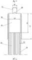
如图1至图6,一种可视医学探针,包括探针外管10、探查镜头20、导像束30、透光镜片40和导光束50,所述探查镜头设置在所述透光镜片上,所述探查镜头的前端露出所述透光镜片,所述透光镜片设置在所述探针外管的前端,所述探查镜头设置在所述透光镜片上,所述探查镜头的前端露出所述透光镜片,所述导光束穿入所述探针外管连接所述透光镜片,所述导像束穿入所述探针外管连接所述探查镜头。As shown in FIG. 1 to FIG. 6, a visual medical probe includes a probe
所述探针外管的外径d1为2mm±0.005mm,所述探针外管的壁厚S1为0.1 mm±0.005mm,所述探针外管的长度L为200mm~500mm,所述探针外管的材质为软钢。The outer diameter d1 of the probe outer tube is 2mm±0.005mm, the wall thickness S1 of the probe outer tube is 0.1mm±0.005mm, the length L of the probe outer tube is 200mm~500mm, the probe The material of the needle outer tube is mild steel.
所述透光镜片的直径d4为1.8 mm±0.005mm,所述透光镜片的厚度S4为3.0mm,所述透光镜片的前面与所述探针外管前端平齐,所述导光束包括3000根直径12um单根光纤。The diameter d4 of the transparent lens is 1.8 mm±0.005 mm, the thickness S4 of the transparent lens is 3.0 mm, the front of the transparent lens is flush with the front end of the probe outer tube, and the light guide includes: 3000 single fibers with a diameter of 12um.
所述探查镜头是微距广角镜头,所述探查镜头的直径d2为0.55 mm±0.005mm,所述探查镜头的厚度S2为2.0mm,所述探查镜头的外径套有镜头护套21,所述镜头护套是壁厚为0.1mm的不锈钢套管,所述探查镜头的前端边缘与所述透光镜片前端面平齐,所述导像束包括8000根直径为7um~8um的单根光纤。The probe lens is a macro wide-angle lens, the diameter d2 of the probe lens is 0.55 mm ± 0.005mm, the thickness S2 of the probe lens is 2.0mm, the outer diameter of the probe lens is covered with a
所述探针外管内设有注射管60,所述注射管的前端插入所述透光镜片,注射管前端与透光镜片前端面平齐。The probe outer tube is provided with an
所述注射管的外径d6为0.9 mm±0.005mm,所述注射管的壁厚S6为0.1 mm±0.005mm,所述注射管是软钢管或塑料软管。The outer diameter d6 of the injection pipe is 0.9 mm±0.005 mm, the wall thickness S6 of the injection pipe is 0.1 mm±0.005 mm, and the injection pipe is a soft steel pipe or a plastic hose.
所述透光镜片40设有镜头安装孔41和注射管安装孔42,所述镜头安装孔和注射管安装孔设置在透光镜片中心的两侧,所述探查镜头安装在所述镜头安装孔内,所述注射管的前端安装在所述注射管安装孔内,所述导光束在所述注射管与导像束余留的空间内穿入所述探针外管,所述导光束的前端紧贴透光镜片40后端面。The light-transmitting
所述探针外管的前端设有头部外套管70,所述头部外套管的前端与所述探针外管的前端平齐,所述头部外套管的前端与所述探针外管的前端打磨圆润,所述头部外套管是长度L7为4.0mm、壁厚S7为0.1mm的钛合金管,所述头部外套管的前端设有向所述探针外管中心方向弯折的喷溅舌71,所述喷溅舌的位置对应于所述注射管的出口。The front end of the probe outer tube is provided with a head
所述喷溅舌的宽度W7为0.7mm,所述喷溅舌探出所述头部外套管的前端E7=0.8mm。The width W7 of the splash tongue is 0.7 mm, and the front end E7 of the splash tongue protruding from the outer sleeve of the head is 0.8 mm.
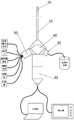
如图7,所述导像束、导光束和注射管的尾部伸出所述探针外管的末端,所述探针外管的末端设有探针连接头80,所述探针连接头设有图像接口81、光源接口82和注射接口83,所述导像束连接所述图像接口,所述导光束连接所述光源接口,所述注射管连接所述注射接口。As shown in FIG. 7 , the tail of the image guide beam, the light guide beam and the injection tube protrude from the end of the probe outer tube, and the end of the probe outer tube is provided with a
实施例一:Example 1:
如图1至图6,一种可视医学探针,包括探针外管10、探查镜头20、导像束30、透光镜片40、导光束50和注射管60。As shown in FIG. 1 to FIG. 6 , a visual medical probe includes a probe
探针外管10的外径d1=2mm±0.005mm,探针外管的壁厚S1=0.1 mm±0.005mm,根据需要探针外管的长度L可选200mm~500mm,在实际制作中可以优选200mm、250mm、300mm、400mm、500mm的不同规格。探针外管的外壁设有单位为1mm的刻度线11。本实施例探针外管的长度L=300mm,为了使探针外管具有适度的弯曲特性,探针外管的材质采用软钢,如低碳钢或304不锈钢经高温退火制成,其机械性能接近直径同为2mm的银线,既能弯曲成一定弧度,又能拉直,且具有良好的韧性不易折断。The outer diameter of the probe
透光镜片40设置在探针外管10的前端,透光镜片的直径d4=1.8 mm±0.005mm,透光镜片的厚度S4=3.0mm,透光镜片40是一个高透光度的圆柱体平面保护镜片,由石英玻璃制成,透光镜片轴向安装于探针外管内孔中,前边缘与探针外管的前缘平齐。透光镜片40设有镜头安装孔41和注射管安装孔42,镜头安装孔41和注射管安装孔42是轴向通孔,镜头安装孔和注射管安装孔对称设置在透光镜片中心的两侧,注射管安装孔直径为0.9±0.005mm,镜头安装孔的直径为0.75±0.005mm。The
探查镜头20是微距广角镜头,探查镜头的直径d2=0.55 mm±0.005mm,探查镜头的厚度S2=2.0mm,探查镜头是石英玻璃凸透镜,前端是设有一定曲率的凸面,镜头后面为平面,即前凸后平的凸透镜,焦距达到微距,焦距≤5mm,视角宽度≥160°,此镜头即为导像束的物镜,也即可视医学探针的物镜,具有视近物清晰放大作用。探查镜头的外径套有镜头护套21,镜头护套是壁厚为0.1±0.005mm的不锈钢套管,镜头护套的外径为0.75mm,对应于透光镜片的镜头安装孔41的直径。探查镜头的前端边缘与透光镜片前端面平齐。将导像束的前端轴向装配于镜头护套内,紧贴探查镜头后面,导像束截面中心与凸透镜同心,进行光路耦合校正,周边缝隙靠环氧树脂粘合固定封装。探查镜头20通过镜头护套安装在镜头安装孔41内,并穿过透光镜片,探查镜头前端的凸面露出透光镜片的端面。镜头护套21可以保护探查镜头,还可使探查镜头与导像束更可靠的连接,镜头护套的另一个重要作用是能够有效防止光晕现象发生,使图像清晰稳定。The
注射管60设置探针外管内,注射管的外径d6=0.9 mm±0.005mm,对应于透光镜片的注射管安装孔42的直径,注射管的壁厚S6=0.1 mm±0.005mm,注射管可采用软钢管或塑料软管。注射管安装在注射管安装孔42内,并穿过透光镜片40,注射管的前端与透光镜片的前端平齐。The
导像束30穿入探针外管连接探查镜头20,导像束的尾部从探针外管10的尾部穿出。导像束30的外轮廓直径不大于0.75mm,导像束包括8000根直径为7um~8um的单根光纤,可传送较高分辨率的探查图像。The
导光束50穿入探针外管连接透光镜片40,导光束的尾部从探针外管10的尾部穿出,。导光束包括3000根直径12um单根光纤。导光束在注射管60与导像束30余留的空间内穿入探针外管,导光束的前端紧贴透光镜片40后端面,如图6所示。The
探针外管的前端设有头部外套管70,头部外套管70套在探针外管10上,头部外套管的前端与探针外管的前端平齐,头部外套管的前端与探针外管的前端打磨圆润,头部外套管的长度L7 =4.0mm,头部外套管的壁厚S7=0.1mm,头部外套管是钛合金管。在头部外套管的前端设有向探针外管中心方向弯折的喷溅舌71,喷溅舌的位置对应于注射管60的出口。喷溅舌71的宽度W7=0.7mm,所述喷溅舌探出所述头部外套管的前端E7约为0.8mm,喷溅舌顶端的弯折量不超过注射管60的中心。本实施例的喷溅舌的制作方法是,选取一个长度4.5mm,内径2±0.005mm,壁厚0.1±0.005mm的钛合金圆管,一端为前端,一端为后端,在前端切割留出宽度为0.7mm,长度为0.8mm管壁,将留有的此部分管壁边缘打磨圆润,向圆管轴心方向做弧形弯折成型,弧形半径为1.10mm,形成喷溅舌71。留有的长度为4mm圆管即为头部外套管,打磨管壁边缘圆润,至此喷溅舌与头部外套管一体结构制作完毕。The front end of the probe outer tube is provided with a head
探针外管10头端部分、探查镜头20、导像束30头端部分、透光镜片40、导光束50头端部分、注射管60头端部分采用精密紧配安装,微小缝隙环氧树脂胶防水密封固定,头部外套管70与探针外管采用焊接或粘接固定。这样做的目的是整个头部防水,该器械可以浸泡水中,以方便药液消毒或气体消毒和临床操作使用。The head end of the probe
本发明的可视医学探针合理的利用了探针外管的内部空间,在直径仅有1.8mm的探针外管内孔中需要穿入导像束、导光束和注射管,本发明创新了导光束的安装方法,采用了导像束和注射管分别对称设置在透光镜片中心的两侧,将导像束30、注射管60并行穿入探针外管内孔中,从截面上看,导像束与注射管为两个近似外切圆,导像束和注射管与探针外管内孔成近似内切圆状态,由于导光束仅传导用于照明的可见光,不传导图像,不要求对应整齐排列的特点,把导光束平均分成两束,呈伞状聚集安装于探针外管的内孔导像束和注射管周围,这样尽最大可能安装更多的导光束,得到理想的照明效果。The visual medical probe of the present invention reasonably utilizes the inner space of the probe outer tube, and needs to penetrate the image guiding beam, the guiding beam and the injection tube in the inner hole of the probe outer tube with a diameter of only 1.8 mm, and the present invention is innovative. The installation method of the light guide adopts that the image guide beam and the injection tube are symmetrically arranged on both sides of the center of the light-transmitting lens, and the
头部外套管70可以增强探针外管10头部的结构强度。头部外套管70另一个重要作用是设置喷溅舌71。喷溅舌的作用包括:其一,通过注射管60喷水时,水流喷射到喷溅舌上,使水流向四周喷洒,还可使水流冲洗探查镜头,使探查镜头视物清晰。其二,向前及四周喷洒的水流或药液对瘘管管壁及内口具有良好的冲洗治疗作用。其三:治疗用激光光纤等从注射管穿出时遇到喷溅舌会向旁侧弯折,有利于对瘘管壁进行治疗。其四:利用喷溅舌可以在探针头部系线,对肛瘘进行挂线手术治疗。The head
本发明的基本原理是通过伸入到瘘管内部观察瘘管管壁和内口的解剖结构和形态特征进行诊断和治疗,插入人体部分的头部外套管优选外径为2.2mm,插入部探针外管优选外径为2.0mm,这种瘘管镜可以插入麻醉后的人体瘘管直径≥1.5mm的瘘管管道中,而且此医学探针身为软体结构,外表光滑,能够随瘘管弯曲走形,到达内口,这比视频辅助内窥镜套件更加纤细巧妙,适合绝大多数肛瘘的诊疗。本发明的可视医学探针为实时彩色光学成像,为0度正视向角,视场角宽度≥160度,与正常人眼的视向角和视场角宽度接近,此参数比8度斜视向角,70°~90°之间的视场角宽度更加优越,相当于把医生的眼睛带入到瘘管内部观察,可以得到瘘管管壁和内口清晰的解剖结构形态实时光学图像和照片。The basic principle of the present invention is to observe the anatomical structure and morphological characteristics of the fistula canal wall and internal opening by extending into the fistula canal for diagnosis and treatment. The outer diameter of the tube is preferably 2.0mm. This kind of fistula endoscope can be inserted into the fistula tube with a diameter of ≥1.5mm in human fistula after anesthesia, and the medical probe is a soft structure with a smooth appearance, which can bend and go along with the fistula tube to reach the inside. It is more slender and ingenious than the video-assisted endoscope kit, and is suitable for the diagnosis and treatment of most anal fistulas. The visual medical probe of the present invention is real-time color optical imaging, with a frontal viewing angle of 0 degrees, and a field angle width of ≥160 degrees, which is close to the viewing direction angle and field angle width of normal human eyes, and this parameter is more than 8 degrees of strabismus. The width of the field of view between 70° and 90° is more superior, which is equivalent to bringing the doctor's eyes into the inside of the fistula to observe, and can obtain clear real-time optical images and photos of the anatomical structure and shape of the fistula wall and internal opening.
本发明的可视医学探针不需要任何套管,可以直接应用。适合肛瘘(包括复杂性肛瘘)的诊断和治疗,还适合其它瘘道(窦道)性疾病比如骶尾部窦道感染(又称骶尾部藏毛窦)、化脓性汗腺炎、身体其他部位的感染或手术切口愈合不良通向体表的瘘道窦道等的诊断和治疗。The visual medical probe of the present invention does not need any cannula and can be directly applied. Suitable for the diagnosis and treatment of anal fistulas (including complex anal fistulas), as well as other fistula (sinus) diseases such as sacrococcygeal sinus infections (also known as sacrococcygeal pilonidal sinus), hidradenitis suppurativa, infections elsewhere in the body Or the diagnosis and treatment of fistulas and sinuses leading to the body surface due to poor healing of surgical incisions.
本发明的技术可以吸取或刮出瘘管及内口组织进行活体组织病理检查(简称:活检)以进一步了解瘘管壁和内口组织的细胞性质,配合医用激光器(如钬激光)或高频电刀可以精确地微创治疗肛瘘,从而不损伤肛门括约肌,保留肛门的功能,伤口小,恢复快。The technology of the present invention can suck or scrape out the fistula and the internal opening tissue for biopsy (abbreviation: biopsy) to further understand the cellular properties of the fistula wall and the internal opening tissue. Anal fistula can be precisely and minimally treated, so that the anal sphincter is not damaged, the function of the anus is preserved, the wound is small, and the recovery is fast.
实施例二:Embodiment 2:
一种可视医学探针接头装置。包括实施例一所述的可视医学探针。A visual medical probe joint device. It includes the visual medical probe described in the first embodiment.
如图7、图8,所述可视医学探针的导像束30、导光束50和注射管60的尾部伸出探针外管10的末端,探针外管的末端设有探针连接头80,探针连接头设有图像接口81、光源接口82和注射接口83。As shown in Fig. 7 and Fig. 8 , the tails of the
探针连接头80的端头连接探针外管10的末端。The end of the
图像接口81正对探针外管10,导像束30连接图像接口。在图像接口81内设有微型摄像头组件,包括摄像头镜头81a,CCD感光元件81b,视频数据电缆81c。摄像头镜头81a对正导像束30的末端。微型摄像头组件具有微距拍摄功能,最小拍摄距离≤5mm,圆柱形摄像头外径≤15mm,分辨率≥1280*1024,摄像头的镜头组件有外螺纹,能够旋转移动调焦。视频数据电缆81c从图像接口81的尾部穿出,可连接视频设备和数据处理设备,如显示器和计算机,实现对瘘管内的实时观察,并记录数据。The
光源接口82设置在探针连接头80的一侧。导光束50连接光源接口82,在光源接口内设有照明光源82a,为可视医学探针提供光源。照明光源82a是一只LED灯,照明光源82a可以采用电源线供电,也可以采用内置电池供电做成一体组件。照明光源通过内外螺纹连接或插卡式连接安装或卸下。照明光源也可以采用光纤传输的医用冷光源,照明光纤输出头通过内外螺纹连接或插卡式连接安装或卸下。The
注射接口83设置在探针连接头80相对于光源接口82的另一侧。注射管60连接注射接口83。注射接口83可以连接多种使用注射管60的设备,如注射器、抽吸泵、细活检钳、细针吸器械、高频刀、医用激光设备。The
采用本发明的技术方案,创新了一种保留括约肌的肛瘘微创治疗手术方式,即“可视探针(瘘管镜)保留括约肌隧道式肛瘘切除根治术” 该术式是利用本研发的可视探针独创的特有微创术式,这种微创术式是从瘘管内部进行治疗,较现有的传统的手术方式从瘘管外部进行治疗更加微创和精准,重点保护了肛门括约肌,伤口小,恢复快,痛苦小,愈合后肛门无畸形。By adopting the technical scheme of the present invention, a minimally invasive surgical method for sphincter-preserving anal fistula is innovated, namely "visual probe (fistula endoscope) sphincter-preserving tunnel type anal fistula resection radical resection". The unique and unique minimally invasive surgery of the probe, this minimally invasive surgery is performed from the inside of the fistula, which is more minimally invasive and precise than the existing traditional surgery from the outside of the fistula. It focuses on protecting the anal sphincter and has a small wound. , Fast recovery, less pain, no deformity of the anus after healing.
已知现有手术基本原理是,在探查清楚瘘管及内口位置基础上从瘘管外部将瘘管连同内口切除或切开或挂线,这将不可避免地会附带切开或切除瘘管到皮肤侧的肌肉、皮下脂肪及皮肤组织(挂线术也是慢性切割),也可以理解为切开或切除一片肉或一块肉,术后疼痛重,经常应用止痛药,伤口愈合时间长,一般一个月至三个月或更长,愈合后形成线状或片状瘢痕,有时瘢痕大会导致肛门或臀部畸形,有时过多的切除组织还有肛门失禁的风险。而应用可视探针的微创手术“可视探针(瘘管镜)保留括约肌隧道式肛瘘切除根治术”是在瘘管内部进行精细微创治疗,完好地保留(不损伤)肛门括约肌,极大地保护肛门功能,进一步还是小切口,创伤小疼痛轻微,愈合也很快,愈合后瘢痕小,可能就是个斑点,愈合时间7天左右。在科学研究中根据创伤理论和临床经验,上述微创手术较传统手术比较测算了几个重要指标。指标一,按伤口大小测算,创伤程度缩小到1/5至1/20;指标二,伤口愈合速度增快,愈合时间缩短到到1/4至1/12。It is known that the basic principle of the existing operation is to excise or incise the fistula together with the internal orifice from the outside of the fistula on the basis of finding out the position of the fistula and the internal opening, which will inevitably lead to the incision or excision of the fistula to the skin side. The muscle, subcutaneous fat and skin tissue (the thread hanging is also a chronic cut) can also be understood as the incision or removal of a piece of meat or a piece of meat, the pain is severe after surgery, painkillers are often used, and the wound healing time is long, usually a month to After three months or more, linear or sheet-like scars form after healing. Sometimes scarring can lead to deformities of the anus or buttocks. Sometimes too much tissue is removed and there is a risk of anal incontinence. The minimally invasive surgery using visual probe "visual probe (fistula endoscope) sphincter-preserving tunnel anal fistula resection" is a fine and minimally invasive treatment inside the fistula, which preserves (does not damage) the anal sphincter, greatly To protect the function of the anus, it is still a small incision, the trauma is small and the pain is mild, and the healing is fast. After healing, the scar is small, it may be a spot, and the healing time is about 7 days. In scientific research, according to trauma theory and clinical experience, the above minimally invasive surgery compared with traditional surgery to calculate several important indicators. Index 1, according to the size of the wound, the degree of trauma is reduced to 1/5 to 1/20; Index 2, the wound healing speed is increased, and the healing time is shortened to 1/4 to 1/12.
将先进的钬激光技术创新应用到瘘管病治疗领域,使手术治疗更加微创和高效。钬激光是以钇铝石榴石(YAG)为激活媒质,掺敏化离子铬(Cr)、传能离子铥(Tm)、激活离子钬(Ho)的激光晶体(Cr:Tm:Ho:YAG)制成的脉冲固体激光装置产生的新型激光。应用于泌尿外科、肝胆外科、脊柱微创外科、五官科、皮肤科、妇科等科室手术。该激光手术为微创手术,病人的治疗痛苦非常小。其主要特点是:激光具有极好的切割能力和组织切除能力,在组织切割过程中止血效果好,即使对于直径为1mm的血管也可以进行止血,而且在水中可以操作治疗,在前列腺增生剜切、尿道狭窄及尿路肿瘤切除、尿道结石、胆道结石、胆道肿瘤治疗中发挥优异效果。钬激光还有一个特点,就是其光波可以经由氧化硅石英光纤传导,这种光纤是可曲性的,因此非常适合在内镜下或微针镜下进行治疗。结合腔镜技术或微针镜技术,钬激光则可通过直径200~1000μm的石英纤维传导,便于进行精细的操作,手术一般需5-30分钟,只一点式即可完成,可(1)大大缩短手术时间;(2)对周围正常组织损伤小,术后反应轻,伤口愈合快,疤痕也小。(3)止血效果好,止血时间是电刀的十四分之一,止血效果是电刀的2~4倍。因此,可望达到术中极少出血甚至无血手术。手术野无渗血且清晰可辨,还可大大缩短手术时间。(4)手术中通过气化进行治疗,修整组织边缘光滑有坡度,不会像机械清理时那样留下台阶,不会形成软组织瘢痕;(5)激光手术对术中的各种监护仪器无干扰。钬激光在胆道外科、泌尿外科、脊柱微创外科领域的广泛应用,使上述学科微创化进一步发展。但是目前为止,还没有钬激光在肛肠科领域中的应用,通过本瘘管微创手术装置,将先进的钬激光技术应用于肛肠科领域瘘道疾病诊治中,开创了钬激光的应用范围,而且开创了可视探针(瘘管镜)结合钬激光微创瘘管手术从内治疗方法的先例。The innovative application of advanced holmium laser technology to the field of fistula treatment makes surgical treatment more minimally invasive and efficient. The holmium laser is based on yttrium aluminum garnet (YAG) as the activation medium, doped with sensitizing ions chromium (Cr), energy-transmitting ions thulium (Tm), and activating ions holmium (Ho) laser crystal (Cr:Tm:Ho:YAG) A new type of laser produced by a pulsed solid-state laser device. It is used in urology, hepatobiliary surgery, minimally invasive spine surgery, ENT, dermatology, gynecology and other departments. The laser surgery is a minimally invasive surgery, and the patient's treatment pain is very small. Its main features are: the laser has excellent cutting ability and tissue removal ability, and the hemostasis effect is good in the process of tissue cutting. It can stop bleeding even for blood vessels with a diameter of 1mm, and it can be operated in water. , urethral stricture and urinary tract tumor resection, urethral calculi, biliary calculi, biliary tract tumors have excellent results. Another feature of holmium laser is that its light waves can be transmitted through silica silica fiber, which is flexible, so it is very suitable for treatment under endoscopy or microneedling. Combined with endoscopic technology or micro-needle mirror technology, holmium laser can be conducted through quartz fibers with a diameter of 200-1000 μm, which is convenient for delicate operations. The operation generally takes 5-30 minutes and can be completed with only one point. It can be (1) greatly Shorten the operation time; (2) The damage to the surrounding normal tissue is small, the postoperative reaction is light, the wound heals quickly, and the scar is small. (3) The hemostatic effect is good, the hemostasis time is one-fourteenth of that of the electric knife, and the hemostatic effect is 2 to 4 times that of the electric knife. Therefore, it is expected to achieve minimal or even bloodless surgery during the operation. There is no oozing blood in the surgical field and can be clearly identified, and the operation time can be greatly shortened. (4) During the operation, gasification is used for treatment, and the edges of the trimmed tissue are smooth and sloping, which will not leave steps like mechanical cleaning, and will not form soft tissue scars; (5) Laser surgery does not interfere with various monitoring instruments during the operation. . The wide application of holmium laser in the field of biliary tract surgery, urology surgery, and minimally invasive spine surgery has further developed the minimally invasive aspects of these disciplines. But so far, there is no application of holmium laser in the field of anorectal. Through this fistula minimally invasive surgical device, the advanced holmium laser technology is applied to the diagnosis and treatment of fistula diseases in the field of anorectal, which has created the application scope of holmium laser, and Created a precedent for the visual probe (fistulascope) combined with holmium laser minimally invasive fistula surgery from the inside.
另外,需说明的是,应用本可视探针除了对瘘管病从内手术治疗外,还可以利用本可视探针在找准瘘管基础上进行传统方法的瘘管手术,比如对高位复杂肛瘘的瘘管中医挂线术等治疗,提高了治愈率,是中医和微创手术结合的范例。In addition, it should be noted that, in addition to the internal surgical treatment of fistula, the visual probe can also be used to perform traditional fistula surgery on the basis of pinpointing the fistula, such as the treatment of high complex anal fistula. Treatments such as traditional Chinese medicine threading of fistulae have improved the cure rate and are an example of the combination of traditional Chinese medicine and minimally invasive surgery.
通过本可视探针对瘘管的观察,将瘘管内部解剖结构实时显示出来,是人们更加详细了解和掌握瘘管内部结构相关知识,进一步提高理论水平和科研水平,有利于传授知识和教学。Through the observation of the fistula by the visual probe, the internal anatomical structure of the fistula can be displayed in real time, which enables people to understand and master the knowledge of the internal structure of the fistula in more detail, further improve the theoretical level and scientific research level, and is conducive to imparting knowledge and teaching.
Claims (10)
Translated fromChinesePriority Applications (1)
| Application Number | Priority Date | Filing Date | Title |
|---|---|---|---|
| CN202210276872.5ACN114532963A (en) | 2022-03-21 | 2022-03-21 | Visual medical probe |
Applications Claiming Priority (1)
| Application Number | Priority Date | Filing Date | Title |
|---|---|---|---|
| CN202210276872.5ACN114532963A (en) | 2022-03-21 | 2022-03-21 | Visual medical probe |
Publications (1)
| Publication Number | Publication Date |
|---|---|
| CN114532963Atrue CN114532963A (en) | 2022-05-27 |
Family
ID=81666374
Family Applications (1)
| Application Number | Title | Priority Date | Filing Date |
|---|---|---|---|
| CN202210276872.5APendingCN114532963A (en) | 2022-03-21 | 2022-03-21 | Visual medical probe |
Country Status (1)
| Country | Link |
|---|---|
| CN (1) | CN114532963A (en) |
Citations (11)
| Publication number | Priority date | Publication date | Assignee | Title |
|---|---|---|---|---|
| US20050070759A1 (en)* | 2003-09-26 | 2005-03-31 | Armstrong David N. | Instrument and method for endoscopic visualization and treatment of anorectal fistula |
| CN202982871U (en)* | 2012-12-27 | 2013-06-12 | 郭明浩 | Multifunctional medical probe |
| CN203693660U (en)* | 2014-02-04 | 2014-07-09 | 王洪波 | Anal fistula probe |
| CN104127210A (en)* | 2014-08-14 | 2014-11-05 | 李春穴 | Anal fistula seton surgery device |
| CN108852430A (en)* | 2018-04-10 | 2018-11-23 | 中国人民解放军陆军军医大学第二附属医院 | Cord holder is used in anal fistula seton operation |
| CN108888304A (en)* | 2018-08-16 | 2018-11-27 | 潍坊高航机械科技有限公司 | anal fistula seton device |
| CN108992137A (en)* | 2018-07-23 | 2018-12-14 | 张家港市中医医院 | Cleaning device strikes off in visualization anal fistula tube wall tissue |
| CN110251062A (en)* | 2019-07-26 | 2019-09-20 | 上海中医药大学附属岳阳中西医结合医院 | A video-assisted endoscopy kit for anal fistula treatment |
| CN211324923U (en)* | 2019-10-07 | 2020-08-25 | 李鹏 | Anorectal enteroscope |
| CN215079197U (en)* | 2021-05-06 | 2021-12-10 | 重庆巴星医疗科技有限公司 | A puncture subassembly for anal fistula hangs line treatment |
| CN217186077U (en)* | 2022-03-21 | 2022-08-16 | 郭明浩 | Visual medical probe |
- 2022
- 2022-03-21CNCN202210276872.5Apatent/CN114532963A/enactivePending
Patent Citations (11)
| Publication number | Priority date | Publication date | Assignee | Title |
|---|---|---|---|---|
| US20050070759A1 (en)* | 2003-09-26 | 2005-03-31 | Armstrong David N. | Instrument and method for endoscopic visualization and treatment of anorectal fistula |
| CN202982871U (en)* | 2012-12-27 | 2013-06-12 | 郭明浩 | Multifunctional medical probe |
| CN203693660U (en)* | 2014-02-04 | 2014-07-09 | 王洪波 | Anal fistula probe |
| CN104127210A (en)* | 2014-08-14 | 2014-11-05 | 李春穴 | Anal fistula seton surgery device |
| CN108852430A (en)* | 2018-04-10 | 2018-11-23 | 中国人民解放军陆军军医大学第二附属医院 | Cord holder is used in anal fistula seton operation |
| CN108992137A (en)* | 2018-07-23 | 2018-12-14 | 张家港市中医医院 | Cleaning device strikes off in visualization anal fistula tube wall tissue |
| CN108888304A (en)* | 2018-08-16 | 2018-11-27 | 潍坊高航机械科技有限公司 | anal fistula seton device |
| CN110251062A (en)* | 2019-07-26 | 2019-09-20 | 上海中医药大学附属岳阳中西医结合医院 | A video-assisted endoscopy kit for anal fistula treatment |
| CN211324923U (en)* | 2019-10-07 | 2020-08-25 | 李鹏 | Anorectal enteroscope |
| CN215079197U (en)* | 2021-05-06 | 2021-12-10 | 重庆巴星医疗科技有限公司 | A puncture subassembly for anal fistula hangs line treatment |
| CN217186077U (en)* | 2022-03-21 | 2022-08-16 | 郭明浩 | Visual medical probe |
Similar Documents
| Publication | Publication Date | Title |
|---|---|---|
| JP5567013B2 (en) | Revolving prism type endoscope | |
| CN101732082B (en) | Soft and hard gallbladder and cholangioscopic system | |
| CN101732027B (en) | Bendable steering endoscope | |
| US20060149129A1 (en) | Catheter with multiple visual elements | |
| CN112842525B (en) | An endoscopic laser ablation catheter | |
| JP2011529724A5 (en) | ||
| JP2001104315A (en) | Ultrasonic-guided paracentesis system device | |
| Carta et al. | Sialendoscopy for salivary stones: principles, technical skills and therapeutic experience | |
| CN217186077U (en) | Visual medical probe | |
| MX2012009035A (en) | Multi-fiber flexible surgical probe. | |
| WO2024001634A1 (en) | Negative-pressure suction ureteral sheath and ureteral insertion apparatus | |
| WO2024159665A1 (en) | Nephroscope | |
| US20210068884A1 (en) | Electrode for prostate surgery and using method thereof | |
| CN217310575U (en) | Fistula minimally invasive surgery device | |
| CN114532963A (en) | Visual medical probe | |
| CN114849021B (en) | Visual intubation device and using method thereof | |
| RU2727460C1 (en) | Instrument - holmium fibre laser conductor for tissue vaporisation in area of natural anastomosis of maxillary sinus | |
| CN114949553B (en) | A visualization catheter and a cutting knife with the visualization catheter | |
| CN113499136B (en) | Hard endoscope externally-matched rotatable auxiliary channel | |
| EP3678556B1 (en) | Imaging device | |
| CN209645054U (en) | The hemostasis electrod assembly of Flexible ureteroscope | |
| CN2605815Y (en) | Three pipe insulative sleeve for endoscope with flexible pipe | |
| CN109330640B (en) | Endoscope working sheath for urological surgery | |
| CN206587013U (en) | A kind of endoscope casing tube formula circular cutter | |
| CN210249789U (en) | Uretero-renoscope outer tube |
Legal Events
| Date | Code | Title | Description |
|---|---|---|---|
| PB01 | Publication | ||
| PB01 | Publication | ||
| SE01 | Entry into force of request for substantive examination | ||
| SE01 | Entry into force of request for substantive examination |