CN114470324B - Novel strategy for the modification of universal bone implants for the intervention of bone defects associated with diabetes - Google Patents
Novel strategy for the modification of universal bone implants for the intervention of bone defects associated with diabetesDownload PDFInfo
- Publication number
- CN114470324B CN114470324BCN202210118813.5ACN202210118813ACN114470324BCN 114470324 BCN114470324 BCN 114470324BCN 202210118813 ACN202210118813 ACN 202210118813ACN 114470324 BCN114470324 BCN 114470324B
- Authority
- CN
- China
- Prior art keywords
- bone
- antibacterial
- implant
- pluronic
- gentamicin
- Prior art date
- Legal status (The legal status is an assumption and is not a legal conclusion. Google has not performed a legal analysis and makes no representation as to the accuracy of the status listed.)
- Expired - Fee Related
Links
- 239000007943implantSubstances0.000titleclaimsabstractdescription80
- 210000000988bone and boneAnatomy0.000titleclaimsabstractdescription78
- 206010012601diabetes mellitusDiseases0.000titleclaimsabstractdescription27
- 230000007547defectEffects0.000titleclaimsabstractdescription18
- 230000004048modificationEffects0.000titleclaimsdescription11
- 238000012986modificationMethods0.000titleclaimsdescription11
- 230000000844anti-bacterial effectEffects0.000claimsabstractdescription33
- TUSDEZXZIZRFGC-UHFFFAOYSA-N1-O-galloyl-3,6-(R)-HHDP-beta-D-glucoseNatural productsOC1C(O2)COC(=O)C3=CC(O)=C(O)C(O)=C3C3=C(O)C(O)=C(O)C=C3C(=O)OC1C(O)C2OC(=O)C1=CC(O)=C(O)C(O)=C1TUSDEZXZIZRFGC-UHFFFAOYSA-N0.000claimsabstractdescription29
- 239000001263FEMA 3042Substances0.000claimsabstractdescription29
- LRBQNJMCXXYXIU-PPKXGCFTSA-NPenta-digallate-beta-D-glucoseNatural productsOC1=C(O)C(O)=CC(C(=O)OC=2C(=C(O)C=C(C=2)C(=O)OC[C@@H]2[C@H]([C@H](OC(=O)C=3C=C(OC(=O)C=4C=C(O)C(O)=C(O)C=4)C(O)=C(O)C=3)[C@@H](OC(=O)C=3C=C(OC(=O)C=4C=C(O)C(O)=C(O)C=4)C(O)=C(O)C=3)[C@H](OC(=O)C=3C=C(OC(=O)C=4C=C(O)C(O)=C(O)C=4)C(O)=C(O)C=3)O2)OC(=O)C=2C=C(OC(=O)C=3C=C(O)C(O)=C(O)C=3)C(O)=C(O)C=2)O)=C1LRBQNJMCXXYXIU-PPKXGCFTSA-N0.000claimsabstractdescription29
- LRBQNJMCXXYXIU-NRMVVENXSA-Ntannic acidChemical compoundOC1=C(O)C(O)=CC(C(=O)OC=2C(=C(O)C=C(C=2)C(=O)OC[C@@H]2[C@H]([C@H](OC(=O)C=3C=C(OC(=O)C=4C=C(O)C(O)=C(O)C=4)C(O)=C(O)C=3)[C@@H](OC(=O)C=3C=C(OC(=O)C=4C=C(O)C(O)=C(O)C=4)C(O)=C(O)C=3)[C@@H](OC(=O)C=3C=C(OC(=O)C=4C=C(O)C(O)=C(O)C=4)C(O)=C(O)C=3)O2)OC(=O)C=2C=C(OC(=O)C=3C=C(O)C(O)=C(O)C=3)C(O)=C(O)C=2)O)=C1LRBQNJMCXXYXIU-NRMVVENXSA-N0.000claimsabstractdescription29
- 229940033123tannic acidDrugs0.000claimsabstractdescription29
- 235000015523tannic acidNutrition0.000claimsabstractdescription29
- 229920002258tannic acidPolymers0.000claimsabstractdescription29
- 229920001992poloxamer 407Polymers0.000claimsabstractdescription22
- 208000015181infectious diseaseDiseases0.000claimsabstractdescription20
- CEAZRRDELHUEMR-URQXQFDESA-NGentamicinChemical compoundO1[C@H](C(C)NC)CC[C@@H](N)[C@H]1O[C@H]1[C@H](O)[C@@H](O[C@@H]2[C@@H]([C@@H](NC)[C@@](C)(O)CO2)O)[C@H](N)C[C@@H]1NCEAZRRDELHUEMR-URQXQFDESA-N0.000claimsabstractdescription17
- 229930182566GentamicinNatural products0.000claimsabstractdescription17
- 229960002518gentamicinDrugs0.000claimsabstractdescription16
- 230000033115angiogenesisEffects0.000claimsabstractdescription14
- 239000011248coating agentSubstances0.000claimsabstractdescription11
- 238000000576coating methodMethods0.000claimsabstractdescription11
- 230000001737promoting effectEffects0.000claimsabstractdescription11
- 238000000338in vitroMethods0.000claimsabstractdescription8
- 238000010883osseointegrationMethods0.000claimsabstractdescription8
- 210000002889endothelial cellAnatomy0.000claimsabstractdescription6
- 238000001727in vivoMethods0.000claimsabstractdescription6
- 241000191967Staphylococcus aureusSpecies0.000claimsabstractdescription3
- 230000002491angiogenic effectEffects0.000claimsabstractdescription3
- 230000036542oxidative stressEffects0.000claimsabstractdescription3
- 239000004696Poly ether ether ketoneSubstances0.000claimsdescription40
- 229920002530polyetherether ketonePolymers0.000claimsdescription40
- 239000000243solutionSubstances0.000claimsdescription25
- 238000000034methodMethods0.000claimsdescription22
- 229920002873PolyethyleniminePolymers0.000claimsdescription13
- 239000011259mixed solutionSubstances0.000claimsdescription13
- 241000700159RattusSpecies0.000claimsdescription12
- 206010031252OsteomyelitisDiseases0.000claimsdescription10
- RDEIXVOBVLKYNT-VQBXQJRRSA-N(2r,3r,4r,5r)-2-[(1s,2s,3r,4s,6r)-4,6-diamino-3-[(2r,3r,6s)-3-amino-6-(1-aminoethyl)oxan-2-yl]oxy-2-hydroxycyclohexyl]oxy-5-methyl-4-(methylamino)oxane-3,5-diol;(2r,3r,4r,5r)-2-[(1s,2s,3r,4s,6r)-4,6-diamino-3-[(2r,3r,6s)-3-amino-6-(aminomethyl)oxan-2-yl]oChemical compoundOS(O)(=O)=O.O1C[C@@](O)(C)[C@H](NC)[C@@H](O)[C@H]1O[C@@H]1[C@@H](O)[C@H](O[C@@H]2[C@@H](CC[C@@H](CN)O2)N)[C@@H](N)C[C@H]1N.O1C[C@@](O)(C)[C@H](NC)[C@@H](O)[C@H]1O[C@@H]1[C@@H](O)[C@H](O[C@@H]2[C@@H](CC[C@H](O2)C(C)N)N)[C@@H](N)C[C@H]1N.O1[C@H](C(C)NC)CC[C@@H](N)[C@H]1O[C@H]1[C@H](O)[C@@H](O[C@@H]2[C@@H]([C@@H](NC)[C@@](C)(O)CO2)O)[C@H](N)C[C@@H]1NRDEIXVOBVLKYNT-VQBXQJRRSA-N0.000claimsdescription9
- 238000002513implantationMethods0.000claimsdescription9
- 238000010171animal modelMethods0.000claimsdescription8
- 239000000463materialSubstances0.000claimsdescription8
- 230000001023pro-angiogenic effectEffects0.000claimsdescription8
- 238000002791soakingMethods0.000claimsdescription6
- 239000002904solventSubstances0.000claimsdescription5
- 238000007920subcutaneous administrationMethods0.000claimsdescription5
- 239000000126substanceSubstances0.000claimsdescription5
- 102000009524Vascular Endothelial Growth Factor AHuman genes0.000claimsdescription4
- 108010073929Vascular Endothelial Growth Factor AProteins0.000claimsdescription4
- 238000011160researchMethods0.000claimsdescription4
- ZSJLQEPLLKMAKR-GKHCUFPYSA-NstreptozocinChemical compoundO=NN(C)C(=O)N[C@H]1[C@@H](O)O[C@H](CO)[C@@H](O)[C@@H]1OZSJLQEPLLKMAKR-GKHCUFPYSA-N0.000claimsdescription4
- 239000000758substrateSubstances0.000claimsdescription4
- 230000000694effectsEffects0.000claimsdescription3
- 238000011156evaluationMethods0.000claimsdescription3
- 206010033675panniculitisDiseases0.000claimsdescription3
- 210000004304subcutaneous tissueAnatomy0.000claimsdescription3
- 210000003606umbilical veinAnatomy0.000claimsdescription3
- 238000002965ELISAMethods0.000claimsdescription2
- 238000007605air dryingMethods0.000claimsdescription2
- 238000001514detection methodMethods0.000claimsdescription2
- 239000012154double-distilled waterSubstances0.000claimsdescription2
- 239000004615ingredientSubstances0.000claimsdescription2
- 238000012360testing methodMethods0.000claimsdescription2
- ZSJLQEPLLKMAKR-UHFFFAOYSA-NStreptozotocinNatural productsO=NN(C)C(=O)NC1C(O)OC(CO)C(O)C1OZSJLQEPLLKMAKR-UHFFFAOYSA-N0.000claims2
- 229960001052streptozocinDrugs0.000claims2
- JUPQTSLXMOCDHR-UHFFFAOYSA-Nbenzene-1,4-diol;bis(4-fluorophenyl)methanoneChemical compoundOC1=CC=C(O)C=C1.C1=CC(F)=CC=C1C(=O)C1=CC=C(F)C=C1JUPQTSLXMOCDHR-UHFFFAOYSA-N0.000claims1
- 238000012258culturingMethods0.000claims1
- 238000007654immersionMethods0.000claims1
- 230000006698inductionEffects0.000claims1
- 238000004519manufacturing processMethods0.000claims1
- 239000003814drugSubstances0.000abstractdescription7
- 238000001338self-assemblyMethods0.000abstractdescription6
- 238000002474experimental methodMethods0.000abstractdescription4
- 230000009286beneficial effectEffects0.000abstractdescription3
- 230000000399orthopedic effectEffects0.000abstractdescription3
- 238000001356surgical procedureMethods0.000abstractdescription2
- 230000001172regenerating effectEffects0.000abstract1
- 238000010586diagramMethods0.000description8
- 239000010410layerSubstances0.000description8
- 230000011164ossificationEffects0.000description8
- 238000010186stainingMethods0.000description7
- 229940079593drugDrugs0.000description6
- 208000035143Bacterial infectionDiseases0.000description5
- 208000022362bacterial infectious diseaseDiseases0.000description5
- 230000006378damageEffects0.000description5
- 230000002458infectious effectEffects0.000description5
- 238000002360preparation methodMethods0.000description5
- 229950003937toloniumDrugs0.000description5
- HNONEKILPDHFOL-UHFFFAOYSA-Mtolonium chlorideChemical compound[Cl-].C1=C(C)C(N)=CC2=[S+]C3=CC(N(C)C)=CC=C3N=C21HNONEKILPDHFOL-UHFFFAOYSA-M0.000description5
- CSCPPACGZOOCGX-UHFFFAOYSA-NAcetoneChemical compoundCC(C)=OCSCPPACGZOOCGX-UHFFFAOYSA-N0.000description4
- 210000004204blood vesselAnatomy0.000description4
- 230000010354integrationEffects0.000description4
- 210000000440neutrophilAnatomy0.000description4
- 238000006243chemical reactionMethods0.000description3
- 239000011148porous materialSubstances0.000description3
- QAOWNCQODCNURD-UHFFFAOYSA-Nsulfuric acidSubstancesOS(O)(=O)=OQAOWNCQODCNURD-UHFFFAOYSA-N0.000description3
- LFQSCWFLJHTTHZ-UHFFFAOYSA-NEthanolChemical compoundCCOLFQSCWFLJHTTHZ-UHFFFAOYSA-N0.000description2
- 206010061218InflammationDiseases0.000description2
- 230000001772anti-angiogenic effectEffects0.000description2
- 210000004027cellAnatomy0.000description2
- 125000004122cyclic groupChemical group0.000description2
- 239000008367deionised waterSubstances0.000description2
- 229910021641deionized waterInorganic materials0.000description2
- 230000004054inflammatory processEffects0.000description2
- 238000010603microCTMethods0.000description2
- 230000007935neutral effectEffects0.000description2
- 238000011552rat modelMethods0.000description2
- 238000001878scanning electron micrographMethods0.000description2
- 239000006228supernatantSubstances0.000description2
- 210000001519tissueAnatomy0.000description2
- 230000009466transformationEffects0.000description2
- 210000000689upper legAnatomy0.000description2
- XLYOFNOQVPJJNP-UHFFFAOYSA-NwaterChemical compoundOXLYOFNOQVPJJNP-UHFFFAOYSA-N0.000description2
- 241000894006BacteriaSpecies0.000description1
- 108010077805Bacterial ProteinsProteins0.000description1
- 238000008157ELISA kitMethods0.000description1
- 102000002045EndothelinHuman genes0.000description1
- 108050009340EndothelinProteins0.000description1
- 206010031264OsteonecrosisDiseases0.000description1
- 208000034189SclerosisDiseases0.000description1
- 102000005789Vascular Endothelial Growth FactorsHuman genes0.000description1
- 108010019530Vascular Endothelial Growth FactorsProteins0.000description1
- 238000010306acid treatmentMethods0.000description1
- 230000001154acute effectEffects0.000description1
- 229940126575aminoglycosideDrugs0.000description1
- 238000004458analytical methodMethods0.000description1
- 239000002870angiogenesis inducing agentSubstances0.000description1
- 230000000845anti-microbial effectEffects0.000description1
- 230000003078antioxidant effectEffects0.000description1
- 238000000429assemblyMethods0.000description1
- 230000000712assemblyEffects0.000description1
- 230000001580bacterial effectEffects0.000description1
- 230000015572biosynthetic processEffects0.000description1
- 210000000170cell membraneAnatomy0.000description1
- 239000003153chemical reaction reagentSubstances0.000description1
- 230000001684chronic effectEffects0.000description1
- 230000001186cumulative effectEffects0.000description1
- 230000009881electrostatic interactionEffects0.000description1
- ZUBDGKVDJUIMQQ-UBFCDGJISA-Nendothelin-1Chemical compoundC([C@@H](C(=O)N[C@@H](CC(C)C)C(=O)N[C@@H](CC(O)=O)C(=O)N[C@@H]([C@@H](C)CC)C(=O)N[C@@H]([C@@H](C)CC)C(=O)N[C@@H](CC=1C2=CC=CC=C2NC=1)C(O)=O)NC(=O)[C@H]1NC(=O)[C@H](CC=2C=CC=CC=2)NC(=O)[C@@H](CC=2C=CC(O)=CC=2)NC(=O)[C@H](C(C)C)NC(=O)[C@H]2CSSC[C@@H](C(N[C@H](CO)C(=O)N[C@@H](CO)C(=O)N[C@H](CC(C)C)C(=O)N[C@@H](CCSC)C(=O)N[C@H](CC(O)=O)C(=O)N[C@@H](CCCCN)C(=O)N[C@@H](CCC(O)=O)C(=O)N2)=O)NC(=O)[C@@H](CO)NC(=O)[C@H](N)CSSC1)C1=CNC=N1ZUBDGKVDJUIMQQ-UBFCDGJISA-N0.000description1
- 210000003714granulocyteAnatomy0.000description1
- 230000035876healingEffects0.000description1
- 238000007490hematoxylin and eosin (H&E) stainingMethods0.000description1
- 229910052739hydrogenInorganic materials0.000description1
- 239000001257hydrogenSubstances0.000description1
- 230000005660hydrophilic surfaceEffects0.000description1
- 230000002209hydrophobic effectEffects0.000description1
- 230000005661hydrophobic surfaceEffects0.000description1
- 206010020718hyperplasiaDiseases0.000description1
- 238000003384imaging methodMethods0.000description1
- 230000006872improvementEffects0.000description1
- 230000008595infiltrationEffects0.000description1
- 238000001764infiltrationMethods0.000description1
- 238000011068loading methodMethods0.000description1
- 230000010534mechanism of actionEffects0.000description1
- 239000002184metalSubstances0.000description1
- 229910052751metalInorganic materials0.000description1
- 239000007769metal materialSubstances0.000description1
- 210000001087myotubuleAnatomy0.000description1
- 230000002093peripheral effectEffects0.000description1
- 150000008442polyphenolic compoundsChemical class0.000description1
- 235000013824polyphenolsNutrition0.000description1
- 230000008569processEffects0.000description1
- 238000001243protein synthesisMethods0.000description1
- 238000004445quantitative analysisMethods0.000description1
- 210000003705ribosomeAnatomy0.000description1
- 230000001568sexual effectEffects0.000description1
- 230000001954sterilising effectEffects0.000description1
- 238000004659sterilization and disinfectionMethods0.000description1
- 230000035882stressEffects0.000description1
- 238000006277sulfonation reactionMethods0.000description1
- 239000002345surface coating layerSubstances0.000description1
- 239000004094surface-active agentSubstances0.000description1
- 229920001864tanninPolymers0.000description1
- 235000018553tanninNutrition0.000description1
- 239000001648tanninSubstances0.000description1
- 230000001550time effectEffects0.000description1
- 230000014616translationEffects0.000description1
- 238000004506ultrasonic cleaningMethods0.000description1
- 230000002792vascularEffects0.000description1
- 238000012795verificationMethods0.000description1
- 238000005406washingMethods0.000description1
Images


Classifications
- A—HUMAN NECESSITIES
- A61—MEDICAL OR VETERINARY SCIENCE; HYGIENE
- A61L—METHODS OR APPARATUS FOR STERILISING MATERIALS OR OBJECTS IN GENERAL; DISINFECTION, STERILISATION OR DEODORISATION OF AIR; CHEMICAL ASPECTS OF BANDAGES, DRESSINGS, ABSORBENT PADS OR SURGICAL ARTICLES; MATERIALS FOR BANDAGES, DRESSINGS, ABSORBENT PADS OR SURGICAL ARTICLES
- A61L27/00—Materials for grafts or prostheses or for coating grafts or prostheses
- A61L27/14—Macromolecular materials
- A61L27/18—Macromolecular materials obtained otherwise than by reactions only involving carbon-to-carbon unsaturated bonds
- A—HUMAN NECESSITIES
- A61—MEDICAL OR VETERINARY SCIENCE; HYGIENE
- A61L—METHODS OR APPARATUS FOR STERILISING MATERIALS OR OBJECTS IN GENERAL; DISINFECTION, STERILISATION OR DEODORISATION OF AIR; CHEMICAL ASPECTS OF BANDAGES, DRESSINGS, ABSORBENT PADS OR SURGICAL ARTICLES; MATERIALS FOR BANDAGES, DRESSINGS, ABSORBENT PADS OR SURGICAL ARTICLES
- A61L27/00—Materials for grafts or prostheses or for coating grafts or prostheses
- A61L27/28—Materials for coating prostheses
- A61L27/34—Macromolecular materials
- A—HUMAN NECESSITIES
- A61—MEDICAL OR VETERINARY SCIENCE; HYGIENE
- A61L—METHODS OR APPARATUS FOR STERILISING MATERIALS OR OBJECTS IN GENERAL; DISINFECTION, STERILISATION OR DEODORISATION OF AIR; CHEMICAL ASPECTS OF BANDAGES, DRESSINGS, ABSORBENT PADS OR SURGICAL ARTICLES; MATERIALS FOR BANDAGES, DRESSINGS, ABSORBENT PADS OR SURGICAL ARTICLES
- A61L27/00—Materials for grafts or prostheses or for coating grafts or prostheses
- A61L27/50—Materials characterised by their function or physical properties, e.g. injectable or lubricating compositions, shape-memory materials, surface modified materials
- A61L27/54—Biologically active materials, e.g. therapeutic substances
- A—HUMAN NECESSITIES
- A61—MEDICAL OR VETERINARY SCIENCE; HYGIENE
- A61L—METHODS OR APPARATUS FOR STERILISING MATERIALS OR OBJECTS IN GENERAL; DISINFECTION, STERILISATION OR DEODORISATION OF AIR; CHEMICAL ASPECTS OF BANDAGES, DRESSINGS, ABSORBENT PADS OR SURGICAL ARTICLES; MATERIALS FOR BANDAGES, DRESSINGS, ABSORBENT PADS OR SURGICAL ARTICLES
- A61L27/00—Materials for grafts or prostheses or for coating grafts or prostheses
- A61L27/50—Materials characterised by their function or physical properties, e.g. injectable or lubricating compositions, shape-memory materials, surface modified materials
- A61L27/56—Porous materials, e.g. foams or sponges
- A—HUMAN NECESSITIES
- A61—MEDICAL OR VETERINARY SCIENCE; HYGIENE
- A61L—METHODS OR APPARATUS FOR STERILISING MATERIALS OR OBJECTS IN GENERAL; DISINFECTION, STERILISATION OR DEODORISATION OF AIR; CHEMICAL ASPECTS OF BANDAGES, DRESSINGS, ABSORBENT PADS OR SURGICAL ARTICLES; MATERIALS FOR BANDAGES, DRESSINGS, ABSORBENT PADS OR SURGICAL ARTICLES
- A61L2300/00—Biologically active materials used in bandages, wound dressings, absorbent pads or medical devices
- A61L2300/20—Biologically active materials used in bandages, wound dressings, absorbent pads or medical devices containing or releasing organic materials
- A61L2300/23—Carbohydrates
- A—HUMAN NECESSITIES
- A61—MEDICAL OR VETERINARY SCIENCE; HYGIENE
- A61L—METHODS OR APPARATUS FOR STERILISING MATERIALS OR OBJECTS IN GENERAL; DISINFECTION, STERILISATION OR DEODORISATION OF AIR; CHEMICAL ASPECTS OF BANDAGES, DRESSINGS, ABSORBENT PADS OR SURGICAL ARTICLES; MATERIALS FOR BANDAGES, DRESSINGS, ABSORBENT PADS OR SURGICAL ARTICLES
- A61L2300/00—Biologically active materials used in bandages, wound dressings, absorbent pads or medical devices
- A61L2300/40—Biologically active materials used in bandages, wound dressings, absorbent pads or medical devices characterised by a specific therapeutic activity or mode of action
- A61L2300/404—Biocides, antimicrobial agents, antiseptic agents
- A61L2300/406—Antibiotics
- A—HUMAN NECESSITIES
- A61—MEDICAL OR VETERINARY SCIENCE; HYGIENE
- A61L—METHODS OR APPARATUS FOR STERILISING MATERIALS OR OBJECTS IN GENERAL; DISINFECTION, STERILISATION OR DEODORISATION OF AIR; CHEMICAL ASPECTS OF BANDAGES, DRESSINGS, ABSORBENT PADS OR SURGICAL ARTICLES; MATERIALS FOR BANDAGES, DRESSINGS, ABSORBENT PADS OR SURGICAL ARTICLES
- A61L2300/00—Biologically active materials used in bandages, wound dressings, absorbent pads or medical devices
- A61L2300/40—Biologically active materials used in bandages, wound dressings, absorbent pads or medical devices characterised by a specific therapeutic activity or mode of action
- A61L2300/45—Mixtures of two or more drugs, e.g. synergistic mixtures
- A—HUMAN NECESSITIES
- A61—MEDICAL OR VETERINARY SCIENCE; HYGIENE
- A61L—METHODS OR APPARATUS FOR STERILISING MATERIALS OR OBJECTS IN GENERAL; DISINFECTION, STERILISATION OR DEODORISATION OF AIR; CHEMICAL ASPECTS OF BANDAGES, DRESSINGS, ABSORBENT PADS OR SURGICAL ARTICLES; MATERIALS FOR BANDAGES, DRESSINGS, ABSORBENT PADS OR SURGICAL ARTICLES
- A61L2420/00—Materials or methods for coatings medical devices
- A61L2420/04—Coatings containing a composite material such as inorganic/organic, i.e. material comprising different phases
- A—HUMAN NECESSITIES
- A61—MEDICAL OR VETERINARY SCIENCE; HYGIENE
- A61L—METHODS OR APPARATUS FOR STERILISING MATERIALS OR OBJECTS IN GENERAL; DISINFECTION, STERILISATION OR DEODORISATION OF AIR; CHEMICAL ASPECTS OF BANDAGES, DRESSINGS, ABSORBENT PADS OR SURGICAL ARTICLES; MATERIALS FOR BANDAGES, DRESSINGS, ABSORBENT PADS OR SURGICAL ARTICLES
- A61L2420/00—Materials or methods for coatings medical devices
- A61L2420/06—Coatings containing a mixture of two or more compounds
- A—HUMAN NECESSITIES
- A61—MEDICAL OR VETERINARY SCIENCE; HYGIENE
- A61L—METHODS OR APPARATUS FOR STERILISING MATERIALS OR OBJECTS IN GENERAL; DISINFECTION, STERILISATION OR DEODORISATION OF AIR; CHEMICAL ASPECTS OF BANDAGES, DRESSINGS, ABSORBENT PADS OR SURGICAL ARTICLES; MATERIALS FOR BANDAGES, DRESSINGS, ABSORBENT PADS OR SURGICAL ARTICLES
- A61L2420/00—Materials or methods for coatings medical devices
- A61L2420/08—Coatings comprising two or more layers
- A—HUMAN NECESSITIES
- A61—MEDICAL OR VETERINARY SCIENCE; HYGIENE
- A61L—METHODS OR APPARATUS FOR STERILISING MATERIALS OR OBJECTS IN GENERAL; DISINFECTION, STERILISATION OR DEODORISATION OF AIR; CHEMICAL ASPECTS OF BANDAGES, DRESSINGS, ABSORBENT PADS OR SURGICAL ARTICLES; MATERIALS FOR BANDAGES, DRESSINGS, ABSORBENT PADS OR SURGICAL ARTICLES
- A61L2430/00—Materials or treatment for tissue regeneration
- A61L2430/02—Materials or treatment for tissue regeneration for reconstruction of bones; weight-bearing implants
Landscapes
- Health & Medical Sciences (AREA)
- Chemical & Material Sciences (AREA)
- Life Sciences & Earth Sciences (AREA)
- Medicinal Chemistry (AREA)
- Public Health (AREA)
- General Health & Medical Sciences (AREA)
- Transplantation (AREA)
- Epidemiology (AREA)
- Veterinary Medicine (AREA)
- Animal Behavior & Ethology (AREA)
- Oral & Maxillofacial Surgery (AREA)
- Dermatology (AREA)
- Dispersion Chemistry (AREA)
- Chemical Kinetics & Catalysis (AREA)
- Engineering & Computer Science (AREA)
- Biomedical Technology (AREA)
- Molecular Biology (AREA)
- Prostheses (AREA)
- Materials For Medical Uses (AREA)
Abstract
Translated fromChinese
Description
Translated fromChinese技术领域technical field
本发明涉及针对糖尿病伴感染性骨缺损修补手术;对骨科植入物进行表面改性的应用领域。The invention relates to the application field of repairing surgery for diabetic bone defect associated with infection and surface modification of orthopedic implants.
背景技术Background technique
(1)骨科植入物作为替代自体移植物的生物医学材料在临床上的应用越来越广泛,但是其生物惰性、应力屏蔽效应等不足限制了其在临床上的使用。通过表面改性技术对相关材料进行功能化改造有利于促进在体内与周围骨组织的整合。尽管已经开发出越来越多的技术手段对植入物进行改造,但是有着诸多限制,如:价格高昂、操作繁琐、对植入物材料有特殊要求等。并且,面临越来越多伴有糖尿病的患者,如何改造植入物促进糖尿病患者骨缺损的愈合具有广泛的应用前景。(1) Orthopedic implants are used more and more clinically as biomedical materials to replace autografts, but their biological inertia and stress shielding effect limit their clinical use. The functional transformation of related materials through surface modification technology is beneficial to promote the integration with surrounding bone tissue in vivo. Although more and more technical means have been developed to modify implants, there are many limitations, such as: high price, cumbersome operation, and special requirements for implant materials. Moreover, in the face of more and more patients with diabetes, how to modify implants to promote the healing of bone defects in diabetic patients has broad application prospects.
(2)聚乙烯亚胺是经典的植入物改性物质,通过简单的浸泡法可以赋予植入物表面生物安全的正电荷改性。(2) Polyethyleneimine is a classic implant modification substance, which can impart biosafe positive charge modification to the implant surface by a simple soaking method.
(3)单宁酸作为多酚类物质中单宁类的一员,具有良好的抗氧化性能、金属螯合能力以及诸多化学键,并且在中性pH条件下呈负电荷,作为植入物表面改性物质可以完美的负载在金属材料和正电荷改性材料表面。(3) Tannic acid, as a member of tannins in polyphenols, has good antioxidant properties, metal chelating ability and many chemical bonds, and is negatively charged under neutral pH conditions. Modified substances can be perfectly loaded on the surface of metal materials and positively charged modified materials.
(4)庆大霉素(GS)是一种氨基糖苷类药物。其作用机制是作用于细菌体内的核糖体,抑制细菌蛋白质合成,并破坏细菌细胞膜的完整性。并且在中性条件下呈正电荷,与单宁酸可以通过静电作用进行连接。(4) Gentamicin (GS) is an aminoglycoside drug. Its mechanism of action is to act on ribosomes in bacteria, inhibit bacterial protein synthesis, and destroy the integrity of bacterial cell membranes. And it is positively charged under neutral conditions, and can be connected with tannic acid through electrostatic interaction.
(5)Pluronic F127是生物安全的表面活性物质,作为两亲性嵌合物,可以与单宁酸进行氢键作用方式自组装,并能够改善植入物表面亲疏水性结构和药物释放。(5) Pluronic F127 is a biologically safe surface active substance. As an amphiphilic chimera, it can self-assemble with tannic acid through hydrogen bonding, and can improve the hydrophilic and hydrophobic structure of the implant surface and drug release.
发明内容Contents of the invention
本发明的目的是利用简单的层层自组装方式,在骨植入物表面进行功能化改造。在起到抗细菌感染的同时能够抵抗糖尿病患者体内特殊微环境的损伤,促进局部血管生成,有利于骨植入物与周围组织的骨整合。The purpose of the present invention is to carry out functional transformation on the surface of bone implants by using a simple layer-by-layer self-assembly method. While resisting bacterial infection, it can resist the damage of the special microenvironment in the body of diabetic patients, promote local angiogenesis, and facilitate the osseointegration of bone implants and surrounding tissues.
本发明的目的是这样实现的,一种骨植入物表面改性对抗菌、成血管生成性能评价以促进骨整合的方法,其特征在于,包括以下步骤:The object of the present invention is achieved like this, a kind of bone implant surface modification is characterized in that, comprises the following steps:
以聚醚醚酮(PEEK)植入材料作为代表性研究对象,构建表面涂层结构进行性能评价;磺化聚醚醚酮后再通过聚乙烯亚胺(PEI)对磺化聚醚醚酮的多孔表面进行改性处理;再分别用单宁酸(TA)溶液和用庆大霉素(GS)与Pluronic F127的混合溶液交替浸泡10分钟完成一次组装;按此组装方式进行一定次数的循环组装;即得到具抗菌和促血管生成性能的改性骨植入物。Taking polyether ether ketone (PEEK) implant material as a representative research object, the surface coating structure was constructed for performance evaluation; after sulfonated polyether ether ketone, polyethyleneimine (PEI) was used to treat the sulfonated polyether ether ketone The porous surface is modified; then soaked alternately with tannic acid (TA) solution and mixed solution of gentamicin (GS) and Pluronic F127 for 10 minutes to complete one assembly; according to this assembly method, a certain number of cyclic assemblies are performed ; That is, a modified bone implant with antibacterial and pro-angiogenic properties is obtained.
所选取的聚醚醚酮均为医用级别,并且制备成份单宁酸(TA)、硫酸庆大霉素(GS)以及Pluronic F127属于分析纯级别。The selected polyetheretherketone is of medical grade, and the preparation ingredients tannic acid (TA), gentamicin sulfate (GS) and Pluronic F127 are of analytical grade.
所述的磺化聚醚醚酮在PEI溶液中浸泡1小时进行表面改性处理;PEI溶液以双蒸水作为溶剂进行配置,浓度为0.2mg/ml。The sulfonated polyether ether ketone is soaked in PEI solution for 1 hour to carry out surface modification treatment; the PEI solution is prepared with double distilled water as a solvent, and the concentration is 0.2 mg/ml.
所述的单宁酸溶液浓度为1mg/ml,庆大霉素和Pluronic F127的混合溶液中,庆大霉素浓度为0.5mg/ml,Pluronic F127浓度为1mg/ml;并且单宁酸溶液、庆大霉素和Pluronic F127的混合溶液均以0.01MPBS作为溶剂进行配置。Described tannic acid solution concentration is 1mg/ml, and in the mixed solution of gentamicin and Pluronic F127, gentamicin concentration is 0.5mg/ml, and Pluronic F127 concentration is 1mg/ml; And tannic acid solution, The mixed solution of gentamicin and Pluronic F127 was prepared with 0.01MPBS as solvent.
当分别用单宁酸(TA)溶液和用庆大霉素(GS)与Pluronic F127的混合溶液交替浸泡10分钟后;再使用0.01M的PBS浸泡1分钟以充分洗涤未粘连的物质、自然环境下风干,再进行后续操作。After soaking alternately with tannic acid (TA) solution and mixed solution of gentamicin (GS) and Pluronic F127 for 10 minutes respectively; then use 0.01M PBS to soak for 1 minute to fully wash unadhesive substances and natural environment Let it air dry before proceeding with subsequent operations.
所述的方法,其特征在于,循环组装固定单宁酸TA溶液和GS与Pluronic F127的混合溶液的次数为3次。The method is characterized in that the number of times of cyclic assembly of the mixed solution of fixed tannic acid TA solution and GS and Pluronic F127 is 3 times.
所述的方法,其特征在于,将制备好的具抗菌和促血管生成性能的样品在体外以金黄色葡萄球菌作为研究对象进行连续抗菌实验;在模拟糖尿病体内氧化应激的微环境条件下与人脐静脉内皮细胞(HUVEC)共培养,通过ELISA检测试剂盒分析VEGF-A表达验证其促进血管生成作用。The method is characterized in that the prepared sample with antibacterial and angiogenesis-promoting properties is used in vitro as a research object to carry out continuous antibacterial experiments; Human umbilical vein endothelial cells (HUVEC) were co-cultured, and the expression of VEGF-A was analyzed by ELISA detection kit to verify its role in promoting angiogenesis.
所述的方法,其特征在于,在链脲佐菌素(STZ)诱导糖尿病大鼠后,建立皮下植入模型,将不同改性植入物样品植入物大鼠背部皮下组织;体内验证改性骨植入物在糖尿病大鼠体内的促血管生成能力。The method is characterized in that after streptozotocin (STZ) induces diabetic rats, a subcutaneous implantation model is established, and different modified implant samples are implanted into the subcutaneous tissue of the back of the rat; Pro-angiogenic capacity of sexual bone implants in diabetic rats.
所述的方法,其特征在于,在链脲佐菌素(STZ)诱导大鼠糖尿病后,建立骨缺损伴骨感染动物模型,将改性的骨植入物用于骨缺损伴骨感染部位,验证其抗细菌感染、促进骨整合性能。The method is characterized in that after streptozotocin (STZ) induces diabetes in rats, an animal model of bone defect with bone infection is established, and the modified bone implant is used at the site of bone defect with bone infection, Verify its anti-bacterial infection, promote osseointegration performance.
本发明上述的方法得到可在植入物表面上制备具有抗菌和促血管生成性能的涂层结构。The method of the present invention described above results in the preparation of a coating structure with antibacterial and pro-angiogenic properties on the surface of the implant.
具体地说,为了实现上述目的,本发明研究了层层自组装在骨植入物表面修饰TA,GS和PF127形成TA-GS/PF127涂层,进行抗菌、促血管化和骨整合研究,其特征在于,包括以下步骤:Specifically, in order to achieve the above-mentioned purpose, the present invention has studied layer by layer self-assembly on the surface of bone implants to modify TA, GS and PF127 to form a TA-GS/PF127 coating for antibacterial, vascularization and osseointegration studies. It is characterized in that it comprises the following steps:
以医用级聚醚醚酮作为骨植入物代表性研究对象,进一步通过浓硫酸磺化形成三维孔径结构表面。在通过丙酮、无水乙醇和去离子水清洗过后,将无菌样品在TA溶液和GS-Pluronic F127混合溶液中循环浸泡10分钟,得到具有抗菌和促血管生成双功能植入物。本发明为了优化植入物在体内外的生物相容性、抗菌时效以及促血管生成性能,改变表面涂层层数。Taking medical-grade polyetheretherketone as the representative research object of bone implants, it was further sulfonated with concentrated sulfuric acid to form a three-dimensional pore structure surface. After being washed with acetone, absolute ethanol and deionized water, the sterile sample was soaked in the mixed solution of TA solution and GS-Pluronic F127 for 10 minutes to obtain an antibacterial and angiogenesis-promoting dual-function implant. In order to optimize the implant's biocompatibility in vivo and in vitro, the antibacterial time effect and the performance of promoting angiogenesis, the number of surface coating layers is changed.
所配置的聚乙烯亚胺溶液浓度为1mg/ml,并以pH=7.5的0.01M PBS作为溶剂,制备1mg/mL的单宁酸溶液和庆大霉素-Pluronic F127混合溶液,其中庆大霉素浓度为0.5mg/mL、Pluronic F127浓度为1mg/mL。在上述反应体系中,单宁酸溶液需避光保存,并为现配溶液,层层自组装循环过程在无菌、室温条件下进行。The concentration of the configured polyethyleneimine solution is 1mg/ml, and 0.01M PBS with pH=7.5 is used as solvent to prepare 1mg/mL tannic acid solution and gentamicin-Pluronic F127 mixed solution, wherein gentamicin The concentration of Pluronic F127 is 0.5mg/mL, and the concentration of Pluronic F127 is 1mg/mL. In the above reaction system, the tannic acid solution needs to be kept away from light, and is a ready-to-use solution, and the layer-by-layer self-assembly cycle process is carried out under sterile and room temperature conditions.
上述反应后生成具有抗菌和促血管生成能力的样品洗涤并室温干燥。After the above reactions, samples with antibacterial and pro-angiogenic properties were produced, washed and dried at room temperature.
本发明将制备好的具有抗菌和促血管生成能力的样品针对金黄色葡萄球菌进行体外连续抗菌实验。In the present invention, the prepared samples with antibacterial and angiogenesis-promoting abilities are subjected to in vitro continuous antibacterial experiments against Staphylococcus aureus.
与人脐静脉内皮细胞共培养,在模拟糖尿病患者氧化应激微环境下,连续孵育3天,取细胞上清液用ELISA试剂检测盒检测各样品上清液中分泌的VEGF-A含量。It was co-cultured with human umbilical vein endothelial cells and incubated continuously for 3 days under the microenvironment simulating the oxidative stress of diabetic patients. The supernatant of the cells was collected to detect the content of VEGF-A secreted in the supernatant of each sample with an ELISA kit.
本发明建立糖尿病大鼠模型,并将具有抗菌及促血管生成性能的样品植入皮下组织,体内验证其促血管生成能力。The invention establishes a diabetic rat model, and implants a sample with antibacterial and angiogenesis-promoting properties into subcutaneous tissue to verify its ability to promote angiogenesis in vivo.
本发明在糖尿病大鼠基础上建立感染性骨缺损动物模型,并将具有抗菌及促血管生成性能的样品植入骨缺损部位,体内验证其具有抗细菌感染及促血管生成能力。The invention establishes an animal model of infectious bone defect on the basis of diabetic rats, implants a sample with antibacterial and angiogenesis-promoting properties into the bone defect site, and verifies in vivo that it has the ability to resist bacterial infection and promote angiogenesis.
本发明上述的方法制得的具有抗菌及促血管生成性能的骨植入物材料。The bone implant material with antibacterial and angiogenesis-promoting properties prepared by the above-mentioned method of the present invention.
简而言之,本发明提供了通过简单的制备工艺,在骨植入物表面制备具抗菌及促血管生成性能表面涂层结构,其特征在于:能够在基底表面上进行多功能涂层制备。In short, the present invention provides a surface coating structure with antibacterial and angiogenic properties prepared on the surface of bone implants through a simple preparation process, and is characterized in that it can be prepared with a multifunctional coating on the surface of the substrate.
其中,试剂容易获得,反应条件易操纵。Among them, the reagents are easy to obtain, and the reaction conditions are easy to manipulate.
将骨植入物材料通过丙酮、无水乙醇以及去离子水超声清洗三遍后,灭菌处理以待后续使用。The bone implant material was ultrasonically cleaned three times with acetone, absolute ethanol, and deionized water, and then sterilized for subsequent use.
为了对骨植入物表面进行正电荷改性,将骨植入物浸泡于聚乙烯亚胺溶液中1小时,后用PBS洗涤、风干。In order to positively modify the surface of the bone implant, the bone implant was soaked in polyethyleneimine solution for 1 hour, washed with PBS, and air-dried.
为对骨植入物进行功能化改性,通过层层自组装将骨植入物在含不同成分得溶液中浸泡10分钟,期间使用PBS洗涤并风干,通过控制组装层数与成份制得不同分组的样品。In order to functionally modify the bone implants, the bone implants were soaked in solutions containing different components for 10 minutes through layer-by-layer self-assembly, during which they were washed with PBS and air-dried, and different components were obtained by controlling the number of assembled layers and components. Grouped samples.
针对上述现有技术,本发明的目的在于克服骨植入物固有生物惰性表面的问题,提供一种操作简便,能有效改善骨植入物的生物惰性的方法。In view of the above-mentioned prior art, the purpose of the present invention is to overcome the problem of inherent bioinert surface of bone implants, and provide a method which is easy to operate and can effectively improve the bioinertness of bone implants.
为了实现上述目的,本发明还建立了在糖尿病基础上进行皮下植入及骨缺损伴骨感染的动物模型,其特征在于,皮下植入模型中,改性骨植入物具有较初始骨植入物更优良的促进血管生成性能,骨缺损伴骨感染的动物模型中改性与未改性样品在抗菌和促血管生成性能的显著差异。In order to achieve the above object, the present invention also establishes an animal model of subcutaneous implantation and bone defect with bone infection on the basis of diabetes. It is characterized in that, in the subcutaneous implantation model, the modified bone implant has a higher The anti-angiogenic and anti-angiogenic properties of the modified and unmodified samples were significantly different in the animal model of bone defect with bone infection.
本发明的有益效果是,本发明利用不同溶液在不同pH条件下其自身所携带的电荷性,固定单宁酸和庆大霉素,并利用Pluronic F127改善表面药物负载率和亲疏水性表面,使得骨植入物具有抗菌和促血管生成性能;本发明的其他特征和优点将在随后的具体实施方式部分予以详细说明。The beneficial effects of the present invention are that the present invention utilizes the electric charges carried by different solutions under different pH conditions to fix tannic acid and gentamicin, and utilizes Pluronic F127 to improve the surface drug loading rate and the hydrophilic and hydrophobic surface, so that The bone implant has antimicrobial and pro-angiogenic properties; other features and advantages of the invention will be described in detail in the detailed description that follows.
附图说明Description of drawings
附图是用来提供对本发明的进一步理解,并且构成说明书的一部分,与下面的具体实施方式一起用于解释本发明,但并不构成对本发明的限制。在附图中:The accompanying drawings are used to provide a further understanding of the present invention, and constitute a part of the description, together with the following specific embodiments, are used to explain the present invention, but do not constitute a limitation to the present invention. In the attached picture:
图1是不同改性样品SEM图。Figure 1 is the SEM images of different modified samples.
图2是药物在样品表面的释放曲线图。Figure 2 is a graph showing the release curve of the drug on the sample surface.
图3是不同样品的体外抗菌结果图。Figure 3 is a diagram of the in vitro antibacterial results of different samples.
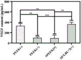
图4不同植入物体外促进内皮细胞释放血管内皮因子图。Fig. 4 Diagram of different implants promoting endothelial cells to release vascular endothelin in vitro.
图5是在糖尿病大鼠模型基础上进行皮下植入实验观察促进血管生长图。Fig. 5 is a graph of promoting blood vessel growth observed by subcutaneous implantation experiment on the basis of diabetic rat model.
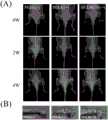
图6是糖尿病伴感染性骨缺损动物模型的X线图;大体标本;Micro-CT图;HE、甲苯胺蓝和Masson染色图。Fig. 6 is an X-ray diagram of an animal model of diabetes with infectious bone defect; gross specimen; Micro-CT diagram; HE, toluidine blue and Masson staining diagram.
具体实施方式Detailed ways
(1)下面结合具体实施方式和附图对本发明进行详细说明。应当理解的是,此处所描述的具体实施方式仅用于说明和解释本发明,并不用于限制本发明。(1) The present invention will be described in detail below in conjunction with specific embodiments and accompanying drawings. It should be understood that the specific embodiments described here are only used to illustrate and explain the present invention, and are not intended to limit the present invention.
(2)实施例(具有抗菌和促血管化骨植入物的制备)(2) Example (Preparation of Antibacterial and Provascularization Bone Implants)
本实施例的样品通过以下步骤制得:The sample of this embodiment is made through the following steps:
以单纯PEEK进行硫酸处理制得具有三维多孔的磺化聚醚醚酮(SPEEK),具有三维多孔的磺化聚醚醚酮(SPEEK)作为基底,在经过超声清洗和灭菌后进行下述的涂层制备。SPEEK在0.2mg/mL的聚乙烯亚胺(PEI)中浸泡一小时,获得表面正电荷改性的SPEEK。经过0.01M PBS洗涤、风干后,依次浸泡在A液(1mg/mL的单宁酸溶液)和B液(0.5mg/mL的庆大霉素和1mg/mL的Pluronic F127混合溶液)中,每次浸泡时间为十分钟。期间以0.01M PBS进行洗涤、风干。一般再按上述步骤三个循环次数后,制得SP/LBL*3的完整样品。The three-dimensional porous sulfonated polyetheretherketone (SPEEK) is obtained by sulfuric acid treatment with simple PEEK, and the three-dimensional porous sulfonated polyetheretherketone (SPEEK) is used as the substrate, and the following steps are carried out after ultrasonic cleaning and sterilization Coating preparation. SPEEK was soaked in 0.2 mg/mL polyethyleneimine (PEI) for one hour to obtain SPEEK with surface positive charge modification. After washing with 0.01M PBS and air-drying, soak in solution A (1 mg/mL tannic acid solution) and B solution (mixed solution of 0.5 mg/mL gentamycin and 1 mg/mL Pluronic F127) in sequence. The first soaking time is ten minutes. During this period, wash with 0.01M PBS and air dry. Generally, the complete sample of SP/LBL*3 is prepared after three cycles of the above steps.
(3)对本实施例中制得的具有抗菌和促血管化的植入物,得到的测试分析结果如下:(3) For the antibacterial and vascularization-promoting implants prepared in this example, the test and analysis results obtained are as follows:
图1是单纯PEEK、SPEEK以及SP/LBL*3的SEM图;如图1所示,单纯聚醚醚酮在经过磺化后会形成大小均匀的孔隙,在层层自组装后表面可负载药物。Figure 1 is the SEM image of pure PEEK, SPEEK and SP/LBL*3; as shown in Figure 1, pure polyether ether ketone will form pores of uniform size after sulfonation, and the surface can be loaded with drugs after layer-by-layer self-assembly .
图2所示在样品负载的单宁酸和庆大霉素累计药物释放曲线。Figure 2 shows the cumulative drug release curves of tannic acid and gentamicin loaded in the samples.
图3是不同样品体外连续抗菌性能评价。观察到其最大抗菌时间可达到6天,并在7天仍保留部分抗菌性能。Figure 3 is the in vitro continuous antibacterial performance evaluation of different samples. It is observed that its maximum antibacterial time can reach 6 days, and still retain some antibacterial properties in 7 days.
图4所示,在经表面涂层改性后的植入物治疗后,内皮细胞释放的VEGF与正常细胞之间无统计学差异。As shown in Figure 4, there was no statistical difference between the VEGF release from endothelial cells and normal cells after treatment with the implants modified by the surface coating.
图5是不同样品在糖尿病大鼠体内,不同植入物表面血管爬行图;如图5中的(A)所示,单纯的PEEK和SPEEK表面只有少量的血管爬行,但SP/LBL*3组表面有大量血管生成,并且血管处于不同层面,血管之间形成分支相互关联。在图5中的(B)中发现PEEK周围有较多的中心粒细胞浸润。在图5中的(C)和(D)中发现SP/LBL*3组明显较PEEK和SPEEK具有更高的成血管因子表达。这表明在糖尿病模型植入部位,SP/LBL*3能够促进周围血管生成。Figure 5 is a picture of blood vessel creeping on the surface of different implants in different samples in diabetic rats; There are a large number of angiogenesis on the surface, and the blood vessels are at different layers, and the branches of the blood vessels are connected with each other. In (B) in Fig. 5, more infiltration of neutrophils around PEEK was found. In (C) and (D) of Figure 5, it was found that SP/LBL*3 group had significantly higher expression of angiogenic factors than PEEK and SPEEK. This indicates that SP/LBL*3 can promote peripheral angiogenesis at the implantation site in diabetic models.
图6是糖尿病大鼠伴感染性骨缺损动物模型的X线图;大体标本;Micro-CT图;HE、甲苯胺蓝和Masson染色图。图6中(A)所示,PEEK(-)组(PEEK植入无感染组)与周围骨界限清楚,未见到明显的骨质破坏。PEEK(+)组(PEEK植入感染组)植入区域周围可见低密度阴影,提示植入物周围发生局部骨质破坏,植入物周围广泛的增生、硬化,骨膜新生骨显著,甚至形成骨包壳,大片骨坏死,说明感染没有得到控制,急性感染性骨髓炎转变成了慢性感染性骨髓炎。SP/LBL*3(+)组(SP/LBL*3植入感染组)植入区域周围未见到明显的感染灶,与周围骨界限清楚;如图6中(B)所示PEEK(+)组植入区域感染扩散到股骨远端,这与在影像学中的改变一致。而在PEEK(-)和SP/LBL*3(+)中则未见明显的骨质破坏;如图6中(C)中的A)从植入物的垂直轴观察植入物和周围骨组织之间的关系,这直观地反映了植入物和周围骨组织的情况。在PEEK(+)周围形成不连续的新骨,并伴有一些缺损。而PEEK(-)组和SP/LBL*3(+)组的植入物被完整的新骨(灰色箭头)包围,这也在植入物和周围骨之间形成了一个“桥”,称为小梁骨(白色箭头)。小梁骨的存在表明植入物可以与周围的骨组织结合;如图6中(C)中的B)显示植入体的局部股骨,使用μ-CT进行三维重建。白色箭头表示植入物上新骨的形成。在PEEK(+)只有极少量的新骨形成,表明存在无法控制的局部感染。在SP/LBL*3(+)组可以看到大量的新骨形成,表明局部感染能够被很好的控制;如图6中(C)中的C)所示从植入物的纵轴上可以观察到植入物与周围骨组织的关系,直观地反应了植入物与周围骨组织的情况。在PEEK(-)周围形成不连续的新骨并伴有一些缺损;如图6中(C)中的D)与PEEK(+)植入物相比,PEEK(-)与SP/LBL*3(+)组的BV/TV较PEEK(+)显著较高,具有统计学意义,而PEEK(-)与SP/LBL*3(+)组之间则没有统计学差异。这表明在SPEEK上负载TA和GS的涂层抑制了细菌感染,并内促进BV/TV的改善。此外,Tb.N的定量分析证实SP/LBL*3(+)植入区有明显的新骨和小梁骨形成。而在Tb.Th和Tb.Sp上进一步验证发现SP/LBL*3能够有效的抑制细菌感染并促进植入物与周围骨组织的整合。图6中(D)是在糖尿病大鼠基础上进行骨感染伴骨缺损动物模型的标本HE、甲苯胺蓝和Masson染色图;通过骨组织病理切片HE染色观察局部炎症、感染控制及新骨形成情况。在PEEK(-)组植入周围可见许多散在的中性粒细胞(黑色方框),表明植入物周围的无菌性炎症没有得到有效控制;PEEK(+)组周围可见许多散在的中性粒细胞(黑色方框),表明植入物周围的感染没有得到有效控制;在SP/LBL*3(+)组,植入物周围仅发现少量局部中性粒细胞,表明感染趋于局限性,局部骨感染得到控制。甲苯胺蓝染色(TB染色)观察新骨形成情况。在PEEK(-)组中,可以发现存在不完全的新骨形成(黑色箭头)。更重要的是PEEK(-)组中植入物和新骨之间出现明显的间隙(灰色箭头)。与PEEK(-)组相比,在PEEK(+)组甲苯胺蓝染色中未看到明显的新骨形成,植入物周围骨破坏严重。而在SP/LBL*3(+)组,新形成的骨能够覆盖住植入物,形成稳定的连接,说明植入物周围感染能够得到很好的控制,骨形成紧密的连接,具有较强的骨整合能力。进一步通过Masson染色评价植入物与周围骨组织的整合效果,从图中可以发现PEEK(-)与周围骨组织主要通过肌纤维进行相连,是一种无效的骨整合方式。而SP/LBL*3(+)则可以发现有明显的新骨与周围骨组织进行整合,并且可观察到新骨长入植入物周围三维孔隙结构中。Fig. 6 is an X-ray diagram of an animal model of diabetic rat with infectious bone defect; gross specimen; Micro-CT diagram; HE, toluidine blue and Masson staining diagram. As shown in Figure 6 (A), the PEEK (-) group (PEEK implanted without infection group) had a clear boundary with the surrounding bone, and no obvious bone destruction was seen. PEEK (+) group (PEEK implantation infection group) showed low-density shadows around the implanted area, suggesting that local bone destruction occurred around the implant, extensive hyperplasia and sclerosis around the implant, and periosteal new bone was prominent, and even bone formation Encrustation, large bone necrosis, indicating that the infection has not been controlled, and acute infectious osteomyelitis has transformed into chronic infectious osteomyelitis. In the SP/LBL*3(+) group (SP/LBL*3 implantation infection group), no obvious infection focus was seen around the implanted area, and the boundary with the surrounding bone was clear; as shown in Figure 6 (B), PEEK (+ ) group, the implantation area infection spread to the distal femur, which was consistent with the changes on imaging. In PEEK (-) and SP/LBL*3 (+), no obvious bone destruction was seen; as shown in Figure 6 (C) in A) The implant and surrounding bone were observed from the vertical axis of the implant The relationship between tissues, which intuitively reflects the condition of the implant and surrounding bone tissue. Discontinuous new bone formed around the PEEK(+) with some defects. While the implants in the PEEK(-) group and the SP/LBL*3(+) group were surrounded by intact new bone (gray arrow), which also formed a "bridge" between the implant and the surrounding bone, said For trabecular bone (white arrow). The presence of trabecular bone indicates that the implant can integrate with the surrounding bone tissue; (B) in (C) in Figure 6 shows the local femur of the implant, which was reconstructed in 3D using μ-CT. White arrows indicate the formation of new bone on the implant. In PEEK(+) there was only minimal new bone formation, indicating an uncontrolled local infection. A large amount of new bone formation can be seen in the SP/LBL*3(+) group, indicating that the local infection can be well controlled; as shown in Figure 6 (C) in the longitudinal axis of the implant The relationship between the implant and the surrounding bone tissue can be observed, which intuitively reflects the situation of the implant and the surrounding bone tissue. Discontinuous new bone was formed around PEEK(-) with some defects; Fig. 6(C) D) Compared with PEEK(+) implant, PEEK(-) with SP/LBL*3 The BV/TV of the (+) group was significantly higher than that of PEEK (+), with statistical significance, while there was no statistical difference between the PEEK (-) and SP/LBL*3 (+) groups. This indicated that the coating loaded with TA and GS on SPEEK inhibited bacterial infection and promoted the improvement of BV/TV. In addition, quantitative analysis of Tb.N confirmed significant new bone and trabecular bone formation in the SP/LBL*3(+) implanted area. Further verification on Tb.Th and Tb.Sp found that SP/LBL*3 could effectively inhibit bacterial infection and promote the integration of implants and surrounding bone tissue. Figure 6 (D) is the HE, toluidine blue, and Masson staining images of specimens from animal models of bone infection with bone defects based on diabetic rats; local inflammation, infection control, and new bone formation were observed through HE staining of bone histopathological sections Condition. Many scattered neutrophils (black boxes) were seen around the implant in the PEEK(-) group, indicating that the aseptic inflammation around the implant was not effectively controlled; many scattered neutrophils were seen around the PEEK(+) group Granulocytes (black box), indicating that the infection around the implant was not effectively controlled; in the SP/LBL*3(+) group, only a few localized neutrophils were found around the implant, indicating that the infection tended to be localized , Local bone infection was controlled. Toluidine blue staining (TB staining) was used to observe the new bone formation. In the PEEK(-) group, incomplete new bone formation could be found (black arrow). More importantly, a clear gap (gray arrow) appeared between the implant and the new bone in the PEEK(-) group. Compared with the PEEK (-) group, no obvious new bone formation was seen in the toluidine blue staining of the PEEK (+) group, and the bone destruction around the implant was severe. In the SP/LBL*3(+) group, the newly formed bone can cover the implant and form a stable connection, indicating that the infection around the implant can be well controlled, the bone forms a tight connection, and has a strong osseointegration ability. The integration effect of the implant and the surrounding bone tissue was further evaluated by Masson staining. It can be seen from the figure that PEEK (-) is connected with the surrounding bone tissue mainly through muscle fibers, which is an ineffective way of osseointegration. In SP/LBL*3 (+), it can be found that there is obvious integration of new bone with the surrounding bone tissue, and the growth of new bone into the three-dimensional pore structure around the implant can be observed.
(4)以上实施例仅为本发明的优选实施例,并非全部。基于实施方式中的实施例,本领域技术人员在没有做出创造性劳动的前提下所获得其它实施例,都属于本发明的保护范围。(4) The above embodiments are only preferred embodiments of the present invention, not all of them. Based on the examples in the implementation manners, other examples obtained by those skilled in the art without making creative efforts all belong to the protection scope of the present invention.
Claims (10)
Priority Applications (1)
| Application Number | Priority Date | Filing Date | Title |
|---|---|---|---|
| CN202210118813.5ACN114470324B (en) | 2022-02-08 | 2022-02-08 | Novel strategy for the modification of universal bone implants for the intervention of bone defects associated with diabetes |
Applications Claiming Priority (1)
| Application Number | Priority Date | Filing Date | Title |
|---|---|---|---|
| CN202210118813.5ACN114470324B (en) | 2022-02-08 | 2022-02-08 | Novel strategy for the modification of universal bone implants for the intervention of bone defects associated with diabetes |
Publications (2)
| Publication Number | Publication Date |
|---|---|
| CN114470324A CN114470324A (en) | 2022-05-13 |
| CN114470324Btrue CN114470324B (en) | 2023-04-04 |
Family
ID=81478200
Family Applications (1)
| Application Number | Title | Priority Date | Filing Date |
|---|---|---|---|
| CN202210118813.5AExpired - Fee RelatedCN114470324B (en) | 2022-02-08 | 2022-02-08 | Novel strategy for the modification of universal bone implants for the intervention of bone defects associated with diabetes |
Country Status (1)
| Country | Link |
|---|---|
| CN (1) | CN114470324B (en) |
Families Citing this family (1)
| Publication number | Priority date | Publication date | Assignee | Title |
|---|---|---|---|---|
| CN114989475B (en)* | 2022-05-30 | 2023-07-18 | 浙江大学 | Preparation method and product application of biological functionalized surface modified polyether-ether-ketone material |
Citations (3)
| Publication number | Priority date | Publication date | Assignee | Title |
|---|---|---|---|---|
| EP2544728A1 (en)* | 2010-03-12 | 2013-01-16 | Carmeda AB | Immobilised biological entities |
| CN106176695A (en)* | 2016-07-27 | 2016-12-07 | 中国人民解放军第四军医大学 | Resveratrol is the application in medical titanium alloy implants under diabetic conditions |
| CN108774335A (en)* | 2018-07-05 | 2018-11-09 | 深圳大学 | A method of polyether-ether-ketone surface is modified using polyaniline nano fiber |
Family Cites Families (6)
| Publication number | Priority date | Publication date | Assignee | Title |
|---|---|---|---|---|
| AU2004212942A1 (en)* | 2003-02-14 | 2004-09-02 | Depuy Spine, Inc. | In-situ formed intervertebral fusion device |
| MX2008000131A (en)* | 2005-07-01 | 2008-04-04 | Cinv Ag | Medical devices comprising a reticulated composite material. |
| KR102109324B1 (en)* | 2014-11-19 | 2020-05-12 | 헤레우스 메디컬 컴포넌츠 엘엘씨 | Conductive polymer coatings for three dimensional substrates |
| CN109913400B (en)* | 2016-09-14 | 2023-03-14 | 四川蓝光英诺生物科技股份有限公司 | Artificial tissue precursors and methods for making same |
| WO2018185770A1 (en)* | 2017-04-05 | 2018-10-11 | Setbone Medical Ltd. | Property changing implant |
| CN112618791A (en)* | 2020-12-25 | 2021-04-09 | 福建医科大学附属协和医院 | Polyether-ether-ketone three-dimensional porous and modified polydopamine/gentamicin for implant antibiosis, anti-inflammation and promotion of osseointegration |
- 2022
- 2022-02-08CNCN202210118813.5Apatent/CN114470324B/ennot_activeExpired - Fee Related
Patent Citations (3)
| Publication number | Priority date | Publication date | Assignee | Title |
|---|---|---|---|---|
| EP2544728A1 (en)* | 2010-03-12 | 2013-01-16 | Carmeda AB | Immobilised biological entities |
| CN106176695A (en)* | 2016-07-27 | 2016-12-07 | 中国人民解放军第四军医大学 | Resveratrol is the application in medical titanium alloy implants under diabetic conditions |
| CN108774335A (en)* | 2018-07-05 | 2018-11-09 | 深圳大学 | A method of polyether-ether-ketone surface is modified using polyaniline nano fiber |
Also Published As
| Publication number | Publication date |
|---|---|
| CN114470324A (en) | 2022-05-13 |
Similar Documents
| Publication | Publication Date | Title |
|---|---|---|
| US20230037048A1 (en) | Extracellular Matrix-Derived Gels and Related Methods | |
| US12005158B2 (en) | Methods for preparation of a terminally sterilized hydrogel derived from extracellular matrix | |
| Li et al. | Elastic porous microspheres/extracellular matrix hydrogel injectable composites releasing dual bio-factors enable tissue regeneration | |
| JP2024161378A (en) | Extracellular Matrix Composition | |
| Shu et al. | Self‐Tandem Bio‐Heterojunctions Empower Orthopedic Implants with Amplified Chemo‐Photodynamic Anti‐Pathogenic Therapy and Boosted Diabetic Osseointegration | |
| Bakhtiar et al. | Human amniotic membrane extracellular matrix scaffold for dental pulp regeneration in vitro and in vivo | |
| US20180200405A1 (en) | Methods of preparing ecm scaffolds and hydrogels from colon | |
| Zhang et al. | Metal phenolic nanodressing of porous polymer scaffolds for enhanced bone regeneration via interfacial gating growth factor release and stem cell differentiation | |
| CN112618791A (en) | Polyether-ether-ketone three-dimensional porous and modified polydopamine/gentamicin for implant antibiosis, anti-inflammation and promotion of osseointegration | |
| Ai et al. | Photo-crosslinked bioactive BG/BMSCs@ GelMA hydrogels for bone-defect repairs | |
| CN113679889B (en) | Acellular matrix composite material and preparation method and application thereof | |
| Changchen et al. | The characterization, cytotoxicity, macrophage response and tissue regeneration of decellularized cartilage in costal cartilage defects | |
| CN105935317B (en) | Plant percutaneous base station and preparation method thereof in a kind of surface carried medicine sustained-release jaw face | |
| CN114470324B (en) | Novel strategy for the modification of universal bone implants for the intervention of bone defects associated with diabetes | |
| Fang et al. | Review on additives in hydrogels for 3D bioprinting of regenerative medicine: from mechanism to methodology | |
| Zhang et al. | A biomimetic multifunctional scaffold for infectious vertical bone augmentation | |
| CN110279890A (en) | Method of modifying and application of the dexamethasone/minocycline based on liposome on the surface PEEK | |
| Lu et al. | Enhancing epithelial regeneration with gelatin methacryloyl hydrogel loaded with extracellular vesicles derived from adipose mesenchymal stem cells for decellularized tracheal patching. | |
| TWI445557B (en) | To induce new bone formation around the implant compositions | |
| US20220096712A1 (en) | Biomaterials comprising a scaffold containing a mineral compound, and uses thereof as bone substitutes | |
| CN114196625B (en) | Preparation method of engineering small extracellular vesicles and application of engineering small extracellular vesicles in artificial vertebral bodies | |
| Kimura et al. | Fabrication of Decellularized Tissue for Biomedical Application | |
| CN116790017A (en) | A surface-modified polyetheretherketone material and its preparation method and application | |
| Li et al. | Janus Porous Polylactic Acid Membranes with Versatile Metal-Phenolic Interface for Biomimetic Periodontal Bone Regeneration | |
| CN120346367A (en) | Piezoelectric nano hydroxyapatite modified titanium-based implant, and preparation method and application thereof |
Legal Events
| Date | Code | Title | Description |
|---|---|---|---|
| PB01 | Publication | ||
| PB01 | Publication | ||
| SE01 | Entry into force of request for substantive examination | ||
| SE01 | Entry into force of request for substantive examination | ||
| GR01 | Patent grant | ||
| GR01 | Patent grant | ||
| CF01 | Termination of patent right due to non-payment of annual fee | ||
| CF01 | Termination of patent right due to non-payment of annual fee | Granted publication date:20230404 |