CN114469124A - Method for identifying abnormal electrocardiosignals in motion process - Google Patents
Method for identifying abnormal electrocardiosignals in motion processDownload PDFInfo
- Publication number
- CN114469124A CN114469124ACN202210114479.6ACN202210114479ACN114469124ACN 114469124 ACN114469124 ACN 114469124ACN 202210114479 ACN202210114479 ACN 202210114479ACN 114469124 ACN114469124 ACN 114469124A
- Authority
- CN
- China
- Prior art keywords
- sequence
- signal
- electrocardiosignals
- noise
- imf
- Prior art date
- Legal status (The legal status is an assumption and is not a legal conclusion. Google has not performed a legal analysis and makes no representation as to the accuracy of the status listed.)
- Granted
Links
- 238000000034methodMethods0.000titleclaimsabstractdescription36
- 230000002159abnormal effectEffects0.000titleclaimsabstractdescription15
- 230000008569processEffects0.000titleclaimsabstractdescription9
- 238000013145classification modelMethods0.000claimsabstractdescription20
- 230000000694effectsEffects0.000claimsabstractdescription13
- 238000004422calculation algorithmMethods0.000claimsabstractdescription12
- 238000004458analytical methodMethods0.000claimsabstractdescription6
- 238000012549trainingMethods0.000claimsdescription21
- 108010076504Protein Sorting SignalsProteins0.000claimsdescription16
- 238000000354decomposition reactionMethods0.000claimsdescription8
- 238000012360testing methodMethods0.000claimsdescription8
- 230000000737periodic effectEffects0.000claimsdescription5
- 238000005070samplingMethods0.000claimsdescription4
- 238000007781pre-processingMethods0.000claimsdescription3
- 238000012795verificationMethods0.000claimsdescription3
- 238000012216screeningMethods0.000claims2
- 238000012935AveragingMethods0.000claims1
- 238000012545processingMethods0.000abstractdescription3
- 238000011161developmentMethods0.000abstractdescription2
- 238000012544monitoring processMethods0.000abstract1
- 230000001737promoting effectEffects0.000abstract1
- 238000010586diagramMethods0.000description7
- 230000000875corresponding effectEffects0.000description6
- 230000006870functionEffects0.000description6
- 238000010200validation analysisMethods0.000description5
- 208000024172Cardiovascular diseaseDiseases0.000description3
- 238000013461designMethods0.000description3
- 230000007246mechanismEffects0.000description3
- 238000003058natural language processingMethods0.000description3
- 238000000718qrs complexMethods0.000description3
- 230000005856abnormalityEffects0.000description2
- 238000004364calculation methodMethods0.000description2
- 238000007635classification algorithmMethods0.000description2
- 238000013135deep learningMethods0.000description2
- 238000010801machine learningMethods0.000description2
- 238000010606normalizationMethods0.000description2
- 238000011160researchMethods0.000description2
- ORILYTVJVMAKLC-UHFFFAOYSA-NAdamantaneNatural productsC1C(C2)CC3CC1CC2C3ORILYTVJVMAKLC-UHFFFAOYSA-N0.000description1
- 230000009286beneficial effectEffects0.000description1
- 230000002596correlated effectEffects0.000description1
- 238000001514detection methodMethods0.000description1
- 230000018109developmental processEffects0.000description1
- 238000003745diagnosisMethods0.000description1
- 238000005516engineering processMethods0.000description1
- 238000011156evaluationMethods0.000description1
- 230000005284excitationEffects0.000description1
- 238000000605extractionMethods0.000description1
- 230000003760hair shineEffects0.000description1
- 238000003709image segmentationMethods0.000description1
- 230000006872improvementEffects0.000description1
- 238000012986modificationMethods0.000description1
- 230000004048modificationEffects0.000description1
- 238000005457optimizationMethods0.000description1
- 238000003672processing methodMethods0.000description1
- 230000009467reductionEffects0.000description1
- 238000001228spectrumMethods0.000description1
- 230000003068static effectEffects0.000description1
- 230000009466transformationEffects0.000description1
Images


Classifications
- A—HUMAN NECESSITIES
- A61—MEDICAL OR VETERINARY SCIENCE; HYGIENE
- A61B—DIAGNOSIS; SURGERY; IDENTIFICATION
- A61B5/00—Measuring for diagnostic purposes; Identification of persons
- A61B5/24—Detecting, measuring or recording bioelectric or biomagnetic signals of the body or parts thereof
- A61B5/316—Modalities, i.e. specific diagnostic methods
- A61B5/318—Heart-related electrical modalities, e.g. electrocardiography [ECG]
- A61B5/346—Analysis of electrocardiograms
- A61B5/349—Detecting specific parameters of the electrocardiograph cycle
- A61B5/352—Detecting R peaks, e.g. for synchronising diagnostic apparatus; Estimating R-R interval
- A—HUMAN NECESSITIES
- A61—MEDICAL OR VETERINARY SCIENCE; HYGIENE
- A61B—DIAGNOSIS; SURGERY; IDENTIFICATION
- A61B5/00—Measuring for diagnostic purposes; Identification of persons
- A61B5/72—Signal processing specially adapted for physiological signals or for diagnostic purposes
- A61B5/7203—Signal processing specially adapted for physiological signals or for diagnostic purposes for noise prevention, reduction or removal
- A—HUMAN NECESSITIES
- A61—MEDICAL OR VETERINARY SCIENCE; HYGIENE
- A61B—DIAGNOSIS; SURGERY; IDENTIFICATION
- A61B5/00—Measuring for diagnostic purposes; Identification of persons
- A61B5/72—Signal processing specially adapted for physiological signals or for diagnostic purposes
- A61B5/7235—Details of waveform analysis
- A61B5/7253—Details of waveform analysis characterised by using transforms
- A61B5/7257—Details of waveform analysis characterised by using transforms using Fourier transforms
- A—HUMAN NECESSITIES
- A61—MEDICAL OR VETERINARY SCIENCE; HYGIENE
- A61B—DIAGNOSIS; SURGERY; IDENTIFICATION
- A61B5/00—Measuring for diagnostic purposes; Identification of persons
- A61B5/72—Signal processing specially adapted for physiological signals or for diagnostic purposes
- A61B5/7235—Details of waveform analysis
- A61B5/7264—Classification of physiological signals or data, e.g. using neural networks, statistical classifiers, expert systems or fuzzy systems
- A61B5/7267—Classification of physiological signals or data, e.g. using neural networks, statistical classifiers, expert systems or fuzzy systems involving training the classification device
Landscapes
- Health & Medical Sciences (AREA)
- Life Sciences & Earth Sciences (AREA)
- Engineering & Computer Science (AREA)
- Physics & Mathematics (AREA)
- Public Health (AREA)
- Surgery (AREA)
- Cardiology (AREA)
- Veterinary Medicine (AREA)
- General Health & Medical Sciences (AREA)
- Biophysics (AREA)
- Pathology (AREA)
- Biomedical Technology (AREA)
- Heart & Thoracic Surgery (AREA)
- Medical Informatics (AREA)
- Molecular Biology (AREA)
- Animal Behavior & Ethology (AREA)
- Artificial Intelligence (AREA)
- Signal Processing (AREA)
- Physiology (AREA)
- Computer Vision & Pattern Recognition (AREA)
- Psychiatry (AREA)
- Mathematical Physics (AREA)
- Evolutionary Computation (AREA)
- Fuzzy Systems (AREA)
- Measurement And Recording Of Electrical Phenomena And Electrical Characteristics Of The Living Body (AREA)
Abstract
Description
Translated fromChinese技术领域technical field
本发明属于动态心电信号处理的技术领域,具体涉及一种运动过程中异常心电信号的识别方法。The invention belongs to the technical field of dynamic electrocardiographic signal processing, and in particular relates to a method for identifying abnormal electrocardiographic signals during exercise.
背景技术Background technique
随着心血管类疾病日益增长的患病率,其逐渐成为研究的热点课题。对于心血管类疾病主要通过心电图(ECG)进行检测,心电图是一种电位波形图,描绘了心脏激动过程中体表上产生周期性的电变化活动。目前常规的心电诊断技术仍是医生通过静态心电图结合临床经验来判断患者是否异常,但是伴随着移动智能设备的快速发展,实时的心电信号异常检测任务亟待解决。比如通过电子手环可以实时监测人体的心电信号,若能动态的对这些信号进行异常识别,便可以实现预防心血管类疾病的功能。With the increasing prevalence of cardiovascular diseases, it has gradually become a hot research topic. Cardiovascular diseases are mainly detected by an electrocardiogram (ECG), which is a potential waveform diagram that depicts periodic electrical changes on the body surface during the excitation of the heart. At present, the conventional ECG diagnosis technology is still that doctors use static ECG combined with clinical experience to determine whether a patient is abnormal. However, with the rapid development of mobile smart devices, the task of real-time ECG abnormality detection needs to be solved urgently. For example, the electronic bracelet can monitor the human body's ECG signals in real time. If these signals can be dynamically identified, the function of preventing cardiovascular diseases can be realized.
在心电信号异常识别的研究中,主要可以分为三个板块:(i)心电信号去噪,主要应用经验模态分解(EMD)、小波阈值去噪以及滤波器等方法;In the research of abnormal ECG signal identification, it can be divided into three sections: (i) ECG signal denoising, mainly using empirical mode decomposition (EMD), wavelet threshold denoising and filter methods;
(ii)心电信号特征提取,时域特征一般提取QRS波群相关特征,频域特征常通过傅里叶变换、小波变换等信号处理方法;(iii)心电信号分类算法,主要应用机器学习和深度学习分类算法。(ii) ECG signal feature extraction, time-domain features generally extract QRS complex-related features, and frequency-domain features are often processed by Fourier transform, wavelet transform and other signal processing methods; (iii) ECG signal classification algorithm, mainly using machine learning and deep learning classification algorithms.
数据质量对分类效果具有直接的影响。EMD方法是信号去噪的常用方法之一,目前研究人员在使用EMD对心电信号进行去噪时,会结合阈值去噪方法对分解后得到的IMF分量系数进行筛选进而重构实现去噪。但是这个过程中往往忽略掉EMD在心电信号上分解时存在的模态混叠问题,即使通过加入白噪声的EEMD方法也无法解决,这是由于QRS波群过窄且过陡的性质,加入的白噪声会被其覆盖。IMF分量中模态混叠问题的存在,会导致后续确定阈值时存在偏差,影响去噪效果,亟需一种新的分解方法来解决EEMD在心电信号分解上的模态混叠问题。Data quality has a direct impact on classification performance. The EMD method is one of the common methods of signal denoising. At present, when researchers use EMD to denoise ECG signals, they will combine the threshold denoising method to screen the decomposed IMF component coefficients and then reconstruct to achieve denoising. However, in this process, the modal aliasing problem that exists when EMD is decomposed on ECG signals is often ignored, which cannot be solved even by adding white noise to the EEMD method. This is due to the narrow and steep nature of the QRS complex. White noise will be covered by it. The existence of the modal aliasing problem in the IMF component will lead to a deviation in the subsequent determination of the threshold value, which will affect the denoising effect. A new decomposition method is urgently needed to solve the modal aliasing problem of EEMD in ECG signal decomposition.
近几年来,研究人员不再局限于序列数据分析,而是提取其二维特征,比如转化为二维图像或者通过相关信号分析手段提取频域二维特征,然后构建基于CNN的图分类模型。深度学习是近年来机器学习领域的热点课题,在自然语言处理和计算机视觉领域取得了重要的成果,其中2017年提出的基于自注意力机制的Transformer模型大放异彩,在自然语言处理上效果显著。近两年,研究人员逐渐将其迁入到计算机视觉领域,提出了VisionTransformer(VIT)模型,也证明其在大批量数据集上表现良好。In recent years, researchers are no longer limited to the analysis of sequence data, but to extract its two-dimensional features, such as converting into two-dimensional images or extracting two-dimensional features in the frequency domain by means of correlated signal analysis, and then constructing a CNN-based graph classification model. Deep learning is a hot topic in the field of machine learning in recent years, and important results have been achieved in the fields of natural language processing and computer vision. Among them, the Transformer model based on the self-attention mechanism proposed in 2017 shines brightly and has remarkable effects in natural language processing. . In the past two years, researchers have gradually moved it into the field of computer vision, and proposed the VisionTransformer (VIT) model, which has also proved to perform well on large-scale datasets.
发明内容SUMMARY OF THE INVENTION
有鉴于此,本发明提供了一种运动过程中异常心电信号的识别方法,能够解决EEMD算法在心电信号分解上的模态混叠问题,通过傅里叶变换自动划分心拍,实现高准确率。In view of this, the present invention provides a method for identifying abnormal ECG signals during exercise, which can solve the modal aliasing problem of EEMD algorithm in the decomposition of ECG signals, and automatically divide heart beats through Fourier transform to achieve high accuracy. .
实现本发明的技术方案如下:The technical scheme that realizes the present invention is as follows:
一种运动过程中异常心电信号的识别方法,包括以下步骤:A method for identifying abnormal electrocardiographic signals during exercise, comprising the following steps:
步骤1:获取用于训练和测试的运动过程中的心电信号,获得心电信号的类别标签,并对其进行数据预处理;Step 1: Acquire the ECG signal during exercise for training and testing, obtain the category label of the ECG signal, and perform data preprocessing on it;
步骤1.1:设计改进EEMD算法,对心电信号进行分解;Step 1.1: Design and improve the EEMD algorithm to decompose the ECG signal;
步骤1.2:通过阈值和阈值函数对分解后的IMF分量进行去噪,并重构得到去噪信号;Step 1.2: Denoise the decomposed IMF components through a threshold and a threshold function, and reconstruct to obtain a denoised signal;
步骤2:心拍自动划分;Step 2: The heart beat is automatically divided;
步骤2.1:对去噪后的心电信号进行傅里叶变换,得到该心电信号的周期特征;Step 2.1: Perform Fourier transform on the denoised ECG signal to obtain the periodic characteristics of the ECG signal;
步骤2.2:通过差分法定位R波位置后进行心拍划分;Step 2.2: Divide the heart beat after locating the R wave position by the difference method;
步骤3:构建VIT分类模型,提取步骤2划分后的心拍的频域特征输入到分类模型中进行训练并分类;Step 3: construct a VIT classification model, extract the frequency domain features of the heart beats divided in
步骤3.1:提取心拍序列的二维频域特征;Step 3.1: Extract the two-dimensional frequency domain features of the heart beat sequence;
步骤3.2:搭建Vision Transformer分类模型;Step 3.2: Build the Vision Transformer classification model;
步骤3.3:将训练数据集划分为80%的训练集、20%的验证集,将训练集输入到VIT模型中进行训练,通过验证集对模型进行调参,得到分类模型;Step 3.3: Divide the training data set into 80% training set and 20% validation set, input the training set into the VIT model for training, and adjust the parameters of the model through the validation set to obtain a classification model;
步骤3.4:计算分类模型在测试集上的准确率、召回率和精准率,评价模型的分类效果。Step 3.4: Calculate the accuracy, recall and precision of the classification model on the test set, and evaluate the classification effect of the model.
进一步地,步骤1.1具体为:Further, step 1.1 is specifically:
(1)得到原始信号采样序列{x(tn)},n=1,2,...,N,其中N为序列长度,计算原始序列的一阶差分序列


(2)计算一阶差分序列的绝对值的中位数,作为是否进行插值的判断阈值,即
(3)对一阶差分序列的值进行判断,若差分值大于判断阈值,即


(4)生成与插值信号序列同等长度N+K的高斯白噪声序列,向插值信号序列

(5)计算序列
(6)对上下包络线求均值,得到局部均值曲线,就是第一个IMF分量
(7)重复步骤(5)和(6),直到余项


进一步地,步骤1.2具体为:Further, step 1.2 is specifically:
(1)对步骤1.1分解后得到的q个IMF分量,确定出以噪声为主导的分量,计算每个IMF分量的过零率pi,i=1,...,q,然后计算相邻两个IMF分量的过零率比值fi’=pi/pi+1,i’=1,..,q-1,当fi’第一次达到极大值点时,定义其为以噪声为主导的分量和以信号为主导的分量的分界处;(1) For the q IMF components obtained after decomposing in step 1.1, determine the noise-dominated component, calculate the zero-crossing rate pi , i=1,...,q of each IMF component, and then calculate the adjacent The zero-crossing ratio ratio of the two IMF components fi' = pi /pi+1 , i' = 1, .., q-1, when fi' reaches the maximum point for the first time, it is defined as the boundary between the noise-dominated and signal-dominated components;
(2)对以噪声为主导的IMF分量进行系数筛选,使用通用阈值


对IMF分量系数进行筛选;Screen the IMF component coefficients;
(3)根据步骤1.1中(3)记录的原始信号的位置,对去噪后的以噪声为主导的IMF分量和以信号为主导的IMF分量进行相应位置重构,得到去噪信号序列{x’(tn)},n=1,2,...,N。(3) According to the position of the original signal recorded in (3) in step 1.1, reconstruct the corresponding positions of the noise-dominated IMF component and the signal-dominated IMF component after denoising, to obtain the denoised signal sequence {x '(tn )}, n=1,2,...,N.
进一步地,步骤2.1具体为:Further, step 2.1 is specifically:
(1)对去噪后的心电信号序列{x’(tn)},n=1,2,...,N进行离散傅里叶变换
(2)找到其傅里叶变换F(ω)的极大值点Fmax(ω)及其对应的频率ωmax,得到心电信号序列的周期T=N/ωmax,其中N为序列长度。(2) Find the maximum point Fmax (ω) of its Fourier transform F(ω) and its corresponding frequency ωmax , and obtain the period of the ECG signal sequence T=N/ωmax , where N is the sequence length .
有益效果:Beneficial effects:
本发明方法根据心电信号的波形特征,设计了基于插值的改进EEMD方法,能实现对心电信号的有效分解,进而提高了去噪效果,在保证原始信号特征的情况下提高信噪比,提高数据质量以为分类打下良好基础;使用傅里叶变换对心拍进行自动划分,打破了以往依赖先验进行划分的限制;通过VIT模型进行分类,实现了较好的心电信号异常识别效果,准确度达到93%。According to the waveform characteristics of the ECG signal, the method of the invention designs an improved EEMD method based on interpolation, which can realize the effective decomposition of the ECG signal, thereby improving the denoising effect, and improving the signal-to-noise ratio while ensuring the original signal characteristics. Improve data quality to lay a good foundation for classification; use Fourier transform to automatically divide heart beats, breaking the previous limitation of relying on prior division; classifying through VIT model, achieves better ECG abnormality recognition effect, accurate The degree reaches 93%.
附图说明Description of drawings
图1为改进EEMD算法对心电信号分解流程图。Figure 1 is a flow chart of the decomposition of ECG signals by the improved EEMD algorithm.
图2为仿真信号与去噪后信号图像。Figure 2 shows the simulated signal and the signal image after denoising.
图2(a)表示仿真带噪信号图;Figure 2(a) shows the simulated noisy signal diagram;
图2(b)表示基于改进EEMD去噪后的信号图像;Figure 2(b) shows the signal image after denoising based on improved EEMD;
图3为去噪效果对比图。Figure 3 is a comparison diagram of the denoising effect.
图4为本发明通过傅里叶变换自动划分出的心拍示意图。FIG. 4 is a schematic diagram of a heart beat automatically divided by Fourier transform according to the present invention.
图4(a)表示傅里叶变换后的频谱;Figure 4(a) shows the spectrum after Fourier transform;
图4(b)表示单个心拍示意图;Figure 4(b) shows a schematic diagram of a single heart beat;
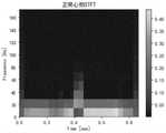
图5为单个心拍的STFT图像。Figure 5 is an STFT image of a single heart beat.
图6为VIT模型的结构图。Figure 6 is a structural diagram of the VIT model.
具体实施方式Detailed ways
下面结合附图并举实施例,对本发明进行详细描述。The present invention will be described in detail below with reference to the accompanying drawings and embodiments.
一种运动过程中异常心电信号的识别方法,包括以下步骤:A method for identifying abnormal electrocardiographic signals during exercise, comprising the following steps:
步骤1:获取用于训练和测试的运动过程中心电信号,获得心电信号的类别标签,并对其进行数据预处理。Step 1: Obtain the ECG signal during the exercise process for training and testing, obtain the category label of the ECG signal, and perform data preprocessing on it.
步骤1.1:设计改进EEMD算法,对心电信号进行分解。Step 1.1: Design and improve the EEMD algorithm to decompose the ECG signal.
具体地:specifically:
针对EEMD算法在心电信号QRS波群的陡脉冲信号中无法添加有效添加白噪声导致出现模态混叠这一问题,提出改进的EEMD算法,具体步骤如下:(1)得到原始信号采样序列{x(tn)},n=1,2,...,N,其中N为序列长度,计算原始序列的一阶差分序列


(2)计算一阶差分序列的绝对值的中位数,作为是否进行插值的判断阈值,即
(3)对一阶差分序列的值进行判断,若差分值大于判断阈值,即


(4)生成与插值信号序列同等长度N+K的高斯白噪声序列,向插值信号序列

(5)计算序列
(6)对上下包络线求均值,得到局部均值曲线,就是第一个IMF分量
(7)重复步骤(5)和(6),直到余项


步骤1.2:通过阈值和阈值函数对分解后的IMF分量进行去噪,并重构得到去噪信号。Step 1.2: De-noise the decomposed IMF components through threshold and threshold function, and reconstruct to obtain a de-noised signal.
具体地:specifically:
(1)对步骤1.1分解后得到的q个IMF分量,确定出以噪声为主导的分量,因为心电信号中噪声主要为高频噪声,主要集中在前面的IMF分量中。计算每个IMF分量的过零率pi,i=1,...,q,然后计算相邻两个IMF分量的过零率比值fi’=pi/pi+1,i’=1,..,q-1,当fi’第一次达到极大值点时,定义其为以噪声为主导的分量和以信号为主导的分量的分界处。(1) For the q IMF components obtained after decomposing in step 1.1, determine the components dominated by noise, because the noise in the ECG signal is mainly high-frequency noise, which is mainly concentrated in the previous IMF components. Calculate the zero-crossing rate pi ,i=1,...,q of each IMF component, and then calculate the zero-crossing rate ratio fi' =pi /pi+1 ,i'= 1,..,q-1, when fi' reaches the maximum point for the first time, it is defined as the boundary between the noise-dominated component and the signal-dominated component.
(2)对以噪声为主导的IMF分量进行系数筛选,使用通用阈值


对IMF分量系数进行筛选。Filter the IMF component coefficients.
(3)根据步骤1.1中(3)记录的原始信号的位置,对去噪后的以噪声为主导的IMF分量和以信号为主导的IMF分量进行相应位置重构,得到去噪信号序列{x’(tn)},n=1,2,...,N,图2为仿真信号与去噪后信号图像。(3) According to the position of the original signal recorded in (3) in step 1.1, reconstruct the corresponding positions of the noise-dominated IMF component and the signal-dominated IMF component after denoising, to obtain the denoised signal sequence {x '(tn )}, n=1,2,...,N, Fig. 2 is the simulated signal and the signal image after denoising.
(4)评价去噪效果,使用指标包括:信噪比


步骤2:心拍自动划分。Step 2: The heart beat is automatically divided.
步骤2.1:对去噪后的心电信号进行傅里叶变换,计算得到该心电信号的周期特征。Step 2.1: Perform Fourier transform on the denoised ECG signal, and calculate the periodic characteristics of the ECG signal.
具体地:specifically:
(1)对去噪后的心电信号序列{x’(tn)},n=1,2,...,N进行离散傅里叶变换
(2)找到其傅里叶变换F(ω)的极大值点Fmax(ω)及其对应的频率ωmax,得到心电信号序列的周期T=N/ωmax,其中N为序列长度。(2) Find the maximum point Fmax (ω) of its Fourier transform F(ω) and its corresponding frequency ωmax , and obtain the period of the ECG signal sequence T=N/ωmax , where N is the sequence length .
步骤2.2:通过差分法定位R波位置后进行心拍划分。Step 2.2: Divide the heart beat after locating the R wave position by the difference method.
具体地:specifically:
(1)通过差分法定位R波位置,步骤如下:(1) To locate the R wave position by the differential method, the steps are as follows:
①计算去噪信号序列{x’(tn)},n=1,2,...,N的一阶差分序列


②从起始点开始以时间窗W截取一段心电数据,求出极大值点记为R1;2. Intercept a section of ECG data with time window W from the starting point, and find the maximum point and record it as R1 ;
③找出一阶差分序列
④以第一个R波位置为起点,按照步骤②和③找出第二个R波位置,依此类推。④ Using the first R wave position as the starting point, follow
(2)对于检测到的R波位置,分别向前向后提取2T/5、3T/5个采样点,记为单个心拍,至此,将心电信号序列全部划分为单个心拍序列

步骤3:构建VIT分类模型,提取步骤2划分后的心拍的频域特征输入到分类模型中进行训练并分类。Step 3: Build a VIT classification model, extract the frequency domain features of the heart beats divided in
步骤3.1:提取心拍序列的二维频域特征。Step 3.1: Extract the two-dimensional frequency domain features of the beat sequence.
具体地:specifically:
搭建对步骤2划分后的心拍序列

步骤3.2:搭建Vision Transformer分类模型。Step 3.2: Build the Vision Transformer classification model.
具体地:specifically:
Vision Transformer是使用基于Self-Attention机制的传统Transformer的架构,对二维图像数据进行分类,对于大批量数据的效果要高于传统的基于CNN的模型。图6为VIT模型的结构图。Vision Transformer uses the traditional Transformer architecture based on the Self-Attention mechanism to classify two-dimensional image data, and its effect on large batches of data is higher than that of traditional CNN-based models. Figure 6 is a structural diagram of the VIT model.
(1)图像分块与降维。采用切块的方式将x∈H×W×C的连续的图像转换为类似NLP任务中的一个个token,变成一个xp∈N’×(P2·C)的展平的二维块序列,其中H×W为二维图像大小,C为通道数,P为块的大小,N’=HW/P2表示二维块的个数,每个块的维度是(P2·C)。(1) Image segmentation and dimensionality reduction. The continuous image of x∈H×W×C is converted into a token similar to the NLP task by dicing, and becomes a flattened two-dimensional block of xp ∈ N'×(P2 ·C) sequence, where H×W is the size of the two-dimensional image, C is the number of channels, P is the size of the block, N'=HW/P2 represents the number of two-dimensional blocks, and the dimension of each block is (P2 ·C) .
(2)Patch Embedding。对每个向量做一个线性变换(全连接层),记为E,将向量压缩,它的输入维度大小是(P2·C),输出维度大小是D,常用256。然后在第一个位置加入一个可学习的嵌入向量xclass,再加上可学习的位置向量Epos,即得到最终的输入
(3)将输入送到Encoder,执行多头注意力机制z’l=MSA(LN(zl-1))+zl-1,l=1,...,L,其中LN(·)为Layer Normalization,是指在通道方向上进行归一化,MSA(·)为多头自注意力过程。然后执行zl=MLP(LN(z’l))+z’l,l=1,...,L和
步骤3.3:将训练数据集划分为80%的训练集、20%的验证集,将训练集输入到VIT模型中进行训练,通过验证集对模型进行调参,得到分类模型。Step 3.3: Divide the training data set into 80% training set and 20% validation set, input the training set into the VIT model for training, and adjust the parameters of the model through the validation set to obtain a classification model.
具体地:specifically:
(1)本发明使用电子设备采集到的运动心电训练数据共375M,将其划分为80%的训练集共300M、20%的验证集75M,使用VIT模型进行大数据的预训练。随机初始化权重,使用Adam优化器,minibatch大小为64,学习率初始化为0.001。(1) The present invention uses 375M of exercise ECG training data collected by electronic equipment, divides it into 80% training set totaling 300M, and 20% verification set 75M, and uses VIT model for big data pre-training. Randomly initialize the weights, use the Adam optimizer, the minibatch size is 64, and the learning rate is initialized to 0.001.
(2)通过20%的验证集进行调参,即通过网格搜索与人工调整的方式来调整网络架构和优化算法的超参数,最终得到训练好的分类模型。(2) Adjust the parameters through 20% of the validation set, that is, adjust the network architecture and the hyperparameters of the optimization algorithm through grid search and manual adjustment, and finally obtain a trained classification model.
步骤3.4:计算分类模型在测试集上的准确率、召回率和精准率,评价模型的分类效果。Step 3.4: Calculate the accuracy, recall and precision of the classification model on the test set, and evaluate the classification effect of the model.
具体地:specifically:
本发明使用电子设备采集到的运动心电测试数据共75M,使用训练好的分类模型对测试数据进行分类预测,计算分类结果的准确率、召回率、精准率和F1,主要以准确率作为评价指标评价模型的分类效果。The present invention uses a total of 75M of exercise ECG test data collected by electronic equipment, uses a trained classification model to classify and predict the test data, calculates the accuracy, recall, precision and F1 of the classification results, and mainly uses the accuracy as the evaluation. The index evaluates the classification effect of the model.
结果表明,通过改进EEMD的去噪方法能更好的去除心电信号中的噪声,为分类模型提供了更高质量的数据;通过傅里叶变换识别周期特征可以很好的划分出单个心拍;搭建的VIT模型能够很好的提取心电信号的特征并实现异常心拍与正常心拍的识别,准确率达到93%,本发明所提供的方法在心电信号动态异常识别领域具有良好的表现。The results show that the noise in the ECG signal can be better removed by improving the denoising method of EEMD, which provides higher-quality data for the classification model; identifying the periodic features through Fourier transform can well divide a single heart beat; The constructed VIT model can well extract the characteristics of the electrocardiogram signal and realize the identification of abnormal heart beats and normal heart beats, with an accuracy rate of 93%.
综上所述,以上仅为本发明的较佳实施例而已,并非用于限定本发明的保护范围。凡在本发明的精神和原则之内,所作的任何修改、等同替换、改进等,均应包含在本发明的保护范围之内。To sum up, the above are only preferred embodiments of the present invention, and are not intended to limit the protection scope of the present invention. Any modification, equivalent replacement, improvement, etc. made within the spirit and principle of the present invention shall be included within the protection scope of the present invention.
Claims (4)


Priority Applications (1)
| Application Number | Priority Date | Filing Date | Title |
|---|---|---|---|
| CN202210114479.6ACN114469124B (en) | 2022-01-30 | 2022-01-30 | Method for identifying abnormal electrocardiosignals in movement process |
Applications Claiming Priority (1)
| Application Number | Priority Date | Filing Date | Title |
|---|---|---|---|
| CN202210114479.6ACN114469124B (en) | 2022-01-30 | 2022-01-30 | Method for identifying abnormal electrocardiosignals in movement process |
Publications (2)
| Publication Number | Publication Date |
|---|---|
| CN114469124Atrue CN114469124A (en) | 2022-05-13 |
| CN114469124B CN114469124B (en) | 2024-04-09 |
Family
ID=81479214
Family Applications (1)
| Application Number | Title | Priority Date | Filing Date |
|---|---|---|---|
| CN202210114479.6AActiveCN114469124B (en) | 2022-01-30 | 2022-01-30 | Method for identifying abnormal electrocardiosignals in movement process |
Country Status (1)
| Country | Link |
|---|---|
| CN (1) | CN114469124B (en) |
Cited By (6)
| Publication number | Priority date | Publication date | Assignee | Title |
|---|---|---|---|---|
| CN115099502A (en)* | 2022-06-29 | 2022-09-23 | 四川大学 | Short-term power load prediction method based on inter-user power consumption behavior similarity |
| CN115337018A (en)* | 2022-09-19 | 2022-11-15 | 广东技术师范大学 | Electrocardiosignal classification method and system based on overall dynamic characteristics |
| CN116712056A (en)* | 2023-08-07 | 2023-09-08 | 合肥工业大学 | Characteristic image generation and identification method, equipment and storage medium for electrocardiogram data |
| EP4331488A1 (en)* | 2022-09-05 | 2024-03-06 | Tata Consultancy Services Limited | Method and system for generating 2d representation of electrocardiogram (ecg) signals |
| CN118051842A (en)* | 2024-02-20 | 2024-05-17 | 山东锋士信息技术有限公司 | Method for identifying abnormal pulse signals based on improved VMD algorithm |
| CN119416117A (en)* | 2024-10-30 | 2025-02-11 | 电子科技大学 | Real-time Capture Method of Occasional Abnormal Signals by Intelligent Oscilloscope |
Citations (4)
| Publication number | Priority date | Publication date | Assignee | Title |
|---|---|---|---|---|
| US20200008696A1 (en)* | 2017-03-07 | 2020-01-09 | Transformative AI Ltd | Analysis of cardiac data |
| CN110680308A (en)* | 2019-11-04 | 2020-01-14 | 北京理工大学 | ECG signal denoising method based on fusion of improved EMD and threshold method |
| CN113408508A (en)* | 2021-08-20 | 2021-09-17 | 中国科学院自动化研究所 | Transformer-based non-contact heart rate measurement method |
| CN113855037A (en)* | 2021-10-15 | 2021-12-31 | 南方医科大学 | Transformer-based atrial fibrillation identification method and device |
- 2022
- 2022-01-30CNCN202210114479.6Apatent/CN114469124B/enactiveActive
Patent Citations (4)
| Publication number | Priority date | Publication date | Assignee | Title |
|---|---|---|---|---|
| US20200008696A1 (en)* | 2017-03-07 | 2020-01-09 | Transformative AI Ltd | Analysis of cardiac data |
| CN110680308A (en)* | 2019-11-04 | 2020-01-14 | 北京理工大学 | ECG signal denoising method based on fusion of improved EMD and threshold method |
| CN113408508A (en)* | 2021-08-20 | 2021-09-17 | 中国科学院自动化研究所 | Transformer-based non-contact heart rate measurement method |
| CN113855037A (en)* | 2021-10-15 | 2021-12-31 | 南方医科大学 | Transformer-based atrial fibrillation identification method and device |
Non-Patent Citations (2)
| Title |
|---|
| LINGXIAO MENG 等: "Enhancing dynamic ECG heartbeat classification with lightweight transformer model", ARTIFICIAL INTELLIGENCEINMEDICINE, no. 124, pages 102236* |
| 尹丽;陈富民;张琦;陈鑫;: "采用集合经验模态分解和改进阈值函数的心电自适应去噪方法", 西安交通大学学报, no. 01, pages 101 - 107* |
Cited By (8)
| Publication number | Priority date | Publication date | Assignee | Title |
|---|---|---|---|---|
| CN115099502A (en)* | 2022-06-29 | 2022-09-23 | 四川大学 | Short-term power load prediction method based on inter-user power consumption behavior similarity |
| EP4331488A1 (en)* | 2022-09-05 | 2024-03-06 | Tata Consultancy Services Limited | Method and system for generating 2d representation of electrocardiogram (ecg) signals |
| CN115337018A (en)* | 2022-09-19 | 2022-11-15 | 广东技术师范大学 | Electrocardiosignal classification method and system based on overall dynamic characteristics |
| CN115337018B (en)* | 2022-09-19 | 2024-01-09 | 广东技术师范大学 | Electrocardiogram signal classification method and system based on overall dynamic characteristics |
| CN116712056A (en)* | 2023-08-07 | 2023-09-08 | 合肥工业大学 | Characteristic image generation and identification method, equipment and storage medium for electrocardiogram data |
| CN116712056B (en)* | 2023-08-07 | 2023-11-03 | 合肥工业大学 | Characteristic image generation and identification method, equipment and storage medium for electrocardiogram data |
| CN118051842A (en)* | 2024-02-20 | 2024-05-17 | 山东锋士信息技术有限公司 | Method for identifying abnormal pulse signals based on improved VMD algorithm |
| CN119416117A (en)* | 2024-10-30 | 2025-02-11 | 电子科技大学 | Real-time Capture Method of Occasional Abnormal Signals by Intelligent Oscilloscope |
Also Published As
| Publication number | Publication date |
|---|---|
| CN114469124B (en) | 2024-04-09 |
Similar Documents
| Publication | Publication Date | Title |
|---|---|---|
| CN114469124B (en) | Method for identifying abnormal electrocardiosignals in movement process | |
| Shi et al. | Lung sound recognition algorithm based on vggish-bigru | |
| CN108714026B (en) | Fine-grained electrocardiosignal classification method based on deep convolutional neural network and online decision fusion | |
| Singh et al. | Classification of unsegmented heart sound recording using KNN classifier | |
| CN100418480C (en) | Heart sound analysis based automatic heart disease classification system and heart sound segmentation method | |
| CN112307959B (en) | Wavelet denoising method for electrocardiosignal analysis | |
| CN105997055A (en) | Automatic classification method, system and device of electrocardiosignal ST band | |
| CN111481192B (en) | A method for detecting R wave of ECG signal based on improved U-Net | |
| CN110598676B (en) | Deep Learning Gesture EMG Recognition Method Based on Confidence Score Model | |
| CN114159079B (en) | Multi-type muscle fatigue detection method based on feature extraction and GRU deep learning model | |
| CN116172531B (en) | Blood pressure change trend estimation method based on wavelet dispersion transformation | |
| CN111914925A (en) | Patient behavior multi-modal perception and analysis system based on deep learning | |
| CN116451110B (en) | Blood glucose prediction model construction method based on signal energy characteristics and pulse period | |
| CN113116300A (en) | Physiological signal classification method based on model fusion | |
| CN112336369B (en) | Coronary heart disease risk index evaluation system of multichannel heart sound signals | |
| CN115251845A (en) | Sleep monitoring method for processing brain wave signals based on TB-TF-BiGRU model | |
| CN115718867B (en) | Classifying method for converting electrocardiosignals into graph structures based on shapelet | |
| Balasubramanian et al. | Machine Learning-Based Classification of Pulmonary Diseases through Real-Time Lung Sounds. | |
| CN115281676A (en) | Fatigue detection method based on GRU neural network and ECG signal | |
| CN110811673A (en) | Heart sound analysis system based on probabilistic neural network model | |
| CN116491956A (en) | Abnormal electroencephalogram automatic detection method based on multi-domain feature fusion | |
| Wang et al. | OFGST-Swin: swin transformer utilizing overlap fusion-based generalized S-transform for respiratory cycle classification | |
| CN113662524B (en) | Method for removing PPG signal motion artifact | |
| CN118902410B (en) | Frequency domain feature extraction method of pulse wave signals | |
| CN112183354A (en) | Single-period pulse wave signal quality evaluation method based on support vector machine |
Legal Events
| Date | Code | Title | Description |
|---|---|---|---|
| PB01 | Publication | ||
| PB01 | Publication | ||
| SE01 | Entry into force of request for substantive examination | ||
| SE01 | Entry into force of request for substantive examination | ||
| GR01 | Patent grant | ||
| GR01 | Patent grant |