CN114463290A - A method and system for intelligent identification of organoid types based on microscopic images - Google Patents
A method and system for intelligent identification of organoid types based on microscopic imagesDownload PDFInfo
- Publication number
- CN114463290A CN114463290ACN202210066327.3ACN202210066327ACN114463290ACN 114463290 ACN114463290 ACN 114463290ACN 202210066327 ACN202210066327 ACN 202210066327ACN 114463290 ACN114463290 ACN 114463290A
- Authority
- CN
- China
- Prior art keywords
- organoid
- alveolar
- image
- model
- type
- Prior art date
- Legal status (The legal status is an assumption and is not a legal conclusion. Google has not performed a legal analysis and makes no representation as to the accuracy of the status listed.)
- Granted
Links
Images


Classifications
- G—PHYSICS
- G06—COMPUTING OR CALCULATING; COUNTING
- G06T—IMAGE DATA PROCESSING OR GENERATION, IN GENERAL
- G06T7/00—Image analysis
- G06T7/0002—Inspection of images, e.g. flaw detection
- G06T7/0012—Biomedical image inspection
- G—PHYSICS
- G06—COMPUTING OR CALCULATING; COUNTING
- G06F—ELECTRIC DIGITAL DATA PROCESSING
- G06F18/00—Pattern recognition
- G06F18/20—Analysing
- G06F18/24—Classification techniques
- G06F18/241—Classification techniques relating to the classification model, e.g. parametric or non-parametric approaches
- G—PHYSICS
- G06—COMPUTING OR CALCULATING; COUNTING
- G06T—IMAGE DATA PROCESSING OR GENERATION, IN GENERAL
- G06T2207/00—Indexing scheme for image analysis or image enhancement
- G06T2207/10—Image acquisition modality
- G06T2207/10056—Microscopic image
- G—PHYSICS
- G06—COMPUTING OR CALCULATING; COUNTING
- G06T—IMAGE DATA PROCESSING OR GENERATION, IN GENERAL
- G06T2207/00—Indexing scheme for image analysis or image enhancement
- G06T2207/30—Subject of image; Context of image processing
- G06T2207/30004—Biomedical image processing
- G06T2207/30061—Lung
Landscapes
- Engineering & Computer Science (AREA)
- Theoretical Computer Science (AREA)
- Computer Vision & Pattern Recognition (AREA)
- Data Mining & Analysis (AREA)
- General Physics & Mathematics (AREA)
- Physics & Mathematics (AREA)
- General Engineering & Computer Science (AREA)
- Health & Medical Sciences (AREA)
- Evolutionary Computation (AREA)
- Bioinformatics & Computational Biology (AREA)
- Bioinformatics & Cheminformatics (AREA)
- Artificial Intelligence (AREA)
- Life Sciences & Earth Sciences (AREA)
- Evolutionary Biology (AREA)
- General Health & Medical Sciences (AREA)
- Medical Informatics (AREA)
- Nuclear Medicine, Radiotherapy & Molecular Imaging (AREA)
- Radiology & Medical Imaging (AREA)
- Quality & Reliability (AREA)
- Investigating Or Analysing Biological Materials (AREA)
Abstract
Description
Translated fromChinese技术领域technical field
本发明属于生物医药技术领域,具体涉及一种基于显微图像的类器官类型 智能识别方法及系统。The invention belongs to the technical field of biomedicine, and in particular relates to a method and system for intelligent identification of organoid types based on microscopic images.
背景技术Background technique
肺组织发育是一个细胞生长和分化严格协调的复杂过程,小鼠肺组织发生 于胚胎发育的第9.5d(Embryonic day9.5,E9.5);随后,肺组织的发育通常 经历假腺期、小管期、囊状期、原始肺泡期和肺泡期;上皮组织不断的生长和 反复分支形成各级气管、支气管至肺泡,其中肺泡化是一个多种细胞及生物分 子协调参与的过程,涵盖原始肺泡期至肺泡期的发育,以肺泡壁的变薄和次级 隔壁的发生及生长为主要的形态特征。肺的发育是从近端到远端的过程,从气 管开始,发展至肺泡,在肺远端形成一个精细的肺泡系统,含有产生表面活性 剂的肺泡II型(AT2)细胞和具有气体交换功能的肺泡I型(AT1)细胞,行使 肺组织的主要功能。Lung tissue development is a complex process in which cell growth and differentiation are strictly coordinated. Mouse lung tissue occurs at the 9.5th day of embryonic development (Embryonic day 9.5, E9.5); subsequently, the development of lung tissue usually goes through the pseudoglandular stage, Canalicular phase, cystic phase, primitive alveolar phase and alveolar phase; the continuous growth and repeated branching of epithelial tissue to form all levels of trachea, bronchi and alveoli, in which alveolarization is a process in which a variety of cells and biomolecules are coordinated and involved, covering primitive alveoli The development from the alveolar stage to the alveolar stage is characterized by the thinning of the alveolar wall and the occurrence and growth of the secondary septum. The development of the lung is a process from proximal to distal, starting from the trachea and progressing to the alveoli, where a fine alveolar system is formed at the distal end of the lung, containing surfactant-producing alveolar type II (AT2) cells and gas exchange functions Alveolar type I (AT1) cells perform the main function of lung tissue.
现在尚未出现肺泡类器官分型智能识别系统,型态和分型都是人为判断, 缺乏公认的标准与能导入AI识别的参数;如根据肺上皮类器官产生了三种形态 上不同的集落类型,一种是大而圆的腔体,管腔和壁较薄,称之为A型;一种 形态较不规则,内部可见疏松或腔体,称之为B型;另一种是圆形或椭圆形的 实心状,无腔体或较小腔体,称之为C型,三种类型对应肺类器官不同的成熟 阶段,免疫标志物也是不一样的;对肺类器官类型加以区分并统计比例,可有 效了解类器官的生长状态与成熟阶段,对建库和质控有所帮助。因此,有必要 开发一种基于显微图像的类器官类型智能识别方法及系统来解决上述问题。There is no intelligent recognition system for alveolar organoid classification, and the type and classification are all human judgments, lacking recognized standards and parameters that can be imported into AI recognition; for example, three morphologically different colony types have been generated based on lung epithelial organoids , one is a large and round cavity with thin lumen and wall, called type A; one is irregular in shape, with loose or cavity visible inside, called type B; the other is round Or oval solid shape, no cavity or small cavity, called C type, the three types correspond to different maturation stages of lung organoids, and the immune markers are also different; the types of lung organoids are distinguished and analyzed. Statistical ratio can effectively understand the growth state and maturity stage of organoids, which is helpful for library construction and quality control. Therefore, it is necessary to develop a method and system for intelligent identification of organoid types based on microscopic images to solve the above problems.
发明内容SUMMARY OF THE INVENTION
针对现有技术存在的问题,本发明旨在提供一种基于显微图像的类器官类 型智能识别方法及系统,该方法及系统具体针对肺泡类器官。本发明的技术方 案为:In view of the problems existing in the prior art, the present invention aims to provide a method and system for intelligent identification of organoid types based on microscopic images, and the method and system are specifically aimed at alveolar organoids. Technical scheme of the present invention is:
第一个方面,本发明提供一种基于显微图像的肺泡类器官类型智能识别方 法,包括:In a first aspect, the present invention provides a method for intelligent identification of alveolar organoid types based on microscopic images, comprising:
步骤1,获取一系列肺泡类器官的显微图像并进行图像合成;Step 1, obtain a series of microscopic images of alveolar organoids and perform image synthesis;
步骤2,将合成的显微图像中的每个肺泡类器官进行框选,并将框选的每个 肺泡类器官图像单独一一提取出来,用xi表示其中一个肺泡类器官图像,i为整 数;Step 2, frame each alveolar organoid in the synthesized microscopic image, and extract each alveolar organoid image selected by the frame individually, and use xi to represent one of the alveolar organoid images, where i is an integer ;
步骤3,将每个肺泡类器官图像分别输入到3种类型智能判断与识别模型 中:VGG、ResNet和GoogleNet,得到该图像中肺泡类器官类型的3种预测结 果,如果该图像中肺泡类器官存在人工标注结果,则将获得的3种预测结果与 人工标注结果进行比较,并通过最小化loss来优化3种类型智能判断与识别模 型;否则将3种预测结果进行比较,至少有两个预测结果相一致时,将该一致 结果作为该图像肺泡类器官类型的伪标签,然后将剩下的一种预测结果对应的 模型通过最小化loss来优化;Step 3: Input each alveolar organoid image into three types of intelligent judgment and recognition models: VGG, ResNet and GoogleNet, and obtain three prediction results of the alveolar organoid type in the image. If the alveolar organoid in the image is If there are manual annotation results, compare the obtained three prediction results with the manual annotation results, and optimize the three types of intelligent judgment and recognition models by minimizing the loss; otherwise, compare the three prediction results, and there are at least two predictions. When the results are consistent, the consistent result is used as the pseudo-label of the alveolar organoid type in the image, and then the model corresponding to the remaining prediction results is optimized by minimizing the loss;
步骤4,待所有肺泡类器官图像处理完毕后,统计3种类型智能判断与识别 模型的精确率和召回率结果,并利用这些结果对三种模型进行模型评价指标F1 值评价,选择评价结果最优的模型为最终的分选模型;Step 4: After all alveolar organoid images are processed, count the precision and recall results of the three types of intelligent judgment and recognition models, and use these results to evaluate the F1 value of the model evaluation index for the three models, and select the most evaluation result. The optimal model is the final sorting model;
步骤5,采用该分选模型进行肺泡类器官类型的识别与判断。Step 5, using the sorting model to identify and judge the type of alveolar organoids.
进一步地,所述最小化loss优化所采用的公式为:Further, the formula used in the described minimization loss optimization is:

式I中,

进一步地,所述F1值的计算公式为:Further, the calculation formula of the F1 value is:

式II中,Precision表示精确率,Recall表示召回率,P/N为模型的判断结果 正/负,T/F为模型的判断结果对错真/假;FP表示假正例,FN表示假负例,TP 表示真正例,TN表示真负例,并且:In formula II, Precision represents the precision rate, Recall represents the recall rate, P/N represents the positive/negative of the judgment result of the model, T/F represents the true/false of the judgment result of the model; FP represents the false positive example, and FN represents the false negative For example, TP represents true examples, TN represents true negative examples, and:
a)precision=TP/(TP+FP)模型正确判断出的正例(TP)占数据集中所有判断 正例的比例;a) The proportion of positive examples (TP) correctly judged by the precision=TP/(TP+FP) model in all the judged positive examples in the data set;
b)recall=TP/(TP+FN)模型正确判断出的正例(TP)占数据集中所有实际正 例的比例;b) The proportion of positive examples (TP) correctly judged by the recall=TP/(TP+FN) model to all actual positive examples in the data set;
c)accuracy=(TP+TN)/(TP+FP+TN+FN)模型判断正确的数据(TP+TN)占总 数据的比例。c) Accuracy=(TP+TN)/(TP+FP+TN+FN) The proportion of the correct data (TP+TN) in the total data determined by the model.
第二个方面,本发明提供一种基于显微图像的肺泡类器官类型智能识别系 统,包括:In a second aspect, the present invention provides an intelligent identification system for alveolar organoid types based on microscopic images, comprising:
显微图像获取装置,用于获取肺泡类器官的显微图像;A microscopic image acquisition device for acquiring microscopic images of alveolar organoids;
类器官类型判断与识别装置,与所述显微图像获取装置连接,所述类器官 类型判断与识别装置包括类器官检测器和类器官分类器,所述类器官检测器用 于提取所述显微图像中各个肺泡类器官;所述肺泡类器官分类器用于对提取出 来的肺泡类器官进行类型判断与识别;An organoid type judging and identifying device is connected to the microscopic image acquisition device, the organoid type judging and identifying device includes an organoid detector and an organoid classifier, and the organoid detector is used to extract the microscopic image. Each alveolar organoid in the image; the alveolar organoid classifier is used for type judgment and identification of the extracted alveolar organoid;
通讯装置,用于将所述显微图像获取装置、所述类器官检测器、所述类器 官分类器通讯连接。A communication device, for connecting the microscopic image acquisition device, the organoid detector, and the organoid classifier in communication.
第三个方面,上述基于显微图像的肺泡类器官类型智能识别方法及系统在 肺泡类器官培养或者肺泡类器官建库中的应用。The third aspect is the application of the above-mentioned method and system for intelligent identification of alveolar organoid types based on microscopic images in alveolar organoid culture or alveolar organoid building.
第四个方面,本发明提供一种肺泡类器官培养方法,包括以下步骤:In a fourth aspect, the present invention provides a method for culturing alveolar organoids, comprising the following steps:
获取待培养组织;Obtain the tissue to be cultured;
培养肺泡类器官;Culture of alveolar organoids;
培养过程中采用上述肺泡类器官类型智能判断与识别方法及系统进行类器 官类型判断与识别。During the culture process, the above-mentioned alveolar organoid type intelligent judgment and identification method and system were used to judge and identify the organoid type.
第五方面,本发明提供一种肺泡类器官的建库方法,包括以下步骤:In a fifth aspect, the present invention provides a method for building a bank of alveolar organoids, comprising the following steps:
将培养获得的肺泡类器官利用上述肺泡类器官类型智能判断与识别方法及 系统进行类器官类型判断与识别;The cultured alveolar organoids are judged and identified by the above-mentioned intelligent judgment and identification method and system of alveolar organoid types;
将满足要求的类器官组件成样本库。Organoid components that meet the requirements are assembled into a sample library.
本发明的有益效果为:本发明以肺泡类器官影像为数据输入,直接导出详 细分型比例数据,拥有统一的判别标准且快速直观,可进行高通量作业,分型 比例数据有多种用途;1.可作为建库和质控标准,达到一定比例的类器官组样本 才可建库或判定为合格产品;2.可作为研究参数,如细胞因子诱导分化效率的研 究,需要分化前后比例变化的对比。The beneficial effects of the present invention are as follows: the present invention uses the alveolar organoid image as data input, and directly derives detailed classification ratio data, has a unified discrimination standard, is fast and intuitive, can perform high-throughput operations, and has various uses for the classification ratio data. ;1. It can be used as a library construction and quality control standard, and only when a certain proportion of organoid samples are reached can a library be established or be judged as a qualified product; 2. It can be used as a research parameter, such as the study of cytokine-induced differentiation efficiency, which requires the ratio before and after differentiation changing contrast.
附图说明Description of drawings
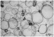
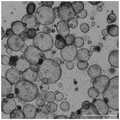
图1为本发明实施例1中获取的其中一张肺泡类器官影像数据。FIG. 1 is one piece of alveolar organoid image data obtained in Example 1 of the present invention.
图2为本发明实施例1中获取的另一张肺泡类器官影像数据。FIG. 2 is another alveolar organoid image data obtained in Example 1 of the present invention.
图3为本发明实施例1中自动标定并分型的其中一张肺泡类器官影像数据。FIG. 3 is one piece of alveolar organoid image data automatically calibrated and typed in Example 1 of the present invention.
图4为本发明实施例1中自动标定并分型的另一张肺泡类器官影像数据。FIG. 4 is another alveolar organoid image data automatically calibrated and typed in Example 1 of the present invention.
图5为本发明实施例2中获取的其中一张肺泡类器官影像数据。FIG. 5 is one piece of alveolar organoid image data obtained in Example 2 of the present invention.
图6为本发明实施例2中获取的另一张肺泡类器官影像数据。FIG. 6 is another alveolar organoid image data obtained in Example 2 of the present invention.
图7为本发明实施例2中自动标定并分型的其中一张肺泡类器官影像数据。FIG. 7 is one piece of alveolar organoid image data automatically calibrated and typed in Example 2 of the present invention.
图8为本发明实施例2中自动标定并分型的另一张肺泡类器官影像数据。FIG. 8 is another alveolar organoid image data automatically calibrated and typed in Example 2 of the present invention.
图9为本发明实施例3中获取的其中一张肺泡类器官影像数据。FIG. 9 is one piece of alveolar organoid image data obtained in Example 3 of the present invention.
图10为本发明实施例3中获取的另一张肺泡类器官影像数据。FIG. 10 is another alveolar organoid image data obtained in Example 3 of the present invention.
图11为本发明实施例3中自动标定并分型的其中一张肺泡类器官影像数据。FIG. 11 is one piece of alveolar organoid image data that is automatically calibrated and typed in Example 3 of the present invention.
图12为本发明实施例3中自动标定并分型的另一张肺泡类器官影像数据。FIG. 12 is another image data of alveolar organoids automatically calibrated and typed in Example 3 of the present invention.
具体实施方式Detailed ways
在本发明的描述中,需要说明的是,实施例中未注明具体条件者,按照常 规条件或制造商建议的条件进行。所用试剂或仪器未注明生产厂商者,均为可 以通过市售购买获得的常规产品。In the description of the present invention, it should be noted that, those who do not specify specific conditions in the examples are carried out according to conventional conditions or conditions suggested by the manufacturer. The reagents or instruments used without the manufacturer's indication are conventional products that can be purchased from the market.
下面结合附图和具体的实施例对本发明做进一步详细说明,所述是对本发 明的解释而不是限定。The present invention will be described in further detail below in conjunction with the accompanying drawings and specific embodiments, which are to explain rather than limit the present invention.
本发明具体实施例提供了一种基于显微图像的肺泡类器官类型智能识别方 法,包括:A specific embodiment of the present invention provides a method for intelligent identification of alveolar organoid types based on microscopic images, including:
步骤1,获取一系列肺泡类器官的显微图像并进行图像合成,即将单幅图像 拼接成整幅图像。Step 1: Obtain a series of microscopic images of alveolar organoids and perform image synthesis, that is, stitch a single image into a whole image.
步骤2,将合成的显微图像中的每个肺泡类器官进行框选,并将框选的每个 肺泡类器官图像单独一一提取出来,用xi表示其中一个肺泡类器官图像,i为整 数。Step 2, frame each alveolar organoid in the synthesized microscopic image, and extract each alveolar organoid image selected by the frame individually, and use xi to represent one of the alveolar organoid images, where i is an integer .
步骤3,将每个肺泡类器官图像分别输入到3种类型智能判断与识别模型 中:VGG、ResNet和GoogleNet,得到该图像中肺泡类器官类型的3种预测结 果,如果该图像中肺泡类器官存在人工标注结果,则将获得的3种预测结果与 人工标注结果进行比较,并通过最小化loss来优化3种类型智能判断与识别模 型;否则将3种预测结果进行比较,至少有两个预测结果相一致时,将该一致 结果作为该图像肺泡类器官类型的伪标签,然后将剩下的一种预测结果对应的 模型通过最小化loss来优化;所述最小化loss优化所采用的公式为:Step 3: Input each alveolar organoid image into three types of intelligent judgment and recognition models: VGG, ResNet and GoogleNet, and obtain three prediction results of the alveolar organoid type in the image. If the alveolar organoid in the image is If there are manual annotation results, compare the obtained three prediction results with the manual annotation results, and optimize the three types of intelligent judgment and recognition models by minimizing the loss; otherwise, compare the three prediction results, and there are at least two predictions. When the results are consistent, the consistent result is used as the pseudo-label of the alveolar organoid type in the image, and then the model corresponding to the remaining one prediction result is optimized by minimizing loss; the formula used for minimizing loss optimization is: :

式I中,

步骤4,待所有肺泡类器官图像处理完毕后,统计3种类型智能判断与识别 模型的精确率和召回率结果,并利用这些结果对三种模型进行F1值评价,选择 评价结果最优的模型为最终的分选模型;所述F1值的计算公式为:Step 4: After all alveolar organoid images are processed, count the precision and recall results of the three types of intelligent judgment and recognition models, and use these results to evaluate the F1 value of the three models, and select the model with the best evaluation result. is the final sorting model; the calculation formula of the F1 value is:

式II中,Precision表示精确率,Recall表示召回率,P/N为模型的判断结果 正/负,T/F为模型的判断结果对错真/假;FP表示假正例,FN表示假负例,TP 表示真正例,TN表示真负例,并且:In formula II, Precision represents the precision rate, Recall represents the recall rate, P/N represents the positive/negative of the judgment result of the model, T/F represents the true/false of the judgment result of the model; FP represents the false positive example, and FN represents the false negative For example, TP represents true examples, TN represents true negative examples, and:
a)precision=TP/(TP+FP)模型正确判断出的正例(TP)占数据集中所有判断 正例的比例;a) The proportion of positive examples (TP) correctly judged by the precision=TP/(TP+FP) model in all the judged positive examples in the data set;
b)recall=TP/(TP+FN)模型正确判断出的正例(TP)占数据集中所有实际正例 的比例;b) The ratio of the positive examples (TP) correctly judged by the recall=TP/(TP+FN) model to all the actual positive examples in the data set;
c)accuracy=(TP+TN)/(TP+FP+TN+FN)模型判断正确的数据(TP+TN)占总 数据的比例。c) Accuracy=(TP+TN)/(TP+FP+TN+FN) The proportion of the correct data (TP+TN) in the total data determined by the model.
最终获得的3种模型的F1值如表1所示。The F1 values of the three models finally obtained are shown in Table 1.
表1Table 1
步骤5,最终选定Googlenet作为本发明的分选模型,采用该分选模型进行 肺泡类器官类型的识别与判断。In step 5, Googlenet is finally selected as the sorting model of the present invention, and the sorting model is used to identify and judge the type of alveolar organoids.
本发明具体实施例还提供一种基于显微图像的肺泡类器官类型智能识别系 统,包括:A specific embodiment of the present invention also provides a microscopic image-based intelligent identification system for alveolar organoid types, including:
显微图像获取装置,用于获取类器官的显微图像。本发明具体实施例中采 用的是光学显微镜,型号为CYTATION5。此外,目前其它型号的光学显微镜以 及电子显微镜也同样可以用作本发明的显微图像获取装置。并采用图像合成调 整软件将图像进行拼接处理。Microscopic image acquisition device for acquiring microscopic images of organoids. In the specific embodiment of the present invention, an optical microscope is used, and the model is CYTATION5. In addition, other types of optical microscopes and electron microscopes can also be used as the microscopic image acquisition device of the present invention. And use the image synthesis adjustment software to stitch the images.
类器官类型判断与识别装置,与所述显微图像获取装置连接,所述类器官 类型判断与识别装置包括类器官检测器和类器官分类器,所述类器官检测器用 于提取所述显微图像中各个肺泡类器官;所述类器官分类器用于对提取出来的 肺泡类器官进行类型判断与识别。本发明具体实施例中采用的显微图像获取装 置是Biotek Cytation5,可支持所有常规微孔板检测仪,图像合成调整软件型号 为GEN5 3.09。An organoid type judging and identifying device is connected to the microscopic image acquisition device, the organoid type judging and identifying device includes an organoid detector and an organoid classifier, and the organoid detector is used to extract the microscopic image. Each alveolar organoid in the image; the organoid classifier is used for type judgment and identification of the extracted alveolar organoid. The microscopic image acquisition device adopted in the specific embodiment of the present invention is Biotek Cytation5, which can support all conventional microplate detectors, and the image synthesis adjustment software model is GEN5 3.09.
通讯装置,用于将所述显微图像获取装置、所述类器官检测器、所述类器 官分类器通讯连接,本实施例中具体采用的是有线连接方式。A communication device is used to communicate and connect the microscopic image acquisition device, the organoid detector, and the organoid classifier. In this embodiment, a wired connection is specifically adopted.
上述肺泡类器官类型智能判断与识别方法及系统可应用在肺泡类器官培养 或者肺泡类器官建库中。并且所涉及的肺泡类器官培养方法,包括以下步骤:The above-mentioned method and system for intelligent judgment and identification of alveolar organoid types can be applied to alveolar organoid culture or alveolar organoid bank building. And the involved alveolar organoid culture method includes the following steps:
获取待培养组织;Obtain the tissue to be cultured;
培养肺泡类器官;培养肺泡类器官的方法可以参考CN202010843695.5;Culture alveolar organoids; the method of culturing alveolar organoids can refer to CN202010843695.5;
培养过程中采用上述基于显微图像的肺泡类器官类型智能识别方法及系统 进行肺泡类器官类型判断与识别。In the culture process, the above-mentioned intelligent identification method and system of alveolar organoid types based on microscopic images were used to judge and identify the types of alveolar organoids.
所述肺泡类器官的建库方法,包括以下步骤:The method for building a bank of alveolar organoids includes the following steps:
将培养获得的肺泡类器官利用上述基于显微图像的肺泡类器官类型智能识 别方法及系统进行类器官类型判断与识别;The cultured alveolar organoids are judged and identified by using the above-mentioned microscopic image-based intelligent identification method and system for alveolar organoid types;
将满足要求的类器官组件成样本库。Organoid components that meet the requirements are assembled into a sample library.
实施例1Example 1
本实施例提供一种人来源肺泡类器官类型智能判断与识别方法,包括以下 步骤:The present embodiment provides a human-derived alveolar organoid type intelligent judgment and identification method, comprising the following steps:
(1)获取人来源肺泡类器官影像数据。具体方法为:使用光学显微镜对 需要计数的人肺泡类器官孔板(或培养皿)进行扫描拍照。显微图像需要包含 三维信息(即层切叠加),如图1和图2为其中两张影像数据。(1) Obtain image data of human alveolar organoids. The specific method is: using an optical microscope to scan and photograph the human alveolar organoid plate (or petri dish) to be counted. Microscopic images need to contain three-dimensional information (ie, slice stacking), as shown in Figure 1 and Figure 2 for two of them.
(2)将获取的一系列肺泡类器官的显微图像中的每个类器官进行框选,并 将框选的每个肺泡类器官图像单独一一提取出来,忽略不需要的类器官(直径 小于30μm的,外壁松散形状溃散的,透光度差内部坏死的);如图3和图4 所示。(2) Frame each organoid in a series of acquired microscopic images of alveolar organoids, and extract each alveolar organoid image selected by the frame individually, ignoring unnecessary organoids (diameter Less than 30μm, the outer wall is loose and the shape is collapsed, and the transmittance is poor and the interior is necrotic); as shown in Figure 3 and Figure 4.
(3)将数据载入所选择的分选模型(类器官类型判断与识别装置) GoogleNet中进行类器官类型的识别与判断。(3) Load the data into the selected sorting model (organoid type judgment and identification device) GoogleNet to identify and judge the organoid type.
(4)对所有判断结果进行统计,导出EXCEL数据表,如表1所示,图3 和图4中,红色表示A型,绿色表示B型,黄色表示C型。(4) Make statistics on all judgment results, and export the EXCEL data table, as shown in Table 1. In Figures 3 and 4, red represents type A, green represents type B, and yellow represents type C.
表1人肺泡类器官的自动分型结果Table 1 Results of automatic typing of human alveolar organoids
表1的结果显示,图1中有30个A型人肺泡类器官,5个B型人肺泡类器 官,8个C型人肺泡类器官;图2中有31个A型人肺泡类器官,2个B型人肺 泡类器官,3个C型人肺泡类器官。该结果和人工标注结果相同。The results in Table 1 show that there are 30 type A human alveolar organoids, 5 type B human alveolar organoids, and 8 type C human alveolar organoids in Figure 1; there are 31 type A human alveolar organoids in Figure 2, 2 type B human alveolar organoids and 3 type C human alveolar organoids. This result is the same as the manual labeling result.
实施例2Example 2
本实施例提供一种小鼠扩增肺泡类器官类型智能判断与识别方法,包括以 下步骤:The present embodiment provides a method for intelligently judging and identifying the type of mouse expanded alveolar organoids, comprising the following steps:
(1)获取小鼠扩增肺泡类器官影像数据。具体方法为:使用光学显微镜 对需要计数的小鼠扩增肺泡类器官孔板(或培养皿)进行扫描拍照。显微图像 需要包含三维信息(即层切叠加),如图5和图6为其中两张影像数据。(1) Obtain the imaging data of mouse expanded alveolar organoids. The specific method is: using a light microscope to scan and take pictures of the mouse expanded alveolar organoid plate (or petri dish) that needs to be counted. Microscopic images need to contain three-dimensional information (ie, slice stacking), as shown in Figure 5 and Figure 6 for two of the image data.
(2)将获取的一系列小鼠扩增肺泡类器官的显微图像中的每个类器官进 行框选,并将框选的每个小鼠肺泡类器官图像单独一一提取出来,忽略不需要 的类器官(直径小于30μm的,外壁松散形状溃散的,透光度差内部坏死的); 如图7和图8所示。(2) Box-select each organoid in a series of microscopic images of mouse expanded alveolar organoids obtained, and extract each mouse alveolar organoid image selected by the box one by one. The desired organoids (less than 30 μm in diameter, loosely shaped outer wall and necrotic inside); as shown in Figure 7 and Figure 8 .
(3)将数据载入所选择的分选模型(类器官类型判断与识别装置) GoogleNet中进行类器官类型的识别与判断。(3) Load the data into the selected sorting model (organoid type judgment and identification device) GoogleNet to identify and judge the organoid type.
(4)对所有判断结果进行统计,导出EXCEL数据表,如表2所示,图7 和图8中,红色表示A型,绿色表示B型,黄色表示C型。(4) Make statistics on all judgment results, and export the EXCEL data table, as shown in Table 2. In Figures 7 and 8, red represents type A, green represents type B, and yellow represents type C.
表2小鼠扩增肺泡类器官的自动分型结果Table 2 Results of automatic typing of expanded alveolar organoids in mice
表2的结果显示,图5中有78个A型小鼠扩增肺泡类器官,63个B型小 鼠扩增肺泡类器官,63个C型小鼠扩增肺泡类器官;图6中有76个A型小鼠 扩增肺泡类器官,49个B型小鼠扩增肺泡类器官,73个C型小鼠扩增肺泡类器 官。该结果和人工标注结果相同。The results in Table 2 show that 78 type A mice expanded alveolar organoids, 63 type B mice expanded alveolar organoids, and 63 type C mice expanded alveolar organoids in Figure 5; Alveolar organoids were expanded from 76 type A mice, 49 type B mice and 73 type C mice. This result is the same as the manual labeling result.
实施例3Example 3
本实施例提供一种小鼠分化肺泡类器官类型智能判断与识别方法,包括以 下步骤:The present embodiment provides a method for intelligent judgment and identification of mouse differentiated alveolar organoid types, comprising the following steps:
(1)获取小鼠分化肺泡类器官影像数据。具体方法为:使用光学显微镜 对需要计数的小鼠分化肺泡类器官孔板(或培养皿)进行扫描拍照。显微图像 需要包含三维信息(即层切叠加),如图9和图10为其中两张影像数据。(1) Obtain the imaging data of mouse differentiated alveolar organoids. The specific method is: using a light microscope to scan and photograph the well plates (or petri dishes) of mouse differentiated alveolar organoids that need to be counted. Microscopic images need to contain three-dimensional information (ie, slice stacking), as shown in Figure 9 and Figure 10 for two of the image data.
(2)将获取的一系列小鼠分化肺泡类器官的显微图像中的每个类器官进 行框选,并将框选的每个小鼠肺泡类器官图像单独一一提取出来,忽略不需要 的类器官(直径小于30μm的,外壁松散形状溃散的,透光度差内部坏死的); 如图11和图12所示。(2) Box-select each organoid in a series of obtained microscopic images of mouse differentiated alveolar organoids, and extract each mouse alveolar organoid image selected by the box one by one. Organoids (less than 30 μm in diameter, with loose outer wall and collapsed shape, poor light transmittance and internal necrosis); as shown in Figure 11 and Figure 12 .
(3)将数据载入所选择的分选模型(类器官类型判断与识别装置) GoogleNet中进行类器官类型的识别与判断。(3) Load the data into the selected sorting model (organoid type judgment and identification device) GoogleNet to identify and judge the organoid type.
(4)对所有判断结果进行统计,导出EXCEL数据表,如表3所示,图11 和图12中,红色表示A型,绿色表示B型,黄色表示C型。(4) Make statistics on all judgment results, and export the EXCEL data table, as shown in Table 3. In Figure 11 and Figure 12, red represents type A, green represents type B, and yellow represents type C.
表3小鼠分化肺泡类器官的自动分型结果Table 3 Results of automatic typing of mouse differentiated alveolar organoids
表3的结果显示,图9中有85个A型小鼠分化肺泡类器官,5个B型小鼠 分化肺泡类器官,6个C型小鼠分化肺泡类器官;图10中有81个A型小鼠分 化肺泡类器官,5个B型小鼠分化肺泡类器官,7个C型小鼠分化肺泡类器官。 该结果和人工标注结果相同。 The results in Table 3 show that there are 85 type A mice differentiated alveolar organoids in Figure 9, 5 type B mice differentiated alveolar organoids, and 6 type C mice differentiated alveolar organoids; there are 81 A mice in Figure 10 Type B mice differentiated alveolar organoids, 5 type B mice differentiated alveolar organoids, and 7 type C mice differentiated alveolar organoids. This result is the same as the manual labeling result.
综上,本发明方法以肺泡类器官影像为数据输入,直接导出详细分型比例 数据,拥有统一的判别标准且快速直观,可进行高通量作业,分型比例数据有 多种用途;1.可作为建库和质控标准,达到一定比例的肺泡类器官组样本才可建 库或判定为合格产品;2.可作为研究参数,如细胞因子诱导分化效率的研究,需 要分化前后比例变化的对比。To sum up, the method of the present invention uses alveolar organoid images as data input, and directly derives detailed classification ratio data, has a unified discrimination standard, is fast and intuitive, and can perform high-throughput operations, and the classification ratio data has a variety of uses; 1. It can be used as a library construction and quality control standard. Only when a certain proportion of alveolar organoid samples are reached can the library be established or be judged as a qualified product; 2. It can be used as a research parameter, such as the study of cytokine-induced differentiation efficiency, which requires a change in the proportion before and after differentiation. Compared.
以上所述实施例仅表达了本发明的几种实施方式,其描述较为具体和详 细,但并不能因此而理解为对本发明专利范围的限制。应当指出的是,对于本 领域的普通技术人员来说,在不脱离本发明构思的前提下,还可以做出若干变 形和改进,这些都属于本发明的保护范围。因此,本发明专利的保护范围应以 所附权利要求为准。The above-described embodiments only represent several embodiments of the present invention, and their descriptions are more specific and detailed, but should not therefore be construed as limiting the scope of the present invention. It should be pointed out that for those of ordinary skill in the art, without departing from the concept of the present invention, several modifications and improvements can also be made, which all belong to the protection scope of the present invention. Therefore, the protection scope of the patent of the present invention should be governed by the appended claims.
Claims (7)
Translated fromChinese


Priority Applications (1)
| Application Number | Priority Date | Filing Date | Title |
|---|---|---|---|
| CN202210066327.3ACN114463290B (en) | 2022-01-20 | 2022-01-20 | Micro-image-based organoid type intelligent identification method and system |
Applications Claiming Priority (1)
| Application Number | Priority Date | Filing Date | Title |
|---|---|---|---|
| CN202210066327.3ACN114463290B (en) | 2022-01-20 | 2022-01-20 | Micro-image-based organoid type intelligent identification method and system |
Publications (2)
| Publication Number | Publication Date |
|---|---|
| CN114463290Atrue CN114463290A (en) | 2022-05-10 |
| CN114463290B CN114463290B (en) | 2024-11-01 |
Family
ID=81408980
Family Applications (1)
| Application Number | Title | Priority Date | Filing Date |
|---|---|---|---|
| CN202210066327.3AActiveCN114463290B (en) | 2022-01-20 | 2022-01-20 | Micro-image-based organoid type intelligent identification method and system |
Country Status (1)
| Country | Link |
|---|---|
| CN (1) | CN114463290B (en) |
Cited By (1)
| Publication number | Priority date | Publication date | Assignee | Title |
|---|---|---|---|---|
| CN116012838A (en)* | 2022-12-30 | 2023-04-25 | 创芯国际生物科技(广州)有限公司 | Artificial intelligence-based organoid activity recognition method and system |
Citations (5)
| Publication number | Priority date | Publication date | Assignee | Title |
|---|---|---|---|---|
| US20170337682A1 (en)* | 2016-05-18 | 2017-11-23 | Siemens Healthcare Gmbh | Method and System for Image Registration Using an Intelligent Artificial Agent |
| CN109326343A (en)* | 2017-10-25 | 2019-02-12 | 首都医科大学附属北京友谊医院 | A method for acquiring lesion data, a method for identifying lesions, and computer equipment |
| CN112862569A (en)* | 2021-03-04 | 2021-05-28 | 上海交通大学 | Product appearance style evaluation method and system based on image and text multi-modal data |
| CN113283352A (en)* | 2021-05-31 | 2021-08-20 | 创芯国际生物科技(广州)有限公司 | Organoid vitality evaluation method and system based on microscopic image |
| US20210353260A1 (en)* | 2018-07-26 | 2021-11-18 | Koninklijke Philips N.V. | Ultrasound system with automated dynamic setting of imaging parameters based on organ detection |
- 2022
- 2022-01-20CNCN202210066327.3Apatent/CN114463290B/enactiveActive
Patent Citations (5)
| Publication number | Priority date | Publication date | Assignee | Title |
|---|---|---|---|---|
| US20170337682A1 (en)* | 2016-05-18 | 2017-11-23 | Siemens Healthcare Gmbh | Method and System for Image Registration Using an Intelligent Artificial Agent |
| CN109326343A (en)* | 2017-10-25 | 2019-02-12 | 首都医科大学附属北京友谊医院 | A method for acquiring lesion data, a method for identifying lesions, and computer equipment |
| US20210353260A1 (en)* | 2018-07-26 | 2021-11-18 | Koninklijke Philips N.V. | Ultrasound system with automated dynamic setting of imaging parameters based on organ detection |
| CN112862569A (en)* | 2021-03-04 | 2021-05-28 | 上海交通大学 | Product appearance style evaluation method and system based on image and text multi-modal data |
| CN113283352A (en)* | 2021-05-31 | 2021-08-20 | 创芯国际生物科技(广州)有限公司 | Organoid vitality evaluation method and system based on microscopic image |
Non-Patent Citations (1)
| Title |
|---|
| 林翊: "基于深度学习的先天性心脏病辅助诊疗研究", 《中国知网硕士论文数据库》, 15 February 2021 (2021-02-15)* |
Cited By (2)
| Publication number | Priority date | Publication date | Assignee | Title |
|---|---|---|---|---|
| CN116012838A (en)* | 2022-12-30 | 2023-04-25 | 创芯国际生物科技(广州)有限公司 | Artificial intelligence-based organoid activity recognition method and system |
| CN116012838B (en)* | 2022-12-30 | 2023-11-07 | 创芯国际生物科技(广州)有限公司 | Artificial intelligence-based organoid activity recognition method and system |
Also Published As
| Publication number | Publication date |
|---|---|
| CN114463290B (en) | 2024-11-01 |
Similar Documents
| Publication | Publication Date | Title |
|---|---|---|
| CN113283352B (en) | Organoid vitality evaluation method and system based on microscopic image | |
| US7796815B2 (en) | Image analysis of biological objects | |
| CN113283353B (en) | Organoid cell counting method and system based on microscopic image | |
| CN107832801B (en) | Model construction method for cell image classification | |
| CN110222681A (en) | A kind of casting defect recognition methods based on convolutional neural networks | |
| CN115266540A (en) | Optical measurements performed on samples | |
| JP2015146747A (en) | Cell determination method | |
| WO2019144700A1 (en) | Deep learning-based quick and precise high-throughput drug screening system | |
| CN114463290A (en) | A method and system for intelligent identification of organoid types based on microscopic images | |
| CN119964156B (en) | Image recognition-based blood cell number counting method, system, equipment and medium | |
| CN110060229A (en) | A kind of cell automatic positioning dividing method of myeloplast | |
| CN118866107A (en) | A tumor microenvironment analysis method integrating spatial multi-omics data | |
| WO2016088243A1 (en) | Determination device, observation system, observation method, program for same, method for manufacturing cell, and cell | |
| CN111833296B (en) | A bone marrow cell morphology automatic detection review system and review method | |
| CN112001315B (en) | Bone marrow cell classification and identification method based on migration learning and image texture characteristics | |
| CN110414317B (en) | Capsule network-based automatic white blood cell classification and counting method | |
| CN112540039A (en) | Method for directly calculating number of adherent living cells | |
| CN112750118B (en) | Novel method and system for identifying cell number in single cell pore plate sequencing based on automatic visual detection | |
| CN111795967B (en) | A kind of smear self-inspection method of bone marrow cell morphology automatic detection system | |
| CN111795919B (en) | Automatic bone marrow cell morphology detection system and working method thereof | |
| US20190340415A1 (en) | Automated system and method for creating and executing a scoring guide to assist in the analysis of tissue specimen | |
| Sun et al. | Color‐based tumor tissue segmentation for the automated estimation of oral cancer parameters | |
| CN118334652A (en) | Dense object fine-grained detection and recognition method and system based on contrastive learning | |
| CN117612161A (en) | Cell phenotype analysis method based on label-free image and deep learning and application | |
| US11908130B2 (en) | Apparatuses and methods for digital pathology |
Legal Events
| Date | Code | Title | Description |
|---|---|---|---|
| PB01 | Publication | ||
| PB01 | Publication | ||
| SE01 | Entry into force of request for substantive examination | ||
| SE01 | Entry into force of request for substantive examination | ||
| GR01 | Patent grant | ||
| GR01 | Patent grant |