CN114445362A - Image processing method, apparatus, device, readable storage medium and program product - Google Patents
Image processing method, apparatus, device, readable storage medium and program productDownload PDFInfo
- Publication number
- CN114445362A CN114445362ACN202210086842.8ACN202210086842ACN114445362ACN 114445362 ACN114445362 ACN 114445362ACN 202210086842 ACN202210086842 ACN 202210086842ACN 114445362 ACN114445362 ACN 114445362A
- Authority
- CN
- China
- Prior art keywords
- image
- target
- sample
- wavelength
- pseudo
- Prior art date
- Legal status (The legal status is an assumption and is not a legal conclusion. Google has not performed a legal analysis and makes no representation as to the accuracy of the status listed.)
- Pending
Links
Images


Classifications
- G—PHYSICS
- G06—COMPUTING OR CALCULATING; COUNTING
- G06T—IMAGE DATA PROCESSING OR GENERATION, IN GENERAL
- G06T7/00—Image analysis
- G06T7/0002—Inspection of images, e.g. flaw detection
- G06T7/0012—Biomedical image inspection
- G—PHYSICS
- G06—COMPUTING OR CALCULATING; COUNTING
- G06T—IMAGE DATA PROCESSING OR GENERATION, IN GENERAL
- G06T7/00—Image analysis
- G06T7/10—Segmentation; Edge detection
- G06T7/11—Region-based segmentation
- G—PHYSICS
- G06—COMPUTING OR CALCULATING; COUNTING
- G06T—IMAGE DATA PROCESSING OR GENERATION, IN GENERAL
- G06T7/00—Image analysis
- G06T7/90—Determination of colour characteristics
- G—PHYSICS
- G06—COMPUTING OR CALCULATING; COUNTING
- G06V—IMAGE OR VIDEO RECOGNITION OR UNDERSTANDING
- G06V10/00—Arrangements for image or video recognition or understanding
- G06V10/10—Image acquisition
- G06V10/12—Details of acquisition arrangements; Constructional details thereof
- G06V10/14—Optical characteristics of the device performing the acquisition or on the illumination arrangements
- G06V10/143—Sensing or illuminating at different wavelengths
- G—PHYSICS
- G06—COMPUTING OR CALCULATING; COUNTING
- G06V—IMAGE OR VIDEO RECOGNITION OR UNDERSTANDING
- G06V10/00—Arrangements for image or video recognition or understanding
- G06V10/20—Image preprocessing
- G06V10/25—Determination of region of interest [ROI] or a volume of interest [VOI]
- G—PHYSICS
- G06—COMPUTING OR CALCULATING; COUNTING
- G06V—IMAGE OR VIDEO RECOGNITION OR UNDERSTANDING
- G06V10/00—Arrangements for image or video recognition or understanding
- G06V10/40—Extraction of image or video features
- G06V10/56—Extraction of image or video features relating to colour
- G—PHYSICS
- G06—COMPUTING OR CALCULATING; COUNTING
- G06V—IMAGE OR VIDEO RECOGNITION OR UNDERSTANDING
- G06V10/00—Arrangements for image or video recognition or understanding
- G06V10/40—Extraction of image or video features
- G06V10/60—Extraction of image or video features relating to illumination properties, e.g. using a reflectance or lighting model
- G—PHYSICS
- G06—COMPUTING OR CALCULATING; COUNTING
- G06V—IMAGE OR VIDEO RECOGNITION OR UNDERSTANDING
- G06V10/00—Arrangements for image or video recognition or understanding
- G06V10/70—Arrangements for image or video recognition or understanding using pattern recognition or machine learning
- G06V10/764—Arrangements for image or video recognition or understanding using pattern recognition or machine learning using classification, e.g. of video objects
- G—PHYSICS
- G06—COMPUTING OR CALCULATING; COUNTING
- G06V—IMAGE OR VIDEO RECOGNITION OR UNDERSTANDING
- G06V20/00—Scenes; Scene-specific elements
- G06V20/70—Labelling scene content, e.g. deriving syntactic or semantic representations
- A—HUMAN NECESSITIES
- A61—MEDICAL OR VETERINARY SCIENCE; HYGIENE
- A61B—DIAGNOSIS; SURGERY; IDENTIFICATION
- A61B5/00—Measuring for diagnostic purposes; Identification of persons
- A61B5/0059—Measuring for diagnostic purposes; Identification of persons using light, e.g. diagnosis by transillumination, diascopy, fluorescence
- A61B5/0075—Measuring for diagnostic purposes; Identification of persons using light, e.g. diagnosis by transillumination, diascopy, fluorescence by spectroscopy, i.e. measuring spectra, e.g. Raman spectroscopy, infrared absorption spectroscopy
- A—HUMAN NECESSITIES
- A61—MEDICAL OR VETERINARY SCIENCE; HYGIENE
- A61B—DIAGNOSIS; SURGERY; IDENTIFICATION
- A61B5/00—Measuring for diagnostic purposes; Identification of persons
- A61B5/72—Signal processing specially adapted for physiological signals or for diagnostic purposes
- A61B5/7235—Details of waveform analysis
- A61B5/7264—Classification of physiological signals or data, e.g. using neural networks, statistical classifiers, expert systems or fuzzy systems
- G—PHYSICS
- G01—MEASURING; TESTING
- G01J—MEASUREMENT OF INTENSITY, VELOCITY, SPECTRAL CONTENT, POLARISATION, PHASE OR PULSE CHARACTERISTICS OF INFRARED, VISIBLE OR ULTRAVIOLET LIGHT; COLORIMETRY; RADIATION PYROMETRY
- G01J3/00—Spectrometry; Spectrophotometry; Monochromators; Measuring colours
- G01J3/02—Details
- G01J3/0264—Electrical interface; User interface
- G—PHYSICS
- G01—MEASURING; TESTING
- G01J—MEASUREMENT OF INTENSITY, VELOCITY, SPECTRAL CONTENT, POLARISATION, PHASE OR PULSE CHARACTERISTICS OF INFRARED, VISIBLE OR ULTRAVIOLET LIGHT; COLORIMETRY; RADIATION PYROMETRY
- G01J3/00—Spectrometry; Spectrophotometry; Monochromators; Measuring colours
- G01J3/28—Investigating the spectrum
- G01J3/2823—Imaging spectrometer
- G—PHYSICS
- G01—MEASURING; TESTING
- G01J—MEASUREMENT OF INTENSITY, VELOCITY, SPECTRAL CONTENT, POLARISATION, PHASE OR PULSE CHARACTERISTICS OF INFRARED, VISIBLE OR ULTRAVIOLET LIGHT; COLORIMETRY; RADIATION PYROMETRY
- G01J3/00—Spectrometry; Spectrophotometry; Monochromators; Measuring colours
- G01J3/28—Investigating the spectrum
- G01J3/30—Measuring the intensity of spectral lines directly on the spectrum itself
- G01J3/32—Investigating bands of a spectrum in sequence by a single detector
- G—PHYSICS
- G01—MEASURING; TESTING
- G01N—INVESTIGATING OR ANALYSING MATERIALS BY DETERMINING THEIR CHEMICAL OR PHYSICAL PROPERTIES
- G01N33/00—Investigating or analysing materials by specific methods not covered by groups G01N1/00 - G01N31/00
- G01N33/48—Biological material, e.g. blood, urine; Haemocytometers
- G01N33/483—Physical analysis of biological material
- G01N33/4833—Physical analysis of biological material of solid biological material, e.g. tissue samples, cell cultures
- G—PHYSICS
- G06—COMPUTING OR CALCULATING; COUNTING
- G06T—IMAGE DATA PROCESSING OR GENERATION, IN GENERAL
- G06T2207/00—Indexing scheme for image analysis or image enhancement
- G06T2207/10—Image acquisition modality
- G06T2207/10032—Satellite or aerial image; Remote sensing
- G06T2207/10036—Multispectral image; Hyperspectral image
- G—PHYSICS
- G06—COMPUTING OR CALCULATING; COUNTING
- G06T—IMAGE DATA PROCESSING OR GENERATION, IN GENERAL
- G06T2207/00—Indexing scheme for image analysis or image enhancement
- G06T2207/10—Image acquisition modality
- G06T2207/10056—Microscopic image
- G—PHYSICS
- G06—COMPUTING OR CALCULATING; COUNTING
- G06T—IMAGE DATA PROCESSING OR GENERATION, IN GENERAL
- G06T2207/00—Indexing scheme for image analysis or image enhancement
- G06T2207/20—Special algorithmic details
- G06T2207/20081—Training; Learning
- G—PHYSICS
- G06—COMPUTING OR CALCULATING; COUNTING
- G06T—IMAGE DATA PROCESSING OR GENERATION, IN GENERAL
- G06T2207/00—Indexing scheme for image analysis or image enhancement
- G06T2207/20—Special algorithmic details
- G06T2207/20084—Artificial neural networks [ANN]
- G—PHYSICS
- G06—COMPUTING OR CALCULATING; COUNTING
- G06T—IMAGE DATA PROCESSING OR GENERATION, IN GENERAL
- G06T2207/00—Indexing scheme for image analysis or image enhancement
- G06T2207/30—Subject of image; Context of image processing
- G06T2207/30004—Biomedical image processing
- G06T2207/30024—Cell structures in vitro; Tissue sections in vitro
- G—PHYSICS
- G06—COMPUTING OR CALCULATING; COUNTING
- G06T—IMAGE DATA PROCESSING OR GENERATION, IN GENERAL
- G06T2207/00—Indexing scheme for image analysis or image enhancement
- G06T2207/30—Subject of image; Context of image processing
- G06T2207/30004—Biomedical image processing
- G06T2207/30096—Tumor; Lesion
- G—PHYSICS
- G06—COMPUTING OR CALCULATING; COUNTING
- G06V—IMAGE OR VIDEO RECOGNITION OR UNDERSTANDING
- G06V2201/00—Indexing scheme relating to image or video recognition or understanding
- G06V2201/03—Recognition of patterns in medical or anatomical images
- G06V2201/031—Recognition of patterns in medical or anatomical images of internal organs
Landscapes
- Engineering & Computer Science (AREA)
- Theoretical Computer Science (AREA)
- Physics & Mathematics (AREA)
- General Physics & Mathematics (AREA)
- Multimedia (AREA)
- Computer Vision & Pattern Recognition (AREA)
- Health & Medical Sciences (AREA)
- General Health & Medical Sciences (AREA)
- Medical Informatics (AREA)
- Software Systems (AREA)
- Radiology & Medical Imaging (AREA)
- Quality & Reliability (AREA)
- Computational Linguistics (AREA)
- Nuclear Medicine, Radiotherapy & Molecular Imaging (AREA)
- Artificial Intelligence (AREA)
- Computing Systems (AREA)
- Databases & Information Systems (AREA)
- Evolutionary Computation (AREA)
- Investigating Or Analysing Materials By Optical Means (AREA)
- Image Analysis (AREA)
- Investigating Or Analysing Biological Materials (AREA)
Abstract
Description
Translated fromChinese技术领域technical field
本申请实施例涉及医学数据处理领域,特别涉及一种图像处理方法、装置、设备、可读存储介质及程序产品。The embodiments of the present application relate to the field of medical data processing, and in particular, to an image processing method, apparatus, device, readable storage medium, and program product.
背景技术Background technique
在通过手术切除包括肿瘤组织的病理标本后,通常会进一步研究采样得到的病理标本,对病理标本中肿瘤组织的边界进行寻找和识别,从而对肿瘤组织进行更正确的分析,得到更有价值的医学分析结果。After the pathological specimen including tumor tissue is surgically removed, the sampled pathological specimen is usually further studied to find and identify the boundary of the tumor tissue in the pathological specimen, so that the tumor tissue can be analyzed more accurately and more valuable Medical analysis results.
相关技术中,在将手术切除的病理标本通过福尔马林固定后,通常由病理医生以肉眼观察的方法对肿瘤组织的边界进行确定,或者采用X光设备对病理标本进行扫描,由医生对X光影像进行解读,确定肿瘤组织的边界并进行肿瘤组织的取材工作。In the related art, after the surgically resected pathological specimen is fixed with formalin, the pathologist usually determines the boundary of the tumor tissue by visual observation, or scans the pathological specimen with X-ray equipment, and the doctor determines the boundary of the tumor tissue by visual observation. X-ray images were interpreted, the boundary of tumor tissue was determined, and tumor tissue sampling was performed.
然而,采用上述方法对肿瘤组织的边界进行确定时,对于一些瘤床不明显的病变,肉眼很难识别,而X光设备也由于价格昂贵,较难达到广泛普及。However, when the boundary of the tumor tissue is determined by the above method, it is difficult to identify the lesions with inconspicuous tumor beds with the naked eye, and X-ray equipment is also difficult to achieve widespread popularity due to its high price.
发明内容SUMMARY OF THE INVENTION
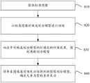
本申请实施例提供了一种图像处理方法、装置、设备、可读存储介质及程序产品,能够利用目标样本在不同波长下的光谱特性对目标样本进行分析,提高病理取材的准确性。所述技术方案如下。The embodiments of the present application provide an image processing method, apparatus, device, readable storage medium, and program product, which can use the spectral characteristics of the target sample at different wavelengths to analyze the target sample and improve the accuracy of pathological material sampling. The technical solution is as follows.
一方面,提供了一种图像处理方法,所述方法包括:In one aspect, an image processing method is provided, the method comprising:
获取样本图像,所述样本图像包括在预设波段内对目标样本进行采集得到的图像;acquiring a sample image, the sample image including an image obtained by collecting the target sample within a preset waveband;
获取所述样本图像中与所述预设波段中至少一个目标波长对应的目标图像,得到伪彩色图像;acquiring a target image corresponding to at least one target wavelength in the preset wavelength band in the sample image to obtain a pseudo-color image;
根据所述样本图像中样本元素类型的差异,对所述样本图像进行区域划分,得到区域划分结果,所述样本元素类型中包括待识别的目标元素类型;According to the difference of the sample element type in the sample image, the sample image is divided into regions to obtain a region division result, and the sample element type includes the target element type to be identified;
基于所述伪彩色图像和所述区域划分结果,在所述样本图像中确定包括所述目标元素类型的目标区域。Based on the pseudo-color image and the area division result, a target area including the target element type is determined in the sample image.
另一方面,提供了一种图像处理装置,所述装置包括:In another aspect, an image processing apparatus is provided, the apparatus comprising:
样本获取模块,用于获取样本图像,所述样本图像包括在预设波段内对目标样本进行采集得到的图像;a sample acquisition module, configured to acquire a sample image, where the sample image includes an image obtained by collecting a target sample within a preset waveband;
图像获取模块,用于获取所述样本图像中与所述预设波段中至少一个目标波长对应的目标图像,得到伪彩色图像;an image acquisition module, configured to acquire a target image corresponding to at least one target wavelength in the preset waveband in the sample image, to obtain a pseudo-color image;
区域划分模块,用于根据所述样本图像中样本元素类型的差异,对所述样本图像进行区域划分,得到区域划分结果,所述样本元素类型中包括待识别的目标元素类型;a region division module, configured to perform region division on the sample image according to the difference of the sample element types in the sample image, and obtain a region division result, where the sample element type includes the target element type to be identified;
区域确定模块,用于基于所述伪彩色图像和所述区域划分结果,在所述样本图像中确定包括所述目标元素类型的目标区域。an area determination module, configured to determine a target area including the target element type in the sample image based on the pseudo-color image and the area division result.
另一方面,提供了一种计算机设备,所述计算机设备包括处理器和存储器,所述存储器中存储有至少一条指令、至少一段程序、代码集或指令集,所述至少一条指令、所述至少一段程序、所述代码集或指令集由所述处理器加载并执行以实现如上述本申请实施例中任一所述图像处理方法。In another aspect, a computer device is provided, the computer device includes a processor and a memory, the memory stores at least one instruction, at least a section of a program, a code set or an instruction set, the at least one instruction, the at least one A piece of program, the code set or the instruction set is loaded and executed by the processor to implement the image processing method as described in any of the foregoing embodiments of the present application.
另一方面,提供了一种计算机可读存储介质,所述存储介质中存储有至少一条指令、至少一段程序、代码集或指令集,所述至少一条指令、所述至少一段程序、所述代码集或指令集由处理器加载并执行以实现如上述本申请实施例中任一所述的图像处理方法。In another aspect, a computer-readable storage medium is provided, wherein the storage medium stores at least one instruction, at least one piece of program, code set or instruction set, the at least one instruction, the at least one piece of program, the code The set or instruction set is loaded and executed by the processor to implement the image processing method described in any of the above embodiments of the present application.
另一方面,提供了一种计算机程序产品或计算机程序,该计算机程序产品或计算机程序包括计算机指令,该计算机指令存储在计算机可读存储介质中。计算机设备的处理器从计算机可读存储介质读取该计算机指令,处理器执行该计算机指令,使得该计算机设备执行上述实施例中任一所述的图像处理方法。In another aspect, a computer program product or computer program is provided, the computer program product or computer program comprising computer instructions stored in a computer-readable storage medium. The processor of the computer device reads the computer instructions from the computer-readable storage medium, and the processor executes the computer instructions, so that the computer device executes the image processing method described in any one of the foregoing embodiments.
本申请实施例提供的技术方案带来的有益效果至少包括:The beneficial effects brought by the technical solutions provided in the embodiments of the present application include at least:
从样本图像中获取目标波段对应的目标图像,并得到伪彩色图像,对样本图像进行区域划分后得到区域划分结果,结合伪彩色图像和区域划分结果确定目标区域。通过上述方法,可以避免仅仅依靠医生的裸眼观测和描述对肿瘤组织的大小、区域等进行判断,从而降低对患者肿瘤组织区域判断的不准确性。基于预先确定的预设波段,对目标样本进行采集,得到样本图像,从预设波段中选取至少一个效果较好的目标波长,并根据至少一个目标波长,从样本图像中确定与目标波长对应的目标图像,对目标图像进行处理后,得到伪彩色图像,伪彩色图像能够较准确地体现目标波长的优势。此外,根据样本图像中样本元素类型的差异,对样本图像进行区域划分后得到区域划分结果。结合伪彩色图像和区域划分结果,确定包括目标元素类型的目标区域,从而确定待识别区域 (如:肿瘤组织)的位置信息,提高病理取材的准确性,降低病理取材的难度,利用目标样本在不同波长下对应的光谱特性对目标样本进行分析,不仅操作较为简便,且成本相对较低,更容易广泛应用。The target image corresponding to the target band is obtained from the sample image, and the pseudo-color image is obtained. The sample image is divided into regions to obtain the region division result, and the target region is determined by combining the pseudo-color image and the region division result. Through the above method, it is possible to avoid judging the size and area of the tumor tissue only by the naked eye observation and description of the doctor, thereby reducing the inaccuracy of judging the patient's tumor tissue area. Based on a predetermined preset wavelength band, the target sample is collected to obtain a sample image, at least one target wavelength with better effect is selected from the preset wavelength band, and according to the at least one target wavelength, the sample image corresponding to the target wavelength is determined from the sample image. After processing the target image, a pseudo-color image is obtained, and the pseudo-color image can more accurately reflect the advantages of the target wavelength. In addition, according to the difference of the sample element types in the sample image, the region division result is obtained after the sample image is divided into regions. Combined with the pseudo-color image and the area division results, determine the target area including the target element type, so as to determine the location information of the area to be identified (such as: tumor tissue), improve the accuracy of pathological sampling, reduce the difficulty of pathological sampling, use the target sample in The spectral characteristics corresponding to different wavelengths are used to analyze the target sample, which is not only easy to operate, but also has a relatively low cost and is easier to be widely used.
附图说明Description of drawings
为了更清楚地说明本申请实施例中的技术方案,下面将对实施例描述中所需要使用的附图作简单地介绍,显而易见地,下面描述中的附图仅仅是本申请的一些实施例,对于本领域普通技术人员来讲,在不付出创造性劳动的前提下,还可以根据这些附图获得其他的附图。In order to illustrate the technical solutions in the embodiments of the present application more clearly, the following briefly introduces the drawings that are used in the description of the embodiments. Obviously, the drawings in the following description are only some embodiments of the present application. For those of ordinary skill in the art, other drawings can also be obtained from these drawings without creative effort.
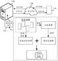
图1是本申请一个示例性实施例提供的实施环境示意图;FIG. 1 is a schematic diagram of an implementation environment provided by an exemplary embodiment of the present application;
图2是本申请一个示例性实施例提供的图像处理方法的流程图;FIG. 2 is a flowchart of an image processing method provided by an exemplary embodiment of the present application;
图3是本申请一个示例性实施例提供的获取样本图像的示意图;3 is a schematic diagram of acquiring a sample image provided by an exemplary embodiment of the present application;
图4是本申请一个示例性实施例提供的预设波段内的样本图像示意图;4 is a schematic diagram of a sample image in a preset band provided by an exemplary embodiment of the present application;
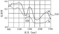
图5是本申请一个示例性实施例提供的样本图像对应的光谱特征曲线图;5 is a spectral characteristic curve diagram corresponding to a sample image provided by an exemplary embodiment of the present application;
图6是本申请一个示例性实施例提供的对目标样本进行图像处理的示意图;6 is a schematic diagram of performing image processing on a target sample provided by an exemplary embodiment of the present application;
图7是本申请一个示例性实施例提供的合成得到伪彩色图像的示意图;7 is a schematic diagram of a pseudo-color image obtained by synthesis provided by an exemplary embodiment of the present application;
图8是本申请另一个示例性实施例提供的图像处理方法的流程图;8 is a flowchart of an image processing method provided by another exemplary embodiment of the present application;
图9是本申请另一个示例性实施例提供的对病理样本进行取材的示意图;9 is a schematic diagram of sampling a pathological sample provided by another exemplary embodiment of the present application;
图10是本申请一个示例性实施例提供的对样本图像进行区域划分的流程图;FIG. 10 is a flowchart of region division of a sample image provided by an exemplary embodiment of the present application;
图11是本申请一个示例性实施例提供的空腔脏器对应的光谱特征曲线图;11 is a spectral characteristic curve diagram corresponding to a cavity organ provided by an exemplary embodiment of the present application;
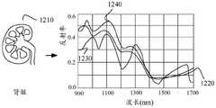
图12是本申请一个示例性实施例提供的肾对应的光谱特征曲线图;12 is a spectral characteristic curve diagram corresponding to a kidney provided by an exemplary embodiment of the present application;
图13是本申请一个示例性实施例提供的乳腺对应的光谱特征曲线图;13 is a spectral characteristic curve diagram corresponding to a breast provided by an exemplary embodiment of the present application;
图14是本申请一个示例性实施例提供的肺对应的光谱特征曲线图;14 is a spectral characteristic curve diagram corresponding to a lung provided by an exemplary embodiment of the present application;
图15是本申请一个示例性实施例提供的单色填充提示的示意图;FIG. 15 is a schematic diagram of a monochrome filling prompt provided by an exemplary embodiment of the present application;
图16是本申请一个示例性实施例提供的获得预测结果的流程图;FIG. 16 is a flowchart of obtaining a prediction result provided by an exemplary embodiment of the present application;
图17是本申请一个示例性实施例提供的四种组织分类图像表现示意图;FIG. 17 is a schematic representation of four types of tissue classification images provided by an exemplary embodiment of the present application;
图18是本申请一个示例性实施例提供的肾癌的不同图像表现示意图;18 is a schematic diagram of different image representations of renal cancer provided by an exemplary embodiment of the present application;
图19是本申请一个示例性实施例提供的乳腺的不同图像表现示意图;FIG. 19 is a schematic diagram of different image representations of the mammary gland provided by an exemplary embodiment of the present application;
图20是本申请另一个示例性实施例提供的乳腺的不同图像表现示意图;FIG. 20 is a schematic diagram of different image representations of the mammary gland provided by another exemplary embodiment of the present application;
图21是本申请一个示例性实施例提供的图像处理装置的结构框图;FIG. 21 is a structural block diagram of an image processing apparatus provided by an exemplary embodiment of the present application;
图22是本申请另一个示例性实施例提供的图像处理装置的结构框图;22 is a structural block diagram of an image processing apparatus provided by another exemplary embodiment of the present application;
图23是本申请一个示例性实施例提供的服务器的结构框图。FIG. 23 is a structural block diagram of a server provided by an exemplary embodiment of the present application.
具体实施方式Detailed ways
为使本申请的目的、技术方案和优点更加清楚,下面将结合附图对本申请实施方式作进一步地详细描述。In order to make the objectives, technical solutions and advantages of the present application clearer, the embodiments of the present application will be further described in detail below with reference to the accompanying drawings.
首先,针对本申请实施例中涉及的名词进行简单介绍。First, the terms involved in the embodiments of the present application are briefly introduced.
人工智能(Artificial Intelligence,AI):是利用数字计算机或者数字计算机控制的机器模拟、延伸和扩展人的智能,感知环境、获取知识并使用知识获得最佳结果的理论、方法、技术及应用系统。换句话说,人工智能是计算机科学的一个综合技术,它企图了解智能的实质,并生产出一种新的能以人类智能相似的方式做出反应的智能机器。人工智能也就是研究各种智能机器的设计原理与实现方法,使机器具有感知、推理与决策的功能。Artificial Intelligence (AI): It is a theory, method, technology and application system that uses digital computers or machines controlled by digital computers to simulate, extend and expand human intelligence, perceive the environment, acquire knowledge and use knowledge to obtain the best results. In other words, artificial intelligence is a comprehensive technique of computer science that attempts to understand the essence of intelligence and produce a new kind of intelligent machine that can respond in a similar way to human intelligence. Artificial intelligence is to study the design principles and implementation methods of various intelligent machines, so that the machines have the functions of perception, reasoning and decision-making.
人工智能技术是一门综合学科,涉及领域广泛,既有硬件层面的技术也有软件层面的技术。人工智能基础技术一般包括如传感器、专用人工智能芯片、云计算、分布式存储、大数据处理技术、操作/交互系统、机电一体化等技术。人工智能软件技术主要包括计算机视觉技术、语音处理技术、自然语言处理技术以及机器学习/深度学习等几大方向。Artificial intelligence technology is a comprehensive discipline, involving a wide range of fields, including both hardware-level technology and software-level technology. The basic technologies of artificial intelligence generally include technologies such as sensors, special artificial intelligence chips, cloud computing, distributed storage, big data processing technology, operation/interaction systems, and mechatronics. Artificial intelligence software technology mainly includes computer vision technology, speech processing technology, natural language processing technology, and machine learning/deep learning.
机器学习(Machine Learning,ML):是一门多领域交叉学科,涉及概率论、统计学、逼近论、凸分析、算法复杂度理论等多门学科。专门研究计算机怎样模拟或实现人类的学习行为,以获取新的知识或技能,重新组织已有的知识结构使之不断改善自身的性能。机器学习是人工智能的核心,是使计算机具有智能的根本途径,其应用遍及人工智能的各个领域。机器学习和深度学习通常包括人工神经网络、置信网络、强化学习、迁移学习、归纳学习、示教学习等技术。Machine Learning (ML): It is a multi-field interdisciplinary subject involving probability theory, statistics, approximation theory, convex analysis, algorithm complexity theory and other subjects. It specializes in how computers simulate or realize human learning behaviors to acquire new knowledge or skills, and to reorganize existing knowledge structures to continuously improve their performance. Machine learning is the core of artificial intelligence and the fundamental way to make computers intelligent, and its applications are in all fields of artificial intelligence. Machine learning and deep learning usually include artificial neural networks, belief networks, reinforcement learning, transfer learning, inductive learning, teaching learning and other techniques.
相关技术中,在将手术切除的病理标本通过福尔马林固定后,通常由病理医生以肉眼观察的方法对肿瘤组织的边界进行确定,或者采用X光设备对病理标本进行扫描,由医生对X光影像进行解读,确定肿瘤组织的边界并进行肿瘤组织的取材工作。然而,采用上述方法对肿瘤组织的边界进行确定时,对于一些瘤床不明显的病变,肉眼很难识别,而X光设备也由于价格昂贵,较难达到广泛普及。In the related art, after the surgically resected pathological specimen is fixed with formalin, the pathologist usually determines the boundary of the tumor tissue by visual observation, or scans the pathological specimen with X-ray equipment, and the doctor determines the boundary of the tumor tissue by visual observation. X-ray images were interpreted, the boundary of tumor tissue was determined, and tumor tissue sampling was performed. However, when the boundary of the tumor tissue is determined by the above method, it is difficult to identify the lesions with inconspicuous tumor beds with the naked eye, and X-ray equipment is also difficult to achieve widespread popularity due to its high price.
本申请实施例中,提供了一种图像处理方法,利用目标样本在不同波长下的光谱特性对目标样本进行分析,提高病理取材的准确性。针对本申请训练得到的方法,在应用时包括如下场景中的至少一种。In the embodiment of the present application, an image processing method is provided, which utilizes the spectral characteristics of the target sample under different wavelengths to analyze the target sample, so as to improve the accuracy of pathological material sampling. The method obtained by training in this application includes at least one of the following scenarios when applied.
一、应用于医学领域中1. Application in the field of medicine
肿瘤切除手术中需要准确地知道肿瘤边缘位置,以实现完整切除肿瘤区域的过程,从而防止患者病情复发和避免二次手术,术后组织病理分析是肿瘤诊断的金标准。为了准确获得患者的病灶信息,医生挑选病理组织块的过程尤为重要,漏选含有病灶的组织块将限制病理医生做出更准确的判断,而过多的选取组织块则会大大增加制片的工作量,降低医疗效率。示意性的,采用上述图像处理方法,以具有病灶的组织(如:肾脏器官、乳腺等)为目标样本,对目标样本在预设波段内进行采集并获取得到样本图像,从样本图像中选择目标波长对应的目标图像后得到伪色彩图像,并根据样本图像的样本元素类型对样本图像进行区域划分,得到区域划分结果,综合分析区域划分结果和伪彩色图像,最终能够较为准确地确定肿瘤组织对应的目标区域,实现对目标区域的识别过程。通过上述方法,可以辅助病理医生更快速地找到病灶区域,也可以减少采用X光等影像设备的仪器成本,运用更广泛、经济的光谱仪器获取样本图像,并对具有光谱信息的样本图像进行分析,从而在降低医学成本的基础上,提高肿瘤组织的判断准确率。In tumor resection, it is necessary to accurately know the position of the tumor edge in order to realize the process of complete tumor resection, thereby preventing the recurrence of the patient's condition and avoiding secondary surgery. Postoperative histopathological analysis is the gold standard for tumor diagnosis. In order to accurately obtain the patient's lesion information, the process of selecting pathological tissue blocks is particularly important. Missing the selection of tissue blocks containing lesions will limit the pathologist to make more accurate judgments, and selecting too many tissue blocks will greatly increase the production time. workload and reduce medical efficiency. Illustratively, the above-mentioned image processing method is used to take the tissue with lesions (such as kidney organs, breast, etc.) as the target sample, collect the target sample in a preset waveband and obtain a sample image, and select the target from the sample image. After obtaining the target image corresponding to the wavelength, a pseudo-color image is obtained, and the sample image is divided into regions according to the sample element type of the sample image to obtain the region division result. After comprehensively analyzing the region division result and the pseudo-color image, the corresponding tumor tissue can be determined more accurately. The target area is realized to realize the identification process of the target area. Through the above method, pathologists can be assisted to find the lesion area more quickly, and the cost of using X-ray and other imaging equipment can also be reduced, and more extensive and economical spectral instruments can be used to obtain sample images, and the sample images with spectral information can be analyzed. , so as to improve the accuracy of tumor tissue judgment on the basis of reducing medical costs.
二、应用于食品检测领域中2. Applied in the field of food testing
食品安全关系到生命安全,食品中往往含有不同的组成成分,不健康的成分或者不正确的成分比例,都可能会造成食品安全事故。示意性的,采用上述图像处理方法,以待检测食品为目标样本,对目标样本在预设波段内进行采集并获取得到样本图像,从样本图像中选择目标波长对应的目标图像后得到伪色彩图像,并根据样本图像的样本元素类型对样本图像进行区域划分,得到区域划分结果,综合分析区域划分结果和伪彩色图像,最终能够较为准确地确定待检测食品中不同成分对应的区域,并确定不健康成分对应的目标区域,实现对目标区域的识别过程。通过上述方法,可以辅助食品监管机构对食品进行更好地监督,结合根据目标波长确定伪彩色图像以及区域划分结果,更准确地对目标区域进行识别。Food safety is related to life safety. Food often contains different ingredients, unhealthy ingredients or incorrect ingredient ratios, which may cause food safety accidents. Schematically, using the above image processing method, taking the food to be detected as the target sample, collecting the target sample within a preset wavelength band to obtain a sample image, and selecting a target image corresponding to the target wavelength from the sample image to obtain a pseudo-color image. , and divide the sample image according to the sample element type of the sample image to obtain the area division result, comprehensively analyze the area division result and the pseudo-color image, and finally can more accurately determine the area corresponding to the different components in the food to be tested, and determine the unhealthy The target area corresponding to the component realizes the identification process of the target area. Through the above method, it is possible to assist the food regulatory agency to better supervise the food, and to identify the target area more accurately in combination with the determination of the pseudo-color image according to the target wavelength and the result of the area division.
值得注意的是,上述应用场景仅为示意性的举例,本实施例提供的图像处理方法还可以应用于其他场景中,本申请实施例对此不加以限定。It should be noted that the above application scenarios are only illustrative examples, and the image processing method provided in this embodiment may also be applied to other scenarios, which are not limited in this embodiment of the present application.
可以理解的是,在本申请的具体实施方式中,涉及到用户信息等相关的数据,当本申请以上实施例运用到具体产品或技术中时,需要获得用户许可或者同意,且相关数据的收集、使用和处理需要遵守相关国家和地区的相关法律法规和标准。It can be understood that, in the specific implementation of this application, related data such as user information is involved. When the above embodiments of this application are applied to specific products or technologies, the user's permission or consent needs to be obtained, and the collection of relevant data. , use and processing need to comply with relevant laws, regulations and standards of relevant countries and regions.
其次,对本申请实施例中涉及的实施环境进行说明,示意性的,请参考图1,该实施环境中涉及终端110、服务器120,终端110和服务器120之间通过通信网络130连接。Next, the implementation environment involved in the embodiments of the present application will be described. For a schematic illustration, please refer to FIG. 1 . The implementation environment involves a terminal 110 and a
在一些实施例中,终端110中安装有具有图像采集功能的应用程序。在一些实施例中,终端110用于向服务器120发送样本图像。服务器120可根据样本图像对应的光谱信息,通过图像处理模型121确定样本图像中包括目标元素类型的目标区域,并将目标区域以特殊方式进行标识并反馈至终端110进行显示。In some embodiments, an application program with an image capture function is installed in the
其中,图像处理模型121的应用方式如下所示:从预设波段中选取目标波长,根据目标波长从样本图像中确定与目标波长对应的目标图像,对目标图像进行处理后得到伪彩色图像;此外,根据样本图像中的样本元素类型,对样本图像进行区域划分,得到样本图像对应的区域划分结果,结合区域划分结果和伪彩色图像,确定样本图像中的目标区域,目标区域可以用于指示目标元素类型的位置信息。例如:目标样本为病理样本,对目标样本进行分析后,确定的目标区域为肿瘤组织对应的区域,由此更为准确地确定肿瘤组织对应的区域信息。上述过程是图像处理模型121应用过程的不唯一情形的举例。The application method of the
值得注意的是,上述终端包括但不限于手机、平板电脑、便携式膝上笔记本电脑、智能语音交互设备、智能家电、车载终端等移动终端,也可以实现为台式电脑等;上述服务器可以是独立的物理服务器,也可以是多个物理服务器构成的服务器集群或者分布式系统,还可以是提供云服务、云数据库、云计算、云函数、云存储、网络服务、云通信、中间件服务、域名服务、安全服务、内容分发网络(Content Delivery Network,CDN)、以及大数据和人工智能平台等基础云计算服务的云服务器。It is worth noting that the above-mentioned terminals include but are not limited to mobile terminals such as mobile phones, tablet computers, portable laptop computers, intelligent voice interaction devices, smart home appliances, vehicle-mounted terminals, etc., and can also be implemented as desktop computers, etc.; the above-mentioned servers can be independent. A physical server can also be a server cluster or distributed system composed of multiple physical servers, and can also provide cloud services, cloud databases, cloud computing, cloud functions, cloud storage, network services, cloud communications, middleware services, and domain name services. , security services, Content Delivery Network (CDN), and cloud servers for basic cloud computing services such as big data and artificial intelligence platforms.
其中,云技术(Cloud technology)是指在广域网或局域网内将硬件、应用程序、网络等系列资源统一起来,实现数据的计算、储存、处理和共享的一种托管技术。云技术基于云计算商业模式应用的网络技术、信息技术、整合技术、管理平台技术、应用技术等的总称,可以组成资源池,按需所用,灵活便利。云计算技术将变成重要支撑。技术网络系统的后台服务需要大量的计算、存储资源,如视频网站、图片类网站和更多的门户网站。伴随着互联网行业的高度发展和应用,将来每个物品都有可能存在自己的识别标志,都需要传输到后台系统进行逻辑处理,不同程度级别的数据将会分开处理,各类行业数据皆需要强大的系统后盾支撑,只能通过云计算来实现。Among them, cloud technology refers to a kind of hosting technology that unifies a series of resources such as hardware, applications, and networks in a wide area network or a local area network to realize the calculation, storage, processing and sharing of data. Cloud technology is based on the general term of network technology, information technology, integration technology, management platform technology, application technology, etc. applied in the cloud computing business model. It can form a resource pool, which can be used on demand and is flexible and convenient. Cloud computing technology will become an important support. Background services of technical network systems require a lot of computing and storage resources, such as video websites, picture websites and more portal websites. With the high development and application of the Internet industry, in the future, each item may have its own identification mark, which needs to be transmitted to the back-end system for logical processing. Data of different levels will be processed separately, and all kinds of industry data need to be strong. The system backing support can only be achieved through cloud computing.
在一些实施例中,上述服务器还可以实现为区块链系统中的节点。区块链(Blockchain)是分布式数据存储、点对点传输、共识机制、加密算法等计算机技术的新型应用模式。区块链,本质上是一个去中心化的数据库,是一串使用密码学方法相关联产生的数据块,每一个数据块中包含了一批次网络交易的信息,用于验证其信息的有效性(防伪)和生成下一个区块。区块链可以包括区块链底层平台、平台产品服务层以及应用服务层。In some embodiments, the above server may also be implemented as a node in a blockchain system. Blockchain is a new application mode of computer technology such as distributed data storage, point-to-point transmission, consensus mechanism, and encryption algorithm. Blockchain, essentially a decentralized database, is a series of data blocks associated with cryptographic methods. Each data block contains a batch of network transaction information to verify the validity of its information. security (anti-counterfeiting) and generating the next block. The blockchain can include the underlying platform of the blockchain, the platform product service layer, and the application service layer.
结合上述名词简介和应用场景,对本申请提供的图像处理方法进行说明,以该方法应用于服务器为例,如图2所示,该方法包括如下步骤210至步骤240。The image processing method provided by the present application is described in conjunction with the above-mentioned nomenclature introduction and application scenarios. Taking the method applied to a server as an example, as shown in FIG. 2 , the method includes the following
步骤210,获取样本图像。
其中,样本图像包括在预设波段内对目标样本进行采集得到的图像。Wherein, the sample image includes an image obtained by collecting the target sample within a preset waveband.
波段用于指示波长的范围,例如:可见光波段用于指示380nm至750nm之间的波长范围;近红外波段用于指示750nm至2500nm之间的波长范围;中红外波段用于指示2500nm至25000nm之间的波长范围等。The band is used to indicate the range of wavelengths, for example: the visible light band is used to indicate the wavelength range between 380nm and 750nm; the near-infrared band is used to indicate the wavelength range between 750nm and 2500nm; the mid-infrared band is used to indicate between 2500nm and 25000nm wavelength range, etc.
可选地,提供波长的照明光源既包括卤素灯,也包括白炽灯、发光二级管 (LED,Light Emitting Diode)光源等。示意性的,预设波段用于指示预先设置的波段,提供波长的照明光源中涵盖预设波段。例如,预设波段为400nm至 1700nm的波段,被选择的照明光源中涵盖400nm至1700nm的波段;或者,预设波段为400nm至1700nm的波段,A照明光源可以提供400nm至1200nm的波段、B照明光源可以提供1100nm至1800nm的波段,采用A照明光源和B照明光源作为提供预设波段的照明光源等。Optionally, the illuminating light source that provides the wavelength includes not only a halogen lamp, but also an incandescent lamp, a light emitting diode (LED, Light Emitting Diode) light source, and the like. Illustratively, the preset wavelength band is used to indicate a preset wavelength band, and the illumination light source providing the wavelength covers the preset wavelength band. For example, the preset wavelength range is from 400nm to 1700nm, and the selected illumination light source covers the wavelength band from 400nm to 1700nm; or, the preset wavelength range is from 400nm to 1700nm, the A illumination source can provide the 400nm to 1200nm wavelength band, the B illumination The light source can provide a wavelength band from 1100 nm to 1800 nm, and the A illumination light source and the B illumination light source are used as the illumination light source for providing the preset wavelength band.
目标样本用于指示待进行分析的样本。可选地,目标样本为手术切除的病理样本,对手术切除的病理样本进行分析后,可以知悉病理样本的位置信息、性质信息等;或者,目标样本为一份化学混合物,对该化学混合物进行分析后,可以知悉混合物中的成分信息、比例信息等;或者,目标样本为一块宝石,对该块宝石进行分析后,可以知悉宝石中的结构信息等。The target sample is used to indicate the sample to be analyzed. Optionally, the target sample is a surgically resected pathological sample, and after analyzing the surgically resected pathological sample, the location information, property information, etc. of the pathological sample can be known; or, the target sample is a chemical mixture, and the chemical mixture is subjected to analysis. After the analysis, the composition information, proportion information, etc. in the mixture can be known; or, the target sample is a gemstone, and the structural information in the gemstone can be known after the analysis of the gemstone.
在一个可选的实施例中,对目标样本进行推扫式采集操作,得到样本图像。In an optional embodiment, a push-broom acquisition operation is performed on the target sample to obtain a sample image.
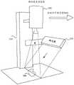
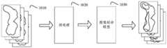
其中,推扫式采集是沿扫描线逐点扫描成像的一种采集方式,推扫式采集操作是基于采集设备进行的。示意性的,如图3所示,为一个推扫式短波红外高光谱成像系统,该系统包括样本台310、短波红外高光谱相机320、线光源330 以及目标样本340。其中,高光谱相机320是成像技术和光谱探测技术的结合,用于采集得到具有光谱信息的样本图像,样本图像为高光谱图像,高光谱图像是三维的,其中x轴与y轴用于表示二维信息的坐标值,z轴用于表示波长信息。与普通成像技术相比,高光谱图像增加了图像的光谱信息,即:样本图像为具有光谱信息的图像。Among them, the push-broom acquisition is an acquisition method in which the imaging is scanned point by point along the scan line, and the push-broom acquisition operation is performed based on the acquisition equipment. Illustratively, as shown in FIG. 3 , it is a push-broom short-wave infrared hyperspectral imaging system, which includes a
可选地,在确定预设波段后,以照明光源照射目标样本,并在预设波段内,以不同的波长照射条件下,对目标样本进行多次拍摄,从而获得多个目标样本对应的样本图像。示意性的,预设波段为900nm~1700nm(从近红外波段中选取的波段),目标样本为手术切除后的病理标本,采用卤素灯作为照明光源,将高光谱相机作为图像采集设备,在不同波长下对手术切除后的病理标本进行采集。例如:在预设波段内,采用高光谱相机,对每一个波长采集一个图像,从而获得不同波长下对应的多个样本图像。Optionally, after the preset wavelength band is determined, the target sample is irradiated with an illumination light source, and the target sample is photographed multiple times within the preset wavelength band under different wavelength illumination conditions, so as to obtain samples corresponding to multiple target samples. image. Schematically, the preset waveband is 900nm-1700nm (the waveband selected from the near-infrared waveband), the target sample is the pathological specimen after surgical resection, the halogen lamp is used as the illumination light source, and the hyperspectral camera is used as the image acquisition device. The pathological specimens after surgical resection were collected under the wavelength. For example, in a preset wavelength band, a hyperspectral camera is used to collect an image for each wavelength, so as to obtain multiple sample images corresponding to different wavelengths.
如图3所示,以线光源330对目标样本340进行照射,并采用短波红外高光谱相机320对不同波长照射下的目标样本340进行拍摄,从而实现获取多个不同波长下多个样本图像的过程。如图4所示,为采用短波红外高光谱相机320 采集到的多个样本图像410,其中,多个样本图像依据波长900nm~1700nm的范围,从上向下依次排列,多个样本图像410为具有光谱信息的三维高光谱图像。As shown in FIG. 3 , the
示意性的,如图5所示,任意选取样本图像410中的M点420以及N点430,基于样本图像410对应的光谱信息,得到M点420对应的光谱特定曲线510,以及N点430对应的光谱特性曲线520。其中,光谱特性曲线图是光反射率与波长之间的关系图,横坐标为波长,纵坐标为反射率,反射率用于指示被目标样本反射的光通量与入射到目标样本的光通量之比。可选地,图5中的光谱特性曲线为经过反射率校正后得到的曲线。Schematically, as shown in FIG. 5 , the
在一个可选的实施例中,在目标波段范围内,采用可调谐滤波器,确定至少一个波长;基于采集设备,对目标样本进行推扫式采集操作,获取至少一个波长对应的样本图像。In an optional embodiment, within the target wavelength range, a tunable filter is used to determine at least one wavelength; based on the acquisition device, a push-broom acquisition operation is performed on the target sample to obtain a sample image corresponding to at least one wavelength.
可选地,在高光谱相机前添加液晶可调谐滤波器(LCTF,Liquid Crystal TunableFilter),液晶可调谐滤波器用于从涵盖目标波段的照明光源中对波长进行选择,从而能够快速而且无振动地选择可见光波段内或者近红外波段内的波长。例如:照明光源涵盖的波段为900nm~1700nm,将照明光源发射的光经过液晶可调谐滤波器后,得到1130nm波长的光,即液晶可调谐滤波器将预设波段内除1130nm波长以外的其他波长的光予以滤除。Optionally, add a Liquid Crystal Tunable Filter (LCTF) in front of the hyperspectral camera, which is used to select wavelengths from illumination sources covering the target band, enabling fast and vibration-free selection Wavelengths in the visible band or in the near-infrared band. For example: the wavelength range covered by the illumination light source is 900nm to 1700nm. After passing the light emitted by the illumination light source through the liquid crystal tunable filter, light with a wavelength of 1130 nm is obtained, that is, the liquid crystal tunable filter will convert the wavelengths other than the 1130 nm wavelength within the preset band. light is filtered out.
可选地,采集设备为高光谱相机,高光谱相机为内置光栅推扫结构的相机,根据光栅的排列方式,对目标样本进行推扫式采集操作,得到样本图像。或者,高光谱相机外置推扫结构的高光谱拍摄方式,如图3所示,移动样品台310进行推扫拍摄等。值得注意的是,以上仅为示意性的举例,本申请实施例对此不加以限定。Optionally, the acquisition device is a hyperspectral camera, and the hyperspectral camera is a camera with a built-in grating push-broom structure. According to the arrangement of the gratings, a push-broom acquisition operation is performed on the target sample to obtain a sample image. Alternatively, in a hyperspectral imaging method with a push-broom structure external to the hyperspectral camera, as shown in FIG. 3 , the
步骤220,获取样本图像中与预设波段中至少一个目标波长对应的目标图像,得到伪彩色图像。Step 220: Acquire a target image corresponding to at least one target wavelength in the preset wavelength band in the sample image to obtain a pseudo-color image.
示意性的,样本图像为对目标样本进行采集得到的多个图像,样本图像对应的波长在预设波段内。在预设波段内,从多个波长中选择至少一个目标波长,并将目标波长对应的样本图像作为目标图像,最终得到伪彩色图像。Illustratively, the sample images are multiple images obtained by collecting the target sample, and the wavelengths corresponding to the sample images are within a preset wavelength band. In the preset wavelength band, at least one target wavelength is selected from multiple wavelengths, and the sample image corresponding to the target wavelength is used as the target image, and finally a pseudo-color image is obtained.
可选地,对于一个目标波长,存在与该目标波长对应的至少一个样本图像。示意性的,当被选择的目标波长对应存在多个样本图像时,既可以从多个样本图像中随机选择一个样本图像作为与该目标波长对应的目标图像,也可以对多个样本图像进行结合分析,从而确定与该目标波长对应的目标图像。Optionally, for a target wavelength, there is at least one sample image corresponding to the target wavelength. Illustratively, when there are multiple sample images corresponding to the selected target wavelength, one sample image may be randomly selected from the multiple sample images as the target image corresponding to the target wavelength, or multiple sample images may be combined. analysis to determine the target image corresponding to the target wavelength.
或者,对于一个目标波长,存在与该目标波长对应的一个样本图像,将该样本图像作为目标图像。可选地,以一个目标波长对应一个目标图像为例,根据所选择的目标波长的数量,对目标图像的处理方式存在差异。示意性的,对选择一个目标波长和选择多个目标波长的情况分别进行分析。Alternatively, for a target wavelength, there is a sample image corresponding to the target wavelength, and the sample image is used as the target image. Optionally, taking one target wavelength corresponding to one target image as an example, according to the number of selected target wavelengths, there are differences in the processing methods of the target image. Illustratively, the cases of selecting one target wavelength and selecting multiple target wavelengths are analyzed respectively.
(1)选择一个目标波长(1) Select a target wavelength
在一个可选的实施例中,对预设波段中一个目标波长对应的目标图像进行赋色处理,得到伪彩色图像。In an optional embodiment, a colorimetric process is performed on a target image corresponding to a target wavelength in a preset wavelength band to obtain a pseudo-color image.
示意性的,预设波段为从近红外波段中选取的900nm至1700nm的波段,由于预设波段为不可见波段,故该波段对应的图像为灰度图像。从预设波段中选择一个目标波长,该目标波长对应的目标图像为一幅灰度图像,对该灰度图像进行赋色处理,得到伪彩色图像。Illustratively, the preset wavelength band is a wavelength band from 900 nm to 1700 nm selected from the near-infrared wavelength band. Since the preset wavelength band is an invisible wavelength band, the image corresponding to this wavelength band is a grayscale image. A target wavelength is selected from the preset wavelength band, and the target image corresponding to the target wavelength is a grayscale image, and the grayscale image is subjected to coloration processing to obtain a pseudo-color image.
伪彩色图像处理用于指示将黑白的灰度图像转换为彩色图像的技术过程,从而提高图像内容的可辨识度。示意性的,采用灰度分成法、灰度变换法等方法,进行伪彩色图像处理。Pseudo-color image processing is used to indicate the technical process of converting a black-and-white grayscale image into a color image, thereby improving the recognizability of the image content. Illustratively, methods such as a grayscale division method, a grayscale transformation method, and the like are used to process pseudo-color images.
可选地,灰度图像为单通道图像,即每个像素点只有一个值表示颜色,其像素值位于0至255之间,0用于指示黑色,255用于指示白色,中间值为不同等级的灰色。或者,当灰度图像为三通道图像时,三个通道的像素值均相同。Optionally, the grayscale image is a single-channel image, that is, each pixel has only one value to represent the color, and its pixel value is between 0 and 255, where 0 is used to indicate black, 255 is used to indicate white, and the intermediate values are different levels. grey. Alternatively, when the grayscale image is a three-channel image, the pixel values of all three channels are the same.
可选地,与单通道图像相对的图像包括三通道图像,即每个像素点都有3 个值表示。示意性的,RGB图像为三通道图像,是通过对红(R)、绿(G)、蓝(B) 三个颜色通道的变化以及它们相互之间的叠加,从而得到各式各样的颜色。其中,每一个像素点由三个值表示。Optionally, the image opposite to the single-channel image includes a three-channel image, that is, each pixel is represented by three values. Schematically, the RGB image is a three-channel image, and various colors are obtained by changing the three color channels of red (R), green (G), and blue (B) and superimposing them on each other. . Among them, each pixel is represented by three values.
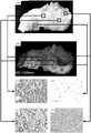
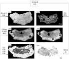
示意性的,目标样本为手术切除的病理样本,如图6所示,为对目标样本进行不同图像处理的示意图。其中,图610用于指示目标样本(为便于表示,采用常规相机进行拍摄得到);图620用于指示波长为1300nm的高光谱图像;图631至图634用于指示采用苏木素伊红染色法(HE,Hematoxylin Eosin)观察到的组织表现,其中图631用于指示癌组织(图610或图620中的A点所示)、图632用于指示脂肪组织(图610或图620中的B点所示)、图633用于指示正常黏膜组织(图610或图620中的B点所示)、图634用于指示肌组织(图610 或图620中的D点所示)。Illustratively, the target sample is a surgically resected pathological sample, as shown in FIG. 6 , which is a schematic diagram of performing different image processing on the target sample. Among them, Figure 610 is used to indicate the target sample (for convenience of representation, it is obtained by using a conventional camera); Figure 620 is used to indicate a hyperspectral image with a wavelength of 1300 nm; Figures 631 to 634 are used to indicate the use of hematoxylin and eosin staining method ( HE, Hematoxylin Eosin), where
示意性的,将波长1300nm作为被选择的目标波长,将图620对应的高光谱图像作为目标波长对应的目标图像,对目标图像进行上述赋色处理,得到伪彩色图像。Illustratively, the wavelength of 1300 nm is used as the selected target wavelength, the hyperspectral image corresponding to FIG. 620 is used as the target image corresponding to the target wavelength, and the above-mentioned colorimetric processing is performed on the target image to obtain a pseudo-color image.
(2)选择多个目标波长(2) Select multiple target wavelengths
示意性的,根据至少两个目标波长,确定至少两个目标波长分别对应的至少两个目标图像。其中,第i个目标波长对应第i个目标图像,i为正整数。Illustratively, at least two target images corresponding to the at least two target wavelengths are determined according to the at least two target wavelengths. Among them, the ith target wavelength corresponds to the ith target image, and i is a positive integer.
在一个可选的实施例中,将预设目标波段中至少两个目标波长对应的至少两个目标图像进行合成处理,对合成的图像以及进行赋色处理后,得到伪彩色图像。In an optional embodiment, at least two target images corresponding to at least two target wavelengths in the preset target wavelength band are synthesized and processed, and a pseudo-color image is obtained after the synthesized images and color assignment processing.
示意性的,从预设目标波段中选择至少两个目标波长,每一个目标波长对应一个目标图像。可选地,对至少两个目标图像进行合成处理,得到候选图像。Illustratively, at least two target wavelengths are selected from the preset target wavelength bands, and each target wavelength corresponds to a target image. Optionally, at least two target images are synthesized to obtain candidate images.
示意性的,对多个目标图像进行合成处理的方式包括如下至少一种方式。Illustratively, the manner of synthesizing multiple target images includes at least one of the following manners.
(1)像素值处理(1) Pixel value processing
在一个可选的实施例中,对至少两个目标图像对应像素点的像素值进行平均处理,得到对应像素点的目标像素值;基于各像素点对应的目标像素值确定候选图像。In an optional embodiment, the pixel values of the corresponding pixel points of at least two target images are averaged to obtain the target pixel value of the corresponding pixel point; the candidate image is determined based on the target pixel value corresponding to each pixel point.
示意性的,在获得至少两个目标波段对应的至少两个目标图像后,将至少两个目标图像对应像素点的像素值进行求和后取平均值,得到对应像素点的目标像素值,即:目标像素值为对不同目标图像对应像素点的像素值进行综合分析后得到的平均值。可选地,在确定各像素点对应的目标像素值后,依据像素点的位置信息,得到候选图像,候选图像中各个像素点的像素值为对应的目标像素值。Illustratively, after obtaining at least two target images corresponding to at least two target bands, the pixel values of the corresponding pixel points of the at least two target images are summed and then averaged to obtain the target pixel value of the corresponding pixel point, that is, : The target pixel value is the average value obtained by comprehensively analyzing the pixel values of the corresponding pixel points of different target images. Optionally, after determining the target pixel value corresponding to each pixel point, a candidate image is obtained according to the position information of the pixel point, and the pixel value of each pixel point in the candidate image corresponds to the corresponding target pixel value.
(2)软件处理(2) software processing
在一个可选的实施例中,在确定目标波长对应的目标图像后,将至少两个目标图像通过软件进行合成处理,得到候选图像。In an optional embodiment, after the target image corresponding to the target wavelength is determined, at least two target images are synthesized through software to obtain candidate images.
示意性的,将至少两个目标图像输入Photoshop中,进行对齐、拼接调色、擦除接缝、导出等操作,得到将至少两个目标图像进行合成后的候选图像。Illustratively, at least two target images are input into Photoshop, and operations such as aligning, splicing toning, erasing seams, and exporting are performed to obtain candidate images obtained by synthesizing the at least two target images.
以上仅为示意性的举例,本申请实施例对此不加以限定。The above are only illustrative examples, which are not limited in the embodiments of the present application.
在一个可选的实施例中,对候选图像进行赋色处理,得到伪彩色图像。In an optional embodiment, color assignment processing is performed on the candidate image to obtain a pseudo-color image.
示意性的,基于候选图像中像素点的亮度值,对候选图像中的像素点进行亮度分级,确定至少两个亮度级别;对至少两个亮度级别分别赋色,得到伪彩色图像。Illustratively, based on the brightness values of the pixels in the candidate image, the brightness of the pixels in the candidate image is classified to determine at least two brightness levels; the at least two brightness levels are assigned colors to obtain a pseudo-color image.
可选地,目标图像为灰度图像,基于目标图像合成得到的候选图像为灰度图像,灰度图像中各个像素点对应的目标像素值用于指示候选图像的亮度。示意性的,目标像素值位于0至255之间,0用于指示黑色(亮度最小),255用于指示白色(亮度最大),即:目标像素值的数值越小,亮度越小;目标像素值的数值越大,亮度越大。Optionally, the target image is a grayscale image, the candidate image synthesized based on the target image is a grayscale image, and the target pixel value corresponding to each pixel in the grayscale image is used to indicate the brightness of the candidate image. Illustratively, the target pixel value is between 0 and 255, 0 is used to indicate black (minimum brightness), and 255 is used to indicate white (maximum brightness), that is: the smaller the value of the target pixel value, the smaller the brightness; The larger the value of the value, the greater the brightness.
示意性的,如图7所示,为将三个目标波长(波长1100nm、波长1300nm 以及波长1450nm)对应的目标图像进行合成处理和赋值处理后得到伪彩色图像的过程示意图。Illustratively, as shown in FIG. 7 , it is a schematic diagram of the process of obtaining a pseudo-color image after synthesizing and assigning target images corresponding to three target wavelengths (
其中,图710用于指示波长为1100nm的高光谱图像;图720用于指示波长为1300nm的高光谱图像;图730用于指示波长为1450nm的高光谱图像。可选地,将上述三个目标波长对应的高光谱图像进行合成处理和赋值处理后,得到图740所示的伪彩色图像。Wherein, the
步骤230,根据样本图像中样本元素类型的差异,对样本图像进行区域划分,得到区域划分结果。Step 230: Divide the sample image into regions according to the difference of the sample element types in the sample image to obtain a region division result.
其中,样本元素类型中包括待识别的目标元素类型。The sample element type includes the target element type to be identified.
可选地,样本元素类型用于指示样本图像中不同样本区域对应的样本性质差异。示意性的,当样本图像为针对病理样本进行拍摄得到的图像,样本元素类型包括:该病理样本中的肿瘤组织、该病理样本中的脂肪组织、该病理样本中的黏膜组织、该病理样本中的肌组织等;当样本图像为针对化学混合物(其中包括A化合物、B化合物以及杂质)进行拍摄得到的图像,样本元素类型包括:A化合物、B化合物以及杂质。Optionally, the sample element type is used to indicate differences in sample properties corresponding to different sample regions in the sample image. Illustratively, when the sample image is an image obtained by photographing a pathological sample, the sample element types include: tumor tissue in the pathological sample, adipose tissue in the pathological sample, mucosal tissue in the pathological sample, and adipose tissue in the pathological sample. When the sample image is an image obtained by photographing a chemical mixture (including A compound, B compound and impurities), the sample element types include: A compound, B compound and impurities.
示意性的,当样本图像为针对病理样本进行拍摄得到病理图像时,目标元素类型为预先确定的、待识别的肿瘤组织(该病理图像对应的样本元素类型中的一种);或者,目标元素类型为预先确定的、待识别的待识别的脂肪组织等。可选地,当样本图像为针对化学混合物进行拍摄得到的化学图像,目标元素类型为预先确定的、待识别的B化合物(该化学图像对应的样本元素类型中的一种)等。Illustratively, when the sample image is a pathological image obtained by photographing a pathological sample, the target element type is a predetermined tumor tissue to be identified (one of the sample element types corresponding to the pathological image); or, the target element The type is a predetermined, to-be-identified, to-be-identified adipose tissue, or the like. Optionally, when the sample image is a chemical image obtained by photographing a chemical mixture, the target element type is a predetermined B compound to be identified (one of the sample element types corresponding to the chemical image) and the like.
示意性的,样本图像为具有光谱信息的图像,根据不同物质的性质差异,光谱信息存在差异,根据样本图像对应的光谱信息,对样本图像进行区域划分,得到区域划分结果。Illustratively, the sample image is an image with spectral information. According to the difference in properties of different substances, the spectral information is different. According to the spectral information corresponding to the sample image, the sample image is divided into regions to obtain a region division result.
示意性的,光谱信息在样本图像上显示不同,例如:样本图像为灰度图像时,A样本元素对应的区域颜色最深,目标样本元素对应的区域颜色最浅,由此得到样本图像对应的区域划分结果。Schematically, the spectral information is displayed differently on the sample image. For example, when the sample image is a grayscale image, the region corresponding to the A sample element has the darkest color, and the region corresponding to the target sample element has the lightest color, thus obtaining the region corresponding to the sample image. Divide the results.
在一个可选的实施例中,为便于区分,可以对不同的区域填充不同的颜色,得到具有颜色的区域划分结果;或者,采用较深的轮廓线,对不同区域进行划分,得到具有较明显分隔的区域划分结果等。In an optional embodiment, in order to facilitate the distinction, different colors can be filled in different regions to obtain a region division result with colors; Delimited area division results, etc.
值得注意的是,以上仅为示意性的举例,本申请实施例对此不加以限定。It should be noted that the above are only illustrative examples, which are not limited in the embodiments of the present application.
步骤240,基于伪彩色图像和区域划分结果,在样本图像中确定包括目标元素类型的目标区域。
示意性的,伪彩色图像是对目标波长对应的目标图像进行处理后得到的图像;区域划分结果是根据样本图像中样本元素类型进行区域划分后的结果。可选地,伪彩色图像中以不同的颜色对伪彩色图像进行划分,例如:样本图像为针对病理样本进行拍摄得到的图像,其中的肿瘤组织呈现为橙色;脂肪组织呈现为亮黄色、黏膜组织呈现为较肿癌组织颜色更浅的浅橙色、肌组织呈现为较肿瘤组织颜色更深的深橙色等。Illustratively, the pseudo-color image is an image obtained after processing the target image corresponding to the target wavelength; the region division result is the result of region division according to the sample element type in the sample image. Optionally, the pseudo-color image is divided into different colors in the pseudo-color image, for example: the sample image is an image obtained by photographing a pathological sample, in which the tumor tissue appears orange; the adipose tissue appears bright yellow, and the mucosal tissue appears The color of the muscle tissue is darker than that of the tumor tissue.
在一个可选的实施例中,确定伪彩色图像与区域划分结果中的重叠区域;在样本图像中,将重叠区域作为包括目标元素类型的目标区域。In an optional embodiment, the overlapping area between the pseudo-color image and the area division result is determined; in the sample image, the overlapping area is used as the target area including the target element type.
示意性的,目标样本为病理样本,对病理样本进行采集后得到具有光谱信息的样本图像,欲观察样本图像中的肿瘤组织,对样本图像进行上述处理过程,得到被选择的目标波长对应的伪彩色图像以及对样本图像的区域划分结果。根据伪彩色图像中确定的肿瘤组织的区域,以及区域划分结果中包括目标元素类型(肿瘤组织)的识别结果,将重叠区域作为包括目标元素类型(肿瘤组织) 的目标区域,实现对肿瘤组织区域的识别过程。Illustratively, the target sample is a pathological sample. After collecting the pathological sample, a sample image with spectral information is obtained. To observe the tumor tissue in the sample image, the above processing process is performed on the sample image to obtain a pseudo-spectrographic image corresponding to the selected target wavelength. Color image and the result of region division of the sample image. According to the area of the tumor tissue determined in the pseudo-color image and the recognition result including the target element type (tumor tissue) in the area division result, the overlapping area is taken as the target area including the target element type (tumor tissue) to realize the identification of the tumor tissue area. identification process.
以上仅为示意性的举例,本申请实施例对此不加以限定。The above are only illustrative examples, which are not limited in the embodiments of the present application.
综上所述,从样本图像中获取目标波段对应的目标图像,并得到伪彩色图像,对样本图像进行区域划分后得到区域划分结果,结合伪彩色图像和区域划分结果确定目标区域。通过上述方法,可以避免仅仅依靠医生的裸眼观测和描述对肿瘤组织的大小、区域等进行判断。基于预先确定的预设波段,对目标样本进行采集,得到样本图像,从预设波段中选取至少一个效果较好的目标波长,并根据至少一个目标波长,从样本图像中确定与目标波长对应的目标图像,对目标图像进行处理后,得到伪彩色图像,伪彩色图像能够较准确地体现目标波长的优势。根据样本图像中样本元素类型的差异,对样本图像进行区域划分后得到区域划分结果。结合伪彩色图像进而区域划分结果,确定包括目标元素类型的目标区域,从而确定待识别区域(如:肿瘤组织)的位置信息,提高病理取材的准确性,降低病理取材的难度,利用目标样本在不同波长下对应的光谱特性对目标样本进行分析,不仅操作较为简便,且成本相对较低,更容易应用并广泛普及。To sum up, the target image corresponding to the target band is obtained from the sample image, and the pseudo-color image is obtained. The sample image is divided into regions to obtain the region division result, and the target region is determined by combining the pseudo-color image and the region division result. By the above method, it can be avoided to judge the size, area, etc. of the tumor tissue only by the naked eye observation and description of the doctor. Based on a predetermined preset wavelength band, the target sample is collected to obtain a sample image, at least one target wavelength with better effect is selected from the preset wavelength band, and according to the at least one target wavelength, the sample image corresponding to the target wavelength is determined from the sample image. After processing the target image, a pseudo-color image is obtained, and the pseudo-color image can more accurately reflect the advantages of the target wavelength. According to the difference of the sample element types in the sample image, the region division result is obtained after the sample image is divided into regions. Combined with the pseudo-color image and the area division results, the target area including the target element type is determined, so as to determine the location information of the area to be identified (such as: tumor tissue), improve the accuracy of pathological sampling, and reduce the difficulty of pathological sampling. The spectral characteristics corresponding to different wavelengths are used to analyze the target sample, which is not only easy to operate, but also has a relatively low cost, and is easier to apply and widely popularize.
在一个可选的实施例中,对样本图像进行区域划分的过程是通过不同样本元素类型对应的不同光谱信息确定的。示意性的,如图8所示,上述图2所示出的实施例中的步骤230还可以实现为如下步骤810至步骤850。In an optional embodiment, the process of dividing the sample image into regions is determined by different spectral information corresponding to different sample element types. Illustratively, as shown in FIG. 8 ,
步骤810,获取标准图像。
其中,标准图像是针对目标样本进行采集得到的具有光谱信息的预先标注图像。The standard image is a pre-labeled image with spectral information obtained by collecting the target sample.
可选地,样本图像和标准图像是对目标样本进行采集得到的图像,在对目标样本进行采集时,确定样本图像的金标准,即确定标准图像。金标准用于指示当前临床医学界公认的诊断疾病的最可靠方法。Optionally, the sample image and the standard image are images obtained by collecting the target sample. When the target sample is collected, the gold standard of the sample image is determined, that is, the standard image is determined. The gold standard is used to indicate the most reliable method of diagnosing disease currently recognized by the clinical medical community.
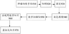
在一个可选的实施例中,目标样本为手术切除的病理样本。示意性的,如图9所示,为对病理样本进行取材的流程示意图,首先,医生通过肿瘤切除手术910对患者的病理样本进行切除(病理样本中包括肿瘤组织),之后,对病理样本进行切割920,得到适当体积的组织块,然后,采用福尔马林浸泡等方法将组织块进行固定930。例如:在手术切除病理样本离体后的30分钟内,将组织块放入足量的3.7%中性福尔马林溶液中进行固定,固定时间12h-48h。随后,对固定后的组织块切取厚度5mm±1mm的组织片(平均约为5mm),其中包括肿瘤组织及周围1-2cm的正常组织。可选地,既可以将对病理样本进行固定后的组织块作为目标样本,也可以将对组织块进行切片后的组织片作为目标样本。In an optional embodiment, the target sample is a surgically resected pathological sample. Illustratively, as shown in FIG. 9 , which is a schematic diagram of the process of taking a pathological sample, first, the doctor removes the patient's pathological sample (the pathological sample includes tumor tissue) through a tumor resection operation 910 , and then performs a tumor resection on the pathological sample. Cut 920 to obtain a tissue block of an appropriate volume, and then fix 930 the tissue block by means of formalin immersion or the like. For example, within 30 minutes after the surgical excision of the pathological sample, the tissue block is placed in a sufficient amount of 3.7% neutral formalin solution for fixation, and the fixation time is 12h-48h. Subsequently, tissue slices with a thickness of 5 mm ± 1 mm (about 5 mm in average) were cut from the fixed tissue block, including tumor tissue and surrounding normal tissue of 1-2 cm. Optionally, either the fixed tissue block of the pathological sample can be used as the target sample, or the tissue slice obtained by slicing the tissue block can be used as the target sample.
在一个可选的实施例中,在获取目标样本后,组织片经大体取材、常规脱水、包埋以及HE染色制片处理后,得到染色图像940,将染色图像940经过数字扫描仪950进行扫描后,得到多个全视野数字切片(WSI,Whole Slide Image) 图像960。其中,WSI是在数字扫描仪950(一种电动显微镜结构)下采集的该病理样本的图像。示意性的,若单张WSI图像的尺寸较小,被分析的WSI图像可以是多张病理切片拼接而成,例如,采用WSI拼接软件对多个WSI部分 (fragments)进行拼接,还原得到虚拟大切片970。In an optional embodiment, after obtaining the target sample, the tissue piece is processed by rough sampling, conventional dehydration, embedding and HE staining to obtain a stained image 940 , and the stained image 940 is scanned by a digital scanner 950 Then, a plurality of Whole Slide Image (WSI, Whole Slide Image) images 960 are obtained. Wherein, WSI is the image of the pathological sample acquired under the digital scanner 950 (a motorized microscope structure). Illustratively, if the size of a single WSI image is small, the analyzed WSI image can be spliced from multiple pathological slices. For example, using WSI splicing software to splicing multiple WSI fragments (fragments), restore a virtual large slice. Sheet 970.
可选地,以在虚拟大切片970上运用高级系统分析程序(ASAP,Advanced SystemsAnalysis Program)进行标注作为金标准,得到多个针对该病理样本扫描得到的、具有标注的WSI图像,其中,标注可以采用对区域标注的方式进行,标注的区域既包括一种或者多种病灶所在的区域,还包括有提示作用的特殊区域等。示意性的,在空腔脏器中,将肿瘤组织标注为红色,将正常黏膜标记为绿色,将脂肪组织标记为黄色,将肌组织标记为蓝色;在实质脏器中,将肿瘤组织标注为红色,将正常组织标注为绿色,将脂肪组织标记为黄色等。可选地,上述颜色标记仅为示意性的举例,也可以采用不同的颜色对被选择的组织进行标记,例如:对实质脏器中的乳腺组织进行标记时,将乳腺组织中的肿瘤组织标记为红色,将脂肪组织标记为黄色,将纤维结缔组织标记为绿色等。可选地,当被观察的脏器中不存在对应颜色的组织时,可以不标注,例如:采用上述颜色标记方式标记实质脏器时,若被观察的脏器中不存在脂肪组织,则不予标记黄色。示意性的,将标注的WSI图像作为标准图像,实现对标准图像的获取过程。Optionally, using the Advanced Systems Analysis Program (ASAP, Advanced Systems Analysis Program) on the virtual large slice 970 for labeling as the gold standard, a plurality of WSI images with labeling obtained by scanning the pathological sample are obtained, wherein the labeling can be The method is used to mark the area, and the marked area includes not only the area where one or more lesions are located, but also the special area with a prompting effect. Schematically, in hollow organs, tumor tissue is marked in red, normal mucosa is marked in green, adipose tissue is marked in yellow, and muscle tissue is marked in blue; in solid organs, tumor tissue is marked in in red, normal tissue in green, adipose tissue in yellow, etc. Optionally, the above color marking is only a schematic example, and different colors can also be used to mark the selected tissue, for example: when marking the breast tissue in the parenchymal organ, mark the tumor tissue in the breast tissue. in red, adipose tissue in yellow, fibrous connective tissue in green, etc. Optionally, when there is no tissue of the corresponding color in the observed organ, it may not be marked. For example, when the above-mentioned color marking method is used to mark the parenchymal organ, if there is no adipose tissue in the observed organ, it will not be marked. I marked yellow. Illustratively, the marked WSI image is used as the standard image to realize the process of acquiring the standard image.
步骤820,以标准图像对候选划分模型进行训练。Step 820: Train the candidate segmentation model with the standard image.
其中,候选划分模型为未训练的、具有一定区域划分功能的模型。示意性的,以标准图像为金标准,对候选划分模型进行训练,在大量标准图像的训练下,候选划分模型进行学习,并能够逐渐自动识别病灶区域等特殊区域,并逐渐具备区域分割功能。Among them, the candidate division model is an untrained model with a certain area division function. Schematically, the candidate segmentation model is trained with standard images as the gold standard. Under the training of a large number of standard images, the candidate segmentation model learns, and can gradually and automatically identify special areas such as lesion areas, and gradually have the function of area segmentation.
步骤830,响应于对候选划分模型的训练达到训练效果,得到图像划分模型。
其中,图像划分模型用于对目标图像进行区域分割。示意性的,在对候选划分模型进行训练的过程中,会因为对候选划分模型的训练达到训练目标而得到图像划分模型,可选地,以损失值判断候选划分模型的训练效果,训练目标至少包括如下一种情况。Among them, the image segmentation model is used to perform region segmentation on the target image. Illustratively, in the process of training the candidate division model, the image division model will be obtained because the training of the candidate division model achieves the training target. Including the following one.
1、响应于损失值达到收敛状态,将最近一次迭代训练得到的候选划分模型作为图像划分模型。1. In response to the loss value reaching the convergence state, the candidate partition model obtained by the latest iteration training is used as the image partition model.
示意性的,损失值达到收敛状态用于指示通过损失函数得到的损失值的数值不再变化或者变化幅度小于预设阈值。例如:第n个标准图像对应的损失值为0.1,第n+1个标准图像对应的损失值也为0.1,可以视为该损失值达到收敛状态,将第n个标准图像或者第n+1个标准图像对应的损失值调整的候选划分模型作为图像划分模型,实现对候选划分模型的训练过程。Illustratively, the loss value reaching a convergence state is used to indicate that the value of the loss value obtained through the loss function no longer changes or the change range is smaller than a preset threshold. For example, the loss value corresponding to the nth standard image is 0.1, and the loss value corresponding to the n+1th standard image is also 0.1. It can be considered that the loss value has reached a convergence state, and the nth standard image or the n+1th The candidate partition model adjusted by the loss value corresponding to each standard image is used as the image partition model to realize the training process of the candidate partition model.
2、响应于损失值的获取次数达到次数阈值,将最近一次迭代训练得到的候选划分模型作为图像划分模型。2. In response to the number of acquisitions of the loss value reaching the number threshold, the candidate division model obtained by the latest iterative training is used as the image division model.
示意性的,一次获取可以得到一个损失值,预先设定用于训练图像划分模型的损失值的获取次数,当一个标准图像对应一个损失值时,损失值的获取次数即为标准图像的个数;或者,当一个标准图像对应多个损失值时,损失值的获取次数即为损失值的个数。例如:预先设定一次获取可以得到一个损失值,损失值获取的次数阈值为10次,即当达到获取次数阈值时,将最近一次损失值调整的候选划分模型作为图像划分模型,或者将损失值10次调整过程中最小损失值调整的候选划分模型作为图像划分模型,实现对候选划分模型的训练过程。Illustratively, one loss value can be obtained at one time, and the number of times of obtaining the loss value for training the image division model is preset. When a standard image corresponds to one loss value, the number of times of obtaining the loss value is the number of standard images. ; or, when a standard image corresponds to multiple loss values, the number of loss values obtained is the number of loss values. For example, a loss value can be obtained by pre-setting one acquisition, and the threshold of the number of acquisitions of the loss value is 10 times, that is, when the threshold of acquisition times is reached, the candidate division model of the latest loss value adjustment is used as the image division model, or the loss value is used as the image division model. The candidate division model adjusted by the minimum loss value in the 10 adjustment processes is used as the image division model to realize the training process of the candidate division model.
值得注意的是,以上仅为示意性的举例,本申请实施例对此不加以限定。It should be noted that the above are only illustrative examples, which are not limited in the embodiments of the present application.
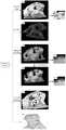
步骤840,将样本图像通过预先训练得到的图像划分模型,确定元素类型的差异表示。Step 840: Determine the difference representation of the element type by passing the sample image through an image division model obtained by pre-training.
在一个可选的实施例中,对样本图像进行图像预处理后,输入预先训练得到的图像划分模型中。In an optional embodiment, after image preprocessing is performed on the sample image, it is input into the image division model obtained by pre-training.
示意性的,如图10所示,在采集得到多个样本图像1010后,将样本图像 1010进行预处理1020。其中,对样本图像进行图像预处理1020的过程包括如下至少一种:对样本图像进行几何变换操作、图像增强操作等(如:图像背景校正、配准、去噪等),从而突出样本图像中的重要特征。之后,将预处理后的样本图像1010通过预先训练得到的图像划分模型1030中,由图像划分模型1030 对样本图像中的区域进行划分。Illustratively, as shown in FIG. 10 , after a plurality of
在一个可选的实施例中,样本图像为具有光谱信息的图像,对目标图像进行光谱分析,得到光谱分析结果;基于光谱分析结果,确定样本图像对应的元素类型的差异表示。In an optional embodiment, the sample image is an image with spectral information, and spectral analysis is performed on the target image to obtain a spectral analysis result; based on the spectral analysis result, a differential representation of element types corresponding to the sample image is determined.
根据样本图像对应的目标样本的差异,得到不同样本图像对应的不同光谱分析结果。示意性的,光谱分析结果采用光谱特征曲线图的形式表示,光谱特征曲线图的横坐标为波长,纵坐标为反射率,不同的光谱曲线用于指示不同的目标样本在不同波长下的反射率变化情况,即光谱分析结果。According to the difference between the target samples corresponding to the sample images, different spectral analysis results corresponding to different sample images are obtained. Schematically, the spectral analysis result is represented in the form of a spectral characteristic curve graph, the abscissa of the spectral characteristic graph is the wavelength, and the ordinate is the reflectance, and different spectral curves are used to indicate the reflectance of different target samples at different wavelengths The change is the result of spectral analysis.
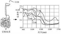
在一个可选的实施例中,对62例不同系统组织的高光谱图像进行分析后,初步确定不同器官中区分肿瘤组织与正常组织的波长在1296-1308nm(该波长范围内效果较好)。示意性的,以存在肿瘤组织的空腔脏器(如:食管,胃,结直肠)、肾、乳腺以及肺作为目标样本为例进行分析,得到空腔脏器、肾、乳腺以及肺对应的样本图像,样本图像为三维高光谱图像,根据三维高光谱图像对应的数据,得到空腔脏器、肾、乳腺以及肺分别对应的光谱特征曲线图。In an optional embodiment, after analyzing the hyperspectral images of 62 different system tissues, it is preliminarily determined that the wavelength for distinguishing tumor tissue from normal tissue in different organs is 1296-1308 nm (the effect is better in this wavelength range). Illustratively, take the hollow organs (such as esophagus, stomach, colorectum), kidney, breast and lung with tumor tissue as the target samples for analysis, and obtain the corresponding hollow organs, kidney, breast and lung. The sample image is a three-dimensional hyperspectral image. According to the data corresponding to the three-dimensional hyperspectral image, spectral characteristic curves corresponding to the hollow organ, kidney, breast and lung respectively are obtained.
如图11所示,为空腔脏器1110对应的光谱特征曲线图,其中,肿瘤组织(癌组织)对应的波长曲线为肿瘤波长曲线1120;脂肪组织对应的波长曲线为脂肪波长曲线1130;正常黏膜对应的波长曲线为黏膜波长曲线1140;肌组织对应的波长曲线为肌组织波长曲线1150。As shown in FIG. 11 , it is the spectral characteristic curve graph corresponding to the
如图12所示,为肾1210对应的光谱特征曲线图,其中,肿瘤组织(癌组织)对应的波长曲线为肿瘤波长曲线1220;脂肪组织对应的波长曲线为脂肪波长曲线1230;正常黏膜对应的波长曲线为黏膜波长曲线1240。As shown in FIG. 12 , it is the spectral characteristic curve graph corresponding to
如图13所示,为乳腺1310对应的光谱特征曲线图,其中,肿瘤组织(癌组织)对应的波长曲线为肿瘤波长曲线1320;脂肪组织对应的波长曲线为脂肪波长曲线1330;正常黏膜对应的波长曲线为黏膜波长曲线1340。As shown in FIG. 13, it is the spectral characteristic curve corresponding to the
如图14所示,为肺1410对应的光谱特征曲线图,其中,肿瘤组织(癌组织)对应的波长曲线为肿瘤波长曲线1420;正常的肺对应的波长曲线为正常波长曲线1430。As shown in FIG. 14 , it is a spectral characteristic curve graph corresponding to
其中,样本图像对应的元素类型的差异表示即为不同组织之间的差异,例如:肿瘤组织和脂肪组织是不同的等。以上仅为示意性的举例,本申请实施例对此不加以限定。Among them, the difference of the element types corresponding to the sample image is expressed as the difference between different tissues, for example, tumor tissue and adipose tissue are different. The above are only illustrative examples, which are not limited in the embodiments of the present application.
综合图11至图14进行分析,在波长为1300nm左右时,在空腔脏器组织的样本中不同组织显示出较好的区分度。实质脏器(如:乳腺、肾及肺)中的肿瘤组织与周围的正常组织及脂肪组织亦显示较好的区分度。Comprehensive analysis of Figures 11 to 14 shows that when the wavelength is about 1300 nm, different tissues show a good degree of discrimination in the hollow organ tissue sample. The tumor tissue in the parenchymal organs (such as breast, kidney and lung) also showed a good degree of differentiation from the surrounding normal tissue and adipose tissue.
示意性的,以结肠癌为例,通过肉眼观察1300nm高光谱图像中肿瘤组织呈灰色,正常肌组织表现出较肿瘤组织颜色深的灰黑色,脂肪组织呈灰白色,正常粘膜显示出较肌层浅,较肿瘤组织稍深的深灰色,1300nm高光谱图像显示出对脂肪、肌层及肿瘤组织较好的区分度。Schematically, taking colon cancer as an example, in the 1300nm hyperspectral image, the tumor tissue is gray in the naked eye, the normal muscle tissue is darker gray than the tumor tissue, the adipose tissue is gray and white, and the normal mucosa is lighter than the muscle layer. , slightly darker than the tumor tissue, and the 1300nm hyperspectral image showed a better distinction between fat, muscle and tumor tissue.
在一个可选的实施例中,抽取高光谱图像中三个波峰波谷1100nm、1300nm 以及1450nm作为特征波段,合成短波红外彩色合成图像,从而提供更符合人眼观察习惯的伪彩色图像,以便于医生识别不同组织。在短波红外彩色合成图像中,癌组织呈现为橙色,肌组织则表现为较肿瘤组织颜色更深的橙色,正常粘膜表现为较癌组织浅的橙色,脂肪组织则呈亮黄色。In an optional embodiment, three peaks and valleys of 1100 nm, 1300 nm and 1450 nm in the hyperspectral image are extracted as characteristic bands, and a short-wave infrared color composite image is synthesized, thereby providing a pseudo-color image that is more in line with the observation habits of the human eye, which is convenient for doctors Identify different organizations. In the short-wave infrared color composite image, cancer tissue appears orange, muscle tissue appears darker orange than tumor tissue, normal mucosa appears lighter orange than cancer tissue, and adipose tissue appears bright yellow.
步骤850,基于元素类型的差异表示,对样本图像进行区域划分,确定样本图像对应的区域划分结果。Step 850 , based on the difference representation of the element types, perform region division on the sample image, and determine a region division result corresponding to the sample image.
示意性的,在得到光谱分析结果后,由图像划分模型在样本图像上给出相应的区域信息提示,区域信息提示包括如下至少一种方式。Illustratively, after obtaining the spectral analysis result, the image segmentation model provides corresponding regional information prompts on the sample image, and the regional information prompts include at least one of the following methods.
(1)轮廓线提示(1) Outline prompt
示意性的,采用轮廓线对样本图像中不同的区域进行划分处理,得到不同划分区域,其中,轮廓线既可以是较深的曲线,也可以是有颜色的曲线等。Illustratively, contour lines are used to divide different regions in the sample image to obtain different divided regions, wherein the contour lines may be dark curves or colored curves.
(2)热力图提示(2) Heat map tips
示意性的,采用特殊高亮的方式,对肿瘤组织所在区域进行表示。Illustratively, the region where the tumor tissue is located is represented by a special highlighting method.
(3)单色填充提示(3) Monochrome filling tips
示意性的,如图15所示,对于不同的区域,以填充的不同颜色予以区分,例如:肿瘤组织区域填充红色1510、脂肪组织区域填充绿色1520等。可选地,对无法准确划分的区域,填充为白色或者不对其进行填充等。Illustratively, as shown in FIG. 15 , different regions are distinguished by different filled colors, for example, the tumor tissue region is filled with red 1510 , the adipose tissue region is filled with green 1520 , and the like. Optionally, the areas that cannot be accurately divided are filled with white or not filled, etc.
在一个可选的实施例中,如图16所示,基于短波红外高光谱图像1610(样本图像)和已标注的WSI1620进行深度学习,最终得到对短波红外高光谱图像 1610进行预测后的预测结果1630,示意性的,预测结果1630采用单色填充的方式进行提示。以上仅为示意性的举例,本申请实施例对此不加以限定。In an optional embodiment, as shown in FIG. 16 , deep learning is performed based on the short-wave infrared hyperspectral image 1610 (sample image) and the
综上所述,基于预先确定的预设波段,对目标样本进行采集,得到样本图像,从预设波段中选取至少一个效果较好的目标波长,并根据至少一个目标波长,从样本图像中确定与目标波长对应的目标图像,对目标图像进行处理后,得到伪彩色图像,伪彩色图像能够较准确地体现目标波长的优势。根据样本图像中样本元素类型的差异,对样本图像进行区域划分后得到区域划分结果。结合伪彩色图像进而区域划分结果,确定包括目标元素类型的目标区域,从而确定待识别区域(如:肿瘤组织)的位置信息。通过上述方法,可以避免仅仅依靠医生的裸眼观测和描述对肿瘤组织的大小、区域等进行判断,降低病理取材的难度,不仅操作较为简便,且成本相对较低。To sum up, based on a predetermined preset wavelength band, the target sample is collected to obtain a sample image, at least one target wavelength with better effect is selected from the preset wavelength band, and according to the at least one target wavelength, the sample image is determined. For the target image corresponding to the target wavelength, after the target image is processed, a pseudo-color image is obtained, and the pseudo-color image can more accurately reflect the advantages of the target wavelength. According to the difference of the sample element types in the sample image, the region division result is obtained after the sample image is divided into regions. Combined with the pseudo-color image and the result of region division, the target region including the target element type is determined, so as to determine the position information of the region to be identified (eg, tumor tissue). Through the above method, it is possible to avoid relying on the doctor's naked eye observation and description to judge the size and area of the tumor tissue, and reduce the difficulty of pathological sampling. Not only is the operation easier, but the cost is relatively low.
在本申请实施例中,对区域划分模型的训练过程和应用过程进行了说明。在训练区域划分模型时,将全视野数字图像作为标准图像,以标准图像对具有未训练的候选划分模型进行训练,直至达到收敛条件得到图像划分模型,以图像划分模型对样本图像进行区域划分,根据样本图像中的元素类型的差异表示,确定样本图像对应的区域划分结果。通过上述方法,以模型对切除的组织病灶区域进行学习,通过模型自动识别病灶区域等特殊区域,并在模型输出的图像上以区域信息提示的方式对目标区域进行特殊标示,从而借助模型更好地对图像进行分析,提高病理取材的准确性。In the embodiments of the present application, the training process and application process of the region division model are described. When training the region division model, the full-view digital image is used as the standard image, and the untrained candidate division model is trained with the standard image until the convergence condition is reached to obtain the image division model, and the sample image is divided by the image division model. According to the difference representation of the element types in the sample image, the region division result corresponding to the sample image is determined. Through the above method, the model is used to learn the excised tissue lesion area, and the special area such as the lesion area is automatically identified by the model, and the target area is specially marked on the image output by the model in the form of regional information prompts, so that it is better to use the model. The image is analyzed to improve the accuracy of pathological sampling.
在一个可选的实施例中,将上述图像处理方法应用于医学领域,对病理图像进行处理。在获取得到不同部位对应的具有红外高光谱信息病理图像后,结合窄带合成伪彩色图像,以及深度学习对切除的组织病灶区域进行预测,从而对术中肿瘤边缘确定和术后辅助病理取材提供了新的解决方案。示意性的,将上述图像处理方法应用于如下至少两种识别过程中:(一)对空腔脏器肿瘤组织进行识别;(二)对实质脏器肿瘤组织进行识别。In an optional embodiment, the above-mentioned image processing method is applied to the medical field to process pathological images. After obtaining the pathological images with infrared hyperspectral information corresponding to different parts, combining the narrow-band synthetic pseudo-color image and deep learning to predict the resected tissue lesion area, thus providing a useful information for intraoperative tumor margin determination and postoperative auxiliary pathological sampling. new solution. Illustratively, the above-mentioned image processing method is applied to at least two identification processes as follows: (1) identifying the tumor tissue of a hollow organ; (2) identifying the tumor tissue of a solid organ.
(一)对空腔脏器肿瘤组织进行识别(1) Identification of hollow organ tumor tissue
空腔脏器,是指管腔状、脏器内部含有大量空间的脏器,如胃、肠、膀胱、胆等;实质性脏器是相对于空腔脏器而言,包括心脏,肺部,肾,肝,脾,等等。不同点就是前者是实心的而后者是空心的。示意性的,腹部实质性脏器有包括肝脏、脾脏、肾脏、肾上腺、胰腺等;腹部空腔脏器包括胆囊、胃、十二指肠、空肠、回肠、阑尾、结肠等。Cavity organs refer to the lumen-shaped organs with a large amount of space inside the organs, such as stomach, intestines, bladder, gallbladder, etc.; solid organs are relative to hollow organs, including heart, lungs, etc. , kidney, liver, spleen, etc. The difference is that the former is solid and the latter is hollow. Illustratively, abdominal solid organs include liver, spleen, kidney, adrenal gland, pancreas, etc.; abdominal cavity organs include gallbladder, stomach, duodenum, jejunum, ileum, appendix, colon, and the like.
在一个可选的实施例中,对空腔脏器中的结肠癌组织、直肠癌组织、胃癌组织及食管癌组织进行研究。在四种不同的肿瘤组织中,HSI1300nm显示出较好的区分度,且成像的颜色类似。In an optional embodiment, colon cancer tissue, rectal cancer tissue, gastric cancer tissue and esophageal cancer tissue in hollow organs are studied. Among the four different tumor tissues, HSI1300nm showed better discrimination, and the imaged colors were similar.
与X线图像相比,高光谱成像对空腔脏器肌层的识别显示出较大的优势。在判断肿瘤边界时,高光谱图像明显较常规彩色图像更加清晰。精选1100nm、 1300nm以及1450nm的HSI图像合成彩色图像能清晰的显示肿瘤组织的范围,不同组织呈现从黄色到橙色不同强度的色彩。Compared with X-ray images, hyperspectral imaging shows great advantages in the identification of the muscular layer of hollow organs. In judging tumor boundaries, hyperspectral images were significantly clearer than conventional color images. Selected 1100nm, 1300nm and 1450nm HSI images to synthesize color images can clearly display the range of tumor tissues, and different tissues present colors with different intensities from yellow to orange.
示意性的,如图17所示,为空腔脏器中所选取的四种组织分类图像表现。其中,样本1为结肠癌组织,样本2为直肠癌组织,样本3为胃癌组织,样本4 为食管癌组织。Illustratively, as shown in FIG. 17 , it is an image representation of four types of tissue classification selected in the hollow organ. Among them, sample 1 is colon cancer tissue, sample 2 is rectal cancer tissue, sample 3 is gastric cancer tissue, and sample 4 is esophageal cancer tissue.
其中,第一行所示的图1710至图1740用于指示采用普通相机拍摄的常规彩色图像(相当于肉眼观察);Among them, the
第二行所示的图1711至图1741用于指示使用X光设备获取得到的X线图像,能显示肿瘤区域的大致轮廓,但效果并不清晰,且无法分辨肌层结构;Figures 1711 to 1741 shown in the second row are used to indicate the X-ray images obtained by using X-ray equipment, which can show the general outline of the tumor area, but the effect is not clear, and the muscle layer structure cannot be distinguished;
第三行所示的图1712至图1742用于指示采用高光谱相机采集得到的波长为1300nm的高光谱图像(HSI 1300nm图像),高光谱图像为灰度图像,且不同组织显示出深浅不一的颜色,肉眼可辨;The graphs 1712 to 1742 shown in the third row are used to indicate the hyperspectral images with a wavelength of 1300 nm (
第四行所示的图1713至图1743用于指示使用波长为1100nm的高光谱图像、波长为1300nm的高光谱图像以及波长为1450nm的高光谱图像合成的伪彩色图像;
第五行所示的图1714至图1744用于指示人工智能分割图像,如采用上述区域划分模型得到的输出图像,该图像能够提供更多详细的取材信息。例如:A 颜色代表肿瘤组织,B颜色代表肌层组织,C颜色代表正常粘膜组织,D颜色代表脂肪组织,可选地,颜色越深则置信度越高。Figures 1714 to 1744 shown in the fifth row are used to indicate the artificial intelligence to segment the image. For example, the output image obtained by using the above-mentioned region division model, the image can provide more detailed material information. For example: A color represents tumor tissue, B color represents muscle tissue, C color represents normal mucosal tissue, D color represents adipose tissue, optionally, the darker the color, the higher the confidence.
第六行所示的图1715至图1745指示WSI图像(金标准),用于显示肿瘤组织的真实范围。The graphs 1715-1745 shown in the sixth row indicate WSI images (gold standard) for showing the true extent of tumor tissue.
(二)对实质脏器肿瘤组织进行识别(2) Identification of solid organ tumor tissue
在一个可选的实施例中,对具有病灶的(如具有肿瘤组织)实质脏器中的肾、肺和乳腺进行研究,在肿瘤组织相对较大的样本中,肉眼识别并不是很困难。示意性的,如图18所示,为肾癌中不同图像的表示形式。In an alternative embodiment, kidneys, lungs, and breasts in parenchymal organs with lesions (eg, tumor tissue) are studied, and visual identification is not very difficult in samples with relatively large tumor tissue. Illustratively, as shown in Figure 18, is a representation of different images in renal cancer.
图1810用于指示肾组织中的常规彩色图像(肉眼观察),在常规彩色图像中,肿瘤组织呈现灰白色,脂肪组织呈现黄色,正常肾组织呈现浅棕色;图1811 用于指示对常规彩色图像进行放大后的肿瘤边界示意图。其中,放大图像中显示的肿瘤组织与正常肾组织交界处的界限不易分辩。Figure 1810 is used to indicate a conventional color image (observed to the naked eye) in kidney tissue, in which tumor tissue appears off-white, adipose tissue appears yellow, and normal kidney tissue appears light brown; Schematic diagram of the enlarged tumor boundary. Among them, the boundary between the tumor tissue and the normal kidney tissue shown in the enlarged image is not easy to distinguish.
图1820用于指示采用X光设备采集得到的X线图像。其中,X线图像虽能显示出肿瘤的轮廓,但境界较不清晰,无法较好地区分出正常肾组织及肿瘤组织。The
图1830用于指示波长为1300nm的高光谱图像;图1831用于指示对波长为 1300nm的高光谱图像进行放大后的肿瘤边界示意图。高光谱1300nm图像成像中,肿瘤组织呈灰白色,正常肾组织呈灰色,脂肪组织呈亮灰白色,放大图中,肿瘤组织与周围正常组织有较清晰的分界。
图1840用于指示伪彩色图像;图1832用于指示对合成的伪彩色图像进行放大后的肿瘤边界示意图。
示意性的,伪彩色图像既可以由一个波长对应的一个高光谱图像进行赋色处理后得到的,也可以是对多个波长对应的多个高光谱图像进行合成处理和赋色处理后得到的。其中,波长的选择既可以是随机选择的,也可以是预先确定的。示意性的,综合实验结果判断,预先选择效果较好的至少一个波长,根据至少一个波长对应的高光谱图像,得到伪彩色图像。Schematically, the pseudo-color image can be obtained by performing coloration processing on a hyperspectral image corresponding to one wavelength, or by synthesizing and coloring multiple hyperspectral images corresponding to multiple wavelengths. . Wherein, the selection of the wavelength can be either randomly selected or predetermined. Illustratively, based on the judgment of the experimental results, at least one wavelength with better effect is preselected, and a pseudo-color image is obtained according to the hyperspectral image corresponding to at least one wavelength.
例如:预先选择效果较好的波长1300nm,并对波长1300nm对应的高光谱图像进行赋色后得到伪彩色图像;或者,随机选择波长1250nm,并对波长1250nm 对应的高光谱图像进行赋色后得到伪彩色图像。或者,预先选择效果较好的三个波长,分别为波长1300nm、波长1100nm以及波长1450nm,将三个波长分别对应的高光谱图像进行合成处理和赋色处理后,得到伪彩色图像。For example: pre-select the wavelength 1300nm with better effect, and colorize the hyperspectral image corresponding to the wavelength 1300nm to obtain a pseudo-color image; or, randomly select the wavelength 1250nm, and colorize the hyperspectral image corresponding to the wavelength 1250nm to obtain a false color image. Pseudo-color image. Alternatively, three wavelengths with better effects are preselected, namely
在短红外合成彩色图像中,肾癌组织呈橘黄色,脂肪组织呈亮黄色,正常肾组织区域显示橘黄偏黑的颜色,在放大图中显示,肿瘤组织与周围组织的边界清晰易于分辨。In the short-infrared composite color image, the renal cancer tissue is orange, the adipose tissue is bright yellow, and the normal kidney tissue area is orange and black. In the enlarged image, the boundary between the tumor tissue and the surrounding tissue is clear and easy to distinguish.
图1850用于指示人工智能分割图像,用于对样本图像进行区域划分。示意性的,不同区域采用不同形式进行区别,如:肿瘤组织呈现为红色,正常肾组织呈现为绿色,脂肪组织呈现为黄色等,颜色越深置信度越高。人工智能分割图像,红色为肿瘤组织,绿色为正常肾组织,黄色为脂肪组织,颜色越深则置信度越高,肿瘤的轮廓与WSI的肿瘤边界更加吻合。The
图1860用于指示WSI肿瘤区域轮廓。
在一个可选的实施例中,以实质脏器中的乳腺为例进行说明。肉眼判断肿瘤的边界似乎并不容易,例如,无法通过普通相机拍摄的照片准确识别肿瘤组织的边界。如图19所示,图1910用于指示普通相机拍摄的常规彩色图像,其中,圈出的部分为肿瘤组织的边界部分,该部分中的肿瘤组织与周围组织的界限在常规彩色图像中无法较清晰地区分。In an optional embodiment, the mammary gland in the parenchymal organ is used as an example for description. It seems that it is not easy to judge the boundary of the tumor with the naked eye, for example, the boundary of the tumor tissue cannot be accurately identified by the pictures taken by ordinary cameras. As shown in Fig. 19, a
X线图像在判断肿瘤组织中具有较好的效果,一直以来在病理的取材中被作为主要的辅助工具,帮助病理医生寻找瘤床范围。图1920用于指示采用X光设备采集得到的X线图像,在显示的病例中,X线图像中显示的肿瘤轮廓的边缘呈现毛刺状,与WSI显示的肿瘤轮廓相比,明显范围较大。X-ray images have a good effect in judging tumor tissue, and have been used as the main auxiliary tool in pathological sampling to help pathologists find the extent of tumor bed. The
图1930用于指示波长为1300nm的高光谱图像,其中,肿瘤组织呈现为深灰色(不规则形状对应的部分),圆圈中为周围正常的乳腺组织,呈现为较浅的灰色。The
图1940用于指示根据至少一个被选择的波长对应的高光谱图像确定的伪彩色图像。示意性的,在短波红外合成的伪彩色图像中,与周围乳腺组织相比肿瘤组织的区域为较深的橙色,脂肪组织呈亮黄色。
图1950用于指示人工智能分割图像(对样本图像经过深度学习模型得到的处理结果),提供了较为准确的肿瘤组织范围的信息参考;图1960用于指示全视野数字切片图像(WSI为金标准)。The
可选地,在乳腺病例中,经过X光设备得到的X线图像,可以显示点状钙化。示意性的,如图20所示,图2010用于指示普通相机拍摄的常规彩色图像,肿瘤区域呈现为灰白色,能够辨认肿瘤组织的大致范围;图2020用于指示采用 X光设备采集得到的X线图像,该X线图像可以大致显示肿瘤组织的边缘,边缘呈现为毛刺状,且其内可见点状钙化(图2020中箭头所指示的位置);图2030 用于指示波长为1300nm的高光谱图像,其中的肿瘤组织显示深灰色,正常乳腺组织相比肿瘤组织区域灰度较浅,脂肪组织呈现为灰白色。图2040用于指示对上述被选择的至少一个波长对应的高光谱图像进行处理后得到短波红外彩色图像,该短波红外彩色图像能够显示更加清晰的肿瘤轮廓;图2050用于指示人工分割的图像结果显示;图2060用于指示作为金标准的全视野数字切片(WSI)。示意性的,结合短波红外彩色图像以及人工分割的图像结果显示,确定包括肿瘤组织的目标区域,该目标区域显示的轮廓与金标准(WSI)的吻合度最高。Optionally, in breast cases, X-ray images obtained by X-ray equipment can show punctate calcifications. Schematically, as shown in Figure 20, Figure 2010 is used to indicate a conventional color image captured by an ordinary camera, the tumor area is gray and white, and the approximate range of the tumor tissue can be identified; Figure 2020 is used to indicate the X-ray obtained by using X-ray equipment. Line image, the X-ray image can roughly show the edge of tumor tissue, the edge is burr-like, and punctate calcification can be seen in it (the position indicated by the arrow in Figure 2020); Figure 2030 is used to indicate the hyperspectral wavelength of 1300nm In the image, the tumor tissue is dark gray, the normal breast tissue is lighter in gray than the tumor tissue, and the adipose tissue is gray. Figure 2040 is used to indicate that a short-wave infrared color image is obtained after processing the hyperspectral image corresponding to the selected at least one wavelength, and the short-wave infrared color image can display a clearer tumor outline; Figure 2050 is used to indicate the image result of manual segmentation Display;
综上所述,基于预先确定的预设波段,对目标样本进行采集,得到样本图像,从预设波段中选取至少一个效果较好的目标波长,并根据至少一个目标波长,从样本图像中确定与目标波长对应的目标图像,对目标图像进行处理后,得到伪彩色图像,伪彩色图像能够较准确地体现目标波长的优势。根据样本图像中样本元素类型的差异,对样本图像进行区域划分后得到区域划分结果。结合伪彩色图像进而区域划分结果,确定包括目标元素类型的目标区域,从而确定待识别区域(如:肿瘤组织)的位置信息。通过上述方法,可以避免仅仅依靠医生的裸眼观测和描述对肿瘤组织的大小、区域等进行判断,降低病理取材的难度,不仅操作较为简便,且成本相对较低。To sum up, based on a predetermined preset wavelength band, the target sample is collected to obtain a sample image, at least one target wavelength with better effect is selected from the preset wavelength band, and according to the at least one target wavelength, the sample image is determined. For the target image corresponding to the target wavelength, after the target image is processed, a pseudo-color image is obtained, and the pseudo-color image can more accurately reflect the advantages of the target wavelength. According to the difference of the sample element types in the sample image, the region division result is obtained after the sample image is divided into regions. Combined with the pseudo-color image and the result of region division, the target region including the target element type is determined, so as to determine the position information of the region to be identified (eg, tumor tissue). Through the above method, it is possible to avoid relying on the doctor's naked eye observation and description to judge the size and area of the tumor tissue, and reduce the difficulty of pathological sampling. Not only is the operation easier, but the cost is relatively low.
在本申请实施例中,将上述图像处理方法应用于医学领域中,对空腔脏器以及实质脏器进行了分析,证明了通过上述图像处理方法的有益性。一方面,上述图像处理方法相比医生裸眼观察和手感触摸的方法更可靠,图像的一致性更有保障。另一方面,高光谱拍摄系统具有无损伤、无接触、无电离辐射的特点,且高光谱拍摄系统的硬件系统成本相比X射线设备的成本更低。In the embodiments of the present application, the above-mentioned image processing method is applied in the medical field, and hollow organs and solid organs are analyzed, which proves the usefulness of the above-mentioned image processing method. On the one hand, the above-mentioned image processing method is more reliable than the doctor's naked eye observation and hand touching method, and the consistency of the image is more guaranteed. On the other hand, the hyperspectral imaging system has the characteristics of no damage, no contact, and no ionizing radiation, and the hardware system cost of the hyperspectral imaging system is lower than that of X-ray equipment.
图21是本申请一个示例性实施例提供的图像处理装置的结构框图,如图21 所示,该装置包括如下部分:FIG. 21 is a structural block diagram of an image processing apparatus provided by an exemplary embodiment of the present application. As shown in FIG. 21 , the apparatus includes the following parts:
样本获取模块2110,用于获取样本图像,所述样本图像包括在预设波段内对目标样本进行采集得到的图像;a
图像获取模块2120,用于获取所述样本图像中与所述预设波段中至少一个目标波长对应的目标图像,得到伪彩色图像;An
区域划分模块2130,用于根据所述样本图像中样本元素类型的差异,对所述样本图像进行区域划分,得到区域划分结果,所述样本元素类型中包括待识别的目标元素类型;A
区域确定模块2140,用于基于所述伪彩色图像和所述区域划分结果,在所述样本图像中确定包括所述目标元素类型的目标区域。A
在一个可选的实施例中,所述图像获取模块2120还用于对所述预设波段中目标波长对应的目标图像进行赋色处理,得到所述伪彩色图像;或者,将所述预设波段中至少两个目标波长对应的至少两个目标图像进行合成处理,对合成的图像进行赋色处理,得到所述伪彩色图像。In an optional embodiment, the
在一个可选的实施例中,所述图像获取模块2120还用于根据所述至少两个目标波长,确定所述至少两个目标波长分别对应的至少两个目标图像,其中,第i个目标波长对应第i个目标图像,i为正整数;对所述至少两个目标图像进行合成处理,得到候选图像;对所述候选图像进行赋色处理,得到所述伪彩色图像。In an optional embodiment, the
在一个可选的实施例中,所述图像获取模块2120还用于对所述至少两个目标图像对应像素点的像素值进行平均处理,得到所述对应像素点的目标像素值;基于各像素点对应的目标像素值确定所述候选图像。In an optional embodiment, the
在一个可选的实施例中,所述图像获取模块2120还用于基于所述候选图像中像素点的亮度值,对所述候选图像中的像素点进行亮度分级,确定至少两个亮度级别;对所述至少两个亮度级别分别赋色,得到所述伪彩色图像。In an optional embodiment, the
如图22所示,在一个可选的实施例中,所述区域划分模块2130包括:As shown in FIG. 22, in an optional embodiment, the
确定单元2131,用于将所述样本图像通过预先训练得到的图像划分模型,确定所述元素类型的差异表示;Determining
划分单元2132,用于基于所述元素类型的差异表示,对所述样本图像进行区域划分,确定所述样本图像对应的所述区域划分结果。A
在一个可选的实施例中,所述样本图像为具有光谱信息的图像;In an optional embodiment, the sample image is an image with spectral information;
所述确定单元2131还用于对所述目标图像进行光谱分析,得到光谱分析结果;基于所述光谱分析结果,确定所述样本图像对应的所述元素类型的差异表示。The determining
在一个可选的实施例中,所述装置还用于获取标准图像,所述标准图像是针对所述目标样本进行采集得到的具有光谱信息的预先标注图像;以所述标准图像对候选划分模型进行训练;响应于对所述候选划分模型的训练达到训练效果,得到图像划分模型,所述图像划分模型用于对所述目标图像进行区域分割。In an optional embodiment, the apparatus is further configured to acquire a standard image, where the standard image is a pre-labeled image with spectral information obtained by collecting the target sample; using the standard image to classify the candidate division model Perform training; in response to the training of the candidate division model achieving a training effect, an image division model is obtained, and the image division model is used to perform region segmentation on the target image.
在一个可选的实施例中,所述样本获取模块2110还用于对所述目标样本进行推扫式采集操作,得到所述样本图像。In an optional embodiment, the
在一个可选的实施例中,所述推扫式采集操作是基于采集设备进行的,所述样本获取模块2110还用于在所述目标波段范围内,采用可调谐滤波器,确定至少一个波长;基于所述采集设备,对所述目标样本进行推扫式采集操作,获取所述至少一个波长对应的样本图像。In an optional embodiment, the push-broom collection operation is performed based on a collection device, and the
在一个可选的实施例中,所述区域确定模块2140还用于确定所述伪彩色图像与所述区域划分结果中的重叠区域;在所述样本图像中,将所述重叠区域作为包括所述目标元素类型的所述目标区域。In an optional embodiment, the
综上所述,基于预先确定的预设波段,对目标样本进行采集,得到样本图像,从预设波段中选取至少一个效果较好的目标波长,并根据至少一个目标波长,从样本图像中确定与目标波长对应的目标图像,对目标图像进行处理后,得到伪彩色图像,伪彩色图像能够较准确地体现目标波长的优势。根据样本图像中样本元素类型的差异,对样本图像进行区域划分后得到区域划分结果。结合伪彩色图像进而区域划分结果,确定包括目标元素类型的目标区域,从而确定待识别区域(如:肿瘤组织)的位置信息。通过上述装置,可以避免仅仅依靠医生的裸眼观测和描述对肿瘤组织的大小、区域等进行判断,降低病理取材的难度,不仅操作较为简便,且成本相对较低。To sum up, based on a predetermined preset wavelength band, the target sample is collected to obtain a sample image, at least one target wavelength with better effect is selected from the preset wavelength band, and according to the at least one target wavelength, the sample image is determined. For the target image corresponding to the target wavelength, after the target image is processed, a pseudo-color image is obtained, and the pseudo-color image can more accurately reflect the advantages of the target wavelength. According to the difference of the sample element types in the sample image, the region division result is obtained after the sample image is divided into regions. Combined with the pseudo-color image and the result of region division, the target region including the target element type is determined, so as to determine the position information of the region to be identified (eg, tumor tissue). With the above device, it is possible to avoid judging the size and area of tumor tissue only by the naked eye observation and description of the doctor, and reduce the difficulty of pathological sampling, which is not only easy to operate, but also relatively low in cost.
需要说明的是:上述实施例提供的图像处理装置,仅以上述各功能模块的划分进行举例说明,实际应用中,可以根据需要而将上述功能分配由不同的功能模块完成,即将设备的内部结构划分成不同的功能模块,以完成以上描述的全部或者部分功能。另外,上述实施例提供的图像处理装置与图像处理方法实施例属于同一构思,其具体实现过程详见方法实施例,这里不再赘述。It should be noted that: the image processing apparatus provided by the above-mentioned embodiments is only illustrated by the division of the above-mentioned functional modules. In practical applications, the above-mentioned functions can be allocated to different functional modules according to needs. It is divided into different functional modules to complete all or part of the functions described above. In addition, the image processing apparatus and the image processing method embodiments provided by the above embodiments belong to the same concept, and the specific implementation process thereof is detailed in the method embodiments, which will not be repeated here.
图23示出了本申请一个示例性实施例提供的服务器的结构示意图。该服务器2300包括中央处理单元(Central Processing Unit,CPU)2301、包括随机存取存储器(RandomAccess Memory,RAM)2302和只读存储器(Read Only Memory,ROM)2303的系统存储器2304,以及连接系统存储器2304和中央处理单元2301的系统总线2305。服务器2300还包括用于存储操作系统2313、应用程序2314和其他程序模块2315的大容量存储设备2306。FIG. 23 shows a schematic structural diagram of a server provided by an exemplary embodiment of the present application. The
大容量存储设备2306通过连接到系统总线2305的大容量存储控制器(未示出)连接到中央处理单元2301。大容量存储设备2306及其相关联的计算机可读介质为服务器2300提供非易失性存储。也就是说,大容量存储设备2306可以包括诸如硬盘或者紧凑型光盘只读存储器(Compact Disc Read Only Memory, CD-ROM)驱动器之类的计算机可读介质(未示出)。
不失一般性,计算机可读介质可以包括计算机存储介质和通信介质。计算机存储介质包括以用于存储诸如计算机可读指令、数据结构、程序模块或其他数据等信息的任何方法或技术实现的易失性和非易失性、可移动和不可移动介质。计算机存储介质包括RAM、ROM、可擦除可编程只读存储器(Erasable Programmable Read Only Memory,EPROM)、带电可擦可编程只读存储器 (Electrically Erasable Programmable Read Only Memory,EEPROM)、闪存或其他固态存储技术,CD-ROM、数字通用光盘(Digital Versatile Disc,DVD)或其他光学存储、磁带盒、磁带、磁盘存储或其他磁性存储设备。当然,本领域技术人员可知计算机存储介质不局限于上述几种。上述的系统存储器2304和大容量存储设备2306可以统称为存储器。Without loss of generality, computer-readable media can include computer storage media and communication media. Computer storage media includes volatile and nonvolatile, removable and non-removable media implemented in any method or technology for storage of information such as computer readable instructions, data structures, program modules or other data. Computer storage media include RAM, ROM, Erasable Programmable Read Only Memory (EPROM), Electrically Erasable Programmable Read Only Memory (EEPROM), flash memory, or other solid-state storage technology, CD-ROM, Digital Versatile Disc (DVD) or other optical storage, cassette, magnetic tape, magnetic disk storage or other magnetic storage device. Of course, those skilled in the art know that the computer storage medium is not limited to the above-mentioned types. The
根据本申请的各种实施例,服务器2300还可以通过诸如因特网等网络连接到网络上的远程计算机运行。也即服务器2300可以通过连接在系统总线2305 上的网络接口单元2311连接到网络2312,或者说,也可以使用网络接口单元 2311来连接到其他类型的网络或远程计算机系统(未示出)。According to various embodiments of the present application, the
上述存储器还包括一个或者一个以上的程序,一个或者一个以上程序存储于存储器中,被配置由CPU执行。The above-mentioned memory also includes one or more programs, and the one or more programs are stored in the memory and configured to be executed by the CPU.
本申请的实施例还提供了一种计算机设备,该计算机设备包括处理器和存储器,该存储器中存储有至少一条指令、至少一段程序、代码集或指令集,至少一条指令、至少一段程序、代码集或指令集由处理器加载并执行以实现上述各方法实施例提供的图像处理方法。Embodiments of the present application also provide a computer device, the computer device includes a processor and a memory, and the memory stores at least one instruction, at least one piece of program, code set or instruction set, at least one instruction, at least one piece of program, code The set or instruction set is loaded and executed by the processor to implement the image processing methods provided by the above method embodiments.
本申请的实施例还提供了一种计算机可读存储介质,该计算机可读存储介质上存储有至少一条指令、至少一段程序、代码集或指令集,至少一条指令、至少一段程序、代码集或指令集由处理器加载并执行,以实现上述各方法实施例提供的图像处理方法。Embodiments of the present application also provide a computer-readable storage medium, on which is stored at least one instruction, at least one piece of program, code set or instruction set, at least one instruction, at least one piece of program, code set or The instruction set is loaded and executed by the processor to implement the image processing methods provided by the above method embodiments.
本申请的实施例还提供了一种计算机程序产品或计算机程序,该计算机程序产品或计算机程序包括计算机指令,该计算机指令存储在计算机可读存储介质中。计算机设备的处理器从计算机可读存储介质读取该计算机指令,处理器执行该计算机指令,使得该计算机设备执行上述实施例中任一所述的图像处理方法。Embodiments of the present application also provide a computer program product or computer program, where the computer program product or computer program includes computer instructions, and the computer instructions are stored in a computer-readable storage medium. The processor of the computer device reads the computer instructions from the computer-readable storage medium, and the processor executes the computer instructions, so that the computer device executes the image processing method described in any one of the foregoing embodiments.
可选地,该计算机可读存储介质可以包括:只读存储器(ROM,Read Only Memory)、随机存取记忆体(RAM,Random Access Memory)、固态硬盘(SSD, Solid State Drives)或光盘等。其中,随机存取记忆体可以包括电阻式随机存取记忆体(ReRAM,ResistanceRandom Access Memory)和动态随机存取存储器 (DRAM,Dynamic Random AccessMemory)。上述本申请实施例序号仅仅为了描述,不代表实施例的优劣。Optionally, the computer-readable storage medium may include: Read Only Memory (ROM, Read Only Memory), Random Access Memory (RAM, Random Access Memory), Solid State Drives (SSD, Solid State Drives), or an optical disc. The random access memory may include a resistive random access memory (ReRAM, Resistance Random Access Memory) and a dynamic random access memory (DRAM, Dynamic Random Access Memory). The above-mentioned serial numbers of the embodiments of the present application are only for description, and do not represent the advantages or disadvantages of the embodiments.
本领域普通技术人员可以理解实现上述实施例的全部或部分步骤可以通过硬件来完成,也可以通过程序来指令相关的硬件完成,所述的程序可以存储于一种计算机可读存储介质中,上述提到的存储介质可以是只读存储器,磁盘或光盘等。Those of ordinary skill in the art can understand that all or part of the steps of implementing the above embodiments can be completed by hardware, or can be completed by instructing relevant hardware through a program, and the program can be stored in a computer-readable storage medium. The storage medium mentioned may be a read-only memory, a magnetic disk or an optical disk, etc.
以上所述仅为本申请的可选实施例,并不用以限制本申请,凡在本申请的精神和原则之内,所作的任何修改、等同替换、改进等,均应包含在本申请的保护范围之内。The above descriptions are only optional embodiments of the present application, and are not intended to limit the present application. Any modifications, equivalent replacements, improvements, etc. made within the spirit and principles of the present application shall be included in the protection of the present application. within the range.
Claims (15)
Priority Applications (5)
| Application Number | Priority Date | Filing Date | Title |
|---|---|---|---|
| CN202210086842.8ACN114445362A (en) | 2022-01-25 | 2022-01-25 | Image processing method, apparatus, device, readable storage medium and program product |
| PCT/CN2022/132171WO2023142615A1 (en) | 2022-01-25 | 2022-11-16 | Image processing method and apparatus, and device, readable storage medium and program product |
| JP2024525788AJP7693112B2 (en) | 2022-01-25 | 2022-11-16 | IMAGE PROCESSING METHOD, APPARATUS, DEVICE, AND PROGRAM |
| EP22923409.1AEP4468236A4 (en) | 2022-01-25 | 2022-11-16 | Image processing method and apparatus, and device, readable storage medium and program product |
| US18/224,201US20230368379A1 (en) | 2022-01-25 | 2023-07-20 | Image processing method and apparatus |
Applications Claiming Priority (1)
| Application Number | Priority Date | Filing Date | Title |
|---|---|---|---|
| CN202210086842.8ACN114445362A (en) | 2022-01-25 | 2022-01-25 | Image processing method, apparatus, device, readable storage medium and program product |
Publications (1)
| Publication Number | Publication Date |
|---|---|
| CN114445362Atrue CN114445362A (en) | 2022-05-06 |
Family
ID=81369562
Family Applications (1)
| Application Number | Title | Priority Date | Filing Date |
|---|---|---|---|
| CN202210086842.8APendingCN114445362A (en) | 2022-01-25 | 2022-01-25 | Image processing method, apparatus, device, readable storage medium and program product |
Country Status (5)
| Country | Link |
|---|---|
| US (1) | US20230368379A1 (en) |
| EP (1) | EP4468236A4 (en) |
| JP (1) | JP7693112B2 (en) |
| CN (1) | CN114445362A (en) |
| WO (1) | WO2023142615A1 (en) |
Cited By (5)
| Publication number | Priority date | Publication date | Assignee | Title |
|---|---|---|---|---|
| CN115236015A (en)* | 2022-07-21 | 2022-10-25 | 华东师范大学 | Pathological analysis system and method of puncture sample based on hyperspectral imaging technology |
| CN115406841A (en)* | 2022-08-30 | 2022-11-29 | 北京理工大学 | Accurate detection method and system for glomerular regions based on hyperspectral and deep learning |
| WO2023142615A1 (en)* | 2022-01-25 | 2023-08-03 | 腾讯科技(深圳)有限公司 | Image processing method and apparatus, and device, readable storage medium and program product |
| CN116798583A (en)* | 2023-06-28 | 2023-09-22 | 华东师范大学 | Pathological tissue macroscopic information acquisition and analysis system and analysis method thereof |
| CN117274236A (en)* | 2023-11-10 | 2023-12-22 | 山东第一医科大学第一附属医院(山东省千佛山医院) | Urine composition abnormality detection method and system based on hyperspectral images |
Families Citing this family (1)
| Publication number | Priority date | Publication date | Assignee | Title |
|---|---|---|---|---|
| CN116956139B (en)* | 2023-08-04 | 2024-09-17 | 深圳优立全息科技有限公司 | Equipment association method and device based on infrared band |
Citations (2)
| Publication number | Priority date | Publication date | Assignee | Title |
|---|---|---|---|---|
| US20120029348A1 (en)* | 2009-04-14 | 2012-02-02 | The Gemeral Hospital Corporation | Method and apparatus for multimodal imaging of biological tissue |
| CN113139628A (en)* | 2021-06-22 | 2021-07-20 | 腾讯科技(深圳)有限公司 | Sample image identification method, device and equipment and readable storage medium |
Family Cites Families (8)
| Publication number | Priority date | Publication date | Assignee | Title |
|---|---|---|---|---|
| WO2020081340A1 (en)* | 2018-10-15 | 2020-04-23 | Ventana Medical Systems, Inc. | Image enhancement to enable improved nuclei detection and segmentation |
| US10902590B2 (en)* | 2018-11-28 | 2021-01-26 | International Business Machines Corporation | Recognizing pathological images captured by alternate image capturing devices |
| CN110555835B (en)* | 2019-09-04 | 2022-12-02 | 郑州大学 | A brain slice image region division method and device |
| WO2021072408A1 (en)* | 2019-10-10 | 2021-04-15 | Georgia Tech Research Corporation | Label-free hematology and histopathology analysis using deep-ultraviolet microscopy |
| CN113450305B (en)* | 2020-03-26 | 2023-01-24 | 太原理工大学 | Medical image processing method, system, equipment and readable storage medium |
| KR20230011390A (en)* | 2020-05-15 | 2023-01-20 | 켐이미지 코포레이션 | Systems and methods for tumor subtyping using molecular chemical imaging |
| CN112907581A (en)* | 2021-03-26 | 2021-06-04 | 山西三友和智慧信息技术股份有限公司 | MRI (magnetic resonance imaging) multi-class spinal cord tumor segmentation method based on deep learning |
| CN114445362A (en)* | 2022-01-25 | 2022-05-06 | 腾讯科技(深圳)有限公司 | Image processing method, apparatus, device, readable storage medium and program product |
- 2022
- 2022-01-25CNCN202210086842.8Apatent/CN114445362A/enactivePending
- 2022-11-16WOPCT/CN2022/132171patent/WO2023142615A1/ennot_activeCeased
- 2022-11-16EPEP22923409.1Apatent/EP4468236A4/enactivePending
- 2022-11-16JPJP2024525788Apatent/JP7693112B2/enactiveActive
- 2023
- 2023-07-20USUS18/224,201patent/US20230368379A1/enactivePending
Patent Citations (2)
| Publication number | Priority date | Publication date | Assignee | Title |
|---|---|---|---|---|
| US20120029348A1 (en)* | 2009-04-14 | 2012-02-02 | The Gemeral Hospital Corporation | Method and apparatus for multimodal imaging of biological tissue |
| CN113139628A (en)* | 2021-06-22 | 2021-07-20 | 腾讯科技(深圳)有限公司 | Sample image identification method, device and equipment and readable storage medium |
Non-Patent Citations (1)
| Title |
|---|
| STOJAN TRAJANOVSKI: "Tongue Tumor Detection in Hyperspectral Images Using Deep Learning Semantic Segmentation", 《IEEE TRANSACTIONS ON BIOMEDICAL ENGINEERING》, 25 September 2020 (2020-09-25)* |
Cited By (7)
| Publication number | Priority date | Publication date | Assignee | Title |
|---|---|---|---|---|
| WO2023142615A1 (en)* | 2022-01-25 | 2023-08-03 | 腾讯科技(深圳)有限公司 | Image processing method and apparatus, and device, readable storage medium and program product |
| CN115236015A (en)* | 2022-07-21 | 2022-10-25 | 华东师范大学 | Pathological analysis system and method of puncture sample based on hyperspectral imaging technology |
| CN115236015B (en)* | 2022-07-21 | 2024-05-03 | 华东师范大学 | Puncture sample pathology analysis system and method based on hyperspectral imaging technology |
| CN115406841A (en)* | 2022-08-30 | 2022-11-29 | 北京理工大学 | Accurate detection method and system for glomerular regions based on hyperspectral and deep learning |
| CN116798583A (en)* | 2023-06-28 | 2023-09-22 | 华东师范大学 | Pathological tissue macroscopic information acquisition and analysis system and analysis method thereof |
| CN117274236A (en)* | 2023-11-10 | 2023-12-22 | 山东第一医科大学第一附属医院(山东省千佛山医院) | Urine composition abnormality detection method and system based on hyperspectral images |
| CN117274236B (en)* | 2023-11-10 | 2024-03-08 | 山东第一医科大学第一附属医院(山东省千佛山医院) | Urine composition abnormality detection method and system based on hyperspectral images |
Also Published As
| Publication number | Publication date |
|---|---|
| US20230368379A1 (en) | 2023-11-16 |
| EP4468236A4 (en) | 2025-05-07 |
| EP4468236A1 (en) | 2024-11-27 |
| JP2024545566A (en) | 2024-12-10 |
| WO2023142615A1 (en) | 2023-08-03 |
| JP7693112B2 (en) | 2025-06-16 |
Similar Documents
| Publication | Publication Date | Title |
|---|---|---|
| CN114445362A (en) | Image processing method, apparatus, device, readable storage medium and program product | |
| AU2019203346B2 (en) | Optical detection of skin disease | |
| US10755406B2 (en) | Systems and methods for co-expression analysis in immunoscore computation | |
| CN110033456B (en) | A medical image processing method, device, equipment and system | |
| JP7047059B2 (en) | Automated Assay Evaluation and Normalization for Image Processing | |
| EP3545491B1 (en) | Method of non-invasive detection of tumour and/or healthy tissue and hyperspectral imaging apparatus | |
| Kho et al. | Broadband hyperspectral imaging for breast tumor detection using spectral and spatial information | |
| JP6800152B2 (en) | Classification of nuclei in histological images | |
| US7027627B2 (en) | Medical decision support system and method | |
| CN119207765A (en) | Computer-implemented method for wound analysis | |
| KR20080016847A (en) | Image analysis method based on chromogen separation | |
| Min et al. | Development and evaluation of an automatic acne lesion detection program using digital image processing | |
| JP7487418B2 (en) | Identifying autofluorescence artifacts in multiplexed immunofluorescence images | |
| Garnavi | Computer-aided diagnosis of melanoma | |
| Domingues et al. | Computer vision in esophageal cancer: a literature review | |
| CN115049666A (en) | Endoscope virtual biopsy device based on color wavelet covariance depth map model | |
| Salvador et al. | HELICoiD: Interdisciplinary and collaborative project for real-time brain cancer detection | |
| Gavrilov et al. | Deep learning based skin lesions diagnosis | |
| CN112270662B (en) | Image processing method, device, equipment and storage medium | |
| CN117274146A (en) | Image processing method, apparatus, device, storage medium, and program product | |
| WO2019092723A1 (en) | System and method for determining pathological status of a laryngopharyngeal area in a patient | |
| KR20240041032A (en) | Artificial intelligence method and apparatus for mapping of endoscopic submucosal dissection specimen | |
| Nicolás-Sáenz | A Colour Wheel to Rule them All: Analysing Colour & Geometry in Medical Microscopy | |
| HK40037747A (en) | Image processing method and apparatus, device and storage medium | |
| CN120707942A (en) | Deep learning model-based renal cancer recurrence risk prediction method |
Legal Events
| Date | Code | Title | Description |
|---|---|---|---|
| PB01 | Publication | ||
| PB01 | Publication | ||
| SE01 | Entry into force of request for substantive examination | ||
| SE01 | Entry into force of request for substantive examination |