CN114414046A - Observation aid device, information processing method, and program - Google Patents
Observation aid device, information processing method, and programDownload PDFInfo
- Publication number
- CN114414046A CN114414046ACN202111536559.2ACN202111536559ACN114414046ACN 114414046 ACN114414046 ACN 114414046ACN 202111536559 ACN202111536559 ACN 202111536559ACN 114414046 ACN114414046 ACN 114414046A
- Authority
- CN
- China
- Prior art keywords
- light
- observation
- microscope
- spectroscopic
- beam splitter
- Prior art date
- Legal status (The legal status is an assumption and is not a legal conclusion. Google has not performed a legal analysis and makes no representation as to the accuracy of the status listed.)
- Pending
Links
- 230000010365information processingEffects0.000titleclaimsdescription4
- 238000003672processing methodMethods0.000titleclaimsdescription4
- 230000005284excitationEffects0.000claimsabstractdescription171
- 238000003384imaging methodMethods0.000claimsabstractdescription80
- 230000002159abnormal effectEffects0.000claimsdescription33
- 230000033001locomotionEffects0.000claimsdescription30
- 239000013307optical fiberSubstances0.000claimsdescription30
- 230000008859changeEffects0.000claimsdescription20
- 230000007246mechanismEffects0.000claimsdescription14
- 239000006185dispersionSubstances0.000claims8
- 230000002194synthesizing effectEffects0.000abstractdescription6
- 230000003595spectral effectEffects0.000description24
- 238000000034methodMethods0.000description23
- 230000003287optical effectEffects0.000description17
- 239000000126substanceSubstances0.000description17
- 238000010586diagramMethods0.000description16
- 230000008569processEffects0.000description16
- 238000012937correctionMethods0.000description12
- 238000001228spectrumMethods0.000description12
- 238000012545processingMethods0.000description11
- 238000004611spectroscopical analysisMethods0.000description11
- 230000006870functionEffects0.000description10
- 230000001678irradiating effectEffects0.000description9
- 206010028980NeoplasmDiseases0.000description7
- 238000005286illuminationMethods0.000description7
- 239000000203mixtureSubstances0.000description7
- ZGXJTSGNIOSYLO-UHFFFAOYSA-N88755TAZ87Chemical compoundNCC(=O)CCC(O)=OZGXJTSGNIOSYLO-UHFFFAOYSA-N0.000description6
- 238000003745diagnosisMethods0.000description6
- 238000001514detection methodMethods0.000description4
- MOVRNJGDXREIBM-UHFFFAOYSA-Naid-1Chemical compoundO=C1NC(=O)C(C)=CN1C1OC(COP(O)(=O)OC2C(OC(C2)N2C3=C(C(NC(N)=N3)=O)N=C2)COP(O)(=O)OC2C(OC(C2)N2C3=C(C(NC(N)=N3)=O)N=C2)COP(O)(=O)OC2C(OC(C2)N2C3=C(C(NC(N)=N3)=O)N=C2)COP(O)(=O)OC2C(OC(C2)N2C(NC(=O)C(C)=C2)=O)COP(O)(=O)OC2C(OC(C2)N2C3=C(C(NC(N)=N3)=O)N=C2)COP(O)(=O)OC2C(OC(C2)N2C3=C(C(NC(N)=N3)=O)N=C2)COP(O)(=O)OC2C(OC(C2)N2C3=C(C(NC(N)=N3)=O)N=C2)COP(O)(=O)OC2C(OC(C2)N2C(NC(=O)C(C)=C2)=O)COP(O)(=O)OC2C(OC(C2)N2C3=C(C(NC(N)=N3)=O)N=C2)COP(O)(=O)OC2C(OC(C2)N2C3=C(C(NC(N)=N3)=O)N=C2)COP(O)(=O)OC2C(OC(C2)N2C3=C(C(NC(N)=N3)=O)N=C2)COP(O)(=O)OC2C(OC(C2)N2C(NC(=O)C(C)=C2)=O)COP(O)(=O)OC2C(OC(C2)N2C3=C(C(NC(N)=N3)=O)N=C2)COP(O)(=O)OC2C(OC(C2)N2C3=C(C(NC(N)=N3)=O)N=C2)COP(O)(=O)OC2C(OC(C2)N2C3=C(C(NC(N)=N3)=O)N=C2)CO)C(O)C1MOVRNJGDXREIBM-UHFFFAOYSA-N0.000description3
- 239000002131composite materialSubstances0.000description3
- 238000001356surgical procedureMethods0.000description3
- 238000012935AveragingMethods0.000description2
- KSFOVUSSGSKXFI-GAQDCDSVSA-NCC1=C/2NC(\C=C3/N=C(/C=C4\N\C(=C/C5=N/C(=C\2)/C(C=C)=C5C)C(C=C)=C4C)C(C)=C3CCC(O)=O)=C1CCC(O)=OChemical compoundCC1=C/2NC(\C=C3/N=C(/C=C4\N\C(=C/C5=N/C(=C\2)/C(C=C)=C5C)C(C=C)=C4C)C(C)=C3CCC(O)=O)=C1CCC(O)=OKSFOVUSSGSKXFI-GAQDCDSVSA-N0.000description2
- 230000009471actionEffects0.000description2
- 239000000654additiveSubstances0.000description2
- 230000000996additive effectEffects0.000description2
- 229960002749aminolevulinic acidDrugs0.000description2
- 230000015572biosynthetic processEffects0.000description2
- 230000001788irregularEffects0.000description2
- 230000003902lesionEffects0.000description2
- 239000004973liquid crystal related substanceSubstances0.000description2
- 238000012986modificationMethods0.000description2
- 230000004048modificationEffects0.000description2
- 229950003776protoporphyrinDrugs0.000description2
- 238000003786synthesis reactionMethods0.000description2
- 208000003174Brain NeoplasmsDiseases0.000description1
- 238000001069Raman spectroscopyMethods0.000description1
- 230000005856abnormalityEffects0.000description1
- 238000000862absorption spectrumMethods0.000description1
- 238000004458analytical methodMethods0.000description1
- 230000002547anomalous effectEffects0.000description1
- 230000005540biological transmissionEffects0.000description1
- 230000004397blinkingEffects0.000description1
- 230000000903blocking effectEffects0.000description1
- 238000004364calculation methodMethods0.000description1
- 238000004891communicationMethods0.000description1
- 238000004590computer programMethods0.000description1
- 239000000470constituentSubstances0.000description1
- 230000007423decreaseEffects0.000description1
- 230000000694effectsEffects0.000description1
- 238000002189fluorescence spectrumMethods0.000description1
- 238000003780insertionMethods0.000description1
- 230000037431insertionEffects0.000description1
- 238000009434installationMethods0.000description1
- 230000010354integrationEffects0.000description1
- 239000007788liquidSubstances0.000description1
- 230000007774longtermEffects0.000description1
- 238000005259measurementMethods0.000description1
- 210000000056organAnatomy0.000description1
- 230000008520organizationEffects0.000description1
- 230000010355oscillationEffects0.000description1
- 238000011160researchMethods0.000description1
- 230000029058respiratory gaseous exchangeEffects0.000description1
- 230000000717retained effectEffects0.000description1
- 230000033764rhythmic processEffects0.000description1
- 239000004065semiconductorSubstances0.000description1
- 238000000926separation methodMethods0.000description1
- 239000000758substrateSubstances0.000description1
- 238000001308synthesis methodMethods0.000description1
- 238000012546transferMethods0.000description1
- 238000002834transmittanceMethods0.000description1
- 210000004881tumor cellAnatomy0.000description1
Images


Classifications
- G—PHYSICS
- G01—MEASURING; TESTING
- G01J—MEASUREMENT OF INTENSITY, VELOCITY, SPECTRAL CONTENT, POLARISATION, PHASE OR PULSE CHARACTERISTICS OF INFRARED, VISIBLE OR ULTRAVIOLET LIGHT; COLORIMETRY; RADIATION PYROMETRY
- G01J3/00—Spectrometry; Spectrophotometry; Monochromators; Measuring colours
- G01J3/02—Details
- G01J3/0205—Optical elements not provided otherwise, e.g. optical manifolds, diffusers, windows
- G01J3/0218—Optical elements not provided otherwise, e.g. optical manifolds, diffusers, windows using optical fibers
- G01J3/0221—Optical elements not provided otherwise, e.g. optical manifolds, diffusers, windows using optical fibers the fibers defining an entry slit
- A—HUMAN NECESSITIES
- A61—MEDICAL OR VETERINARY SCIENCE; HYGIENE
- A61B—DIAGNOSIS; SURGERY; IDENTIFICATION
- A61B90/00—Instruments, implements or accessories specially adapted for surgery or diagnosis and not covered by any of the groups A61B1/00 - A61B50/00, e.g. for luxation treatment or for protecting wound edges
- A61B90/20—Surgical microscopes characterised by non-optical aspects
- G—PHYSICS
- G01—MEASURING; TESTING
- G01J—MEASUREMENT OF INTENSITY, VELOCITY, SPECTRAL CONTENT, POLARISATION, PHASE OR PULSE CHARACTERISTICS OF INFRARED, VISIBLE OR ULTRAVIOLET LIGHT; COLORIMETRY; RADIATION PYROMETRY
- G01J3/00—Spectrometry; Spectrophotometry; Monochromators; Measuring colours
- G01J3/28—Investigating the spectrum
- G—PHYSICS
- G01—MEASURING; TESTING
- G01N—INVESTIGATING OR ANALYSING MATERIALS BY DETERMINING THEIR CHEMICAL OR PHYSICAL PROPERTIES
- G01N21/00—Investigating or analysing materials by the use of optical means, i.e. using sub-millimetre waves, infrared, visible or ultraviolet light
- G01N21/62—Systems in which the material investigated is excited whereby it emits light or causes a change in wavelength of the incident light
- G01N21/63—Systems in which the material investigated is excited whereby it emits light or causes a change in wavelength of the incident light optically excited
- G01N21/64—Fluorescence; Phosphorescence
- G01N21/645—Specially adapted constructive features of fluorimeters
- G01N21/6456—Spatial resolved fluorescence measurements; Imaging
- G01N21/6458—Fluorescence microscopy
- G—PHYSICS
- G02—OPTICS
- G02B—OPTICAL ELEMENTS, SYSTEMS OR APPARATUS
- G02B21/00—Microscopes
- G02B21/0004—Microscopes specially adapted for specific applications
- G02B21/0012—Surgical microscopes
- G—PHYSICS
- G02—OPTICS
- G02B—OPTICAL ELEMENTS, SYSTEMS OR APPARATUS
- G02B21/00—Microscopes
- G02B21/0004—Microscopes specially adapted for specific applications
- G02B21/002—Scanning microscopes
- G02B21/0024—Confocal scanning microscopes (CSOMs) or confocal "macroscopes"; Accessories which are not restricted to use with CSOMs, e.g. sample holders
- G02B21/0028—Confocal scanning microscopes (CSOMs) or confocal "macroscopes"; Accessories which are not restricted to use with CSOMs, e.g. sample holders specially adapted for specific applications, e.g. for endoscopes, ophthalmoscopes, attachments to conventional microscopes
- G—PHYSICS
- G02—OPTICS
- G02B—OPTICAL ELEMENTS, SYSTEMS OR APPARATUS
- G02B21/00—Microscopes
- G02B21/0004—Microscopes specially adapted for specific applications
- G02B21/002—Scanning microscopes
- G02B21/0024—Confocal scanning microscopes (CSOMs) or confocal "macroscopes"; Accessories which are not restricted to use with CSOMs, e.g. sample holders
- G02B21/0052—Optical details of the image generation
- G02B21/0076—Optical details of the image generation arrangements using fluorescence or luminescence
- G—PHYSICS
- G02—OPTICS
- G02B—OPTICAL ELEMENTS, SYSTEMS OR APPARATUS
- G02B21/00—Microscopes
- G02B21/36—Microscopes arranged for photographic purposes or projection purposes or digital imaging or video purposes including associated control and data processing arrangements
- G02B21/365—Control or image processing arrangements for digital or video microscopes
- G02B21/367—Control or image processing arrangements for digital or video microscopes providing an output produced by processing a plurality of individual source images, e.g. image tiling, montage, composite images, depth sectioning, image comparison
- G—PHYSICS
- G02—OPTICS
- G02B—OPTICAL ELEMENTS, SYSTEMS OR APPARATUS
- G02B27/00—Optical systems or apparatus not provided for by any of the groups G02B1/00 - G02B26/00, G02B30/00
- G02B27/10—Beam splitting or combining systems
- G—PHYSICS
- G01—MEASURING; TESTING
- G01J—MEASUREMENT OF INTENSITY, VELOCITY, SPECTRAL CONTENT, POLARISATION, PHASE OR PULSE CHARACTERISTICS OF INFRARED, VISIBLE OR ULTRAVIOLET LIGHT; COLORIMETRY; RADIATION PYROMETRY
- G01J3/00—Spectrometry; Spectrophotometry; Monochromators; Measuring colours
- G01J2003/003—Comparing spectra of two light sources
Landscapes
- Physics & Mathematics (AREA)
- General Physics & Mathematics (AREA)
- Health & Medical Sciences (AREA)
- Chemical & Material Sciences (AREA)
- Analytical Chemistry (AREA)
- Optics & Photonics (AREA)
- General Health & Medical Sciences (AREA)
- Spectroscopy & Molecular Physics (AREA)
- Surgery (AREA)
- Life Sciences & Earth Sciences (AREA)
- Nuclear Medicine, Radiotherapy & Molecular Imaging (AREA)
- Pathology (AREA)
- Engineering & Computer Science (AREA)
- Immunology (AREA)
- Biochemistry (AREA)
- Multimedia (AREA)
- Ophthalmology & Optometry (AREA)
- Radiology & Medical Imaging (AREA)
- Computer Vision & Pattern Recognition (AREA)
- Oral & Maxillofacial Surgery (AREA)
- Biomedical Technology (AREA)
- Heart & Thoracic Surgery (AREA)
- Medical Informatics (AREA)
- Molecular Biology (AREA)
- Animal Behavior & Ethology (AREA)
- Public Health (AREA)
- Veterinary Medicine (AREA)
- Microscoopes, Condenser (AREA)
- Investigating, Analyzing Materials By Fluorescence Or Luminescence (AREA)
Abstract
Translated fromChinese
Description
Translated fromChinese本申请是申请日为2016年5月24日提交的名为“观察辅助装置、信息处理方法以及程序”的中国发明专利申请201680045313.6的分案申请。其母案的相关申请作为参考并入本文中。This application is a divisional application of Chinese invention patent application 201680045313.6 named "Observation Aid, Information Processing Method and Program", filed on May 24, 2016. The related applications of its parent case are incorporated herein by reference.
技术领域technical field
本发明涉及安装于能够利用激发光作为光源的显微镜上并能够利用于诊断的装置等。The present invention relates to a device or the like which is mounted on a microscope capable of utilizing excitation light as a light source and can be used for diagnosis.
背景技术Background technique
作为现有技术,已知有如下荧光手术用立体显微镜:其是用于识别观察对象区域内的观察对象的荧光区域的荧光手术用立体显微镜,包括:第一照明装置,其在激发操作状态下,通过至少一个照明光路采用激发波长区域内的光照射所述观察对象区域,并在手术操作状态下,通过所述至少一个照明光路使用照明波长区域内的光照射所述观察对象区域;观察光路,其用于引导所述观察对象区域所带来的反射光和发出的光;以及第一观察滤波器,其在所述激发波长区域内及发光波长区域内为透光性且位于所述观察光路内(优选能够选择性地插入的)(例如,参照专利文献1)。As a prior art, there is known a stereomicroscope for fluorescence surgery, which is a stereomicroscope for fluorescence surgery for recognizing the fluorescence region of the observation object within the observation object region, and includes a first illumination device which is in an excitation operation state , irradiating the observation object area with light in the excitation wavelength region through at least one illumination light path, and in a surgical operation state, irradiating the observation object area with light in the illumination wavelength region through the at least one illumination light path; observation light path , which is used to guide the reflected light and the emitted light brought by the observation object region; and a first observation filter, which is light-transmitting in the excitation wavelength region and emission wavelength region and located in the observation In the optical path (preferably selectively insertable) (for example, refer to Patent Document 1).
现有技术文献prior art literature
专利文献Patent Literature
专利文献1:日本特开2012-58732号公报(第1页、图1等)Patent Document 1: Japanese Patent Application Laid-Open No. 2012-58732 (page 1, FIG. 1, etc.)
发明内容SUMMARY OF THE INVENTION
发明要解决的问题Invention to solve problem
然而,在现有技术中,存在使用激发光作为光源而利用显微镜对观察对象进行观察时不能正确且容易地进行观察的问题。However, in the prior art, there is a problem that the observation object cannot be accurately and easily observed with a microscope using excitation light as a light source.
例如,在使用激发光作为光源的观察中,由于仅能够观察激发光所激发的部分,因此无法确认未被激发的部分变得如何,并且无法同时把握未被激发的部分的情况。另外,仅使用激发光进行观察,医师等难以判断是否正在观察正确的区域。For example, in observation using excitation light as a light source, since only the portion excited by the excitation light can be observed, it is not possible to confirm how the unexcited portion becomes, and it is impossible to simultaneously grasp the condition of the unexcited portion. In addition, only the excitation light is used for observation, and it is difficult for a physician or the like to judge whether or not the correct region is being observed.
例如,在使用激发光作为光源的观察中,由于观察的区域的外观与使用自然光等作为光源的情况大不相同,因此也存在以下问题:医师等难以判断是否正在观察正确的区域,难以掌握荧光的部分与自然光图像的位置对应关系。即,仅通过荧光图像难以获得与解剖学位置的统一性,也需要同时把握荧光发色部分和自然光下的解剖学上的手术区这两者。For example, in observation using excitation light as the light source, since the appearance of the observed area is very different from that in the case of using natural light or the like as the light source, there are also problems in that it is difficult for physicians to judge whether the correct area is being observed, and it is difficult to grasp the fluorescence The part corresponds to the position of the natural light image. That is, it is difficult to obtain uniformity with the anatomical position only by the fluorescent image, and it is necessary to grasp both the fluorescent color part and the anatomical operation area under natural light at the same time.
本发明是为了消除上述问题而作出的,其目的在于,提供一种观察辅助装置等,其能够正确且容易地进行使用激发光作为光源的观察。The present invention has been made in order to solve the above-mentioned problems, and an object thereof is to provide an observation assistance device or the like which can accurately and easily perform observation using excitation light as a light source.
用于解决问题的方案solution to the problem
本发明的观察辅助装置为如下观察辅助装置,其具备:拍摄部,其使用从能够切换地利用激发光和观察光作为光源且具备第二分束器的显微镜的第二分束器射出的光,拍摄分别使用激发光和观察光作为光源时的显微镜的相同观察区域的图像,其中,所述观察光包含激发光以外的波长的光;以及输出部,其将拍摄部分别使用激发光和观察光作为光源而拍摄的图像叠加合成后输出。An observation assisting device of the present invention is an observation assisting device including an imaging unit that uses light emitted from a second beam splitter of a microscope that can switchably utilize excitation light and observation light as light sources and includes a second beam splitter , taking images of the same observation area of the microscope when using excitation light and observation light as light sources, respectively, wherein the observation light contains light of wavelengths other than the excitation light; Images captured with light as a light source are superimposed and synthesized and output.
通过所述构成,能够正确且容易地进行使用激发光作为光源的观察。例如,通过将分别使用激发光和观察光作为光源而拍摄的图像叠加合成后输出,能够同时观察在使用观察光而拍摄的图像中能够观察的而使用激发光不能观察的部分、和在使用激发光而拍摄的图像中能够观察的发出荧光的部位。另外,例如,通过查看使用观察光能够观察的而使用激发光不能观察的部分,能够确认是否正在观察正确的位置,并能够进行利用激发光的观察。另外,由于使用激发光而拍摄的图像和使用观察光拍摄成的图像叠加合成后输出,因此能够一目了然地容易地把握两者的图像中所示的部位等的对应关系。另外,由于不需要在两者的图像间移动视线,因此可视性优异。With the above configuration, observation using excitation light as a light source can be performed accurately and easily. For example, by superimposing and synthesizing images captured using excitation light and observation light as light sources, and outputting, it is possible to simultaneously observe parts of an image captured using observation light that cannot be observed using excitation light, and parts that cannot be observed using excitation light. Fluorescent areas that can be observed in an image captured with light. In addition, for example, by checking a portion that can be observed with observation light but cannot be observed with excitation light, it can be confirmed whether or not the correct position is being observed, and observation with excitation light can be performed. In addition, since the image captured using the excitation light and the image captured using the observation light are superimposed and synthesized and output, it is possible to easily grasp the correspondence between the parts and the like shown in the two images at a glance. In addition, since there is no need to move the line of sight between the two images, visibility is excellent.
另外,本发明的观察辅助装置为如下观察辅助装置:在所述观察辅助装置中,显微镜还具备第一分束器,所述观察辅助装置还具备分光部,该分光部在利用激发光作为光源时对从显微镜的第一分束器射出的光进行分光,输出部还进行与分光部所进行的分光结果相应的输出。In addition, the observation assisting device of the present invention is an observation assisting device in which the microscope further includes a first beam splitter, and the observation assisting device further includes a spectroscopic section that uses excitation light as a light source At this time, the light emitted from the first beam splitter of the microscope is split, and the output unit also performs output according to the result of the split by the spectroscopic unit.
通过所述构成,在利用显微镜对观察对象进行观察时,能够同时对显微镜的观察区域进行分光分析。With the above configuration, when the observation object is observed with a microscope, a spectroscopic analysis can be simultaneously performed on the observation area of the microscope.
另外,本发明的观察辅助装置为如下观察辅助装置:在所述观察辅助装置中,还具备光纤,从第一分束器射出的光之中的至少一部分的光入射至所述光纤,并且所述光纤使入射的光对分光部射出,分光部对从第一分束器射出的光之中的通过光纤入射的光进行分光。Further, an observation assistance device of the present invention is an observation assistance device further comprising an optical fiber, at least a part of the light emitted from the first beam splitter entering the optical fiber, and the optical fiber is The optical fiber emits the incident light to the spectroscopic portion, and the spectroscopic portion splits the light incident through the optical fiber among the lights emitted from the first beam splitter.
通过所述构成,在利用显微镜对观察对象进行观察时,能够同时对显微镜的观察区域进行分光分析。另外,通过利用光纤进行连接,例如能够提高配置观察辅助装置尤其是配置分光部的位置的自由度With the above configuration, when the observation object is observed with a microscope, a spectroscopic analysis can be simultaneously performed on the observation area of the microscope. In addition, by connecting with an optical fiber, for example, the degree of freedom in arranging the observation assisting device, especially the position of the spectroscopic portion, can be improved.
另外,本发明的观察辅助装置为如下观察辅助装置:在所述观察辅助装置中,还具备接收部,该接收部接收显微镜的放大率及焦距中的至少一方,输出部根据接收部接收的放大率及焦距中的至少一方,对分光部的分光结果进行校正。In addition, the observation assisting device of the present invention is an observation assisting device in which the observation assisting device further includes a receiving unit that receives at least one of the magnification and focal length of the microscope, and the output unit is based on the magnification received by the receiving unit. At least one of the ratio and the focal length is used to correct the spectroscopic result of the spectroscopic section.
通过所述构成,能够获得对放大率、焦距的变化所导致的入射至分光部的光的变化进行校正后的分光结果。With the above-described configuration, it is possible to obtain a spectroscopic result corrected for changes in light incident on the spectroscopic portion due to changes in magnification and focal length.
另外,本发明的观察辅助装置为如下观察辅助装置:在所述观察辅助装置中,显微镜具有自动变焦机构,输出部根据由自动变焦机构输出的变焦的放大率对分光部的分光结果进行校正。In addition, the observation assistance device of the present invention is an observation assistance device in which the microscope has an automatic zoom mechanism, and the output unit corrects the spectroscopic result of the spectroscopic unit according to the zoom magnification output by the automatic zoom mechanism.
通过所述构成,能够自动地进行与显微镜的变焦的放大率相应的校正。With the above configuration, it is possible to automatically perform correction according to the magnification of the zoom of the microscope.
另外,本发明的观察辅助装置为如下观察辅助装置:在所述观察辅助装置中,输出部在分光部的分光结果满足预先指定的条件时进行预先指定的输出。In addition, the observation assisting device of the present invention is an observation assisting device in which the output unit performs a predetermined output when the spectroscopic result of the spectroscopic unit satisfies a predetermined condition.
通过所述构成,能够判断在观察区域所观察的观察对象是否满足预先指定的条件。With the above configuration, it can be determined whether or not the observation object observed in the observation area satisfies the predetermined condition.
另外,本发明的观察辅助装置为如下观察辅助装置:在所述观察辅助装置中,分光部分别进行以激发光作为光源观察通常组织时从第一分束器射出的光的分光、以及观察异常组织时从第一分束器射出的光的分光,输出部输出对通常组织的分光结果与对异常组织的分光结果的比较的相关信息。In addition, the observation assisting device of the present invention is an observation assisting device in which the spectroscopic section performs the spectral separation of the light emitted from the first beam splitter when the normal tissue is observed using excitation light as a light source, and the observation of abnormality, respectively. The output unit outputs information related to the comparison of the spectral result of normal tissue and the spectral result of abnormal tissue for the spectral splitting of the light emitted from the first beam splitter in the case of tissue.
通过所述构成,例如能够参考对正常组织的分光结果来分析对异常组织的分光结果。With this configuration, for example, the spectroscopic result of abnormal tissue can be analyzed with reference to the spectroscopic result of normal tissue.
另外,本发明的观察辅助装置为如下观察辅助装置:在所述观察辅助装置中,分光部分别对以观察光作为光源观察相同观察对象时从第一分束器射出的光、以及以激发光作为光源观察相同观察对象时从第一分束器射出的光进行分光,输出部使用分光部的以观察光作为光源时的分光结果,对以激发光作为光源而获得的分光结果进行校正。In addition, the observation assisting device of the present invention is an observation assisting device in which the spectroscopic section separates the light emitted from the first beam splitter and the excitation light when the same observation object is observed with observation light as a light source, respectively. When the same observation object is observed as the light source, the light emitted from the first beam splitter is split, and the output section uses the spectroscopic result of the spectroscopic section when the observation light is used as the light source to correct the spectral result obtained by using the excitation light as the light source.
通过所述构成,能够从以激发光作为光源时所获得的分光结果中去除观察光的影响。With the above configuration, the influence of observation light can be removed from the spectroscopic result obtained when excitation light is used as the light source.
另外,本发明的观察辅助装置为如下观察辅助装置:在所述观察辅助装置中,所述拍摄部使用从显微镜的第二分束器射出的光,拍摄使用相同光源的情况下的显微镜的相同观察区域的多个图像,输出部还输出将拍摄部所拍摄的多个图像合成后的图像。In addition, the observation assisting device of the present invention is an observation assisting device in which the imaging unit uses the light emitted from the second beam splitter of the microscope to photograph the same light source of the microscope when the same light source is used. The output unit also outputs an image obtained by combining the plurality of images captured by the imaging unit for the plurality of images of the observation area.
通过所述构成,能够输出将多个图像合成并强调图像内的特定的部分等例如发出荧光的部分等的图像。With the above configuration, it is possible to output an image in which a plurality of images are synthesized to emphasize a specific part in the image, for example, a part that emits fluorescence.
另外,本发明的观察辅助装置为如下观察辅助装置:在所述观察辅助装置中,输出部还使拍摄部分别使用激发光和观察光作为光源而拍摄的图像以彼此不叠加的方式同时输出。In addition, the observation assisting device of the present invention is an observation assisting device in which the output unit further causes the images captured by the imaging unit to respectively use excitation light and observation light as light sources to simultaneously output without overlapping each other.
通过所述构成,除了合成后的图像以外,还能够输出未合成的以观察光作为光源时的观察区域的图像、以及以激发光作为光源时的观察区域的图像。由此,例如,能够对以观察光作为光源时的观察区域的图像和以激发光作为光源时的观察区域的图像进行比较。另外,能够将以观察光作为光源的图像中所显示的部分和以激发光作为光源的图像中所显示的部分明确地分离并看到。With the above configuration, in addition to the combined image, an uncombined image of the observation area with observation light as the light source and an image of the observation area with excitation light as the light source can be output. Thereby, for example, it is possible to compare the image of the observation area when the observation light is used as the light source and the image of the observation area when the excitation light is used as the light source. In addition, a portion displayed in an image using observation light as a light source and a portion displayed in an image using excitation light as a light source can be clearly separated and viewed.
另外,本发明的观察辅助装置为如下观察辅助装置:在所述观察辅助装置中,还具备判断部,该判断部在使用激发光作为光源时,针对拍摄部拍摄的图像,判断拍摄的图像是否移动、或图像的放大率是否发生变化,拍摄部在判断部检测到图像的移动或放大率的变化时,将光源切换为观察光来拍摄显微镜的观察区域的图像,输出部更新拍摄部以观察光作为光源而拍摄的图像的输出。In addition, the observation assisting device of the present invention is an observation assisting device in which the observation assisting device further includes a judging unit that, when using excitation light as a light source, judges whether or not the photographed image is photographed with respect to the image photographed by the photographing unit. Whether the movement or the magnification of the image has changed, when the determination unit detects the movement of the image or the change of the magnification, the imaging unit switches the light source to observation light to capture an image of the observation area of the microscope, and the output unit updates the imaging unit to observe The output of an image captured with light as a light source.
通过所述构成,对使用激发光正在观察时的观察区域的改变进行检测,能够自动获取对于改变后的观察区域的使用观察光作为光源的拍摄图像。With the above configuration, a change in the observation area during observation using excitation light is detected, and a captured image using observation light as a light source for the changed observation area can be automatically acquired.
另外,本发明的观察辅助装置如下观察辅助装置:在所述观察辅助装置中,观察光为自然光。Moreover, the observation assistance apparatus of this invention is an observation assistance apparatus in which the observation light is natural light.
通过所述构成,能够正确且容易地进行使用激发光作为光源的观察。例如,通过将分别使用激发光和自然光作为光源而拍摄的图像叠加合成后输出,能够同时观察在使用自然光而拍摄的图像中能够观察的而使用激发光不能观察的部分、和在使用激发光而拍摄的图像中能够观察的发出荧光的部位。另外,例如,通过查看使用自然光能够观察的而使用激发光不能观察的部分,能够确认是否正在观察正确的位置,并能够进行利用激发光的观察。另外,由于使用激发光而拍摄的图像和使用自然光而拍摄的图像叠加合成后输出,因此能够一目了然地容易地把握两者的图像中所示的部位等的对应关系。另外,由于不需要在两者的图像间移动视线,因此可视性优异。With the above configuration, observation using excitation light as a light source can be performed accurately and easily. For example, by superimposing and synthesizing images captured using excitation light and natural light as light sources, and outputting, it is possible to simultaneously observe parts that can be observed in images captured using natural light but cannot be observed using excitation light, and parts that cannot be observed using excitation light. Parts that emit fluorescence that can be observed in the captured image. In addition, for example, by checking a portion that can be observed with natural light but cannot be observed with excitation light, it can be confirmed whether or not the correct position is being observed, and observation with excitation light can be performed. In addition, since the image captured using excitation light and the image captured using natural light are superimposed and synthesized and output, it is possible to easily grasp the correspondence between the parts and the like shown in the two images at a glance. In addition, since there is no need to move the line of sight between the two images, visibility is excellent.
发明效果Invention effect
利用根据本发明的观察辅助装置等,能够正确且容易地进行使用激发光作为光源的观察。With the observation assisting device or the like according to the present invention, observation using excitation light as a light source can be accurately and easily performed.
附图说明Description of drawings
图1是示出本发明的实施方式中的诊断系统的构成的一例的示意图。FIG. 1 is a schematic diagram showing an example of the configuration of a diagnosis system in an embodiment of the present invention.
图2是对该诊断系统的观察辅助装置的动作进行说明的流程图。FIG. 2 is a flowchart for explaining the operation of the observation assistance device of the diagnostic system.
图3是示出该诊断系统的观察辅助装置所获取的分光结果的一例的图。FIG. 3 is a diagram showing an example of a spectroscopic result obtained by an observation assistance device of the diagnostic system.
图4是示出该诊断系统的观察辅助装置所获取的曲线图的一例的图。FIG. 4 is a diagram showing an example of a graph acquired by the observation assistance device of the diagnostic system.
图5是用于说明该诊断系统的观察辅助装置所得到的多个图像合成的处理的、示出作为合成对象的4张帧图像的示意图(图5(a))、以及示出合成后的图像的示意图(图5(b))。5 is a schematic diagram ( FIG. 5( a )) showing four frame images to be synthesized, and showing a synthesized image for explaining the process of synthesizing a plurality of images obtained by the observation assisting device of the diagnosis system. Schematic representation of the image (Fig. 5(b)).
图6是示出根据该诊断系统的观察辅助装置的显示例的图。FIG. 6 is a diagram showing a display example of the observation assistance device according to the diagnosis system.
图7是示出该诊断系统的观察辅助装置所获取的曲线图的一例的图。FIG. 7 is a diagram showing an example of a graph acquired by the observation assistance device of the diagnostic system.
图8是示出本发明的实施方式中的计算机系统的外观的一例的图。FIG. 8 is a diagram showing an example of the appearance of the computer system in the embodiment of the present invention.
图9是示出该计算机系统的构成的一例的图。FIG. 9 is a diagram showing an example of the configuration of the computer system.
具体实施方式Detailed ways
下面参照附图对观察辅助装置等的实施方式进行说明。需要说明的是,实施方式中赋予了相同附图标记的构成部件进行同样的动作,因此有时省略再次的说明。Hereinafter, embodiments of the observation assistance device and the like will be described with reference to the drawings. In addition, in embodiment, since the same operation|movement is performed by the component which attached|subjected the same code|symbol in embodiment, it may abbreviate|omit the repeated description.
(实施方式)(Embodiment)
图1为示出本发明的实施方式中的诊断系统1000的一例的示意图。FIG. 1 is a schematic diagram showing an example of a
诊断系统1000具备观察辅助装置1以及显微镜2。The
观察辅助装置1具备分光部101、光纤102、接收部103、拍摄部104、判断部105、以及输出部106。The observation assistance device 1 includes a
显微镜2具备第一分束器201、第二分束器202、透镜组203、自动变焦机构204、目镜部205、光源206、以及偏转棱镜207。The microscope 2 includes a
显微镜2为能够利用激发光作为光源的显微镜。显微镜2例如为光学显微镜。显微镜2例如为手术用立体显微镜。激发光例如为使观察对象中所存储的荧光物质激发的波长的光。通过对包含存储有荧光物质的部分的区域照射激发光,能够从存储有荧光物质的部分发出(放射)荧光,从而能够通过显微镜看到存储有荧光物质的部分。The microscope 2 is a microscope capable of using excitation light as a light source. The microscope 2 is, for example, an optical microscope. The microscope 2 is, for example, a surgical stereo microscope. The excitation light is, for example, light of a wavelength that excites the fluorescent substance stored in the observation object. By irradiating excitation light to the region including the portion where the fluorescent substance is stored, fluorescence can be emitted (radiated) from the portion where the fluorescent substance is stored, so that the portion where the fluorescent substance is stored can be seen through a microscope.
显微镜2具备:配置于同一光路20上的透镜组203、第一分束器201、第二分束器202和目镜部205,通过透镜组203、第一分束器201以及第二分束器202,能从目镜部205观察配置在与透镜组203相对的位置上的观察对象。此处的同一光路20例如为从观察对象入射到透镜组203的光前进至目镜部205为止的光路。第一分束器201和第二分束器202相对于该光路20的配置可以相反。The microscope 2 includes: a
透镜组203由一个以上的透镜构成,通常由多个透镜构成。此处作为一例,示出了透镜组203具有配置于观察对象侧的物镜203a和变焦镜头203b的情况。物镜203a由未图示的一个以上的透镜(通常为多个透镜)构成。物镜203a由多个透镜构成的情况下,对其个数、所构成的透镜的种类、组合等没有限制。The
变焦镜头203b由多个透镜(未图示)构成,通过使一个以上的透镜的位置前后移动,从而能够改变焦距、放大率。放大率可以认为是倍率。在此,变焦镜头203b通过自动变焦机构204根据用户对未图示的操作部进行的操作,使构成的一个以上的透镜的位置电动地移动,从而能够改变放大率。但是,变焦镜头203b也可以为能够手动操作的变焦镜头。自动变焦机构204例如可以获取表示变焦镜头203b当前的放大率的信息、因变焦而改变的焦距等的信息,并对观察辅助装置1输出。此外,自动变焦机构204的结构、自动变焦机构204用于获取放大率的信息等的结构是公知的,因此在此省略详细的说明。The zoom lens 203b is composed of a plurality of lenses (not shown), and the focal length and magnification can be changed by moving the position of one or more lenses back and forth. Magnification can be thought of as magnification. Here, in the zoom lens 203b, the
另外,物镜203a可以获取物镜203a自身的焦距的信息、放大率的信息等,并对观察辅助装置1输出。关于用于获取物镜203a的放大率的信息等的结构是公知的,因此在此省略详细的说明。这在后面叙述的目镜部205所具有的目镜透镜(未图示)等中也同样。In addition, the objective lens 203a can acquire information on the focal length of the objective lens 203a itself, information on the magnification, and the like, and can output it to the observation assisting device 1 . The configuration for acquiring information and the like of the magnification of the objective lens 203a is well known, and therefore detailed descriptions are omitted here. This also applies to an eyepiece lens (not shown) and the like included in the
此外,本实施方式中,透镜组203可以为与变焦镜头同样的具有能够移动透镜位置的结构的由多个透镜构成的物镜。另外,透镜组203可以不具有变焦镜头203b。另外,对变焦镜头203b的设置位置没有限制。In addition, in the present embodiment, the
第一分束器201将从观察对象经透镜组203入射的光分离为两束,并将分离后的一方的光射出至上述到达目镜部205为止的光路20。另外,将分离后的另一方的光对观察辅助装置1的分光部101射出。即,将分离后的另一方的光对从第一分束器201入射至分光部101的光路21射出。在此,具体而言,使分离后的光入射至与分光部101光学结合的光纤102。在光纤102的第一分束器201侧的端面附近可以具备一个以上的透镜(未图示),该一个以上的透镜用于对第一分束器201所分离的光进行聚光,并使其入射至光纤102的端面。例如,第一分束器201通过使入射的光的一部分反射并使剩余的光透过而将光分离为两束。通常,将反射光对分光部101射出。作为第一分束器201,例如已知有棱镜型、平面型、楔形基板型等。在此,作为一例,使用棱镜型的第一分束器201的情况作为例子而示出。此外,第一分束器201可以为半透半反镜。此外,分束器的结构为公知的,因此,在此省略详细说明。The
第二分束器202将从观察对象经透镜组203和第一分束器201而入射的光分离为两束,将分离后的一方的光射出至上述到达目镜部205为止的光路20。另外,将分离后的另一方的光对观察辅助装置1的拍摄部104射出。即,将分离后的另一方的光对从第一分束器201入射至分光部101的光路21射出。拍摄部104与第二分束器202之间通过例如连接管、固定构件等的连接部202b而连接。在拍摄部104与第二分束器202之间可以具备一个以上的透镜(未图示),该一个以上的透镜用于对第二分束器202所分离的光进行聚光,并使其入射至拍摄部104的拍摄面(未图示)等。关于第二分束器202的结构等,可利用与第一分束器201同样的结构,因此在此省略详细的说明。The
显微镜2通常具备两个目镜部205。但是,目镜部205也可以为一个。在目镜部205上例如设置有目镜透镜(未图示)。The microscope 2 generally includes two
此外,显微镜2可以具备上述以外的一个以上的透镜等的光学系统。例如,在上述同一光路上可以具备成像透镜(未图示)、偏转棱镜(未图示)等。在具备成像透镜的情况下,优选第一分束器201、第二分束器202各自设置在物镜203a与成像透镜之间。Further, the microscope 2 may be provided with an optical system such as one or more lenses other than those described above. For example, an imaging lens (not shown), a deflection prism (not shown) and the like may be provided on the same optical path. When an imaging lens is provided, it is preferable that each of the
另外,在此省略了说明,但显微镜2可以具备聚焦机构等。In addition, although description is abbreviate|omitted here, the microscope 2 may be equipped with a focus mechanism etc.. FIG.
另外,显微镜2可以具备显微镜信息输出部(未图示)等,该显微镜信息输出部获取焦距的信息和放大率的信息中的至少一方,并输出至观察辅助装置1。此外,显微镜的获取并输出焦距、放大率的信息的结构为公知技术,因此在此省略详细的说明。Further, the microscope 2 may include a microscope information output unit (not shown) that acquires at least one of focal length information and magnification information, and outputs it to the observation assisting device 1 . In addition, the structure of acquiring and outputting the information of a focal length and a magnification of a microscope is a well-known technique, and a detailed description is abbreviate|omitted here.
光源206对显微镜2的观察对象照射激发光。光源206例如对观察对象照射由偏转棱镜207所偏转的激发光。观察对象例如为试样。具体而言,观察对象为生物体的产生肿瘤的患部等。对观察对象照射激发光可以认为是对观察区域照射激发光。此外,可以不通过偏转棱镜207,而直接从光源206对观察对象照射光。另外,可以在观察对象与光源206之间配置一个以上的透镜(未图示)、一个以上的凹面镜等的镜(未图示)等。The
光源206可以从观察对象的表面照射光,也可以从背面照射光。例如,显微镜2为手术用显微镜的情况下,光源206从观察对象的正面照射光。The
光源206照射的激发光的波长为能激发观察对象所具有的荧光物质并使其发出荧光的波长或波长带的光,该波长或波长带根据作为激发对象的荧光物质来确定。例如,在观察对象为肿瘤的情况下,投与5-ALA(5-氨基乙酰丙酸,5-Aminolevulinic Acid)时,该5-ALA聚集在肿瘤细胞内而成为荧光物质(原卟啉IX,ProtoporphyrinIX:PpIX),为了使从该荧光物质发出荧光,例如使用波长为400nm的激发光。The wavelength of the excitation light irradiated by the
在显微镜2中,例如通过点亮光源206,从而能够利用激发光作为向观察对象照射的光的光源,通过熄灭光源206,从而能够利用自然光作为向观察对象照射的光的光源。即,显微镜2通过光源206的点亮与熄灭的切换,从而能切换并利用激发光和自然光作为向观察对象照射的光的光源。或者,可以通过改变光源206的照射方向、偏转棱镜207的位置来切换对观察对象照射或不照射光源206的光。此处的自然光可以认为是环境光、可见光、白色光等,可以不一定是太阳光。自然光例如可以认为是设置于室内的照明等所发出的室内光等。例如,自然光可以是在手术室等中在手术过程中对观察对象照射的照明光。此外,显微镜2可以具有用于对观察对象照射自然光的光源(未图示)、偏转棱镜(未图示)等。In the microscope 2 , by turning on the
光源206可以为能切换射出的激发光的强度的光源。例如,光源206可以由照射相同波长带的光的多个光源(未图示)构成,在该情况下,通过多个光源的打开、关闭的组合,能改变射出的光的强度。The
此外,显微镜2可以为能根据观察辅助装置1所输出的信息例如控制信息,适当切换利用激发光作为光源的情况和利用自然光作为光源的情况的显微镜。例如,显微镜2可以为根据观察辅助装置1所输出的信息,能够切换照射激发光的光源206的点亮、熄灭的显微镜。Further, the microscope 2 may be a microscope that can appropriately switch between the case of using excitation light as the light source and the case of using natural light as the light source according to information output from the observation assisting device 1 , such as control information. For example, the microscope 2 may be a microscope that can switch on and off the
此外,图1中示出的显微镜2为一例,在本实施方式中,只要为具备所叙述的功能、结构等的显微镜,也可以为其它结构的显微镜。In addition, the microscope 2 shown in FIG. 1 is an example, In this embodiment, as long as it is a microscope which has the function, structure, etc. which were described, the microscope of other structures may be used.
观察辅助装置1例如为用于辅助显微镜2所进行的观察的装置。观察辅助装置1优选以可拆装的方式安装于显微镜2上。但是,观察辅助装置1也可以以安装于显微镜2上的状态而被固定。观察辅助装置1例如为在用于医疗用途的装置。用于医疗用途是指例如用于诊断、手术灯的医疗行为、与医疗行为相关的检查等。The observation assisting device 1 is, for example, a device for assisting the observation by the microscope 2 . The observation aid 1 is preferably detachably mounted on the microscope 2 . However, the observation assisting device 1 may be fixed in a state of being attached to the microscope 2 . The observation aid device 1 is, for example, a device used in medical use. The use for medical use refers to, for example, use for diagnosis, medical action of an operating light, examination related to medical action, and the like.
分光部101对从显微镜2的第一分束器201射出的光进行分光。例如,分光部101对从第一分束器201射出的光之中的经由光纤102入射的光进行分光。例如,分光部101通过光纤102与第一分束器201光学连接。光纤102例如可以认为是观察辅助装置1的一部分。The
分光部101例如对入射的光进行分光并获取每个波长的光强度。此外,此处的波长可以适当置换为频率来考虑。这在下面的叙述中也同样。分光部101获取的光强度例如为相对强度。对光强度的单位等没有限制。光强度可以为表示光强度的信号强度。分光部101例如获取入射光的光谱。光谱例如为分光光谱。分光光谱例如为针对观察对象的荧光光谱、反射光谱。例如,分光部101获取从光源206照射的激发光使从观察对象的荧光物质发光且由第一分束器201分离并入射的荧光的光谱。另外,例如,分光部101获取由观察对象反射且由第一分束器201分离并入射的自然光的光谱。此外,分光部101获取的光谱例如可以为吸收光谱或振荡光谱。此外,分光部101获取的光强度例如为相对强度。例如,分光部101获取的光谱为具有将多个波长和相对的光强度相对应的信息。The
例如,分光部101可以分别,对在以自然光作为光源观察相同的观察对象时从第一分束器201射出的光,以及在以激发光作为光源206观察相同的观察对象时从第一分束器201射出的光进行分光,并获取各自的分光结果。分光结果为每个波长的光强度的值、光谱。获取的各自的分光结果例如存储在未图示的存储部等中。此外,仅使用激发光作为光源进行观察时,优选将配置有显微镜2的房间等的照明熄灭、或对自外部向房间等的光进行遮光。不如此进行熄灭、遮光而使用激发光作为光源进行观察的情况下,分光部101会对由激发光所产生的荧光和基于自然光的反射光这两者进行分光。For example, the
另外,分光部101可以分别对在以激发光作为光源206观察通常组织时第一分束器201射出的光,以及在观察异常组织时从第一分束器201射出的光进行分光,并获取各自的分光结果。例如,用户将作为观察对象的组织切换为通常组织和异常组织,分光部101获取对各自的组织的分光结果。组织通常是指生物体的组织。异常组织例如为病变部位的组织。病变部位的组织是指肿瘤组织。通常组织是指除异常组织以外的组织。但是,用作通常组织和异常组织的组织优选为相同的生物体,例如相同的被试验者的同样的部位,例如相同的脏器、邻近的部位等的组织。获取的各自的分光结果例如存储在未图示的存储部等中。In addition, the
此外,关于分光部101的结构、分光部101用于进行分光的动作等,与以下的URL等中所示的分光器之类的通常的分光器是同样的,因此,在此省略详细的说明(“迷你分光器:拉曼分光装置用|Hamamatsu Photonics K.K.”、[online]、[2015年5月29日检索]、网址<URL:http://www.hamamatsu.com/jp/ja/product/application/1505/4517/4387/index.html>。In addition, the configuration of the
从第一分束器201射出的光之中的至少一部分的光入射至光纤102。而且,光纤102使入射的光对分光部101射出。由第一分束器201所分离的光之中的与射出至到达目镜部205为止的光路20的光不同的光可以全部入射至光纤102中,也可以仅其一部分入射至光纤102中。例如,可以通过未图示的透镜等仅对一部分的光进行聚光并使其入射至光纤102。例如,可以仅使从显微镜2的观察区域内的一部分区域、优选观察区域的中央附近获得的光入射至光纤102。观察区域的中央附近例如是指为包含观察区域的中心位置的对象区域,且占观察区域的50%以下的面积,优选占7~13%的面积的区域。例如,通过设为7~13%的面积,从而接收与通常的摄像机的中央重点测光为同样的面积的光,能够接收观察区域的正确的部位的光。显微镜2的观察区域例如可以认为为显微镜2的观察场、视场等。At least a part of the light emitted from the
接收部103接收显微镜2的放大率及焦距中的至少一方。例如,接收部103可以接收如上所述的显微镜2的自动变焦机构204、物镜203a、目镜部205、未图示的显微镜信息输出部等所输出的放大率及焦距中的至少一方的信息。另外,接收部103可以接收如上所述的显微镜2的自动变焦机构204、物镜203a、目镜部205、未图示的显微镜信息输出部等所输出的表示显微镜2正在使用的透镜的信息、表示变焦镜头的情况的信息等,并利用该接收的信息计算出放大率、焦距的信息。在这种情况下,实质上也可以认为是接收放大率及焦距中的至少一方。另外,用户可以通过未图示的输入设备等接收输入的放大率及焦距的信息。显微镜2的放大率与通常的显微镜的放大率(或倍率)同样,例如,为显微镜2的透镜组203的放大率与目镜部205所具有的目镜透镜(未图示)的放大率的积。此外,关于显微镜2的放大率、焦距的计算方法、获取方法为公知技术,因此在此省略详细的说明。此处的接收为包括通过输入接口(未图示)的信息的输入的接收、从键盘、鼠标、触摸面板等输入设备输入的信息的接收等的概念。对于输入单元,十键数字键、键盘、鼠标、菜单画面者等均可以。接收部103可以利用十键数字键、键盘等输入单元的设备驱动程序、菜单画面的控制软件等来实现。The receiving
拍摄部104使用从显微镜2的第二分束器202射出的光,来拍摄显微镜2的相同观察区域的图像。拍摄部104具备CCD、CMOS等拍摄元件(未图示)。对拍摄部104所具备的拍摄元件的像素数、尺寸等没有限制。拍摄部104所拍摄的图像通常为彩色的图像,但也可以为黑白的图像。拍摄部104所拍摄的图像可以为运动图像,也可以为静止图像,也可以为其两者。拍摄部104可以每隔固定或不固定的时间间隔连续拍摄2个以上的静止图像。例如,拍摄部104对从第二分束器202射出的、通过未图示的连接管、固定构件等入射的光进行分光。例如,拍摄部104通过连接管、固定构件等与第二分束器202光学连接。在拍摄部104与第二分束器202之间可以适当设置有未图示的一个以上的透镜、镜、棱镜等。The
拍摄部104例如可以使用从显微镜2的第二分束器202射出的光,拍摄显微镜2的相同观察区域的多个图像。例如,拍摄部104可以每隔预先指定的时间间隔连续多次地拍摄相同观察区域的图像,以拍摄显微镜2的相同观察区域的多个图像。另外,该多个图像可以是对相同观察区域所拍摄的运动图像的连续的多个帧图像或从运动图像每隔预先指定的一个以上的帧图像数依次获取的多个帧图像。例如,拍摄部104可以获取运动图像并从该运动图像获取该多个帧图像。相同观察区域的多个图像是指例如不使观察区域移动且不对观察区域进行放大率的改变等而拍摄的多个图像。例如,拍摄部104可以在未图示的接收部接收到拍摄相同观察区域的多个图像的指示时拍摄多个图像,也可以经常拍摄多个图像。The
另外,拍摄部104可以使用从显微镜2的第二分束器202射出的光,拍摄分别使用激发光和自然光作为光源206时的显微镜2的相同观察区域的图像。相同观察区域的图像是指与上述同样地不使观察区域移动且不进行放大率的改变而拍摄的图像。此处拍摄的图像可以均为静止图像,也可以均为运动图像,也可以一方为静止图像,另一方为运动图像。In addition, the
另外,拍摄部104可以在后面叙述的判断部105检测到图像的移动或放大率的变化时,将光源切换为自然光来拍摄显微镜2的观察区域的图像。作为移动或放大率的变化的检测对象的图像例如为使用激发光作为光源而拍摄的运动图像或2个以上的静止图像,优选为2个以上的以连续的顺序所拍摄的静止图像。图像的移动是指例如图像内的拍摄对象的移动、或者对应的像素、像素组的移动、或者对应的运动对象的移动。这对于放大率的变化也是同样的。另外,此处的图像的移动可以是伴随显微镜2的移动等的观察区域的移动所引起的移动,也可以是观察对象的移动所引起的移动。这对于放大率的变化也是同样的。此处的图像的移动、放大率的变化为阈值以上的图像的移动、放大率的变化。例如,拍摄部104在对使用激发光作为光源的图像进行拍摄的情况下,可以在后述的判断部105检测到图像的移动或放大率的变化时,临时将光源切换为自然光来拍摄显微镜2的观察区域的图像,拍摄后再次将光源切换为激发光来拍摄显微镜2的观察区域的图像。由此,在观察区域的设定有变化的情况下,能够获取以自然光对变化的观察区域所拍摄的图像,并能够获取以自然光对变化后的观察区域所拍摄的图像。In addition, the
判断部105在使用激发光作为光源206的情况下,对拍摄部104所拍摄的图像判断所拍摄的图像是否移动、或图像的放大率是否发生变化。例如,判断部105对使用激发光所拍摄的运动图像或者2个以上的静止图像,优选2个以上的以连续的顺序所拍摄的静止图像,判断图像是否移动或是否要改变图像的放大率。例如,在运动图像的帧图像之间或者2个以上的静止图像之间,使用差分图像等对像素、像素块、像素对象等像素组进行运动检测,并根据该运动检测图像的移动、图像的放大率的变化。例如,对于像素、像素组,检测到的运动为阈值以上时,则判断图像移动了。另外,通过运动检测而检测到的像素组、运动对象的大小的变化、例如面积的变化为阈值以上时,则判断图像的放大率变化了。此外,根据所拍摄的图像检测图像的运动、或判断图像的放大率变化的有无的处理作为检测被摄体的运动等的技术等是公知的技术,因此,在此省略详细的说明。When using excitation light as the
输出部106进行与分光部101所进行的分光结果相应的输出。输出部106例如输出分光部101所进行的分光结果。例如,输出部106以曲线图或表输出分光部101作为分光结果而获取的每个波长的光强度的值。例如,输出部106以横轴为波长、纵轴为光强度的曲线图表示每个波长的光强度的值。例如,输出部106可以使用作为分光结果所获取的每个波长的光强度之中的预先指定的波长带(波长的范围)的光强度而设定基准值,作为针对该波长带以外的其它波长所获取的光强度,可以输出将该基准值作为基准而获取的值。将基准值作为基准而获取的光强度的值是指例如以分光部101所获取的每个波长的光强度与基准值的差所表示的光强度的值。例如,输出部106可以算出并输出将与设定的基准值对应的光强度设为0时的每个波长的光强度。基准值根据预先指定的波长带的光强度来设定。预先指定的波长带例如优选为在对异常组织的分光结果和对通常组织的分光结果中均在光强度的值上没有大幅不同的波长带。预先指定的波长带例如为设定为500~530nm的范围内的波长带。例如,对于输出部106,为分光结果之中的对预先指定的波长带中所包含的波长所获取的光强度的平均值或中间值等。但是,根据预先指定的波长带中所包含的波长如何获取基准值均可。The
另外,输出部106使用分光部101作为分光结果所获取的每个波长的光强度的值,检测光强度取最大值的波长,并输出该波长的值以作为与分光结果相应的输出。此处的最大值可以为预先指定的波长带即波长的范围内的最大值。例如,波长可以为550~750nm的范围内的最大值。另外,可以获取该最大值的光强度的值并输出作为与分光结果相应的输出。另外,可以计算并输出该上述的曲线图的光强度为最大的波长的附近的面积。该面积怎样计算均可,例如,可以利用积分等计算。另外,输出部106可以使用分光部101作为分光结果而获取的每个波长的光强度的值,检测2个以上的峰,计算并输出该峰的光强度的值的比。由于自动检测峰的技术为公知的,因此省略详细的说明。此外,作为此处的光强度,可以使用分光部101所获取的光强度的值,也可以使用如上所述的分光结果所表示的光强度与基准值的差。这在以下也同样。In addition, the
另外,输出部106可以根据接收部103所接收的放大率及焦距中的至少一方对分光部101的分光结果进行校正。此处的校正可以对分光结果整体进行,也可以对一部分进行。而且,输出部106可以进行与校正后的分光结果相应的输出。具体而言,可以根据接收部103所接收的放大率及焦距中的至少一方,对分光结果所具有的一个以上的波长的光强度的值进行校正。例如,若放大率变大(或焦距变长),则入射至显微镜2的观察区域(例如,观察场)的光的量减少,因此输出部106可以随着放大率增加(或随着焦距变长)而连续或非连续地使一个以上的波长的光强度的值增加。此处的一个以上的波长可以为分光部101获取光强度的值的全部的波长、也可以为其一部分的波长。例如,可以仅对获得了最大值的光强度的波长的光强度、或者获得了峰的光强度的波长的光强度进行校正。或者,可以仅对获得了最大值的光强度的波长及其附近的波长的光强度、或者获得了峰的光强度的波长及其附近的波长的光强度进行校正。此处的最大值可以为预先指定的波长带,即波长的范围中的最大值。In addition, the
例如,若放大率变为n倍,则从观察区域入射的光减少至1/n2倍,因此输出部106在放大率变为n倍时将分光结果所具有的一个以上的波长的光强度的值校正为n2倍。例如,输出部106将光强度的最大值校正为n2倍。作为该情况下的n倍基准的放大率只要预先指定即可。只要考虑作为该基准的放大率的情况下的放大率为1倍即可。此外,关于焦距也是,为了对焦距的变化所带来的观察区域的面积的变化所导致的光量的变化进行补偿,只要校正光强度即可。For example, when the magnification is increased to n times, the incident light from the observation area is reduced to 1/n2 times, so the
此外,例如可以如下:若将分光部101所获取的分光结果的光强度的最大值设为L1,将如上所述求出的基准值设为LB,则对于该最大值L1,根据放大率的n倍将光强度校正为n2倍而使校正后的光强度为n2L1,并且对其它波长X的光强度LX,以该基准值LB的光强度在校正的前后不改变的方式,获取(n2L1-LB)Lx/n2L1作为校正值。即,可以将各波长的光强度校正在最大值与基准值之间,以分配分光结果的各波长的光强度。In addition, for example, if the maximum value of the light intensity of the spectroscopic result acquired by the
此外,输出部106可以根据自动变焦机构204输出的变焦的放大率对分光部101的分光结果进行校正。例如,从自动变焦机构204获得变焦的放大率的信息即表示显微镜2的放大率的信息,并根据该放大率,进行与上述放大率相应的校正同样的校正。In addition, the
输出部106可以在使用激发光作为光源时的分光部101的分光结果满足预先指定的条件时,进行预先指定的输出。例如,输出部106可以判断在作为分光结果的每个波长的光强度之中是否存在预先设定的阈值以上的光强度,如果存在,则判断满足条件并进行预先指定的输出。另外,例如,输出部106可以判断作为分光结果的每个波长的光强度的最大值是否为预先设定的阈值以上的光强度,如果为阈值以上,则判断满足条件并进行预先指定的输出。另外,例如,输出部106可以判断对作为分光结果的每个波长的光强度的最大值如上所述那样所获取的面积是否为预先设定的阈值以上,如果为阈值以上,则判断满足条件并进行预先指定的输出。此外,此处的光强度的最大值、阈值等可以为分光部101所获取的光强度的最大值、阈值,也可以为将如上所述的基准值作为基准而获取的光强度的最大值、阈值。The
此处的预先指定的输出例如为用于将设定了正确的观察区域以作为显微镜2的观察区域(例如观察场)等通知给观察者的输出。此处的预先指定的输出例如为预先指定的声音的输出、预先指定的图像的显示、预先指定的灯等的点亮、闪烁。预先指定的声音例如可以为蜂鸣器音、报警器音,也可以为预先准备的音频文件的播放等。通过输出声音,从而能够对满足条件进行通知而不妨碍利用显微镜2的观察。例如可以在显微镜2的目镜部205内设置透光型的液晶面板(未图示)、或能局部发光的透明的面板等,输出部106在该液晶面板上进行预先指定的显示、或使其发出预先指定的光。此外,进行预先指定的输出可以认为是在输出中添加预先指定的变化,例如,为也包括使在不满足预先指定的条件时输出的输出在满足条件时结束等的概念。The pre-specified output here is, for example, an output for notifying the observer that an accurate observation area is set as the observation area (eg, observation field) of the microscope 2 . The pre-designated output here is, for example, the output of a pre-designated sound, the display of a pre-designated image, and the lighting and blinking of a pre-designated lamp or the like. The pre-designated sound may be, for example, a buzzer sound, an alarm sound, or playback of a pre-prepared audio file. By outputting a sound, it is possible to notify that the condition is satisfied without hindering the observation by the microscope 2 . For example, a light-transmitting liquid crystal panel (not shown) or a transparent panel capable of partially emitting light may be provided in the
此外,输出部106可以在使用激发光作为光源的时的分光部101所进行的分光结果中,判断在预先指定的波长、波长带中是否存在光强度的峰,并根据判断结果,输出表示观察对象是否为异常组织的信息。例如如以下所述的非专利文献1所示,已知,使用荧光物质PpIX对观察对象照射激发光而得到的光进行分光分析时,在肿瘤组织中在636nm处存在峰,因此检测到峰的频率为636nm的情况下,可以输出表示观察区域中包含异常组织的信息、例如声音。(非专利文献1:岛谷浩二、另外1名、“手术显微镜下基于5-ALA感应荧光测量的脑肿瘤的手术中鉴定的相关研究”、[online]、[2015年5月26日检索]、网址<URL:http://repository.dl.itc.u-tokyo.ac.jp/dspace/bitstream/2261/20382/1/K-01362-a.pdf>)。In addition, the
此外,输出部106可以分别输出如上所述的校正后的分光结果和未校正的分光结果。另外,可以分别输出如上所述的校正后的分光结果和未校正的分光结果中的至少一方、以及如上所述的预先指定的输出。In addition, the
输出部106例如输出分光部101所获取的对通常组织的分光结果与对异常组织的分光结果的比较的相关信息。比较的相关信息是指例如将对通常组织的分光结果和对异常组织的分光结果以能够比较的方式而示出的信息。例如,优选将分光部101所获取并存储的这些分光结果的相关信息同时输出在相同画面中。例如为将对通常组织的分光结果的曲线图和对异常组织的分光结果的曲线图叠加而示出的曲线图。另外,比较的相关信息为示出对通常组织的分光结果与对异常组织的分光结果的差的信息,例如为示出每个波长的值的差的曲线图。The
输出部106可以使用分光部101的以自然光作为光源时的分光结果,对以激发光作为光源而得到的分光结果进行校正。而且,可以与上述未校正的分光结果同样地输出与校正后的分光结果相应的信息。例如,输出部106可以获取并输出以激发光作为光源而得到的分光结果与以自然光作为光源时的分光结果的差作为校正后的分光结果。例如,输出部106从以激发光作为光源而得到的分光结果所表示的每个频率的光强度中,减去以自然光作为光源时的分光结果所表示的与上述各频率相对应的每个频率的光强度,并输出经减法计算而得到的每个频率的光强度的值。The
输出部106例如还可以输出将如上所述拍摄部104对相同观察区域所拍摄的多个图像的合成后的图像。此处的对相同观察区域所拍摄的多个图像例如为使用相同光源例如激发光的光源206拍摄的多个图像。例如,输出部106对拍摄部104所拍摄的多个图像进行合成。而且,输出合成后的图像,例如显示在未图示的显示器等上。输出部106例如从对相同观察区域所拍摄的运动图像中依次获取预先指定的数量的多个帧图像,并每当获取预先指定的数量的帧图像时,则将该帧图像进行合成,并依次显示合成后的图像。在该情况下,例如,在显示新合成的图像时,只要以新合成的图像更新正在显示的图像即可。The
多个图像的合成例如优选强调由荧光物质发出荧光的部分、或变得易于看到的合成。例如,优选亮度高的部分(明亮的部分)变得更明亮的合成。例如,多个图像的合成为多个图像的相加平均。另外,多个图像的合成可以为多个图像的屏幕合成。关于图像的相加平均、屏幕合成,为公知技术,因此在此省略详细的说明。但是,也可以进行上述以外的合成。合成方法用户可以适当设定。It is preferable to combine a plurality of images, for example, to emphasize the part that fluoresces from the fluorescent substance, or to make it easier to see. For example, it is preferable to synthesize that a high-brightness part (bright part) becomes brighter. For example, the composition of a plurality of images is an additive average of the plurality of images. In addition, the composition of the multiple images may be a screen composition of the plurality of images. Image averaging and screen synthesis are well-known techniques, so detailed descriptions are omitted here. However, synthesis other than the above can also be performed. The synthesis method can be appropriately set by the user.
输出部106例如还可以输出拍摄部104分别使用激发光和自然光作为光源而拍摄的图像(例如显示)。例如,输出部106可以同时显示使用各个光源所拍摄的图像。例如,可以将使用各个光源拍摄的图像配置在一个画面内而同时显示。在该情况下,例如,图像彼此可以不叠加。另外,输出部106可以使图像彼此完全重叠地显示。完全重叠是指图像彼此的四个边、朝向、角一致地重叠。重叠时,例如,可以将配置于上方的图像的透光度设置为0%以外的值、例如50%左右而配置,以使配置于下方的图像显示。也可以将配置于上方的图像以叠加合成、屏幕合成、刻入合成、相乘合成等合成模式与下方的图像合成。另外,输出部106例如可以将拍摄部104分别使用激发光和自然光作为光源而拍摄的图像叠加合成后输出,并且以拍摄部104分别使用激发光和自然光作为光源而拍摄的该图像彼此不叠加的方式输出。输出部106例如可以:在拍摄部104当前正在使用激发光作为光源拍摄图像(例如运动图像)的情况下,依次显示所拍摄的最新的图像,以作为使用激发光拍摄的图像,对于使用自然光拍摄的图像,则显示存储在未图示的存储部等中的即将在使用激发光拍摄之前以自然光作为光源对相同拍摄区域所拍摄的图像。另外,输出部106可以:在拍摄部104当前正在使用自然光作为光源拍摄图像(例如,运动图像)的情况下,依次显示所拍摄的最新的图像,以作为使用自然光拍摄的图像,对于使用激发光拍摄的图像,则显示存储在未图示的存储部等中的即将在使用自然光拍摄之前以激发光作为光源对相同拍摄区域所拍摄的图像。另外,输出部106还可以输出将拍摄部104使用相同光源对显微镜2的相同观察区域所拍摄的多个图像的合成后的图像。例如,输出部106可以将使用相同光源拍摄的相同区域的多个图像的合成后的图像与将分别使用激发光和自然光作为光源对相同观察区域所拍摄的图像叠加合成后的图像一起输出。For example, the
此外,在当前正在以激发光作为光源而拍摄图像(例如运动图像)的情况下,也可以每隔预先指定的固定或不固定的时间,依次以自然光作为光源进行拍摄,每当以自然光作为光源进行拍摄时,则以该图像更新即将显示的以自然光作为光源而拍摄的图像。例如,拍摄部104每当以激发光作为光源拍摄29帧的帧图像,则以自然光作为光源拍摄1帧的帧图像,输出部106依次显示以激发光作为光源而拍摄的帧图像,同时作为以自然光作为光源而拍摄的图像,依次更新并显示每隔29帧所拍摄的帧图像即可。另外,拍摄部104例如可以每当以激发光作为光源进行预先确定的时间(例如,20秒钟等)的拍摄时,一瞬间照射自然光来进行拍摄。此外,拍摄部104例如通过对显微镜2输出切换光源的指示,能够适当切换拍摄中所利用的光源。In addition, when an image (for example, a moving image) is currently being captured using excitation light as the light source, it is also possible to sequentially capture images using natural light as the light source at predetermined fixed or irregular time intervals, and each time the natural light is used as the light source When shooting, the image to be displayed that was shot with natural light as the light source is updated with this image. For example, every time the
另外,输出部106例如根据判断部105所进行的图像的移动或放大率的变化的检测,在拍摄部104以自然光作为光源206拍摄图像时,可以以该图像更新拍摄部104以自然光作为光源206而拍摄的图像的输出。尤其是在拍摄部104当前正在使用激发光作为光源进行拍摄时,优选如此。由此,在观察区域改变时,能够更新使用自然光作为光源而拍摄的图像,也能够在使用自然光拍摄的图像中示出使用激发光而拍摄的部分。In addition, the
此外,在分光部101通过光纤102而接收光的范围为如上所述显微镜2的观察区域的一部分的范围的情况下,输出部106可以将该显微镜2的观察区域之中的表示分光部101通过光纤102而接收光的一部分的范围的图像显示在拍摄部104所拍摄的观察区域的图像内。例如,可以在图像中显示表示范围的轮廓的图像等。另外,输出部106可以在设置于目镜部205的透光型的液层面板等上显示表示分光部101通过该光纤102接收光的一部分的范围的图像。In addition, when the range in which the
另外,输出部106在对以自然光作为光源而摄影的图像和以激发光作为光源而摄影的图像进行合成时,可以将以激发光作为光源而摄影的图像的荧光显示范围(例如,病性区域等)的亮度设定为默认(default)的亮度,也可以根据用户等的指示手动地设定。另外,输出部106可以自动设定拍摄的图像的荧光显示范围的亮度,以成为与分光部101的分光结果相对应的亮度。例如,输出部106可以以拍摄的图像的荧光显示范围的亮度成为分光部106所获取的分光结果所示出的荧光值的最大值附近的值的预先确定的比率的值的方式设定荧光显示范围的亮度值。例如,输出部106可以以荧光显示范围的亮度的最大值成为分光部106所获取的分光结果所示出的荧光值的最大值附近的值的预先确定的比率的值的方式来设定分配在拍摄的图像的荧光显示范围内的亮度值的范围。最大值附近的值可以为最大值。预先确定的比率例如可以为1%以上且小于100%的值。In addition, when the
输出为包括在显示器上的显示、使用投影仪的投影、在打印机上的打字、声音输出、灯等的点亮、向外部的装置的发送、在记录介质上的存储、处理结果向其它处理装置、其它程序等的传送等的概念。此处的显示为也包括在有线连接、无线连接的显示器上的显示、在无线连接的其它装置的显示器上的显示等的概念。Output includes display on a monitor, projection using a projector, typing on a printer, sound output, lighting of lamps, etc., transmission to an external device, storage on a recording medium, and processing results to other processing devices , transfer of other programs, etc. The display here is a concept that also includes a display on a wired connection, a display on a wirelessly connected display, a display on a display of another device wirelessly connected, and the like.
输出部106可以认为包括显示器、扬声器等输出设备,也可以认为不包括。另外,输出部106可以具备多个输出设备。该多个输出设备可以包括相同种类的输出设备,也可以包括不同种类的输出设备。输出部106通过1个或2个以上的输出设备的驱动软件或输出设备的驱动软件和输出设备等能够实现。The
接着,利用图2的流程图对诊断系统1000的观察辅助装置1的动作的一例进行说明。Next, an example of the operation of the observation assistance device 1 of the
(步骤S101)分光部101判断是否为接收从使用自然光作为光源的状态的显微镜2的第一分束器201射出的光的时机。接收光的时机可以为预先指定的固定或不固定的时间间隔,也可以为即将以激发光作为光源进行观察前的时机。如果为接收光的时机,则通过光纤102等接收光,并前进到步骤S102,如果不为接收光时机,则前进到步骤S106。(Step S101 ) The
(步骤S102)分光部101对在步骤S101中接收的光进行分光,获取作为分光结果的每个波长的光强度的值。(Step S102 ) The
(步骤S103)分光部101将在步骤S101中接收的光存储在未图示的存储部等中。此外,如果不需要使用自然光作为光源时的分光结果,则可以省略步骤S102和步骤S103的处理。(Step S103 ) The
(步骤S104)拍摄部101使用从第二分束器202输出的光,拍摄以自然光作为光源时的拍摄区域的图像。(Step S104 ) The
(步骤S105)拍摄部101将在步骤S104中拍摄的图像存储在未图示的存储部中,并返回至步骤S101。(Step S105 ) The
(步骤S106)分光部101判断是否利用显微镜2进行使用激发光作为光源的观察。例如,从显微镜2接收到表示射出激发光的光源206点亮了或正在点亮的信息时,可以判断为进行使用激发光作为光源的观察,未图示的接收部等从用户接收到表示将激发光用于光源的信息时,可以判断进行使用激发光作为光源的观察。如果进行使用激发光作为光源的观察,则前进到步骤S107,如果不进行,则返回到步骤S101。(Step S106 ) The
(步骤S107)分光部101判断是否为接收从使用激发光作为光源的状态的显微镜2的第一分束器201射出的光的时机。例如,分光部101自电源接通后或观察辅助装置1启动后,每经过预先指定的时间,判断为接收光的时机。如果为接收光的时机,则通过光纤102等接收光并前进到步骤S108,如果不为接收光的时机,则前进到步骤S116。(Step S107 ) The
(步骤S108)分光部101对在步骤S107中接收的光进行分光,获取作为分光结果的每个波长的光强度的值。(Step S108 ) The
(步骤S109)接收部103从显微镜2接收放大率、焦距的信息。另外,可以读取利用自然光作为光源而在步骤S102等中获取并存储的每个波长的光强度的值。(Step S109 ) The receiving
(步骤S110)输出部106,对于在步骤S109中获取的放大率、焦距的信息,使用利用自然光作为光源而获取的每个波长的光强度的值,对在步骤S108中所获取的分光结果进行校正。(Step S110 ) The
(步骤S111)输出部106判断在步骤S110中校正后的分光结果是否满足预先指定的条件。例如,判断分光结果所示出的预先指定的波长带(波长范围)中的光强度的最大值是否超过预先指定的阈值,如果超过,则判断满足条件,如果不超过,则判断不满足条件。如果判断满足条件,则前进到步骤S112,如果判断不满足条件的情况下,则前进到步骤S113。(Step S111 ) The
(步骤S112)输出部106进行预先指定的输出。预先指定的输出例如为声音的输出、灯等的点亮。(Step S112 ) The
(步骤S113)输出部106显示表示分光结果的曲线图。此外,在后述的步骤S114中,存储针对正常的组织所获取的分光结果的信息时,可以将该针对正常的组织所获取的分光结果的曲线图例如与上述的分光结果的曲线图叠加地显示。例如,上述的分光结果的曲线图为例如针对异常的组织所获取的分光结果的曲线图时,能够显示将针对正常的组织和异常的组织分别所获取的分光结果进行比较的曲线图。(Step S113 ) The
(步骤S114)输出部106判断是否将在步骤108中所获取的分光结果、在步骤S110中校正后的分光结果的信息存储为针对正常的组织所获取的分光结果的信息。例如,输出部106判断未图示的操作接收部等是否从用户等接收到指示,所述指示为将在步骤S110中校正后的分光结果的信息存储为针对正常的组织所获取的分光结果的信息。如果存储为针对正常的组织所获取的分光结果的信息,则前进到步骤S115,如果未存储,则返回到步骤S101。(Step S114 ) The
(步骤S115)输出部106将在步骤S108中所获取的分光结果、在步骤S110中校正后的分光结果等存储在未图示的存储部等。然后,返回到步骤S101。(Step S115 ) The
(步骤S116)拍摄部104使用从显微镜2的第二分束器202射出的光,依次拍摄利用激发光作为光源的时的观察区域的图像。例如,拍摄部104拍摄观察区域的运动图像。(Step S116 ) Using the light emitted from the
(步骤S117)判断部105对在步骤S116中拍摄部104依次拍摄的多个图像(例如,构成运动图像的连续的帧图像)进行检测图像的移动、放大率的变化的处理,如果检测到预先指定的阈值以上的图像的移动、放大率的变化,则前进到步骤S121,如果未检测到,则前进到步骤S118。(Step S117 ) The
(步骤S118)输出部106判断在步骤S116中是否拍摄了预先指定数量的多个图像。如果拍摄了,则前进到步骤S119,如果未拍摄,则返回到步骤S116。(Step S118 ) The
(步骤S119)输出部106对在步骤S116中拍摄的多个图像之中的预先指定数量的多个图像进行合成。例如,进行多个图像的相加平均,从而获取一个图像。(Step S119 ) The
(步骤S120)输出部106将在步骤S119中通过合成而获取的图像显示在未图示的显示器等上。例如,输出部106可以将拍摄部104利用自然光作为光源而拍摄的最新的观察区域的图像与在步骤S119中合成的图像同时显示在同一画面中。然后,返回到步骤S101。(Step S120 ) The
(步骤S121)拍摄部104例如向显微镜2发送用于使光源切换为自然光的指示等从而将显微镜2的光源切换为自然光,并使用从第二分束器202射出的光,拍摄利用自然光作为光源时的显微镜2的观察区域的图像。此处拍摄的图像可以为静止图像,也可以为运动图像,也可以为运动图像的1帧图像或连续的多帧图像。(Step S121 ) The
(步骤S122)拍摄部104将拍摄的图像存储在未图示的存储部等。(Step S122 ) The
(步骤S123)分光部101通过光纤102等而接收将光源切换为自然光的状态的从第一分束器201射出的光,并获取每个波长的光强度作为分光结果。(Step S123 ) The
(步骤S124)分光部101将在步骤S123中所获取的分光结果存储在未图示的存储部等。然后,返回到步骤S116。(Step S124 ) The
此外,在图2的流程图中,通过切断电源、处理结束的插入来结束处理。In addition, in the flowchart of FIG. 2, the process is terminated by the insertion of turning off the power and ending the process.
下面对本实施方式中的诊断系统1000的具体的动作进行说明。在此,显微镜2为手术用显微镜,举将该手术用显微镜用于在手术室中以口服投与了5-ALA的患者的患部为观察对象进行观察的情况作为例子进行说明。另外,设定光源206射出的激发光的波长为400nm,自然光为手术室内的照明。The specific operation of the
首先,若用户使显微镜2的透镜组203的物镜203a在作为观察对象的肿瘤组织上移动,并对显微镜2的未图示的操作部等进行使用自然光作为光源进行观察的操作,则显微镜2的光源206不点亮,自然光中的在观察对象中反射的光经过透镜组203等射出至到达目镜部205的光路20,从显微镜2的目镜部205用肉眼观察到由透镜组203等放大的观察对象。由此,能够利用自然光对观察对象进行观察。First, when the user moves the objective lens 203a of the
从观察对象经过透镜组203射出的光由第一分束器201分离为两束,分离后的一方光入射至光纤102,经过光纤102而入射至观察辅助装置1的分光部101。The light emitted from the observation object through the
另外,由第一分束器201所分离的光之中的射出至朝向目镜部205的光路的光由第二分束器202分离为两束,分离后的一方的光入射至观察辅助装置1的拍摄部104。In addition, among the lights split by the
分光部101对是否为使用自然光作为光源时入射的光的接收时机进行判断。在此,例如,判断从未图示的钟表等所获取的当前的时刻为表示自上述的操作起经过了预先指定的时间的接收光的时机的时刻,从而从经由光纤102入射的光获取每个波长的光强度。该每个波长的光强度为使用自然光作为光源时的分光结果。获取的分光结果存储在未图示的存储部等。The
另外,拍摄部104使用入射的光拍摄显微镜2的观察区域的静止图像。该图像为使用自然光对观察区域所拍摄的图像。拍摄的图像存储在未图示的存储部等。In addition, the
在此,若用户将手术室的照明熄灭,并对显微镜2的未图示的操作部等进行用于使用激发光作为光源进行观察的操作,则光源206对患部照射预先指定的亮度(例如1800勒克斯)的激发光。在此,例如,显微镜2的放大率设定为作为基准的放大率,焦距也设定为作为基准的焦距。通过照射激发光,作为观察对象的肿瘤组织的存储有荧光物质的部分发出荧光,从显微镜2的观察对象发出的荧光经过透镜组203等射出至到达目镜部205的光路,从显微镜2的目镜部205用肉眼观察到被透镜组203等放大的观察对象的荧光。另外,显微镜2例如向观察辅助装置1发送表示进行了利用激发光作为光源的观察的信息。另外,显微镜2的透镜组203等向观察辅助装置1发送当前的放大率、焦距等的信息。Here, when the user turns off the lighting in the operating room and performs an operation for observation using excitation light as a light source on an operation unit or the like of the microscope 2 (not shown), the
从观察对象经过透镜组203射出的光(荧光)由第一分束器201分离为两束,分离后的一方的光入射至光纤102,经过光纤102入射至观察辅助装置1的分光部101。The light (fluorescence) emitted from the observation object through the
另外,由第一分束器201分离的光之中的射出至朝向目镜部205的光路的光被第二分束器202分离为两束,分离后的一方的光入射至观察辅助装置1的拍摄部104。In addition, among the lights split by the
若观察辅助装置1的未图示的接收部等从显微镜2接收到表示进行了利用激发光作为光源的观察的信息,则分光部101从通过光纤102入射的光获取每个波长的光强度。When a receiver or the like (not shown) of the observation assisting device 1 receives information from the microscope 2 indicating that observation using excitation light as a light source is performed, the
另外,接收部103接收显微镜2所发送的放大率等的信息。In addition, the receiving
图3为示出分光部101所获取的每个波长的光强度的光谱的曲线图的一例。图3中,横轴表示波长,纵轴表示光强度。此处的光强度为相对强度。FIG. 3 is an example of a graph showing a spectrum of light intensity for each wavelength acquired by the
输出部106根据分光部101所获取的分光结果的500nm~520nm的范围的每个波长的光强度获取基准值,并利用分光部101所获取的分光结果,获取将该基准值所示出的光强度的值设为0时每个频率的光强度。而且,获取示出如此获取的分光结果即每个频率的光强度的曲线图的图像。在此,作为一例,获取示出每个波长的光强度的近似曲线的曲线图的图像。The
图4为示出输出部106所获取的曲线图的图像的图。图4中,横轴表示波长、纵轴表示光强度。此处的光强度为相对强度。FIG. 4 is a diagram showing an image of the graph acquired by the
此外,可以在使用自然光作为光源的情况下,使用分光部101针对相同观察区域所获取的如上所述的分光结果,对使用激发光作为光源时所得到的分光结果进行校正。但在此,由于在进行使用激发光作为光源的观察时要熄灭自然光,因此自然光对观察的影响少,因此预先进行设定以不进行这样的校正。In addition, in the case of using natural light as the light source, the spectral results obtained when the excitation light is used as the light source can be corrected using the above-described spectral results obtained by the
另外,对通过改变分光部101所获取的分光结果以使基准值所表示的光强度的值成为0而获得的分光结果之中的550nm~750nm间的波长中的光强度的最大值进行检测。而且,根据需要对检测到的光强度进行校正。但是,在此,由于从显微镜2接收的放大率、焦距的值均与预先指定的作为基准的值一致,因此不根据放大率、焦距等进行校正。此外,接收的放大率等与基准值不同的情况下,只要根据放大率相对于该基准值的差异等对最大值进行校正即可。而且,输出部106对分光部101所获取的光强度的最大值(进行了校正的情况下,为最大值校正后的值)是否为预先设定的光强度的阈值以上进行判断。例如,为阈值以上时,可以认为在显微镜2的观察区域中正确地配置有含有荧光物质的部分、即存储有荧光物质的肿瘤组织。在此,若为阈值以上,则输出部106输出预先指定的声音。通过该声音,用户能够从目镜部205确认观察区域的同时,识别出当前设定的观察区域为正确的观察区域。此外,该声音优选仅在小于阈值的光强度最初达到阈值以上时发出。In addition, the maximum value of the light intensity in wavelengths between 550 nm and 750 nm among the spectral results obtained by changing the spectral result obtained by the
未发出声音时,可以认为未设置正确的观察区域,因此用户只要使显微镜2的观察区域移动直到发出声音为止来寻找正确的观察区域即可。When no sound is emitted, it can be considered that the correct observation area is not set, so the user only needs to move the observation area of the microscope 2 until the sound is emitted to search for the correct observation area.
另外,拍摄部104通过接收从第二分束器202输出的光来拍摄显微镜2的观察区域的图像。在此,拍摄由连续的多个帧图像构成的运动图像。而且,从拍摄的帧图像获取预先指定数量的多个连续的帧图像例如4张连续的帧图像,并进行获取的帧图像的合成,在此进行相加平均。In addition, the
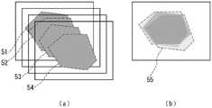
图5是用于说明输出部106进行的合成多个图像的处理的、示出作为合成对象的4张帧图像的示意图(图5(a))、以及示出合成后的图像的示意图(图5(b))。在图中,区域51~54为各帧图像的荧光部分。另外,区域55为合成后的图像之中的荧光部分合成的区域。FIG. 5 is a schematic diagram ( FIG. 5( a )) showing four frame images to be combined, and a schematic diagram showing a combined image ( FIG. 5(b)). In the figure,
通过进行相加平均,例如,即使在相同观察区域进行观察时观察对象的被观察的部分稍微移动的情况下,在拍摄的多个图像间有变化的部分的像素在多个图像间被平均化,从而值发生变化,无变化的部分即使被平均化,值也不发生变化,因此无变化的部分被强调地显示出来。观察对象为生物体的情况下,不能避免由心律、呼吸等所导致的观察对象的小的移动,但如图5所示通过进行相加平均,表示荧光部分的像素之中的变化少的部分被强调地显示出来,结果能够获得强调荧光部分的图像。By performing the additive averaging, for example, even when the observed portion of the observation object moves slightly when the same observation area is observed, the pixels of the portion that varies among the captured images are averaged among the plurality of images. , the value changes, and even if the unchanged part is averaged, the value does not change, so the unchanged part is highlighted. When the observation object is a living body, small movements of the observation object due to heart rhythm, respiration, etc. cannot be avoided. However, as shown in FIG. 5 , the average is performed to show the part with little change among the pixels of the fluorescent part. is displayed with emphasis, and as a result, an image in which the fluorescent portion is emphasized can be obtained.
而且,输出部106将图5(b)中所示的以激发光作为光源而拍摄的多个图像进行合成而得到的图像、和上述中以自然光作为光源时拍摄部104所拍摄的观察区域的图像排列于显示器106a上并同时显示。在此,例如,图4中示出的分光结果的曲线图也同时显示。Then, the
图6是表示示出输出部106所输出的分光结果的曲线图和拍摄观察区域而得到的图像的显示例的图,图像61为示出分光结果的曲线图,图像62为使用激发光作为光源时所拍摄的多个图像的合成后的图像,图像63为使用自然光作为光源是所拍摄的多个图像的合成后的图像。6 is a diagram showing a graph showing a spectroscopic result output by the
此外,判断部105利用拍摄部104在使用激发光作为光源时所拍摄的多个帧图像适当进行运动检测等,从而判断帧图像间的图像的移动、放大率的变化的有无,并在检测到图像的移动、放大率的变化时,不如上所述对连续的4个帧图像进行合成,而是将显微镜2的光源206熄灭,通过将光源切换为自然光,使拍摄部104暂时使用自然光拍摄有变化的观察区域的图像,并以该拍摄的图像更新输出部106所显示的观察区域的图像。而且,通过再次使用激发光拍摄观察区域的图像,从而在所拍摄的图像有变化时,能够将使用自然光作为光源对变化后的观察区域所拍摄的图像和使用激发光作为光源对变化后的观察区域所拍摄的图像输出,例如显示。Further, the
此外,分光部101等进行与上述同样的处理,从而预先获取使用激发光作为光源对正常组织进行观察时的分光结果,输出部106等根据用户等的操作,将该分光结果作为正常的组织的分光结果,预先存储在未图示的存储部等。而且,制作如图4所示的示出异常组织的分光结果的曲线图时,可以制作根据该正常的组织的分光结果所制作的图叠加而成的曲线图,并将该曲线图显示在显示器106a等上,以代替图4所示的曲线图。In addition, the
图7是表示将示出异常组织的分光结果的曲线图和示出正常组织的分光结果的曲线图叠加的曲线图的图,实线71为示出异常组织的分光结果的曲线图,虚线72为示出正常组织的分光结果的曲线图。7 is a diagram showing a graph in which a graph showing the spectroscopic result of abnormal tissue and a graph showing the spectroscopic result of normal tissue are superimposed, the
以上,根据本实施方式,在使用激发光作为光源,利用显微镜对观察对象进行观察时,能够同时对观察区域进行分光分析。由此,利用分光分析结果,例如能够实时地识别显微镜2的观察区域是否设定在正确的位置,能够使用激发光进行正确的观察。另外,利用分光分析结果也能够实时地检测观察区域中的异常组织。As described above, according to the present embodiment, when an observation object is observed with a microscope using excitation light as a light source, it is possible to simultaneously perform a spectroscopic analysis of the observation area. Thereby, it is possible to recognize in real time whether or not the observation area of the microscope 2 is set at the correct position, for example, based on the results of the spectroscopic analysis, and it is possible to perform accurate observation using excitation light. In addition, abnormal tissues in the observation area can also be detected in real time using the results of the spectroscopic analysis.
例如,在现有技术中,由于不能定量地掌握利用显微镜观察的区域内的荧光物质根据激发光而发出的光量,因此观察者自身必须主观地判断是否正在观察光量多的部分、即荧光物质多的部分,其结果是,不能正确判断显微镜的观察区域是否设定在荧光物质多的区域,从而存在不能进行正确的观察的问题、以及为了寻找正确的观察区域而需要长时间的问题。但根据本实施方式,利用分光结果,能够实时且容易地确认是否正在观察正确的观察区域。For example, in the prior art, since it is impossible to quantitatively grasp the amount of light emitted by a fluorescent substance in an area observed with a microscope due to excitation light, the observer must subjectively determine whether a portion with a large amount of light, that is, a large amount of fluorescent substance is being observed. As a result, it is not possible to accurately determine whether the observation area of the microscope is set in an area with many fluorescent substances, so that accurate observation cannot be performed, and there is a problem that it takes a long time to find the correct observation area. However, according to the present embodiment, whether or not the correct observation area is being observed can be easily confirmed in real time using the spectroscopic result.
另外,在现有技术中,用显微镜进行观察时,不能利用分光结果判断观察对象是否为异常组织等。例如存在如下问题:由于为了输出分光结果,必须暂时中断观察,而不能进行实时的判断等,因此在手术等想要在短时间内进行判断的情况下难以利用。与此相对,在本实施方式中,能够利用分光分析结果实时地对观察区域中的异常组织进行检测,即使在想要在短时间内进行判断的情况等中也能够利用。In addition, in the prior art, when observing with a microscope, it is impossible to judge whether the observation object is abnormal tissue or the like based on the spectroscopic result. For example, in order to output a spectroscopic result, observation must be temporarily interrupted, and real-time determination cannot be performed. Therefore, it is difficult to use it when a determination is desired in a short time, such as in surgery. On the other hand, in the present embodiment, the abnormal tissue in the observation area can be detected in real time using the spectroscopic analysis result, and it can be used even when it is desired to make a judgment in a short time.
另外,在本实施方式中,例如,通过将分别使用激发光和自然光作为光源而拍摄的图像叠加合成后输出,能够在使用自然光而拍摄的图像中确认是否正在观察正确的观察部位,并且也能够同时在使用激发光而拍摄的图像中观察发出荧光的部位。另外,由于将使用激发光而拍摄的图像和使用自然光而拍摄的图像叠加合成后输出,因此能够一目了然地容易地把握两者的图像中所示出的部位等的对应关系。另外,由于用户不必在两者的图像间移动视线,因此能够提高可视性。由此,在本实施方式中,能够正确且容易地进行使用激发光作为光源的观察。In addition, in this embodiment, for example, by superimposing and synthesizing images captured using excitation light and natural light as light sources, and outputting, it is possible to confirm whether or not the correct observation part is being observed in the image captured using natural light, and it is also possible to At the same time, the portion emitting fluorescence was observed in the image captured with excitation light. In addition, since the image captured using excitation light and the image captured using natural light are superimposed and synthesized and output, it is possible to easily grasp the correspondence between the parts and the like shown in the two images at a glance. In addition, since the user does not have to move the line of sight between the two images, visibility can be improved. Thus, in the present embodiment, observation using excitation light as a light source can be accurately and easily performed.
此外,在上述实施方式中,对使用激发光和自然光作为光源的情况进行了说明,但本发明也能够适用于使用激发光和包含激发光以外的波长的光(以下,称为观察光)作为光源的情况。由于使用观察光而拍摄的图像中包含在使用激发光拍摄的图像中看不见的部分,因此如上所述,通过使用激发光和观察光作为光源,由此将各自作为光源而拍摄的图像叠加合成并后输出,从而也能够同时观察仅用激发光不能观察的部分,能够正确且容易地进行观察。In addition, in the above-mentioned embodiment, the case where excitation light and natural light are used as light sources has been described, but the present invention can also be applied to the use of excitation light and light containing wavelengths other than excitation light (hereinafter, referred to as observation light) as light sources. the condition of the light source. Since the image captured using observation light includes parts that are not visible in the image captured using excitation light, the images captured as light sources are superimposed and synthesized by using excitation light and observation light as light sources as described above. It is possible to simultaneously observe parts that cannot be observed with only excitation light by outputting them in parallel, thereby enabling accurate and easy observation.
包含激发光以外的波长的光可以认为是至少包含激发光的波长以外的波长的光的光。观察光只要包含激发光以外的波长的光,则可以包含激发光,也可以不包含激发光。观察光可以为上述实施方式中所说明的自然光,可以包含自然光,也可以不包含自然光。观察光可以为单色光,也可以为复色光。观察光可以包含可见光,也可以不包含可见光。例如,激发光不是红色光的情况下,观察光可以为红色光或包含红色光的光。另外,激发光为波长800nm以外的光的情况下,观察光可以为作为非可见光的波长800nm的光。这样,观察光为非可见光的情况下,作为拍摄部等,优选使用红外线摄像机等之类的具备能使用非可见光作为光源进行拍摄的结构的拍摄部等。Light containing wavelengths other than excitation light can be considered to be light containing at least light of wavelengths other than the wavelength of excitation light. The observation light may or may not include excitation light as long as it includes light of wavelengths other than excitation light. The observation light may be the natural light described in the above embodiment, and may or may not include natural light. The observation light may be monochromatic light or polychromatic light. The observation light may or may not contain visible light. For example, when the excitation light is not red light, the observation light may be red light or light containing red light. In addition, when the excitation light is light having a wavelength other than 800 nm, the observation light may be light having a wavelength of 800 nm, which is invisible light. In this way, when the observation light is invisible light, it is preferable to use an imaging unit or the like having a structure capable of capturing images using invisible light as a light source, such as an infrared camera or the like.
此外,在上述各实施方式中,各处理(各功能)可以通过利用单一的装置(系统)进行集中处理来实现,或者也可以通过利用多个装置进行分散处理来实现。In addition, in each of the above-described embodiments, each process (each function) may be implemented by centralized processing using a single device (system), or may be implemented by performing distributed processing using a plurality of devices.
另外,在上述实施方式中,各构成要素所执行的处理的有关信息、例如各构成要素接收、或获取、或选择、或生成、或发送、或接收的信息、各构成要素在处理中使用的阈值、公式、地址等的信息等,即使在上述说明中没有明确说明的情况下,也暂时或长期保持在未图示的记录介质中。另外,各构成要素或未图示的存储部可以进行信息在该未图示的记录介质中的存储。另外,各构成要素或未图示的读取部可以进行从该未图示的记录介质的信息读取。In addition, in the above-described embodiment, information about the processing performed by each component, such as information received, or acquired, or selected, or generated, or transmitted, or received by each component, or information used by each component for processing Information such as thresholds, formulas, addresses, and the like are temporarily or long-term retained in a recording medium not shown, even if not explicitly described in the above description. In addition, each constituent element or a storage unit (not shown) may store information in a recording medium (not shown). In addition, each component or a reading unit (not shown) can read information from the recording medium (not shown).
另外,在上述各实施方式中,对观察辅助装置为独立的情况进行了说明,但观察辅助装置可以为独立的装置,也可以为服务器、客户端系统中的服务器装置。在后者的情况下,输出部、接收部通过通信线路接收输入、或输出画面。In addition, in each of the above-described embodiments, the case where the observation assisting device is independent has been described, but the observation assisting device may be an independent device, or may be a server or a server device in a client system. In the latter case, the output unit and the receiving unit receive the input or output the screen through the communication line.
另外,在上述各实施方式中,各构成要素可以由专用的硬件构成,或者对于利用软件能够实现的构成要素,也可以通过执行程序来实现。例如,能够通过CPU等程序执行部读取并执行记录于硬盘、半导体存储器等记录介质中的软件、程序来实现各构成要素。在该执行时,程序执行部可以在访问存储部(例如,硬盘、存储器等记录介质)的同时执行程序。In addition, in each of the above-described embodiments, each component may be constituted by dedicated hardware, or components that can be implemented by software may be implemented by executing a program. For example, each component can be realized by a program execution unit such as a CPU reading and executing software or programs recorded on a recording medium such as a hard disk or a semiconductor memory. During this execution, the program execution unit may execute the program while accessing a storage unit (eg, a recording medium such as a hard disk and a memory).
此外,实现上述各实施方式的观察辅助装置的软件为如下的程序。即,该程序为使计算机作为拍摄部和输出部而发挥功能的程序,所述拍摄部使用从能够切换地利用激发光和观察光作为光源且具备第二分束器的显微镜的该第二分束器射出的光,拍摄分别使用激发光和观察光作为光源时的所述显微镜的相同观察区域的图像,其中所述观察光包含激发光以外的波长的光;所述输出部将所述拍摄部分别使用激发光和观察光作为光源而拍摄的图像叠加合成后输出。In addition, the software which implements the observation assistance apparatus of each said embodiment is the following program. That is, this program is a program for causing a computer to function as an imaging unit and an output unit using the second part of a microscope that can switchably use excitation light and observation light as light sources and includes a second beam splitter The light emitted by the beamer is used to capture images of the same observation area of the microscope when excitation light and observation light are used as light sources, wherein the observation light contains light of wavelengths other than excitation light; Part of the images captured using excitation light and observation light as light sources are superimposed and synthesized and output.
此外,在上述程序中,上述程序实现的功能中不包括只能由硬件实现的功能。例如,只能由获取信息的获取部、输出信息的输出部等中的调制解调器、接口卡等硬件实现的功能不包括在上述程序所实现的功能中。In addition, in the above program, the functions realized by the above program do not include functions that can only be realized by hardware. For example, functions that can only be realized by hardware such as modems, interface cards, etc. in the acquisition unit for acquiring information, the output unit for outputting information, etc., are not included in the functions realized by the above-mentioned programs.
另外,执行该程序的计算机可以为单数,也可以为复数。即可以进行集中处理,或者可以进行分散处理。In addition, the computer which executes the program may be singular or plural. That is, centralized processing can be performed, or distributed processing can be performed.
图8为示出执行上述程序而实现基于上述实施方式的观察辅助装置的计算机的外观的一例的示意图。上述实施方式由计算机硬件及在其上执行的计算机程序能够实现。FIG. 8 is a schematic diagram showing an example of the appearance of a computer that executes the above-described program to realize the observation assistance device according to the above-described embodiment. The above-described embodiments can be realized by computer hardware and a computer program executed thereon.
在图8中,计算机系统900具备:包括CD-ROM(光盘只读存储器,Compact Disk ReadOnly Memory)驱动器905的计算机901、键盘902、鼠标903、以及显示器904。In FIG. 8 , a
图9为示出计算机系统900的内部构成的图。在图9中,计算机901除了CD-ROM驱动器905以外,还具备MPU(微处理单元,Micro Processing Unit)911、用于存储启动程序等程序的ROM912、与MPU911连接且临时存储应用程序指令并提供临时存储空间的RAM(随机存取存储器,Random Access Memory)913、存储应用程序、系统程序及数据的硬盘914、以及将MPU911、ROM912等彼此连接的总线915。此外,计算机901可以包括提供与LAN连接的未图示的网卡。FIG. 9 is a diagram showing the internal configuration of the
使计算机系统900执行根据上述实施方式的观察辅助装置等的功能的程序可以存储在CD-ROM921中、插入至CD-ROM驱动器905中、传送至硬盘914中。该程序还可以通过未图示的网络发送至计算机901并存储在硬盘914中以取而代之。程序在执行时被下载到RAM913中。此外,程序可以从CD-ROM921、或网络直接下载。A program for causing the
程序可以不一定包括使计算机901执行根据上述实施方式的观察辅助装置的功能的操作系统(OS)、或第三方程序等。程序可以只包含以受控的方式调用合适的功能(模块),以获得所需的结果的指令的一部分。关于计算机系统900是如何运行是公知的,省略详细的说明。The program may not necessarily include an operating system (OS) that causes the
本发明不限定于以上的实施方式,能够进行各种改变,当然所述改变也包含在本发明的范围内。The present invention is not limited to the above-described embodiments, and various modifications can be made, and of course the modifications are also included in the scope of the present invention.
工业实用性Industrial Applicability
如上所述,本发明所述的观察辅助装置等适于作为辅助使用显微镜的观察的装置等,尤其是作为对从显微镜的观察区域所获得的光进行分光的装置等是有用的。As described above, the observation assisting device or the like according to the present invention is suitable as a device or the like for assisting observation using a microscope, and is particularly useful as a device or the like for spectroscopic light obtained from an observation region of a microscope.
Claims (13)
Applications Claiming Priority (4)
| Application Number | Priority Date | Filing Date | Title |
|---|---|---|---|
| JP2015-112179 | 2015-06-02 | ||
| JP2015112179 | 2015-06-02 | ||
| PCT/JP2016/065272WO2016194699A1 (en) | 2015-06-02 | 2016-05-24 | Observation assisting device, information processing method, and program |
| CN201680045313.6ACN107923851A (en) | 2015-06-02 | 2016-05-24 | Observation assistance device, information processing method, and program |
Related Parent Applications (1)
| Application Number | Title | Priority Date | Filing Date |
|---|---|---|---|
| CN201680045313.6ADivisionCN107923851A (en) | 2015-06-02 | 2016-05-24 | Observation assistance device, information processing method, and program |
Publications (1)
| Publication Number | Publication Date |
|---|---|
| CN114414046Atrue CN114414046A (en) | 2022-04-29 |
Family
ID=57441510
Family Applications (3)
| Application Number | Title | Priority Date | Filing Date |
|---|---|---|---|
| CN202111537082.XAPendingCN114324268A (en) | 2015-06-02 | 2016-05-24 | Observation aid device, information processing method, and program |
| CN202111536559.2APendingCN114414046A (en) | 2015-06-02 | 2016-05-24 | Observation aid device, information processing method, and program |
| CN201680045313.6APendingCN107923851A (en) | 2015-06-02 | 2016-05-24 | Observation assistance device, information processing method, and program |
Family Applications Before (1)
| Application Number | Title | Priority Date | Filing Date |
|---|---|---|---|
| CN202111537082.XAPendingCN114324268A (en) | 2015-06-02 | 2016-05-24 | Observation aid device, information processing method, and program |
Family Applications After (1)
| Application Number | Title | Priority Date | Filing Date |
|---|---|---|---|
| CN201680045313.6APendingCN107923851A (en) | 2015-06-02 | 2016-05-24 | Observation assistance device, information processing method, and program |
Country Status (5)
| Country | Link |
|---|---|
| US (2) | US10852517B2 (en) |
| EP (1) | EP3306307A4 (en) |
| JP (1) | JP6859554B2 (en) |
| CN (3) | CN114324268A (en) |
| WO (1) | WO2016194699A1 (en) |
Families Citing this family (6)
| Publication number | Priority date | Publication date | Assignee | Title |
|---|---|---|---|---|
| CN114324268A (en)* | 2015-06-02 | 2022-04-12 | 国立大学法人旭川医科大学 | Observation aid device, information processing method, and program |
| WO2018042413A1 (en)* | 2016-08-28 | 2018-03-08 | Siegel Gabriel | A system for histological examination of tissue specimens |
| JP7290449B2 (en)* | 2019-04-03 | 2023-06-13 | 株式会社ディスコ | Ultra-high-speed imaging device |
| GB201907953D0 (en)* | 2019-06-04 | 2019-07-17 | Smi Drug Discovery Ltd | An optical microscope |
| WO2020254287A1 (en)* | 2019-06-18 | 2020-12-24 | Surgvision B.V. | Luminescence imaging with refining loop based on combination of partial illuminations |
| DE102020124224A1 (en)* | 2020-09-17 | 2022-03-17 | Carl Zeiss Microscopy Gmbh | Device for installation in a microscope, method for contacting microscope components on a rotor of a microscope and microscope |
Citations (16)
| Publication number | Priority date | Publication date | Assignee | Title |
|---|---|---|---|---|
| JP2005319211A (en)* | 2004-05-11 | 2005-11-17 | Pentax Corp | Fluorescence observation endoscope device |
| CN1882276A (en)* | 2003-11-20 | 2006-12-20 | 浜松光子学株式会社 | Lymph node detector |
| JP2008139796A (en)* | 2006-12-05 | 2008-06-19 | Keyence Corp | Fluorescence microscope, method of operating fluorescence microscope, fluorescence microscope operation program, computer-readable recording medium, and recorded equipment |
| CN101583304A (en)* | 2007-01-18 | 2009-11-18 | 奥林巴斯株式会社 | Fluorescence observing device and fluorescence observing method |
| US20100103250A1 (en)* | 2007-01-31 | 2010-04-29 | Olympus Corporation | Fluorescence observation apparatus and fluorescence observation method |
| CN102216827A (en)* | 2008-09-13 | 2011-10-12 | 独立行政法人科学技术振兴机构 | Microscope device and fluorescent observing method using same |
| CN102361580A (en)* | 2009-03-24 | 2012-02-22 | 奥林巴斯株式会社 | Fluorescence observation device, fluorescence observation system, and fluorescence image processing method |
| CN102370462A (en)* | 2010-07-13 | 2012-03-14 | 索尼公司 | Imaging apparatus, imaging system, surgical navigation system, and imaging method |
| CN102374983A (en)* | 2010-08-16 | 2012-03-14 | 萨伊姆公司 | Apparatus for receiving raman scattering signals and method of doing the same |
| US20120259174A1 (en)* | 2011-04-11 | 2012-10-11 | Fujifilm Corporation | Endoscopic diagnosis system |
| CN102939555A (en)* | 2010-06-11 | 2013-02-20 | 株式会社尼康 | Microscope device, observation method |
| CN103460026A (en)* | 2011-03-29 | 2013-12-18 | 奥林巴斯株式会社 | Photometric analysis device, photometric analysis method, and computer program for photometric analysis, using single light-emitting particle detection |
| JP2014059448A (en)* | 2012-09-18 | 2014-04-03 | Olympus Corp | Microscope system and microscopic observation method |
| CN104548387A (en)* | 2013-10-29 | 2015-04-29 | 株式会社日立制作所 | Charged particle beam system |
| CN104597590A (en)* | 2014-12-30 | 2015-05-06 | 深圳先进技术研究院 | Super-resolution fluorescence spectrum imaging microscope |
| CN107923851A (en)* | 2015-06-02 | 2018-04-17 | 国立大学法人旭川医科大学 | Observation assistance device, information processing method, and program |
Family Cites Families (20)
| Publication number | Priority date | Publication date | Assignee | Title |
|---|---|---|---|---|
| DE4111903A1 (en)* | 1991-04-12 | 1992-10-15 | Bayer Ag | SPECTROSCOPY-CORRECTED LIGHT-SCREEN MICROSCOPY |
| US5528368A (en)* | 1992-03-06 | 1996-06-18 | The United States Of America As Represented By The Department Of Health And Human Services | Spectroscopic imaging device employing imaging quality spectral filters |
| JPH11103471A (en)* | 1997-09-25 | 1999-04-13 | Olympus Optical Co Ltd | Video signal processor |
| AU5235299A (en)* | 1998-07-27 | 2000-02-21 | Cedars-Sinai Medical Center | Spectral topography of mammalian matter |
| JP2000097859A (en)* | 1998-09-24 | 2000-04-07 | Olympus Optical Co Ltd | Fluorescence observing method and device |
| AU2003288491A1 (en) | 2002-12-13 | 2004-07-09 | Image Enhancement Technologies, Llc | Optical examination method and apparatus particularly useful for real-time discrimination of tumors from normal tissues during surgery |
| CN100416333C (en)* | 2004-03-31 | 2008-09-03 | 奥林巴斯株式会社 | observation device |
| JP2006171024A (en) | 2004-12-10 | 2006-06-29 | Olympus Corp | Multipoint fluorescence spectrophotometric microscope and multipoint fluorescence spectrophotometric method |
| JP5123660B2 (en)* | 2005-03-30 | 2013-01-23 | オリンパス株式会社 | measuring device |
| ATE555711T1 (en)* | 2007-12-19 | 2012-05-15 | Kantonsspital Aarau Ag | METHOD FOR ANALYZING AND PROCESSING FLUORESCENCE IMAGES |
| KR101061004B1 (en)* | 2008-12-10 | 2011-09-01 | 한국전기연구원 | Device for photodynamic therapy and light detection |
| DE102008062650B9 (en)* | 2008-12-17 | 2021-10-28 | Carl Zeiss Meditec Ag | Surgical microscope for observing infrared fluorescence and methods for this |
| JP5393216B2 (en)* | 2009-03-24 | 2014-01-22 | オリンパス株式会社 | Fluorescence observation system and method of operating fluorescence observation system |
| JP5393215B2 (en)* | 2009-03-24 | 2014-01-22 | オリンパス株式会社 | Fluorescence observation apparatus, fluorescence observation system, and operation method of fluorescence observation apparatus |
| KR101172745B1 (en)* | 2010-01-29 | 2012-08-14 | 한국전기연구원 | Combined apparatus for detection of multi-spectrum optical imaging coming out of organic body and light therapy |
| EP2359745A1 (en) | 2010-02-12 | 2011-08-24 | Helmholtz Zentrum München Deutsches Forschungszentrum für Gesundheit und Umwelt (GmbH) | Method and device for multi-spectral photonic imaging |
| DE102010044503B4 (en)* | 2010-09-06 | 2017-11-02 | Leica Microsystems (Schweiz) Ag | Operations fluorescence stereomicroscope |
| WO2012147820A1 (en)* | 2011-04-28 | 2012-11-01 | オリンパス株式会社 | Fluorescent observation device and image display method therefor |
| US20140378843A1 (en)* | 2012-01-20 | 2014-12-25 | The Trustees Of Dartmouth College | Method And Apparatus For Quantitative Hyperspectral Fluorescence And Reflectance Imaging For Surgical Guidance |
| KR102069723B1 (en)* | 2013-04-23 | 2020-01-23 | 세다르스-신나이 메디칼 센터 | Systems and methods for recording simultaneously visible light image and infrared light image from fluorophores |
- 2016
- 2016-05-24CNCN202111537082.XApatent/CN114324268A/enactivePending
- 2016-05-24CNCN202111536559.2Apatent/CN114414046A/enactivePending
- 2016-05-24JPJP2017521836Apatent/JP6859554B2/enactiveActive
- 2016-05-24EPEP16803134.2Apatent/EP3306307A4/enactivePending
- 2016-05-24CNCN201680045313.6Apatent/CN107923851A/enactivePending
- 2016-05-24WOPCT/JP2016/065272patent/WO2016194699A1/ennot_activeCeased
- 2016-05-24USUS15/578,528patent/US10852517B2/enactiveActive
- 2020
- 2020-11-05USUS17/089,818patent/US11719920B2/enactiveActive
Patent Citations (17)
| Publication number | Priority date | Publication date | Assignee | Title |
|---|---|---|---|---|
| CN1882276A (en)* | 2003-11-20 | 2006-12-20 | 浜松光子学株式会社 | Lymph node detector |
| JP2005319211A (en)* | 2004-05-11 | 2005-11-17 | Pentax Corp | Fluorescence observation endoscope device |
| JP2008139796A (en)* | 2006-12-05 | 2008-06-19 | Keyence Corp | Fluorescence microscope, method of operating fluorescence microscope, fluorescence microscope operation program, computer-readable recording medium, and recorded equipment |
| CN101583304A (en)* | 2007-01-18 | 2009-11-18 | 奥林巴斯株式会社 | Fluorescence observing device and fluorescence observing method |
| US20100103250A1 (en)* | 2007-01-31 | 2010-04-29 | Olympus Corporation | Fluorescence observation apparatus and fluorescence observation method |
| CN102216827A (en)* | 2008-09-13 | 2011-10-12 | 独立行政法人科学技术振兴机构 | Microscope device and fluorescent observing method using same |
| CN102361580A (en)* | 2009-03-24 | 2012-02-22 | 奥林巴斯株式会社 | Fluorescence observation device, fluorescence observation system, and fluorescence image processing method |
| CN102939555A (en)* | 2010-06-11 | 2013-02-20 | 株式会社尼康 | Microscope device, observation method |
| CN102370462A (en)* | 2010-07-13 | 2012-03-14 | 索尼公司 | Imaging apparatus, imaging system, surgical navigation system, and imaging method |
| CN102374983A (en)* | 2010-08-16 | 2012-03-14 | 萨伊姆公司 | Apparatus for receiving raman scattering signals and method of doing the same |
| CN103460026A (en)* | 2011-03-29 | 2013-12-18 | 奥林巴斯株式会社 | Photometric analysis device, photometric analysis method, and computer program for photometric analysis, using single light-emitting particle detection |
| US20120259174A1 (en)* | 2011-04-11 | 2012-10-11 | Fujifilm Corporation | Endoscopic diagnosis system |
| JP2014059448A (en)* | 2012-09-18 | 2014-04-03 | Olympus Corp | Microscope system and microscopic observation method |
| CN104548387A (en)* | 2013-10-29 | 2015-04-29 | 株式会社日立制作所 | Charged particle beam system |
| CN104597590A (en)* | 2014-12-30 | 2015-05-06 | 深圳先进技术研究院 | Super-resolution fluorescence spectrum imaging microscope |
| CN107923851A (en)* | 2015-06-02 | 2018-04-17 | 国立大学法人旭川医科大学 | Observation assistance device, information processing method, and program |
| CN114324268A (en)* | 2015-06-02 | 2022-04-12 | 国立大学法人旭川医科大学 | Observation aid device, information processing method, and program |
Also Published As
| Publication number | Publication date |
|---|---|
| US10852517B2 (en) | 2020-12-01 |
| CN107923851A (en) | 2018-04-17 |
| EP3306307A1 (en) | 2018-04-11 |
| US20180157020A1 (en) | 2018-06-07 |
| WO2016194699A1 (en) | 2016-12-08 |
| US20210109333A1 (en) | 2021-04-15 |
| US11719920B2 (en) | 2023-08-08 |
| CN114324268A (en) | 2022-04-12 |
| JP6859554B2 (en) | 2021-04-14 |
| JPWO2016194699A1 (en) | 2018-04-12 |
| EP3306307A4 (en) | 2019-02-27 |
Similar Documents
| Publication | Publication Date | Title |
|---|---|---|
| US11719920B2 (en) | Observation assisting device, information processing method, and program | |
| JP5735031B2 (en) | Compact fluorescent endoscope imaging system | |
| KR100411631B1 (en) | Fluorescence endoscope apparatus and a method for imaging tissue within a body using the same | |
| US20120056996A1 (en) | Special-illumination surgical video stereomicroscope | |
| WO2015186225A1 (en) | Scan-type projection device, projection method, and surgery support system | |
| US11160442B2 (en) | Endoscope apparatus | |
| US10805512B2 (en) | Dual path endoscope | |
| EP3009098A1 (en) | Microscope system for surgery | |
| EP1728464A1 (en) | Endoscope image pickup system | |
| WO2017043539A1 (en) | Image processing system, image processing device, projecting device, and projecting method | |
| JP2022179746A (en) | Medical control apparatus, medical observation system, control apparatus, and observation system | |
| US12108128B2 (en) | Apparatus, system and method to capture images of a medical site in white light and fluorescent light | |
| JP2004504072A (en) | Portable system for detecting skin abnormalities | |
| WO2018003263A1 (en) | Observation device and control method for observation device | |
| US20180092519A1 (en) | Infrared fluorescence observation device | |
| JP2021035549A (en) | Endoscope system | |
| WO2019100449A1 (en) | Imaging fiber based surgical navigation system | |
| WO2012002320A1 (en) | Image processing apparatus and image processing method | |
| JP2002345739A (en) | Image display device | |
| JP2002291682A (en) | Endoscope system for fluorescent observation | |
| RU2290855C1 (en) | Method and device for carrying out fluorescent endoscopy | |
| JP2003000528A (en) | Method and device for imaging fluorescent diagnostic image | |
| CN208319313U (en) | Optical operation navigation system | |
| JP2005152130A (en) | Endoscope imaging system | |
| WO2010023983A1 (en) | In vivo observation device |
Legal Events
| Date | Code | Title | Description |
|---|---|---|---|
| PB01 | Publication | ||
| PB01 | Publication | ||
| SE01 | Entry into force of request for substantive examination | ||
| SE01 | Entry into force of request for substantive examination | ||
| TA01 | Transfer of patent application right | ||
| TA01 | Transfer of patent application right | Effective date of registration:20220819 Address after:Swiss Hill Brugger Applicant after:LEICA MICROSYSTEMS (SCHWEIZ) AG Address before:Hokkaido, Asahikawa, Japan Applicant before:NATIONAL UNIVERSITY CORPORATION ASAHIKAWA MEDICAL University |