CN114373371A - A simulation head model for tooth extraction training - Google Patents
A simulation head model for tooth extraction trainingDownload PDFInfo
- Publication number
- CN114373371A CN114373371ACN202110388996.8ACN202110388996ACN114373371ACN 114373371 ACN114373371 ACN 114373371ACN 202110388996 ACN202110388996 ACN 202110388996ACN 114373371 ACN114373371 ACN 114373371A
- Authority
- CN
- China
- Prior art keywords
- model
- edge
- cavity
- tooth extraction
- shaped cavity
- Prior art date
- Legal status (The legal status is an assumption and is not a legal conclusion. Google has not performed a legal analysis and makes no representation as to the accuracy of the status listed.)
- Pending
Links
Images


Classifications
- G—PHYSICS
- G09—EDUCATION; CRYPTOGRAPHY; DISPLAY; ADVERTISING; SEALS
- G09B—EDUCATIONAL OR DEMONSTRATION APPLIANCES; APPLIANCES FOR TEACHING, OR COMMUNICATING WITH, THE BLIND, DEAF OR MUTE; MODELS; PLANETARIA; GLOBES; MAPS; DIAGRAMS
- G09B23/00—Models for scientific, medical, or mathematical purposes, e.g. full-sized devices for demonstration purposes
- G09B23/28—Models for scientific, medical, or mathematical purposes, e.g. full-sized devices for demonstration purposes for medicine
- G09B23/283—Models for scientific, medical, or mathematical purposes, e.g. full-sized devices for demonstration purposes for medicine for dentistry or oral hygiene
Landscapes
- Engineering & Computer Science (AREA)
- General Physics & Mathematics (AREA)
- Health & Medical Sciences (AREA)
- Physics & Mathematics (AREA)
- Algebra (AREA)
- Mathematical Analysis (AREA)
- General Health & Medical Sciences (AREA)
- Medical Informatics (AREA)
- Medicinal Chemistry (AREA)
- Public Health (AREA)
- Oral & Maxillofacial Surgery (AREA)
- Computational Mathematics (AREA)
- Epidemiology (AREA)
- Chemical & Material Sciences (AREA)
- Mathematical Optimization (AREA)
- Mathematical Physics (AREA)
- Pure & Applied Mathematics (AREA)
- Business, Economics & Management (AREA)
- Educational Administration (AREA)
- Educational Technology (AREA)
- Theoretical Computer Science (AREA)
- Instructional Devices (AREA)
- Dental Tools And Instruments Or Auxiliary Dental Instruments (AREA)
Abstract
Description
Translated fromChinese技术领域technical field
本发明属于口腔医学临床教学训练教具应用领域,涉及一种可重复使用、可结合真牙的立体打印牙颌模具,尤其涉及一种能仿真模拟临床复杂阻生牙拔除术过程、能根据教学训练阶段设置拔牙操作难度、并能评价拔牙手术创伤及可能引起的相关并发症的头模模型。The invention belongs to the application field of clinical teaching and training teaching aids in stomatology, relates to a three-dimensional printing dental jaw mold that can be used repeatedly and can be combined with real teeth, and in particular, relates to a method capable of simulating the clinical complex impacted tooth extraction process and can be trained according to teaching and training. The stage sets the difficulty of tooth extraction operation, and can evaluate the head model of tooth extraction surgery trauma and related complications that may be caused.
背景技术Background technique
牙拔除术是口腔医学临床最为基本和常见的治疗手术,并且基于人类牙颌系统进化特点,国人出现智齿(上下颌第三磨牙)阻生的机率逐渐升高,有统计表明有接近三分之一的中国人需要拔除至少一颗阻生牙,因而阻生牙拔除术是口腔医生特别是口腔外科医生需要掌握的必要临床手术技术。但由于口腔局部解剖结构复杂,需要拔除牙与周围组织的关系变化多端,牙拔除术并不是一个简单的手术,其基本特点包括:拔牙难度的个体差异性显著;拔牙难度相关影响因素众多,包括患者自身局部和全身性因素,以及术者手术技术和相关经验积累因素等;拔牙操作受到口腔复杂立体结构的多种条件限制,可引起牙、骨、粘膜、神经、血管、关节等多种组织损伤,周围组织的过度损伤或重要结构如神经、血管等损伤,会导致较为严重的并发症,严重影响患者正常生活工作,甚至有生命危险。基于以上特点,拔牙医生需要在理论知识、操作技术、临床经验、器械设备等方面不断提高与完善,才能完善拔牙技术,有效控制或正确处理各种拔牙并发症。Tooth extraction is the most basic and common clinical treatment in stomatology, and based on the evolutionary characteristics of the human dental system, the probability of impacted wisdom teeth (maxillary third molars) in Chinese people is gradually increasing, and statistics show that nearly one-third One percent of Chinese people need to extract at least one impacted tooth, so the extraction of impacted teeth is a necessary clinical surgical technique that dentists, especially oral surgeons, need to master. However, due to the complex anatomical structure of the oral cavity, the relationship between tooth extraction and surrounding tissues is varied. Tooth extraction is not a simple operation. Its basic characteristics include: the individual differences in the difficulty of tooth extraction are significant; The patient's own local and systemic factors, as well as the surgeon's surgical technique and related experience accumulation factors, etc. The extraction operation is limited by various conditions of the complex three-dimensional structure of the oral cavity, which can cause various tissues such as teeth, bones, mucosa, nerves, blood vessels, and joints. Injury, excessive damage to surrounding tissues or damage to important structures such as nerves and blood vessels, can lead to more serious complications, seriously affecting the normal life and work of patients, and even life-threatening. Based on the above characteristics, dental extraction doctors need to continuously improve and perfect their theoretical knowledge, operating techniques, clinical experience, and equipment in order to improve tooth extraction techniques and effectively control or correctly handle various tooth extraction complications.
头模模型训练是医学生临床前训练不可或缺的部分,目前临床教学所用模型教具在牙拔除术训练方面有以下问题。Head model training is an indispensable part of preclinical training for medical students. At present, the model teaching aids used in clinical teaching have the following problems in tooth extraction training.
现有模型难以模拟真牙的复杂组织质地。一般采用仿真人工树脂制作的牙,由于真牙由牙釉质、牙本质、牙骨质等不同种类硬组织构成,并且存在多变的牙冠、牙根、牙髓腔等外形结构,不同部位的硬度、质地等有较为多样的变化,人工牙难以完全模拟真牙形态结构与质地,常不能承受牙拔除术所用牙钳、牙挺、牙钻等作用力,进而在实践过程中难以获得磨切真牙的确切手感,而这种手感对手术医生熟悉牙拔除术所使用器械的感觉、判断磨切深度、避免意外创伤是非常重要的。Existing models are difficult to simulate the complex tissue texture of real teeth. Generally, teeth made of artificial artificial resin are made of artificial resin. Since real teeth are composed of different types of hard tissues such as enamel, dentin, and cementum, and there are variable shape structures such as crown, root, and pulp cavity, the hardness of different parts There are various changes in the shape, texture, etc. of artificial teeth. It is difficult for artificial teeth to completely simulate the shape, structure and texture of real teeth, and they often cannot withstand the forces of forceps, elevators, and dental drills used in tooth extraction, so it is difficult to obtain true grinding in practice. The exact feel of the tooth is very important for the surgeon to familiarize himself with the feel of the instruments used in tooth extraction, to judge the depth of grinding, and to avoid accidental trauma.
现有模型难以模拟真牙固位的稳定性,以及脱位时的松动过程。人体上,牙通过牙周韧带与周围骨组织连接并固位,牙拔除术中,通过牙挺牙钳等拔牙器械作用,使牙根周围牙槽骨局部形变、牙周韧带撕裂而使牙根松动、脱离牙槽窝,牙槽骨形变的程度与拔牙创伤相关,理想的牙拔除术只产生细微的牙槽骨形变,创伤小,术后恢复快,无明显疼痛等并发症,也不会造成周围重要组织器官、解剖结构的损伤。但牙拔除术操作不当,损伤较大时,牙槽骨变形、缺损、甚至折裂,则会出现出血、疼痛、神经损伤、伤口愈合困难等问题。随年龄增长,牙与牙周支持结构的关系会有增龄性变化,牙周韧带等软组织构成的牙周膜间隙变窄,牙根与周围牙槽骨的结合更为紧密,临床上表现为“根骨粘连”,牙根不易松动脱位,拔牙难度增大,对手术医生的操作技术要求更高。Existing models are difficult to simulate the stability of the retention of real teeth and the loosening process during dislocation. In the human body, the tooth is connected and retained by the periodontal ligament and the surrounding bone tissue. During tooth extraction, the tooth root is loosened by local deformation of the alveolar bone around the tooth root and tearing of the periodontal ligament by the action of tooth extraction instruments such as tooth lift forceps. , out of the alveolar socket, the degree of alveolar bone deformation is related to the trauma of tooth extraction, ideal tooth extraction only produces slight alveolar bone deformation, small trauma, fast postoperative recovery, no obvious pain and other complications, and will not cause Injury to surrounding vital tissues, organs, and anatomical structures. However, when the tooth extraction is performed improperly, when the damage is large, the alveolar bone is deformed, deficient, or even fractured, which may cause problems such as bleeding, pain, nerve damage, and difficulty in wound healing. With the increase of age, the relationship between the teeth and the periodontal supporting structure will change with age, the periodontal ligament space composed of soft tissues such as the periodontal ligament becomes narrower, and the root of the tooth is more closely combined with the surrounding alveolar bone. Root bone adhesion", the root is not easy to loosen and dislocate, the difficulty of tooth extraction increases, and the operating skills of the surgeon are higher.
现有模型难以反复使用。牙拔除术是破坏性手术,导致模型破损后需要采用新的模型用于进一步训练,成本较高。Existing models are difficult to reuse. Tooth extraction is a destructive operation, which requires a new model for further training after the model is damaged, which is costly.
现有模型难以模拟不同难度复杂牙阻生牙拔除过程。阻生牙方向与深度等变化多样,个体差异性显著,并且阻生位置是拔牙难度的关键影响因素。Existing models are difficult to simulate the extraction process of impacted teeth with different difficulty and complexity. The direction and depth of impacted teeth vary widely, with significant individual differences, and the location of impacted teeth is a key factor affecting the difficulty of tooth extraction.
现有模型难以模拟临床牙拔除术术中术后可能出现的各种并发症,不能帮助学员了解并发症可能出现原因、表现、和预防措施。Existing models are difficult to simulate various complications that may occur during and after surgery in clinical tooth extraction, and cannot help students understand the possible causes, manifestations, and preventive measures of complications.
鉴于现有模型对评价牙拔除术手术操作规范性、术后损伤微创性等方面存在较明显不足,急需研究一种新型的放置模拟拔牙训练头模模型。In view of the obvious deficiencies of the existing models in evaluating the standardization of tooth extraction surgery and the minimally invasiveness of postoperative injury, it is urgent to develop a new model for placing simulated tooth extraction training headforms.
发明内容SUMMARY OF THE INVENTION
本发明的目的在于克服现有技术中存在的缺点,利用三维打印工业技术结合临床废弃真牙,有针对性设计牙拔除术头模训练模型。The purpose of the present invention is to overcome the shortcomings in the prior art, and to design a head mold training model for tooth extraction in a targeted manner by using three-dimensional printing industrial technology combined with clinically discarded real teeth.
为了实现上述目的,本发明提供如下技术方案:In order to achieve the above object, the present invention provides the following technical solutions:
一种仿真模拟拔牙训练头模模型,其包括牙科训练头模、上颌模型和下颌模型,其特征在于,所述上颌模型和下颌模型可拆卸地安装在所述牙科训练头模中,所述上颌模型的颌骨上部具有上颌窦且所述上颌模型的第二磨牙和第三磨牙位置处分别形成有一个上槽型窝洞,所述上槽型窝洞的底部与所述上颌窦的底部相通,每个所述上槽型窝洞中埋置有一颗上真牙,所述上颌模型的牙齿上设置有上硅胶牙龈,所述下颌模型的颌骨内部具有下颌管,所述下颌管的两端分别具有颏孔和下颌孔,所述下颌模型的第三磨牙位置处形成有下槽型窝洞、第一磨牙位置处形成有桶形窝洞,所述下槽型窝洞和所述桶形窝洞的底部都与所述下颌管相通且所述下槽型窝洞和所述桶形窝洞中分别埋置有一颗下真牙,所述下颌管中具有灌满红色染料的橡皮管,所述下颌模型的牙齿上设置有下硅胶牙龈。A simulated tooth extraction training head model, comprising a dental training head model, an upper jaw model and a lower jaw model, characterized in that the upper jaw model and the lower jaw model are detachably installed in the dental training head model, and the upper jaw model and the lower jaw model are detachably installed in the dental training head model. The upper part of the jawbone of the model has a maxillary sinus, and the second molar and the third molar of the maxillary model are respectively formed with an upper groove-shaped cavity, and the bottom of the upper groove-shaped cavity communicates with the bottom of the maxillary sinus. , an upper real tooth is embedded in each of the upper groove-shaped sockets, the upper silicone gums are arranged on the teeth of the upper jaw model, the jawbone of the lower jaw model has a mandibular canal, and the two parts of the mandibular canal are The ends respectively have a mental foramen and a mandibular foramen, a lower groove-shaped cavity is formed at the position of the third molar of the mandibular model, and a barrel-shaped cavity is formed at the position of the first molar, and the lower groove-shaped cavity and the barrel are formed. The bottom of the cavity is communicated with the mandibular canal, and a lower true tooth is embedded in the lower groove-shaped cavity and the barrel cavity respectively, and the mandibular canal has a rubber tube filled with red dye. , the teeth of the mandible model are provided with lower silicone gums.
优选地,其中,所述上颌模型和下颌模型利用三维打印技术制备而成。Preferably, the upper jaw model and the lower jaw model are prepared by three-dimensional printing technology.
优选地,其中,所述上颌模型和下颌模型通过其磁性基底吸附和卡位固定于所述牙科训练头模中。Preferably, wherein, the upper jaw model and the lower jaw model are fixed in the dental training head model through their magnetic bases by adsorption and clamping.
优选地,其中,所述上槽型窝洞中镶衬有橡皮片。Preferably, wherein, the upper groove-shaped cavity is lined with a rubber sheet.
优选地,其中,所述上真牙通过石膏固定在所述上槽型窝洞中,且所述下真牙也通过石膏固定在所述下槽型窝洞和所述桶形窝洞中。Preferably, the upper real teeth are fixed in the upper groove-shaped cavity by plaster, and the lower real teeth are also fixed in the lower groove-shaped cavity and the barrel-shaped cavity by gypsum.
优选地,其中,所述下槽型窝洞的近中边缘位于第二磨牙的远中牙颈部、远中边缘位于下颌升支前缘后方、颊侧边缘位于外斜嵴、舌侧边缘位于舌侧牙槽嵴。Preferably, the mesial edge of the inferior groove-shaped cavity is located at the neck of the distal tooth of the second molar, the distal edge is located behind the anterior edge of the mandibular ascending ramus, the buccal edge is located at the external oblique ridge, and the lingual edge is located at the lingual alveolar ridge.
优选地,其中,所述下槽型窝洞的近远中直径为3cm,颊舌侧直径为1.5cm。Preferably, the mesial-distal diameter of the inferior groove-shaped cavity is 3 cm, and the buccal-lingual diameter is 1.5 cm.
优选地,其中,所述桶形窝洞的近中边缘位于第二磨牙的远中牙颈部、远中边缘位于第二磨牙的近中牙颈部、颊侧边缘位于颊侧牙槽嵴、舌侧边缘位于舌侧牙槽嵴。Preferably, wherein the mesial edge of the barrel-shaped cavity is located at the distal neck of the second molar, the distal edge is located at the mesial neck of the second molar, the buccal edge is located at the buccal alveolar ridge, The lingual edge is located at the lingual alveolar ridge.
优选地,其中,所述桶形窝洞的近远中直径和颊舌侧直径均为1.2cm。Preferably, the mesio-distal diameter and the buccal-lingual diameter of the barrel-shaped cavity are both 1.2 cm.
优选地,其中,所述上槽型窝洞的颊侧边缘位于颧牙槽嵴、舌侧边缘位于舌侧牙槽嵴、近中边缘位于第一磨牙的远中牙颈部、远中边缘位于上颌窦结节上方,且所述上槽型窝洞的近远中直径为3cm,颊舌侧直径为1.5cm。Preferably, wherein the buccal edge of the upper alveolar cavity is located at the zygomatic alveolar ridge, the lingual edge is located at the lingual alveolar ridge, the mesial edge is located at the neck of the distal tooth of the first molar, and the distal edge is located at the lingual alveolar ridge. Above the maxillary sinus tubercle, and the mesial and distal diameter of the upper groove cavity is 3 cm, and the diameter of the buccal and lingual side is 1.5 cm.
与现有技术相比,本发明的仿真模拟拔牙训练头模模型具有如下有益技术效果:其通过收集临床拔牙后废弃的真牙,可反复使用的仿真拔牙头模模型,完整模拟临床复杂牙拔除过程,拔牙操作练习手感与临床实践高度贴近,利于学员真实掌握临床拔牙基本技术,建立微创手术关健意识,适于各阶段实习、实训学员对不同难度复杂牙拔除的训练要求,可促进学习效果,减少临床实践过程中在患者实际操作时发生意外的可能性。Compared with the prior art, the simulated tooth extraction training headform model of the present invention has the following beneficial technical effects: by collecting the discarded real teeth after clinical tooth extraction, a reusable simulation tooth extraction headform model can completely simulate the extraction of complex clinical teeth. During the process, the feeling of tooth extraction operation and practice is highly close to the clinical practice, which is beneficial for students to truly master the basic techniques of clinical tooth extraction and establish the key awareness of minimally invasive surgery. The learning effect reduces the possibility of accidents occurring during the actual operation of the patient during clinical practice.
附图说明Description of drawings
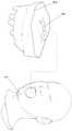
图1是本发明的仿真模拟拔牙训练头模模型的构成示意图,其中,未示出下颌模型。FIG. 1 is a schematic diagram of the structure of the simulation model of the tooth extraction training head model of the present invention, wherein the mandible model is not shown.
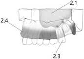
图2是本发明的仿真模拟拔牙训练头模模型的上颌模型的示意图。FIG. 2 is a schematic diagram of the maxillary model of the simulation model of the tooth extraction training head model of the present invention.
图3是本发明的仿真模拟拔牙训练头模模型的上颌模型装饰完成后的示意图。Fig. 3 is a schematic diagram showing the decoration of the maxillary model of the simulated tooth extraction training head model according to the present invention.
图4是本发明的仿真模拟拔牙训练头模模型的下颌模型的示意图。FIG. 4 is a schematic diagram of the mandible model of the simulation model of the tooth extraction training head model of the present invention.
图5是本发明的仿真模拟拔牙训练头模模型的下颌模型装饰完成后的示意图。FIG. 5 is a schematic diagram after the decoration of the mandible model of the simulated tooth extraction training head model of the present invention is completed.
具体实施方式Detailed ways
下面结合附图和实施例对本发明进一步说明,实施例的内容不作为对本发明的保护范围的限制。The present invention will be further described below with reference to the accompanying drawings and embodiments, and the contents of the embodiments are not intended to limit the protection scope of the present invention.
本发明涉及一种仿真模拟拔牙训练头模模型,其是可重复使用,并可结合真牙的立体打印牙颌模具,能仿真模拟临床复杂阻生牙拔除术过程、能根据教学训练阶段设置拔牙操作难度、并能评价拔牙手术创伤及可能引起的相关并发症。The invention relates to a head mold model for simulating tooth extraction training, which is reusable and can be combined with a three-dimensional printing dental jaw mold of real teeth, which can simulate and simulate the clinical complex impacted tooth extraction process, and can set the tooth extraction according to the teaching and training stage. Difficulty of operation, and can evaluate the trauma of tooth extraction and related complications that may be caused.
如图1-5所示,本发明的仿真模拟拔牙训练头模模型包括牙科训练头模1、上颌模型2和下颌模型4。As shown in FIGS. 1-5 , the simulated tooth extraction training head model of the present invention includes a dental
其中,所述上颌模型2和下颌模型4可拆卸地安装在所述牙科训练头模1中。这样,使得所述上颌模型2、下颌模型4和牙科训练头模1都可重复利用。Wherein, the
在本发明中,优选地,所述上颌模型2和下颌模型4的基底都由磁性材料制成,由此,使得所述上颌模型2和下颌模型4可以通过其磁性基底3吸附固定于所述牙科训练头模1中。In the present invention, preferably, the bases of the
更优选地,所述上颌模型2和下颌模型4的基底中都设有卡位,由此,所述上颌模型2和下颌模型4可以通过卡位卡扣在所述牙科训练头模1中。由于采用了双重固定方式,使得所述上颌模型2和下颌模型4可以更稳固地固定在所述牙科训练头模1中。More preferably, the bases of the
在本发明中,如图2和3所示,所述上颌模型2的颌骨上部具有上颌窦2.1且所述上颌模型2的第二磨牙和第三磨牙位置处分别形成有一个上槽型窝洞2.2,用于埋置上真牙2.3。所述上槽型窝洞2.2的底部与所述上颌窦2.1的底部相通,使所述上颌窦2.1部分开放于所述上槽型窝洞2.2底部。每个所述上槽型窝洞2.2中埋置有一颗上真牙2.3。并且,所述上颌模型2的牙齿上设置有上硅胶牙龈2.4。In the present invention, as shown in FIGS. 2 and 3 , the upper part of the jawbone of the
优选地,所述上槽型窝洞2.2的颊侧边缘位于颧牙槽嵴、舌侧边缘位于舌侧牙槽嵴、近中边缘位于第一磨牙的远中牙颈部、远中边缘位于上颌窦结节上方。更优选地,所述上槽型窝洞2.2的近远中直径为3cm,颊舌侧直径为1.5cm。Preferably, the buccal edge of the upper alveolar cavity 2.2 is located at the zygomatic alveolar ridge, the lingual edge is located at the lingual alveolar ridge, the mesial edge is located at the neck of the distal teeth of the first molar, and the distal edge is located in the maxilla above the sinus tubercle. More preferably, the mesial-distal diameter of the upper grooved cavity 2.2 is 3 cm, and the buccal-lingual diameter is 1.5 cm.
如图4和5所示,所述下颌模型4的颌骨内部具有下颌管4.1。所述下颌管4.1的两端分别具有颏孔4.2和下颌孔4.3。所述下颌模型4的第三磨牙位置处形成有下槽型窝洞4.4、第一磨牙位置处形成有桶形窝洞4.5,用于埋置下真牙2.6。所述下槽型窝洞4.4和所述桶形窝洞4.5的底部都与所述下颌管4.1相通,使所述下颌管4.1半开放于所述下槽型窝洞4.4和桶形窝洞4.5的底部。所述下槽型窝洞4.4和所述桶形窝洞4.5中分别埋置有一颗下真牙4.6。所述下颌管4.1中具有灌满红色染料的橡皮管4.7,用于模拟血管。所述下颌模型4的牙齿上设置有下硅胶牙龈4.8。As shown in Figures 4 and 5, the jawbone of the
优选地,所述下槽型窝洞4.4的近中边缘位于第二磨牙的远中牙颈部、远中边缘位于下颌升支前缘后方、颊侧边缘位于外斜嵴、舌侧边缘位于舌侧牙槽嵴。更优选地,所述下槽型窝洞4.4的近远中直径为3cm,颊舌侧直径为1.5cm。Preferably, the mesial edge of the inferior groove 4.4 is located at the neck of the distal tooth of the second molar, the distal edge is located behind the anterior edge of the ascending mandibular ramus, the buccal edge is located at the external oblique ridge, and the lingual edge is located at the tongue Lateral alveolar ridge. More preferably, the mesial-distal diameter of the lower groove-shaped cavity 4.4 is 3 cm, and the buccal-lingual diameter is 1.5 cm.
此外,优选地,所述桶形窝洞4.5的近中边缘位于第二磨牙的远中牙颈部、远中边缘位于第二磨牙的近中牙颈部、颊侧边缘位于颊侧牙槽嵴、舌侧边缘位于舌侧牙槽嵴。更优选地,所述桶形窝洞4.5的近远中直径和颊舌侧直径均为1.2cm。In addition, preferably, the mesial edge of the barrel-shaped cavity 4.5 is located at the distal neck of the second molar, the distal edge is located at the mesial neck of the second molar, and the buccal edge is located at the buccal alveolar ridge The lingual edge is located in the lingual alveolar ridge. More preferably, both the mesial-distal diameter and the buccal-lingual diameter of the barrel-shaped cavity 4.5 are 1.2 cm.
并且,在本发明中,优选地,所述上槽型窝洞2.2中镶衬有橡皮片。由此,可评价实际临床操作时是否造成口腔上颌窦穿通。Moreover, in the present invention, preferably, the upper groove-shaped cavity 2.2 is lined with a rubber sheet. Thus, it is possible to evaluate whether the oral and maxillary sinus perforation is caused during actual clinical operations.
更优选地,所述上真牙2.3通过石膏固定在所述上槽型窝洞2.2中。且所述下真牙4.6也通过石膏固定在所述下槽型窝洞4.4和所述桶形窝洞4.5中。More preferably, the upper real teeth 2.3 are fixed in the upper groove cavity 2.2 by plaster. And the lower real teeth 4.6 are also fixed in the lower groove-shaped cavity 4.4 and the barrel-shaped cavity 4.5 by plaster.
在本发明中,所述上颌模型2和下颌模型4利用三维打印技术制备而成。In the present invention, the
其中,所述下颌模型4具有符合下颌管解剖学结构特征的中空管道。所述上颌模型2具有符合上颌窦底解剖学结构特征的凹型空腔.Wherein, the
在打印时,设计打印对映上下颌牙颌模型的牙槽突的阴模模型,在牙列位置留出适合空隙,使阴模模型能穿过上下颌牙颌模型的牙列,戴入上下颌牙颌模型的牙槽突表面。When printing, design and print the female mold model that corresponds to the alveolar process of the upper and lower jaw models, leaving a suitable gap in the dentition position, so that the female mold model can pass through the dentition of the upper and lower jaw models, and be inserted into the upper and lower jaw models. The alveolar surface of the mandibular dental model.
其中,设计打印下颌模型4时,去除第三磨牙,并在第三磨牙部位设计凹入下颌体的下槽型窝洞4.4。所述下槽型窝洞4.4的近中边缘位于第二磨牙的远中牙颈部,窝洞的远中边缘位于下颌升支前缘后方,窝洞的颊侧边缘位于外斜嵴,窝洞的舌侧边缘位于舌侧牙槽嵴,窝洞的弧形底部位于所述下颌管4.1水平,并与下颌管4.1相通,使局部下颌管半开放于窝洞底部,窝洞近远中直径3cm,颊舌侧直径1.5cm。Among them, when designing and printing the
设计打印下颌模型4时,去除第一磨牙,并在第一磨牙部位设计凹入下颌体的桶形窝洞4.5。所述桶形窝洞4.5的近中边缘位于第二前磨牙远中牙颈部,窝洞的远中边缘位于第二磨牙近中牙颈部,窝洞的颊侧边缘位于颊侧牙槽嵴,窝洞的舌侧边缘位于舌侧牙槽嵴,窝洞的半球形底部位于下颌管水平,并与下颌管相通,使局部下颌管半开放于窝洞底部。所述桶形窝洞4.5的近远中直径、颊舌侧直径均为1.2cm。When designing and printing the
其中,使用薄壁橡皮管(或细长气球)灌满红色染料,两头封扎,从下颌孔4.2穿入下颌管4.1,途径第三磨牙和第一磨牙窝洞底部再穿出下颌颏孔4.3,模拟下牙槽神经血管束,下颌第三磨牙和第一磨牙窝洞底部局部破损后会喷出红色染料.Among them, a thin-walled rubber tube (or a slender balloon) is filled with red dye, sealed at both ends, passed through the mandibular canal 4.1 from the mandibular foramen 4.2, and then passed through the bottom of the third molar and the first molar cavity and then passed through the mandibular mental foramen 4.3 , simulating the inferior alveolar neurovascular bundle, the bottom of the mandibular third molar and the first molar cavity is partially damaged, and red dye will be ejected.
设计打印上颌模型2时,去除第二磨牙和第三磨牙,并在第二三磨牙部位设计凹入上颌牙槽突的上槽型窝洞4.2。所述上槽型窝洞4.2的颊侧边缘位于颧牙槽嵴,窝洞的舌侧边缘位于舌侧牙槽嵴,窝洞的近中边缘位于第一磨牙远中牙颈部,窝洞的远中边缘位于上颌窦结节上方,窝洞的弧形底部与位于上颌窦底水平,并与上颌窦凹型空腔小部分相通。所述上槽型窝洞4.2的近远中直径3cm,窝洞的颊舌侧直径1.5cm。When designing and printing the
在打印完成上颌模型2和下颌模型4之后,首先,使用薄壁橡皮片衬垫第二三磨牙部位的所述上槽型窝洞4.2的底部。After printing the
接着,收集临床拔除的完整上下颌第三磨牙、以及因牙周病拔除的完整上下颌第一磨牙和第二磨牙,双氧水洗涤,净水冲洗,多聚甲醛固定保存。Next, the complete maxillary and maxillary third molars extracted clinically, and the complete maxillary and maxillary first and second molars extracted due to periodontal disease were collected, washed with hydrogen peroxide, rinsed with clean water, and fixed in paraformaldehyde.
之后,于上下颌模型预留窝洞内填入调拌好的硬质石膏浆体,再将收集保存的上下颌磨牙置入石膏内,其中上颌第二磨牙与下颌第一磨牙正位置入,上下颌第三磨牙根据训练阶段难度要求,分别以不同的阻生方向和深度置入,戴入上下颌牙槽突阴模模型,使充填固定真牙的石膏表面成型为与牙槽突外形一致的形态,静置待其结固。After that, the prepared cavity of the upper and lower mandibular models is filled with the prepared anhydrite slurry, and then the collected and preserved upper and lower molars are placed in the plaster, wherein the upper second molar and the lower first molar are placed in the correct position. The maxillary and mandibular third molars were placed in different impacted directions and depths according to the difficulty requirements of the training stage, and the maxillary and mandibular alveolar process female mold models were placed, so that the plaster surface for filling and fixing the real teeth was shaped to be consistent with the shape of the alveolar process. form, and wait for it to solidify.
待石膏结固后,后牙区表面涂布胶水,再涂布牙科硅胶印膜料,戴入上下颌牙槽突阴模模型,成型硅胶印模材料,使其呈薄层覆盖后牙区牙槽突表面,静置待其结固,取下阴模模型。After the plaster is solidified, the surface of the posterior teeth area is coated with glue, and then the dental silicone impression material is applied, and the female mold model of the upper and lower alveolar processes is put in, and the silicone impression material is formed to cover the posterior teeth area in a thin layer. The groove protruding surface, let it stand and wait for it to be solidified, and then remove the female mold model.
在使用时,模拟临床拔牙过程,切开翻起薄层硅胶印模料,模拟临床牙龈切开翻瓣过程,外科钻磨除真牙表面和周围的部分石膏,模拟临床去骨显露和消减阻力过程,外科钻分切真牙拔除,模拟临床分牙消减阻力拔除过程,完全拔除埋置真牙后,缝合薄层硅胶印膜料,模拟临床缝合牙龈伤口。When in use, simulate the process of clinical tooth extraction, cut and lift a thin layer of silicone impression material, simulate the process of clinical gingival incision and flap, surgical drill to grind the surface of real teeth and some surrounding plaster, simulate clinical bone removal and reduce resistance In the process, the surgical drill cuts the real teeth and extracts the real teeth, simulating the clinical division of the teeth to reduce the resistance of the extraction process. After the implanted real teeth are completely extracted, a thin layer of silicone printing material is sutured to simulate the clinical suture of the gingival wound.
拔牙操作完成后,进行检查,包括:检查薄层硅胶印膜料形态是否完整整齐,可评价实际临床操作时,是否可能造成牙龈撕裂或破损;检查拼合分切磨开的各部分牙体组织是否完整,可评价实际临床操作时,是否可能造成牙拔除不完全;检查牙拔除后的石膏窝洞形态,是否有过度缺损,可评价实际临床操作时是否可能造成骨过度创伤;检查下颌管灌注染料的气球是否破损,可评价实际临床操作时是否可能造成神经损伤风险;检查邻牙有无松动或磨切损伤,可评价实际临床操作时是否可能造成邻牙损伤;检查上颌窝洞底部橡皮薄片是否破损,可评价实际临床操作时是否造成口腔上颌窦穿通。After the tooth extraction operation is completed, inspections are carried out, including: checking whether the shape of the thin layer of silicone printing material is complete and tidy, which can evaluate whether the gingival tear or damage may be caused during the actual clinical operation; check the parts of the tooth tissue that have been split and ground apart. Whether it is complete can evaluate whether the actual clinical operation may cause incomplete tooth extraction; check the shape of the plaster cavity after tooth extraction, whether there is excessive defect, can evaluate whether the actual clinical operation may cause excessive bone trauma; check the mandibular canal perfusion Whether the balloon of the dye is damaged can evaluate whether the risk of nerve damage may be caused during the actual clinical operation; check whether the adjacent teeth are loose or grinding damage, which can evaluate whether the adjacent teeth may be damaged during the actual clinical operation; check the rubber sheet at the bottom of the maxillary cavity Whether it is damaged, it can be evaluated whether the oral maxillary sinus is perforated during the actual clinical operation.
全部牙拔除完成并检查评价后,去除残留石膏材料和硅胶印模材料,清理上下颌模型,可重复使用,同上述方法置入真牙,准备下一次训练应用。After all the teeth are extracted and inspected and evaluated, the residual gypsum materials and silicone impression materials are removed, and the upper and lower jaw models are cleaned, which can be reused. The same method is used to place real teeth and prepare for the next training application.
本发明的仿真模拟拔牙训练头模模型可以提供良好的仿真模拟效果,埋置固定真牙的石膏硬度与牙槽骨接近,拔牙训练中的松动脱位阻力与临床实际情况接近,如训练操作不当,造成石膏大量破损,可较为准确的模拟临床牙槽骨损伤情况,而拔牙后石膏固位窝璧完整,则可以表示牙槽骨损伤小,变形少,基本达到“微创”手术目标。The simulation model of the tooth extraction training head model of the present invention can provide a good simulation effect, the hardness of the plaster of the embedded and fixed real teeth is close to the alveolar bone, and the loosening and dislocation resistance in the tooth extraction training is close to the actual clinical situation, such as improper training operation, If the plaster is damaged a lot, it can more accurately simulate the clinical alveolar bone injury, and the plaster-retained socket wall is complete after tooth extraction, which means that the alveolar bone has less damage and less deformation, and basically achieves the goal of "minimally invasive" surgery.
同时,本发明使用三维打印模型预先设计容留真牙的窝洞,并使用硬质石膏填埋窝洞和固定真牙,拔牙手术完成后,可去除残余石膏材料,三维打印模型保有原始形态,可重复使用,降低训练成本,避免过多不可降解废物产生,较为绿色环保。At the same time, the present invention uses the three-dimensional printing model to pre-design the cavity for accommodating real teeth, and uses hard plaster to fill the cavity and fix the real teeth. After the tooth extraction operation is completed, the residual gypsum material can be removed, and the three-dimensional printing model retains the original shape, which can be Reuse, reduce training costs, avoid excessive non-degradable waste, and are more environmentally friendly.
而且,本发明可根据训练需要,调整真牙埋置固定的位置,包括阻生方向、阻生深度等,从而预先设定牙拔除难度,有的放矢,可实现阶段性、步进式训练,使受训者从简入繁,通过反复练习和体会,逐步熟练牙拔除基本技术、不同阻生方向牙拔除操作要点、不同阻生深度牙拔除关键难点和相关手术技巧,达到较好的训练效果Moreover, according to the training needs, the present invention can adjust the imbedded and fixed position of the real teeth, including the impaction direction, impaction depth, etc., so as to preset the difficulty of tooth extraction, and can achieve staged and step-by-step training, so that the trained Through repeated practice and experience, the students gradually become proficient in the basic techniques of tooth extraction, the key points of tooth extraction in different impacted directions, the key difficulties of tooth extraction with different impacted depths and related surgical skills, so as to achieve a better training effect.
最后,本发明采用三维打印包含下颌管、上颌窦等重要结构的头模训练模型,采用石膏材料埋置固定真牙,采用硅胶材料仿真牙龈组织;牙拔除后,通过观察硅胶材料的完整性、包埋石膏材料窝洞的完整性、下颌管染料橡皮管的完整性、上颌窦底薄橡皮片的完整性等,可以直观判断牙拔除操作是否造成软组织意外损伤、过度骨组织损伤、重要神经血管损伤、重要结构损伤等,可以较为准确的仿真判断相关损伤是否在临床上会导致较为严重的并发症,教员从而可以判读学员手术操作存在的问题,学员也可以自我审视出现问题的可能原因,达到教学相长目的。Finally, the present invention adopts three-dimensional printing of a head mold training model including mandibular canal, maxillary sinus and other important structures, uses gypsum material to embed and fix real teeth, and uses silicone material to simulate gingival tissue; The integrity of the cavity embedded in the gypsum material, the integrity of the mandibular canal dye rubber tube, and the integrity of the thin rubber sheet at the bottom of the maxillary sinus, etc., can intuitively judge whether the tooth extraction operation causes accidental soft tissue damage, excessive bone tissue damage, and important neurovascular damage. Injury, important structural damage, etc., can be simulated more accurately to determine whether the related injury will lead to more serious complications clinically, so that the instructor can interpret the problems existing in the students' surgical operation, and the students can also self-examine the possible causes of the problems, so as to achieve Teaching and learning purposes.
本发明的上述实施例仅仅是为清楚地说明本发明所作的举例,而并非是对本发明的实施方式的限定。对于所属领域的普通技术人员来说,在上述说明的基础上还可以做出其它不同形式的变化或变动。这里无法对所有的实施方式予以穷举。凡是属于本发明的技术方案所引伸出的显而易见的变化或变动仍处于本发明的保护范围之列。The above-mentioned embodiments of the present invention are only examples for clearly illustrating the present invention, and are not intended to limit the embodiments of the present invention. For those of ordinary skill in the art, changes or modifications in other different forms can also be made on the basis of the above description. Not all implementations can be exhaustive here. Any obvious changes or changes derived from the technical solutions of the present invention are still within the protection scope of the present invention.
Claims (10)
Translated fromChinesePriority Applications (1)
| Application Number | Priority Date | Filing Date | Title |
|---|---|---|---|
| CN202110388996.8ACN114373371A (en) | 2021-04-12 | 2021-04-12 | A simulation head model for tooth extraction training |
Applications Claiming Priority (1)
| Application Number | Priority Date | Filing Date | Title |
|---|---|---|---|
| CN202110388996.8ACN114373371A (en) | 2021-04-12 | 2021-04-12 | A simulation head model for tooth extraction training |
Publications (1)
| Publication Number | Publication Date |
|---|---|
| CN114373371Atrue CN114373371A (en) | 2022-04-19 |
Family
ID=81138635
Family Applications (1)
| Application Number | Title | Priority Date | Filing Date |
|---|---|---|---|
| CN202110388996.8APendingCN114373371A (en) | 2021-04-12 | 2021-04-12 | A simulation head model for tooth extraction training |
Country Status (1)
| Country | Link |
|---|---|
| CN (1) | CN114373371A (en) |
Cited By (2)
| Publication number | Priority date | Publication date | Assignee | Title |
|---|---|---|---|---|
| WO2024159260A1 (en)* | 2023-01-30 | 2024-08-08 | Oratomic4D Pty Ltd | Dental training device |
| CN120299350A (en)* | 2025-05-12 | 2025-07-11 | 首都医科大学附属北京口腔医院 | A bionic model system for periodontal surgery training and its preparation method and application |
- 2021
- 2021-04-12CNCN202110388996.8Apatent/CN114373371A/enactivePending
Cited By (2)
| Publication number | Priority date | Publication date | Assignee | Title |
|---|---|---|---|---|
| WO2024159260A1 (en)* | 2023-01-30 | 2024-08-08 | Oratomic4D Pty Ltd | Dental training device |
| CN120299350A (en)* | 2025-05-12 | 2025-07-11 | 首都医科大学附属北京口腔医院 | A bionic model system for periodontal surgery training and its preparation method and application |
Similar Documents
| Publication | Publication Date | Title |
|---|---|---|
| KR101256655B1 (en) | Tooth model for hands-on education of implant, regenerative surgeries and basic surgical techniques | |
| RU2349966C1 (en) | Stomatological phantom | |
| EP3828870B1 (en) | Jaw model, tooth model and system for practicing techniques of operative dentistry | |
| JP3182224U (en) | Dental training model | |
| CN114373371A (en) | A simulation head model for tooth extraction training | |
| CN111951652B (en) | A teaching model for removing impacted wisdom teeth | |
| Silverstein et al. | Aesthetic enhancement of anterior dental implants with the use of tapered osteotomes and soft tissue manipulation | |
| CN219476238U (en) | A dental model for teaching that can be combined and reused for simulation | |
| CN214475922U (en) | A simulation head model for tooth extraction training | |
| CN215265258U (en) | Lower jaw wisdom tooth extraction simulation training ware | |
| CN212112949U (en) | Upper tooth root canal therapy and micro apical operation integrated model | |
| CN212322511U (en) | Integrated model for lower dental canal treatment and micro apical operation | |
| CN213070311U (en) | Teaching is with hindering living wisdom tooth and pull out model | |
| CN113487947A (en) | Lower jaw wisdom tooth extraction simulation training ware | |
| CN204463650U (en) | A periodontal resection bone surgery model | |
| CN221040314U (en) | Microscopic root tip operation model | |
| RU2771506C1 (en) | Simulator for teaching surgery on the lateralization of the inferior alveolar nerve | |
| CN111568595A (en) | Gum-imitating type resin-supported stopper | |
| RU2652745C1 (en) | Use of pig lower jaw as imitator of the human lower jaw | |
| CN220121369U (en) | Root cyst operation model | |
| CN2749003Y (en) | Simulation teaching aid for oral root canal therapy | |
| CN221040315U (en) | Teaching model based on anterior tooth invagination | |
| RU2738043C1 (en) | Dentomodel for training in treatment of circular caries and oral hygiene | |
| KR102022360B1 (en) | Educational Tooth Model Manufacturing Method and Educational Tooth Model | |
| CN218676306U (en) | Autologous tooth transplantation teaching model based on pressure feedback |
Legal Events
| Date | Code | Title | Description |
|---|---|---|---|
| PB01 | Publication | ||
| PB01 | Publication | ||
| SE01 | Entry into force of request for substantive examination | ||
| SE01 | Entry into force of request for substantive examination |