CN114099683B - Application of NOX2 specific inhibitor in preparation of retinal degeneration medicine - Google Patents
Application of NOX2 specific inhibitor in preparation of retinal degeneration medicineDownload PDFInfo
- Publication number
- CN114099683B CN114099683BCN202111636147.6ACN202111636147ACN114099683BCN 114099683 BCN114099683 BCN 114099683BCN 202111636147 ACN202111636147 ACN 202111636147ACN 114099683 BCN114099683 BCN 114099683B
- Authority
- CN
- China
- Prior art keywords
- nox2
- mice
- retinal degeneration
- microglia
- agent
- Prior art date
- Legal status (The legal status is an assumption and is not a legal conclusion. Google has not performed a legal analysis and makes no representation as to the accuracy of the status listed.)
- Active
Links
Images


Classifications
- A—HUMAN NECESSITIES
- A61—MEDICAL OR VETERINARY SCIENCE; HYGIENE
- A61K—PREPARATIONS FOR MEDICAL, DENTAL OR TOILETRY PURPOSES
- A61K45/00—Medicinal preparations containing active ingredients not provided for in groups A61K31/00 - A61K41/00
- A—HUMAN NECESSITIES
- A61—MEDICAL OR VETERINARY SCIENCE; HYGIENE
- A61K—PREPARATIONS FOR MEDICAL, DENTAL OR TOILETRY PURPOSES
- A61K31/00—Medicinal preparations containing organic active ingredients
- A61K31/33—Heterocyclic compounds
- A61K31/395—Heterocyclic compounds having nitrogen as a ring hetero atom, e.g. guanethidine or rifamycins
- A61K31/435—Heterocyclic compounds having nitrogen as a ring hetero atom, e.g. guanethidine or rifamycins having six-membered rings with one nitrogen as the only ring hetero atom
- A61K31/4353—Heterocyclic compounds having nitrogen as a ring hetero atom, e.g. guanethidine or rifamycins having six-membered rings with one nitrogen as the only ring hetero atom ortho- or peri-condensed with heterocyclic ring systems
- A61K31/437—Heterocyclic compounds having nitrogen as a ring hetero atom, e.g. guanethidine or rifamycins having six-membered rings with one nitrogen as the only ring hetero atom ortho- or peri-condensed with heterocyclic ring systems the heterocyclic ring system containing a five-membered ring having nitrogen as a ring hetero atom, e.g. indolizine, beta-carboline
- A—HUMAN NECESSITIES
- A61—MEDICAL OR VETERINARY SCIENCE; HYGIENE
- A61K—PREPARATIONS FOR MEDICAL, DENTAL OR TOILETRY PURPOSES
- A61K31/00—Medicinal preparations containing organic active ingredients
- A61K31/33—Heterocyclic compounds
- A61K31/395—Heterocyclic compounds having nitrogen as a ring hetero atom, e.g. guanethidine or rifamycins
- A61K31/495—Heterocyclic compounds having nitrogen as a ring hetero atom, e.g. guanethidine or rifamycins having six-membered rings with two or more nitrogen atoms as the only ring heteroatoms, e.g. piperazine or tetrazines
- A61K31/505—Pyrimidines; Hydrogenated pyrimidines, e.g. trimethoprim
- A61K31/519—Pyrimidines; Hydrogenated pyrimidines, e.g. trimethoprim ortho- or peri-condensed with heterocyclic rings
- A—HUMAN NECESSITIES
- A61—MEDICAL OR VETERINARY SCIENCE; HYGIENE
- A61K—PREPARATIONS FOR MEDICAL, DENTAL OR TOILETRY PURPOSES
- A61K38/00—Medicinal preparations containing peptides
- A61K38/16—Peptides having more than 20 amino acids; Gastrins; Somatostatins; Melanotropins; Derivatives thereof
- A61K38/17—Peptides having more than 20 amino acids; Gastrins; Somatostatins; Melanotropins; Derivatives thereof from animals; from humans
- A61K38/1703—Peptides having more than 20 amino acids; Gastrins; Somatostatins; Melanotropins; Derivatives thereof from animals; from humans from vertebrates
- A61K38/1709—Peptides having more than 20 amino acids; Gastrins; Somatostatins; Melanotropins; Derivatives thereof from animals; from humans from vertebrates from mammals
- A—HUMAN NECESSITIES
- A61—MEDICAL OR VETERINARY SCIENCE; HYGIENE
- A61P—SPECIFIC THERAPEUTIC ACTIVITY OF CHEMICAL COMPOUNDS OR MEDICINAL PREPARATIONS
- A61P27/00—Drugs for disorders of the senses
- A61P27/02—Ophthalmic agents
- A—HUMAN NECESSITIES
- A61—MEDICAL OR VETERINARY SCIENCE; HYGIENE
- A61P—SPECIFIC THERAPEUTIC ACTIVITY OF CHEMICAL COMPOUNDS OR MEDICINAL PREPARATIONS
- A61P9/00—Drugs for disorders of the cardiovascular system
- A61P9/10—Drugs for disorders of the cardiovascular system for treating ischaemic or atherosclerotic diseases, e.g. antianginal drugs, coronary vasodilators, drugs for myocardial infarction, retinopathy, cerebrovascula insufficiency, renal arteriosclerosis
Landscapes
- Health & Medical Sciences (AREA)
- Life Sciences & Earth Sciences (AREA)
- Veterinary Medicine (AREA)
- Chemical & Material Sciences (AREA)
- Medicinal Chemistry (AREA)
- Pharmacology & Pharmacy (AREA)
- Animal Behavior & Ethology (AREA)
- General Health & Medical Sciences (AREA)
- Public Health (AREA)
- Bioinformatics & Cheminformatics (AREA)
- Engineering & Computer Science (AREA)
- Epidemiology (AREA)
- General Chemical & Material Sciences (AREA)
- Chemical Kinetics & Catalysis (AREA)
- Organic Chemistry (AREA)
- Nuclear Medicine, Radiotherapy & Molecular Imaging (AREA)
- Cardiology (AREA)
- Urology & Nephrology (AREA)
- Vascular Medicine (AREA)
- Gastroenterology & Hepatology (AREA)
- Heart & Thoracic Surgery (AREA)
- Proteomics, Peptides & Aminoacids (AREA)
- Marine Sciences & Fisheries (AREA)
- Immunology (AREA)
- Zoology (AREA)
- Ophthalmology & Optometry (AREA)
- Medicines That Contain Protein Lipid Enzymes And Other Medicines (AREA)
- Pharmaceuticals Containing Other Organic And Inorganic Compounds (AREA)
Abstract
Description
Translated fromChinese技术领域technical field
本发明涉及NOX2特异性抑制剂在制备视网膜变性药物中的应用,属于生物技术领域。The invention relates to the application of NOX2 specific inhibitors in the preparation of retinal degeneration medicines, and belongs to the field of biotechnology.
背景技术Background technique
原发性视网膜色素变性(retinitis pigmentosa,RP)是引起进行性感光细胞凋亡的一组遗传性眼病,发病率为1/3000~4000,已导致包括全球超过150万成年者致盲。虽然基因及干细胞疗法在治疗RP上取得了一定的进展,但由于RP表型的复杂性或技术本身存在的潜在问题,其在临床上广泛应用还不现实,因此,对继发于基因变异下游共同病理机制的研究仍十分重要。即使不能根本改变病变的致病原因(基因突变),但以其共同致病机制核心环节为靶点进行药物治疗仍可延缓感光细胞丢失,推迟患者致盲时间,提高其生活质量。Primary retinitis pigmentosa (RP) is a group of hereditary eye diseases that cause progressive photoreceptor cell apoptosis, with an incidence rate of 1/3000-4000, and has led to blindness of more than 1.5 million adults worldwide. Although gene and stem cell therapy have made some progress in the treatment of RP, due to the complexity of the RP phenotype or the potential problems of the technology itself, its widespread clinical application is not yet realistic. The study of the common pathological mechanism is still very important. Even if the cause of the disease (gene mutation) cannot be fundamentally changed, drug treatment targeting the core link of its common pathogenic mechanism can still delay the loss of photoreceptor cells, delay the blinding time of patients, and improve their quality of life.
大量研究成果表明,慢性神经元炎症可导致多种中枢神经系统(central nervoussystem,CNS)变性疾病的发展。小胶质细胞是CNS中天然的宿主细胞,活化后可产生多种神经元毒性因子诱导细胞死亡。视网膜是延伸的脑组织。RP局部的炎症反应曾被认为是基因变异导致感光细胞死亡的继发事件。然而,越来越多的证据显示,慢性炎症反应可能导致杆锥细胞变性,RP病人视网膜炎症水平与其视功能负相关。在RP实验动物模型中,以往的研究显示,小胶质细胞在感光细胞调亡发生之前即外移至视网膜外核层中,活化并释放大量神经元毒性因子TNF-α。抑制小胶质细胞活化可减轻感光细胞丢失。可见,小胶质细胞不只是感光细胞凋亡的旁观者,而是更显著地参与了视网膜变性的启动及持续过程,是导致感光细胞最终凋亡的重要致病途径。现阶段,RP中小胶质细胞活化的机制还不十分清楚。A large number of research results have shown that chronic neuronal inflammation can lead to the development of a variety of central nervous system (central nervous system, CNS) degenerative diseases. Microglia is a natural host cell in the CNS, and after activation, it can produce a variety of neuron toxicity factors to induce cell death. The retina is an extension of brain tissue. The local inflammatory response in RP has been considered as a secondary event of gene mutation leading to photoreceptor cell death. However, more and more evidence shows that chronic inflammatory response may lead to the degeneration of rod and cone cells, and the level of retinal inflammation in RP patients is negatively correlated with its visual function. In the experimental animal model of RP, previous studies have shown that microglia migrate to the outer nuclear layer of the retina before photoreceptor cell apoptosis occurs, activate and release a large amount of neuron toxicity factor TNF-α. Inhibition of microglial activation attenuates photoreceptor loss. It can be seen that microglia are not just bystanders of photoreceptor cell apoptosis, but more significantly participate in the initiation and continuous process of retinal degeneration, and are an important pathogenic pathway leading to the final apoptosis of photoreceptor cells. At this stage, the mechanism of microglial activation in RP is still not very clear.
CNS近期的研究结果显示,NADPH氧化酶(nicotinamide adeninedinucleotidephosphate oxidases;NOXs)活化是激活小胶质细胞的重要机制。尽管多种病理因素通过小胶质细胞活化发挥毒性作用,但产生细胞内外活性氧(reactive oxidativespecies,ROS)是大多数神经元变性疾病共同的结局及特征。通常在巨噬细胞系统中ROS主要有三种来源:细胞内过氧化物酶、细胞膜表面NOXs或线粒体的氧化过程。其中,NOX2是小胶质细胞受刺激后产生细胞外ROS的主要来源。在NOX的7种异构体(包括NOX1-5、DUOX1以及DUOX2)中,NOX2主要存在于小胶质细胞及巨噬细胞中。NOX2是将氧气分解成O2-的膜绑定酶。该酶复合体在静止的吞噬细胞中处于休眠状态,受到刺激后活化。在静止的吞噬细胞中,NOX2的细胞浆亚单位(P47phox、P67phox、P40phox及Rac2)活化时向细胞膜移位,与细胞膜中的亚单位(gp91phox、P22phox)结合,表现为可产生O2-的活化酶的状态。小胶质细胞中NOX2活化通过两种途径对神经元细胞产生毒性作用。一方面,通过产生细胞外ROS直接产生神经元毒性;另一方面,启动细胞内ROS信号通路(redox signaling)。细胞内ROS可作为第二信使调节多种下游信号分子,包括丝裂原激活蛋白激酶(mitogen-acivated protein kinase,MAPK)、NF-κB等,从而促进大量致炎或神经元毒性因子产生,包括TNF-α、IL-1β、IL-6、iNOS等。而ROS激活凋亡前期信号通路MAPK,如SAPK/JNK、ERK1/2和p38MARK,P53可直接诱导细胞凋亡。Recent research results in the CNS have shown that the activation of NADPH oxidases (nicotinamide adeninedinucleotidephosphate oxidases; NOXs) is an important mechanism for the activation of microglia. Although a variety of pathological factors play a toxic role through the activation of microglia, the production of intracellular and extracellular reactive oxygen species (reactive oxidative species, ROS) is a common outcome and feature of most neuron degenerative diseases. Generally, there are three main sources of ROS in the macrophage system: intracellular peroxidase, NOXs on the cell membrane surface or mitochondrial oxidation process. Among them, NOX2 is the main source of extracellular ROS produced by microglia after stimulation. Among the seven isoforms of NOX (including NOX1-5, DUOX1 and DUOX2), NOX2 mainly exists in microglia and macrophages. NOX2 is a membrane- bound enzyme that breaks down oxygen into O2-. This enzyme complex is dormant in quiescent phagocytes and is activated upon stimulation. In resting phagocytes, the cytoplasmic subunits of NOX2 (P47phox , P67phox , P40phox and Rac2) translocate to the cell membrane when activated, and combine with the subunits in the cell membrane (gp91phox , P22phox ), showing that they can The state of the activating enzyme that producesO2- . NOX2 activation in microglia produces toxic effects on neurons through two pathways. On the one hand, it directly produces neuronal toxicity by producing extracellular ROS; on the other hand, it activates the intracellular ROS signaling pathway (redox signaling). Intracellular ROS can act as a second messenger to regulate a variety of downstream signaling molecules, including mitogen-activated protein kinase (MAPK), NF-κB, etc., thereby promoting the production of a large number of inflammatory or neuron toxic factors, including TNF-α, IL-1β, IL-6, iNOS, etc. While ROS activates the pre-apoptotic signaling pathway MAPK, such as SAPK/JNK, ERK1/2 and p38MARK, and P53 can directly induce apoptosis.
氧化应激是RP发展过程中重要的生物学过程。大分子物质如脂质、蛋白及核酸的氧化反应在各种RP动物模型中明显提高。Campochiro团队的研究显示,氧化损伤是遗传性视网膜变性视杆细胞死亡后视锥细胞凋亡的主要致病因素。他们认为在各种RP动物模型中由于基因变异导致视杆细胞死亡会造成外层视网膜的高氧状态,氧气水平升高引起视锥细胞进行性氧化损伤,而NADPH氧化酶是rd1小鼠及Q344ter转基因小鼠视锥细胞变性过程中ROS产生的主要来源。众所周知,与视锥细胞凋亡相比,视杆细胞丢失更代表视网膜变性的早期阶段,对该阶段凋亡机制的研究将有利于疾病的早期干预,延缓最终影响视力的视锥细胞凋亡的发生。而对视杆细胞凋亡过程中ROS产生及NOX2活化的研究显示,在rd1小鼠视网膜变性过程中,小胶质细胞中NOX2明显活化,ROS产生增加,早于视杆细胞丢失并与凋亡存在明显平行的时间和空间关系;以及,NOX2抑制剂香荚兰乙酮(apocynin)全身使用可减少ROS产生,延缓视杆细胞的丢失。因此,可以提出假说:在遗传性视网膜变性过程中,NOX2活化可能在小胶质细胞介导的视杆细胞凋亡中发挥核心致病作用,并且,可以认为apocynin是一种可能具有治疗遗传性视网膜变性前景的药物。Oxidative stress is an important biological process in the development of RP. Oxidation of macromolecules such as lipids, proteins, and nucleic acids was significantly enhanced in various animal models of RP. Studies by Campochiro's group have shown that oxidative damage is a major causative factor for cone cell apoptosis following rod cell death in inherited retinal degeneration. They believe that in various RP animal models, the death of rod cells due to genetic mutations will cause the hyperoxic state of the outer retina. Major source of ROS production during cone degeneration in transgenic mice. It is well known that compared with cone cell apoptosis, the loss of rod cells represents the early stage of retinal degeneration. The research on the mechanism of apoptosis in this stage will be beneficial to the early intervention of the disease and delay the progress of cone cell apoptosis that ultimately affects vision. occur. The study of ROS production and NOX2 activation during rod cell apoptosis showed that in the process of retinal degeneration in rd1 mice, NOX2 was significantly activated in microglial cells, and ROS production increased, which was earlier than the loss of rod cells and was associated with apoptosis. Apparently parallel temporal and spatial relationships exist; and, systemic administration of the NOX2 inhibitor apocynin reduces ROS production and delays rod loss. Therefore, it can be hypothesized that NOX2 activation may play a central pathogenic role in microglia-mediated rod apoptosis during hereditary retinal degeneration, and that apocynin can be considered as a possible therapeutically inherited Drugs for the Prospect of Retinal Degeneration.
然而,近期,多项研究指出,apocynin并非NOX2或其他种类NOXs的特异性抑制剂,其对神经元凋亡的减轻作用很可能是基于其氧化清除活性,而并非NADPH氧化酶抑制活性(具体可见文献“Augsburger,F.,et al.Pharmacological characterization of theseven human NOX isoforms and their inhibitors.Redox Biol.2019;26:101272.”以及“Chocry,M.and L.Leloup.The NADPH Oxidase Family and Its Inhibitors.AntioxidRedox Signal.2020;33(5):332-353.”)。这不仅对上述“在遗传性视网膜变性过程中,NOX2活化可能在小胶质细胞介导的视杆细胞凋亡中发挥核心致病作用”的假说提出了挑战,而且严重阻碍了apocynin在治疗神经元变性疾病,包括视网膜变性方面的商业开发和临床应用。文献“Sorce,S.,K.H.Krause,and V.Jaquet.Targeting NOX enzymes in the centralnervous system:therapeutic opportunities.Cell Mol Life Sci.2012;69(14):2387-407.”中也提到,apocynin具有作用机制不确切、缺乏特异性和对NOX2抑制效果差的缺陷,并且,以往使用抗氧化药物治疗遗传性视网膜变性的失败率很高,也进一步证明了apocynin在治疗视网膜变性方面的不可实施。亟需确切了解小胶质细胞中NOX2的活化致病机制以发现新的治疗和/或预防视网膜变性的特异性靶向药物进而减缓视网膜变性的病程并最终治疗视网膜变性。However, a number of recent studies have pointed out that apocynin is not a specific inhibitor of NOX2 or other types of NOXs, and its mitigating effect on neuronal apoptosis is likely to be based on its oxidative scavenging activity rather than NADPH oxidase inhibitory activity (see Literature "Augsburger, F., et al. Pharmacological characterization of these seven human NOX isoforms and their inhibitors. Redox Biol. 2019; 26:101272." and "Chocry, M. and L. Leloup. The NADPH Oxidase Family and Its Inhibitors. Antioxid Redox Signal. 2020;33(5):332-353.”). This not only challenges the above-mentioned hypothesis that "NOX2 activation may play a central pathogenic role in microglia-mediated rod apoptosis in the process of inherited retinal degeneration", but also seriously hinders the role of apocynin in the treatment of neuronal Metadegenerative diseases, including commercial development and clinical applications in retinal degeneration. The document "Sorce, S., K.H. Krause, and V. Jaquet. Targeting NOX enzymes in the central nervous system: therapeutic opportunities. Cell Mol Life Sci. 2012; 69(14): 2387-407." also mentioned that apocynin has The inaccurate mechanism of action, lack of specificity and poor inhibitory effect on NOX2, and the high failure rate of the previous use of antioxidant drugs in the treatment of hereditary retinal degeneration further prove that apocynin is not feasible in the treatment of retinal degeneration. It is urgent to understand the pathogenic mechanism of NOX2 activation in microglia in order to discover new therapeutic and/or specific targeted drugs to prevent retinal degeneration, so as to slow down the course of retinal degeneration and ultimately treat retinal degeneration.
发明内容Contents of the invention
为解决上述问题,本发明提供了NOX2特异性抑制剂在制备治疗和/或预防视网膜变性的药物中的应用。To solve the above problems, the present invention provides the application of NOX2-specific inhibitors in the preparation of drugs for treating and/or preventing retinal degeneration.
在本发明的一种实施方式中,所述视网膜变性为遗传性视网膜变性。In one embodiment of the present invention, the retinal degeneration is hereditary retinal degeneration.
在本发明的一种实施方式中,所述NOX2特异性抑制剂为gp91phox-tat(NOX2ds-tat)、GSK-2795039(CAS 1415925-18-6)或Vas2870(CAS 722456-31-7)中的一种或一种以上。In one embodiment of the present invention, the NOX2 specific inhibitor is gp91phox-tat (NOX2ds-tat), GSK-2795039 (CAS 1415925-18-6) or Vas2870 (CAS 722456-31-7) One or more than one.
在本发明的一种实施方式中,所述NOX2特异性抑制剂为gp91phox-tat或GSK-2795039。In one embodiment of the present invention, the NOX2 specific inhibitor is gp91phox-tat or GSK-2795039.
本发明还提供了一种治疗和/或预防视网膜变性的药物,所述药物含有活性组分;所述活性组分为NOX2特异性抑制剂。The present invention also provides a medicament for treating and/or preventing retinal degeneration, the medicament contains an active component; the active component is a NOX2 specific inhibitor.
在本发明的一种实施方式中,所述NOX2特异性抑制剂为gp91phox-tat、GSK-2795039或Vas2870中的一种或一种以上。In one embodiment of the present invention, the NOX2-specific inhibitor is one or more of gp91phox-tat, GSK-2795039 or Vas2870.
在本发明的一种实施方式中,所述NOX2特异性抑制剂为gp91phox-tat或GSK-2795039。In one embodiment of the present invention, the NOX2 specific inhibitor is gp91phox-tat or GSK-2795039.
在本发明的一种实施方式中,所述药物还含有药物载体和/或药用辅料。In one embodiment of the present invention, the drug further contains a drug carrier and/or a pharmaceutical excipient.
在本发明的一种实施方式中,所述药物载体包含微囊、微球、纳米粒或脂质体中的一种或一种以上。In one embodiment of the present invention, the drug carrier comprises one or more of microcapsules, microspheres, nanoparticles or liposomes.
在本发明的一种实施方式中,所述药用辅料包含溶剂、抛射剂、增溶剂、助溶剂、乳化剂、着色剂、黏合剂、崩解剂、填充剂、润滑剂、润湿剂、渗透压调节剂、稳定剂、助流剂、矫味剂、防腐剂、助悬剂、包衣材料、芳香剂、抗黏合剂、整合剂、渗透促进剂、pH值调节剂、缓冲剂、增塑剂、表面活性剂、发泡剂、消泡剂、增稠剂、包合剂、保湿剂、吸收剂、稀释剂、絮凝剂与反絮凝剂、助滤剂或释放阻滞剂中的一种或一种以上。In one embodiment of the present invention, the pharmaceutical excipients include solvents, propellants, solubilizers, co-solvents, emulsifiers, colorants, binders, disintegrants, fillers, lubricants, wetting agents, Osmotic pressure regulators, stabilizers, glidants, flavoring agents, preservatives, suspending agents, coating materials, fragrances, anti-adhesive agents, integration agents, penetration enhancers, pH regulators, buffers, enhancers One of plasticizers, surfactants, foaming agents, defoamers, thickeners, inclusion agents, humectants, absorbents, diluents, flocculants and deflocculants, filter aids or release retardants or more than one.
在本发明的一种实施方式中,所述药物的剂型为散剂、片剂、颗粒剂、胶囊剂、溶液剂、乳剂、混悬剂或注射剂。In one embodiment of the present invention, the dosage form of the drug is powder, tablet, granule, capsule, solution, emulsion, suspension or injection.
本发明还提供了NOX2基因作为治疗和/或预防视网膜变性的药物的治疗靶点的应用。The present invention also provides the application of NOX2 gene as the therapeutic target of the medicine for treating and/or preventing retinal degeneration.
在本发明的一种实施方式中,所述视网膜变性为遗传性视网膜变性。In one embodiment of the present invention, the retinal degeneration is hereditary retinal degeneration.
本发明技术方案,具有如下优点:The technical solution of the present invention has the following advantages:
本发明提供了NOX2特异性抑制剂在制备治疗和/或预防视网膜变性的药物中的应用;研究发现,与遗传性视网膜变性动物模型rd1小鼠相比,NOX2基因缺陷rd1小鼠模型视网膜外核层感光细胞丢失明显延迟,小胶质细胞活化显著抑制,小胶质细胞中NOX2表达明显减少,并且,研究发现,与rd1小鼠模型相比,体内注射NOX2特异性抑制剂的rd1小鼠模型视网膜外核层厚度明显增加,小胶质细胞活化明显抑制,小胶质细胞中NOX2表达明显减少,另外,与香荚兰乙酮(apocynin)相比,NOX2特异性抑制剂作用机制更明确、特异性更强,因此,NOX2特异性抑制剂在制备治疗和/或预防视网膜变性的药物中极具应用前景。The invention provides the application of NOX2-specific inhibitors in the preparation of drugs for the treatment and/or prevention of retinal degeneration; the study found that compared with the rd1 mouse model of hereditary retinal degeneration, the outer nucleus of the retina of the NOX2 gene-deficient rd1 mouse model Laminar photoreceptor cell loss was significantly delayed, microglial activation was significantly suppressed, NOX2 expression was significantly reduced in microglia, and, the study found that the rd1 mouse model injected with a NOX2-specific inhibitor in vivo compared with the rd1 mouse model The thickness of the outer nuclear layer of the retina was significantly increased, the activation of microglial cells was significantly inhibited, and the expression of NOX2 in microglial cells was significantly reduced. In addition, compared with apocynin, the specific inhibitor of NOX2 has a clearer mechanism of action. The specificity is stronger, therefore, the NOX2 specific inhibitor has a great application prospect in the preparation of drugs for treating and/or preventing retinal degeneration.
附图说明Description of drawings
图1:NOX2基因缺陷rd1小鼠模型的交配流程示意图。Figure 1: Schematic diagram of the mating process of the NOX2 gene-deficient rd1 mouse model.
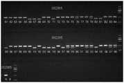
图2:F1代小鼠NOX2基因型的鉴定结果。其中,野生型为一条240bp的条带;纯合型为一条195bp的条带;杂合型为240bp及195bp两条条带;002365为NOX2基因代码。Figure 2: Identification results of NOX2 genotypes in F1 generation mice. Among them, the wild type is a band of 240bp; the homozygous type is a band of 195bp; the heterozygous type is two bands of 240bp and 195bp; 002365 is the NOX2 gene code.
图3:F2代小鼠NOX2基因型的鉴定结果。其中,野生型为一条240bp的条带;纯合型为一条195bp的条带;杂合型为240bp及195bp两条条带;002365为NOX2基因代码。Figure 3: Identification results of NOX2 genotypes in F2 generation mice. Among them, the wild type is a band of 240bp; the homozygous type is a band of 195bp; the heterozygous type is two bands of 240bp and 195bp; 002365 is the NOX2 gene code.
图4:F2代小鼠Pde6b基因型的鉴定结果。其中,野生型为一条400bp条带;纯合型为一条550bp条带;杂合型为550bp及400b两条条带;Pde6b基因为rd1小鼠变异基因(等位基因变异)。Fig. 4: Identification results of Pde6b genotype of F2 generation mice. Among them, the wild type has a 400bp band; the homozygous type has a 550bp band; the heterozygous type has two bands of 550bp and 400b; the Pde6b gene is the rd1 mouse variant gene (allelic variation).
图5:F3代小鼠NOX2基因型的鉴定结果。其中,野生型为一条240bp的条带;纯合型为一条195bp的条带;杂合型为240bp及195bp两条条带;002365为NOX2基因代码。Figure 5: Identification results of NOX2 genotypes in F3 generation mice. Among them, the wild type is a band of 240bp; the homozygous type is a band of 195bp; the heterozygous type is two bands of 240bp and 195bp; 002365 is the NOX2 gene code.
图6:不同小鼠视网膜外核层感光细胞的丢失情况。Figure 6: Loss of photoreceptor cells in the outer nuclear layer of different mouse retinas.
图7:不同小鼠视网膜外核层感光细胞的凋亡情况。Figure 7: Apoptosis of photoreceptor cells in the outer nuclear layer of the retina of different mice.
图8:不同小鼠视网膜NOX2主要亚单位gp91phox蛋白的表达及小胶质细胞的活化情况。Figure 8: Expression of NOX2 main subunit gp91phox protein and activation of microglia in different mouse retinas.
图9:rd1小鼠视网膜外核层感光细胞的丢失情况(玻璃体腔注射DMSO)。Figure 9: Loss of photoreceptor cells in the outer nuclear layer of the retina of rd1 mice (intravitreal injection of DMSO).
图10:rd1小鼠视网膜外核层感光细胞的丢失情况(玻璃体腔注射DMSO+VAS2870)。Figure 10: Loss of photoreceptor cells in the outer nuclear layer of the retina of rd1 mice (intravitreal injection of DMSO+VAS2870).
图11:rd1小鼠视网膜外核层感光细胞的丢失情况(腹腔注射PBS)。Figure 11: Loss of photoreceptor cells in the retinal outer nuclear layer of rd1 mice (intraperitoneal injection of PBS).
图12:rd1小鼠视网膜外核层感光细胞的丢失情况(腹腔注射PBS+gp91phox-tat)。Figure 12: Loss of photoreceptor cells in the retinal outer nuclear layer of rd1 mice (intraperitoneal injection of PBS+gp91phox-tat).
图13:rd1小鼠视网膜外核层感光细胞的丢失情况(腹腔注射PBS+GSK-2795039)。Figure 13: Loss of photoreceptor cells in the retinal outer nuclear layer of rd1 mice (intraperitoneal injection of PBS+GSK-2795039).
图14:rd1小鼠视网膜NOX2主要亚单位gp91phox蛋白的表达及小胶质细胞的活化情况(腹腔注射PBS)。Fig. 14: Expression of NOX2 major subunit gp91phox protein in retina of rd1 mice and activation of microglial cells (intraperitoneal injection of PBS).
图15:rd1小鼠视网膜NOX2主要亚单位gp91phox蛋白的表达及小胶质细胞的活化情况(腹腔注射PBS+gp91phox-tat)。Figure 15: Expression of NOX2 main subunitgp91phox protein in retina of rd1 mice and activation of microglial cells (intraperitoneal injection of PBS+gp91phox-tat).
图16:rd1小鼠视网膜NOX2主要亚单位gp91phox蛋白的表达及小胶质细胞的活化情况(腹腔注射PBS+GSK-2795039)。Figure 16: Expression of main subunit gp91phox protein of retinal NOX2 in rd1 mice and activation of microglial cells (intraperitoneal injection of PBS+GSK-2795039).
具体实施方式detailed description
提供下述实施例是为了更好地进一步理解本发明,并不局限于所述最佳实施方式,不对本发明的内容和保护范围构成限制,任何人在本发明的启示下或是将本发明与其他现有技术的特征进行组合而得出的任何与本发明相同或相近似的产品,均落在本发明的保护范围之内。The following examples are provided in order to further understand the present invention better, are not limited to the best implementation mode, and do not limit the content and protection scope of the present invention, anyone under the inspiration of the present invention or use the present invention Any product identical or similar to the present invention obtained by combining features of other prior art falls within the protection scope of the present invention.
下述实施例中未注明具体实验步骤或条件者,按照本领域内的文献所描述的常规实验步骤的操作或条件即可进行。所用试剂或仪器未注明生产厂商者,均为可以通过市购获得的常规试剂产品。If no specific experimental steps or conditions are indicated in the following examples, it can be carried out according to the operation or conditions of the conventional experimental steps described in the literature in this field. The reagents or instruments used, whose manufacturers are not indicated, are all commercially available conventional reagent products.
实验例1:NOX2基因作为治疗和/或预防遗传性视网膜变性的药物的治疗靶点的验证Experimental Example 1: Verification of the NOX2 gene as a therapeutic target of drugs for the treatment and/or prevention of hereditary retinal degeneration
1、NOX2基因敲除(NOX2KO)小鼠与rd1小鼠杂交获取F1代小鼠1. Crossbreeding NOX2 knockout (NOX2KO) mice with rd1 mice to obtain F1 generation mice
将四只基因型为gp91phox-/Y rd1W/W的雄性NOX2基因敲除小鼠(即NOX2基因在X染色体敲除的B6.129S-Cybbtm1Din/J小鼠,购自杰克逊实验室,JAX编码为002365)和八只基因型为gp91phox+/+rd1+/+的雌性rd1小鼠(购自北京维通利华实验动物技术有限公司)杂交,得到基因型为gp91phox+/Y rd1W/+的雄性杂交F1代小鼠和基因型为gp91phox+/-rd1w+的雌性杂交F1代小鼠。Four male NOX2 gene knockout mice with gp91phox-/Y rd1W/W genotype (that is, B6.129S-Cybbtm1Din /J mice with NOX2 gene knockout on the X chromosome, purchased from Jackson Laboratory, JAX code 002365) and eight female rd1 mice with gp91phox+/+rd1+/+ genotype (purchased from Beijing Weitong Lihua Experimental Animal Technology Co., Ltd.) to obtain male hybrid F1 with gp91phox+/Y rd1W/+ genotype generation mice and female hybrid F1 generation mice of gp91phox+/-rd1w+ genotype.
为了验证F1代小鼠基因型的正确性,使用表1的引物对F1代小鼠的基因组进行PCR扩增后,对扩增产物进行凝胶电泳分析,分析结果见图2。In order to verify the correctness of the genotype of the F1 generation mice, the primers in Table 1 were used to amplify the genome of the F1 generation mice by PCR, and then the amplified products were analyzed by gel electrophoresis. The analysis results are shown in Figure 2.
由图2可知,雄性杂交F1代小鼠的基因型为gp91phox+/Y rdW/+,雌性杂交F1代小鼠的基因型为Gp91phox+/-rdw+,F1代小鼠基因型制备成功。It can be seen from Figure 2 that the genotype of the male hybrid F1 generation mice is gp91phox+/Y rdW/+, and the genotype of the female hybrid F1 generation mice is Gp91phox+/-rdw+, and the genotype of the F1 generation mice was successfully prepared.
表1 002365的引物序列Table 1 The primer sequence of 002365

注:002365基因型的判断标准:Note: Judgment criteria for 002365 genotype:
Mutant(纯合)=195bpMutant (homozygous) = 195bp
Heterozygote(杂合)=195bp and 240bpHeterozygote (heterozygous) = 195bp and 240bp
Wild type(野生)=240bpWild type (wild) = 240bp
2、F1代小鼠交配获得杂交F2代小鼠2. F1 generation mice were mated to obtain hybrid F2 generation mice
将基因型为gp91phox+/Y rd1W/+的雄性杂交F1代小鼠和基因型为gp91phox+/-rd1w+的雌性杂交F1代小鼠杂交,得到F2代小鼠(共有48只,编号分别为#61~#108)。The male hybrid F1 generation mice with gp91phox+/Y rd1W/+ and the female hybrid F1 generation mice with gp91phox+/-rd1w+ genotype were crossed to obtain F2 generation mice (48 in total, numbered #61~ #108).
为了筛选基因型正确的小鼠,使用表1和表2的引物对F2代小鼠的基因组进行PCR扩增后,对扩增产物进行凝胶电泳分析,分析结果见图3~4。并按照gp91phox及Pde6b的基因型对F2代小鼠进行分类,分类结果见表3~4。In order to screen mice with the correct genotype, the primers in Table 1 and Table 2 were used to amplify the genome of the F2 generation mice by PCR, and then the amplified products were analyzed by gel electrophoresis. The analysis results are shown in Figures 3-4. The F2 generation mice were classified according to gp91phox and Pde6b genotypes, and the classification results are shown in Tables 3-4.
根据分类结果,筛选出基因型为gp91phox-/Y rd1+/+的#67,#81,#85作为雄性杂交F2代小鼠,筛选出基因型为gp91phox+/-rd1+/+的#76,#79,#102作为雌性杂交F2代小鼠。According to the classification results, #67, #81, and #85 with gp91phox-/Y rd1+/+ genotypes were screened out as male hybrid F2 mice, and #76, #79 with gp91phox+/-rd1+/+ genotypes were screened out , #102 was used as a female hybrid F2 generation mouse.
表2 Pde6b的引物序列Table 2 The primer sequence of Pde6b

注:Pde6b基因型的判断标准:Note: Judgment criteria for Pde6b genotype:
Mutant(纯合)=500bpMutant (homozygous) = 500bp
Heterozygote(野生)=550bp and 400bpHeterozygote (wild) = 550bp and 400bp
Wild type(杂合)=400bpWild type (heterozygous) = 400bp
表3 002365基因型的分类结果Table 3 Classification results of 002365 genotype

表4 Pde6b基因型的分类结果Table 4 Classification results of Pde6b genotypes

3、F2代小鼠交配获得杂交F3代小鼠3. F2 generation mice were mated to obtain hybrid F3 generation mice
将基因型为gp91phox-/Y rd1+/+的雄性杂交F2代小鼠和基因型为gp91phox+/-rd1+/+的雌性杂交F2代小鼠杂交,得到F3代小鼠(共有18只,编号分别为#126~#143)。The male hybrid F2 generation mice with gp91phox-/Y rd1+/+ were crossed with the female hybrid F2 generation mice with gp91phox+/-rd1+/+ genotype to obtain F3 generation mice (a total of 18 mice, numbered respectively #126~#143).
为了筛选基因型正确的小鼠,使用表1的引物对F3代小鼠的基因组进行PCR扩增后,对扩增产物进行凝胶电泳分析,分析结果见图5。并按照gp91phox的基因型对F3代小鼠进行分类,分类结果见表5。In order to screen mice with the correct genotype, the primers in Table 1 were used to amplify the genome of the F3 generation mice by PCR, and then the amplified products were analyzed by gel electrophoresis. The analysis results are shown in Figure 5. The F3 generation mice were classified according to the gp91phox genotype, and the classification results are shown in Table 5.
根据分类结果,筛选出基因型为gp91phox-/Y rd1+/+的#126,#138,#139,#140作为雄性杂交F3代小鼠,筛选出基因型为gp91phox+/-rd1+/+的#129,#131,#132,#142作为雌性杂交F3代小鼠。According to the classification results, #126, #138, #139, and #140 with gp91phox-/Y rd1+/+ genotype were screened out as male hybrid F3 mice, and #129 with gp91phox+/-rd1+/+ genotype was screened out , #131, #132, and #142 were used as female hybrid F3 generation mice.
基因型为gp91phox-/Y rd1+/+的雄性杂交F2代小鼠和雄性杂交F3代小鼠即为双纯合的雄性NOX2基因缺陷rd1小鼠模型;基因型为gp91phox-/-rd1+/+的雌性杂交F3代小鼠即为双纯合的雌性NOX2基因缺陷rd1小鼠模型。雄性NOX2基因缺陷rd1小鼠模型和雌性NOX2基因缺陷rd1小鼠模型可通过杂交进行扩繁。Male hybrid F2 mice and male hybrid F3 mice with gp91phox-/Y rd1+/+ genotype are double homozygous male NOX2 gene-deficient rd1 mouse models; gp91phox-/-rd1+/+ genotype The female hybrid F3 mice are the double homozygous female NOX2 gene-deficient rd1 mouse model. The male NOX2 gene-deficient rd1 mouse model and the female NOX2 gene-deficient rd1 mouse model can be amplified by crossbreeding.
表5 002365基因型的分类结果Table 5 Classification results of 002365 genotypes
4、NOX2基因缺陷rd1小鼠模型的视网膜组织细胞学鉴定4. Retinal histocytological identification of NOX2 gene-deficient rd1 mouse model
4.1、实验试剂及仪器4.1. Experimental reagents and instruments
Hoechst33258荧光染料(美国Sigma Alorich公司);樱花冷冻切片包埋剂(美国McMormick公司);TUNEL法细胞凋亡检测试剂盒(国产诺唯赞公司);含DAPI抗淬灭封片剂(美国Sigma Alorich公司);小鼠抗小鼠gp91phox单克隆抗体(美国BD公司);大鼠抗小鼠CD11b抗体(美国伯乐公司);FITC标记抗大鼠IgG、TRITC标记抗小鼠荧光二抗(美国Invitrogen公司);CM1850型快速冰冻样品制备切片机、DM4000B型光学显微镜、Confocal激光共聚焦显微镜(SPE)、LAS X照相系统(德国Leica公司)。Hoechst33258 fluorescent dye (Sigma Alorich, USA); Cherry Blossom Frozen Section Embedding Agent (McMormick, USA); TUNEL Apoptosis Detection Kit (Novizan, China); DAPI-containing anti-fade mountant (Sigma Alorich, USA) company); mouse anti-mouse gp91phox monoclonal antibody (BD, USA); rat anti-mouse CD11b antibody (Bio-Rad, USA); FITC-labeled anti-rat IgG, TRITC-labeled anti-mouse fluorescent secondary antibody (Invitrogen, USA) company); CM1850 fast frozen sample preparation microtome, DM4000B optical microscope, Confocal laser confocal microscope (SPE), LAS X camera system (Germany Leica company).
4.2、实验方法4.2. Experimental method
4.2.1、视网膜组织切片4.2.1. Retinal tissue section
以C57BL/6N小鼠(购自北京维通利华实验动物技术有限公司)、NOX2基因敲除小鼠和rd1小鼠(日龄14d)作为对照组小鼠,以步骤3获得的NOX2基因缺陷rd1小鼠模型(日龄14d)作为实验组小鼠;将各组小鼠经水合氯醛过量麻醉处死,快速摘除眼球,至于OCT包埋剂中,迅速置于液氮中骤冷,-80℃冰箱保存待用;使用时经视盘及锯齿缘做7μm冰冻切片,每组6只小鼠12只眼;将获得的小鼠眼球冰冻切片自然晾干,用4%(w/v,g/100mL)多聚甲醛室温固定15min,0.1mol/L PBS缓冲液(pH 7.4)漂洗3次,每次5min,苏木精染色20min,1%(v/v)盐酸乙醇分化20s,自来水浸泡20min,伊红染色2min,梯度乙醇脱水,二甲苯透明,中性树胶封片,光学显微镜下观察并照相。每个眼球选取3张切片,每张切片选取后极部相同部位视网膜进行拍照并比较各组外核层细胞层数。实验结果见图6。With C57BL/6N mice (purchased from Beijing Weitong Lihua Experimental Animal Technology Co., Ltd.), NOX2 knockout mice and rd1 mice (day-old 14d) as control mice, the NOX2 gene deficiency obtained in step 3 The rd1 mouse model (age 14 days) was used as the mice in the experimental group; the mice in each group were anesthetized and killed by overdose of chloral hydrate, and the eyeballs were quickly removed. Store in the refrigerator at ℃ for later use; make 7 μm frozen sections through the optic disc and ora serrata when used, 12 eyes of 6 mice in each group; dry the obtained frozen sections of mouse eyeballs in 4% (w/v, g/ 100mL) fixed in paraformaldehyde at room temperature for 15min, rinsed 3 times with 0.1mol/L PBS buffer (pH 7.4), 5min each time, stained with hematoxylin for 20min, differentiated with 1% (v/v) hydrochloric acid ethanol for 20s, soaked in tap water for 20min, Stain with eosin for 2 minutes, dehydrate with graded ethanol, clear with xylene, mount with neutral gum, observe and take pictures under an optical microscope. Three slices were selected for each eyeball, and the same part of the retina at the posterior pole was selected for each slice to be photographed and the number of outer nuclear layer cells in each group was compared. The experimental results are shown in Figure 6.
4.2.2、视网膜感光细胞凋亡分析4.2.2. Analysis of apoptosis of retinal photoreceptor cells
采用TUNEL细胞凋亡原位检测试剂盒,将步骤4.2.1获得的小鼠眼球冰冻切片自然晾干,用4%(w/v,g/100mL)多聚甲醛室温固定15min,0.1mol/L PBS缓冲液(pH 7.4)漂洗3次,每次5min,经蛋白酶K 37℃消化15min,TBS冲洗,滴加FITC荧光标记的检测液,湿盒内37℃避光孵育2h,TBS冲洗,含DAPI防荧光淬灭封片剂封片,荧光显微镜488nm波长下观察并拍照。绿色荧光信号为TUNEL阳性细胞。实验结果见图7。Using the TUNEL Cell Apoptosis In Situ Detection Kit, dry the mouse eyeball frozen section obtained in step 4.2.1, and fix it with 4% (w/v, g/100mL) paraformaldehyde at room temperature for 15min, 0.1mol/L Rinse with PBS buffer (pH 7.4) for 3 times, 5min each time, digest with proteinase K at 37°C for 15min, rinse with TBS, add FITC fluorescently labeled detection solution dropwise, incubate in a wet box at 37°C for 2h in the dark, wash with TBS, containing DAPI The slides were sealed with anti-fluorescence quenching mounting medium, observed and photographed under a fluorescence microscope at a wavelength of 488nm. Green fluorescent signal is TUNEL positive cell. The experimental results are shown in Figure 7.
4.2.3、gp91phox与CD11b免疫荧光染色及共定位4.2.3 Immunofluorescence staining and colocalization of gp91phox and CD11b
gp91phox是NOX2的主要亚单位,CD11b单克隆抗体标记小胶质细胞。二者免疫荧光染色共定位观察NOX2在小胶质细胞中的表达情况。将步骤4.2.1获得的小鼠眼球冰冻切片自然晾干,冷丙酮固定10min,0.1mol/L PBS缓冲液(pH 7.4)漂洗3次,每次5min,正常羊血清工作液封闭,室温(25℃)孵育10min,弃血清,勿洗;孵育来源不同种属的两个一抗(gp91phox来源于小鼠,CD11b来源于大鼠),4℃孵育过夜(16h),次日复温30min后经0.1mol/L PBS缓冲液(pH 7.4)漂洗,孵育与其相对应的FITC和TRITC(1:600,v/v),室温(25℃)孵育1h后经0.1mol/L PBS缓冲液(pH 7.4)漂洗3次,每次5min;含DAPI防荧光淬灭封片剂封片,荧光显微镜488nm波长下观察FITC绿色荧光、532nm波长下观察TRITC红色荧光并照相。实验结果见图8。gp91-phox is the major subunit of NOX2, and CD11b monoclonal antibody labels microglia. The expression of NOX2 in microglia was observed by immunofluorescence staining co-localization. Dry the mouse eyeball frozen sections obtained in step 4.2.1 naturally, fix with cold acetone for 10 min, rinse with 0.1mol/L PBS buffer (pH 7.4) for 3 times, each time for 5 min, seal with normal sheep serum working solution, and store at room temperature (25 ℃) for 10 min, discard the serum, do not wash; incubate with two primary antibodies from different species (gp91phox is from mouse, CD11b is from rat), incubate overnight at 4 °C (16 h), and rewarm for 30 min the next day Rinse with 0.1mol/L PBS buffer (pH 7.4), incubate with corresponding FITC and TRITC (1:600, v/v), incubate at room temperature (25°C) for 1 hour, then wash with 0.1mol/L PBS buffer (pH 7.4) Rinse 3 times, 5 min each time; mount the slide with DAPI anti-fluorescence quenching mounting agent, observe FITC green fluorescence at 488nm wavelength and TRITC red fluorescence at 532nm wavelength under a fluorescence microscope, and take pictures. The experimental results are shown in Figure 8.
上述实验的实验结果采用SPSS 20.0统计学软件进行统计分析。计量资料经Shapiro-Wilk检验呈正态分布,以mean±SD表示。应用单因素方差(ANOVA)分析比较不同组小鼠视网膜外核层厚度及TUNEL细胞百分比的差别,组间两两比较采用LSD-t检验。P<0.05为差异有统计学意义。The experimental results of the above experiments were statistically analyzed using SPSS 20.0 statistical software. The measurement data were normally distributed by the Shapiro-Wilk test, expressed as mean±SD. One-way analysis of variance (ANOVA) was used to compare the thickness of retinal outer nuclear layer and the percentage of TUNEL cells in different groups of mice, and LSD-t test was used for pairwise comparison between groups. P<0.05 means the difference is statistically significant.
4.3、实验结果4.3. Experimental results
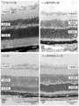
由图6可知,C57BL/6N小鼠及NOX2基因缺陷小鼠视网膜结构完整,内外核层细胞排列规则、致密,外核层平均厚度分别为54.44±2.33μm及52.96±1.31μm;而同龄rd1小鼠视网膜外核层明显变薄,外核层厚度为21.45±1.33μm,细胞核数量明显减少,且排列稀疏杂乱,形态大小不一;与rd1小鼠相比,Nox2基因缺陷rd1小鼠视网膜外核层厚度明显增加(36.18±2.59μm,t=8.770,p=0.001),内外核层细胞较之排列规则、致密。此结果表明NOX2因缺陷的rd1小鼠感光细胞丢失明显延缓。It can be seen from Figure 6 that the retinal structures of C57BL/6N mice and NOX2 gene-deficient mice are complete, the inner and outer nuclear layer cells are arranged regularly and densely, and the average thickness of the outer nuclear layer is 54.44±2.33μm and 52.96±1.31μm respectively; The outer nuclear layer of the mouse retina was significantly thinner, the thickness of the outer nuclear layer was 21.45±1.33 μm, the number of nuclei was significantly reduced, and the arrangement was sparse and disorderly, with different shapes and sizes; The layer thickness increased significantly (36.18±2.59μm, t=8.770, p=0.001), and the inner and outer nuclear layer cells were arranged more regularly and densely. This result indicates that the loss of NOX2-deficient photoreceptor cells in rd1 mice is significantly delayed.
由图7可知,C57BL/6N小鼠及NOX2基因缺陷小鼠视网膜外核层偶见TUNEL阳性细胞(分别为0.17±0.07%及0.08±0.03%);与C57BL/6N小鼠及NOX2基因缺陷小鼠相比,rd1小鼠外核层TUNEL细胞数量明显增多(5.37±0.75%)(t=19.645,P<0.01);与rd1小鼠相比,NOX2基因缺陷rd1小鼠视网膜外核层厚度增加,TUNEL细胞数量明显减少(1.5±0.3%)(t=8.42,P<0.01)。此结果进一步表明NOX2因缺陷的rd1小鼠感光细胞丢失明显延缓。It can be seen from Figure 7 that TUNEL-positive cells were occasionally seen in the retinal outer nuclear layer of C57BL/6N mice and NOX2 gene-deficient mice (0.17±0.07% and 0.08±0.03% respectively); Compared with mice, the number of TUNEL cells in the outer nuclear layer of rd1 mice was significantly increased (5.37±0.75%) (t=19.645, P<0.01); compared with rd1 mice, the thickness of the retinal outer nuclear layer of rd1 mice with NOX2 gene deficiency increased , the number of TUNEL cells was significantly reduced (1.5±0.3%) (t=8.42, P<0.01). This result further suggests that NOX2-deficient photoreceptor cell loss is significantly delayed in rd1 mice.
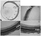
由图8可知,在C57BL/6N小鼠中,仅在内层视网膜(从视网膜内界膜到外丛状层)偶见gp91phox阳性细胞及分支状CD11b阳性小胶质细胞;在NOX2基因缺陷小鼠中,视网膜相似位置同样发现gp91phox阳性细胞及少量分枝状CD11b阳性染色细胞;在上述对照鼠中,部分gp91phox表达于CD11b小胶质细胞中;与上述对照鼠比较,rd1小鼠视网膜gp91phox表达量明显增加,染色加重,部分向外核层侵入,CD11b阳性小胶质细胞数量明显增加,细胞核增大,部分呈阿米巴状,且多数向外核层及视杆、视锥层浸润,部分外丛状层及外核层内的小胶质细胞明显表达gp91phox;与rd1小鼠相比,NOX2基因缺陷rd1小鼠视网膜外核层明显增厚,gp91phox蛋白及CD11b阳性细胞数量显著减少,以外核层明显。此结果表明NOX2因缺陷的rd1小鼠小胶质细胞活化显著抑制,小胶质细胞中NOX2表达明显减少。It can be seen from Figure 8 that in C57BL/6N mice,gp91phox -positive cells and branched CD11b-positive microglial cells were only occasionally seen in the inner retina (from the inner limiting membrane of the retina to the outer plexiform layer); In mice, gp91phox positive cells and a small amount of branched CD11b positive staining cells were also found in similar positions of the retina; in the above control mice, part of gp91phox was expressed in CD11b microglial cells; compared with the above control mice, rd1 mice The expression of gp91phox in the retina was significantly increased, the staining was aggravated, and some of them invaded the outer nuclear layer. The number of CD11b-positive microglial cells increased significantly, and the nuclei were enlarged. The cone layer infiltrates, part of the outer plexiform layer and microglial cells in the outer nuclear layer obviously express gp91phox ; compared with rd1 mice, the retinal outer nuclear layer of NOX2 gene-deficient rd1 mice is significantly thickened, gp91phox protein and CD11b The number of positive cells was significantly reduced, evident in the outer nuclear layer. This result indicated that NOX2 was significantly inhibited by microglia activation in deficient rd1 mice, and the expression of NOX2 in microglia was significantly reduced.
上述结果一方面证明了NOX2因缺陷的rd1小鼠的表型正确,NOX2因缺陷的rd1小鼠构建成功,另一方面确切验证了小胶质细胞中NOX2活化是遗传性视网膜变性的致病机制,为筛选和研发作用机制更明确、特异性更强、更有上市和临床应用前景的治疗和/或预防视网膜变性的药物提供了基础,极具应用前景。The above results prove that the phenotype of the NOX2-deficient rd1 mice is correct, and the NOX2-deficient rd1 mice were successfully constructed. On the other hand, it is confirmed that the activation of NOX2 in microglia is the pathogenic mechanism of hereditary retinal degeneration , which provides a basis for the screening and development of drugs for the treatment and/or prevention of retinal degeneration with a clearer mechanism of action, stronger specificity, and more marketable and clinical application prospects, and has great application prospects.
实验例2:NOX2特异性抑制剂作为制备治疗和/或预防遗传性视网膜变性的药物的验证Experimental Example 2: Verification of NOX2-specific inhibitors as drugs for the treatment and/or prevention of hereditary retinal degeneration
1、实验方法1. Experimental method
1.1、视网膜组织切片(VAS2870)1.1. Retinal tissue slice (VAS2870)
用PI-100微量注射器在rd1小鼠(日龄9d)玻璃体腔内注射0.5μg溶解于1μL 10%(w/v,g/100mL)DMSO的VAS2870(购自Sigma公司,产品号为SML2967),使其在玻璃体腔内的最终有效浓度为50μg/mL,对照组为对侧眼注射同等剂量的10%DMSO。注射后第5天(凋亡高峰期),将rd1小鼠经水合氯醛过量麻醉处死,快速摘除眼球,至于OCT包埋剂中,迅速置于液氮中骤冷,-80℃冰箱保存待用;使用时经视盘及锯齿缘做7μm冰冻切片,每组6只小鼠12只眼;将获得的小鼠眼球冰冻切片自然晾干,用4%(w/v,g/100mL)多聚甲醛室温固定15min,0.1mol/L PBS缓冲液(pH 7.4)漂洗3次,每次5min,苏木精染色20min,1%(v/v)盐酸乙醇分化20s,自来水浸泡20min,伊红染色2min,梯度乙醇脱水,二甲苯透明,中性树胶封片,光学显微镜下观察并照相。每个眼球选取3张切片,每张切片选取后极部相同部位视网膜进行拍照并比较各组外核层厚度。实验结果见图9~10。Inject 0.5 μg of VAS2870 dissolved in 1 μL of 10% (w/v, g/100mL) DMSO (purchased from Sigma, product number SML2967) into the vitreous cavity of rd1 mice (day-old 9d) with a PI-100 microinjector, The final effective concentration in the vitreous cavity was 50 μg/mL, and the contralateral eye was injected with the same dose of 10% DMSO as the control group. On the 5th day after the injection (the peak of apoptosis), the rd1 mice were anesthetized and killed by overdose of chloral hydrate, and the eyeballs were quickly removed. As for the OCT embedding medium, they were quickly placed in liquid nitrogen and stored in a -80°C refrigerator until later. Use; make 7 μm frozen sections through the optic disc and ora serrata, 12 eyes of 6 mice in each group; dry the obtained mouse eyeball frozen sections naturally, and use 4% (w/v, g/100mL) polymer Formaldehyde was fixed at room temperature for 15 minutes, rinsed 3 times with 0.1mol/L PBS buffer (pH 7.4), 5 minutes each time, stained with hematoxylin for 20 minutes, differentiated with 1% (v/v) hydrochloric acid ethanol for 20 seconds, soaked in tap water for 20 minutes, and stained with eosin for 2 minutes , dehydrated with gradient ethanol, transparent with xylene, mounted with neutral gum, observed and photographed under an optical microscope. Three slices were selected for each eyeball, and the same part of the retina at the posterior pole was selected for each slice to be photographed and the thickness of the outer nuclear layer in each group was compared. The experimental results are shown in Figures 9-10.
1.2、视网膜组织切片(gp91phox-tat、GSK-2795039)1.2. Retinal tissue slices (gp91phox-tat, GSK-2795039)
取18只rd1小鼠(日龄9d),随机分为三组,三组分别为PBS对照组、gp91phox-tat实验组以及GSK-2795039实验组。分组结束后,在gp91phox-tat实验组rd1小鼠腹腔内注射50μg溶于50μL 0.1mol/L PBS缓冲液(pH 7.4)的gp91phox-tat(购自Sigma公司),在GSK-2795039实验组小鼠腹腔内注射50μg溶于100μL 0.1mol/L PBS缓冲液(pH 7.4)的GSK-2795039(购自MCE公司),在PBS对照组小鼠腹腔内注射同等剂量的0.1mol/L PBS缓冲液(pH7.4),连续注射5天。注射5天后,将rd1小鼠经水合氯醛过量麻醉处死,快速摘除眼球,至于OCT包埋剂中,迅速置于液氮中骤冷,-80℃冰箱保存待用;使用时经视盘及锯齿缘做7μm冰冻切片,每组6只小鼠12只眼;将获得的小鼠眼球冰冻切片自然晾干,用4%(w/v,g/100mL)多聚甲醛室温固定15min,0.1mol/L PBS缓冲液(pH 7.4)漂洗3次,每次5min,苏木精染色20min,1%(v/v)盐酸乙醇分化20s,自来水浸泡20min,伊红染色2min,梯度乙醇脱水,二甲苯透明,中性树胶封片,光学显微镜下观察并照相。每个眼球选取3张切片,每张切片选取后极部相同部位视网膜进行拍照并比较各组外核层厚度。实验结果见图11~13。Eighteen rd1 mice (9 days old) were randomly divided into three groups, the three groups were PBS control group, gp91phox-tat experimental group and GSK-2795039 experimental group. After grouping, the rd1 mice in the gp91phox-tat experimental group were intraperitoneally injected with 50 μg of gp91phox-tat (purchased from Sigma) dissolved in 50 μL 0.1mol/L PBS buffer (pH 7.4), and the mice in the GSK-2795039 experimental group were injected intraperitoneally. Intraperitoneal injection of 50 μg GSK-2795039 (purchased from MCE Company) dissolved in 100 μL 0.1mol/L PBS buffer (pH 7.4), intraperitoneal injection of the same dose of 0.1mol/L PBS buffer (pH 7.4) in the PBS control group mice .4), continuous injection for 5 days. Five days after the injection, the rd1 mice were anesthetized by overdose of chloral hydrate and sacrificed, and the eyeballs were quickly removed. As for the OCT embedding medium, they were quickly placed in liquid nitrogen and stored in a -80°C refrigerator until use; 7 μm frozen sections were made, 12 eyes of 6 mice in each group; the obtained mouse eyeball frozen sections were dried naturally, fixed with 4% (w/v, g/100mL) paraformaldehyde at room temperature for 15min, 0.1mol/ Rinse with L PBS buffer (pH 7.4) for 3 times, 5 min each time, stain with hematoxylin for 20 min, differentiate with 1% (v/v) hydrochloric acid ethanol for 20 s, soak in tap water for 20 min, stain with eosin for 2 min, dehydrate with gradient ethanol, and transparent with xylene , mounted with neutral gum, observed and photographed under an optical microscope. Three slices were selected for each eyeball, and the same part of the retina at the posterior pole was selected for each slice to be photographed and the thickness of the outer nuclear layer in each group was compared. The experimental results are shown in Figures 11-13.
1.3、gp91phox与CD11b免疫荧光染色及共定位1.3 Immunofluorescent staining and colocalization of gp91phox and CD11b
gp91phox是NOX2的主要亚单位,CD11b单克隆抗体标记小胶质细胞。二者免疫荧光染色共定位观察NOX2在小胶质细胞中的表达情况。将步骤1.2获得的小鼠眼球冰冻切片自然晾干,冷丙酮固定10min,0.1mol/L PBS缓冲液(pH7.4)漂洗3次,每次5min,正常羊血清工作液封闭,室温(25℃)孵育10min,弃血清,勿洗;孵育来源不同种属的两个一抗(gp91phox来源于小鼠,CD11b来源于大鼠),4℃孵育过夜(16h),次日复温30min后经0.1mol/L PBS缓冲液(pH 7.4)漂洗,孵育与其相对应的FITC和TRITC(1:600,v/v),室温(25℃)孵育1h后经0.1mol/L PBS缓冲液(pH 7.4)漂洗3次,每次5min;含DAPI防荧光淬灭封片剂封片,荧光显微镜488nm波长下观察FITC绿色荧光、532nm波长下观察TRITC红色荧光并照相。实验结果见图14~16。gp91-phox is the major subunit of NOX2, and CD11b monoclonal antibody labels microglia. The expression of NOX2 in microglia was observed by immunofluorescence staining co-localization. Dry the mouse eyeball frozen section obtained in step 1.2 naturally, fix with cold acetone for 10 min, rinse with 0.1mol/L PBS buffer (pH7.4) for 3 times, each time for 5 min, seal with normal sheep serum working solution, and store at room temperature (25°C) ) for 10 minutes, discard the serum, and do not wash; incubate two primary antibodies from different species (gp91phox comes from mice, CD11b comes from rats), incubate overnight at 4°C (16h), and rewarm for 30 minutes the next day. Rinse with 0.1mol/L PBS buffer (pH 7.4), incubate with the corresponding FITC and TRITC (1:600, v/v), incubate at room temperature (25°C) for 1h, then wash with 0.1mol/L PBS buffer (pH 7.4 ) rinsed 3 times, 5 min each time; mounted with DAPI anti-fluorescence quenching mounting medium, observed FITC green fluorescence at 488nm wavelength and TRITC red fluorescence at 532nm wavelength under a fluorescence microscope, and took pictures. The experimental results are shown in Figures 14-16.
上述实验的实验结果采用SPSS 20.0统计学软件进行统计分析。计量资料经Shapiro-Wilk检验呈正态分布,以mean±SD表示。应用t检验比较给药组及对照组相同部位小鼠视网膜外核层厚度的差别。P<0.05为差异有统计学意义。The experimental results of the above experiments were statistically analyzed using SPSS 20.0 statistical software. The measurement data were normally distributed by the Shapiro-Wilk test, expressed as mean±SD. The t-test was used to compare the difference in the thickness of the retinal outer nuclear layer in the same part of the mice in the administration group and the control group. P<0.05 means the difference is statistically significant.
2、实验结果2. Experimental results
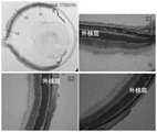
由图9~10可知,rd1小鼠玻璃体腔内注射VAS2870外核层厚度(22.33±1.42)较注射DMSO对照眼(20.16±2.08)并未无明显差异(P>0.05)。It can be seen from Figures 9-10 that the thickness of the outer nuclear layer of rd1 mice injected with VAS2870 into the vitreous cavity (22.33±1.42) was not significantly different from that of the DMSO-injected control eyes (20.16±2.08) (P>0.05).
由图11~13可知,与对照组(注射PBS)相比(23.08±1.23),腹腔内注射gp91phox-tat及GSK2795039使rd1小鼠外核层厚度明显增加(分别为37.10±1.67及38.23±2.02,P<0.05),即感光细胞丢失明显延迟。It can be seen from Figures 11 to 13 that compared with the control group (injection of PBS) (23.08±1.23), intraperitoneal injection of gp91phox-tat and GSK2795039 significantly increased the thickness of the outer nuclear layer of rd1 mice (respectively 37.10±1.67 and 38.23±2.02 , P<0.05), that is, the loss of photoreceptor cells was significantly delayed.
由图14~16可知,与对照组(PBS)相比,腹腔内注射gp91phox-tat及GSK2795039CD11b阳性小胶质细胞活化及细胞内gp91phox表达明显减弱。It can be seen from Figures 14 to 16 that compared with the control group (PBS), the activation of gp91phox-tat and GSK2795039CD11b positive microglial cells and the expression of intracellular gp91phox were significantly weakened after intraperitoneal injection.
显然,上述实施例仅仅是为清楚地说明所作的举例,而并非对实施方式的限定。对于所属领域的普通技术人员来说,在上述说明的基础上还可以做出其它不同形式的变化或变动。这里无需也无法对所有的实施方式予以穷举。而由此所引伸出的显而易见的变化或变动仍处于本发明创造的保护范围之中。Apparently, the above-mentioned embodiments are only examples for clear description, rather than limiting the implementation. For those of ordinary skill in the art, other changes or changes in different forms can be made on the basis of the above description. It is not necessary and impossible to exhaustively list all the implementation manners here. And the obvious changes or changes derived therefrom are still within the scope of protection of the present invention.
序列表sequence listing
<110> 首都医科大学附属北京同仁医院<110> Beijing Tongren Hospital Affiliated to Capital Medical University
<120> NOX2特异性抑制剂在制备视网膜变性药物中的应用<120> Application of NOX2-specific inhibitors in the preparation of retinal degeneration drugs
<160> 6<160> 6
<170> PatentIn version 3.3<170> PatentIn version 3.3
<210> 1<210> 1
<211> 23<211> 23
<212> DNA<212>DNA
<213> 人工序列<213> Artificial sequence
<400> 1<400> 1
aagagaaact cctctgctgt gaa 23aagagaaact cctctgctgt gaa 23
<210> 2<210> 2
<211> 23<211> 23
<212> DNA<212>DNA
<213> 人工序列<213> Artificial sequence
<400> 2<400> 2
cgcactggaa cccctgagaa agg 23cgcactggaa cccctgagaa agg 23
<210> 3<210> 3
<211> 26<211> 26
<212> DNA<212>DNA
<213> 人工序列<213> Artificial sequence
<400> 3<400> 3
gttctaattc catcagaagc ttatcg 26gttctaattc catcagaagc ttatcg 26
<210> 4<210> 4
<211> 28<211> 28
<212> DNA<212>DNA
<213> 人工序列<213> Artificial sequence
<400> 4<400> 4
gtaaacagca agaggcttta ttgggaac 28gtaaacagca agaggcttta ttgggaac 28
<210> 5<210> 5
<211> 28<211> 28
<212> DNA<212>DNA
<213> 人工序列<213> Artificial sequence
<400> 5<400> 5
tgacaattac tccttttccc tcagtctg 28tgacaattac tccttttccc tcagtctg 28
<210> 6<210> 6
<211> 27<211> 27
<212> DNA<212>DNA
<213> 人工序列<213> Artificial sequence
<400> 6<400> 6
tacccaccct tcctaatttt tctcagc 27tacccaccct tcctaatttt tctcagc 27
Claims (4)
Translated fromChinesePriority Applications (2)
| Application Number | Priority Date | Filing Date | Title |
|---|---|---|---|
| CN202310078462.4ACN115969844B (en) | 2021-12-28 | 2021-12-28 | Application of NOX2 specific inhibitors in the preparation of retinal degeneration drugs |
| CN202111636147.6ACN114099683B (en) | 2021-12-28 | 2021-12-28 | Application of NOX2 specific inhibitor in preparation of retinal degeneration medicine |
Applications Claiming Priority (1)
| Application Number | Priority Date | Filing Date | Title |
|---|---|---|---|
| CN202111636147.6ACN114099683B (en) | 2021-12-28 | 2021-12-28 | Application of NOX2 specific inhibitor in preparation of retinal degeneration medicine |
Related Child Applications (1)
| Application Number | Title | Priority Date | Filing Date |
|---|---|---|---|
| CN202310078462.4ADivisionCN115969844B (en) | 2021-12-28 | 2021-12-28 | Application of NOX2 specific inhibitors in the preparation of retinal degeneration drugs |
Publications (2)
| Publication Number | Publication Date |
|---|---|
| CN114099683A CN114099683A (en) | 2022-03-01 |
| CN114099683Btrue CN114099683B (en) | 2022-12-16 |
Family
ID=80363002
Family Applications (2)
| Application Number | Title | Priority Date | Filing Date |
|---|---|---|---|
| CN202310078462.4AActiveCN115969844B (en) | 2021-12-28 | 2021-12-28 | Application of NOX2 specific inhibitors in the preparation of retinal degeneration drugs |
| CN202111636147.6AActiveCN114099683B (en) | 2021-12-28 | 2021-12-28 | Application of NOX2 specific inhibitor in preparation of retinal degeneration medicine |
Family Applications Before (1)
| Application Number | Title | Priority Date | Filing Date |
|---|---|---|---|
| CN202310078462.4AActiveCN115969844B (en) | 2021-12-28 | 2021-12-28 | Application of NOX2 specific inhibitors in the preparation of retinal degeneration drugs |
Country Status (1)
| Country | Link |
|---|---|
| CN (2) | CN115969844B (en) |
Citations (6)
| Publication number | Priority date | Publication date | Assignee | Title |
|---|---|---|---|---|
| US20090176745A1 (en)* | 2007-12-31 | 2009-07-09 | Emory University | Triarylmethane analogs and their use in treating cancers |
| CN103357017A (en)* | 2013-03-07 | 2013-10-23 | 首都医科大学附属北京同仁医院 | Application of met-RANTES in preparing medicine used for treating inherited retinal degeneration |
| CN109512831A (en)* | 2013-04-05 | 2019-03-26 | 勃林格殷格翰国际有限公司 | Yi Palie net therapeutical uses |
| CN112423753A (en)* | 2018-04-25 | 2021-02-26 | A·马特内尔 | Methods and compositions for reducing the risk of relapse and prolonging survival of acute myeloid leukemia |
| WO2021050980A1 (en)* | 2019-09-13 | 2021-03-18 | The United States Of America, As Represented By The Secretary, Department Of Health & Human Services | Druggable target to treat retinal degeneration |
| CN113438945A (en)* | 2019-02-08 | 2021-09-24 | 德国癌症研究公共权益基金会 | Annexin coated particles |
Family Cites Families (2)
| Publication number | Priority date | Publication date | Assignee | Title |
|---|---|---|---|---|
| CA2842034C (en)* | 2011-07-18 | 2023-09-05 | University Of Kentucky Research Foundation | Protection of cells from alu-rna-induced degeneration and inhibitors for protecting cells |
| EP3732180A4 (en)* | 2017-12-27 | 2022-05-11 | Emory University | COMBINED MODALITIES FOR NUCLEOSIDES AND/OR NADPH OXIDASE (NOX) INHIBITORS AS MYELOID-SPECIFIC ANTIVIRALS |
- 2021
- 2021-12-28CNCN202310078462.4Apatent/CN115969844B/enactiveActive
- 2021-12-28CNCN202111636147.6Apatent/CN114099683B/enactiveActive
Patent Citations (6)
| Publication number | Priority date | Publication date | Assignee | Title |
|---|---|---|---|---|
| US20090176745A1 (en)* | 2007-12-31 | 2009-07-09 | Emory University | Triarylmethane analogs and their use in treating cancers |
| CN103357017A (en)* | 2013-03-07 | 2013-10-23 | 首都医科大学附属北京同仁医院 | Application of met-RANTES in preparing medicine used for treating inherited retinal degeneration |
| CN109512831A (en)* | 2013-04-05 | 2019-03-26 | 勃林格殷格翰国际有限公司 | Yi Palie net therapeutical uses |
| CN112423753A (en)* | 2018-04-25 | 2021-02-26 | A·马特内尔 | Methods and compositions for reducing the risk of relapse and prolonging survival of acute myeloid leukemia |
| CN113438945A (en)* | 2019-02-08 | 2021-09-24 | 德国癌症研究公共权益基金会 | Annexin coated particles |
| WO2021050980A1 (en)* | 2019-09-13 | 2021-03-18 | The United States Of America, As Represented By The Secretary, Department Of Health & Human Services | Druggable target to treat retinal degeneration |
Non-Patent Citations (4)
| Title |
|---|
| NADPH氧化酶抑制剂对遗传性视网膜色素变性感光细胞凋亡的抑制作用;丁敏等;《中华实验眼科杂志》;20140430;第32卷(第4期);第313-317页* |
| Neuroprotection from Retinal Ischemia/Reperfusion Injury by NOX2 NADPH Oxidase Deletion;Harumasa Yokota et al.;《Retinal Cell Biology》;20111011;第52卷(第11期);第8123-8131页* |
| NOX2基因缺陷对rd1小鼠感光细胞凋亡的保护作用;刘谦等;《眼科》;20220325;第31卷(第2期);第140-145页* |
| Oxidative Stress and the Role of NADPH Oxidase in Glaucoma;Jennifer C et al.;《Antioxidants (Basel)》;20210204;第10卷(第2期);第238页* |
Also Published As
| Publication number | Publication date |
|---|---|
| CN114099683A (en) | 2022-03-01 |
| CN115969844B (en) | 2024-04-02 |
| CN115969844A (en) | 2023-04-18 |
Similar Documents
| Publication | Publication Date | Title |
|---|---|---|
| Pusic et al. | Spreading depression requires microglia and is decreased by their M2a polarization from environmental enrichment | |
| Zhang et al. | Oxidative stress-involved mitophagy of retinal pigment epithelium and retinal degenerative diseases | |
| Sun et al. | SNAI1, an endothelial–mesenchymal transition transcription factor, promotes the early phase of ocular neovascularization | |
| Zhang et al. | Selenium restores synaptic deficits by modulating NMDA receptors and selenoprotein K in an Alzheimer's disease model | |
| Roger et al. | Preservation of cone photoreceptors after a rapid yet transient degeneration and remodeling in cone-only Nrl−/− mouse retina | |
| Lutty et al. | Proceedings of the Third International Symposium on Retinopathy of Prematurity: an update on ROP from the lab to the nursery (November 2003, Anaheim, California) | |
| Lin et al. | HDAC2 (histone deacetylase 2): a critical factor in environmental enrichment‐mediated stroke recovery | |
| Scaramuzza et al. | The enhancement of activity rescues the establishment of Mecp2 null neuronal phenotypes | |
| Lee et al. | Ciliary neurotrophic factor derived from astrocytes protects retinal ganglion cells through PI3K/AKT, JAK/STAT, and MAPK/ERK pathways | |
| Shi et al. | Pathological high intraocular pressure induces glial cell reactive proliferation contributing to neuroinflammation of the blood-retinal barrier via the NOX2/ET-1 axis-controlled ERK1/2 pathway | |
| Zaidi et al. | Histone deacetylases regulation by δ-opioids in human optic nerve head astrocytes | |
| Sekar et al. | Impairing Gasdermin D-mediated pyroptosis is protective against retinal degeneration | |
| Xiang et al. | Depletion of miR-96 delays, but does not arrest, photoreceptor development in mice | |
| Masuda et al. | GPR3 expression in retinal ganglion cells contributes to neuron survival and accelerates axonal regeneration after optic nerve crush in mice | |
| Shiga et al. | Endoplasmic reticulum stress-related deficits in calcium clearance promote neuronal dysfunction that is prevented by SERCA2 gene augmentation | |
| Chen et al. | Human‐Derived Induced GABAergic Progenitor Cells Improve Cognitive Function in Mice and Inhibit Astrocyte Activation with Anti‐Inflammatory Exosomes | |
| Qiu et al. | Syndromic deafness gene ATP6V1B2 controls degeneration of spiral ganglion neurons through modulating proton flux | |
| Lozano et al. | Optic nerve head gene transcription sequelae to a single elevated IOP exposure provides insights into known responses to chronically elevated IOP | |
| Gao et al. | Hearing improvement in A/J mice via the mouse nerve growth factor | |
| Narayan et al. | Investigations into bioenergetic neuroprotection of cone photoreceptors: relevance to retinitis pigmentosa | |
| Chidlow et al. | Investigations into photoreceptor energy metabolism during experimental retinal detachment | |
| CN114099683B (en) | Application of NOX2 specific inhibitor in preparation of retinal degeneration medicine | |
| Aguirre et al. | Epithelial membrane protein 2 (EMP2) blockade attenuates pathological neovascularization in murine oxygen-induced retinopathy | |
| CN114532296B (en) | NOX2 gene deficient rd1 mouse model and its preparation method and application | |
| Yetemian et al. | Neovascularization, enhanced inflammatory response, and age-related cone dystrophy in the Nrl−/− Grk1−/− mouse retina |
Legal Events
| Date | Code | Title | Description |
|---|---|---|---|
| PB01 | Publication | ||
| PB01 | Publication | ||
| SE01 | Entry into force of request for substantive examination | ||
| SE01 | Entry into force of request for substantive examination | ||
| GR01 | Patent grant | ||
| GR01 | Patent grant |