CN114098936B - Degradable bionic double-layer humerus bone fracture plate - Google Patents
Degradable bionic double-layer humerus bone fracture plateDownload PDFInfo
- Publication number
- CN114098936B CN114098936BCN202111437484.2ACN202111437484ACN114098936BCN 114098936 BCN114098936 BCN 114098936BCN 202111437484 ACN202111437484 ACN 202111437484ACN 114098936 BCN114098936 BCN 114098936B
- Authority
- CN
- China
- Prior art keywords
- plate
- bone
- screw
- degradable
- group
- Prior art date
- Legal status (The legal status is an assumption and is not a legal conclusion. Google has not performed a legal analysis and makes no representation as to the accuracy of the status listed.)
- Active
Links
- 210000002758humerusAnatomy0.000titleclaimsabstractdescription15
- 239000011664nicotinic acidSubstances0.000titleclaimsabstractdescription9
- 208000010392Bone FracturesDiseases0.000titledescription21
- 239000000463materialSubstances0.000claimsabstractdescription16
- 239000002639bone cementSubstances0.000claimsabstractdescription5
- GVALZJMUIHGIMD-UHFFFAOYSA-Hmagnesium phosphateChemical compound[Mg+2].[Mg+2].[Mg+2].[O-]P([O-])([O-])=O.[O-]P([O-])([O-])=OGVALZJMUIHGIMD-UHFFFAOYSA-H0.000claimsabstractdescription4
- 229960002261magnesium phosphateDrugs0.000claimsabstractdescription4
- 229910000157magnesium phosphateInorganic materials0.000claimsabstractdescription4
- 239000004137magnesium phosphateSubstances0.000claimsabstractdescription4
- 235000010994magnesium phosphatesNutrition0.000claimsabstractdescription4
- 229920000747poly(lactic acid)Polymers0.000claimsdescription8
- 239000004626polylactic acidSubstances0.000claimsdescription8
- 239000003462bioceramicSubstances0.000claimsdescription5
- 229920001610polycaprolactonePolymers0.000claimsdescription5
- 239000004632polycaprolactoneSubstances0.000claimsdescription4
- 150000002148estersChemical class0.000claimsdescription2
- 238000009958sewingMethods0.000claims1
- 210000000988bone and boneAnatomy0.000abstractdescription46
- 238000013461designMethods0.000abstractdescription6
- 230000015556catabolic processEffects0.000abstractdescription5
- 238000006731degradation reactionMethods0.000abstractdescription5
- 230000035876healingEffects0.000abstractdescription5
- 230000000399orthopedic effectEffects0.000abstractdescription2
- 230000001054cortical effectEffects0.000abstract1
- 206010017076FractureDiseases0.000description23
- 229910052751metalInorganic materials0.000description9
- 239000000956alloySubstances0.000description8
- 239000002184metalSubstances0.000description8
- 229910045601alloyInorganic materials0.000description7
- 206010020462Humerus fractureDiseases0.000description6
- 229920000954PolyglycolidePolymers0.000description4
- 238000000034methodMethods0.000description4
- 239000004633polyglycolic acidSubstances0.000description4
- 238000001356surgical procedureMethods0.000description4
- 239000002861polymer materialSubstances0.000description3
- 230000000694effectsEffects0.000description2
- 238000002594fluoroscopyMethods0.000description2
- 239000007769metal materialSubstances0.000description2
- 229920003229poly(methyl methacrylate)Polymers0.000description2
- 229920000642polymerPolymers0.000description2
- 239000004926polymethyl methacrylateSubstances0.000description2
- 229920001343polytetrafluoroethylenePolymers0.000description2
- 239000004810polytetrafluoroethyleneSubstances0.000description2
- 238000011084recoveryMethods0.000description2
- 230000000638stimulationEffects0.000description2
- 238000011282treatmentMethods0.000description2
- 206010020649HyperkeratosisDiseases0.000description1
- 208000001132OsteoporosisDiseases0.000description1
- 229920001244Poly(D,L-lactide)Polymers0.000description1
- 229910001069Ti alloyInorganic materials0.000description1
- 208000027418Wounds and injuryDiseases0.000description1
- 230000002411adverseEffects0.000description1
- 238000013459approachMethods0.000description1
- 230000009286beneficial effectEffects0.000description1
- 230000003592biomimetic effectEffects0.000description1
- 239000002131composite materialSubstances0.000description1
- 238000013329compoundingMethods0.000description1
- 229920001577copolymerPolymers0.000description1
- 230000006378damageEffects0.000description1
- 238000011161developmentMethods0.000description1
- 238000002695general anesthesiaMethods0.000description1
- 230000007062hydrolysisEffects0.000description1
- 238000006460hydrolysis reactionMethods0.000description1
- 239000007943implantSubstances0.000description1
- 238000002513implantationMethods0.000description1
- 238000001727in vivoMethods0.000description1
- 208000015181infectious diseaseDiseases0.000description1
- 208000014674injuryDiseases0.000description1
- 238000010883osseointegrationMethods0.000description1
- -1polytetrafluoroethylenePolymers0.000description1
- 238000011160researchMethods0.000description1
- 210000000513rotator cuffAnatomy0.000description1
- 231100000241scarToxicity0.000description1
- 210000000323shoulder jointAnatomy0.000description1
- 210000004872soft tissueAnatomy0.000description1
- 239000010935stainless steelSubstances0.000description1
- 229910001256stainless steel alloyInorganic materials0.000description1
- 210000002435tendonAnatomy0.000description1
- 210000001519tissueAnatomy0.000description1
- 231100000331toxicToxicity0.000description1
- 230000002588toxic effectEffects0.000description1
- 238000011277treatment modalityMethods0.000description1
- 210000003857wrist jointAnatomy0.000description1
Images


Classifications
- A—HUMAN NECESSITIES
- A61—MEDICAL OR VETERINARY SCIENCE; HYGIENE
- A61B—DIAGNOSIS; SURGERY; IDENTIFICATION
- A61B17/00—Surgical instruments, devices or methods
- A61B17/56—Surgical instruments or methods for treatment of bones or joints; Devices specially adapted therefor
- A61B17/58—Surgical instruments or methods for treatment of bones or joints; Devices specially adapted therefor for osteosynthesis, e.g. bone plates, screws or setting implements
- A61B17/68—Internal fixation devices, including fasteners and spinal fixators, even if a part thereof projects from the skin
- A61B17/80—Cortical plates, i.e. bone plates; Instruments for holding or positioning cortical plates, or for compressing bones attached to cortical plates
- A61B17/8061—Cortical plates, i.e. bone plates; Instruments for holding or positioning cortical plates, or for compressing bones attached to cortical plates specially adapted for particular bones
- A—HUMAN NECESSITIES
- A61—MEDICAL OR VETERINARY SCIENCE; HYGIENE
- A61B—DIAGNOSIS; SURGERY; IDENTIFICATION
- A61B17/00—Surgical instruments, devices or methods
- A61B17/56—Surgical instruments or methods for treatment of bones or joints; Devices specially adapted therefor
- A61B17/58—Surgical instruments or methods for treatment of bones or joints; Devices specially adapted therefor for osteosynthesis, e.g. bone plates, screws or setting implements
- A61B17/68—Internal fixation devices, including fasteners and spinal fixators, even if a part thereof projects from the skin
- A61B17/84—Fasteners therefor or fasteners being internal fixation devices
- A61B17/86—Pins or screws or threaded wires; nuts therefor
- A—HUMAN NECESSITIES
- A61—MEDICAL OR VETERINARY SCIENCE; HYGIENE
- A61B—DIAGNOSIS; SURGERY; IDENTIFICATION
- A61B17/00—Surgical instruments, devices or methods
- A61B2017/00004—(bio)absorbable, (bio)resorbable or resorptive
Landscapes
- Health & Medical Sciences (AREA)
- Orthopedic Medicine & Surgery (AREA)
- Surgery (AREA)
- Life Sciences & Earth Sciences (AREA)
- Heart & Thoracic Surgery (AREA)
- Nuclear Medicine, Radiotherapy & Molecular Imaging (AREA)
- Engineering & Computer Science (AREA)
- Biomedical Technology (AREA)
- Neurology (AREA)
- Medical Informatics (AREA)
- Molecular Biology (AREA)
- Animal Behavior & Ethology (AREA)
- General Health & Medical Sciences (AREA)
- Public Health (AREA)
- Veterinary Medicine (AREA)
- Surgical Instruments (AREA)
Abstract
Translated fromChinese
Description
Translated fromChinese技术领域technical field
本发明属骨科植入医疗器械技术领域,具体涉及一种可降解的仿生双层肱骨接骨板。The invention belongs to the technical field of orthopedic implanted medical devices, in particular to a degradable bionic double-layer humerus bone plate.
背景技术Background technique
骨折是骨科临床常见的损伤形式。其中,肱骨骨折在众多常见骨折中位列第3位,在65岁以上患者群体中,肱骨近端骨折占骨折总数的10%,肱骨近端骨折严重影响了老年人的生活质量。根据骨折部位和骨折程度,现有的肱骨近端骨折治疗方式主要包括外固定架固定、克氏针内固定、缝线缝合固定、锁定接骨板系统内固定、髓内钉内固定、肩关节置换、植骨或植入骨水泥应用等。在这些临床治疗方式中,金属或合金型锁定接骨板被广泛使用。Fractures are a common injury form in orthopaedic clinics. Among them, humerus fractures ranks third among many common fractures. In patients over 65 years old, proximal humerus fractures account for 10% of the total number of fractures. Proximal humerus fractures seriously affect the quality of life of the elderly. According to the fracture site and degree of fracture, the existing treatment methods for proximal humerus fractures mainly include external fixator fixation, internal Kirschner wire fixation, suture fixation, internal fixation with locking plate system, internal fixation with intramedullary nails, and shoulder joint replacement. , bone grafting or implantation of bone cement applications, etc. In these clinical treatment modalities, metal or alloy locking plates are widely used.
首先,金属或合金型接骨板用于对骨折裂缝的固定,其提供了足够的力学稳定性,以维持损伤骨良好的复位。当前接骨板的材料几乎均为不锈钢和钛合金,但由于金属或合金型接骨板刚度远大于骨骼的刚度,当载荷施加于接骨板和骨组成的系统时,接骨板往往要比骨承受更多的载荷,对骨折部位易造成应力遮挡,导致愈合的骨骼比发生骨折前的骨骼强度偏弱,易产生骨质疏松、二次骨折等问题。文献调研发现,目前降低应力遮挡的方法主要是通过改变接骨板材料和结构以降低刚度来实现的。在材料领域,研究者发现,某些非金属型接骨板具有良好的生物相容性,应用于颌骨、颧骨、腕关节等非承重骨骨折的内固定,如聚乙醇酸(PGA)、聚乳酸(PLLA)、聚四氟乙烯(PTFE)、聚甲基丙烯酸甲酯(PMMA)、聚乳酸-羟基乙酸的共聚物(PLGA)、生物陶瓷等,这些非金属材料刚度远低于金属或合金材质,这对降低接骨板的应力遮挡具有重要意义。First, metal or alloy type bone plates are used to fix fracture cracks, which provide sufficient mechanical stability to maintain good reduction of the injured bone. The materials of current bone plates are almost all stainless steel and titanium alloys, but because the stiffness of metal or alloy bone plates is much greater than that of bones, when the load is applied to the system composed of bone plates and bone, the bone plate often bears more than the bone. The load is easy to cause stress shielding on the fracture site, resulting in the strength of the healed bone is weaker than that of the bone before the fracture, which is prone to osteoporosis, secondary fractures and other problems. The literature survey found that the current method of reducing stress shielding is mainly achieved by changing the material and structure of the bone plate to reduce the stiffness. In the field of materials, researchers have found that some non-metallic bone plates have good biocompatibility and are used in the internal fixation of non-load-bearing bone fractures such as jawbone, zygomatic bone, and wrist joint, such as polyglycolic acid (PGA), Polylactic acid (PLLA), polytetrafluoroethylene (PTFE), polymethyl methacrylate (PMMA), polylactic acid-glycolic acid copolymer (PLGA), bioceramics, etc. These non-metallic materials are much less rigid than metal or Alloy material, which is of great significance to reduce the stress shielding of the bone plate.
其次,金属或合金型接骨板和螺钉一般均需要进行二次手术移除,在体内随着时间的迁移,易产生生长干扰和感染等不利影响。文献调研表明,某些具有良好生物相容性的高分子材料还具有可降解的性能,在体内可通过水解反应,无任何毒性反应,排异性小,如聚乙醇酸(PGA)、聚己内酯(PCL)、消旋聚乳酸(PDLLA)、外消旋聚乳酸(D,L-PLA)等。但是,聚合物材料力学强度远低于金属或合金型接骨板,不能单独用于肱骨近端接骨板,可与其他高强材料复合。分析发现,而生物陶瓷材料经过处理后可降解,与聚合物材质复合后形成的复合接骨板刚度介于金属型和高分子型材质之间,不仅具有良好的生物相容性、骨结合性以及骨传导性,还具有合适的力学性能,这对接骨板的设计开发带来了重要启发。Secondly, metal or alloy type bone plates and screws generally require secondary surgical removal, and are prone to adverse effects such as growth interference and infection over time in the body. Literature research has shown that some polymer materials with good biocompatibility also have degradable properties, which can pass hydrolysis in vivo without any toxic reaction and have low rejection, such as polyglycolic acid (PGA), polycaprolactone ester (PCL), racemic polylactic acid (PDLLA), racemic polylactic acid (D, L-PLA), etc. However, the mechanical strength of polymer materials is much lower than that of metal or alloy plates, so it cannot be used alone for proximal humerus plates, but can be combined with other high-strength materials. The analysis found that the bioceramic material can be degraded after treatment, and the composite bone plate formed by compounding with the polymer material has a stiffness between the metal type and the polymer type, which not only has good biocompatibility, osseointegration and Osteoconductivity and appropriate mechanical properties have brought important inspiration to the design and development of bone plates.
所以,金属或合金型接骨板系统易于对骨折愈合产生应力遮挡,同时由于不可生物降解,通常还需要进行二次手术将其从体内移除。Therefore, metal or alloy plate systems are prone to stress shielding of fracture healing, and because they are not biodegradable, they often require a secondary surgery to remove them from the body.
综上所述,急需一种可降解的仿生双层肱骨接骨板。In conclusion, there is an urgent need for a degradable biomimetic double-layer humerus bone plate.
发明内容SUMMARY OF THE INVENTION
本发明的目的是设计一种可降解的仿生双层肱骨接骨板,可以对肱骨近端骨折进行内固定,根据可降解材料的力学特性、生物相容性和降解性能,设计双层可降解的接骨板系统,实现了可变刚度设计,可以降低金属或合金型接骨板带来的应力遮挡,还可以避免二次手术。The purpose of the present invention is to design a degradable bionic double-layer humerus bone plate, which can be used for internal fixation of proximal humerus fractures. The bone plate system realizes a variable stiffness design, which can reduce the stress shielding caused by metal or alloy bone plates, and can also avoid secondary surgery.
在骨折固定的最初1个月里,接骨板系统稳定,可提供稳定的力学性能;之后,接骨板上板开始降解,2个月内骨折恢复基本完成,骨折端通过接骨板需要承受较多的力学刺激,接骨板上板通过其特有的降解性能实现了应力更多地向骨折断端传递;术后3个月,接骨板的力学强度降低30%,骨折端可以接收一定的应力,加速骨痂的生长,促进愈合;术后6个月,骨折愈合已经完成,接骨板系统已经失去固定意义,此时只需较低的力学强度和较高的降解速率,接骨螺钉和接骨板下板逐渐降解;术后10个月左右,接骨板螺钉降解消失,接骨板力学强度仅有最初的20%;术后12个月,接骨板系统几乎降解完成,之后,逐渐降解消失。所以,由于接骨板上下板的降解速率不同,对接骨板进行了可变刚度设计,极大地改善了不同愈合阶段骨折端所受的力学刺激,降低了接骨板系统的应力遮挡,促进了骨折区域的良好生长。In the first month of fracture fixation, the bone plate system is stable and can provide stable mechanical properties; after that, the plate begins to degrade, and the fracture recovery is basically completed within 2 months, and the fracture end needs to bear more stress through the plate. Mechanical stimulation, the plate on the bone plate can transmit more stress to the fracture end through its unique degradation performance; 3 months after the operation, the mechanical strength of the bone plate is reduced by 30%, and the fracture end can receive a certain amount of stress and accelerate the bone fracture. The growth of callus promotes healing; 6 months after the operation, the fracture healing has been completed, and the bone plate system has lost its meaning of fixation. At this time, only a lower mechanical strength and a higher degradation rate are needed, and the bone screws and the lower plate of the bone plate are gradually reduced. Degradation; about 10 months after the operation, the bone plate screw degraded and disappeared, and the mechanical strength of the bone plate was only 20% of the initial value; 12 months after the operation, the bone plate system was almost completely degraded, and then gradually degraded and disappeared. Therefore, due to the different degradation rates of the upper and lower plates on the bone plate, the variable stiffness design of the bone plate greatly improves the mechanical stimulation of the fracture end at different healing stages, reduces the stress shielding of the bone plate system, and promotes the fracture area. good growth.
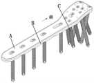
本发明的一种可降解的仿生双层肱骨接骨板,由上板A、下板B和螺钉组C组成,所述的上板A由上前板7和上后板2和凸板17组成,其中上前板7、上后板2圆滑连接而成,凸板17设于上前板7、上后板2下面;所述的下板B由下前板9、下后板16和凹槽18组成,下前板9和下后板16圆滑连接而成,凹槽18设于下前板9和下后板16上面;上板A的的凸板17与下板B的凹槽18契合,并经可降解磷酸镁骨水泥粘接。A degradable bionic double-layer humerus bone plate of the present invention is composed of an upper plate A, a lower plate B and a screw group C. The upper plate A is composed of an upper
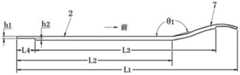
上前板7和上后板2、下前板9和下后板16的夹角θ1均为165-170度;上板A和下板B的垂直投影总长L1均为:110-115mm;其中上后板2和下后板16的垂直投影长度L2为60-75mm;上板A的厚度h1为1.6-2mm;下板B的厚度h3为2-2.4mm。The included angle θ1 of the
凸板17和凹槽18的宽度b3均为:9-12mm;凸板17和凹槽18的垂直投影总长L3均为:90-100mm,凸板17和凹槽18的后端与上后板2和下后板16后端的间距L4均为:5-10mm;凸板17的厚度和凹槽18的深度h2均为:0.4-0.6mm。The width b3 of the
上后板2和下后板16的宽度b1均为:13-15mm;上前板7和下前板9的宽度b2均为:22-24mm。The width b1 of the upper
上板A和下板B固接为一体后,上后板2和下后板16前后向均布八字形孔组1的四个孔,上后板2和下后板16前端设有长形孔3;上前板7和下前板9后部设有螺纹孔对4的两个螺纹孔,上前板7和下前板9中部设有八字形孔5,上前板7和下前板9前部设有螺纹孔组6的六个螺纹孔,上前板7和下前板9前部还设有缝合孔组8的十个孔;下后板16的下边缘均布圆弧缺口组14的十个圆弧缺口,每个圆弧槽的圆弧角θ2为50-58度,圆弧半径r为8-10mm。After the upper plate A and the lower plate B are fixedly connected as a whole, the four holes of the figure-eight
所述的螺钉组C由前螺钉组10的六个螺钉、前螺钉11、螺钉对12的两个螺钉、中螺钉13和后螺钉组15的四个螺钉组成,且上述各位置的螺钉顺序与上板A和下板B上的螺纹孔组6的六个螺纹孔、八字形孔5、螺纹孔对4的两个螺纹孔、长形孔3和八字形孔组1的四个孔对应设置,螺钉的长度L5为25-40mm。The screw group C is composed of six screws of the
所述的上板A的材料为可降解生物陶瓷;下板B的材料为聚己内酯;螺钉组C中螺钉的材料为外消旋聚乳酸。The material of the upper plate A is degradable bioceramics; the material of the lower plate B is polycaprolactone; the material of the screws in the screw group C is racemic polylactic acid.
本发明的工作过程如下:The working process of the present invention is as follows:
1.将肱骨近端骨折患者进行全身麻醉放在沙滩椅上,采取标准三角肌胸大肌间沟入路,用手术刀逐层剖开,直到骨折处显露。1. The patient with proximal humerus fracture was placed under general anesthesia on a beach chair, and a standard deltoid interpectoralis groove approach was adopted, and a scalpel was used to cut it layer by layer until the fracture was exposed.
2.对肱二头肌长腱和肩袖进行确认,清除骨折间软组织和疤痕组织,直到骨折面新鲜化,用克氏针进行临时固定,通过透视观察骨折复位满意。2. Confirm the biceps tendon and rotator cuff, remove the soft tissue and scar tissue between fractures, until the fracture surface is fresh, use Kirschner wire for temporary fixation, and observe the fracture reduction by fluoroscopy.
3.植入接骨板和螺钉进行固定,如果肱骨大结节骨折,需使用缝合线与接骨板固定,并再次通过透视观察骨折受伤位置和螺钉深度,满意后可冲洗切口,并放置1根引流管,逐层缝合包扎切口。3. Implant bone plates and screws for fixation. If the greater tuberosity of the humerus is fractured, use sutures and bone plates for fixation, and observe the location of the fracture and the depth of the screw through fluoroscopy again. After satisfactory, the incision can be washed and a drain is placed. Tube, layer by layer suture bandage the incision.
4.通过良好的功能康复锻炼,并随访恢复效果,透视观察骨骼恢复良好。4. Through good functional rehabilitation exercises and follow-up of the recovery effect, the skeleton is well recovered by fluoroscopic observation.
本发明的有益效果在于:The beneficial effects of the present invention are:
1.接骨板通过高分子生物降解型材料,极大地降低了接骨板的应力遮挡。1. The bone plate is made of polymer biodegradable material, which greatly reduces the stress shielding of the bone plate.
2.接骨板系统通过可降解型材料设计,避免了二次手术。2. The bone plate system is designed with degradable materials to avoid secondary surgery.
3.接骨板通过双层拼接设计,实现了可变刚度。3. The bone plate realizes variable stiffness through double-layer splicing design.
附图说明Description of drawings
图1为可降解的双层肱骨接骨板一轴测图;Figure 1 is an axonometric view of a degradable double-layer humerus bone plate;
图2为可降解的双层肱骨接骨板二轴测图;Figure 2 is a biaxial view of a degradable double-layer humerus bone plate;
图3为图1的俯视图;Fig. 3 is the top view of Fig. 1;
图4为上板A的侧视图;Figure 4 is a side view of the upper plate A;
图5为螺钉的剖视图;Figure 5 is a sectional view of the screw;
图6为图1的仰视图;Fig. 6 is the bottom view of Fig. 1;
图7为上板A的仰视图;Fig. 7 is the bottom view of upper plate A;
图8为下板B的俯视图;8 is a top view of the lower plate B;
图9为下板B的侧视图;Figure 9 is a side view of the lower plate B;
其中:A.上板;B.下板;C.螺钉组;1.八字形孔组;2.上后板;3.长形孔;4.螺纹孔对;5.八字形孔;6.螺纹孔组;7.上前板;8.缝合孔组;9.下前板;10.前螺钉组;11.前螺钉;12.螺钉对;13.中螺钉;14.圆弧缺口组;15.后螺钉组;16.下后板;17.凸板;18.凹槽。Among them: A. upper plate; B. lower plate; C. screw group; 1. figure-eight hole group; 2. upper back plate; 3. long hole; 4. pair of threaded holes; 5. figure-eight hole; 6. Threaded hole group; 7. Upper front plate; 8. Suture hole group; 9. Lower front plate; 10. Front screw group; 11. Front screw; 12. Screw pair; 13. Middle screw; 14. Arc notch group; 15. Rear screw group; 16. Lower rear plate; 17. Convex plate; 18. Groove.
具体实施方式Detailed ways
下面结合附图描述本发明。The present invention will be described below with reference to the accompanying drawings.
如图1至图3所示,本发明由上板A、下板B和螺钉组C组成,该接骨板外边缘光滑,内表面为仿生曲面,与损伤肱骨有很好的贴合性;所述的上板A的材料为可降解生物陶瓷;下板B的材料为聚己内酯;螺钉组C中螺钉的材料为外消旋聚乳酸;上板A的的凸板17与下板B的凹槽18契合,并经可降解磷酸镁骨水泥粘接;所述上后板2和下后板16的宽度b1均为:13-15mm;上前板7和下前板9的宽度b2均为:22-24mm。As shown in Figures 1 to 3, the present invention consists of an upper plate A, a lower plate B and a screw group C, the outer edge of the bone plate is smooth, and the inner surface is a bionic curved surface, which has a good fit with the injured humerus; The material of the upper plate A is degradable bioceramics; the material of the lower plate B is polycaprolactone; the material of the screws in the screw group C is racemic polylactic acid; the
如图4所示,所述上前板7和上后板2、下前板9和下后板16的夹角θ1均为165-170度;上板A和下板B的垂直投影总长L1均为:110-115mm;其中上后板2和下后板16的垂直投影长度L2为60-75mm;凸板17和凹槽18的垂直投影总长L3均为:90-100mm,凸板(17)和凹槽(18)的后端与上后板(2)和下后板16后端的间距L4均为:5-10mm;上板A的厚度h1为1.6-2mm;凸板17的厚度和凹槽18的深度h2均为:0.4-0.6mm。As shown in FIG. 4 , the angle θ1 between the upper
如图5和图6所示,所述上板A和下板B固接为一体后,上后板2和下后板16前后向均布八字形孔组1的四个孔,上后板2和下后板16前端设有长形孔3;上前板7和下前板9后部设有螺纹孔对4的两个螺纹孔,上前板7和下前板9中部设有八字形孔5,上前板7和下前板9前部设有螺纹孔组6的六个螺纹孔,上前板7和下前板9前部还设有缝合孔组8的十个孔;下后板16的下边缘均布圆弧缺口组14的十个圆弧缺口,每个圆弧槽的圆弧角θ2为50-58度,圆弧半径r为8-10mm;所述的螺钉组C由前螺钉组10的六个螺钉、前螺钉11、螺钉对12的两个螺钉、中螺钉13和后螺钉组15的四个螺钉组成,且上述各位置的螺钉顺序与上板A和下板B上的螺纹孔组6的六个螺纹孔、八字形孔5、螺纹孔对4的两个螺纹孔、长形孔3和八字形孔组1的四个孔对应设置,螺钉的长度L5为25-40mm。As shown in FIG. 5 and FIG. 6 , after the upper plate A and the lower plate B are fixed as one, the upper
如图7至图9所示,所述的上板A由上前板7和上后板2和凸板17组成,其中上前板7、上后板2圆滑连接而成,凸板17设于上前板7、上后板2下面;所述的下板B由下前板9、下后板16和凹槽18组成,下前板9和下后板16圆滑连接而成,凹槽18设于下前板9和下后板16上面;下板B的厚度h3为2-2.4mm;凸板17和凹槽18的宽度b3均为:9-12mm。As shown in FIGS. 7 to 9 , the upper plate A is composed of an upper
Claims (2)
Translated fromChinesePriority Applications (1)
| Application Number | Priority Date | Filing Date | Title |
|---|---|---|---|
| CN202111437484.2ACN114098936B (en) | 2021-11-30 | 2021-11-30 | Degradable bionic double-layer humerus bone fracture plate |
Applications Claiming Priority (1)
| Application Number | Priority Date | Filing Date | Title |
|---|---|---|---|
| CN202111437484.2ACN114098936B (en) | 2021-11-30 | 2021-11-30 | Degradable bionic double-layer humerus bone fracture plate |
Publications (2)
| Publication Number | Publication Date |
|---|---|
| CN114098936A CN114098936A (en) | 2022-03-01 |
| CN114098936Btrue CN114098936B (en) | 2022-09-20 |
Family
ID=80367680
Family Applications (1)
| Application Number | Title | Priority Date | Filing Date |
|---|---|---|---|
| CN202111437484.2AActiveCN114098936B (en) | 2021-11-30 | 2021-11-30 | Degradable bionic double-layer humerus bone fracture plate |
Country Status (1)
| Country | Link |
|---|---|
| CN (1) | CN114098936B (en) |
Family Cites Families (8)
| Publication number | Priority date | Publication date | Assignee | Title |
|---|---|---|---|---|
| BR0215966A (en)* | 2002-12-02 | 2005-09-13 | Mathys Medizinaltechnik Ag | Bone fixation implant |
| JP5976647B2 (en)* | 2010-08-31 | 2016-08-24 | シンセス ゲーエムベーハー | Degradation control of bioabsorbable metal implants |
| CN201920886U (en)* | 2010-12-24 | 2011-08-10 | 常州市康辉医疗器械有限公司 | Bone fracture plate for locking and pressurizing near end of shoulder bone |
| US9480510B2 (en)* | 2011-03-23 | 2016-11-01 | Spinecraft, LLC | Devices, systems and methods of attaching same to the spine |
| CN207785264U (en)* | 2017-06-01 | 2018-08-31 | 周顺刚 | A kind of proximal humerus bone plate |
| CN208851618U (en)* | 2018-03-30 | 2019-05-14 | 西安卓恰医疗器械有限公司 | The degradable miniature interior fixation kit of low alloying |
| CN111407387A (en)* | 2020-03-10 | 2020-07-14 | 昶盛(物料应用制品)有限公司 | Screw assembly for bone fixation and bone fixation device |
| CN111658116B (en)* | 2020-07-02 | 2025-09-16 | 董谢平 | Bone fracture plate capable of automatically converting from firm fixation to elastic fixation |
- 2021
- 2021-11-30CNCN202111437484.2Apatent/CN114098936B/enactiveActive
Also Published As
| Publication number | Publication date |
|---|---|
| CN114098936A (en) | 2022-03-01 |
Similar Documents
| Publication | Publication Date | Title |
|---|---|---|
| Rosa et al. | Intramedullary nailing biomechanics: evolution and challenges | |
| Bostman et al. | Biodegradable internal fixation for malleolar fractures. A prospective randomised trial | |
| Taljanovic et al. | Fracture fixation | |
| Suzuki et al. | Resorbable poly-L-lactide plates and screws for the treatment of mandibular condylar process fractures: a clinical and radiologic follow-up study | |
| Southerland et al. | McGlamry's comprehensive textbook of foot and ankle surgery | |
| Kim et al. | Treatment of mandible fractures using bioabsorbable plates | |
| Lovald et al. | Applications of polyetheretherketone in trauma, arthroscopy, and cranial defect repair | |
| Mehmood et al. | Internal fixation: An evolutionary appraisal of methods used for long bone fractures | |
| Thakur | The elements of fracture fixation, 4e | |
| Suuronen et al. | New generation biodegradable plate for fracture fixation: Comparison of bending strengths of mandibular osteotomies fixed with absorbable self-reinforced multi-layer poly-L-lactide plates and metallic plates—An experimental study in sheep | |
| Awati et al. | Limitations of current metallic bone plates: towards development of composite bone plates | |
| Arata et al. | Arthrodesis of the distal interphalangeal joint using a bioabsorbable rod as an intramedullary nail | |
| CN114098936B (en) | Degradable bionic double-layer humerus bone fracture plate | |
| RU2691326C1 (en) | Absorbable intramedullary nail for fixing fractures of long tubular bones | |
| Bibbo et al. | Orthoplastic management of complex bone and soft tissue pathology with a fully radiolucent circular external fixation system | |
| RU2691329C1 (en) | Method of combined osteosynthesis of fractures of long tubular bones | |
| Brunelli et al. | A personal technique for treatment of scaphoid non-union | |
| RU2349278C1 (en) | Method of broken intramedullar metal rod removal from long bone | |
| Dudko et al. | 30-year experience of open reduction internal fixation of limb fractures using biodegradable polymeric devices | |
| CN113786233B (en) | Coupling bionic humerus bone fracture plate capable of effectively reducing stress shielding | |
| Hardy et al. | Fracture Healing: An Evolving Perspective | |
| Reshma et al. | Biodegradable materials used in orthopaedics | |
| RU233557U1 (en) | Pin-cage for intramedullary osteosynthesis of the tibia with the ability to deliver active substances | |
| Krishna et al. | Study of surgical management of distal femoral fractures by distal femoral locking compression plate osteosynthesis | |
| RU2802152C1 (en) | Method of surgical treatment of osteoarthritis of the knee joint |
Legal Events
| Date | Code | Title | Description |
|---|---|---|---|
| PB01 | Publication | ||
| PB01 | Publication | ||
| SE01 | Entry into force of request for substantive examination | ||
| SE01 | Entry into force of request for substantive examination | ||
| GR01 | Patent grant | ||
| GR01 | Patent grant |