CN114022462A - Method, system, device, processor and computer-readable storage medium for realizing lesion segmentation of multi-parameter nuclear magnetic resonance images - Google Patents
Method, system, device, processor and computer-readable storage medium for realizing lesion segmentation of multi-parameter nuclear magnetic resonance imagesDownload PDFInfo
- Publication number
- CN114022462A CN114022462ACN202111326387.6ACN202111326387ACN114022462ACN 114022462 ACN114022462 ACN 114022462ACN 202111326387 ACN202111326387 ACN 202111326387ACN 114022462 ACN114022462 ACN 114022462A
- Authority
- CN
- China
- Prior art keywords
- convolution
- neural network
- output
- magnetic resonance
- nuclear magnetic
- Prior art date
- Legal status (The legal status is an assumption and is not a legal conclusion. Google has not performed a legal analysis and makes no representation as to the accuracy of the status listed.)
- Granted
Links
Images


Classifications
- G—PHYSICS
- G06—COMPUTING OR CALCULATING; COUNTING
- G06T—IMAGE DATA PROCESSING OR GENERATION, IN GENERAL
- G06T7/00—Image analysis
- G06T7/0002—Inspection of images, e.g. flaw detection
- G06T7/0012—Biomedical image inspection
- G—PHYSICS
- G06—COMPUTING OR CALCULATING; COUNTING
- G06F—ELECTRIC DIGITAL DATA PROCESSING
- G06F18/00—Pattern recognition
- G06F18/20—Analysing
- G06F18/24—Classification techniques
- G06F18/241—Classification techniques relating to the classification model, e.g. parametric or non-parametric approaches
- G06F18/2415—Classification techniques relating to the classification model, e.g. parametric or non-parametric approaches based on parametric or probabilistic models, e.g. based on likelihood ratio or false acceptance rate versus a false rejection rate
- G—PHYSICS
- G06—COMPUTING OR CALCULATING; COUNTING
- G06N—COMPUTING ARRANGEMENTS BASED ON SPECIFIC COMPUTATIONAL MODELS
- G06N3/00—Computing arrangements based on biological models
- G06N3/02—Neural networks
- G06N3/04—Architecture, e.g. interconnection topology
- G06N3/045—Combinations of networks
- G—PHYSICS
- G06—COMPUTING OR CALCULATING; COUNTING
- G06N—COMPUTING ARRANGEMENTS BASED ON SPECIFIC COMPUTATIONAL MODELS
- G06N3/00—Computing arrangements based on biological models
- G06N3/02—Neural networks
- G06N3/04—Architecture, e.g. interconnection topology
- G06N3/048—Activation functions
- G—PHYSICS
- G06—COMPUTING OR CALCULATING; COUNTING
- G06N—COMPUTING ARRANGEMENTS BASED ON SPECIFIC COMPUTATIONAL MODELS
- G06N3/00—Computing arrangements based on biological models
- G06N3/02—Neural networks
- G06N3/08—Learning methods
- G—PHYSICS
- G06—COMPUTING OR CALCULATING; COUNTING
- G06T—IMAGE DATA PROCESSING OR GENERATION, IN GENERAL
- G06T7/00—Image analysis
- G06T7/10—Segmentation; Edge detection
- G06T7/11—Region-based segmentation
- G—PHYSICS
- G06—COMPUTING OR CALCULATING; COUNTING
- G06T—IMAGE DATA PROCESSING OR GENERATION, IN GENERAL
- G06T2207/00—Indexing scheme for image analysis or image enhancement
- G06T2207/10—Image acquisition modality
- G06T2207/10072—Tomographic images
- G06T2207/10088—Magnetic resonance imaging [MRI]
- G—PHYSICS
- G06—COMPUTING OR CALCULATING; COUNTING
- G06T—IMAGE DATA PROCESSING OR GENERATION, IN GENERAL
- G06T2207/00—Indexing scheme for image analysis or image enhancement
- G06T2207/20—Special algorithmic details
- G06T2207/20081—Training; Learning
- G—PHYSICS
- G06—COMPUTING OR CALCULATING; COUNTING
- G06T—IMAGE DATA PROCESSING OR GENERATION, IN GENERAL
- G06T2207/00—Indexing scheme for image analysis or image enhancement
- G06T2207/20—Special algorithmic details
- G06T2207/20092—Interactive image processing based on input by user
- G06T2207/20104—Interactive definition of region of interest [ROI]
- G—PHYSICS
- G06—COMPUTING OR CALCULATING; COUNTING
- G06T—IMAGE DATA PROCESSING OR GENERATION, IN GENERAL
- G06T2207/00—Indexing scheme for image analysis or image enhancement
- G06T2207/30—Subject of image; Context of image processing
- G06T2207/30004—Biomedical image processing
- G06T2207/30096—Tumor; Lesion
Landscapes
- Engineering & Computer Science (AREA)
- Theoretical Computer Science (AREA)
- Physics & Mathematics (AREA)
- General Physics & Mathematics (AREA)
- Data Mining & Analysis (AREA)
- General Engineering & Computer Science (AREA)
- Health & Medical Sciences (AREA)
- Life Sciences & Earth Sciences (AREA)
- Artificial Intelligence (AREA)
- Evolutionary Computation (AREA)
- General Health & Medical Sciences (AREA)
- Computational Linguistics (AREA)
- Computing Systems (AREA)
- Molecular Biology (AREA)
- Biophysics (AREA)
- Mathematical Physics (AREA)
- Software Systems (AREA)
- Biomedical Technology (AREA)
- Computer Vision & Pattern Recognition (AREA)
- Bioinformatics & Computational Biology (AREA)
- Bioinformatics & Cheminformatics (AREA)
- Probability & Statistics with Applications (AREA)
- Evolutionary Biology (AREA)
- Medical Informatics (AREA)
- Nuclear Medicine, Radiotherapy & Molecular Imaging (AREA)
- Radiology & Medical Imaging (AREA)
- Quality & Reliability (AREA)
- Magnetic Resonance Imaging Apparatus (AREA)
- Image Analysis (AREA)
Abstract
Description
Translated fromChinese技术领域technical field
本发明涉及医学图像自动分割的技术领域,尤其涉及图像处理中的语义分割领域,具体是指一种基于深度神经网络模型实现多参数核磁共振图像病灶分割的方法、系统、装置、处理器及其计算机可读存储介质。The invention relates to the technical field of automatic segmentation of medical images, in particular to the field of semantic segmentation in image processing, and in particular to a method, system, device, processor and the same for a multi-parameter nuclear magnetic resonance image lesion segmentation based on a deep neural network model. computer readable storage medium.
背景技术Background technique
前列腺癌(Prostate Cancer,PCa)是仅次于肺癌的男性第二大致命疾病,而前列腺癌若能被尽早发现并进行及时治疗,可以有效提高患者的生存率。多参数MRI(Multi-parametric MRI,mpMRI)是一种先进的前列腺成像方法,将前列腺MRI的常规序列与一种或多种功能成像技术联合应用,被认为是用于前列腺癌临床诊断的最佳影像检查技术。但是基于mpMRI的前列腺癌临床诊断需要有放射科医生的专业知识作为基础,不同医师的判断会出现一定的偏差。Prostate cancer (PCa) is the second most deadly disease in men after lung cancer, and if prostate cancer is detected early and treated in time, it can effectively improve the survival rate of patients. Multi-parametric MRI (mpMRI) is an advanced prostate imaging method that combines conventional sequences of prostate MRI with one or more functional imaging techniques, and is considered to be the best for the clinical diagnosis of prostate cancer. imaging technology. However, the clinical diagnosis of prostate cancer based on mpMRI requires the professional knowledge of radiologists, and there will be certain deviations in the judgments of different doctors.
医学图像分割是医疗影像分析领域的热点研究内容,众多学者针对不同的挑战提出了许多不同的分割算法。针对PCa检测与分割的早期工作主要集中在手工特征选择方法上,这些方法利用预定义的图像特征来构建特征经验模型实现PCa病灶的分割;接着深度学习方法被大量用在医学图像分割领域,但是,从前列腺mpMRI中使用CNN进行PCa病灶分割的方法较少。现有的前列腺癌病灶分割方法中,有基于T2W图像的PCa检测方法。然而,采用MRI的单一序列可能会忽略不同形式的互信息,从而阻碍模型实现更好的分割性能;也有设计了基于mpMRI的多通道编解码器网络以实现PCa的检测和分类,但是依然有网络参数冗余,小目标分割困难等问题。Medical image segmentation is a hot research topic in the field of medical image analysis, and many scholars have proposed many different segmentation algorithms for different challenges. Early work on PCa detection and segmentation mainly focused on manual feature selection methods, which used predefined image features to construct feature empirical models to achieve PCa lesion segmentation; then deep learning methods were widely used in the field of medical image segmentation, but , there are fewer methods for PCa lesion segmentation using CNNs from prostate mpMRI. Among the existing prostate cancer lesion segmentation methods, there is a PCa detection method based on T2W images. However, a single sequence using MRI may ignore different forms of mutual information, preventing the model from achieving better segmentation performance; mpMRI-based multi-channel encoder-decoder networks have also been designed to achieve PCa detection and classification, but there are still networks Parameter redundancy, small target segmentation difficulties, etc.
发明内容SUMMARY OF THE INVENTION
本发明的目的是克服了上述现有技术的缺点,提供了一种检测维度多样且应用范围广泛的基于深度神经网络模型实现多参数核磁共振图像病灶分割的方法、系统、装置、处理器及其计算机可读存储介质。The purpose of the present invention is to overcome the above-mentioned shortcomings of the prior art, and to provide a method, system, device, processor and the same for a multi-parameter nuclear magnetic resonance image lesion segmentation based on a deep neural network model with various detection dimensions and a wide range of applications. computer readable storage medium.
为了实现上述目的,本发明的基于深度神经网络模型实现多参数核磁共振图像病灶分割的方法、系统、装置、处理器及其计算机可读存储介质如下:In order to achieve the above object, the method, system, device, processor and computer-readable storage medium thereof for implementing multi-parameter nuclear magnetic resonance image lesion segmentation based on a deep neural network model of the present invention are as follows:
基于深度神经网络模型实现多参数核磁共振图像病灶分割的方法,其主要特点是,所述的方法包括以下步骤:A method for implementing multi-parameter MRI lesion segmentation based on a deep neural network model, the main feature of which is that the method includes the following steps:
(1)输入包含前列腺多参数核磁共振图像的成像序列ADC、T2W、DWI的任意组合样本进行刚性匹配操作;(1) Input any combination sample of the imaging sequence ADC, T2W, and DWI containing the prostate multi-parameter MRI image for rigid matching operation;
(2)对处理后的图像提取感兴趣区域,并输送至前列腺癌病灶分割网络通过编码器进行特征处理;(2) Extract the region of interest from the processed image, and send it to the prostate cancer lesion segmentation network for feature processing through the encoder;
(3)所述的编码器输出特征图并将其输入至级联金字塔卷积处理模块的跨越连接层进行卷积和特征图采样处理;(3) described encoder output feature map and input it to the cross-connection layer of the cascade pyramid convolution processing module to carry out convolution and feature map sampling processing;
(4)解码器进行特征图上采样后与经过所述的跨越连接层输出的特征输送至双输入通道注意力模块进行特征融合处理;(4) After the decoder performs the up-sampling of the feature map, the features output through the cross-connection layer are sent to the dual-input channel attention module for feature fusion processing;
(5)训练所述的前列腺多参数核磁共振图像的前列腺癌病灶分割网络,以获取病灶分割结果。(5) Train the prostate cancer lesion segmentation network of the prostate multi-parameter MRI image to obtain lesion segmentation results.
较佳地,所述的步骤(2)具体为:Preferably, the step (2) is specifically:
采用预训练的ResNeXt网络中的预设个数的卷积模块通过编码器将各个卷积模块中的每一个下采样层的特征图保留,获得相应特征图的通道数。Using a preset number of convolution modules in the pre-trained ResNeXt network, the encoder retains the feature map of each downsampling layer in each convolution module to obtain the number of channels of the corresponding feature map.
较佳地,所述的预设个数的卷积模块设定为ResNeXt网络中的前五个卷积模块,其中,第一个卷积模块使用的卷积核大小为7×7,其余四个卷积模块分别使用了大小为3×3和1×1的卷积核。Preferably, the preset number of convolution modules is set as the first five convolution modules in the ResNeXt network, wherein the size of the convolution kernel used by the first convolution module is 7×7, and the remaining four The convolution modules use convolution kernels of
较佳地,五次下采样所获得的特征图的通道数依次增加,且各个特征图大小依次减小,分别为原图的1/2、1/4、1/8、1/16以及1/32。Preferably, the number of channels of the feature maps obtained by five downsampling increases sequentially, and the size of each feature map decreases sequentially, which are 1/2, 1/4, 1/8, 1/16, and 1 of the original image, respectively. /32.
较佳地,所述的步骤(3)具体包括以下步骤:Preferably, the step (3) specifically includes the following steps:
(3.1)所述的编码器从第一层至第四层分别对应四个级联金字塔卷积模块进行特征图输出,其中每一个大核卷积的输出都与原输入特征图经过1×1卷积后的特征图进行逐像素相加的融合操作,作为下一个卷积的输入;(3.1) The encoder from the first layer to the fourth layer corresponds to four concatenated pyramid convolution modules to output feature maps, wherein the output of each large kernel convolution is 1×1 with the original input feature map. The convolved feature map is subjected to a pixel-by-pixel addition fusion operation as the input of the next convolution;
(3.2)所述的级联金字塔卷积处理模块使用卷积分解将一个大核卷积分解成双支路结构,其中一条支路以x×1和1×y的顺序串联组成,另一条支路的卷积顺序为1×y和x×1,两条支路的输出进行逐元素相加得到最后的输出;(3.2) The cascaded pyramid convolution processing module uses convolution decomposition to decompose a large kernel convolution into a double-branch structure, wherein one branch is formed in series in the order of x×1 and 1×y, and the other branch’s The convolution order is 1×y and x×1, and the outputs of the two branches are added element by element to obtain the final output;
(3.3)对多个大核卷积的结果在通道上进行拼接,最大程度保留小目标对象的特征信息;(3.3) The results of multiple large kernel convolutions are spliced on the channel, and the feature information of the small target object is preserved to the greatest extent;
(3.4)根据编码器前四层输出的对应的四组级联金字塔使用的卷积核的个数与尺寸的差异,以适应编码器不同大小特征图的尺寸。(3.4) According to the difference in the number and size of the convolution kernels used by the corresponding four groups of cascaded pyramids output by the first four layers of the encoder, to adapt to the size of feature maps of different sizes of the encoder.
较佳地,所述的步骤(4)具体包括以下步骤:Preferably, the step (4) specifically includes the following steps:
(4.1)所述的双输入通道注意力模块的第一通道通过跨越连接路径中的级联金字塔卷积模块输入第一特征图


(4.2)将所述的第一特征图与第二特征图在通道维度上拼接,得到
(4.3)所述的双输入通道注意力模块将输入的第一特征图与第二特征图在通道维度上融合后进行全局平均池化操作,并根据以下公式得到全局信息特征向量:The dual-input channel attention module described in (4.3) fuses the input first feature map and the second feature map in the channel dimension and performs a global average pooling operation, and obtains the global information feature vector according to the following formula:

其中,i,j,k,f分别表示特征图的高、宽、深度和通道。Among them, i, j, k, f represent the height, width, depth and channel of the feature map, respectively.
(4.4)通过1×1卷积将特征向量维度降为通道数c,并使用Sigmoid激活函数对特征向量进行归一化操作,根据以下公式得到通道注意力向量CA:(4.4) Reduce the feature vector dimension to the number of channels c by 1×1 convolution, and use the Sigmoid activation function to normalize the feature vector, and obtain the channel attention vector CA according to the following formula:
CA=σ(W×vf+b);CA=σ(W×vf +b);
其中,W和b为卷积核参数,vf为特征图。Among them, W and b are the convolution kernel parameters, and vf is the feature map.
(4.5)所述的注意力向量与级联金字塔卷积模块的输出特征图在通道维度上相乘,以增强网络浅层特征的判别性,再将深层特征通过残差连接的方式连接到输出端,成为双输入通道注意力模块的输出,具体为通过以下公式实现:The attention vector described in (4.5) is multiplied by the output feature map of the cascaded pyramid convolution module in the channel dimension to enhance the discrimination of the shallow features of the network, and then the deep features are connected to the output through residual connections. end, it becomes the output of the dual-input channel attention module, which is realized by the following formula:

其中,
尤佳地,所述的解码器具体为由四个串联的双输入通道注意力模块组成,所述的解码器对每层输出的特征向量通过双线性插值操作进行上采样,与上一层的级联金字塔卷积处理模块输出的特征向量进行融合,从而逐渐将特征图恢复至原始输入尺寸,且输出层使用Softmax函数输出每个特征图的像素所属类别的概率。Preferably, the decoder is specifically composed of four concatenated dual-input channel attention modules, and the decoder up-samples the feature vector output by each layer through bilinear interpolation operation, and compares it with the previous layer. The feature vector output by the cascaded pyramid convolution processing module is fused to gradually restore the feature map to the original input size, and the output layer uses the Softmax function to output the probability of the category to which the pixels of each feature map belong.
更佳地,所述的步骤(5)为通过损失函数获取所述的病灶分割结果,具体为:其中,总的损失函数为:More preferably, the step (5) is to obtain the lesion segmentation result through a loss function, specifically: wherein, the total loss function is:
L′total=L′bce+L′dice;L′total = L′bce + L′dice ;
其中,L′bce为像素级二进制交叉熵损失,L′dice为Dice损失,两个损失分别为:Among them, L'bce is the pixel-level binary cross-entropy loss, L'dice is the Dice loss, and the two losses are:


其中,xi,j为预测类别的概率,yi,j为真实标注。Among them, xi, j are the probabilities of the predicted categories, andyi, j are the true labels.
该利用上述方法实现基于深度神经网络模型实现多参数核磁共振图像病灶分割的系统,其主要特点是,所述的系统包括:The system for implementing multi-parameter nuclear magnetic resonance image lesion segmentation based on a deep neural network model using the above method is mainly characterized in that the system includes:
目标提取处理模块,用于对输入的前列腺多参数核磁共振图像的成像序列ADC、T2W、DWI进行感兴趣区域的目标提取;The target extraction processing module is used to extract the target of the region of interest from the input imaging sequence ADC, T2W and DWI of the prostate multi-parameter MRI image;
尺寸统一处理模块,与所述的目标提取处理模块相连接,用于对提取后的目标图像进行刚性配准操作,并将各个目标图像的大小统一到相同尺寸;A size unification processing module, connected with the target extraction processing module, is used to perform a rigid registration operation on the extracted target images, and unify the size of each target image to the same size;
病灶分割神经网络处理模块,与所述的尺寸统一处理模块相连接,用于将经过尺寸统一处理后的目标图像输入至前列腺多参数核磁共振图像的前列腺癌病灶分割网络中通过编码器进行卷积和特征图采样处理;The lesion segmentation neural network processing module is connected with the size unification processing module, and is used for inputting the target image after size unification processing into the prostate cancer lesion segmentation network of the prostate multi-parameter nuclear magnetic resonance image to perform convolution through the encoder and feature map sampling processing;
级联金字塔卷积处理模块,与所述的病灶分割神经网络处理模块相连接,用于接收所述的编码器输出的特征图,并将其输入至所述的级联金字塔卷积处理模块的跨越连接层进行分组卷积和残差拼接,以保留相应目标对象的特征信息;以及The cascade pyramid convolution processing module is connected with the lesion segmentation neural network processing module, and is used for receiving the feature map output by the encoder, and inputting it into the cascade pyramid convolution processing module. Group convolution and residual concatenation across connected layers to preserve the feature information of the corresponding target object; and
双输入通道注意力模块,与所述的级联金字塔卷积处理模块相连接,用于将通过跨越连接路径中的所述的级联金字塔卷积处理模块的输出特征图以及解码层进行特征上采样后的输出特征图在通道维度上融合后进行全局平均池化操作,以获取对应通道的特征向量。The dual-input channel attention module is connected with the cascaded pyramid convolution processing module, and is used to perform feature mapping on the output feature map and the decoding layer of the cascaded pyramid convolution processing module across the connection path. The sampled output feature maps are fused in the channel dimension and then subjected to a global average pooling operation to obtain the feature vector of the corresponding channel.
该基于深度神经网络模型实现多参数核磁共振图像病灶分割的装置,其主要特点是,所述的装置包括:The main features of the device for implementing multi-parameter MRI lesion segmentation based on a deep neural network model are that the device includes:
处理器,被配置成执行计算机可执行指令;a processor configured to execute computer-executable instructions;
存储器,存储一个或多个计算机可执行指令,所述计算机可执行指令被所述处理器执行时,实现上述所述的基于深度神经网络模型实现多参数核磁共振图像病灶分割的方法的各个步骤。The memory stores one or more computer-executable instructions. When the computer-executable instructions are executed by the processor, each step of the above-mentioned method for implementing multi-parameter MRI lesion segmentation based on a deep neural network model is implemented.
该基于深度神经网络模型实现多参数核磁共振图像病灶分割的处理器,其主要特点是,所述的处理器被配置成执行计算机可执行指令,所述的计算机可执行指令被所述的处理器执行时,实现上述所述的基于深度神经网络模型实现多参数核磁共振图像病灶分割的方法的各个步骤。The main feature of the processor for implementing multi-parameter MRI lesion segmentation based on a deep neural network model is that the processor is configured to execute computer-executable instructions, and the computer-executable instructions are executed by the processor. During execution, each step of the above-mentioned method for implementing multi-parameter MRI lesion segmentation based on a deep neural network model is implemented.
该计算机可读存储介质,其主要特点是,其上存储有计算机程序,所述的计算机程序可被处理器执行以实现上述所述的基于深度神经网络模型实现多参数核磁共振图像病灶分割的方法的各个步骤。The computer-readable storage medium is mainly characterized in that a computer program is stored thereon, and the computer program can be executed by a processor to realize the above-mentioned method for realizing multi-parameter MRI lesion segmentation based on a deep neural network model. of the various steps.
采用了本发明的该基于深度神经网络模型实现多参数核磁共振图像病灶分割的方法、系统、装置、处理器及其计算机可读存储介质,提供了一种基于编解码结构深度神经网络的多参数序列前列腺癌病灶分割方法及框架。该分割方法融合多种MRI模态数据,采用级联金字塔卷积、通道注意力等模块,充分融合深浅层特征信息,降低噪声干扰,分割出多尺度的前列腺癌病灶目标,为前列腺疾病的临床诊断和治疗提供有力支持,降低诊断耗费的时间,为前列腺癌的筛查、检测、诊断提供更加有效的信息处理方法和手段。可以实现对MRI前列腺癌病灶的自动分割。By adopting the method, system, device, processor and computer-readable storage medium for implementing multi-parameter nuclear magnetic resonance image lesion segmentation based on the deep neural network model of the present invention, a multi-parameter deep neural network based encoding and decoding structure is provided. Sequence prostate cancer lesion segmentation method and framework. The segmentation method fuses multiple MRI modal data, adopts cascade pyramid convolution, channel attention and other modules, fully integrates deep and shallow feature information, reduces noise interference, and segmentes multi-scale prostate cancer lesion targets, which is a clinical tool for prostate diseases. Provide strong support for diagnosis and treatment, reduce the time spent on diagnosis, and provide more effective information processing methods and means for prostate cancer screening, detection and diagnosis. Automatic segmentation of MRI prostate cancer lesions can be achieved.
同时,考虑到采用MRI的单一序列可能会忽略不同形式的互信息,从而阻碍模型实现更好的分割性能,我们采用进行通道合并的三个序列数据:T2W、ADC和DWI,使用ADC和DWI序列能够补充病灶特征信息,大大提高分割性能;针对不同病例的PCa病灶形状、尺寸差异较大,小目标区域较多的问题,设计的级联金字塔卷积模块,级联金字塔卷积模块可以多尺度的捕获编码器生成的特征图中携带的详细空间定位信息,融合多个尺度的局部和全局信息,以减少空间定位信息的损失,提高模型对像素点的分类能力,同时,使用级联金字塔卷积模块后也可以改善模型欠分割的情况;为了增强模型对目标区域的关注度,设计了双输入通道注意力模块,利用网络深层特征的语义信息来指导浅层输出,以获得具有更高判别能力的特征,加强网络各阶段的特征提取能力。At the same time, considering that a single sequence using MRI may ignore different forms of mutual information, thus hindering the model from achieving better segmentation performance, we adopt three sequence data for channel merging: T2W, ADC and DWI, using ADC and DWI sequences It can supplement the feature information of lesions and greatly improve the segmentation performance; for the problem that the shape and size of PCa lesions in different cases are quite different, and there are many small target areas, the designed cascade pyramid convolution module, the cascade pyramid convolution module can be multi-scale. The detailed spatial positioning information carried in the feature map generated by the encoder is captured, and the local and global information of multiple scales is fused to reduce the loss of spatial positioning information and improve the model's ability to classify pixels. At the same time, the cascade pyramid volume is used. The under-segmentation of the model can also be improved after the integration module; in order to enhance the model's attention to the target area, a dual-input channel attention module is designed, which uses the semantic information of the deep features of the network to guide the shallow output to obtain higher discrimination. It can enhance the feature extraction capability of each stage of the network.
附图说明Description of drawings
图1为本发明的基于深度神经网络模型实现多参数核磁共振图像病灶分割的方法的流程图。FIG. 1 is a flowchart of a method for implementing multi-parameter MRI lesion segmentation based on a deep neural network model according to the present invention.
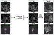
图2为本发明的基于深度神经网络的多参数核磁共振图像病灶分割方法的提取感兴趣区域以及进行统一尺寸的示意图。FIG. 2 is a schematic diagram of extracting a region of interest and unifying the size of the multi-parameter MRI lesion segmentation method based on the deep neural network of the present invention.
图3为本发明的级联金字塔卷积处理模块。FIG. 3 is a cascade pyramid convolution processing module of the present invention.
图4为本发明的级联金字塔卷积处理模块的大核卷积的拆分示意图。FIG. 4 is a schematic diagram of splitting a large kernel convolution of a cascaded pyramid convolution processing module of the present invention.
图5为本发明的双输入通道注意力模块的结构示意图。FIG. 5 is a schematic structural diagram of the dual-input channel attention module of the present invention.
图6为本发明实现多参数核磁共振图像病灶分割方法的分割结果示意图。FIG. 6 is a schematic diagram of the segmentation result of the multi-parameter nuclear magnetic resonance image lesion segmentation method implemented by the present invention.
具体实施方式Detailed ways
为了能够更清楚地描述本发明的技术内容,下面结合具体实施例来进行进一步的描述。In order to describe the technical content of the present invention more clearly, further description will be given below with reference to specific embodiments.
在详细说明根据本发明的实施例前,应该注意到的是,在下文中,第一和第二之类的关系术语仅仅用来区分一个实体或动作与另一个实体或动作,而不一定要求或暗示这种实体或动作之间的任何实际的这种关系或顺序。术语“包括”、“包含”或任何其他变体旨在涵盖非排他性的包含,由此使得包括一系列要素的过程、方法、物品或者设备不仅包含这些要素,而且还包含没有明确列出的其他要素,或者为这种过程、方法、物品或者设备所固有的要素。Before describing embodiments in accordance with the present invention in detail, it should be noted that in the following, relational terms such as first and second are only used to distinguish one entity or action from another, and do not necessarily require or Any actual such relationship or sequence between such entities or actions is implied. The terms "comprising", "comprising" or any other variation are intended to encompass a non-exclusive inclusion whereby a process, method, article or device comprising a list of elements includes not only those elements but also other not expressly listed elements, or elements inherent to such a process, method, article or device.
请参阅图1所示,该基于深度神经网络模型实现多参数核磁共振图像病灶分割的方法,其中,所述的方法包括以下步骤:Please refer to FIG. 1 , the method for implementing multi-parameter MRI lesion segmentation based on a deep neural network model, wherein the method includes the following steps:
(1)输入包含前列腺多参数核磁共振图像(mpMRI)的成像序列ADC、T2W、DWI的任意组合样本进行刚性匹配操作,并将大小统一到相同尺寸,如图2所示;(1) Input any combination samples of the imaging sequence ADC, T2W, and DWI including prostate multi-parameter nuclear magnetic resonance image (mpMRI) to perform rigid matching operation, and unify the size to the same size, as shown in Figure 2;
上述所使用的刚性匹配操作,即为本技术领域的普通技术人员所公知的常规图像处理方式,属于本领域的公知常识,在此不另行说明。The rigid matching operation used above is a conventional image processing method known to those of ordinary skill in the art, and belongs to the common knowledge in the art, and will not be described here.
(2)对处理后的图像提取感兴趣区域,并输送至前列腺癌病灶分割网络通过编码器进行特征处理;(2) Extract the region of interest from the processed image, and send it to the prostate cancer lesion segmentation network for feature processing through the encoder;
在实际应用当中,所设计的mpMRI的前列腺癌(PCa)病灶分割网络中的编码器可以是任意常用深度神经网络结构,以预训练的ResNeXt101为例;In practical applications, the encoder in the designed mpMRI prostate cancer (PCa) lesion segmentation network can be any common deep neural network structure, taking the pre-trained ResNeXt101 as an example;
(3)所述的编码器输出特征图并将其输入至级联金字塔卷积处理模块的跨越连接层进行卷积和特征图采样处理;(3) described encoder output feature map and input it to the cross-connection layer of the cascade pyramid convolution processing module to carry out convolution and feature map sampling processing;
(4)解码器进行特征图上采样后与经过所述的跨越连接层输出的特征输送至双输入通道注意力模块进行特征融合处理;(4) After the decoder performs the up-sampling of the feature map, the features output through the cross-connection layer are sent to the dual-input channel attention module for feature fusion processing;
(5)训练所述的前列腺多参数核磁共振图像的前列腺癌病灶分割网络,以获取病灶分割结果。(5) Train the prostate cancer lesion segmentation network of the prostate multi-parameter MRI image to obtain lesion segmentation results.
作为本发明的优选实施方式,所述的步骤(2)具体为:As a preferred embodiment of the present invention, the step (2) is specifically:
采用预训练的ResNeXt网络中的预设个数的卷积模块通过编码器将各个卷积模块中的每一个下采样层的特征图保留,获得相应特征图的通道数。Using a preset number of convolution modules in the pre-trained ResNeXt network, the encoder retains the feature map of each downsampling layer in each convolution module to obtain the number of channels of the corresponding feature map.
作为本发明的优选实施方式,所述的预设个数的卷积模块设定为ResNeXt网络中的前五个卷积模块,其中,第一个卷积模块使用的卷积核大小为7×7,其余四个卷积模块分别使用了大小为3×3和1×1的卷积核。As a preferred embodiment of the present invention, the preset number of convolution modules is set as the first five convolution modules in the ResNeXt network, wherein the size of the convolution kernel used by the first convolution module is 7× 7. The remaining four convolution modules use convolution kernels of
作为本发明的优选实施方式,五次下采样所获得的特征图的通道数依次增加,且各个特征图大小依次减小,分别为原图的1/2、1/4、1/8、1/16以及1/32。As a preferred embodiment of the present invention, the number of channels of the feature maps obtained by five downsampling increases sequentially, and the size of each feature map decreases sequentially, which are 1/2, 1/4, 1/8, and 1 of the original image, respectively. /16 and 1/32.
在实际应用当中,采用预训练的ResNeXt网络中的前五个卷积块,每个卷积模块都使用了分组卷积和残差连接,可以在不增加(甚至降低)模型复杂度的前提下提升网络精度,第一个卷积模块使用的卷积核大小为7×7,其余四个卷积模块都分别使用了大小为3×3和1×1的卷积核;In practical applications, the first five convolution blocks in the pre-trained ResNeXt network are used, and each convolution block uses grouped convolution and residual connections, which can be used without increasing (or even reducing) the complexity of the model. To improve the network accuracy, the size of the convolution kernel used by the first convolution module is 7×7, and the remaining four convolution modules use convolution kernels of
通过预训练的编码器,将每个下采样层的特征图保留,为五次下采样,获得五份特征图的通道数依次增加,特征图大小依次减小,分别为原图的1/2、1/4、1/8、1/16、1/32。Through the pre-trained encoder, the feature map of each downsampling layer is retained for five downsampling, the number of channels to obtain the five feature maps increases in turn, and the size of the feature map decreases in turn, which are 1/2 of the original image respectively. , 1/4, 1/8, 1/16, 1/32.
请参阅图3所示,作为本发明的优选实施方式,所述的步骤(3)具体包括以下步骤:Please refer to Figure 3, as a preferred embodiment of the present invention, the step (3) specifically includes the following steps:
(3.1)所述的编码器从第一层至第四层分别对应四个级联金字塔卷积模块进行特征图输出,其中每一个大核卷积的输出都与原输入特征图经过1×1卷积后的特征图进行逐像素相加的融合操作,作为下一个卷积的输入;(3.1) The encoder from the first layer to the fourth layer corresponds to four concatenated pyramid convolution modules to output feature maps, wherein the output of each large kernel convolution is 1×1 with the original input feature map. The convolved feature map is subjected to a pixel-by-pixel addition fusion operation as the input of the next convolution;
(3.2)所述的级联金字塔卷积处理模块使用卷积分解将一个大核卷积分解成双支路结构,其中一条支路以x×1和1×y的顺序串联组成,另一条支路的卷积顺序为1×y和x×1,两条支路的输出进行逐元素相加得到最后的输出;(3.2) The cascaded pyramid convolution processing module uses convolution decomposition to decompose a large kernel convolution into a double-branch structure, wherein one branch is formed in series in the order of x×1 and 1×y, and the other branch’s The convolution order is 1×y and x×1, and the outputs of the two branches are added element by element to obtain the final output;
(3.3)对多个大核卷积的结果在通道上进行拼接,最大程度保留小目标对象的特征信息;(3.3) The results of multiple large kernel convolutions are spliced on the channel, and the feature information of the small target object is preserved to the greatest extent;
(3.4)根据编码器前四层输出的对应的四组级联金字塔使用的卷积核的个数与尺寸的差异,以适应编码器不同大小特征图的尺寸。(3.4) According to the difference in the number and size of the convolution kernels used by the corresponding four groups of cascaded pyramids output by the first four layers of the encoder, to adapt to the size of feature maps of different sizes of the encoder.
在实际应用当中,编码器从第一到第四层输出分别对应四个级联金字塔卷积模块,每个大核卷积的输出都与原输入特征图经过1×1卷积后的特征图进行逐像素相加的融合操作,作为下一个卷积的输入。我们使用的大核卷积的大小包括15×15、9×9和5×5。In practical applications, the outputs of the encoder from the first to fourth layers correspond to four concatenated pyramid convolution modules respectively, and the output of each large kernel convolution is the feature map after 1×1 convolution with the original input feature map. Perform a pixel-by-pixel addition fusion operation as the input to the next convolution. The sizes of the large kernel convolutions we use include 15×15, 9×9, and 5×5.
请参阅图4所示,级联金字塔卷积模块中使用了大核卷积,为减少计算复杂度,使用卷积分解将一个大核卷积分解成双支路结构,其中一条支路以x×1和1×y的顺序串联组成,另一条支路的卷积顺序为1×y和x×1,两条支路的输出进行逐元素相加得到最后的输出。Please refer to Figure 4. Large-kernel convolution is used in the cascaded pyramid convolution module. In order to reduce the computational complexity, a large-kernel convolution is decomposed into a double-branch structure using convolution decomposition. One of the branches is x×1 It is formed in series with the order of 1 × y, and the convolution order of the other branch is 1 × y and x × 1. The outputs of the two branches are added element by element to obtain the final output.
双输入通道注意力模块的输入由两部分组成,一部分是通过跨越连接路径中的级联金字塔卷积模块的输出特征图


请参阅图5所示,作为本发明的优选实施方式,所述的步骤(4)具体包括以下步骤:Please refer to Fig. 5, as a preferred embodiment of the present invention, the step (4) specifically includes the following steps:
(4.1)所述的双输入通道注意力模块的第一通道通过跨越连接路径中的级联金字塔卷积模块输入第一特征图


(4.2)将所述的第一特征图与第二特征图在通道维度上拼接,得到
(4.3)所述的双输入通道注意力模块将输入的第一特征图与第二特征图在通道维度上融合后进行全局平均池化操作,并根据以下公式得到全局信息特征向量:The dual-input channel attention module described in (4.3) fuses the input first feature map and the second feature map in the channel dimension and performs a global average pooling operation, and obtains the global information feature vector according to the following formula:

其中,i,j,k,f分别为表示高、宽、深度和通道。Among them, i, j, k, f represent height, width, depth and channel, respectively.
(4.4)通过1×1卷积将特征向量维度降为通道数c,并使用Sigmoid激活函数对特征向量进行归一化操作,根据以下公式得到通道注意力向量CA:(4.4) Reduce the feature vector dimension to the number of channels c by 1×1 convolution, and use the Sigmoid activation function to normalize the feature vector, and obtain the channel attention vector CA according to the following formula:
CA=σ(W×vf+b);CA=σ(W×vf +b);
其中,W和b为卷积核参数,vf为特征图。Among them, W and b are the convolution kernel parameters, and vf is the feature map.
得到的注意力向量每个元素的值介于0到1之间:CA∈[0,1],且和为1,即|CA|=1。The value of each element of the resulting attention vector is between 0 and 1: CA ∈ [0, 1], and the sum is 1, ie |CA|=1.
(4.5)所述的注意力向量与级联金字塔卷积模块的输出特征图在通道维度上相乘,以增强网络浅层特征的判别性,再将深层特征通过残差连接的方式连接到输出端,成为双输入通道注意力模块的输出,具体为通过以下公式实现:The attention vector described in (4.5) is multiplied by the output feature map of the cascaded pyramid convolution module in the channel dimension to enhance the discrimination of the shallow features of the network, and then the deep features are connected to the output through residual connections. end, it becomes the output of the dual-input channel attention module, which is realized by the following formula:

其中,
双输入通道注意力模块利用网络浅层和深层两个部分的特征图,对编码器生成的特征图进一步提取重要内容,使模型更加关注目标区域的同时,对不同类别的区域具有更高的判别能力。The dual-input channel attention module uses the feature maps of the shallow and deep parts of the network to further extract important content from the feature map generated by the encoder, so that the model pays more attention to the target area and has higher discrimination for different categories of areas. ability.
作为本发明的优选实施方式,所述的解码器具体为由四个串联的双输入通道注意力模块组成,所述的解码器对每层输出的特征向量通过双线性插值操作进行上采样,与上一层的级联金字塔卷积处理模块输出的特征向量进行融合,从而逐渐将特征图恢复至原始输入尺寸,且输出层使用Softmax函数输出每个特征图的像素所属类别的概率。As a preferred embodiment of the present invention, the decoder is specifically composed of four concatenated dual-input channel attention modules, and the decoder upsamples the feature vector output by each layer through a bilinear interpolation operation, It is fused with the feature vector output by the cascaded pyramid convolution processing module of the previous layer, so as to gradually restore the feature map to the original input size, and the output layer uses the Softmax function to output the probability of the category of each feature map pixel.
作为本发明的优选实施方式,所述的步骤(5)为通过损失函数获取所述的病灶分割结果,具体为:其中,总的损失函数为:As a preferred embodiment of the present invention, the step (5) is to obtain the lesion segmentation result through a loss function, specifically: wherein, the total loss function is:
L′total=L′bce+L′dice;L′total = L′bce + L′dice ;
其中,L′bce为像素级二进制交叉熵损失,L′dice为Dice损失,两个损失分别为:Among them, L'bce is the pixel-level binary cross-entropy loss, L'dice is the Dice loss, and the two losses are:


其中,xi,j为预测类别的概率,yi,j为真实标注。Among them, xi, j are the probabilities of the predicted categories, andyi, j are the true labels.
在一种较佳的实施方式中,所述的步骤(5)中所述的前列腺癌分割网络训练过程中的损失函数具体包括以下步骤:In a preferred embodiment, the loss function in the prostate cancer segmentation network training process described in the step (5) specifically includes the following steps:
模型输出的预测概率图

L′total=L′bce+L′diceL'total =L'bce +L'dice
其中,L′bce为像素级二进制交叉熵损失,L′dice为Dice损失,两个损失分别为:Among them, L'bce is the pixel-level binary cross-entropy loss, L'dice is the Dice loss, and the two losses are:


得到有监督信息的损失函数后,使用后向传播算法和参数为β1=0.9,β2=0.999的ADAM优化算法去最小化有监督信息的损失函数从而训练步骤(5)中的目标分割模型。After obtaining the loss function with supervised information, use the back propagation algorithm and the ADAM optimization algorithm with parameters of β1 =0.9, β2 =0.999 to minimize the loss function with supervised information to train the target segmentation model in step (5). .
以上述方式得到的模型即为基于深度神经网络的多参数核磁共振图像病灶分割模型。在使用训练好的模型时,将待分割的前列腺MR图像输入至深度神经网络中得到分割结果图。该方法在我们的数据集的分割结果如下表所示:The model obtained in the above manner is a multi-parameter MRI lesion segmentation model based on a deep neural network. When using the trained model, the prostate MR image to be segmented is input into the deep neural network to obtain the segmentation result map. The segmentation results of this method in our dataset are shown in the following table:
分割示意图如图6所示,在数据集上的实验使用了5折交叉验证,计算每种评价指标的均值和标准差,其中,使用的评价指标包括:Dice相似系数(Dice SimilarityCoefficient,DSC)、平均边界距离(Average Boundary Distance,ABD)和相对体积差异(Relative Volume Difference,RVD)。The schematic diagram of the segmentation is shown in Figure 6. The experiment on the data set used 5-fold cross-validation to calculate the mean and standard deviation of each evaluation index. The evaluation indicators used include: Dice Similarity Coefficient (DSC), Average Boundary Distance (ABD) and Relative Volume Difference (RVD).
Dice相似系数(DSC)是医学图像分割领域评价分割结果的最主要指标,用于评价分割结果与真实分割标签之间的相似度,计算公式为:The Dice Similarity Coefficient (DSC) is the most important indicator for evaluating the segmentation results in the field of medical image segmentation. It is used to evaluate the similarity between the segmentation results and the real segmentation labels. The calculation formula is:

其中,X和Y分别表示模型输出分割图与真实分割标签,DSC的取值范围介于0到1之间,DSC的值越大表明预测结果与真实标签越接近。Among them, X and Y represent the model output segmentation map and the real segmentation label, respectively. The value range of DSC is between 0 and 1. The larger the value of DSC, the closer the prediction result is to the real label.
平均边界距离(ABD)用于计算预测分割结果边界与真实分割标签边界之间距离的平均值,可以反映分割结果边缘的准确度,计算公式为:The average boundary distance (ABD) is used to calculate the average distance between the predicted segmentation result boundary and the real segmentation label boundary, which can reflect the accuracy of the segmentation result edge. The calculation formula is:

其中,Xs和Ys分别表示预测结果与真实分割标签图边缘点的集合,d(·,·)为两点之间的欧式距离(Euclidean Distance),n维空间中两点的欧式距离可以表示为:Among them, Xs and Ys represent the set of prediction results and the edge points of the real segmentation label map respectively, d( , ) is the Euclidean Distance between the two points, and the Euclidean distance between the two points in the n-dimensional space can be Expressed as:

ABD的计算方式可以概括为:对给定边缘点集中的每一个点,计算与另外一个边缘点集的最小欧式距离,对所有结果求平均。The calculation method of ABD can be summarized as: for each point in a given edge point set, calculate the minimum Euclidean distance with another edge point set, and average all the results.
相对体积差异可以反映模型的欠分割或过分割状态,由预测分割结果体素量与真实分割标签体素量的比值决定,计算公式为:The relative volume difference can reflect the under-segmentation or over-segmentation state of the model, and is determined by the ratio of the voxel volume of the predicted segmentation result to the actual segmentation label voxel volume. The calculation formula is:

可以看出,RVD的值为负数说明模型预测结果为欠分割,为正数说明预测结果为过分割。It can be seen that a negative value of RVD indicates that the prediction result of the model is under-segmented, and a positive value indicates that the prediction result is over-segmented.
该利用上述方法实现基于深度神经网络模型实现多参数核磁共振图像病灶分割的系统,其中,所述的系统包括:The system for implementing multi-parameter nuclear magnetic resonance image lesion segmentation based on a deep neural network model using the above method, wherein the system includes:
目标提取处理模块,用于对输入的前列腺多参数核磁共振图像的成像序列ADC、T2W、DWI进行感兴趣区域的目标提取;The target extraction processing module is used to extract the target of the region of interest from the input imaging sequence ADC, T2W and DWI of the prostate multi-parameter MRI image;
尺寸统一处理模块,与所述的目标提取处理模块相连接,用于对提取后的目标图像进行刚性配准操作,并将各个目标图像的大小统一到相同尺寸;A size unification processing module, connected with the target extraction processing module, is used to perform a rigid registration operation on the extracted target images, and unify the size of each target image to the same size;
病灶分割神经网络处理模块,与所述的尺寸统一处理模块相连接,用于将经过尺寸统一处理后的目标图像输入至前列腺多参数核磁共振图像的前列腺癌病灶分割网络中通过编码器进行卷积和特征图采样处理;The lesion segmentation neural network processing module is connected with the size unification processing module, and is used for inputting the target image after size unification processing into the prostate cancer lesion segmentation network of the prostate multi-parameter nuclear magnetic resonance image to perform convolution through the encoder and feature map sampling processing;
级联金字塔卷积处理模块,与所述的病灶分割神经网络处理模块相连接,用于接收所述的编码器输出的特征图,并将其输入至所述的级联金字塔卷积处理模块的跨越连接层进行分组卷积和残差拼接,以保留相应目标对象的特征信息;以及The cascade pyramid convolution processing module is connected with the lesion segmentation neural network processing module, and is used for receiving the feature map output by the encoder, and inputting it into the cascade pyramid convolution processing module. Group convolution and residual concatenation across connected layers to preserve the feature information of the corresponding target object; and
双输入通道注意力模块,与所述的级联金字塔卷积处理模块相连接,用于将通过跨越连接路径中的所述的级联金字塔卷积处理模块的输出特征图以及解码层进行特征上采样后的输出特征图在通道维度上融合后进行全局平均池化操作,以获取对应通道的特征向量。The dual-input channel attention module is connected with the cascaded pyramid convolution processing module, and is used to perform feature mapping on the output feature map and the decoding layer of the cascaded pyramid convolution processing module across the connection path. The sampled output feature maps are fused in the channel dimension and then subjected to a global average pooling operation to obtain the feature vector of the corresponding channel.
该基于深度神经网络模型实现多参数核磁共振图像病灶分割的装置,其中,所述的装置包括:The device for implementing multi-parameter MRI lesion segmentation based on a deep neural network model, wherein the device includes:
处理器,被配置成执行计算机可执行指令;a processor configured to execute computer-executable instructions;
存储器,存储一个或多个计算机可执行指令,所述计算机可执行指令被所述处理器执行时,实现上述所述的基于深度神经网络模型实现多参数核磁共振图像病灶分割的方法的各个步骤。The memory stores one or more computer-executable instructions. When the computer-executable instructions are executed by the processor, each step of the above-mentioned method for implementing multi-parameter MRI lesion segmentation based on a deep neural network model is implemented.
该基于深度神经网络模型实现多参数核磁共振图像病灶分割的处理器,其中,所述的处理器被配置成执行计算机可执行指令,所述的计算机可执行指令被所述的处理器执行时,实现上述所述的基于深度神经网络模型实现多参数核磁共振图像病灶分割的方法的各个步骤。The processor for implementing multi-parameter MRI lesion segmentation based on a deep neural network model, wherein the processor is configured to execute computer-executable instructions, and when the computer-executable instructions are executed by the processor, Each step of the above-mentioned method for implementing multi-parameter MRI lesion segmentation based on a deep neural network model is implemented.
该计算机可读存储介质,其中,其上存储有计算机程序,所述的计算机程序可被处理器执行以实现上述所述的基于深度神经网络模型实现多参数核磁共振图像病灶分割的方法的各个步骤。The computer-readable storage medium, wherein a computer program is stored thereon, and the computer program can be executed by a processor to implement the above-mentioned steps of the method for implementing multi-parameter MRI lesion segmentation based on a deep neural network model .
流程图中或在此以其他方式描述的任何过程或方法描述可以被理解为,表示包括一个或更多个用于实现特定逻辑功能或过程的步骤的可执行指令的代码的模块、片段或部分,并且本发明的优选实施方式的范围包括另外的实现,其中可以不按所示出或讨论的顺序,包括根据所涉及的功能按基本同时的方式或按相反的顺序,来执行功能,这应被本发明的实施例所属技术领域的技术人员所理解。Any description of a process or method in the flowcharts or otherwise described herein may be understood to represent a module, segment or portion of code comprising one or more executable instructions for implementing a specified logical function or step of the process , and the scope of the preferred embodiments of the invention includes alternative implementations in which the functions may be performed out of the order shown or discussed, including performing the functions substantially concurrently or in the reverse order depending upon the functions involved, which should It is understood by those skilled in the art to which the embodiments of the present invention belong.
应当理解,本发明的各部分可以用硬件、软件、固件或它们的组合来实现。在上述实施方式中,多个步骤或方法可以用存储在存储器中且由合适的指令执行装置执行的软件或固件来实现。It should be understood that various parts of the present invention may be implemented in hardware, software, firmware or a combination thereof. In the above-described embodiments, various steps or methods may be implemented in software or firmware stored in memory and executed by suitable instruction execution means.
本技术领域的普通技术人员可以理解实现上述实施例方法携带的全部或部分步骤是可以通过程序来指令相关的硬件完成的,程序可以存储于一种计算机可读存储介质中,该程序在执行时,包括方法实施例的步骤之一或其组合。Those skilled in the art can understand that all or part of the steps carried by the methods of the above embodiments can be completed by instructing the relevant hardware through a program, and the program can be stored in a computer-readable storage medium, and when the program is executed , including one or a combination of the steps of the method embodiment.
上述集成的模块既可以采用硬件的形式实现,也可以采用软件功能模块的形式实现。集成的模块如果以软件功能模块的形式实现并作为独立的产品销售或使用时,也可以存储在一个计算机可读取存储介质中。The above-mentioned integrated modules can be implemented in the form of hardware, and can also be implemented in the form of software function modules. If the integrated modules are implemented in the form of software functional modules and sold or used as independent products, they may also be stored in a computer-readable storage medium.
上述提到的存储介质可以是只读存储器,磁盘或光盘等。The above-mentioned storage medium may be a read-only memory, a magnetic disk or an optical disk, and the like.
在本说明书的描述中,参考术语“一实施例”、“一些实施例”、“示例”、“具体示例”、或“实施例”等的描述意指结合该实施例或示例描述的具体特征、结构、材料或者特点包含于本发明的至少一个实施例或示例中。在本说明书中,对上述术语的示意性表述不一定指的是相同的实施例或示例。而且,描述的具体特征、结构、材料或者特点可以在任何的一个或多个实施例或示例中以合适的方式结合。In the description of this specification, reference to the terms "an embodiment", "some embodiments", "example", "specific example", or "an embodiment", etc., means the specific features described in connection with the embodiment or example , structure, material or feature is included in at least one embodiment or example of the present invention. In this specification, schematic representations of the above terms do not necessarily refer to the same embodiment or example. Furthermore, the particular features, structures, materials or characteristics described may be combined in any suitable manner in any one or more embodiments or examples.
尽管上面已经示出和描述了本发明的实施例,可以理解的是,上述实施例是示例性的,不能理解为对本发明的限制,本领域的普通技术人员在本发明的范围内可以对上述实施例进行变化、修改、替换和变型。Although the embodiments of the present invention have been shown and described above, it should be understood that the above embodiments are exemplary and should not be construed as limiting the present invention. Embodiments are subject to variations, modifications, substitutions and variations.
采用了本发明的该基于深度神经网络模型实现多参数核磁共振图像病灶分割的方法、系统、装置、处理器及其计算机可读存储介质,提供了一种基于编解码结构深度神经网络的多参数序列前列腺癌病灶分割方法及框架。该分割方法融合多种MRI模态数据,采用级联金字塔卷积、通道注意力等模块,充分融合深浅层特征信息,降低噪声干扰,分割出多尺度的前列腺癌病灶目标,为前列腺疾病的临床诊断和治疗提供有力支持,降低诊断耗费的时间,为前列腺癌的筛查、检测、诊断提供更加有效的信息处理方法和手段。可以实现对MRI前列腺癌病灶的自动分割。By adopting the method, system, device, processor and computer-readable storage medium for implementing multi-parameter nuclear magnetic resonance image lesion segmentation based on the deep neural network model of the present invention, a multi-parameter deep neural network based encoding and decoding structure is provided. Sequence prostate cancer lesion segmentation method and framework. The segmentation method fuses multiple MRI modal data, adopts cascade pyramid convolution, channel attention and other modules, fully integrates deep and shallow feature information, reduces noise interference, and segmentes multi-scale prostate cancer lesion targets, which is a clinical tool for prostate diseases. Provide strong support for diagnosis and treatment, reduce the time spent on diagnosis, and provide more effective information processing methods and means for prostate cancer screening, detection and diagnosis. Automatic segmentation of MRI prostate cancer lesions can be achieved.
同时,考虑到采用MRI的单一序列可能会忽略不同形式的互信息,从而阻碍模型实现更好的分割性能,我们采用进行通道合并的三个序列数据:T2W、ADC和DWI,使用ADC和DWI序列能够补充病灶特征信息,大大提高分割性能;针对不同病例的PCa病灶形状、尺寸差异较大,小目标区域较多的问题,设计的级联金字塔卷积模块,级联金字塔卷积模块可以多尺度的捕获编码器生成的特征图中携带的详细空间定位信息,融合多个尺度的局部和全局信息,以减少空间定位信息的损失,提高模型对像素点的分类能力,同时,使用级联金字塔卷积模块后也可以改善模型欠分割的情况;为了增强模型对目标区域的关注度,设计了双输入通道注意力模块,利用网络深层特征的语义信息来指导浅层输出,以获得具有更高判别能力的特征,加强网络各阶段的特征提取能力。At the same time, considering that a single sequence using MRI may ignore different forms of mutual information, thus hindering the model from achieving better segmentation performance, we adopt three sequence data for channel merging: T2W, ADC and DWI, using ADC and DWI sequences It can supplement the feature information of lesions and greatly improve the segmentation performance; for the problem that the shape and size of PCa lesions in different cases are quite different, and there are many small target areas, the designed cascade pyramid convolution module, the cascade pyramid convolution module can be multi-scale. The detailed spatial positioning information carried in the feature map generated by the encoder is captured, and the local and global information of multiple scales is fused to reduce the loss of spatial positioning information and improve the model's ability to classify pixels. At the same time, the cascade pyramid volume is used. The under-segmentation of the model can also be improved after the integration module; in order to enhance the model's attention to the target area, a dual-input channel attention module is designed, which uses the semantic information of the deep features of the network to guide the shallow output to obtain higher discrimination. It can enhance the feature extraction capability of each stage of the network.
在此说明书中,本发明已参照其特定的实施例作了描述。但是,很显然仍可以作出各种修改和变换而不背离本发明的精神和范围。因此,说明书和附图应被认为是说明性的而非限制性的。In this specification, the invention has been described with reference to specific embodiments thereof. However, it will be evident that various modifications and changes can still be made without departing from the spirit and scope of the invention. Accordingly, the specification and drawings are to be regarded in an illustrative rather than a restrictive sense.
Claims (12)


Priority Applications (1)
| Application Number | Priority Date | Filing Date | Title |
|---|---|---|---|
| CN202111326387.6ACN114022462B (en) | 2021-11-10 | 2021-11-10 | Method, system, device, processor and computer-readable storage medium for implementing multi-parameter MRI image lesion segmentation |
Applications Claiming Priority (1)
| Application Number | Priority Date | Filing Date | Title |
|---|---|---|---|
| CN202111326387.6ACN114022462B (en) | 2021-11-10 | 2021-11-10 | Method, system, device, processor and computer-readable storage medium for implementing multi-parameter MRI image lesion segmentation |
Publications (2)
| Publication Number | Publication Date |
|---|---|
| CN114022462Atrue CN114022462A (en) | 2022-02-08 |
| CN114022462B CN114022462B (en) | 2025-05-13 |
Family
ID=80063327
Family Applications (1)
| Application Number | Title | Priority Date | Filing Date |
|---|---|---|---|
| CN202111326387.6AActiveCN114022462B (en) | 2021-11-10 | 2021-11-10 | Method, system, device, processor and computer-readable storage medium for implementing multi-parameter MRI image lesion segmentation |
Country Status (1)
| Country | Link |
|---|---|
| CN (1) | CN114022462B (en) |
Cited By (11)
| Publication number | Priority date | Publication date | Assignee | Title |
|---|---|---|---|---|
| CN114565655A (en)* | 2022-02-28 | 2022-05-31 | 上海应用技术大学 | A Pyramid Segmentation Attention-Based Depth Estimation Method and Device |
| CN114820520A (en)* | 2022-04-24 | 2022-07-29 | 广东工业大学 | Prostate image segmentation method and intelligent prostate cancer auxiliary diagnosis system |
| CN114937025A (en)* | 2022-06-10 | 2022-08-23 | 联仁健康医疗大数据科技股份有限公司 | Image segmentation method, model training method, device, equipment and medium |
| CN115272250A (en)* | 2022-08-01 | 2022-11-01 | 深圳技术大学 | Method, apparatus, computer equipment and storage medium for determining lesion location |
| CN115330811A (en)* | 2022-08-10 | 2022-11-11 | 太原理工大学 | Method and system for segmenting focus imaging data |
| CN115588107A (en)* | 2022-10-31 | 2023-01-10 | 上海安路信息科技股份有限公司 | Convolution method and convolution system |
| CN115601356A (en)* | 2022-11-16 | 2023-01-13 | 山东大学(Cn) | Method and system for identifying multiple sclerosis lesions based on sparse convolutional self-encoding |
| CN115601562A (en)* | 2022-11-03 | 2023-01-13 | 沈阳工业大学(Cn) | Fancy carp detection and identification method using multi-scale feature extraction |
| CN116703896A (en)* | 2023-08-02 | 2023-09-05 | 神州医疗科技股份有限公司 | Multi-mode-based prostate cancer and hyperplasia prediction system and construction method |
| CN116758039A (en)* | 2023-06-21 | 2023-09-15 | 广东工业大学 | Method for processing prostate cancer in multiparameter magnetic resonance image and related equipment |
| CN116758039B (en)* | 2023-06-21 | 2025-10-17 | 广东工业大学 | Method for processing prostate cancer in multiparameter magnetic resonance image and related equipment |
Citations (3)
| Publication number | Priority date | Publication date | Assignee | Title |
|---|---|---|---|---|
| CN111192245A (en)* | 2019-12-26 | 2020-05-22 | 河南工业大学 | A brain tumor segmentation network and segmentation method based on U-Net network |
| CN112102321A (en)* | 2020-08-07 | 2020-12-18 | 深圳大学 | Focal image segmentation method and system based on deep convolutional neural network |
| CN113034505A (en)* | 2021-04-30 | 2021-06-25 | 杭州师范大学 | Glandular cell image segmentation method and device based on edge perception network |
- 2021
- 2021-11-10CNCN202111326387.6Apatent/CN114022462B/enactiveActive
Patent Citations (3)
| Publication number | Priority date | Publication date | Assignee | Title |
|---|---|---|---|---|
| CN111192245A (en)* | 2019-12-26 | 2020-05-22 | 河南工业大学 | A brain tumor segmentation network and segmentation method based on U-Net network |
| CN112102321A (en)* | 2020-08-07 | 2020-12-18 | 深圳大学 | Focal image segmentation method and system based on deep convolutional neural network |
| CN113034505A (en)* | 2021-04-30 | 2021-06-25 | 杭州师范大学 | Glandular cell image segmentation method and device based on edge perception network |
Cited By (17)
| Publication number | Priority date | Publication date | Assignee | Title |
|---|---|---|---|---|
| CN114565655B (en)* | 2022-02-28 | 2024-02-02 | 上海应用技术大学 | Depth estimation method and device based on pyramid segmentation attention |
| CN114565655A (en)* | 2022-02-28 | 2022-05-31 | 上海应用技术大学 | A Pyramid Segmentation Attention-Based Depth Estimation Method and Device |
| CN114820520A (en)* | 2022-04-24 | 2022-07-29 | 广东工业大学 | Prostate image segmentation method and intelligent prostate cancer auxiliary diagnosis system |
| CN114937025A (en)* | 2022-06-10 | 2022-08-23 | 联仁健康医疗大数据科技股份有限公司 | Image segmentation method, model training method, device, equipment and medium |
| CN114937025B (en)* | 2022-06-10 | 2025-02-18 | 联仁健康医疗大数据科技股份有限公司 | Image segmentation method, model training method, device, equipment and medium |
| CN115272250A (en)* | 2022-08-01 | 2022-11-01 | 深圳技术大学 | Method, apparatus, computer equipment and storage medium for determining lesion location |
| CN115272250B (en)* | 2022-08-01 | 2024-06-04 | 深圳技术大学 | Method, device, computer equipment and storage medium for determining lesion location |
| CN115330811A (en)* | 2022-08-10 | 2022-11-11 | 太原理工大学 | Method and system for segmenting focus imaging data |
| CN115588107A (en)* | 2022-10-31 | 2023-01-10 | 上海安路信息科技股份有限公司 | Convolution method and convolution system |
| CN115601562A (en)* | 2022-11-03 | 2023-01-13 | 沈阳工业大学(Cn) | Fancy carp detection and identification method using multi-scale feature extraction |
| CN115601562B (en)* | 2022-11-03 | 2025-08-01 | 沈阳工业大学 | Method for detecting and identifying fancy carp by using multi-scale feature extraction |
| CN115601356B (en)* | 2022-11-16 | 2023-03-31 | 山东大学 | Multiple sclerosis focus identification method and system based on sparse convolution self-coding |
| CN115601356A (en)* | 2022-11-16 | 2023-01-13 | 山东大学(Cn) | Method and system for identifying multiple sclerosis lesions based on sparse convolutional self-encoding |
| CN116758039A (en)* | 2023-06-21 | 2023-09-15 | 广东工业大学 | Method for processing prostate cancer in multiparameter magnetic resonance image and related equipment |
| CN116758039B (en)* | 2023-06-21 | 2025-10-17 | 广东工业大学 | Method for processing prostate cancer in multiparameter magnetic resonance image and related equipment |
| CN116703896A (en)* | 2023-08-02 | 2023-09-05 | 神州医疗科技股份有限公司 | Multi-mode-based prostate cancer and hyperplasia prediction system and construction method |
| CN116703896B (en)* | 2023-08-02 | 2023-10-24 | 神州医疗科技股份有限公司 | Multi-mode-based prostate cancer and hyperplasia prediction system and construction method |
Also Published As
| Publication number | Publication date |
|---|---|
| CN114022462B (en) | 2025-05-13 |
Similar Documents
| Publication | Publication Date | Title |
|---|---|---|
| Adegun et al. | Deep learning techniques for skin lesion analysis and melanoma cancer detection: a survey of state-of-the-art | |
| CN114022462A (en) | Method, system, device, processor and computer-readable storage medium for realizing lesion segmentation of multi-parameter nuclear magnetic resonance images | |
| Yuan et al. | Multi-level attention network for retinal vessel segmentation | |
| Basak et al. | MFSNet: A multi focus segmentation network for skin lesion segmentation | |
| CN110674866B (en) | Method for detecting X-ray breast lesion images by using transfer learning characteristic pyramid network | |
| EP3921776B1 (en) | Method and system for classification and visualisation of 3d images | |
| CN110189308B (en) | Tumor detection method and device based on fusion of BM3D and dense convolution network | |
| CN109977955B (en) | Cervical carcinoma pre-lesion identification method based on deep learning | |
| CN111898432B (en) | Pedestrian detection system and method based on improved YOLOv3 algorithm | |
| Abinaya et al. | Cascading autoencoder with attention residual U-Net for multi-class plant leaf disease segmentation and classification | |
| CN110288611A (en) | Coronary vessel segmentation method based on attention mechanism and fully convolutional neural network | |
| CN116563285A (en) | Focus characteristic identifying and dividing method and system based on full neural network | |
| Lai et al. | Toward accurate polyp segmentation with cascade boundary-guided attention | |
| Khattar et al. | Computer assisted diagnosis of skin cancer: A survey and future recommendations | |
| Shoaib et al. | YOLO object detector and inception-V3 convolutional neural network for improved brain tumor segmentation. | |
| Zeeshan Aslam et al. | AML‐Net: Attention‐based multi‐scale lightweight model for brain tumour segmentation in internet of medical things | |
| Amiri et al. | Skin lesion classification via ensemble method on deep learning | |
| Nastase et al. | Deep learning-based segmentation of breast masses using convolutional neural networks | |
| Shi et al. | MAST-UNet: More adaptive semantic texture for segmenting pulmonary nodules | |
| Shreeharsha | Detection of brain tumor using Hybridized 3D U-Net model on MRI images | |
| Quiñones et al. | OSC-CO2: Coattention and cosegmentation framework for plant state change with multiple features | |
| Adegun et al. | Deep convolutional network-based framework for melanoma lesion detection and segmentation | |
| Shen et al. | DSKCA-UNet: Dynamic selective kernel channel attention for medical image segmentation | |
| Kalyani et al. | Deep learning-based detection and classification of adenocarcinoma cell nuclei | |
| CN116977325A (en) | 3DV-Net lung nodule detection method integrating attention mechanism |
Legal Events
| Date | Code | Title | Description |
|---|---|---|---|
| PB01 | Publication | ||
| PB01 | Publication | ||
| SE01 | Entry into force of request for substantive examination | ||
| SE01 | Entry into force of request for substantive examination | ||
| GR01 | Patent grant | ||
| GR01 | Patent grant |