CN113674253A - Rectal cancer CT image automatic segmentation method based on U-transducer - Google Patents
Rectal cancer CT image automatic segmentation method based on U-transducerDownload PDFInfo
- Publication number
- CN113674253A CN113674253ACN202110981706.0ACN202110981706ACN113674253ACN 113674253 ACN113674253 ACN 113674253ACN 202110981706 ACN202110981706 ACN 202110981706ACN 113674253 ACN113674253 ACN 113674253A
- Authority
- CN
- China
- Prior art keywords
- feature map
- image
- transformer
- rectal cancer
- patch
- Prior art date
- Legal status (The legal status is an assumption and is not a legal conclusion. Google has not performed a legal analysis and makes no representation as to the accuracy of the status listed.)
- Granted
Links
Images


Classifications
- G—PHYSICS
- G06—COMPUTING OR CALCULATING; COUNTING
- G06T—IMAGE DATA PROCESSING OR GENERATION, IN GENERAL
- G06T7/00—Image analysis
- G06T7/0002—Inspection of images, e.g. flaw detection
- G06T7/0012—Biomedical image inspection
- G—PHYSICS
- G06—COMPUTING OR CALCULATING; COUNTING
- G06N—COMPUTING ARRANGEMENTS BASED ON SPECIFIC COMPUTATIONAL MODELS
- G06N3/00—Computing arrangements based on biological models
- G06N3/02—Neural networks
- G06N3/04—Architecture, e.g. interconnection topology
- G06N3/045—Combinations of networks
- G—PHYSICS
- G06—COMPUTING OR CALCULATING; COUNTING
- G06N—COMPUTING ARRANGEMENTS BASED ON SPECIFIC COMPUTATIONAL MODELS
- G06N3/00—Computing arrangements based on biological models
- G06N3/02—Neural networks
- G06N3/08—Learning methods
- G—PHYSICS
- G06—COMPUTING OR CALCULATING; COUNTING
- G06T—IMAGE DATA PROCESSING OR GENERATION, IN GENERAL
- G06T7/00—Image analysis
- G06T7/10—Segmentation; Edge detection
- G06T7/11—Region-based segmentation
- G—PHYSICS
- G06—COMPUTING OR CALCULATING; COUNTING
- G06T—IMAGE DATA PROCESSING OR GENERATION, IN GENERAL
- G06T2207/00—Indexing scheme for image analysis or image enhancement
- G06T2207/10—Image acquisition modality
- G06T2207/10072—Tomographic images
- G06T2207/10081—Computed x-ray tomography [CT]
- G—PHYSICS
- G06—COMPUTING OR CALCULATING; COUNTING
- G06T—IMAGE DATA PROCESSING OR GENERATION, IN GENERAL
- G06T2207/00—Indexing scheme for image analysis or image enhancement
- G06T2207/20—Special algorithmic details
- G06T2207/20081—Training; Learning
- G—PHYSICS
- G06—COMPUTING OR CALCULATING; COUNTING
- G06T—IMAGE DATA PROCESSING OR GENERATION, IN GENERAL
- G06T2207/00—Indexing scheme for image analysis or image enhancement
- G06T2207/20—Special algorithmic details
- G06T2207/20084—Artificial neural networks [ANN]
- G—PHYSICS
- G06—COMPUTING OR CALCULATING; COUNTING
- G06T—IMAGE DATA PROCESSING OR GENERATION, IN GENERAL
- G06T2207/00—Indexing scheme for image analysis or image enhancement
- G06T2207/30—Subject of image; Context of image processing
- G06T2207/30004—Biomedical image processing
- G06T2207/30028—Colon; Small intestine
- G—PHYSICS
- G06—COMPUTING OR CALCULATING; COUNTING
- G06T—IMAGE DATA PROCESSING OR GENERATION, IN GENERAL
- G06T2207/00—Indexing scheme for image analysis or image enhancement
- G06T2207/30—Subject of image; Context of image processing
- G06T2207/30004—Biomedical image processing
- G06T2207/30096—Tumor; Lesion
- Y—GENERAL TAGGING OF NEW TECHNOLOGICAL DEVELOPMENTS; GENERAL TAGGING OF CROSS-SECTIONAL TECHNOLOGIES SPANNING OVER SEVERAL SECTIONS OF THE IPC; TECHNICAL SUBJECTS COVERED BY FORMER USPC CROSS-REFERENCE ART COLLECTIONS [XRACs] AND DIGESTS
- Y02—TECHNOLOGIES OR APPLICATIONS FOR MITIGATION OR ADAPTATION AGAINST CLIMATE CHANGE
- Y02A—TECHNOLOGIES FOR ADAPTATION TO CLIMATE CHANGE
- Y02A90/00—Technologies having an indirect contribution to adaptation to climate change
- Y02A90/10—Information and communication technologies [ICT] supporting adaptation to climate change, e.g. for weather forecasting or climate simulation
Landscapes
- Engineering & Computer Science (AREA)
- Physics & Mathematics (AREA)
- Theoretical Computer Science (AREA)
- General Physics & Mathematics (AREA)
- General Health & Medical Sciences (AREA)
- Health & Medical Sciences (AREA)
- Molecular Biology (AREA)
- Computational Linguistics (AREA)
- Software Systems (AREA)
- Mathematical Physics (AREA)
- General Engineering & Computer Science (AREA)
- Life Sciences & Earth Sciences (AREA)
- Artificial Intelligence (AREA)
- Biomedical Technology (AREA)
- Biophysics (AREA)
- Computer Vision & Pattern Recognition (AREA)
- Data Mining & Analysis (AREA)
- Evolutionary Computation (AREA)
- Computing Systems (AREA)
- Medical Informatics (AREA)
- Nuclear Medicine, Radiotherapy & Molecular Imaging (AREA)
- Radiology & Medical Imaging (AREA)
- Quality & Reliability (AREA)
- Image Analysis (AREA)
- Apparatus For Radiation Diagnosis (AREA)
Abstract
Description
Translated fromChinese技术领域technical field
本发明涉及一种基于U-Transformer直肠癌肿瘤CT影像自动分割方法,属于直肠癌的精准分割技术领域。The invention relates to an automatic segmentation method for rectal cancer tumor CT images based on U-Transformer, and belongs to the technical field of accurate segmentation of rectal cancer.
背景技术Background technique
2018年,直肠癌的发病率和死亡率在全球所有癌症中排名第四。根据临床医学指南,直肠癌患者的生存和预后与肿瘤分期高度相关。但总的来说,大多数有症状的患者发展到晚期,晚期患者的5年生存率远低于早期患者。早期发现肿瘤对提高患者生存时间非常重要。In 2018, rectal cancer had the fourth highest incidence and mortality rate of all cancers in the world. According to clinical medical guidelines, the survival and prognosis of patients with rectal cancer are highly correlated with tumor stage. But in general, most symptomatic patients progress to an advanced stage, and the 5-year survival rate of patients with advanced stage is much lower than that of patients with early stage. Early detection of tumors is very important to improve the survival time of patients.
目前,直肠癌的早期筛查方法主要有:粪便潜血检查、肠镜检查和医学影像学检查。隐血物质在检测和分辨中很容易获得,但分辨中通常含有食物残渣,导致假阳性率高,灵敏度低。肠镜检查在检测肠道病变方面具有最高的灵敏度和准确性,但它会给患者带来不适,并且可能需要较长的肠道准备时间。此外,肠镜检查有一定机会导致胃肠道穿孔。医学成像中常见的成像技术包括超声成像、核磁共振成像、计算机断层扫描等。由于CT具有诊断快速、肿瘤观察全面、无创性诊断直肠癌等优点,因此CT在临床上得到了广泛的应用,CT成像在提高直肠癌患者的诊断准确率、提供个性化的诊断和治疗方案、支持临床决策方面具有巨大潜力。当使用CT图像进行辅助诊断时,只有分割直肠癌的肿瘤区域,才能进行下一步的肿瘤诊断,预后分析和制定个性化治疗计划。准确分割直肠癌肿瘤区域是治疗的关键步骤。如果分割不准确,将对后续分析产生很大影响。在传统的分割步骤中,分割过程是由具有丰富专业知识和临床经验的影像科医生完成的。然而,由于医生的主观判断和经验差距,这一过程非常耗时,分割的准确性会有很大的个体差异。据统计,普通放射科医生对直肠癌分割的Dice相似系数为0.71,分割时间为600s/例。自动分割可以大大减轻成像医生的负担,提高分割的鲁棒性和分割的一致性。因此,实现直肠癌的自动分割在临床治疗上非常具有意义。At present, the early screening methods for rectal cancer mainly include: fecal occult blood examination, colonoscopy and medical imaging examination. Occult blood material is easy to obtain in detection and discrimination, but discrimination usually contains food residues, resulting in high false positive rate and low sensitivity. Colonoscopy has the highest sensitivity and accuracy in detecting bowel lesions, but it is uncomfortable for the patient and may require a longer bowel preparation time. In addition, colonoscopy has a chance of perforating the gastrointestinal tract. Common imaging techniques in medical imaging include ultrasound imaging, magnetic resonance imaging, and computed tomography. Because CT has the advantages of rapid diagnosis, comprehensive tumor observation, and non-invasive diagnosis of rectal cancer, CT has been widely used in clinical practice. CT imaging can improve the diagnostic accuracy of rectal cancer patients, provide personalized diagnosis and treatment plans, There is great potential to support clinical decision-making. When using CT images for auxiliary diagnosis, only by segmenting the tumor area of rectal cancer can the next step of tumor diagnosis, prognostic analysis and individualized treatment plan be made. Accurate segmentation of rectal cancer tumor regions is a critical step in treatment. If the segmentation is not accurate, it will have a great impact on subsequent analysis. In the traditional segmentation step, the segmentation process is performed by radiologists with extensive professional knowledge and clinical experience. However, due to the subjective judgment and experience gap of doctors, this process is very time-consuming, and the accuracy of segmentation will vary greatly among individuals. According to statistics, the Dice similarity coefficient for rectal cancer segmentation by general radiologists is 0.71, and the segmentation time is 600s/case. Automatic segmentation can greatly reduce the burden on imaging physicians and improve segmentation robustness and segmentation consistency. Therefore, the realization of automatic segmentation of rectal cancer is of great significance in clinical treatment.
为了解决放射科医生在分割直肠癌时遇到的问题,我们提出了一个名为U-Transformer的深度学习模型。U-Transformer是一个基于Transformer和全尺度跳跃连接的U形架构,由编码器和解码器组成。首先将一维的图像patch送入Swin-Transformer中提取上下文特征,然后利用Patch Merging进行下采样,用于缩小每个token的分辨率并且调整其通道数,进而形成层次化的设计。在解码器部分,使用全尺度跳跃连接作为解码器,解码器中融合了低层细节和高层语义,充分利用了多尺度特征。与普通CNN相比,该方法可以学习到更多全局信息,具有更大的感受野,从而实现高精度的医学分割。与一些研究中使用的Astrous卷积、自我注意机制和图像金字塔相比,U-Transformer在建模长期依赖性方面没有限制,并且能够很好地分割肿瘤内的非肿瘤区域,分割的Dice系数达到了0.87。To address the problem radiologists encounter when segmenting rectal cancer, we propose a deep learning model named U-Transformer. U-Transformer is a U-shaped architecture based on Transformer and full-scale skip connections, consisting of encoder and decoder. First, the one-dimensional image patch is sent to Swin-Transformer to extract contextual features, and then Patch Merging is used for downsampling to reduce the resolution of each token and adjust the number of channels to form a hierarchical design. In the decoder part, full-scale skip connections are used as the decoder, which integrates low-level details and high-level semantics, making full use of multi-scale features. Compared with ordinary CNN, this method can learn more global information and have a larger receptive field, thereby achieving high-precision medical segmentation. Compared with Astrous convolution, self-attention mechanism and image pyramid used in some studies, U-Transformer has no limitation in modeling long-term dependencies, and can segment non-tumor regions within tumors well, with Dice coefficients up to 0.87.
发明内容SUMMARY OF THE INVENTION
为克服现有技术不足,本发明旨在提出一种基于U-Transformer的直肠癌CT影像自动分割方法,实现了直肠癌肿瘤CT影像的分割,避免了人工分割存在的低效率及不稳定的缺陷,从而为相关疾病的诊断、治疗和手术引导提供准确的依据。In order to overcome the deficiencies of the prior art, the present invention aims to propose an automatic segmentation method for rectal cancer CT images based on U-Transformer, which realizes the segmentation of rectal cancer CT images and avoids the inefficiency and instability of manual segmentation. , so as to provide an accurate basis for the diagnosis, treatment and surgical guidance of related diseases.
为此,本发明解决其技术问题所采用的具体步骤如下:For this reason, the concrete steps adopted by the present invention to solve its technical problems are as follows:
步骤1,实验数据的预处理;
步骤2,构建U-Transformer网络模型;
步骤3,训练U-Transformer网络模型;
步骤4,采用训练好的U-Transformer网络模型进行CT影像中直肠癌的分割,并对分割效果进行评估。Step 4: Use the trained U-Transformer network model to segment the rectal cancer in the CT image, and evaluate the segmentation effect.
进一步的,所述步骤1具体过程实现如下:Further, the specific process of
步骤1.1,为了提高图像的对比图和泛化能力,我们首先对CT影像进行直方图均衡化和归一化处理,归一化公式为:Step 1.1, in order to improve the contrast map and generalization ability of the image, we first perform histogram equalization and normalization on the CT image. The normalization formula is:

其中,I为原始图像,Inorm为归一化后的图像,Imin为I的最小强度值,Imax为I的最大强度值;Wherein, I is the original image, Inorm is the normalized image, Imin is the minimum intensity value of I, and Imax is the maximum intensity value of I;
步骤1.2:对肿瘤区域进行裁剪,构建规约数据库;Step 1.2: Crop the tumor area to construct a protocol database;
步骤1.3:对CT影像进行旋转、镜像、水平翻转等几何变换方法,进行数据增强.扩增训练样本,以减少过拟合现象;Step 1.3: Perform geometric transformation methods such as rotation, mirroring, and horizontal flipping on the CT image to enhance data and expand training samples to reduce overfitting;
步骤1.4:将每个CT影像和标记图的尺寸进行统一;Step 1.4: Unify the size of each CT image and marker map;
步骤1.5:按照0.8:0.1:0.1的比例划分训练集、验证集和测试集。Step 1.5: Divide the training set, validation set and test set according to the ratio of 0.8:0.1:0.1.
进一步的,所述步骤2的具体过程包括:Further, the specific process of
步骤2.1:构建Patch Embedding层;将二维的CT影像变换得到M个大小为P2·C的一维patch embeddings;Step 2.1: Build the Patch Embedding layer; transform the two-dimensional CT image to obtain M one-dimensional patch embeddings of size P2 ·C;
步骤2.2:构建Swin Transforner Block。先使用窗口多头注意力机制W-MSA,计算窗口内部的自注意力得分,通过window reverse操作将得到的特征图还原为与输入特征一样的大小。再使用滑动窗口注意力机制SW-MSA计算不同窗口之间的注意力得分,通过window reverse操作将得到的特征图还原为与输入特征一样的大小;Step 2.2: Build the Swin Transformer Block. First, the window multi-head attention mechanism W-MSA is used to calculate the self-attention score inside the window, and the obtained feature map is restored to the same size as the input feature through the window reverse operation. Then use the sliding window attention mechanism SW-MSA to calculate the attention score between different windows, and restore the obtained feature map to the same size as the input feature through the window reverse operation;
步骤2.3:将Patch Merging层与Swin Transformer Block共同构成编码器。在第一层编码器中,使用两个Swin Transformer Block进行特征提取;在第二层编码器中,使用六个Swin Transformer Block进行特征;在第三层编码器中,使用两个Swin TransformerBlock进行特征提取;Step 2.3: Combine the Patch Merging layer with the Swin Transformer Block to form the encoder. In the first layer encoder, two Swin Transformer Blocks are used for feature extraction; in the second layer encoder, six Swin Transformer Blocks are used for features; in the third layer encoder, two Swin TransformerBlocks are used for feature extraction extract;
步骤2.4:在每个解码器中构建全尺度跳跃连接,融合低层细节和高层语义,充分利用多尺度特征;Step 2.4: Construct full-scale skip connections in each decoder, fuse low-level details and high-level semantics, and make full use of multi-scale features;
步骤2.5:对三层编码后的特征图进行三层解码操作;Step 2.5: perform a three-layer decoding operation on the three-layer encoded feature map;
步骤2.6:使用双线性插值将经过三层解码操作后的特征图进行扩展;Step 2.6: Use bilinear interpolation to expand the feature map after the three-layer decoding operation;
步骤2.7:构建线性投影操作,实现像素级分割。Step 2.7: Build a linear projection operation to achieve pixel-level segmentation.
进一步的,所述步骤3的具体过程包括:Further, the specific process of
步骤3.1:采用Adam优化方式;Step 3.1: Adopt Adam optimization method;
步骤3.2:引入二分类的交叉熵损失函数;Step 3.2: Introduce the cross-entropy loss function of the binary classification;
步骤3.3:使用CIFAR-100数据集的权重对U-Transformer网络模型进行预训练。Step 3.3: Pre-train the U-Transformer network model with the weights of the CIFAR-100 dataset.
进一步的,所述步骤4的具体过程包括:Further, the specific process of
步骤4.1:引入Dice相似系数、PPV系数和灵敏度系数来评价分割的效果。Step 4.1: Introduce Dice similarity coefficient, PPV coefficient and sensitivity coefficient to evaluate the effect of segmentation.
进一步的,所述步骤2.1中的Patch Embedding层具体实施方式为:Further, the specific implementation of the Patch Embedding layer in the step 2.1 is:
2.1.1对输入输入的2D医学影像记为
2.1.2将图像分割为多个大小相同的patchs,patchs的表达式为

2.1.3通过线性变换得到M个向量长度为P2·C的一维patch embeddings。2.1.3 Obtain M one-dimensional patch embeddings with vector length P2 ·C through linear transformation.
2.1.4对每一个patch embeddings设置一个一维的位置编码,最终patchembeddings表示如下:2.1.4 Set a one-dimensional position encoding for each patch embeddings, and the final patchembeddings are expressed as follows:

其中,
进一步的,所述步骤2.2中的W-MSA的具体实施方式为:Further, the specific implementation of the W-MSA in the step 2.2 is:
2.2.1使用window partition对输入的patch emdeddings划分窗口。2.2.1 Use window partition to divide the input patch emdeddings into windows.
2.2.2构建多头注意力机制MLP计算每个窗口内部的自注意力得分,得到输出特征图Ⅰ;2.2.2 Build a multi-head attention mechanism MLP to calculate the self-attention score inside each window, and obtain the output feature map I;
2.2.3通过window reverse操作将输出的特征图Ⅰ还原成跟输入特征图一样的大小。2.2.3 Restore the output feature map I to the same size as the input feature map through the window reverse operation.
所述W-MSA的计算公式如下:The calculation formula of the W-MSA is as follows:


其中,

进一步的,所述步骤2.2的SW-MSA的具体实施方式为:Further, the specific implementation of the SW-MSA in step 2.2 is:
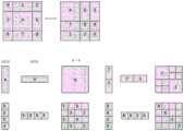
2.2.4通过特征图移位并构建多图注意力机制MLP来实现不同窗口之间注意力得分计算,由于对特征图移位会导致窗口的个数变化,因此通过给Attention设置mask机制来实现注意力得分计算的等价;得到输出特征图Ⅱ;2.2.4 The calculation of the attention score between different windows is realized by shifting the feature map and building a multi-image attention mechanism MLP. Since the shift of the feature map will cause the number of windows to change, it is achieved by setting a mask mechanism for Attention. Equivalent of attention score calculation; get output feature map II;
2.2.5通过window reverse操作将输出特征图Ⅱ还原成跟输入特征图一样的大小。2.2.5 Restore the output feature map II to the same size as the input feature map through the window reverse operation.
所述SW-MSA的计算公式如下:The calculation formula of the SW-MSA is as follows:


其中,

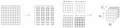
进一步的,所述步骤2.3的Patch Merging的具体实施方式为:对H和W维度进行间隔采样后拼接在一起,达到下采样的目的。Further, the specific implementation of Patch Merging in step 2.3 is as follows: H and W dimensions are sampled at intervals and then spliced together to achieve the purpose of downsampling.
进一步的,所述步骤2.4的全尺度跳跃连接的具体实施方式为:Further, the specific implementation of the full-scale skip connection in step 2.4 is:
2.4.1对于高层语义特征图,先使用最大池化缩小该高层语义特征图的大小,然后使用3×3的卷积核改变其维度。2.4.1 For the high-level semantic feature map, first use max pooling to reduce the size of the high-level semantic feature map, and then use a 3×3 convolution kernel to change its dimension.
2.4.2对于低层细节特征图,先使用双线性插值扩大该低层细节特征图的大小,然后使用3×3的卷积核改变其维度。2.4.2 For the low-level detail feature map, first use bilinear interpolation to expand the size of the low-level detail feature map, and then use a 3×3 convolution kernel to change its dimension.
2.4.3对经过步骤2.4.1处理后的高层语义特征图与经过步骤2.4.2处理后的低层细节特征图进行融合,融合的特征图使用3×3的卷积核,再经过批标准化和ReLU函数激活。2.4.3 Integrate the high-level semantic feature map processed in step 2.4.1 with the low-level detail feature map processed in step 2.4.2. The fused feature map uses a 3 × 3 convolution kernel, and then undergoes batch normalization and summation. ReLU function activation.
进一步的,所述步骤2.6的双线性插值的具体实施方式为:使用双线性插值将经过三层解码操作后的特征图从
进一步的,所述优化方法为Adam,所述损失函数为交叉熵,所述预训练数据集为CIFAR-100。Further, the optimization method is Adam, the loss function is cross entropy, and the pre-training data set is CIFAR-100.
所述交叉熵定义如下:The cross-entropy is defined as follows:

其中,yi为表注的真实眼膜图中像素点i的值,取值为0或1;
进一步的,步骤4中评价方式为Dice相似系数、PPV系数和灵敏度系数。Further, the evaluation methods in
其定义如下:It is defined as follows:


其中,TP表示被正确分割成直肠肿瘤区域的像素数量;TN表示被正确分割成背景区域的像素数量;FP表示将背景区域预测成肿瘤区域的像素数量;FN表示将肿瘤区域预测成背景区域的像素数量。Among them, TP represents the number of pixels correctly segmented into the rectal tumor area; TN represents the number of pixels correctly segmented into the background area; FP represents the number of pixels that predict the background area as the tumor area; FN represents the number of pixels that predict the tumor area as the background area number of pixels.
现有技术相比,本发明的有益结果使:Compared with the prior art, the beneficial results of the present invention make:
本发明通过建立U-Transformer的深度学习模型,实现了直肠癌肿瘤CT影像的分割,避免了人工分割存在的低效率及不稳定的缺陷,从而为相关疾病的诊断、治疗和手术引导提供准确的依据。相比于其他U型网络结构,该方法可以学习到全局特征,具有更大的视觉感知范围,从而实现高精度的医学分割。The present invention realizes the segmentation of rectal cancer tumor CT images by establishing a deep learning model of U-Transformer, avoids the defects of low efficiency and instability in manual segmentation, and thus provides accurate diagnosis, treatment and surgical guidance for related diseases. in accordance with. Compared with other U-shaped network structures, this method can learn global features and have a larger visual perception range, thereby achieving high-precision medical segmentation.
附图说明Description of drawings
图1为CT影像预处理的示意图。Figure 1 is a schematic diagram of CT image preprocessing.
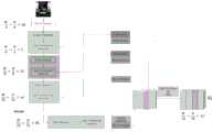
图2为U-Transformer的网络结构图。Figure 2 is the network structure diagram of U-Transformer.
图3为U-Transformer的全尺度跳跃连接示意图。Figure 3 is a schematic diagram of the full-scale skip connection of U-Transformer.
图4为Swin Transformer Block的原理图。Figure 4 is a schematic diagram of the Swin Transformer Block.
图5为U-Transformer分割直肠癌肿瘤的实例效果。Figure 5 shows an example effect of U-Transformer on rectal cancer tumor segmentation.
图6为本发明效果对比实例示意图。FIG. 6 is a schematic diagram of an effect comparison example of the present invention.
图7为U-Transformer的Patch Merging层示意图Figure 7 is a schematic diagram of the Patch Merging layer of U-Transformer
图8为SW-MSA机制的特征图移位和mask机制示意图Figure 8 is a schematic diagram of the feature map shift and mask mechanism of the SW-MSA mechanism
具体实施方式Detailed ways
下面结合附图和具体实施例对本发明作进一步的说明。The present invention will be further described below with reference to the accompanying drawings and specific embodiments.
如图1-8所示,基于U-Transformer的直肠癌CT影像自动分割方法。该方法包括下述过程:对实验数据进行预处理;构建U-Transformer网络模型;训练U-Transformer网络模型;用训练好的U-Transformer网络模型进行CT影像中直肠癌的分割,并对分割效果进行评估。本发明提出的U-Transformer网络模型可以很好的对直肠癌肿瘤的CT影像进行分割。As shown in Figure 1-8, the automatic segmentation method of rectal cancer CT images based on U-Transformer. The method includes the following processes: preprocessing the experimental data; building a U-Transformer network model; training the U-Transformer network model; using the trained U-Transformer network model to segment rectal cancer in CT images, and evaluating the segmentation effect to evaluate. The U-Transformer network model proposed by the present invention can well segment the CT images of rectal cancer tumors.
实施例1:数据预处理模块:对实验数据的预处理Example 1: Data preprocessing module: preprocessing of experimental data
(1)对数据进行直方图均衡化和归一化处理,如图1所示。(1) Perform histogram equalization and normalization processing on the data, as shown in Figure 1.
(2)对CT影像进行数据规约,构建规约数据库。(2) Carry out data reduction of CT images and build a reduction database.
(3)对CT影像进行旋转、镜像、水平翻转等几何变换方法,进行数据增强.扩增训练样本,以减少过拟合现象。(3) Perform geometric transformation methods such as rotation, mirroring, and horizontal flipping of CT images to enhance data and expand training samples to reduce overfitting.
(4)将每个CT影像和标记图的尺寸进行统一。(4) Unify the size of each CT image and marker map.
(5)按照0.8:0.1:0.1的比例划分数据集为训练集、验证集和测试集。(5) Divide the data set into training set, validation set and test set according to the ratio of 0.8:0.1:0.1.
实施例2:U-Transformer网络模型构建模块:构建U-Transformer网络模型。Example 2: U-Transformer network model building module: building a U-Transformer network model.
(1)构建Patch Embedding层。将二维的CT影像变换得到M个大小为P2·C的一维patch embeddings。(1) Build the Patch Embedding layer. Transform the two-dimensional CT image to obtain M one-dimensional patch embeddings of size P2 ·C.
(2)构建Swin Transformer Block。具体的Swin Transformer Block如图4所示:1.使用W-MSA机制计算窗口内部的自注意力得分2.使用SW-MSA机制计算不同窗口之间的注意力得分。(2) Build the Swin Transformer Block. The specific Swin Transformer Block is shown in Figure 4: 1. Use the W-MSA mechanism to calculate the self-attention score inside the
(3)构建Patch Merging。具体的Patch Merging对H和W维度进行间隔采样后拼接在一起,可以达到下采样的目的。(3) Build Patch Merging. The specific Patch Merging samples the H and W dimensions at intervals and splices them together, which can achieve the purpose of downsampling.
(4)构建全尺度跳跃连接。具体的全尺度跳跃连接机制如图3所示:1.对于高层语义特征,我们先使用最大池化缩小其特征图大小,然后使用3×3的卷积核改变其维度。2.对于低层细节特征,我们先使用双线性插值扩大其特征图大小,然后使用3×3的卷积核改变其维度。3.对融合的特征图使用3×3的卷积核,再经过批标准化和ReLU函数激活。(4) Construct full-scale skip connections. The specific full-scale skip connection mechanism is shown in Figure 3: 1. For high-level semantic features, we first use max pooling to reduce the size of its feature map, and then use a 3×3 convolution kernel to change its dimension. 2. For low-level detail features, we first use bilinear interpolation to expand the size of their feature maps, and then use a 3×3 convolution kernel to change their dimensions. 3. Use a 3×3 convolution kernel on the fused feature map, and then go through batch normalization and ReLU function activation.
(5)使用双线性插值将经过三层解码操作后的特征图从

(6)构建线性投影操作。将W×H×C的特征图变为W×H×2,从而实现像素级分割。(6) Constructing a linear projection operation. The feature map of W×H×C is changed to W×H×2 to achieve pixel-level segmentation.
实施例3:U-Transformer网络模型训练模块:训练U-Transformer网络模型Embodiment 3: U-Transformer network model training module: training U-Transformer network model
(1)采用Adam优化方式作为优化方法,同时采用交叉熵作为损失函数进行训练。所述交叉熵公式如下:(1) The Adam optimization method is used as the optimization method, and the cross entropy is used as the loss function for training. The cross-entropy formula is as follows:

其中,yi为表注的真实眼膜图中像素点i的值,取值为0或1;
(2)将U-Transformer网络模型在CIFAR-100数据集上进行预训练。(2) The U-Transformer network model is pre-trained on the CIFAR-100 dataset.
实施例4:分割评估模块:采用训练好的U-Transformer网络模型进行CT影像中直肠癌的分割,并对分割效果进行评估Example 4: Segmentation evaluation module: use the trained U-Transformer network model to segment rectal cancer in CT images, and evaluate the segmentation effect
(1)图5为U-Transformer分割的实例。引入Dice相似系数、PPV系数和灵敏度系数来评价分割的效果,U-Transformer的Dice系数、PPV系数和灵敏度系数分别0.8700、0.8042和0.9481。三个系数的定义如下:(1) Figure 5 is an example of U-Transformer segmentation. Dice similarity coefficient, PPV coefficient and sensitivity coefficient are introduced to evaluate the effect of segmentation. The Dice coefficient, PPV coefficient and sensitivity coefficient of U-Transformer are 0.8700, 0.8042 and 0.9481, respectively. The three coefficients are defined as follows:


其中,TP表示被正确分割成直肠肿瘤区域的像素数量;TN表示被正确分割成背景区域的像素数量;FP表示将背景区域预测成肿瘤区域的像素数量;FN表示将肿瘤区域预测成背景区域的像素数量。Among them, TP represents the number of pixels correctly segmented into the rectal tumor area; TN represents the number of pixels correctly segmented into the background area; FP represents the number of pixels that predict the background area as the tumor area; FN represents the number of pixels that predict the tumor area as the background area number of pixels.
实施例5:效果对比表Example 5: Effect comparison table
如图6和表1所示,U-Transformer分割肿瘤的Dice系数、PPV系数和灵敏度系数分别为0.8700、0.8042和0.9481,均高于其他模型。同时,比放射科医生手动绘制肿瘤的Dice系数高出了18.39%。我们选择了几个具有代表性的分割图进行展示,可以看出U-Transformer可以很好地分割肿瘤的边缘区域,而V-Net、U-Net和R2U-Net不能准确识别肿瘤的位置,这是由于直肠癌位置较为特殊,普通的卷积神经网络很容易将背景区域识别为肿瘤区域。对于部分CT影像,U-Net虽然可以稳定地分割肿瘤区域,但肿瘤的边缘仍然比较粗糙,有时无法识别被肿瘤区域包裹的非肿瘤区域。由Swin Transformer作为编码器和全尺度跳跃连接作为解码器的U-Transformer模型具有比卷积神经网络更大的感受野;并且U-Transformer可以有效地对肿瘤边缘和肿瘤内部的非肿瘤区域进行分割。As shown in Figure 6 and Table 1, the Dice coefficient, PPV coefficient and sensitivity coefficient of U-Transformer segmented tumors were 0.8700, 0.8042 and 0.9481, respectively, which were higher than other models. At the same time, the Dice coefficient was 18.39% higher than that of radiologists manually mapping tumors. We have selected several representative segmentation maps for display. It can be seen that U-Transformer can segment the edge region of the tumor well, while V-Net, U-Net and R2U-Net cannot accurately identify the location of the tumor. It is because the location of rectal cancer is relatively special, and the ordinary convolutional neural network can easily identify the background area as the tumor area. For some CT images, although U-Net can stably segment the tumor area, the edge of the tumor is still relatively rough, and sometimes it cannot identify the non-tumor area surrounded by the tumor area. The U-Transformer model with Swin Transformer as the encoder and full-scale skip connections as the decoder has a larger receptive field than the convolutional neural network; and the U-Transformer can effectively segment the tumor margin and the non-tumor area inside the tumor .
表1:Dice系数、PPV系数和灵敏度系数对比表Table 1: Dice coefficient, PPV coefficient and sensitivity coefficient comparison table

Claims (9)
Translated fromChinese


Priority Applications (1)
| Application Number | Priority Date | Filing Date | Title |
|---|---|---|---|
| CN202110981706.0ACN113674253B (en) | 2021-08-25 | 2021-08-25 | Automatic segmentation method for rectal cancer CT image based on U-transducer |
Applications Claiming Priority (1)
| Application Number | Priority Date | Filing Date | Title |
|---|---|---|---|
| CN202110981706.0ACN113674253B (en) | 2021-08-25 | 2021-08-25 | Automatic segmentation method for rectal cancer CT image based on U-transducer |
Publications (2)
| Publication Number | Publication Date |
|---|---|
| CN113674253Atrue CN113674253A (en) | 2021-11-19 |
| CN113674253B CN113674253B (en) | 2023-06-30 |
Family
ID=78546128
Family Applications (1)
| Application Number | Title | Priority Date | Filing Date |
|---|---|---|---|
| CN202110981706.0AActiveCN113674253B (en) | 2021-08-25 | 2021-08-25 | Automatic segmentation method for rectal cancer CT image based on U-transducer |
Country Status (1)
| Country | Link |
|---|---|
| CN (1) | CN113674253B (en) |
Cited By (24)
| Publication number | Priority date | Publication date | Assignee | Title |
|---|---|---|---|---|
| CN113870258A (en)* | 2021-12-01 | 2021-12-31 | 浙江大学 | Counterwork learning-based label-free pancreas image automatic segmentation system |
| CN114066870A (en)* | 2021-11-23 | 2022-02-18 | 重庆大学 | Gear pitting corrosion measuring method based on visual transducer |
| CN114119585A (en)* | 2021-12-01 | 2022-03-01 | 昆明理工大学 | Method for identifying key feature enhanced gastric cancer image based on Transformer |
| CN114187278A (en)* | 2021-12-14 | 2022-03-15 | 山东众阳健康科技集团有限公司 | Method and system for detecting small rib fractures |
| CN114419054A (en)* | 2022-01-19 | 2022-04-29 | 新疆大学 | Retinal blood vessel image segmentation method and device and related equipment |
| CN114445422A (en)* | 2022-01-13 | 2022-05-06 | 同济大学 | Transform-based medical image segmentation method, system, medium and electronic device |
| CN114529507A (en)* | 2021-12-30 | 2022-05-24 | 广西慧云信息技术有限公司 | Shaving board surface defect detection method based on visual transducer |
| CN114612416A (en)* | 2022-03-04 | 2022-06-10 | 桂林医学院 | A Swin-Unet-based segmentation method for gastric adenocarcinoma lesions |
| CN114758121A (en)* | 2022-03-04 | 2022-07-15 | 杭州隐捷适生物科技有限公司 | CBCT alveolar bone segmentation system and method based on deep learning |
| CN114912575A (en)* | 2022-04-06 | 2022-08-16 | 西安交通大学 | Medical Image Segmentation Model and Method Based on Connected Swin Transformer Pathway |
| CN115222734A (en)* | 2022-09-20 | 2022-10-21 | 山东大学齐鲁医院 | Image analysis method and system for gastric mucosa intestinal metaplasia |
| CN115578406A (en)* | 2022-12-13 | 2023-01-06 | 四川大学 | CBCT jaw bone region segmentation method and system based on context fusion mechanism |
| CN115588013A (en)* | 2022-10-08 | 2023-01-10 | 华东师范大学 | Image segmentation method based on full-scale fusion and flow field attention |
| CN115690127A (en)* | 2022-11-09 | 2023-02-03 | 齐鲁工业大学 | Two-stage CT image segmentation method based on sliding window transform |
| CN115880691A (en)* | 2023-03-02 | 2023-03-31 | 国网山东省电力公司东营供电公司 | A method for estimating rooftop photovoltaic potential based on computer vision |
| CN116072127A (en)* | 2022-12-30 | 2023-05-05 | 国网浙江省电力有限公司营销服务中心 | Voiceprint recognition method and system based on maximum pooling self-attention mechanism |
| CN116245144A (en)* | 2023-02-20 | 2023-06-09 | 南京邮电大学 | Lightweight window pyramid network model and application thereof |
| CN116433697A (en)* | 2023-06-13 | 2023-07-14 | 南京航空航天大学 | Segmentation method of abdominal multi-organ CT image based on eye tracker |
| CN116584955A (en)* | 2023-04-27 | 2023-08-15 | 中国人民解放军战略支援部队信息工程大学 | Brain electricity cognitive load assessment method and system based on multi-feature domain attention network |
| CN116797605A (en)* | 2023-04-11 | 2023-09-22 | 杭州电子科技大学 | Pulmonary embolism CT image identification segmentation method based on SCUNet++ neural network |
| WO2024098318A1 (en)* | 2022-11-10 | 2024-05-16 | 中国科学院深圳先进技术研究院 | Medical image segmentation method |
| CN118644503A (en)* | 2024-06-06 | 2024-09-13 | 浙江工业大学 | A feature-fusion attention-based adrenal tumor segmentation method |
| CN120612488A (en)* | 2025-08-06 | 2025-09-09 | 中南大学 | Tumor image segmentation method, device, electronic device and storage medium |
| CN120612488B (en)* | 2025-08-06 | 2025-10-10 | 中南大学 | Tumor image segmentation method, device, electronic device and storage medium |
Citations (9)
| Publication number | Priority date | Publication date | Assignee | Title |
|---|---|---|---|---|
| US20110116698A1 (en)* | 2009-11-18 | 2011-05-19 | Siemens Corporation | Method and System for Segmentation of the Prostate in 3D Magnetic Resonance Images |
| CN108010041A (en)* | 2017-12-22 | 2018-05-08 | 数坤(北京)网络科技有限公司 | Human heart coronary artery extracting method based on deep learning neutral net cascade model |
| CN111145170A (en)* | 2019-12-31 | 2020-05-12 | 电子科技大学 | Medical image segmentation method based on deep learning |
| CN112102321A (en)* | 2020-08-07 | 2020-12-18 | 深圳大学 | Focal image segmentation method and system based on deep convolutional neural network |
| CN112150429A (en)* | 2020-09-18 | 2020-12-29 | 南京师范大学 | Attention mechanism guided kidney CT image segmentation method |
| CN112164069A (en)* | 2020-07-29 | 2021-01-01 | 南通大学 | A deep learning-based method for CT abdominal blood vessel segmentation |
| CN112348769A (en)* | 2020-08-20 | 2021-02-09 | 盐城工学院 | Intelligent kidney tumor segmentation method and device in CT (computed tomography) image based on U-Net depth network model |
| CN112949648A (en)* | 2021-03-12 | 2021-06-11 | 上海眼控科技股份有限公司 | Method and equipment for acquiring training sample data set of image segmentation model |
| CN112990219A (en)* | 2021-03-25 | 2021-06-18 | 北京百度网讯科技有限公司 | Method and apparatus for image semantic segmentation |
- 2021
- 2021-08-25CNCN202110981706.0Apatent/CN113674253B/enactiveActive
Patent Citations (9)
| Publication number | Priority date | Publication date | Assignee | Title |
|---|---|---|---|---|
| US20110116698A1 (en)* | 2009-11-18 | 2011-05-19 | Siemens Corporation | Method and System for Segmentation of the Prostate in 3D Magnetic Resonance Images |
| CN108010041A (en)* | 2017-12-22 | 2018-05-08 | 数坤(北京)网络科技有限公司 | Human heart coronary artery extracting method based on deep learning neutral net cascade model |
| CN111145170A (en)* | 2019-12-31 | 2020-05-12 | 电子科技大学 | Medical image segmentation method based on deep learning |
| CN112164069A (en)* | 2020-07-29 | 2021-01-01 | 南通大学 | A deep learning-based method for CT abdominal blood vessel segmentation |
| CN112102321A (en)* | 2020-08-07 | 2020-12-18 | 深圳大学 | Focal image segmentation method and system based on deep convolutional neural network |
| CN112348769A (en)* | 2020-08-20 | 2021-02-09 | 盐城工学院 | Intelligent kidney tumor segmentation method and device in CT (computed tomography) image based on U-Net depth network model |
| CN112150429A (en)* | 2020-09-18 | 2020-12-29 | 南京师范大学 | Attention mechanism guided kidney CT image segmentation method |
| CN112949648A (en)* | 2021-03-12 | 2021-06-11 | 上海眼控科技股份有限公司 | Method and equipment for acquiring training sample data set of image segmentation model |
| CN112990219A (en)* | 2021-03-25 | 2021-06-18 | 北京百度网讯科技有限公司 | Method and apparatus for image semantic segmentation |
Non-Patent Citations (2)
| Title |
|---|
| KEYU WEN等: "Learning Dual Semantic Relations With Graph Attention for Image-Text Matching", 《IEEE TRANSACTIONS ON CIRCUITS AND SYSTEMS FOR VIDEO TECHNOLOGY》, vol. 31, no. 7, pages 2866 - 2879, XP011864212, DOI: 10.1109/TCSVT.2020.3030656* |
| 田应仲等: "基于注意力机制与Swin Transformer模型的腰椎图像分割方法", 《计量与测试技术》, vol. 48, no. 12, pages 57 - 61* |
Cited By (32)
| Publication number | Priority date | Publication date | Assignee | Title |
|---|---|---|---|---|
| CN114066870A (en)* | 2021-11-23 | 2022-02-18 | 重庆大学 | Gear pitting corrosion measuring method based on visual transducer |
| CN114119585A (en)* | 2021-12-01 | 2022-03-01 | 昆明理工大学 | Method for identifying key feature enhanced gastric cancer image based on Transformer |
| CN113870258B (en)* | 2021-12-01 | 2022-03-25 | 浙江大学 | An automatic segmentation system for unlabeled pancreatic images based on adversarial learning |
| CN114119585B (en)* | 2021-12-01 | 2022-11-29 | 昆明理工大学 | Method for identifying key feature enhanced gastric cancer image based on Transformer |
| CN113870258A (en)* | 2021-12-01 | 2021-12-31 | 浙江大学 | Counterwork learning-based label-free pancreas image automatic segmentation system |
| CN114187278A (en)* | 2021-12-14 | 2022-03-15 | 山东众阳健康科技集团有限公司 | Method and system for detecting small rib fractures |
| CN114529507B (en)* | 2021-12-30 | 2024-05-17 | 广西慧云信息技术有限公司 | Visual transducer-based particle board surface defect detection method |
| CN114529507A (en)* | 2021-12-30 | 2022-05-24 | 广西慧云信息技术有限公司 | Shaving board surface defect detection method based on visual transducer |
| CN114445422A (en)* | 2022-01-13 | 2022-05-06 | 同济大学 | Transform-based medical image segmentation method, system, medium and electronic device |
| CN114419054B (en)* | 2022-01-19 | 2024-12-17 | 新疆大学 | Retina blood vessel image segmentation method and device and related equipment |
| CN114419054A (en)* | 2022-01-19 | 2022-04-29 | 新疆大学 | Retinal blood vessel image segmentation method and device and related equipment |
| CN114758121A (en)* | 2022-03-04 | 2022-07-15 | 杭州隐捷适生物科技有限公司 | CBCT alveolar bone segmentation system and method based on deep learning |
| CN114612416A (en)* | 2022-03-04 | 2022-06-10 | 桂林医学院 | A Swin-Unet-based segmentation method for gastric adenocarcinoma lesions |
| CN114912575B (en)* | 2022-04-06 | 2024-04-09 | 西安交通大学 | Medical image segmentation model and method based on connection Swin transducer path |
| CN114912575A (en)* | 2022-04-06 | 2022-08-16 | 西安交通大学 | Medical Image Segmentation Model and Method Based on Connected Swin Transformer Pathway |
| CN115222734A (en)* | 2022-09-20 | 2022-10-21 | 山东大学齐鲁医院 | Image analysis method and system for gastric mucosa intestinal metaplasia |
| CN115588013A (en)* | 2022-10-08 | 2023-01-10 | 华东师范大学 | Image segmentation method based on full-scale fusion and flow field attention |
| CN115690127A (en)* | 2022-11-09 | 2023-02-03 | 齐鲁工业大学 | Two-stage CT image segmentation method based on sliding window transform |
| WO2024098318A1 (en)* | 2022-11-10 | 2024-05-16 | 中国科学院深圳先进技术研究院 | Medical image segmentation method |
| CN115578406A (en)* | 2022-12-13 | 2023-01-06 | 四川大学 | CBCT jaw bone region segmentation method and system based on context fusion mechanism |
| CN116072127B (en)* | 2022-12-30 | 2025-08-12 | 国网浙江省电力有限公司营销服务中心 | Voiceprint recognition method and system based on maximum pooling self-attention mechanism |
| CN116072127A (en)* | 2022-12-30 | 2023-05-05 | 国网浙江省电力有限公司营销服务中心 | Voiceprint recognition method and system based on maximum pooling self-attention mechanism |
| CN116245144A (en)* | 2023-02-20 | 2023-06-09 | 南京邮电大学 | Lightweight window pyramid network model and application thereof |
| CN115880691B (en)* | 2023-03-02 | 2023-05-23 | 国网山东省电力公司东营供电公司 | A method for estimating rooftop photovoltaic potential based on computer vision |
| CN115880691A (en)* | 2023-03-02 | 2023-03-31 | 国网山东省电力公司东营供电公司 | A method for estimating rooftop photovoltaic potential based on computer vision |
| CN116797605A (en)* | 2023-04-11 | 2023-09-22 | 杭州电子科技大学 | Pulmonary embolism CT image identification segmentation method based on SCUNet++ neural network |
| CN116584955A (en)* | 2023-04-27 | 2023-08-15 | 中国人民解放军战略支援部队信息工程大学 | Brain electricity cognitive load assessment method and system based on multi-feature domain attention network |
| CN116433697B (en)* | 2023-06-13 | 2023-09-12 | 南京航空航天大学 | Segmentation method of abdominal multi-organ CT images based on eye tracker |
| CN116433697A (en)* | 2023-06-13 | 2023-07-14 | 南京航空航天大学 | Segmentation method of abdominal multi-organ CT image based on eye tracker |
| CN118644503A (en)* | 2024-06-06 | 2024-09-13 | 浙江工业大学 | A feature-fusion attention-based adrenal tumor segmentation method |
| CN120612488A (en)* | 2025-08-06 | 2025-09-09 | 中南大学 | Tumor image segmentation method, device, electronic device and storage medium |
| CN120612488B (en)* | 2025-08-06 | 2025-10-10 | 中南大学 | Tumor image segmentation method, device, electronic device and storage medium |
Also Published As
| Publication number | Publication date |
|---|---|
| CN113674253B (en) | 2023-06-30 |
Similar Documents
| Publication | Publication Date | Title |
|---|---|---|
| CN113674253A (en) | Rectal cancer CT image automatic segmentation method based on U-transducer | |
| CN113870258B (en) | An automatic segmentation system for unlabeled pancreatic images based on adversarial learning | |
| JP7312510B1 (en) | Whole-slide pathological image classification system and construction method considering tumor microenvironment | |
| CN109523521B (en) | Pulmonary nodule classification and lesion location method and system based on multi-slice CT images | |
| WO2023071531A1 (en) | Liver ct automatic segmentation method based on deep shape learning | |
| CN109919230B (en) | Pulmonary nodule detection method in medical images based on circular feature pyramid | |
| CN112150428A (en) | Medical image segmentation method based on deep learning | |
| CN113223005A (en) | Thyroid nodule automatic segmentation and grading intelligent system | |
| CN112489061A (en) | Deep learning intestinal polyp segmentation method based on multi-scale information and parallel attention mechanism | |
| CN107240102A (en) | Malignant tumour area of computer aided method of early diagnosis based on deep learning algorithm | |
| CN110310289A (en) | Lung tissue image segmentation method based on deep learning | |
| CN117746119A (en) | Ultrasound image breast tumor classification method based on feature fusion and attention mechanism | |
| CN118314350A (en) | MRI brain tumor segmentation method based on attention bottleneck fusion | |
| CN112396605B (en) | Network training method and device, image recognition method and electronic equipment | |
| CN114998265A (en) | A Liver Tumor Segmentation Method Based on Improved U-Net | |
| CN114202545A (en) | UNet + + based low-grade glioma image segmentation method | |
| CN114565601A (en) | Improved liver CT image segmentation algorithm based on DeepLabV3+ | |
| CN109215035B (en) | Brain MRI hippocampus three-dimensional segmentation method based on deep learning | |
| CN112263217B (en) | A Lesion Area Detection Method Based on Improved Convolutional Neural Network in Pathological Images of Non-melanoma Skin Cancer | |
| CN116934683B (en) | Artificial intelligence-assisted ultrasound diagnosis of spleen trauma | |
| CN117132774A (en) | Multi-scale polyp segmentation method and system based on PVT | |
| CN116596890A (en) | Dynamic image thyroid cancer risk layering prediction method based on graph convolution network | |
| CN117635625A (en) | Pancreatic tumor segmentation method based on automatic data enhancement strategy and multi-attention-assisted UNet | |
| CN120107236A (en) | A method for predicting benign and malignant thyroid nodules based on automatic segmentation and semi-supervised learning | |
| Wang et al. | Multi-scale boundary neural network for gastric tumor segmentation |
Legal Events
| Date | Code | Title | Description |
|---|---|---|---|
| PB01 | Publication | ||
| PB01 | Publication | ||
| SE01 | Entry into force of request for substantive examination | ||
| SE01 | Entry into force of request for substantive examination | ||
| GR01 | Patent grant | ||
| GR01 | Patent grant |